
Exercise 32
41
Exercise 32 Anatomy of Blood Vessels
-
Upload
desiderio-hierro -
Category
Documents
-
view
37 -
download
0
description
Exercise 32. Anatomy of Blood Vessels. Objectives. Tunics of blood vessel walls Artery, vein, capillary structural differences correlated to functional differences Artery and vein cross-sections Major arteries and veins. Tunics of Blood Vessel (Artery & Vein) Walls. Fig. 21-1. - PowerPoint PPT Presentation
Transcript of Exercise 32

Exercise 32
Anatomy of Blood Vessels

ObjectivesObjectives
• Tunics of blood vessel wallsTunics of blood vessel walls
• Artery, vein, capillary structural Artery, vein, capillary structural differences correlated to functional differences correlated to functional differencesdifferences
• Artery and vein cross-sectionsArtery and vein cross-sections
• Major arteries and veinsMajor arteries and veins

Tunics of Blood Vessel (Artery & Tunics of Blood Vessel (Artery & Vein) WallsVein) Walls
Fig. 21-1

Tunics of Blood Vessel (Artery & Tunics of Blood Vessel (Artery & Vein) WallsVein) Walls
• Tunica intima (interna)Tunica intima (interna)– Lines the lumenLines the lumen– Single, smooth endothelial layer Single, smooth endothelial layer
(squamous)(squamous)– Continuous with heart’s Continuous with heart’s
endocardiumendocardium

Tunics of Blood Vessel (Artery & Tunics of Blood Vessel (Artery & Vein) WallsVein) Walls
• Tunica mediaTunica media– Bulky middle layerBulky middle layer– Smooth muscle, elastinSmooth muscle, elastin– Smooth muscle regulates diameter of Smooth muscle regulates diameter of
vessel vessel blood pressure blood pressure

Tunics of Blood Vessel (Artery & Tunics of Blood Vessel (Artery & Vein) WallsVein) Walls
Tunica adentitia (externa)Tunica adentitia (externa)– Outermost layerOutermost layer– Areolar or fibrous connective tissueAreolar or fibrous connective tissue– Support/protectionSupport/protection

ArteryArtery, Vein, , Vein, oror CapillaryCapillary??
• ArteriesArteries– Walls generally thicker vs. veinsWalls generally thicker vs. veins– Tunica media has more muscle & elastic Tunica media has more muscle & elastic
tissue:tissue:• Large arteries actually Large arteries actually closercloser to the to the
pumping of the heartpumping of the heart• Able to expand as large amts of blood Able to expand as large amts of blood
enter them, recoil as blood passes enter them, recoil as blood passes through through ELASTIC arteries ELASTIC arteries
• Smaller arteries less elastic tissue, Smaller arteries less elastic tissue, moderate muscle moderate muscle MUSCULAR arteries MUSCULAR arteries

• VeinsVeins– Far removed from the heartFar removed from the heart– Low-pressure, not that big pressure Low-pressure, not that big pressure
change situation found in arterieschange situation found in arteries– Thinner walledThinner walled– Often flows against gravity—modification:Often flows against gravity—modification:
• Lumens of veins largerLumens of veins larger• Valves prevent backflowValves prevent backflow• Skeletal muscles “pump” (“milk”) blood Skeletal muscles “pump” (“milk”) blood
toward the hearttoward the heart
Artery,Artery, Vein,Vein, or or CapillaryCapillary??

Fig. 21-1

Artery,Artery, Vein, or Vein, or CapillaryCapillary??
• CapillariesCapillaries– Transparent walls, Transparent walls, one cell thickone cell thick– Tunica intima onlyTunica intima only– Easy gas exchange between blood and Easy gas exchange between blood and
tissue’s cellstissue’s cells

Fig. 21-4

Fig. 21-2

Major ArteriesMajor Arteries
Pulmonary TrunkPulmonary Trunk
Right pulmonary Right pulmonary arteryartery
Left pulmonary Left pulmonary arteryartery
Fig. 21-21Fig. 21-21
To lungs, capillary To lungs, capillary exchange COexchange CO22/O/O22

Fig. 21-22Fig. 21-22
Major Arteries: The AortaMajor Arteries: The Aorta
Thoracic aortaThoracic aorta
Abdominal aortaAbdominal aorta

Fig. 21-23Fig. 21-23
Major ArteriesMajor Arteries
BrachiocephalicBrachiocephalic
R, L subclaviansR, L subclavians

Fig. 21-26Fig. 21-26
R, LR, L

Fig. 21-26Fig. 21-26
FacialFacial
Superficial Superficial temporaltemporal
FacialFacial

(R, L) (R, L) Internal Internal thoracicthoracic
(mammary)(mammary)
Fig. 21-23Fig. 21-23

Axillary Axillary arteryartery
Fig. 21-23Fig. 21-23
Brachial Brachial arteryartery

Fig. 21-23Fig. 21-23
Radial Radial arteryartery
Ulnar arteryUlnar artery

Fig. 21-23Fig. 21-23
Superficial palmar Superficial palmar archarch
Digital Digital arteriesarteries

ASCENDING AORTA
AORTIC ARCH
DESCENDING AORTA
THORACIC AORTA
ABDOMINAL AORTA
BRACHIOCEPHALIC RIGHT SUBCLAVIAN
LEFT SUBCLAVIAN
RIGHT COMMON CAROTID
LEFT COMMON CAROTID
RIGHT FACIAL
LEFT FACIAL
RIGHT SUPERFICIAL TEMPORAL
LEFT SUPERFICIAL TEMPORAL
ARTERIES
RIGHT INTERNAL THORACIC
LEFT INTERNAL THORACIC
RIGHT AXILLARY
LEFT AXILLARY
RIGHT BRACHIAL
LEFT BRACHIAL
RIGHT RADIAL
RIGHT ULNAR
LEFT RADIAL
LEFT ULNAR
SUPERFICIAL PALMAR ARCH
SUPERFICIAL PALMAR ARCH
RIGHT DIGITAL ARTERIES
LEFT DIGITAL ARTERIES
(OVER)

Branches from Abdominal AortaBranches from Abdominal Aorta
Fig. 21-26Fig. 21-26
Celiac Celiac TrunkTrunk
Left Left gastric gastric arteryartery
Splenic Splenic arteryartery
Hepatic Hepatic arteryartery

Fig. 21-27
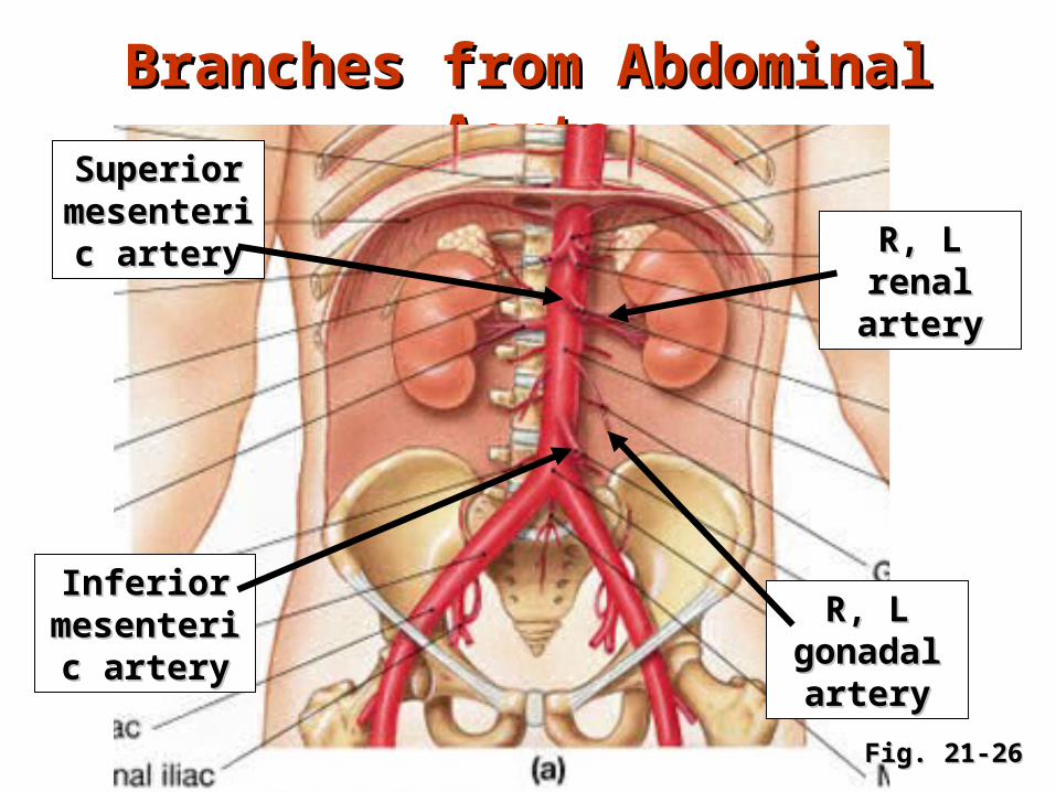
Branches from Abdominal AortaBranches from Abdominal Aorta
Fig. 21-26Fig. 21-26
Superior Superior mesenteric mesenteric
arteryartery R, L renal R, L renal arteryartery
R, L R, L gonadal gonadal arteryartery
Inferior Inferior mesenteric mesenteric
arteryartery


Fig. 21-26Fig. 21-26
R, L common R, L common iliac arteriesiliac arteries
R, L R, L external external
iliac iliac arteriesarteries
R, L R, L internal internal
iliac iliac arteriesarteries


Fig. 21-28Fig. 21-28
Femoral arteryFemoral artery
Deep femoral Deep femoral arteryartery
Lateral Lateral circumflex circumflex
femoral arteryfemoral artery
Popliteal arteryPopliteal artery
Posterior Posterior tibial arterytibial artery
Anterior tibial Anterior tibial arteryartery
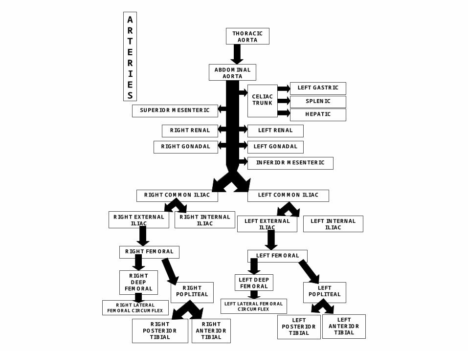
THORACIC AORTA
ABDOMINAL AORTA
CELIAC TRUNK
LEFT GASTRIC
SPLENIC
HEPATIC SUPERIOR MESENTERIC
INFERIOR MESENTERIC
RIGHT RENAL LEFT RENAL
RIGHT GONADAL LEFT GONADAL
RIGHT COMMON ILIAC LEFT COMMON ILIAC
RIGHT EXTERNAL ILIAC
RIGHT INTERNAL ILIAC
RIGHT FEMORAL
RIGHT DEEP
FEMORAL RIGHT POPLITEAL
RIGHT POSTERIOR
TIBIAL
RIGHT ANTERIOR
TIBIAL
LEFT EXTERNAL ILIAC
LEFT INTERNAL ILIAC
LEFT FEMORAL
LEFT DEEP FEMORAL LEFT
POPLITEAL
LEFT POSTERIOR
TIBIAL
LEFT ANTERIOR
TIBIAL
ARTERIES
RIGHT LATERAL FEMORAL CIRCUMFLEX
LEFT LATERAL FEMORAL CIRCUMFLEX

MAJOR VEINSMAJOR VEINS
From lungs, From lungs, capillary capillary
exchange COexchange CO22/O/O22
R, L pulmonary R, L pulmonary veinsveins
Superior vena Superior vena cavacava
Inferior vena Inferior vena cavacava

R, L R, L brachiocephalic brachiocephalic
veinsveins
Fig. 21-30

R, L subclavian R, L subclavian veinsveins
Fig. 21-31

Facial veinFacial vein
Fig. 21-30
Superficial Superficial temporal veintemporal vein
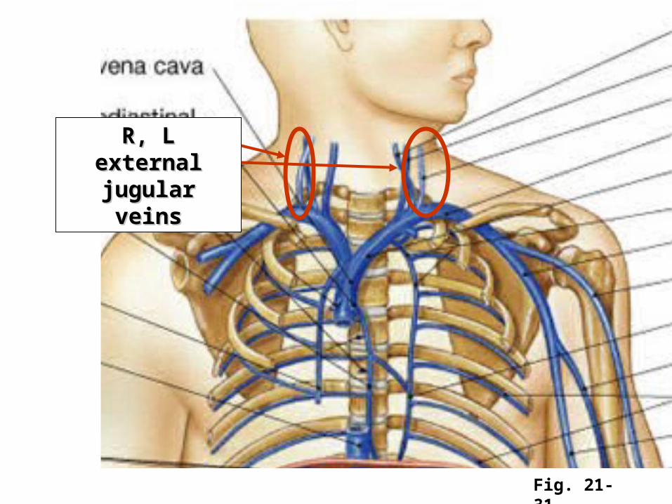
R, L external R, L external jugular veinsjugular veins
Fig. 21-31

R, L internal R, L internal jugular veinsjugular veins
Fig. 21-31

Branches of Inferior Vena CavaBranches of Inferior Vena Cava
Fig. 21-31
R, LR, L
R, LR, L

Fig. 21-34
Hepatic Portal SystemHepatic Portal SystemLeft gastric Left gastric
veinvein
Right gastric Right gastric veinvein
Splenic Splenic veinvein
Superior Superior mesenteric mesenteric
veinvein
Inferior Inferior mesenteric mesenteric
veinvein

Fig. 21-34
R, L R, L common common iliac veiniliac vein
R, L external R, L external iliac veiniliac vein
R, L R, L internal internal
iliac veiniliac vein
R, L R, L femoral femoral
veinvein
Great Great saphenous saphenous
veinvein

Website with interactive A & P model photos
http://daphne.palomar.edu/ccarpenter/Models/model%20index.htm
http://www.uky.edu/LCC/BSN/BIO/BiologyLabs/BSL111/111Lab1/Lab1VesselManFrameSet.html

LEFT RENAL VEIN
LEFT GASTRIC
VEIN
RIGHT COMMON ILIAC VEIN
RIGHT BRACHIOCEPHALIC
RIGHT SUBCLAVIAN
RIGHT EXTERNAL JUGULAR
RIGHT FACIAL
LEFT FACIAL
RIGHT SUPERFICIAL TEMPORAL
LEFT SUPERFICIAL TEMPORAL
VEINS
HEPATIC VEINS
RIGHT SAPHENOUS
RIGHT FEMORAL VEIN
RIGHT EXTERNAL ILIAC VEIN
INFERIOR VENA CAVA
SUPERIOR VENA CAVA
LEFT BRACHIOCEPHALIC
LEFT SUBCLAVIAN
RIGHT INTERNAL JUGULAR
LEFT INTERNAL JUGULAR
LEFT EXTERNAL JUGULAR
RIGHT RENAL VEIN
LEFT GONADAL VEIN
RIGHT GONADAL VEIN
SPLENIC VEIN
SUPERIOR MESENTERIC
VEIN
INFERIOR MESENTERIC
VEIN
LEFT COMMON ILIAC VEIN
LEFT EXTERNAL ILIAC VEIN
RIGHT INTERNAL ILIAC VEIN
LEFT INTERNAL ILIAC VEIN
LEFT FEMORAL VEIN
LEFT SAPHENOUS

![Exercise in Patients Pranissa - thaiheart.org · Microsoft PowerPoint - Exercise in Patients Pranissa [Compatibility Mode] Author: Administrator Created Date: 10/31/2012 3:32:48 PM](https://static.fdocuments.us/doc/165x107/5fd76adae66f2626e435bed3/exercise-in-patients-pranissa-microsoft-powerpoint-exercise-in-patients-pranissa.jpg)


![STATEWIDE MEDICAL AND HEALTH EXERCISE SWMHE EXERCISE DEBRIEF [Exercise Name/Exercise Date] SWMHE EXERCISE DEBRIEF.](https://static.fdocuments.us/doc/165x107/56649d755503460f94a56498/statewide-medical-and-health-exercise-swmhe-exercise-debrief-exercise-nameexercise.jpg)


