Evaluation of the COBAS TaqMan MTB Test for Direct...
26
1 Evaluation of the COBAS TaqMan MTB Test for Direct Detection of Mycobacterium tuberculosis Complex in Respiratory Specimens 5 Yuan-Chieh Yang 1 , Po-Liang Lu 1,2 , Su Chiao Huang, 1 Yi-Shan Jenh 3 , Ruwen Jou 4 , Tsung Chain Chang 3 Department of Laboratory Medicine, 1 and Department of Internal Medicine, 2 Kaohsiung Medical University Hospital, Kaohsiung, Taiwan; Department of Medical 10 Laboratory Science and Biotechnology, College of Medicine, National Cheng Kung University, Tainan, Taiwan; 3 Reference Laboratory of Mycobacteriology, Center for Disease Control, Department of Health, Taipei, Taiwan 4 Running title: EVALUATION OF COBAS TAQMAN MTB TEST 15 Corresponding author: Tsung Chain Chang Mailing address: 1 University Road, Department of Medical Laboratory Science and Biotechnology, College of Medicine, National Cheng Kung University, Tainan 701, Taiwan. 20 Phone: 886-6-2353535 ext. 5790. Fax: 886-6-2363956. E-mail: [email protected] Copyright © 2010, American Society for Microbiology and/or the Listed Authors/Institutions. All Rights Reserved. J. Clin. Microbiol. doi:10.1128/JCM.01839-10 JCM Accepts, published online ahead of print on 22 December 2010 on August 30, 2018 by guest http://jcm.asm.org/ Downloaded from
Transcript of Evaluation of the COBAS TaqMan MTB Test for Direct...

1
Evaluation of the COBAS TaqMan MTB Test for Direct Detection of
Mycobacterium tuberculosis Complex in Respiratory Specimens
5
Yuan-Chieh Yang1, Po-Liang Lu
1,2, Su Chiao Huang,
1 Yi-Shan Jenh
3, Ruwen Jou
4,
Tsung Chain Chang3
Department of Laboratory Medicine,1 and Department of Internal Medicine,
2
Kaohsiung Medical University Hospital, Kaohsiung, Taiwan; Department of Medical 10
Laboratory Science and Biotechnology, College of Medicine, National Cheng Kung
University, Tainan, Taiwan;
3 Reference Laboratory of Mycobacteriology, Center for
Disease Control, Department of Health, Taipei, Taiwan4
Running title: EVALUATION OF COBAS TAQMAN MTB TEST 15
Corresponding author: Tsung Chain Chang
Mailing address: 1 University Road, Department of Medical Laboratory Science and
Biotechnology, College of Medicine, National Cheng Kung University, Tainan 701,
Taiwan. 20
Phone: 886-6-2353535 ext. 5790. Fax: 886-6-2363956.
E-mail: [email protected]
Copyright © 2010, American Society for Microbiology and/or the Listed Authors/Institutions. All Rights Reserved.J. Clin. Microbiol. doi:10.1128/JCM.01839-10 JCM Accepts, published online ahead of print on 22 December 2010
on August 30, 2018 by guest
http://jcm.asm
.org/D
ownloaded from

2
ABSTRACT
The COBAS TaqMan MTB Test, based on real-time PCR technology, was
evaluated for the direct detection of Mycobacterium tuberculosis complex (MTBC) in
respiratory specimens. A total of 1093 samples from 446 patients, including 118
acid-fast smear positive and 975 acid-fast smear negative specimens, were 5
investigated. Diagnostic cultures performed with 7H11 agar, Löwenstein-Jensen
medium, and BACTEC MGIT 960 system, were considered as the reference methods.
When discrepant results between the COBAS TaqMan MTB Test and culture occurred,
additional results from the BD MGIT TBc Identification Test and GenoType
Mycobacterium CM performed on growth-positive and acid-fast-positive MGIT tubes 10
and review of patient’s medical history were used for discrepant analysis. The overall
sensitivity, specificity, positive, and negative predictive values for the COBAS
TaqMan MTB Test were 91.5%, 98.7%, 91.5%, and 98.7%, respectively. In general,
the performance of the new COBAS TaqMan MTB Test was comparable to the
replaced COBAS AMPLICOR MTB system. The most prominent feature of the new 15
system was its extraordinary high sensitivity (79.5%) for detecting MTBC in
smear-negative specimens; out of 44 smear negative but culture positive specimens,
35 were positive by the new system. The COBAS TaqMan MTB assay, including
DNA extraction, can be completed within 3 h.
on August 30, 2018 by guest
http://jcm.asm
.org/D
ownloaded from

3
INTRODUCTION
Tuberculosis (TB) is one of the most threatening curable infectious diseases.
The disease afflicts approximately 8.6 million patients and causes about 2 million
deaths annually (25). The increasing global burden of mycobacteriosis is associated
with improper antibiotic therapy and immunocompromised patients, such as those 5
with AIDS (1). Early diagnosis of TB and the prompt use of adequate antibiotics to
interrupt transmission remain the top priorities for TB control (6).
The conventional diagnosis of mycobacterial infections is based primarily on
demonstrating the presence of the acid-fast bacilli (AFB) in the smear, followed by a
positive culture and identification of the isolate
by biochemical characteristics. In the 10
past decades, several commercial systems are gaining popular for direct detection of
MTBC in clinical specimens. For instance, the COBAS AMPLICOR MTB Test
(Roche, Basel, Switzerland), the Amplified M. tuberculosis Direct Test (Gen-Probe,
San Diego, Ca.), the BDProbe Tec ET system (Becton, Dickinson and Company,
Sparks, Md.) and the GenXpert MTB/RIF system (Cepheid, Sunnyvale, Ca.). These 15
systems, being able to reduce the diagnostic time from weeks to hours, have been
acquiring great attention in TB diagnosis. In general, the specificity of these systems
is very high while the sensitivity varied widely (18). For most commercial tests, the
assay sensitivities (87.5%-100%) seem to be satisfactory for AFB smear-positive
on August 30, 2018 by guest
http://jcm.asm
.org/D
ownloaded from
4
specimens, but the sensitivities (50.0% to 70.8%) varied greatly for AFB
smear-negative samples (18).
The COBAS AMPLICOR MTB assay for direct detection of MTBC in
pulmonary specimens is popular in the developing and developed countries and many
studies have been conducted worldwide to evaluate the system (10, 11, 15, 18). The 5
COBAS AMPLICOR MTB assay is based on amplification of a segment of the 16S
rRNA gene followed by colorimetric detection of the PCR product by probe
hybridization (18). The AMPLICOR assay was approved by the FDA for testing on
smear-positive respiratory samples. Recently, Roche Diagnostics (Taipei, Taiwan)
introduced a new system (COBAS TaqMan MTB Test), based on real-time PCR 10
technology, to replace the COBAS AMPLICOR MTB assay. According to the user’s
instructions from Roche (5), the total agreement rate was 98.3% (95% confidence
interval 97.1-99.1%) between the new and old systems. The new system is
recommended for analyzing respiratory samples, including smear-positive and
-negative specimens. However, so far, there is only one study published to evaluate 15
the performance of the COBAS TaqMan MTB Test by testing a limited number of
specimens (13). The aim of this study was to evaluate the new system by using a large
number of respiratory specimens.
on August 30, 2018 by guest
http://jcm.asm
.org/D
ownloaded from
5
MATERIALS AND METHODS
Clinical specimens and processing. In an open prospective study in the spring of
2010, a total of 1093 respiratory specimens (1036 sputum, 39 bronchial and tracheal
aspirate, and 18 bronchial alveolar lavage samples) were sent to the Division of
Clinical Microbiology, Department of Laboratory Medicine, Kaohsiung Medical 5
University Hospital (KMUH) for mycobacterial testing. The collection of these
clinical samples for this study was approved by the Review Board Committee of
KUMH. The specimens were collected from 446 patients with clinical signs of
pulmonary TB or in order to exclude the possibility of TB infection. Specimens were
digested and decontaminated by the N-acetyl-L-cysteine-NaOH method, and 10
neutralized with phosphate buffer (67 mM, pH 6.8) and centrifuged (12). The
sediment was resuspended in 2.0 ml of the same phosphate buffer. An aliquot of the
suspension was stained with an auramine fluorescent stain. The status of those
deemed positive was confirmed by the Kinyoun acid fast staining method and
classified into AFB +/-, 1+, 2+, 3+ or 4+ based on standard procedures (2). Portions 15
(0.5 ml) of the sediment from each specimen were used to inoculate a
Löwenstein-Jensen (LJ) tube, a 7H11 agar plate, and a BACTEC MGIT 960 tube
(Becton, Dickinson and Company, Taipei, Taiwan) supplemented with oleic acid,
albumin, dextrose, catalase (BBL MGIT OADC) and PANTA Plus (both products
on August 30, 2018 by guest
http://jcm.asm
.org/D
ownloaded from
6
from Becton, Dickinson and Company) (14).
Culture conditions and MTBC identification. The LJ tubes and 7H11 agar
plates were incubated at 37°C for 8 weeks and examined weekly for positive cultures.
The identification of the mycobacterial isolates as MTBC is based mainly on routine
morphological and biochemical assays (24) and confirmed by amplification of the 5
65-kDa heat shock protein followed by restriction enzyme analysis (23). The MGIT
tubes were incubated at 35°C and monitored automatically every 60 min for
fluorescence intensity. The tubes were incubated until positive or for 42 days. Positive
tubes were removed from the MGIT 960 instrument, and smears for AFB stain were
prepared. The smears were first screened with the auramine fluorescent stain and 10
confirmed by the Kinyoun acid fast stain. If the smear was AFB positive, subculture
was made on the 7H11 agar plate and LJ slant for recovery of mycobacteria, and an
aliquot (0.1 ml) of the broth was used for testing of the presence of MTBC specific
protein (MPT64) using the BD MGIT TBc Identification Test (Becton, Dickinson and
Company) according to the manufacturer’s instructions. The immunochromatographic 15
test provides results in 15 minutes. In addition, an 0.2 ml aliquot of the
growth-positive and AFB-positive broth from each MGIT tube was used for DNA
extraction using the Gentra Puregene DNA extraction kit (QIAGENE, Valencia, Ca.)
following the manufacturer’s instructions, except that a preceding step of heat
on August 30, 2018 by guest
http://jcm.asm
.org/D
ownloaded from
7
inactivation (80°C for 5 min) was included. The DNA was kept for further testing by
using a line probe hybridization kit (GenoType Mycobacterium CM, Hain Lifesience
GmbH, Germany). In addition to MTBC, the GenoType kit also can detect more than
10 species of nontuberculous mycobacteria (16). The results of BD MGIT TBc
Identification and Mycobacterium CM assays were used as supporting evidences for 5
the presence of MTBC when discrepant results between the COBAS TaqMan MTB
Test and culture occurred.
COBAS TaqMan MTB Test. The COBAS TaqMan MTB Test is used for the
detection of MTBC in liquefied, decontaminated, and concentrated respiratory 10
specimens. The test utilizes the TaqMan 48 Analyzer for automated amplification and
detection. The test includes two major steps: (1) preparation of specimen DNA, and (2)
real-time PCR. The assay permits the detection of amplified MTBC amplicon and
internal control DNA, which is amplified and detected simultaneously with the
specimen. A 100 µl aliquot of the liquefied, decontaminated, and concentrated 15
respiratory specimen from each specimen was used for testing. One MYCO (–)
control (Mycobacterium negative control) and one MTB (+) control (M. tuberculosis
positive control) were included in each test run. The TaqMan 48 Analyzer determined
the cycle threshold value (Ct) for the DNA of MTBC, and checked whether the Ct
on August 30, 2018 by guest
http://jcm.asm
.org/D
ownloaded from
8
values of the internal control DNA, MTB (+) control, and MYCO (–) control were
within normal ranges. The internal control DNA was used to detect polymerase
inhibitors that might present in specimens. In this study, PCR inhibitors were found in
about 1% specimens (10 samples), and these specimens were 1:10 diluted and retested
as recommended (5). 5
Analysis of discrepant results. Conventional culture was primarily considered as
the “gold standard” for performance calculation. When discrepant results between the
COBAS TaqMan MTB Test and culture occurred, the test results from the adjunct BD
MGIT TBc Identification and GenoType Mycobacterium CM assays were taken into 10
consideration for resolving discrepancies. For example, if a specimen was TaqMan
MTB test positive but culture negative, the specimen was considered to contain
MTBC if the BD MGIT TBc Identification assay and/or the GenoType
Mycobacterium CM test were also positive for MTBC. In addition, patient’s clinical
pictures, including the chest symptoms, X ray, and history of antibiotics administered, 15
whenever those data were available, were taken into account for discrepant analysis.
Patient’s clinical history was classified into five groups according to the
recommendations of the American Thoracic Society (3) with small modifications. The
five groups were: group 1 (exclusion of TB), negative tuberculin skin test, smear and
on August 30, 2018 by guest
http://jcm.asm
.org/D
ownloaded from

9
culture negative, definitive other diagnosis obtained by culture, or on the basis of
clinical presentation; group 2 (TB infection), smear and culture negative, not
clinically active (positive tuberculin skin test and/or history of tuberculosis); group 3
(TB infection), smear positive or negative, culture negative, clinically active (positive
tuberculin skin test, history of TB, radiological or clinical signs of active TB, 5
exclusion of other definitive cultures, improvement under antimycobacterial
chemotherapy); group 4 (TB infection), smear negative or positive, culture positive;
and group 5 (TB infection), smear and culture positive. If a specimen was positive by
the COBAS test but was negative by culture and the two adjunct tests, the specimen
was still determined to contain MTBC if the patient clinical history was classified in 10
group 3.
Performance analysis. The sensitivity, specificity, positive predictive value
(PPV), and negative predictive value (NPV) of the COBAS TaqMan MTB Test were
calculated after discrepant analysis. The 95% confidence interval was calculated
according to Gardner and Altman (9). 15
on August 30, 2018 by guest
http://jcm.asm
.org/D
ownloaded from
10
RESULTS
Smear-positive specimens. A total of 118 specimens from 52 patients were AFB
smear-positive. Of these samples, the COBAS TaqMan MTB assay yielded 115
concordant results (94 positives and 21 negatives) with culture after resolving
discrepancies (Table 1). The TaqMan MTB assay produced three false negatives 5
(T03092, T03609, and T04270), since MTBC was isolated from these samples by
culture. In addition, clinical history indicated that the three specimens were from
patients in clinical group 5. Before discrepancy resolving, 10 specimens were found to
be false positive by the COBAS TaqMan MTB assay (Table 2). Of the 10 samples,
specimens T03876 and T03951 were determined to be true positives by the COBAS 10
system, since both samples were MTBC positive by the GenoType CM test and
specimen T03951 was also BD MGIT TBc test positive. Clinical history also
indicated that the two specimens were from patients with TB infection (group 4).
However, M. abscessus was detected in specimen T03951 by culture and by the
GenoType CM test. Therefore, specimen T03951 was recognized as a mixed culture 15
of MTBC and M. abscessus, with MTBC being detected by the COBAS TaqMan,
GenoType, and BD MGIT TBc assays while M. abscessus being detected by culture
and the GenoType assay.
The remaining eight false positives (T03148, T03587, T04256, T04869, T05989,
on August 30, 2018 by guest
http://jcm.asm
.org/D
ownloaded from
11
T05990, T06228, and T06229) produced by the COBAS TaqMan MTB test were also
considered to be true positives after reviewing patient’s medical history, although
these samples were MTBC negative as determined by three other methods (culture,
the GenoType and BD MGIT TBc assays). The eight specimens were from patients
who had TB history and/or typical signs of TB, and were classified in clinical group 3 5
(TB infection) (Table 2). After resolving discrepancies, the sensitivity, specificity,
PPV, and NPV for the COBAS system for smear-positive specimens were 96.9%,
100%, 100%, and 87.5%, respectively (Table 1). It was noted that specimens T05989
and T05990 were from the same patient, while specimens T06228 and T06229 from
another patient. 10
Smear-negative specimens. A total of 975 specimens from 394 patients were
AFB smear-negative. Of these specimens, the COBAS TaqMan MTB assay yielded
954 concordant results (35 positives and 919 negatives) with culture after resolving
discrepancies. Before discrepant analysis, the COBAS assay produced nine false
negatives and 20 false positives (Table 2). The nine false negatives (specimens 15
T03266, T03281, T03323, T03434, T03577, T03919, T03960, T04265, and T04270)
were true false negatives as MTBC was recovered from these samples by culture; six
of these specimens were also positive by the BD MGIT TBc assay (Table 2). The
false-positive number reduced from 20 to 12 after resolving discrepancies, eight false
on August 30, 2018 by guest
http://jcm.asm
.org/D
ownloaded from

12
positives (T03116, T03218, T03258, T03949, T04077, T04118, T04302, and T04332 )
were considered to be true positives, as clinical data revealed that these specimens
were from patients with TB history or from highly suspected TB patients (medical
group 3) (Table 2). Twelve specimens (T03090, T03215, T03329, T03389, T03464,
T03508, T03798, T03897, T04043, T04058, T04193, and T04277) were from patients 5
without any sign of TB (medical group 1) and these specimens were considered to be
true false-positive by the COBAS TaqMan MTB assay. Therefore, for smear-negative
specimens, the sensitivity, specificity, PPV, and NPV for the COBAS TaqMan MTB
Test were 79.5%, 98.7%, 74.5%, and 99.0%, respectively, after discrepant resolution.
The most prominent feature of the COBAS TaqMan MTB Test was its extraordinary 10
high sensitivity (79.5%) for detecting MTBC in smear-negative samples. Out of 44
specimens that were culture positive, 35 were also positive by the COBAS system.
The less satisfactory PPV (74.5%) for smear-negative specimens was due to the fact
that among the 47 COBAS TaqMan MTB-positive specimens, 12 were false positives
(Table 1). 15
Overall performance of the COBAS TaqMan MTB TEST. If smear-positive
and smear-negative specimens were taken together, a total of 1093 specimens were
analyzed. The overall sensitivity, specificity, PPV, and NPV of the COBAS system
were 91.5%, 98.7%, 91.5%, and 98.7%, respectively (Table 1).
on August 30, 2018 by guest
http://jcm.asm
.org/D
ownloaded from
13
DISCUSSION
The COBAS TaqMan MTB Test is a new system used to substitute for the
COBAS AMPLICOR MTB assay that was the first automated nucleic acid
amplification test for direct detection of MTBC in respiratory specimens. Since the
introduction of nucleic acid-based test systems, the mostly concerned question has 5
been the sensitivity of these assays, especially with smear-negative samples (18). In
this study, we evaluated the performance of the new COBAS system by testing 1093
respiratory samples. During the evaluation, BD MGIT TBc Identification Test and
GenoType Mycobacterium CM assays were also performed on growth-positive and
AFB-positive MGIT broth. The two adjunct tests were intended to compensate culture 10
results that were not available due to no visible growth or outgrowth by other bacteria
(contamination) on the LJ slants and 7H11 agar plates. The sensitivities of the new
COBAS system were 96.9%, 79.5%, and 91.5%, respectively, for smear-positive,
smear-negative, and overall specimens (Table 1). For smear-positive samples, the
sensitivity (96.9%) was comparable to those (87.5%-100%) of the COBAS 15
AMPLICOR MTB test (4, 7, 8, 18-20). For detection of MTBC in smear-negative
specimens, the sensitivity (79.5%) of the new system was the highest among those
(51% to 71.7%, average 59.5%) reported for the COBAS AMPLICOR MTB system
(Table 2). In other words, the newly launched COBAS system has a prominent ability
on August 30, 2018 by guest
http://jcm.asm
.org/D
ownloaded from

14
to detect lower loads of MTBC in smear-negative samples. However, the performance
determined in this study may vary according to the prevalence of tuberculosis in
different countries, especially the positive and negative predictive values.
In this study, the new system was able to detect MTBC in 35 out of 44
smear-negative specimens that were culture positive (Table 1). According to the 5
manufactures’ instructions (5), the detection limit of the COBAS TaqMan MTB assay
is 0.33-0.83 CFU (95% confidence interval) per PCR reaction. The high sensitivity for
smear-negative specimens seems to be a great improvement of the new system, since
smear-negative samples normally represent a major portion (>90%) of clinical
specimens sent to the routine laboratory for initial diagnosis or follow-up of 10
mycobacterial infections. Recently, a study was conducted to compare the
performance of the COBAS TaqMan MTB Test and the COBAS AMPLICOR MTB
system (13). The sensitivity, specificity, PPV, and NPV were 79.1%, 98.2%, 73.1%,
and 98.7%, respectively, for the Cobas TaqMan MTB Test. The performance was less
satisfactory then those obtained in this study (Table 1); this might be due to a small 15
sample size (406 specimens with only 24 being MTBC positive), discrepant results
being not resolved by other molecular methods, and patient’s medical data being not
taken into consideration for discrepancy resolving.
Instead of the Ziehl-Neelsen acid-fast staining method that is popular worldwide,
on August 30, 2018 by guest
http://jcm.asm
.org/D
ownloaded from
15
the cold staining technique (Kinyoun staining) was used in this study. A recent paper
(21) indicated that the positive yields of the Ziehl-Neelsen (14.2%) and Kinyoun
(13.8%) staining techniques were comparable. Therefore, the use of Kinyoun staining
method might not be able to alter the proportion of smear positive to smear negative
specimens in this study (Table 1). 5
In this study, a total of 12 false negatives were produced by the COBAS TaqMan
MTB assay (Table 2), with nine of these specimens being smear negative. Of the 12
specimens, 10 exhibited positive culture results after a period of ≧28 days of
incubation, indicating the low counts of mycobacteria in these samples. Negative
results obtained from culture-positive specimens by molecular amplification assays 10
are normally explained by a low load of mycobacteria, the presence of polymerase
inhibitors, and an unequal distribution of mycobacteria in the test specimens (20). A
total volume of 1.5 ml of the processed sample sediment was inoculated to the LJ tube,
7H11 agar plate, and BACTEC MGIT 960 tube (each 0.5 ml). If MTBC was finally
found in any of the three media, the sample was declared as “culture positive”. 15
Therefore, the total volume used for culture was 1.5 ml; this volume was 15 times of
that (0.1 ml) processed for real-time PCR. Therefore, “sample volume effect” may
contribute to some false-negative results of the COBAS TaqMan MTB system.
The overall specificity of the COBAS TaqMan MTB system was 98.7% (Table 1);
on August 30, 2018 by guest
http://jcm.asm
.org/D
ownloaded from
16
this value was comparable to those (91.3%-100%) reported for the COBAS
AMPLICOR MTB assay (4, 7, 8, 17-20, 22). The high specificity indicated that the
chance of producing false-positives in overall samples by the new system was low.
The PPV of the COBAS TaqMan MTB assay for smear-positive specimens was 100%
(Table 1), demonstrating the superiority of the new system for detection MTBC in 5
AFB-positive samples. It was noted that the extraordinary high sensitivity (79.5%) of
the new system for smear-negative samples was accompanied with a relatively poor
PPV (74.5%) for smear-negative specimens, suggesting the possibility of cross
reactions caused by non-mycobacterial microorganisms. The overall PPV (91.5%) of
the new assay was, in general, comparable to the performance (73.3%-100%) of the 10
COBAS AMPLICOR MTB system (4, 7, 8, 18-20).
The overall NPV (98.7%) of the COBAS TaqMan MTB assay was satisfactory as
comparing with those (80.8%-99.2%) obtained by the COBAS AMPLICOR MTB
system (4, 7, 8, 18-20). This indicates that the new system is also reliable for
excluding non-TB cases. It should be noted that the new COBAS system tends to 15
produce a higher rate of false negative in smear-positive specimens versus
smear-negative samples. For smear-positive specimens, the NPV was 87.5% versus
99.0% of smear-negative specimens (Table 1). A possible explanation of the result is
that some nontuberculous mycobacteria might compete for the primers used to
on August 30, 2018 by guest
http://jcm.asm
.org/D
ownloaded from
17
amplify the DNA of MTBC during the assay and thus caused false negatives. But, the
amplified nontuberculous mycobacterial DNA was unable to react with the
hybridization probes used in the COBAS TaqMan MTB system since 100%
specificity was obtained by the COBAS system for smear-positive specimens.
In conclusion, the COBAS TaqMan MTB Test had comparable performance with 5
the replaced COBAS AMPLICOR MTB system. The most important characteristic of
the new system is its high sensitivity (79.5%) for smear-negative specimens. From the
results of this study and previous reports, it is obvious that molecular methods are still
not as sensitive as culture. Commercially automatic system for MTBC detection
should always be performed in conjunction with microscopy and culture, and the 10
results should be interpreted alongside the patient’s clinical data, as recommended by
other authors (18). Moreover, the reagents used in the COBAS TaqMan MTB kit are
packaged in 12-test, single use vials. For the most efficient use of reagents, specimens
and controls should be processed in batches that are multiples of 12. The use of an
expensive and automatic instrument should depend on several issues, such as the daily 15
samples processed, engineering and technical support from the hospital, and the
prevalence of TB or other mycobacteria-related diseases (18). The major strengths
and weaknesses of an automatic system should be fully understood before adapting
the system in the routine laboratory.
on August 30, 2018 by guest
http://jcm.asm
.org/D
ownloaded from
18
ACKNOWLEDGMENTS
This project was supported by grants from the National Science Council (NSC
99-2321-B-006-007-) and the Department of Health (DOH99-TD-B-111-102). The
authors also thank the statistical help from Tsung-Hsueh Lu, Department of Public 5
Health, College of Medicine, National Cheng Kung University.
on August 30, 2018 by guest
http://jcm.asm
.org/D
ownloaded from
19
REFERENCES
1. Abdool Karim, S. S., G. J. Churchyard, Q. Abdool Karim, and S. D. Lawn.
2009. HIV infection and tuberculosis in South Africa: an urgent need to escalate
the public health response. Lancet 374:921-933.
2. American Thoracic Society. 1981. Diagnostic standards and classifications of 5
tuberculosis and other mycobacterial disease. Am. Rev. Respir. Dis. 123:343-358.
3. American Thoracic Society. 1990. Diagnostic standards and classification of
tuberculosis. Am. Rev. Respir. Dis. 142:725-735.
4. Bogard, M., J. Vincelette, R. Antinozzi, R. Alonso, T. Fenner, J. Schirm, D.
Aubert, C. Gaudreau, E. Sala, M. J. Ruiz-Serrano, H. Petersen, L. A. B. 10
Oostendorp, and H. Burkardt. 2001. Multicenter study of a commercial,
automated polymerase chain reaction system for the rapid detection of
Mycobacterium tuberculosis in respiratory specimens in routine clinical practice.
Eur. J. Clin. Microbiol. Infect. Dis. 20:724-731.
5. COBAS TaqMan MTB Test. Roche Molecular Systems, Inc., Branchburg, NJ. 15
6. Dye, C., and B. G. Williams. 2010. The population dynamics and control of
tuberculosis. Science 328:856-861.
7. Eing, B. R., A. Becker, A. Sohns, and R. Ringelmann. 1998. Comparison of
Roche Cobas Amplicor Mycobacterium tuberculosis assay with in-house PCR and
culture for detection of M. tuberculosis. J. Clin. Microbiol. 36:2023-2029. 20
8. Gamboa, F., J. M. Manterola, J. Lonca, L. Matas, P. J. Cardona, E. Padilla,
B. Vinado, J. Dominguez, A. Hernandez, and V. Ausina. 1998. Comparative
evaluation of two commercial assays for direct detection of Mycobacterium
tuberculosis in respiratory specimens. Eur. J. Clin. Microbiol. Infect. Dis.
17:151-156. 25
on August 30, 2018 by guest
http://jcm.asm
.org/D
ownloaded from

20
9. Gardner, M. J. and D. G. Altman. 1989. Statistics with confidence. British
Medical Journal, London.
10. Goessens, W. H. F., P. de Man, J. G. M. Koeleman, A. Luijendijk, R. te Witt,
H. P. Russell, D. G., C. E. Barry 3rd. and J. L. Flynn. 2010. Tuberculosis: what
we don't know can, and does, hurt us. Science 328:852-856. 5
11. Greco, S., E. Girardi, A. Navarra, and C. Saltini. 2006. Current evidence on
diagnostic accuracy of commercially based nucleic acid amplification tests for the
diagnosis of pulmonary tuberculosis. Thorax 61:783-790.
12. Kent, P. T., and G. P. Kubica. 1985. Public health mycobacteriology: a guide for
the level III laboratory. U.S. Department of Health and Human Services, Centers 10
for Disease Control, Atlanta, Ga.
13. Kim, J. H., Y. J. Kim, C. S. Ki, J. Y. Kim, and N. Y. Lee. 2010. Evaluation of
COBAS TaqMan® MTB PCR for the detection of Mycobacterium tuberculosis. J
Clin. Microbiol. Nov. 3. [Epub ahead of print]
14. Lee, J. J., J. Suo, C. B. Lin, J. D. Wang, T. Y. Lin, and Y. C. Tsai. 2003. 15
Comparative evaluation of the BACTEC MGIT 960 system with solid medium for
isolation of mycobacteria. Int. J. Tuberc. Lung Dis. 7:569-574.
15. Ling, D. I., L. L. Flores, L. W. Riley, and M. Pai. 2008. Commercial
nucleic-acid amplification tests for diagnosis of pulmonary tuberculosis in
respiratory specimens: meta-analysis and meta-regression. PLoS ONE. 3:e1536. 20
16. Makinen, J., M. Marjamaki, H. Marttila, and H. Soini. 2006. Evaluation of a
novel strip test, GenoType Mycobacterium CM/AS, for species identification of
mycobacterial cultures. Clin. Microbiol. Infect. 12:481-483.
17. Mitarai, S., H. Shishido, A. Kurashima, A. Tamura, and H. Nagai. 2000.
Comparative study of Amplicor Mycobacterium PCR and conventional methods 25
on August 30, 2018 by guest
http://jcm.asm
.org/D
ownloaded from

21
for the diagnosis of pleuritis caused by mycobacterial infection. Int. J. Tuberc.
Lung Dis. 4:871-876.
18. Piersimoni, C., and C. Scarparo. 2003. Relevance of commercial amplification
methods for direct detection of Mycobacterium tuberculosis complex in clinical
samples. J. Clin. Microbiol. 41:5355-5365. 5
19. Rajalahti, I., P. Vuorinen, M. M. Nieminen, and A. Miettinen. 1998. Detection
of Mycobacterium tuberculosis complex by the automated Roche COBAS
Amplicor Mycobacterium tuberculosis test. J. Clin. Microbiol. 36:975-978.
20. Reischl, U., N. Lehn, H. Wolf, and L. Naumann. 1998. Clinical evaluation of
the automated COBAS Amplicor MTB assay for testing respiratory and 10
nonrespiratory specimens. J. Clin. Microbiol. 36:2853-2860.
21. Shrestha, D., S. K. Bhattacharya, B. Lekhak, and Rajendra Kumar, B. C.
2005. Evaluation of different staining techniques (Ziehl Neelsen stain, Kinyoun
stain, modified cold stain, fluorochrome stain) for the diagnosis of pulmonary
tuberculosis. J. Nepal Health Res. Council Vol. 3 No. 2. 15
22. Shah, S., A. Miller, A. Mastellone, K. Kim, P. Colaninno, L. Hochstein, and R.
D'Amato. 1998. Rapid diagnosis of tuberculosis in various biopsy and body fluid
specimens by the Amplicor Mycobacterium tuberculosis polymerase chain
reaction test. Chest 113:1190-1194.
23. Telenti, A., F. Marchesi, M. Balz, F. Bally, E. C. Bottger, and T. Bodmer. 1993. 20
Rapid identification of mycobacteria to the species level by polymerase chain
reaction and restriction enzyme analysis. J. Clin. Microbiol. 31:175-178.
24. Vincent, V., B. A. Brown-Elliott, K. C. Jost, Jr., and R. J. Wallace, Jr. 2003.
Mycobacterium: phenotypic and genotypic identification, p. 560–584. In P. R.
Murray, E. J. Baron, E. J. Baron, M. A. Pfaller, and R. H. Yolken (ed.), Manual of 25
clinical microbiology, 8th ed. American Society for Microbiology, Washington,
on August 30, 2018 by guest
http://jcm.asm
.org/D
ownloaded from
22
DC.
25. World Health Organization. 2008. Global tuberculosis control: surveillance,
planning, financing. World Health Organization, Geneva, Switzerland.
on August 30, 2018 by guest
http://jcm.asm
.org/D
ownloaded from
23
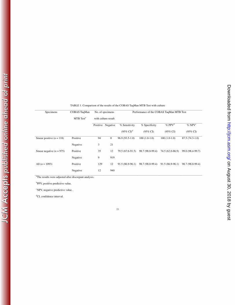
TABLE 1. Comparison of the results of the COBAS TaqMan MTB Test with culture
Specimens COBAS TaqMan
MTB Testa
No. of specimens
with culture result
Performance of the COBAS TaqMan MTB Test
Positive Negative % Sensitivity
(95% CI)d
% Specificity
(95% CI)
% PPVb
(95% CI)
% NPVc
(95% CI)
Smear positive (n = 118) Positive 94 0 96.9 (93.5-1.0) 100 (1.0-1.0) 100 (1.0-1.0) 87.5 (74.3-1.0)
Negative 3 21
Smear negative (n = 975) Positive 35 12 79.5 (67.6-91.5) 98.7 (98.0-99.4) 74.5 (62.0-86.9) 99.0 (98.4-99.7)
Negative 9 919
All (n = 1093) Positive 129 12 91.5 (86.9-96.1) 98.7 (98.0-99.4) 91.5 (86.9-96.1) 98.7 (98.0-99.4)
Negative 12 940
aThe results were adjusted after discrepant analysis.
bPPV, positive predictive value.
cNPV, negative predictive value.
dCI, confidence interval.
on August 30, 2018 by guest
http://jcm.asm
.org/D
ownloaded from
24
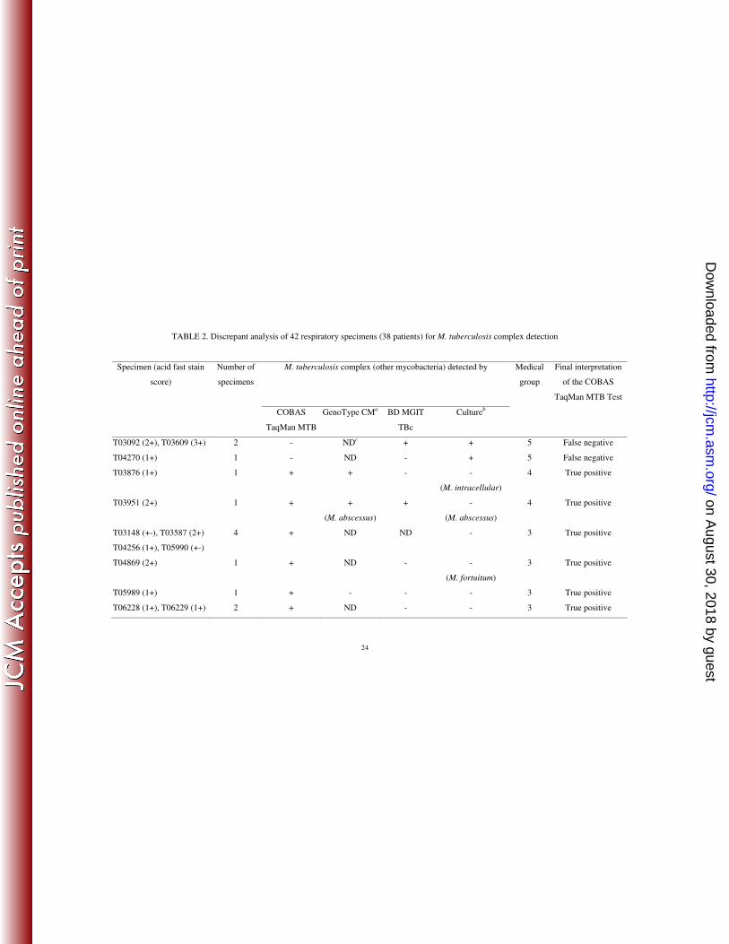
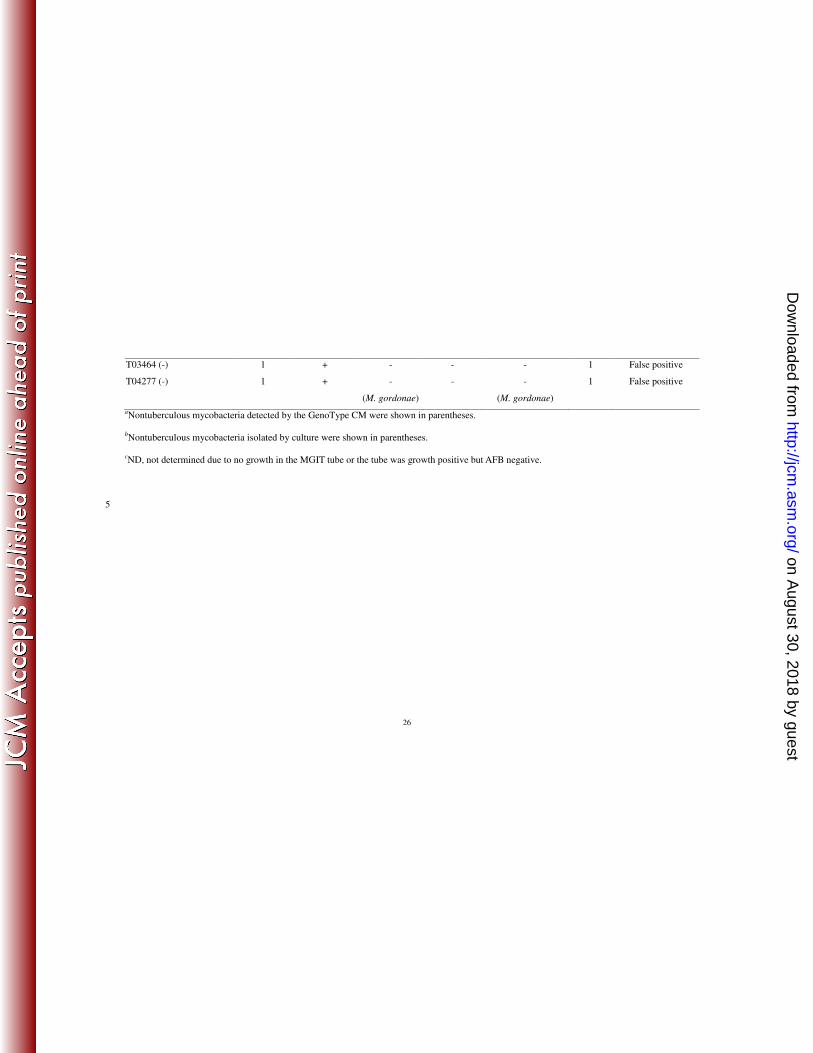
TABLE 2. Discrepant analysis of 42 respiratory specimens (38 patients) for M. tuberculosis complex detection
Specimen (acid fast stain
score)
M. tuberculosis complex (other mycobacteria) detected by Medical
group
Number of
specimens
COBAS
TaqMan MTB
GenoType CMa BD MGIT
TBc
Cultureb
Final interpretation
of the COBAS
TaqMan MTB Test
T03092 (2+), T03609 (3+) 2 - NDc + + 5 False negative
T04270 (1+) 1 - ND - + 5 False negative
T03876 (1+) 1 + + - -
(M. intracellular)
4 True positive
T03951 (2+) 1 + +
(M. abscessus)
+ -
(M. abscessus)
4 True positive
T03148 (+-), T03587 (2+)
T04256 (1+), T05990 (+-)
4 + ND ND - 3 True positive
T04869 (2+) 1 + ND - -
(M. fortuitum)
3 True positive
T05989 (1+) 1 + - - - 3 True positive
T06228 (1+), T06229 (1+) 2 + ND - - 3 True positive
on August 30, 2018 by guest
http://jcm.asm
.org/D
ownloaded from

25
T03266 (-), T03960 (-)
T04270 (-)
3 - ND - + 4 False negative
T03281 (-), T03323 (-),
T03434 (-), T03577 (-),
T03919 (-), T04265 (-)
6 - ND + + 4 False negative
T03116 (-), T03218 (-)
T03258 (-), T03949 (-)
T04077 (-), T04302 (-)
6 + ND ND - 3 True positive
T04118 (-) 1 + ND ND -
(M. kumamotonense)
3 True positive
T04332 (-) 1 + -
(M. abscessus)
- -
(M. abscessus)
3 True positive
T03090 (-), T03389 (-) 2 + ND - - 1 False positive
T03215 (-)
1 + ND ND -
(an unidentified
mycobacterium)
1 False positive
T03329 (-), T03508 (-)
T03798 (-), T03897 (-)
T04043 (-), T04058 (-)
T04193 (-)
7 + ND ND - 1 False positive
on August 30, 2018 by guest
http://jcm.asm
.org/D
ownloaded from
26
T03464 (-) 1 + - - - 1 False positive
T04277 (-) 1 + -
(M. gordonae)
- -
(M. gordonae)
1 False positive
aNontuberculous mycobacteria detected by the GenoType CM were shown in parentheses.
bNontuberculous mycobacteria isolated by culture were shown in parentheses.
cND, not determined due to no growth in the MGIT tube or the tube was growth positive but AFB negative.
5
on August 30, 2018 by guest
http://jcm.asm
.org/D
ownloaded from