Evaluation of lameness in horses
22
EVALUATION AND GRADING OF LAMENESS IN HORSES -Conducted by -Ali saqlain
-
Upload
ali-saqlain -
Category
Health & Medicine
-
view
679 -
download
3
Transcript of Evaluation of lameness in horses

EVALUATION AND GRADING OF LAMENESS IN HORSES
-Conducted by-Ali saqlain

Possible causes of Lameness • Physical• Laminitis• Tendon damage.• Ligament injuries.• Bruises or abscesses in the hoof.• Fractures.• Fibrotic myopathy• Poor foot balance.• Back and neck problems.• Degenerative joint diseases (Navicular Syndrome)• Hoof deformities • Nutritional causes• Grain overload (laminitis)• Infectious• Foot rot ( Fusobacterium necrophorum ) refered as Canker.
•

Foremost step for evaluation of Lameness is “History”
• Use• Shoeing and diet history• Previous lameness/back problems/other• Current lameness problem including:• Trauma/Duration/Recent management • Previous/current
management/medication and response

Lameness Evaluation • Observe for changes in
symmetry, posture, abnormal foot wear, etc.
• Muscles, ligaments, tendons, and joints are palpated and assessed for discomfort or stiffness .
• Hoof testers can be used to check pain response.
• Local Anesthetics can be used as desensitization will relief from pain temporarily
so affected tissue can be diagnosed.

Gait analysis• To begin your
evaluation ,choose a flat ,hard and straight surface
• Horse should be kept on loose rein to avoid head movement restriction
• Check for overall gait balance .• Check for dragging of leg. • Leg should be suspected to be
lammed which is not used for putting weight by horse.
• Check for pelvis movement moving with symmetrical fashion or tilted one side.

LungingHorse is being lunged in a circle on ground to evaluate subtle lameness. It puts more pressure on the inside of leg (front orback) and makes subtle lameness more obvious.

Flexion testing
• The horse is trotted for a baseline lameness
• Then the limbs are individually held in a flexed position as shown
• When the leg is released the horse trots away and any exacerbations in lameness are noted
• Flexing the joints in this manner may reveal problems that are not otherwise readily apparent

Palpation and Manipulation
• Hoof examination• Note the size and shape of the foot.
• Compare the normal with the abnormal. • Look for any abnormal hoof wear
ring formation, heel bulb contraction,
• Hoof wall cracks and swellings.• Abnormal hoof growth• long curled-up toes and collapsed
heels

Club hoof
• Hyperextension of Metacarpophalangeal joint (MCP hyperextension)
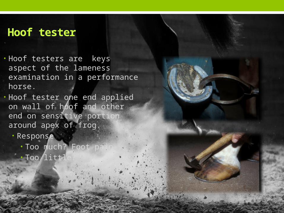
• Hoof testers are keys aspect of the lameness examination in a performance horse.
• Hoof tester one end applied on wall of hoof and other end on sensitive portion around apex of frog.• Response
• Too much? Foot pain• Too little?
Hoof tester

Fetlock
• Palpate both the dorsal and palmar aspect for any thickening and swelling of the joint capsule.
• Palpate the superficial and deep digital flexors for heat, pain or swelling.
• Palpate the sesamoid bones and the associated ligaments.

Pastern
• Palpate this region for heat and or enlargement• Compare any suspected abnormalities with the opposite pastern.• Check for any thickening of the tendons. • Rotate the joint to test for pain in the collateral ligaments.
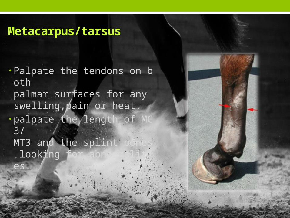
Metacarpus/tarsus
• Palpate the tendons on both palmar surfaces for any swelling,pain or heat.
• palpate the length of MC3/MT3 and the splint bones looking for abnormalities.

Nerve and joint blocks These techniques are perhaps the
most important tools used to identify the location of lameness.
Temporarily numbs sensation to specific segments of the limb, one area at a time, until the lameness disappears.
This procedure isolates the area of pain causing the lameness.
Joint blocks can also help determine whether the condition is treatable or not by intra-articular medications.

Distal interphalangeal joint analgesia
• Palmer digital nerve block ( lower branches)
• Palmer digital nerve block (supplies to Deep digital tendon)
• Abaxial (Basisesamoid) Nerve Block

Radiographs/X-rays
• First choice for imaging bone
• Tarsus/hock • By use of Radiography
we can detect bone changes when 30% difference from normal has occurred.
• Digital radiographs have a wider latitude than conventional radiography.
Arthritis

Diagnostic Ultrasound
Suspensory ligament (Desmitis)
17
First choice for imaging soft tissuesCommonly used to image the fetus in human pregnancy
Classical sound waves that operate at frequencies far above human hearing, generally spanning 1MHz.
Utilizes the transfer and propagation of sound waves into soft tissue to provide an image.

Thermography
• Thermography is a noninvasive diagnostic imaging technique used to detect variations in body-surface temperature by measuring emitted infrared radiation can easily reveal the area of inflammation.

Magnetic Resonance Imaging (MRI)
• MRI provides the ability to see bone pathology and soft tissue structures frequently not seen by other imaging techniques
• We can see damage (edema) in bone• Soft tissue injury• Suspensory ligament• Deep digital flexor tendon• Superficial digital flexor• Soft tissues of the foot

Grading of Lameness
• Grade-1:Lameness is difficult to observe and is not consistently apparent, regardless of circumstances (e.g. under saddle, circling, inclines, hard surface, etc.).
• Grade-2: Lameness is not consistently observed at a walk or when trotting in a straight line but consistently apparent under certain circumstances (e.g. weight-carrying, circling, inclines, hard surface, etc.).
• Grade-3: Lameness is consistently observable at a trot under all circumstances.
• Grade-4: Lameness is obvious at a walk.• Grade-5: Lameness produces minimal weight bearing in motion
and/or at rest or a complete inability to move.

References :Diagnosis and Management of Lameness in the HorseBy Michael W. RossAdams and Stashak's Lameness in Horses By Gary OBJECTIVE MEASURES OF LAMENESS EVALUATION Kevin G. Keegan DVM, MS, DACVShttp://www.equisearch.com/article/the-equine-lameness-exam
http://www.centaurbiomechanics.co.uk/gait-analysis/ Horses-Health-Lameness-by-Oliver-DavisInside a Lameness Exam By Marcia King http://www.vetmed.ucdavis.edu/vmth/diagnostic_imaging/la_ultrasound/activities.cfm Merck veterinary Manual

• Conducted by • Ali Saqlain • 2012-ag-2628