Evaluación de la capacidad fecundante de espermatozoides ...
105
DEPARTAMENTO DE ZOOLOGÍA Y ANTROPOLOGÍA FÍSICA Evaluación de la capacidad fecundante de espermatozoides porcinos refrigerados y congelados Fertilizing capacity evaluation of refrigerated and frozen-thawed boar spermatozoa Elena Sellés Soriano 2008
Transcript of Evaluación de la capacidad fecundante de espermatozoides ...

DEPARTAMENTO DE ZOOLOGÍA Y ANTROPOLOGÍA FÍSICA
Evaluación de la capacidad fecundante de espermatozoides
porcinos refrigerados y congelados
Fertilizing capacity evaluation of refrigerated and frozen-thawed boar
spermatozoa
Elena Sellés Soriano 2008

Tesis Doctoral como compendio de publicaciones
Analysis of in vitro fertilizing capacity to evaluate the freezing procedures of
boar semen and to predict the subsequent fertility.
Sellés E, Gadea J, Romar R, Matás C, Ruiz S.
Reproduction in Domestic Animals. 38:66-72. 2003.
Decrease in glutathione content in boar sperm after cryopreservation. Effect
of the addition of reduced glutathione to the freezing and thawing
extenders.
Gadea J, Sellés E, Marco MA, Coy P, Matás C, Romar R, Ruiz S.
Theriogenology. 62:690-701. 2004.
The predictive value of porcine seminal parameters on fertility outcome
under commercial conditions
Gadea J, Sellés E, Marco MA.
Reproduction in Domestic Animals. 39: 303–308. 2004.


A mi marido y a mis niños

A mi madre, a mi hermano y a mi tía

AGRADECIMIENTOS
Al Dr. Joaquín Gadea por su gran empeño en sacar esta tesis adelante y por animarme y hacerme luchar por ella hasta el final a pesar de mi trabajo y mis tareas personales. Gracias a él esta tesis se ha hecho realidad.
Al Dr. Salvador Ruíz por su gran apoyo y la inestimable ayuda que siempre me ha prestado con cariño desde donde estuviera en cualquier momento.
A estos dos grandes directores que me han enseñado tanto y me han ayudado a encontrar mi salida profesional.
A las Dras. Pilar Coy, Carmen Matás y Rakel Romar por ser, además de compañeras y colaboradoras, buenas amigas.
A Empar García, Manuel Sansegundo y Marco Antonio Marco que en su día emprendieron conmigo un trabajo en común donde compartimos muy buenos momentos, y aunque nuestros caminos ya se separaron aún los sigo recordando.
Al departamento de Fisiología Veterinaria y a todo su personal que me han hecho y aún me hacen sentir ese lugar como mi segunda casa.
Al Dr. Heriberto Rodríguez-Martínez por acogerme en mi estancia en Uppsala y por permitirme aprender mucho de él.
A la empresa Lo Navarro, a la ADS Cordillera Sur y a Fadesporm que en su día nos cedió las muestras de semen y los animales de granja para desarrollar nuestras experiencias.
Al matadero El Pozo por cedernos las muestras biológicas para obtener los ovocitos.
A la empresa IVI-Alicante, que de algún modo me ha concedido finalizar este trabajo.
De nuevo a mi familia, por ser lo mejor que tengo y porque gracias a sus esfuerzos me han permitido dedicar el tiempo necesario para la realización de este trabajo. Gracias a mi marido y a mis hijos por haberme prestado el tiempo que les pertenecía. Y gracias a mi madre, a mi hermano y a mi tía por haber estado ahí siempre que me han hecho falta.

Índice ÍNDICE
1. INTRODUCCIÓN GENERAL 2
2. TRABAJOS PUBLICADOS DE QUE CONSTA LA TESIS
Trabajo 1. Analysis of in vitro fertilizing capacity to evaluate the freezing
procedures of boar semen and to predict the subsequent fertility.
Sellés E, Gadea J, Romar R, Matás C, Ruiz S.
Reproduction in Domestic Animals. 38:66-72. 2003.
15
Trabajo 2. Decrease in glutathione content in boar sperm after
cryopreservation. Effect of the addition of reduced glutathione to the
freezing and thawing extenders.
Gadea J, Sellés E, Marco MA, Coy P, Matás C, Romar R, Ruiz S.
Theriogenology. 62:690-701. 2004.
23
Trabajo 3. The predictive value of porcine seminal parameters on fertility
outcome under commercial conditions.
Gadea J, Sellés E, Marco MA.
Reproduction in Domestic Animals. 39: 303–308. 2004.
36
3. SUMARIO 43
4. RESUMEN GENERAL
4.1. OBJETIVOS 46
4.2 MATERIAL Y MÉTODOS 47
4.3. RESULTADOS 59
4.4. DISCUSIÓN 61
4.5. CONCLUSIONES 72
5. REFERENCIAS BIBLIOGRÁFICAS 73
6. ABSTRACT 83
7. ANEXOS 92
Documentos acreditativos del índice de impacto de las
publicaciones incluidas en la tesis
Declaración de conformidad de los coautores

1
INTRODUCCIÓN GENERAL

Introducción
2
1. INTRODUCCIÓN
El sector porcino ha venido evolucionando en las últimas décadas
hasta convertirse en una de las principales fuentes de proteínas de origen
animal en los países desarrollados y con una gran proyección en los países
emergentes. El merecido puesto que esta industria ha alcanzado
actualmente en los mercados internacionales está estrechamente
relacionado con la evolución que este sector ha sufrido. Este desarrollo no
habría sido posible sin una amplia visión de futuro por parte de los
productores que han sabido adaptarse a los avances tecnológicos y aplicar a
tiempo las mejoras que permiten rentabilizar o mejorar las explotaciones.
Dada la importancia económica y social del sector porcino, las
investigaciones que faciliten o mejoren las producciones en esta especie
tienen una gran repercusión económica y social, tanto en nuestro país como
en el marco de la Unión Europea y en general en un mundo con una
economía globalizada.
En los últimos años se ha producido un gran desarrollo en el campo
de la reproducción porcina en las técnicas de gestión y control reproductivo,
que han ido íntimamente ligadas a la aplicación de la inseminación artificial
(IA) (Levis, 2000). Esta técnica ha permitido la máxima utilización del
potencial genético de reproductores de alto valor, ha sido una herramienta
fundamental en la prevención y lucha contra las enfermedades porcinas
(Phillpott, 1993) y ha supuesto, en definitiva, un mejor control de todo el
proceso reproductivo (Hurtgen, 1986).
En la aplicación de las técnicas de IA porcina se han realizado
numerosos e importantes avances, lo que ha permitido alcanzar una amplia
difusión en las explotaciones con unos resultados equiparables o superiores
a los obtenidos con la monta natural (Colenbrander et al., 1993). La IA
como técnica reproductiva aporta una serie de ventajas, entre las que se
encuentran (Gadea, 2004):

Introducción
3
• La amplia difusión del material genético del verraco seleccionado que
permite inseminar un mayor número de hembras.
• Mejoras sanitarias en la explotación, al evitar el contacto directo
macho-hembras, por lo que se impide la transmisión de
enfermedades por vía venérea y por contacto.
• Evaluación continua de la producción y de la calidad espermática lo
que permite monitorizar la fertilidad de los verracos a lo largo del
tiempo productivo.
• Mejora del control de los resultados reproductivos de la explotación
de forma indirecta
• La reducción en el número de verracos por hembra, con la
consiguiente reducción en costes de adquisición, alojamiento,
alimentación, etc.
La IA porcina es una técnica reproductiva de amplia aplicación en los
países desarrollados, aunque el grado de utilización es muy variable (Levis,
2000). En el contexto europeo, la tasa de aplicación de la IA es muy
elevada, llegando a porcentajes superiores al 80% en algunos países como
Holanda, Francia, Alemania, España, Noruega, Finlandia, etc. Mientras que,
por el contrario, en los Estados Unidos el porcentaje de utilización de la IA
es aún reducido (del orden del 50%), aunque en los últimos años se ha
producido un incremento muy destacable. Según algunas estimaciones, en
el mundo se realizan unos 19 millones de inseminaciones por año, de las
cuales la práctica totalidad (99%), se realiza con semen refrigerado a 15–
20ºC (Johnson et al., 2000). De estas inseminaciones, más del 85% se
realizan en el mismo día de recogida o al día siguiente.
La utilización de la IA con semen refrigerado se ha extendido gracias
al desarrollo de diluyentes que permiten la conservación del semen con
excelentes resultados. Pero su principal limitación radica en la vida útil del
semen que varía entre 2-5 días. Actualmente, se consigue una conservación
del semen refrigerado a 15ºC de hasta 7 días con diluyentes denominados
de larga duración (Johnson et al., 2000; Gadea, 2003).

Introducción
4
La utilización de la IA con semen congelado queda hoy día limitada a
casos muy específicos, bien asociada a la introducción en las explotaciones
de nuevo material genético de alto valor para inseminar determinados
animales de razas puras en las granjas de selección, o bien asociada a
labores de investigación (Johnson, 1985; Waberski et al., 1994). Sin
embargo, el uso de semen congelado puede aportar ciertas ventajas sobre
el semen refrigerado, como son (Gadea, 2004):
• El intercambio de material genético a larga distancia y durante un
periodo muy largo (años). Este periodo de tiempo puede ser crucial para
efectuar un control sanitario o genético del semen/verraco antes de su
uso. Eso es posible hoy con el diagnóstico de enfermedades infecciosas
basado en el estudio de la presencia de ADN del agente infeccioso
mediante técnicas de PCR (Reacción de la polimerasa en cadena) y
además, el desarrollo de marcadores genéticos asociados a la producción
es un proceso creciente.
• La creación de bancos genéticos. De evidente interés en el caso de la
conservación de razas en peligro de extinción y de grandes posibilidades
para la conservación de líneas o estirpes de especial interés. Estos
bancos de genes pueden ser realmente importantes a nivel comercial,
por ejemplo, para asegurar la conservación de líneas genéticas valiosas
ante posibles situaciones desfavorables (epizootía, incapacidad para
recogida, infertilidad/subfertilidad por altas temperaturas, etc.).
• La introducción de material genético de alto valor sin los riesgos
derivados de la incorporación de nuevos animales en la explotación.
Especialmente aplicable a las líneas genéticas puras que se utilizan en
las zonas altas de la pirámide de selección (“abuelas”).
La reducción en el rendimiento reproductivo ha sido el gran limitante
del uso del semen congelado (Johnson, 1985; Waberski et al., 1994). Sin

Introducción
5
embargo, en los últimos años se ha realizado un gran esfuerzo para mejorar
la fertilidad del semen congelado en inseminación artificial obteniéndose
resultados a nivel experimental muy prometedores, que suponen tasas de
partos por encima del 70% y para algunos machos por encima del 80%
(Thilmant, 1998; Eriksson et al., 2002; Sellés et al., 2003; Roca et al.,
2003; Bolarin et al., 2006). Por tanto, hoy en día con estos resultados sería
conveniente evaluar si es rentable comercialmente el uso del semen
congelado para determinadas aplicaciones específicas. En cualquier caso se
debe desterrar la idea muy difundida entre el sector que afirma que los
espermatozoides porcinos no se pueden congelar de forma adecuada y su
uso no permite unos rendimientos reproductivos óptimos. Esta misma idea
incierta se difunde en sentido contrario cuando hacemos referencia al
semen congelado en el ganado vacuno de leche, donde la mayor parte de la
inseminación artificial se realiza con semen congelado, cuando la fertilidad
media obtenida no supera en el mejor de los casos el 70% (Kastelic y
Thundathil, 2008).
El éxito en el proceso de congelación de los espermatozoides porcinos
dependen de un gran número de factores que, de forma conjunta,
determinan la calidad del semen descongelado (Roca et al., 2006; Mazur et
al., 2008). En numerosos estudios se han descrito grandes diferencias en la
capacidad de congelación que presentan los espermatozoides de distintos
machos (Johnson, 1985; Medrano et al., 2002; Hernández et al., 2006), que
afectan tanto a la viabilidad de los espermatozoides tras la descongelación
como a la fertilidad in vivo (Johnson et al., 1981; Bwanga, 1991; Roca et al.,
2006). De manera tradicional, los machos han sido clasificados como
“buenos” o “malos congeladores” (Medrano et al., 2002). Hasta hace muy
poco tiempo se desconocían las posibles causas ciertas de esta variabilidad,
de manera que el único planteamiento viable era la de optimizar los procesos
de congelación para reducir al máximo la variabilidad y descartar aquellos
machos realmente malos congeladores. Recientemente, se han encontrado los
principios de una base genética que justifica estas diferencias (Thurston et al.,

Introducción
6
2002) y que abre nuevas posibilidades en la selección de reproductores de
acuerdo a su capacidad de congelación.
Se han estudiado diversos factores que pueden mejorar la eficiencia
de la técnica: la curva de congelación (con sistemas automatizados o
manuales) así como las condiciones óptimas para la descongelación (Fiser
et al., 1993; Hernández et al., 2007), las diferencias en la capacidad de
congelación de las diversas fracciones del eyaculado (Sellés et al., 2001;
Peña et al., 2006), el método de preparación de las muestras seminales
(Matás et al., 2007), las condiciones en las que se produce la inseminación
(Wabersky et al., 1994; Bertani et al., 1997; Bolarín et al., 2006), así como
el efecto de la estación, la línea genética y el verraco en la capacidad de
congelación (Gadea et al., 2003, Roca et al., 2006).
En los últimos años, los estudios se han centrado en estudiar las
alteraciones que produce la congelación en la célula espermática, lo que
permite conocer y comprender el proceso lesivo y la manera práctica de
reducir o minimizar los cambios en la función de la célula espermática. En
este sentido, se conoce desde hace décadas que el choque frío induce
severas alteraciones en la estructura de la membrana espermática que
llevan a la muerte celular (Pursel et al., 1973). Estas alteraciones de
membrana están relacionadas con la formación de cristales durante el
proceso de congelación (Mazur et al., 1997). El daño que ocasionan los
cristales es proporcional a la cantidad y el tamaño de los mismos (Watson,
1995).
Posteriormente, se puso de manifiesto que el proceso de congelación
además de producir una alteración sobre la estructura de la membrana
espermática producía una alteración en sus componentes que podrían llevar
a modificaciones severas de la funcionalidad espermática. Uno de los
mayores efectos de la congelación se produce sobre la composición,
distribución y estabilidad de los lípidos de la membrana espermática que se

Introducción
7
alteran de forma similar a los cambios producidos durante la capacitación
espermática (Cerolini et al., 2001). Es por ello que a estas modificaciones
se le han denominado “tipo capacitación (“capacitation like changes”,
Watson, 1995; Green & Watson, 2001; Kaneto et al., 2002; Bravo et al.,
2005). Del mismo modo recientemente se han relacionado los cambios que
se producen en las membranas espermáticas durante la congelación a los
que se producen en el proceso de apoptosis (Peña et al., 2003; Trzcinska et
al., 2006). Esta información sobre los procesos básicos puede ser vital para
que posteriormente pueda ser aplicada en el diseño de nuevos protocolos y
diluyentes que permitan minimizar el daño espermático.
Por último mencionaremos el especial interés que tiene el estudio de
la desestabilización del sistema antioxidante del espermatozoide durante la
congelación. En este sentido, se conoce que los procesos de refrigeración y
congelación producen una alteración física y química de las membranas
espermáticas que tiene como consecuencia la reducción de la viabilidad
celular y de su capacidad fecundante. Las alteraciones producidas por la
reducción de la temperatura están asociadas con el denominado estrés
oxidativo, estando inducido por la generación de agentes oxidantes (ROS)
(Chatterjee et al., 2001). La consecuencia final es la peroxidación de los
lípidos y una alteración grave de la funcionalidad espermática.
El semen representa un complejo sistema redox que combina el
potencial antioxidante del plasma seminal y de los espermatozoides con el
potencial pro-oxidante del espermatozoide a través de la generación de
ROS. El sistema defensivo antioxidante incluye una actividad enzimática
(superóxido dismutasa, glutatión reductasa, glutatión peroxidasa y
catalasa), así como la presencia de diversas sustancias con actividad
antioxidante (glutatión reducido (GSH), urato, ácido ascórbico, vitamina E,
taurina, hipotaurina, carotenoides y ubiquinonas). El glutatión (L-g-
glutamil-L-cisteinilglicina) es un tri-péptido distribuido en todas las células
del organismo y juega un papel decisivo en el mecanismo de defensa

Introducción
8
intracelular frente el estrés oxidativo. La enzima glutatión peroxidasa usa
GSH como agente para reducir el peróxido de hidrógeno hasta agua y el
lipoperóxido hasta alquil-alcohol. Por otro lado, la forma oxidada del
glutatión (GSSG) se reduce hasta GSH mediante la enzima glutatión
reductasa usando NADPH como cofactor. El contenido de glutatión (principal
agente antioxidante no enzimático) se reduce durante el proceso de
congelación (Bilodeau et al., 2000, Gadea et al., 2004; Molla et al., 2004),
así como se alteran las proteínas de la membrana espermática (Gadea et
al., 2004).
Para evitar el proceso de oxidación producido durante la congelación
se ha estudiado la adición de antioxidantes en los medios de congelación y
descongelación con relativo éxito. Así se ha ensayado el uso de diversas
sustancias como el tocoferol y análogos (Peña et al., 2003; Breinenger et
al., 2005; Satorre et al., 2007), superóxido dismutasa (Roca et al., 2005) y
el glutatión reducido (Funahashi y Sano, 2005; Gadea et al., 2005).
Los resultados de estas investigaciones podrán permitir en un futuro
próximo conocer con profundidad los daños que se producen en la célula
espermática durante la congelación-descongelación. Con esta información
básica se podrá diseñar sistemas de congelación (curvas de congelación,
diluyentes, crioprotectores, envases, etc…) que mejoren la calida seminal
tras la descongelación y mejoren los rendimientos reproductivos.
En todo este proceso de mejora en la congelación de
espermatozoides porcinos es necesario el empleo de determinadas técnicas
analíticas que miden parámetros de calidad seminal. Por calidad seminal se
entiende el conjunto de parámetros que caracterizan la viabilidad de la
célula espermática. En un principio se hacía referencia a los caracteres que
definen la morfología y el movimiento de los espermatozoides (Larsson,
1986), pero posteriormente se le han añadido otra serie de parámetros que
tienen por objetivo cuantificar de algún modo la funcionalidad del
espermatozoide (Berger et al., 1996).

Introducción
9
Mucho se ha avanzado en las últimas décadas en el campo de la
evaluación de la calidad seminal y sobre el valor predictivo de la fertilidad
que presentan las pruebas de análisis espermático. Sin embargo, es una
tarea aún no resuelta. En este sentido, la calidad del eyaculado ha sido
tradicionalmente evaluada con el espermiograma clásico, basado en la
aplicación de una serie de pruebas de una ejecución relativamente simple y
que pueden ser realizadas con un coste moderado (Gadea, 2005). En el
análisis rutinario se incluye un examen macroscópico y microscópico del
eyaculado en los que se mide el volumen, la concentración, la motilidad, el
estado del acrosoma y las morfoanomalías espermáticas.
En el trabajo diario en los centros de inseminación artificial se
detectan animales que tienen una fertilidad reducida pero que al realizar un
análisis de rutina presentan un espermiograma normal. Por ello, podemos
deducir que ninguno de los parámetros del espermiograma clásico por sí
solo parece ser suficiente para predecir adecuadamente la fertilidad, aunque
la información combinada de todos ellos ofrece una buena estimación de la
calidad seminal (Woelders, 1991).
Para dar una solución a este problema se han desarrollado nuevas
técnicas que pretenden alcanzar un mejor conocimiento de la célula
espermática (Gadea, 2005; Silva y Gadella, 2006; Rodríguez-Martínez y
Barth, 2007). Con este objetivo se puede evaluar la estructura y
funcionalidad del espermatozoide. Entre estas nuevas técnicas, el estudio de
la membrana parece ser un buen procedimiento para evaluar la funcionalidad
del gameto masculino, ya que ésta interviene activamente en la mayoría de
las fases del proceso reproductivo. La membrana puede ser estudiada desde
el punto de vista estructural mediante la utilización de tinciones, o bien
valorando su funcionalidad, para lo que se ha aplicado el test hipoosmótico
(Jeyendran et al., 1984; Vázquez et al., 1997) y distintas técnicas con
fluorocromos que permiten evaluar otros aspectos de la funcionalidad
espermática como son los procesos de capacitación espermática y reacción

Introducción
10
acrosómica, la actividad mitocondrial, la permeabilidad de membrana, la
estructura y estabilidad del núcleo y la cromatina, etc… (Harrison y Vickers,
1990; Garner y Johnson, 1995; Silva y Gadella, 2006).
Los estudios bioquímicos se desarrollaron con la intención de tener una
medición objetiva y fácilmente reproducible de la calidad seminal y que
fueran reflejo de su actividad funcional. Se han realizado estudios
metabólicos y enzimáticos, cuantificándose los diferentes componentes
químicos presentes en el eyaculado y que pueden condicionar la actividad del
espermatozoide como son el contenido en iones, la liberación de enzimas o el
contenido de ATP (Strzezek y Skaweta, 1984; Ciereszko et al., 1994; Gadea
et al., 1998). Posteriormente, los estudios de la composición lipídica de la
membrana espermática porcina han puesto de manifiesto que tanto los
procesos de capacitación (Shadan et al., 2004) como los de congelación
(Cerolini et al., 2001; Maldjian et al., 2005; Waterhouse et al., 2006) están
íntimamente relacionados con los cambios que se producen en el
componente lipídico de la membrana.
Por otra parte, el estudio del núcleo del espermatozoide permite
valorar la madurez y la estabilidad del mismo, condiciones necesarias para
que pueda llegar a producirse la descondensación cromosómica y la
singamia. Estos estudios se desarrollaron cuando se asociaron problemas de
fertilidad con alteraciones de la estructura del núcleo (Evenson et al., 1994).
Estas alteraciones del núcleo han sido clasificadas como factores
incompensables de la fertilidad (Saacke et al., 1994), ya que no es posible
mejorar la fertilidad aumentando el número de espermatozoides por dosis de
inseminación.
Los procesos de capacitación y reacción acrosómica son pasos
fundamentales para que se lleve a cabo la fecundación (Flesh y Gadella,
2000). Por ello, se ha realizado un importante esfuerzo en estudiar los
Introducción
11
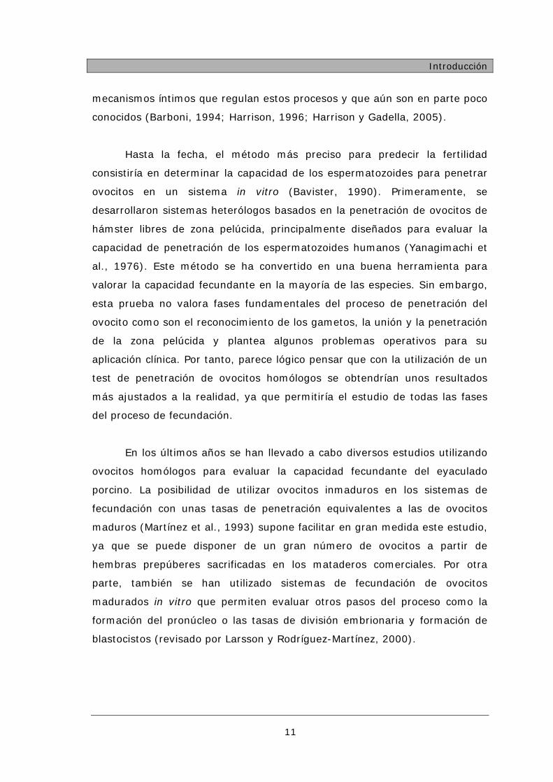
mecanismos íntimos que regulan estos procesos y que aún son en parte poco
conocidos (Barboni, 1994; Harrison, 1996; Harrison y Gadella, 2005).
Hasta la fecha, el método más preciso para predecir la fertilidad
consistiría en determinar la capacidad de los espermatozoides para penetrar
ovocitos en un sistema in vitro (Bavister, 1990). Primeramente, se
desarrollaron sistemas heterólogos basados en la penetración de ovocitos de
hámster libres de zona pelúcida, principalmente diseñados para evaluar la
capacidad de penetración de los espermatozoides humanos (Yanagimachi et
al., 1976). Este método se ha convertido en una buena herramienta para
valorar la capacidad fecundante en la mayoría de las especies. Sin embargo,
esta prueba no valora fases fundamentales del proceso de penetración del
ovocito como son el reconocimiento de los gametos, la unión y la penetración
de la zona pelúcida y plantea algunos problemas operativos para su
aplicación clínica. Por tanto, parece lógico pensar que con la utilización de un
test de penetración de ovocitos homólogos se obtendrían unos resultados
más ajustados a la realidad, ya que permitiría el estudio de todas las fases
del proceso de fecundación.
En los últimos años se han llevado a cabo diversos estudios utilizando
ovocitos homólogos para evaluar la capacidad fecundante del eyaculado
porcino. La posibilidad de utilizar ovocitos inmaduros en los sistemas de
fecundación con unas tasas de penetración equivalentes a las de ovocitos
maduros (Martínez et al., 1993) supone facilitar en gran medida este estudio,
ya que se puede disponer de un gran número de ovocitos a partir de
hembras prepúberes sacrificadas en los mataderos comerciales. Por otra
parte, también se han utilizado sistemas de fecundación de ovocitos
madurados in vitro que permiten evaluar otros pasos del proceso como la
formación del pronúcleo o las tasas de división embrionaria y formación de
blastocistos (revisado por Larsson y Rodríguez-Martínez, 2000).

Introducción
12
La predicción de la capacidad fertilizante del semen tiene una gran
importancia en las producciones ganaderas cuando se hace uso de la IA, así
como para la selección de machos con mejor valor reproductivo
(Hammerstedt, 1996). Este hecho es de especial importancia cuando se hace
uso de semen congelado, cuya viabilidad y funcionalidad está modificada por
el proceso de conservación. De momento, sólo unos pocos estudios
correlacionan de manera significativa los factores espermáticos con la
fertilidad. Esto es debido a que hay un gran número de factores que modulan
esta relación y que están asociados con el potencial fértil de las hembras
(Gadea, 2005). Algunos autores proponen como otra causa de la
inconsistencia de los resultados conseguidos, la evaluación de un número
relativamente reducido de células, la gran influencia de la subjetividad del
observador y la alta variabilidad que presentan muchos análisis seminales
(Evenson et al., 1994; Saacke et al., 1994). Además, el número de
espermatozoides aplicados en cada inseminación es un factor muy
importante a la hora de evaluar la fertilidad y su relación con los parámetros
de calidad seminal (Fearon y Wegener, 2000).
La mayoría de los análisis in vitro utilizados hasta el momento ofrecen
información sobre la calidad seminal, que es fundamental para los estudios
en general de la fisiología del espermatozoide y en particular para la
conservación del semen. Sin embargo, lo realmente importante para el sector
productivo porcino es el estudio de los caracteres relacionados con el proceso
de fecundación, es decir la determinación in vitro de la capacidad fecundante
de los eyaculados in vivo (Woelders, 1991).
La evaluación de la calidad seminal es una parte importante y un
punto crítico en el proceso de la inseminación artificial, ya que en muchos
casos, verracos asociados con una fertilidad reducida presentan alteraciones
detectables mediante un examen rutinario del semen. No obstante, aunque
es necesaria una buena calidad seminal para alcanzar unos niveles de
fertilidad aceptables, no todos los eyaculados con buena calidad seminal

Introducción
13
mantienen niveles de fertilidad dentro de la normalidad (Berger y Parker,
1989; Martínez et al., 1993).
En resumen, podemos afirmar que la IA porcina se ha desarrollado de
manera muy importante pero sin duda quedan aún importantes temas en los
que es posible trabajar para mejorar los resultados productivos. Esta
problemática nos obliga a profundizar en el estudio de técnicas que permitan
avances en la crioconservación espermática desarrollando investigaciones en
la mejora de los sistemas de congelación-descongelación y en los sistemas
de evaluación de la calidad del semen tanto refrigerado como congelado-
descongelado en la eficacia del uso en condiciones de campo.

2
TRABAJOS PUBLICADOS DE QUE CONSTA LA
TESIS

Analysis of in vitro fertilizing capacity to evaluate the
freezing procedures of boar semen and to predict the
subsequent fertility.
Sellés E, Gadea J, Romar R, Matás C, Ruiz S.
Reproduction in Domestic Animals. 38:66-72. 2003.

Analysis of In vitro Fertilizing Capacity to Evaluate the Freezing Procedures of BoarSemen and to Predict the Subsequent Fertility
E Selles, J Gadea, R Romar, C Matas and S Ruiz
Department of Physiology, Faculty of Veterinary Medicine, University of Murcia, Spain
Contents
A porcine in vitro fertilization (IVF) system and seminalquality parameters of frozen–thawed boar semen were used toassess the effectiveness of two different thawing rates of frozenboar semen, and to address the question of whether differencesbetween fertility of ejaculates could be predicted in a limitedfield trial.
In the first experiment, two thawing procedures wereanalysed (37�C, 30 s; 50�C, 12 s) and no differences in spermquality were found. However, when the procedure was 50�C,12 s the IVF results showed a higher number of sperm perpenetrated oocyte and a near 10 points higher rate ofpronuclear formation.
In the second experiment, the fertility results obtained in thelimited field trial show to be efficient enough for application ina commercial use, especially for three of the employed boars(fertility ‡80%). In this limited study, the conventional seminalparameters are not accurate enough to discriminate good andbad boars in relation to fertility. On the contrary, parametersof in vitro penetrability are more precise to predict subsequentfertilities.
As conclusion, the IVF fertilization system seems to be agood tool to evaluate the quality of frozen–thawed boar semenprevious to its commercial way, to verify the bank semenstorage quality and a good way to assay new sperm freezingprocedures, as it is the more precise evaluating method inestimating the potential fertilizing ability.
Introduction
Despite almost 40 years of research in freezing of boarsemen, the fertility results are not satisfactory enoughfor commercial use as it is in other domestic animals.However boar frozen–thawed semen is still a valuabletool as a complement to artificial insemination (AI) withfresh semen in some conditions. As it can be stored for along time, it facilitates the supply of genetic material, aswell as building up gene banks to encourage breeds orvaluable individuals. In the last years, an increasingeffort has been made to improve the fertility resultsmainly by to ways: first the design of better freezingmethods in order to obtain acceptable semen quality(freezing procedures, diluents and cryoprotectants,packages, etc.) (reviewed by Bwanga 1991; Johnsonet al. 2000), secondly with optimal routines for heatdetection and timing of insemination close to theovulation (Waberski et al. 1994).
Evaluation of the quality of frozen–thawed semen isan important goal and great deals of assays have beendeveloped (Johnson et al. 1996). Besides, assays inclu-ding the study of gamete interaction might lead to abetter way of predicting male fertility than routinelaboratory evaluation of semen (Gadea et al. 1998;Larsson and Rodriguez-Martinez 2000). Some of these
assays have been shown to be good tools for evaluatingthe fertilizing capacity of diluted boar semen (Ivanovaand Mollova 1993; Gadea et al. 1998; Xu et al., 1998).However, little information is available about frozenboar semen (Hammitt et al. 1989; Gadea et al. 2001;Pelaez et al. 2001), but it would be very useful toevaluate freezing procedures (Eriksson et al. 2000).
One of the most important factors related with thesuccess of freezing procedures seems to be the thawingrate of semen (Johnson et al. 2000). So, the criticaltemperature range during thawing is an importantfactor affecting spermatozoan viability (Fiser et al.1993). However, it has been demonstrated that theeffectiveness of thawing rate also depends on theoriginal rate of freezing (Mazur 1985). Different studieshave previously described that when thawing rate isincreased (in an optimum range), the motility andacrosome integrity are improved (Pursel and Johnson1975; Fiser et al. 1993).
For these reasons the objectives of this study were tostudy the application of the in vitro fertilization (IVF)systems: (1) to assess the effectiveness of two differentthawing rates of frozen boar semen, and (2) to addressthe question of whether differences between fertility ofejaculates could be predicted with semen qualityparameters and with IVF fertilization systems in alimited field trial.
Material and Methods
Semen collection and freezing
Semen was regularly collected from five mature fertileboars (one Belgium Landrace and four Pietrain18–30 month-old boars) using the hand method and adummy. Sperm-rich fraction was collected in a pre-warmed thermos flask and the gel-fraction was held on agauze tissue covering the thermos opening. The volumeof the sperm-rich fraction of the ejaculate was measuredin a graduated cylinder and sperm concentrationmeasured with a haemacytometer (Neubauer, Brand,Wertheim, Germany) within 20 min after collection andprior to extension of the semen with isothermal Betsvillethawing solution (BTS; Minitub, Tiefenbach, Germany)extender at a ratio of 1 : 1. Semen was stored at 22�C for2 h, and processed according to the straw-freezingprocedure described by Westendorf et al. (1975) andAlmlid and Johnson (1988). Briefly, diluted semen wasplaced at 15�C for 150 min (in a water bath placed into afreezer) and later centrifuged at 800 · g for 10 min. Thesupernatant was discarded and the semen pellet wasre-suspended with lactose–egg yolk extender (LEY;80 ml of 11% lactose and 20 ml egg yolk) to provide
Reprod Dom Anim 38, 66–72 (2003)
� 2003 Blackwell Verlag, Berlin
ISSN 0936-6768
U.S. Copyright Clearance Centre Code Statement: 0936–6768/2003/3801–0066$15.000/0 www.blackwell.de/synergy

1.5 · 109 spermatozoa per ml. Then, it was cooled at5�C for 90 min (in a water bath placed into a freezer)and two parts of LEY extender semen were mixed withLEY extender with 9% glycerol and 1.5% Orvus EsPaste (Equex-Paste; Minitub, Tiefenbach, Germany).The final concentration of semen to be frozen was1 · 109 spermatozoa per ml and 3% glycerol. Thecooled semen was leaded into 0.5 ml. straws (Minitub,Tiefenbach, Germany) and sealed with polyvinyl alco-hol. Straws were wiped dry and the air bubble wasbrought to the centre of the straw. The straws wereplaced in contact with nitrogen vapour about 3 cmabove the liquid nitrogen level for 20 min in anexpandable polystyrene box, plunged into the nitrogentank and stored until use (1–3 months later). Strawswere thawed in a circulating water bath at 50�C for 12 sor at 37�C for 30 s and immediately diluted in 10 mlBTS at 37�C.
Seminal parameters
Sperm motility and movement quality were determinedplacing two sub-samples on warm glass slides (39�C)and examined under a light microscope (100 · magni-fication). The percentage of motile sperm cells wassubjectively estimated to the nearest 5% and theforward progressive motility (FPM) using an arbitraryscale of 0–5.
The proportion of spermatozoa with a normal apicalridge (NAR) was evaluated after fixed in buffered 2%glutaraldehyde solution and examined under a phase-contrast microscope (Leica DMR, Wetzlar, Germany)(1000 · magnification) to analyse acrosomes (Purselet al. 1972). The NAR was determined on two slidesper sample and a total of 300 spermatozoa per sample.
Eosin–nigrosin (EN) viability staining of sperm wasalso studied. It was diluted at a ratio of 1 : 1 a semensample with staining solution (5% yellow eosin, 10%nigrosin in a citrate solution pH ¼ 7.4) and smeared.After air-fixed stained spermatozoa were observed andevaluated 200 sperm per sample (Bamba 1988).
Sperm membrane integrity was evaluated applying acombination of the fluorophores carboxyfluoresceindiacetate (DCF) and propidium iodide (Harrison andVickers 1990) on at least 200 cells per sample using anepifluorescence microscope.
In vitro maturation and in vitro fertilization
Ovaries from pre-puberal gilts were obtained at a localslaughterhouse, and transported to the laboratory insaline (0.9% w/v NaCl) with 100 mg/l kanamycin at35�C. Oocytes surrounded by cumulus cells, were slicingfrom 3 to 6 mm diameter follicles and washed twice in35 mm plastic Petri dishes containing Dulbecco’sphosphate-buffered saline modified supplemented with1 mg/ml polyvinyl alcohol and 0.005 mg/ml red phenol.They were washed twice again in maturation mediumpreviously equilibrated for a minimum of 3 h at 38.5�Cunder 5% CO2 in maximally humidified air.
The medium used for oocyte maturation was Way-mouth-supplemented as previously described (Coy et al.1999) with 10 UI/ml pregnant mare serum gonadotro-
phin (PMSG), 10 UI/ml human chorionic gonadotropin(hCG), 1 lg/ml oestradiol-17b, 10% (v/v) foetal calfserum and 10% porcine follicular fluid (v/v). Thematuration medium was disposed in three droplets of100 ll covered with paraffin oil per dish, 20 oocytes perdroplet and kept at 38�C under 5% CO2 in air. After 20–22 h of culture in maturation medium the oocytes weretransferred to fresh maturation medium without hor-monal supplements, washed twice and cultured for anadditional 20–22 h (Funahashi and Day 1993).
The in vitro fertilization medium was TCM199-supplemented as previously described (Coy et al. 1999)with 12% heat-inactivated foetal calf serum, 0.9 mMsodium pyruvate, 3.05 mM D-glucose, 8.75 mM calciumlactate, 0.68 mM penicillin G, 3.6 mM caffeine and0.068 mM streptomycin sulphate at pH ¼ 7.4.
After thawing, the sperm samples were centrifugedat 50 ·g for 3 min and the supernatants at 1200 · gfor 3 min. Resulting pellets of spermatozoa werediluted in TCM199-supplemented medium but withoutcalcium lactate nor caffeine. A semen volume of100 ll was introduced into Petri dishes containing2 ml of fertilization medium (final concentration of1 · 106 spermatoza/ml) and 20 in vitro maturedoocytes previously washed twice in equilibrated fertil-ization medium. After 18 h the cultured oocytes werefixed in 3 : 1 ethanol : acetic acid for 24 h, stainedwith 1% lacmoid and examined under a phase-contrast microscope to asses penetration (PEN) rate,mean number of sperm per penetrated oocyte (S/O),monospermy (MON) rate and rate of male pronuclearformation (MPF).
Fertility trial
The fertility study was conducted on a commercial farm,using a total of 45 multiparous (two to seven pregnan-cies) crossbred sows. Oestrus was checked daily in thepresence of a mature teaser boar. Occurrence of oestruswas defined by the standing reflex in front of a boar(back pressure test) and reddening and swelling of thevulva. The sows were inseminated immediately afterthawing the semen at 50�C for 12 s and diluted withBTS to prepare insemination doses containing at least5 · 109 spermatozoa in 80 ml. Insemination took placeon 12 h after the diagnosis of oestrus and was repeated12 h later, using disposable AI catheters.
Pregnancy diagnosis was performed 23–25 days afterAI by ultrasonography. Fertility was measured for everyejaculate as the percentage of sows farrowing to AI. Foreach sow that farrowed, the number of dead and livepiglets was counted and the sum was defined as the totalnumber of piglets born.
Experimental design
Experiment 1. Thawing process
Five ejaculates from the same boar were frozenaccording to the methods described before and werethawed at each of the following test velocities resultingfrom immersion in water to 37�C for 30 s or at 50�Cfor 12 s. Seminal parameters and IVF capacity wereassayed.
Analysis of In vitro Fertilizing Capacity 67

Experiment 2. Fertility trial
To evaluate the capacity for fertility prediction ofdifferent assays and to verify the quality of the frozen–thawed semen, ejaculates from five fertile boars werefrozen–thawed and they were used both in an IVFsystem and in a field assay by AI with at least5 · 109 sperm per dose. Seminal parameters were eval-uated and differences between boars were investigated inblind fashion.
Statistical analysis
Data are presented as mean ± SEM. Data for all rateswere modelled according to the binomial model ofparameters and were analysed by two-way ANOVA;considering the thawing procedure and sperm batch asmain effects in experience 1 and one-way ANOVA inexperience 2. When ANOVA revealed a significant effect,values were compared by the Tukey test. Differenceswere considered statistically different at p<0.05.
Linear regression was used to further investigaterelationships between litter size and measured semenparameters (Pearson correlation and multiple regres-sion), and logistic regression was used to relate thedichotomous farrowing rate data to the sperm param-eters, as previously described by Holt et al. (1997).
Results
Experiment 1
The studied thawing velocities had no effect on thesperm quality (Table 1), but a significant difference on
the motility and NAR (p<0.001) was detected betweenbatches. For both treatments the motility and mem-brane integrity (measured with EN or carboxifluoresceinstaining) were over 60%.
In relation to the in vitro fertilizing capacity of thesperm, thawed under two different procedures, theresults obtained showed a higher number of S/O (3.91vs 3.06, p<0.001) (Table 2) and a near 10 points higherrate of MPF (75.47 vs 65.73, p ¼ 0.020) when theprocedure was 50�C for 12 s. Besides, the sperm batchhad a significant effect on the PEN, number of S/O andMON rate (p<0.001).
Experiment 2
The result of sperm assays showed a significant lowernumber of intact membrane (EN and DCF) andacrosomal integrity (NAR) for the frozen–thawed spermfrom boar PI4 (p<0.001, Table 3). However, all the fourIVF parameters were significantly affected by the boarstudied and showed a significant lower values forpenetrability (PEN and S/O) in PI4 and PI779 boarsagainst the other three (Table 4). In the same way, theMON was affected by boar and showed the highestvalues for the boars with less penetrability. The MPFwas higher than 82% in all the boars studied and thisparameter was not related with fertility (Tables 4 and 5).
The in vitro penetrability results are consistent withthe limited data from the fertility field trial, as fertilitywas significantly affected by boar (p ¼ 0.019), beingthose with lower penetrability (boars PI4 and PI779)these are with lower fertility (33%) than the other three
Table 1. Seminal parameters measured in boar semen thawed under two different procedures (mean ± SEM)
Motility (%) FPM (0–5) EN (%) NAR (%) DCF (%)
Thawing velocity
50�C for 12 s 62.27 ± 3.53 3.64 ± 0.15 76.55 ± 1.74 49.36 ± 4.34 66.64 ± 2.53
37�C for 30 s 60.45 ± 4.01 3.73 ± 0.14 72.36 ± 1.86 48.91 ± 3.57 63.45 ± 2.41
Source of variability
Thawing velocity 0.462 0.671 0.058 0.961 0.117
Sperm batch < 0.001 0.125 0.221 < 0.001 0.082
Interaction 0.459 0.658 0.132 0.850 0.053
FPM: forward progressive motility (0–5), EN: eosin–nigrosine stain, NAR: normal apical ridge, DCF: sperm membrane integrity assessed with carboxyfluorescein
diacetate.
Table 2. The IVF results (mean ± SEM) for mature oocytes fertilized with frozen–thawed boar semen under two different procedures: rate ofpenetration (% PEN), mean number of spermatozoa per penetrated oocyte (S/O), monospermy rate (% MON) and rate of male pronuclearformation (% MPF)
n % PEN S/O* % MON* % MPF*
Thawing velocity
50�C for 12 s 230 69.13 ± 3.05 3.91 ± 0.27a 33.96 ± 3.77 75.47 ± 3.42a
37�C for 30 s 254 70.08 ± 2.88 3.06 ± 0.19b 34.83 ± 3.58 65.73 ± 3.57b
Source of variability
Thawing velocity 0.840 <0.001 0.641 0.020
Sperm batch <0.001 <0.001 <0.001 0.654
Interaction 0.949 <0.001 0.519 0.019
* Based on penetrated oocytes.a,b Numbers within columns with different superscripts differ (p<0.05).
n: number of oocytes.
68 E Selles, J Gadea, R Romar, C Matas and S Ruiz

with fertility rates over 80% (Table 4). No differencesbetween boars were detected for litter size.
When the logistic regression was analysed betweenin vitro penetration and seminal parameters with fertilityresults, significant regression coefficients were found forPEN rate and S/O, and all the quality seminal param-eters (motility, FPM, EN, NAR, DCF; Table 5,p<0.05). Later, when studying all semen parametersthrough stepwise on multiple logistic regression for-ward, only three parameters (PEN, MPF and motility)were included (Table 6, R2 ¼ 0.492; p<0.001).
In relation with litter size, significant Pearson corre-lation was found for all the IVF parameters (exceptMPF) and two quality parameters (FPM and DCF,Table 7). When a stepwise multivariate analysis wasmade a significant model (p<0.0001) constructed onlywith fertilization parameters of penetrability (PEN andS/O) that may explain the nearly 80% of variability(Table 8).
Discussion
To improve the viability and fertilizing capacity of theboar frozen–thawed semen is necessary to optimize allthe factors that have an effect in the freezing procedure.In this way we have focused our attention on one ofthem, the thawing process, and in the assessmentmethods for frozen semen evaluation.
Table 3. Seminal parameters measured in sperm thawed from five different boars used in the fertility trial (mean ± SEM)
Motility (%) FPM (0–5) EN (%) NAR (%) DCF (%)
Boar
BB 62.00 ± 2.00 3.95 ± 0.05 72.50 ± 2.88a 42.50 ± 3.49a 69.75 ± 3.17a
PI4 59.37 ± 2.17 3.75 ± 0.06 53.69 ± 2.51b 33.34 ± 2.48b 41.71 ± 3.78b
PI67 63.70 ± 1.55 3.88 ± 0.04 66.31 ± 1.40a 39.97 ± 1.44a 53.69 ± 3.48ab
PI779 58.92 ± 0.82 3.85 ± 0.04 70.73 ± 1.61a 46.76 ± 1.38a 57.41 ± 2.52a
PI89 59.30 ± 0.80 3.86 ± 0.03 69.20 ± 1.18a 51.16 ± 1.30a 56.47 ± 2.41a
Source of variability
Boar 0.053 0.208 <0.001 <0.001 <0.001
FPM: forward progressive motility (0–5), EN: eosin–nigrosine stain, NAR: normal apical ridge, DCF: Sperm membrane integrity assessed with carboxyfluorescein
diacetate.a,bNumbers within columns with different superscripts differ (p < 0.05).
Table 4. The IVF and in vivo fertility results (mean ± SEM) for mature oocytes fertilized with frozen–thawed semen from five different boars:rate of penetration (PEN), mean number of spermatozoa per penetrated oocyte (S/O), monospermy rate (% MON) and rate of male pronuclearformation (MPF)
n PEN (%) S/O* MON* (%) MPF* (%) Fertility (%) Litter size
Boar
BB 199 62.81 ± 3.43cd 2.78 ± 0.28a 48.80 ± 4.49ab 88.80 ± 2.83ab 80 (4/5) 11.5 ± 1.19
PI4 212 44.34 ± 3.42b 1.65 ± 0.13b 61.70 ± 5.04ab 82.98 ± 3.90b 33.33 (2/6) 6 ± 0
PI67 329 67.17 ± 2.59d 2.21 ± 0.11ab 40.72 ± 3.31b 87.33 ± 2.24b 84.62 (11/13) 9.18 ± 1.54
PI779 320 23.13 ± 2.36a 1.63 ± 0.13b 63.51 ± 5.63a 94.59 ± 2.65ab 33.33 (3/9) 10.67 ± 0.67
PI89 308 52.27 ± 2.85bc 2.54 ± 0.18a 50.93 ± 3.95b 98.14 ± 1.07a 83.33 (10/12) 7.3 ± 1.46
Source of variability
Boar <0.001 <0.001 0.001 <0.001 0.019 0.405
* Based on penetrated oocytes.a,b,c,d Numbers within columns with different superscripts differ (p<0.05).
n: number of oocytes.
Table 5. Logistic regression of seminal parameters with fertility result(farrowing rate)
Variable Coefficient B SE t Significance (p)
PEN 0.0208 0.0071 2.9402 0.0033
S/O 0.4384 0.1758 2.4935 0.0127
MPF 0.0063 0.0035 1.8046 0.0711
MON 0.0043 0.0050 0.8577 0.3910
Motility 0.0115 0.0051 2.2360 0.0253
FPM 0.1842 0.0821 2.2445 0.0248
EN 0.0107 0.0047 2.2654 0.0235
NAR 0.0164 0.0072 2.2870 0.0222
DCF 0.0135 0.0058 2.3482 0.0189
PEN: rate of penetration, S/O: mean number of spermatozoa per penetrated
oocyte, MPF: rate of male pronuclear formation, MON: monospermy rate,
FPM: forward progressive motility (0–5), EN: eosin–nigrosine stain, NAR:
normal apical ridge, DCF: Sperm membrane integrity assessed with carboxy-
fluorescein diacetate, SE: standard error.
Table 6. Multiple logistic regression of seminal measurements withfertility result (farrowing rate)
Variable Coefficient SE t Significance (p)
Constant )46.1846 24.9668 )1.8498 0.0643
PEN 0.3747 0.2228 1.6813 0.0927
MPF 0.8055 0.4882 1.6499 0.0990
Motility )0.6857 0.5216 )1.3145 0.1887
McFadden’s R2 ¼ 0.492; log likelihood: )27.373; SE: standard error.
Significance of statistical model p<0.001.
Analysis of In vitro Fertilizing Capacity 69

In the first experiment, we have found no differencesin sperm quality with two thawing procedures near tothe optimal conditions and previously accepted to thawfrozen boar semen in ministraws (Maxwell and Johnson,1997; Erkisson et al. 2000). However, Fiser et al. (1993)found that after freezing at optimal rate, the percentageof motile spermatozoa and sperm with NAR increasedgradually while increasing warming velocity. Theystudied a wide range of velocities, some of them out ofoptimal conditions and those results were related tocryoinjuries made during a slow thawing process by there-growth of ice crystals (Mazur 1985). Besides this,another problem would be present in the rapid thawingprocess, as Bamba and Cran (1985) showed that boarspermatozoa might be also severely damaged by warmshock.
Concerning the in vitro PEN capacity only thenumber of S/O and the rate of MPF were significantlyaffected by the thawing temperature. The capacity ofspermatozoa to penetrate oocyte might be assessed byPEN rate as well as S/O (Matttioli et al. 1988). In thisstudy, we have obtained a higher number of S/O in thethawing method with the higher temperature. Thissituation might be caused by a better sperm functionnot correctly evaluated by classical sperm analysis(Gadea and Matas 2000). Apart from this, it has beenreported that a fast velocity is associated to decrease thecellular damage, so that the effect of a fast velocity couldminimize the damages associated to structure and DNAstability and a significant decrease on the growth of icecrystals (Mazur 1984). In the same way, the higher rate
of MPF obtained with warmer temperature might be inrelation with a less intense alteration of sperm chroma-tin decondensation (White 1993), or the alteration in thenumber of thiol groups during freezing process (Chat-terjee et al. 2001). However, MPF has also been relatedwith others factors such as the content of glutathione inoocyte (Funahashi et al. 1994), the maturation system(Coy et al. 1999) and boar (Xu et al. 1996).
Significant differences associated to batches (motility,NAR, PEN, S/O and MON) would be related to the useof manual methods (nitrogen vapour) difficulty repeat-able. This trouble could be solved with the use ofprogrammable freezing procedures and the optimalcurve of freezing (Ruiz et al. 2002). Finally, it is obviousthat to obtain the best viability after thawing isextremely important to adapt the thawing velocity tothe freezing velocity and to the straw volume (Hofmoand Almlid 1990).
The fertility results obtained in the limited field trial(total 66.67%, 30/45) are efficient enough for applica-tion in a commercial use, specially for three of theemployed boars (80, 83.33 and 84.32%). These resultsare comparable with those obtained by other authors(Almlid and Hofmo 1996; Bertani et al. 1997; Hofmoand Grevle 1999; Erkisson, 2000). However, the ques-tion of whether differences between fertility of ejaculatescould be predicted with semen quality parameters andwith IVF systems is not yet answered. A wide variabilityamong boars in fertility rates obtained from their frozensemen have been demonstrated in several studies(reviewed by Johnson 1985), and recently a geneticbasis of boar semen freezability have been demonstrated(Thurston et al. 2002). However, till now, no method ofevaluating the quality of frozen–thawed semen is yetavailable when attempting to predict fertility.
In this limited study, the conventional seminalparameters are not enough efficient to discriminategood and bad boars in relation to fertility. Only one ofthe two bad freezer boars with a low fertility wasdetected by acrosome and membrane integrity assess-ment. On the contrary, parameters of penetrability(PEN and S/O), measured in an IVF system, are moreprecise to predict subsequent fertilities. The S/O doesnot reflect the normal fertilization events in vivo, butmay provide a useful estimate of spermatozoa with highfertilizing ability.
The results obtained in the IVF system are consistentwith in vivo fertility and significant logistic regressionwas found for PEN and this parameter was included in amultiple model. In this way, in a previous work usingdiluted fresh semen and a higher number of sowsinseminated, a significant relationship was foundbetween in vitro PEN rate and S/O with in vivo fertilityand with a similar multiple logistic regression to fertility,where PEN rate and motility were included (Gadea et al.1998). In this model the motility also appears, butsurprisingly the motility change the sign from logisticregression (positive 0.0115, Table 5) to negative sign inmultiple logistic regression ()0.6857, Table 6). Thesenon-sense signs must be related with co-linearity amongdecisive variables as previously we detected in applica-tions of multiple discriminant analysis model for prog-nosis of in vitro fertility (Gadea and Matas, 2000). The
Table 7. Linear regression of seminal measurements with average littersizea
Variable Pearson correlation coefficient Significance (p)
PEN 0.4461 0.0031
S/O 0.4356 0.0039
MPF )0.1421 0.3692
MON )0.4014 0.0084
Motility 0.2789 0.0635
FPM 0.3317 0.0260
EN 0.1968 0.1951
NAR 0.1181 0.4399
DCF 0.3450 0.0203
a Litter sizes included failed conceptions as zero values.
PEN: rate of penetration, S/O: mean number of spermatozoa per penetrated
oocyte, MPF: rate of male pronuclear formation, MON: monospermy rate,
FPM: forward progressive motility (0–5), EN: eosin–nigrosine stain, NAR:
normal apical ridge, DCF: Sperm membrane integrity assessed with carboxy-
fluorescein diacetate.
Table 8. Multiple linear regression of seminal measurements withlitter sizea
Variable Coefficient SE t p
PEN 0.0609 0.0356 1.7089 0.0952
S/O 1.4749 0.9276 1.5900 0.1197
a Litter sizes included failed conceptions as zero values.
R2 ¼ 79.54%; p<0.0001.
PEN: rate of penetration, S/O: mean number of spermatozoa per penetrated
oocyte.
70 E Selles, J Gadea, R Romar, C Matas and S Ruiz
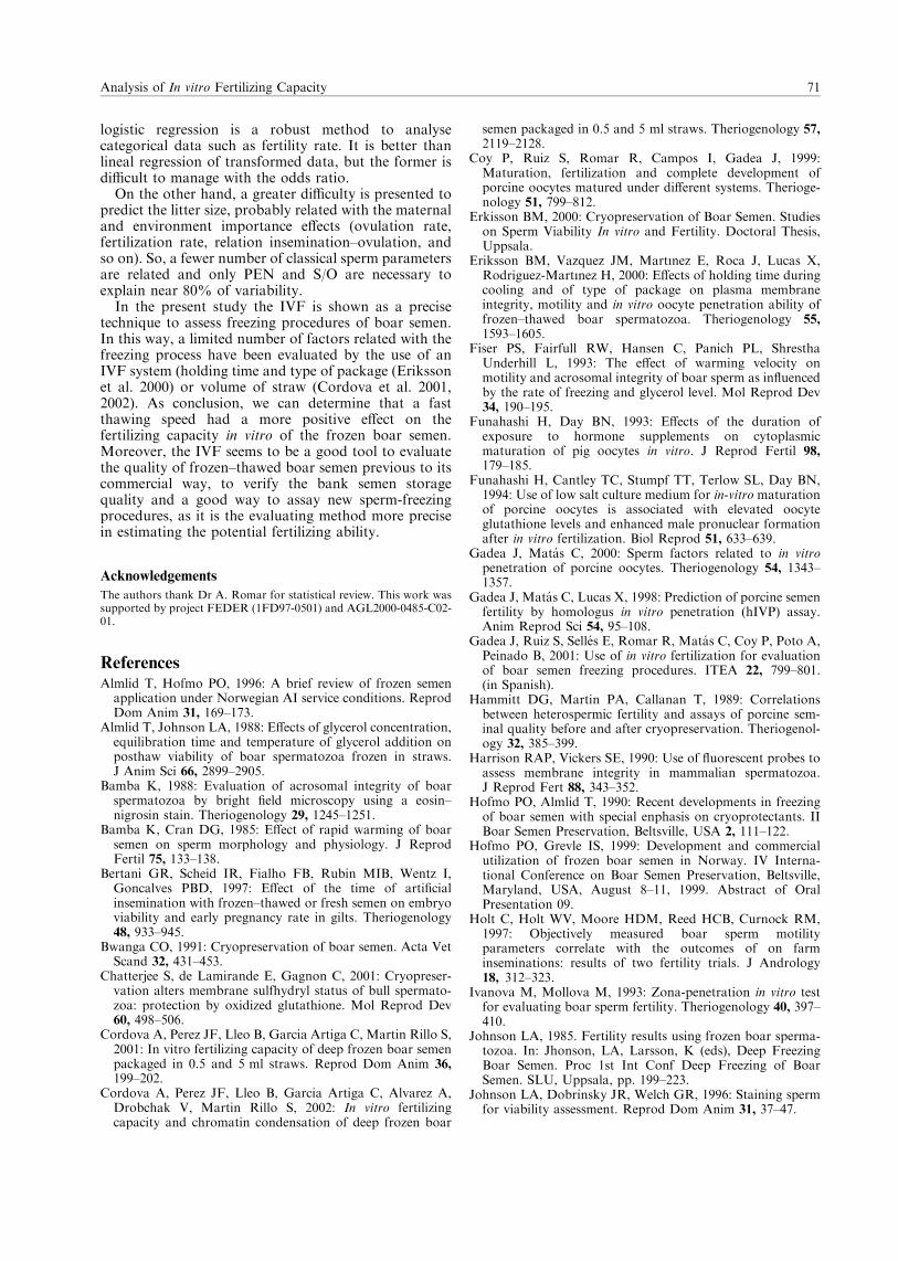
logistic regression is a robust method to analysecategorical data such as fertility rate. It is better thanlineal regression of transformed data, but the former isdifficult to manage with the odds ratio.
On the other hand, a greater difficulty is presented topredict the litter size, probably related with the maternaland environment importance effects (ovulation rate,fertilization rate, relation insemination–ovulation, andso on). So, a fewer number of classical sperm parametersare related and only PEN and S/O are necessary toexplain near 80% of variability.
In the present study the IVF is shown as a precisetechnique to assess freezing procedures of boar semen.In this way, a limited number of factors related with thefreezing process have been evaluated by the use of anIVF system (holding time and type of package (Erikssonet al. 2000) or volume of straw (Cordova et al. 2001,2002). As conclusion, we can determine that a fastthawing speed had a more positive effect on thefertilizing capacity in vitro of the frozen boar semen.Moreover, the IVF seems to be a good tool to evaluatethe quality of frozen–thawed boar semen previous to itscommercial way, to verify the bank semen storagequality and a good way to assay new sperm-freezingprocedures, as it is the evaluating method more precisein estimating the potential fertilizing ability.
Acknowledgements
The authors thank Dr A. Romar for statistical review. This work wassupported by project FEDER (1FD97-0501) and AGL2000-0485-C02-01.
ReferencesAlmlid T, Hofmo PO, 1996: A brief review of frozen semen
application under Norwegian AI service conditions. ReprodDom Anim 31, 169–173.
Almlid T, Johnson LA, 1988: Effects of glycerol concentration,equilibration time and temperature of glycerol addition onposthaw viability of boar spermatozoa frozen in straws.J Anim Sci 66, 2899–2905.
Bamba K, 1988: Evaluation of acrosomal integrity of boarspermatozoa by bright field microscopy using a eosin–nigrosin stain. Theriogenology 29, 1245–1251.
Bamba K, Cran DG, 1985: Effect of rapid warming of boarsemen on sperm morphology and physiology. J ReprodFertil 75, 133–138.
Bertani GR, Scheid IR, Fialho FB, Rubin MIB, Wentz I,Goncalves PBD, 1997: Effect of the time of artificialinsemination with frozen–thawed or fresh semen on embryoviability and early pregnancy rate in gilts. Theriogenology48, 933–945.
Bwanga CO, 1991: Cryopreservation of boar semen. Acta VetScand 32, 431–453.
Chatterjee S, de Lamirande E, Gagnon C, 2001: Cryopreser-vation alters membrane sulfhydryl status of bull spermato-zoa: protection by oxidized glutathione. Mol Reprod Dev60, 498–506.
Cordova A, Perez JF, Lleo B, Garcia Artiga C, Martin Rillo S,2001: In vitro fertilizing capacity of deep frozen boar semenpackaged in 0.5 and 5 ml straws. Reprod Dom Anim 36,199–202.
Cordova A, Perez JF, Lleo B, Garcia Artiga C, Alvarez A,Drobchak V, Martin Rillo S, 2002: In vitro fertilizingcapacity and chromatin condensation of deep frozen boar
semen packaged in 0.5 and 5 ml straws. Theriogenology 57,
2119–2128.Coy P, Ruiz S, Romar R, Campos I, Gadea J, 1999:
Maturation, fertilization and complete development ofporcine oocytes matured under different systems. Therioge-nology 51, 799–812.
Erkisson BM, 2000: Cryopreservation of Boar Semen. Studieson Sperm Viability In vitro and Fertility. Doctoral Thesis,Uppsala.
Eriksson BM, Vazquez JM, Martınez E, Roca J, Lucas X,Rodriguez-Martınez H, 2000: Effects of holding time duringcooling and of type of package on plasma membraneintegrity, motility and in vitro oocyte penetration ability offrozen–thawed boar spermatozoa. Theriogenology 55,
1593–1605.Fiser PS, Fairfull RW, Hansen C, Panich PL, Shrestha
Underhill L, 1993: The effect of warming velocity onmotility and acrosomal integrity of boar sperm as influencedby the rate of freezing and glycerol level. Mol Reprod Dev34, 190–195.
Funahashi H, Day BN, 1993: Effects of the duration ofexposure to hormone supplements on cytoplasmicmaturation of pig oocytes in vitro. J Reprod Fertil 98,179–185.
Funahashi H, Cantley TC, Stumpf TT, Terlow SL, Day BN,1994: Use of low salt culture medium for in-vitro maturationof porcine oocytes is associated with elevated oocyteglutathione levels and enhanced male pronuclear formationafter in vitro fertilization. Biol Reprod 51, 633–639.
Gadea J, Matas C, 2000: Sperm factors related to in vitropenetration of porcine oocytes. Theriogenology 54, 1343–1357.
Gadea J, Matas C, Lucas X, 1998: Prediction of porcine semenfertility by homologus in vitro penetration (hIVP) assay.Anim Reprod Sci 54, 95–108.
Gadea J, Ruiz S, Selles E, Romar R, Matas C, Coy P, Poto A,Peinado B, 2001: Use of in vitro fertilization for evaluationof boar semen freezing procedures. ITEA 22, 799–801.(in Spanish).
Hammitt DG, Martin PA, Callanan T, 1989: Correlationsbetween heterospermic fertility and assays of porcine sem-inal quality before and after cryopreservation. Theriogenol-ogy 32, 385–399.
Harrison RAP, Vickers SE, 1990: Use of fluorescent probes toassess membrane integrity in mammalian spermatozoa.J Reprod Fert 88, 343–352.
Hofmo PO, Almlid T, 1990: Recent developments in freezingof boar semen with special enphasis on cryoprotectants. IIBoar Semen Preservation, Beltsville, USA 2, 111–122.
Hofmo PO, Grevle IS, 1999: Development and commercialutilization of frozen boar semen in Norway. IV Interna-tional Conference on Boar Semen Preservation, Beltsville,Maryland, USA, August 8–11, 1999. Abstract of OralPresentation 09.
Holt C, Holt WV, Moore HDM, Reed HCB, Curnock RM,1997: Objectively measured boar sperm motilityparameters correlate with the outcomes of on farminseminations: results of two fertility trials. J Andrology18, 312–323.
Ivanova M, Mollova M, 1993: Zona-penetration in vitro testfor evaluating boar sperm fertility. Theriogenology 40, 397–410.
Johnson LA, 1985. Fertility results using frozen boar sperma-tozoa. In: Jhonson, LA, Larsson, K (eds), Deep FreezingBoar Semen. Proc 1st Int Conf Deep Freezing of BoarSemen. SLU, Uppsala, pp. 199–223.
Johnson LA, Dobrinsky JR, Welch GR, 1996: Staining spermfor viability assessment. Reprod Dom Anim 31, 37–47.
Analysis of In vitro Fertilizing Capacity 71

Johnson LA, Weitze KF, Fiser P, Maxwell WMC, 2000:Storage of boar semen. Anim Reprod Sci 62, 143–172.
Larsson B, Rodriguez-Martinez H, 2000: Can we use in vitrofertilization tests to predict semen fertility? Anim Reprod Sci60–61, 327–336.
Matttioli M, Galeati G, Bacci ML, Seren E, 1988: Follicularfactors influence oocyte pig oocyte penetrability and corticalgranule distribution. Gamete Res 21, 223–232.
Maxwell WMC, Johnson LA, 1997: Membrane status of boarspermatozoa after cooling or cryopreservation. Therioge-nology 48, 209–219.
Mazur P, 1984: Freezing of living cells: mechanisms andimplications. Anim J Physio 247, 125–142.
Mazur P, 1985: Basic concepts in freezing cells. In: Johnson,LA, Larsson, K (eds), Deep Freezing Boar Semen. Proc 1stInt Conf Deep Freezing of Boar Semen. SLU, Uppsala,pp. 91–111.
Pelaez J, Breininger E, Gonzalez C, Martınez E, Riol JA, PenaFJ, Alegre B, Dominguez JC, 2001: Good quality of post-thaw frozen boar semen may not lead to acceptable repro-ductive performances as evidenced by an homologous in vitrofertilization test. 5th Conference ESDAR, Wienn, 73 pp.
Pursel VG, Johnson LA, 1975: Freezing of boar spermatozoa.Fertilizing capacity with concentrated semen and newthawing procedure. J Anim Sci 40, 99–102.
Pursel VG, Johnson LA, Rampacek GB, 1972: Acrosomemorphology of boar spermatozoa incubated before coldshock. J Anim Sci 34, 278–283.
Ruiz S, Selles E, Gadea J, Marco MA, Murgas L, 2002: Effectof freezing rate on boar semen frozen: preliminary results ofAI. Theriogenology 57, 480. (abstract).
Thurston LM, Siggins K, Mileham AJ, Watson PF, Holt WV,2002: Identification of amplified restriction fragment lengthpolymorphism markers linked to genes controlling boarsperm viability following cryopreservation. Biol Reprod 66,545–554.
Waberski D, Weitze KF, Gleumes T, Schwarz M, Willmen T,Petzoldt R, 1994: Effect of time of insemination relative toovulation on fertility with liquid and frozen boar semen.Theriogenology 42, 831–840.
Westendorf P, Richter L, Treu H, 1975: Deep freezing of boarsperma. Laboratory and insemination results using theHulsenberger paillete method. Dtsch Tierarztl Wochenschr82, 261–267. (in German).
White IG, 1993: Lipids and calcium uptake of sperm inrelation to cold shock and preservation: a review. ReprodFertil Dev 5, 639–658.
Xu X, Ding J, Seth PC, Harbison DS, Foxcroft GR, 1996:In vitro fertilization of in vitro matured pig oocytes: effects ofboar and ejaculate fraction. Theriogenology 45, 745–755.
Xu X, Pommier S, Arbov T, Hutchings B, Sotto W, FoxcroftGR, 1998: In vitro maturation and fertilization techniquesfor assessment of semen quality and boar fertility. J AnimSci 76, 3079–3089.
Submitted: 05.07.2002
Author’s address (for correspondence): J Gadea, Dept. Fisiologıa,Facultad de Veterinaria, Universidad de Murcia, 30, 100 Murcia,Spain. E-mail: [email protected]
72 E Selles, J Gadea, R Romar, C Matas and S Ruiz

Decrease in glutathione content in boar sperm after
cryopreservation. Effect of the addition of reduced
glutathione to the freezing and thawing extenders.
Gadea J, Sellés E, Marco MA, Coy P, Matás C, Romar R,
Ruiz S.
Theriogenology. 62:690-701. 2004.

Decrease in glutathione content in boar spermafter cryopreservation
Effect of the addition of reduced glutathioneto the freezing and thawing extenders
Joaquın Gadea*, Elena Selles, Marco Antonio Marco, Pilar Coy,Carmen Matas, Raquel Romar, Salvador Ruiz
Department of Physiology, Facultad de Veterinaria, School of Veterinary, Campus de Espinardo,
University of Murcia, Murcia 30071, Spain
Received 9 July 2003; received in revised form 15 September 2003; accepted 17 November 2003
Abstract
Although glutathione content in boar spermatozoa has been previously reported, the effect of
reduced glutathione (GSH) on semen parameters and the fertilizing ability of boar spermatozoa after
cryopreservation has never been evaluated. In this study, GSH content was determined in ejaculated
boar spermatozoa before and after cryopreservation. Semen samples were centrifuged and GSH
content in the resulting pellet monitored spectrophotometrically. The fertilizing ability of frozen–
thawed boar sperm was also tested in vitro by incubating sperm with in vitro matured oocytes
obtained from gilts. GSH content in fresh semen was 3:84 � 0:21 nM GSH/108 sperm. Following
semen cryopreservation, there was a 32% decrease in GSH content (P < 0:0001). There were
significant differences in sperm GSH content between different boars and after various preservation
protocols (P ¼ 0:0102). The effect of addition of GSH to the freezing and thawing extenders was also
evaluated. Addition of 5 mM GSH to the freezing extender did not have a significant effect on
standard semen parameters or sperm fertilizing ability after thawing. In contrast, when GSH was
added to the thawing extender, a dose-dependent tendency to increase in sperm fertilizing ability was
observed, although no differences were observed in standard semen parameters. In summary, (i) there
was a loss in GSH content after cryopreservation of boar semen; (ii) addition of GSH to the freezing
extender did not result in any improvement in either standard semen parameters or sperm fertilizing
ability; and (iii) addition of GSH to the thawing extender resulted in a significant increase in sperm
fertilizing ability. Nevertheless, future studies must conclude if this is the case for all boars.
Furthermore, since addition of GSH to the thawing extender did not result in an improvement in
standard semen parameters, this suggests that during the thawing process, GSH prevents damage of a
Theriogenology 62 (2004) 690–701
* Corresponding author. Tel.:þ34-968-364655; fax: þ34-968-364147.
E-mail address: [email protected] (J. Gadea).URL: http://www.um.es/grupo-fisiovet.
0093-691X/$ – see front matter # 2003 Elsevier Inc. All rights reserved.
doi:10.1016/j.theriogenology.2003.11.013

sperm property that is critical in the fertilization process but that is not measured in the routine semen
analysis.
# 2003 Elsevier Inc. All rights reserved.
Keywords: Pig spermatozoa; Glutathione; Freezing; IVF
1. Introduction
The process of cooling and freeze–thaw produces physical and chemical stress on the
sperm membrane that reduces sperm viability and fertilizing ability. Cold shock of sperma-
tozoa is associated with oxidative stress and reactive oxygen species (ROS) generation [1].
ROS-induced damage to sperm is mediated by oxidative attack of bis-allylic methylene
groups of sperm phospholipid-bound polyunsaturated fatty acids, leading to lipid peroxida-
tion [2]. Since boar sperm have a high polyunsaturated fatty acid content they are very
susceptible to lipid peroxidation [3,4]. The effects of lipid peroxidation include irreversible
loss of motility, inhibition of respiration, leakage of intracellular enzymes, damage to sperm
DNA [5], or deficiencies in oocyte penetration and sperm–oocyte fusion [6]. Semen
represents a complex redox system that combines the antioxidant potential of seminal plasma
and spermatozoa with the pro-oxidant potential of sperm through the production of ROS.
Enzymatic antioxidant defense mechanisms in seminal plasma and spermatozoa include
superoxide dismutase, glutathione reductase, gluthathione peroxidase and catalase. Non-
enzymatic antioxidants include reduced glutathione (GSH), urate, ascorbic acid, Vitamin E,
taurine, hypotaurine, carotenoids, and ubiquinones. The interplay of antioxidant and pro-
oxidant mechanisms in semen determines the overall rate of lipid peroxidation in sperm.
Glutahione (L-g-glutamyl-L-cysteinylglycine) is a tripeptide ubiquitously distributed in
living cells. It plays an important role in the intracellular defense mechanism against
oxidative stress [2]. Glutathione peroxidase uses GSH as the reducing equivalent to reduce
hydrogen peroxide to H2O and lipoperoxides to alkyl alcohols. The resulting oxidized
glutathione (GSSG) is reduced to GSH by glutathione reductase using NADPH as the
co-factor.
GSH content has been reported in mammalian sperm [7,8], including boar sperm [9].
However, GSH content in boar sperm before and after cryopreservation has never been
evaluated.
The main objectives of this study were (i) to determine GSH content in boar sperm
before and after cryopreservation; and (ii) to assess the effect of GSH supplementation of
freezing and thawing extenders on standard semen parameters and sperm fertilizing ability
in IVF.
2. Material and methods
Semen was routinely collected from mature fertile boars using the manual method and a
dummy. The sperm-rich fraction was collected in a pre-warmed thermo flask and the gel-
fraction was held on a gauze tissue covering the thermo opening. The semen was then
diluted with isothermal Beltsville thawing solution (BTS) extender at a ratio of 1:1 (v/v).
J. Gadea et al. / Theriogenology 62 (2004) 690–701 691

2.1. Freezing and thawing protocol
Semen samples were processed using the straw freezing procedure described by
Westendorf et al. [10] with minor modifications indicated in the following. Diluted semen
was placed at 15 8C for 2 h and later centrifuged at 800 � g for 10 min. The supernatant
was discarded and the semen pellet was re-suspended with lactose–egg yolk extender
(LEY, 80 ml of 11% lactose and 20 ml egg yolk) to provide 1:5 � 109 spermatozoa/ml.
After further cooling to 5 8C over a 90-min period, two parts of LEY–extender semen were
mixed with LEY extender with 1.5% Orvus Es Paste (Equex-Paste, Minitub, Tiefenbach,
Germany) and 9% glycerol. The final concentration of semen to be frozen was
1 � 109 spermatozoa/ml and 3% glycerol. The diluted and cooled semen was loaded into
0.5 ml straws (Minitub) and placed in liquid nitrogen vapor approximately 3 cm above the
level of the liquid nitrogen for 20 min. The straws were then stored in liquid nitrogen until
thawing.
Thawing was achieved by immersing the straws in a circulating water bath at 50 8C for
12 s [11]. Immediately after thawing, the semen was diluted in BTS.
2.2. Determination of GSH content in spermatozoa
Semen samples were centrifuged at 1000 � g for 5 min at room temperature and the
resulting pellet resuspended in BTS and centrifuged again. The supernatant was discarded,
the pellet resuspended in BTS, and sperm concentration adjusted to 1–5 � 108 sperm/ml.
To release intracellular GSH, the sperm cells were lysed following three cycles of rapid
cooling in liquid nitrogen and thawing at 37 8C. The resulting suspensions were
centrifuged at 7000 � g for 10 min in order to remove membrane fragments.
Glutathione content was determined using a modified coupled optical test system [12].
In this system glutathione is oxidized by 5,5-dithiobis-(2-nitrobenzoic acid) (DTNB) and
then reduced by glutathione reductase with NADPH as hydrogen donor. During the
oxidation of glutathione by DTNB, 2-nitro-5 thiobenzoeic acid is formed, which can
be detected photometrically by a change of absorption at 412 nm. The total glutathione
content (oxidized glutathione (GSSG) and reduced glutathione (GSH)) is calculated
according to a standard curve.
2.3. Analysis of standard semen parameters
Percent motility and progression were determined by placing two sample aliquots on
warm glass slides (39 8C) and examined under light microscopy (magnification 100�).
The percentage of motile sperm was estimated to the nearest 5% and the forward
progressive motility (FPM) using an arbitrary scale from 0 to 5.
The proportion of spermatozoa with a normal apical ridge (NAR) was evaluated after
fixation in a buffered 2% glutaraldehyde solution and examined under phase-contrast
microscopy (magnification 1000�) to analyse acrosomes [13]. NAR was determined on
two slides per sample and a total of 200 spermatozoa per sample.
Eosin–nigrosin viability staining of sperm was also performed (EN). Semen was diluted
1:1 (v/v), with the staining solution (5% yellow eosin, 10% nigrosin in a citrate solution,
692 J. Gadea et al. / Theriogenology 62 (2004) 690–701
pH 7.4) and smeared onto slides. After being air-fixed, the stained spermatozoa were
observed under brightfield microscopy and 200 sperms per sample were evaluated [14].
Sperm membrane integrity was evaluated applying a combination of the fluorophores
carboxyfluorescein diacetate (DCF) and propidium iodide [15] on at least 200 cells per
sample using an epifluorescence microscope.
2.4. In vitro fertilization protocol
Ovaries from prepuberal gilts were obtained at a local slaughterhouse and transported to
the laboratory in saline solution (0.9%, w/v, NaCl) with 100 mg/l kanamicyn at 35 8C.
Oocytes surrounded by cumulus cells, were obtained from 3 to 6 mm diameter follicles and
washed twice in 35 mm plastic Petri-dishes containing modified Dulbecco’s phosphate
buffered saline (mPBSD) supplemented with 1 mg/ml polyvinyl alcohol and 0.005 mg/ml
phenol red. They were washed twice again in maturation medium previously equilibrated
for a minimum of 3 h at 38.5 8C under 5% CO2 in 95% humidified air.
The culture media used for oocyte maturation was Waymouth medium supplemented
with 10 UI/ml PMSG, 10 UI/ml hCG, 1 mg/ml estradiol-17b, 10% (v/v) foetal calf serum
and 10% porcine follicular fluid (v/v), as previously described by Coy et al. [16]. The
maturation medium was added to the Petri-dish in 3 � 100 ml droplets covered with
paraffin oil and 20 oocytes introduced in each droplet and incubated at 38 8C under 5%
CO2 in air.
The in vitro fertilisation medium was TCM199 supplemented with 12% heat inactivated
foetal calf serum, 0.9 mM sodium pyruvate, 3.05 mM D-glucose, 8.75 mM calcium lactate,
0.68 mM penicillin G, 3.6 mM caffeine and 0.068 mM streptomicyn sulphate at pH 7.4, as
previously described [16].
After thawing, the sperm samples were centrifuged at 50 � g for 3 min and the
supernatants centrifuged at 1200 � g for 3 min. The resulting pellets were diluted in
supplemented TCM199 without calcium lactate and caffeine. Aliquots of 100 ml of semen
were placed on Petri-dishes containing 2 ml of fertilization medium (final concentration of
1 � 106 spermatozoa/ml) and 20 in vitro matured oocytes previously washed twice in
equilibrated fertilization medium. After 18 h, the cultured oocytes were fixed in 3:1 (v/v)
ethanol:acetic acid for 24 h, stained with 1% lacmoid and examined under a phase contrast
microscope to assess penetration rate (PEN), mean number of sperm per penetrated oocyte
(S/O), monospermy rate (MON) and rate of male pronuclear formation (MPF).
2.5. Experimental design
2.5.1. Experiment 1: evaluation of GSH content in ejaculated boar spermatozoa
Semen parameters and GSH content in boar ejaculated spermatozoa were determined in
44 ejaculates from 27 boars.
2.5.2. Experiment 2: determination of GSH content in fresh, refrigerated
or cryopreserved boar spermatozoa
Semen parameters and GSH content were determined in boar spermatozoa from 25
ejaculates from 10 boars, after refrigeration for 24 h at 15 8C and after cryopreservation.
J. Gadea et al. / Theriogenology 62 (2004) 690–701 693
2.5.3. Experiment 3: effect of GSH supplementation of the freezing extender on the
viability and in vitro fertilizing ability of cryopreserved boar spermatozoa
Ejaculates from four boars (four per boar) were processed with or without addition of
5 mM GSH to the freezing extender. Standard semen parameters and in vitro sperm
fertilizing ability were evaluated.
2.5.4. Experiment 4: effect of GSH supplementation of the thawing extender on
the viability and in vitro fertilizing ability of cryopreserved boar spermatozoa
To examine the effect of GSH supplementation during the thawing process, frozen
straws from two different boars (four ejaculates per boar) were diluted immediately after
thawing in BTS (control), BTS þ 1 mM GSH and BTS þ 5 mM of GSH. After 30 min of
equilibration, standard semen parameters were evaluated and the sperm fertilizing ability
tested in the IVF system.
2.6. Statistical analysis
Data are expressed as the mean � S:E:M: and analysed by two-way ANOVA, con-
sidering the specific sperm treatment and boar samples as the main variables in experiments
2–4. IVF data (experiments 3 and 4) were modelled according to the binomial model of
parameters and were analysed by two-way ANOVA. When ANOVA revealed a significant
effect, values were compared by the Tukey test. Differences were considered statistically
significant at P < 0:05.
Linear regression analysis was used to further investigate relationships between semen
parameters (Pearson correlation) in experiment 2.
3. Results
3.1. Experiment 1: evaluation of GSH content in ejaculated boar spermatozoa
GSH content in ejaculated boar spermatozoa was determined in 44 ejaculates from 27
boars. The mean value was 3:84 � 0:21 nM GSH/108 cells. There were significant
differences in GSH content between different boars (P < 0:001) ranging from 1.05 to
6.16 nM GSH/108 cells.
3.2. Experiment 2: determination of GSH content in fresh, refrigerated or
cryopreserved boar spermatozoa
Standard semen parameters and GSH content were determined in freshly ejaculated
spermatozoa, refrigerated sperm suspensions for 24 h at 15 8C, and frozen–thawed
spermatozoa. GSH content was significantly lower in cryopreserved sperm as compared
to fresh ejaculated spermatozoa (P < 0:0001, Table 1), with a 32% decrease after
cryopreservation. Significant differences were also observed between boars, and preserva-
tion protocols (interaction P ¼ 0:0102).
694 J. Gadea et al. / Theriogenology 62 (2004) 690–701

All semen parameters were affected by preservation method with significant decrease in
frozen–thawed semen, except forward motility, which showed an increased value in frozen
semen. Although there were differences in semen parameters between different boars
(FPM, NAR and DCF), the interaction value was not significant (Table 1).
No significant correlation was observed between semen parameters and GSH content
after different preservation treatments. Pearson correlation coefficient analysis indicated an
inverse correlation between GSH and NAR in ejaculated spermatozoa (r ¼ �0:4263,
P ¼ 0:0002) and a direct correlation between GSH and forward progressive motility in
cryopreserved sperm (r ¼ 0:2960, P ¼ 0:0411, Table 2).
3.3. Experiment 3: effect of GSH supplementation to the freezing extender on the viability
and in vitro fertilizing ability of cryopreserved boar spermatozoa
Addition of 5 mM GSH to the freezing extender had no a significant effect on semen
parameters (data not shown) or sperm fertilizing ability (Table 3). However, significant
Table 1
Glutathione content (nM GSH/108 cells) and sperm quality parameters in fresh, stored and cryopreserved
spermatozoa (ejaculates ¼ 25) (mean � S:E:M:)
GSH nM/108
cells
Motility (%) FPM (0–5) EN (%) NAR (%) DCF (%)
Treatment
Fresh 4.99 � 0.23a 74.61 � 1.27a 3.21 � 0.07a 84.42 � 1.39a 92.53 � 0.59a 90.42 � 1.31a
Stored for 24 h at 15 8C 4.72 � 0.18a 69.03 � 1.59a 3.08 � 0.04a 83.15 � 1.99a 91.23 � 1.08a 84.08 � 1.68a
Frozen–thawed 3.36 � 0.34b 50.00 � 2.31b 3.74 � 0.07b 56.68 � 2.98b 33.24 � 2.13b 43.36 � 3.47b
Source of variability
Boar 0.0319 0.4351 0.0157 0.1041 0.0399 0.0095
Treatment <0.0001 <0.0001 <0.0001 <0.0001 <0.0001 <0.0001
Boar � treatment 0.0102 0.2339 0.1552 0.7794 0.1059 0.3911
Numbers within columns with different superscripts (a and b) differ (P < 0:05). FPM: forward progressive motility (0–5). EN:
eosine–nigrosine stain. NAR: normal apical ridge. DCF: sperm membrane integrity assessed with carboxyfluorescein diacetate.
ANOVA, P-values.
Table 2
Relationships between seminal parameters and GSH content in fresh, stored and cryopreserved spermatozoa (r:
Pearson correlation coefficient; P: significance)
Fresh Stored for 24 h at 15 8C Frozen–thawed
r P r P r P
Motility 0.2006 0.0911 0.0949 0.5121 �0.1135 0.4425
FPM 0.0923 0.4404 �0.1284 0.3741 0.2960 0.0411
EN 0.1457 0.2221 0.2124 0.1386 �0.1439 0.3293
NAR �0.4263 0.0002 0.1807 0.2091 �0.1197 0.4179
DCF 0.0031 0.9792 0.2842 0.0502 0.1006 0.4962
Morphology 0.0784 0.5124
FPM: forward progressive motility (0–5). EN: eosine–nigrosine stain. NAR: normal apical ridge. DCF: sperm
membrane integrity assessed with carboxyfluorescein diacetate.
J. Gadea et al. / Theriogenology 62 (2004) 690–701 695

differences between boars were observed for each IVF parameter (P < 0:015, Table 3),
except for %MPN. Concerning penetration rate and sperm number per penetrated oocyte,
ANOVA indicated that there were significant differences between boars, as also shown by
the significant value obtained for the interaction of preservation treatment and individual
boars (P ¼ 0:020 and P ¼ 0:017, respectively).
Table 3
IVF results for mature oocytes fertilized with frozen–thawed boar sperm in two different treatments, with (GSH)
or without (control) addition of GSH to the freezing extender: rate of penetration (%PEN), mean number of
spermatozoa per penetrated oocyte (S/O), monospermy rate (%MON) and rate of male pronuclear formation
(%MPN)
Boar Treatment Number of
oocytes
%PEN S/Oa %MONa %MPNa
67 Control 91 87.91 � 3.44a 2.41 � 0.17a 36.25 � 5.41a 72.50 � 5.02
67 GSH 97 69.07 � 4.72a,b 1.83 � 0.12a,b 43.28 � 6.10a,b 73.13 � 5.46
A Control 87 56.32 � 5.35b 1.81 � 0.15a,b 53.06 � 7.20a,b 83.67 � 5.33
A GSH 96 66.67 � 4.84b 2.25 � 0.21a,b 43.75 � 6.25a,b 79.69 � 5.07
BB Control 117 75.94 � 3.72a 2.08 � 0.17a,b 52.48 � 4.99a,b 71.29 � 4.52
BB GSH 115 72.06 � 3.86a 2.25 � 0.17a 47.96 � 5.07a,b 73.40 � 4.58
PI774 Control 124 33.06 � 4.24c 1.75 � 0.16a,b 56.10 � 7.85a,b 65.85 � 7.50
PI774 GSH 123 31.71 � 4.21c 1.36 � 0.09b 69.23 � 7.49b 74.36 � 7.08
Source of variability
Treatment 0.264 0.479 0.727 0.743
Boar <0.001 0.012 0.011 0.190
Interaction 0.020 0.017 0.320 0.815
Numbers within columns with different superscripts (a–c) differ (P < 0:05).a Based on penetrated oocytes.
Table 4
IVF results for mature oocytes fertilized with frozen–thawed boar sperm in three different treatments, with (1
and 5 mM GSH) or without (control) addition of GSH to the thawing extender: rate of penetration (%PEN),
mean number of spermatozoa per penetrated oocyte (S/O), monospermy rate (%MON) and rate of male
pronuclear formation (%MPN)
Boar Treatment Number of
oocytes
%PEN S/Oa %MONa %MPNa
PI774 Control 135 18.52 � 3.36a 1.20 � 0.08a 80.00 � 8.16a 48.00 � 10.20a
PI774 1 mM GSH 154 25.97 � 3.54a,b 1.32 � 0.10a 77.50 � 6.69a 60.00 � 7.84a,b
PI774 5 mM GSH 148 31.76 � 3.84b 1.36 � 0.10a 74.47 � 6.43a 72.34 � 6.60a,c
BB Control 96 83.33 � 3.82c 3.16 � 0.29b 31.25 � 5.21b 81.25 � 4.39b,c
BB 1 mM GSH 107 85.05 � 3.46c 3.44 � 0.17b 14.29 � 3.69b 84.62 � 3.80c
BB 5 mM GSH 118 97.46 � 1.46c 4.01 � 0.28b 23.48 � 3.97b 84.35 � 3.40c
Source of variability
Treatment <0.001 0.254 0.292 0.062
Boar <0.001 <0.001 <0.001 <0.001
Interaction 0.578 0.480 0.409 0.167
Numbers within columns with different superscripts (a–c) differ (P < 0:05). ANOVA, P-values.a Based on penetrated oocytes.
696 J. Gadea et al. / Theriogenology 62 (2004) 690–701

3.4. Experiment 4: effect of GSH supplementation to the thawing extender on the
viability and in vitro fertilizing ability of cryopreserved boar spermatozoa
No significant differences in semen parameters were found between control and
GSH-supplemented thawing extender (data not shown). In contrast, GSH addition to
the thawing extender resulted in an significant increase in sperm in %PEN (P < 0:001)
and also an increase in %MPN, although it did not reach statistical significance
(P ¼ 0:062) (Table 4). For all the studied IVF variables a significant boar effect was
detected (P < 0:001).
4. Discussion
The main findings emerging from this study are the decrease in GSH content observed in
boar sperm after cryopreservation and a tendency to increase in boar sperm fertilizing
ability after addition of GSH to the thawing extender.
Glutathione is the major non-protein thiol compound in mammalian cells. GSH
participates in a number of cell functions including amino acid transport, DNA and
protein synthesis, reduction of disulfide bonds and protection against oxidative stress. The
sulphydryl groups of GSH have been shown to confer protection against cell damage by
oxidants, electrophiles and free radicals (reviewed by Irvine [2]).
The results obtained for GSH content in ejaculated boar semen (3:84 � 0:21 nM/108
cells) are in good agreement with previous reports (3 nM GSH/108 cells [9]) and with the
results obtained in other mammalian species including human sperm (6:7 � 0:4 nM GSH/
108 cells [7]; 6:2 � 0:6 nM GSH/108 cells [17]; 3:49 � 0:87 nM GSH/108 cells [8]), and
bull sperm (2:73 � 0:42 nM GSH/108 cells [18]). As previously reported, GSH values in
mouse sperm are significantly higher than in other mammalian species (90 nM GSH/108
cells [7]). It is noteworthy that comparisons between different studies are often difficult to
interpret due to differences in the method of analysis [19] and sperm processing protocols
[20].
The results of experiment 1 indicated significant differences in GSH content between
different boars. These differences could be related to differences in the fertility potential of
these boars [21]. A relationship between GSH content in sperm and seminal plasma and
infertility has been previously found in human sperm [8,22] and also between GSH content
in sperm and their ability to penetrate bovine cervical mucus [8].
The results also show that cryopreservation of boar sperm is associated with a
significant decrease in GSH content (Table 1), as previously reported in bovine [20]
and human sperm (Molla and Gadea, data not published). This would be expected to
result in changes in the sulphydryl content of proteins from the sperm membranes and
nucleus [23]. Similarly, cryopreservation of boar sperm is associated with a reduction in
the sperm motility and viability. Interestingly, forward progressive motility was
increased in cryopreserved spermatozoa. This could be related to changes brought about
by the media used in boar sperm cryopreservation named capacitation like alterations
[1,24]. However, these preliminary results must be confirmed in future studies where
sperm motility will be monitored by computer-assisted semen analysis (CASA). In
J. Gadea et al. / Theriogenology 62 (2004) 690–701 697

contrast, preservation of boar spermatozoa at 15 8C did not result in a decrease in GSH
content or sperm viability. This suggests that the effects of cryopreservation on GSH
content and sperm motility and viability observed are specific to the process of freezing
and thawing at low temperatures.
Once we detected a decrease in the GSH content in the boar frozen–thawed spermatozoa,
we assessed the effect of GSH supplementation of freezing and thawing extenders on
standard semen parameters and sperm fertilizing ability in IVF (experiments 3 and 4). One
question that remains to be answered is why addition of GSH to the thawing extender and
not to the freezing extender increases boar sperm fertilizing ability in vitro. A plausible
explanation for this question would be that cryodamage of antioxidant enzymes during the
freezing process results in an overall decrease of enzymatic antioxidant defenses in boar
sperm. This damage could selectively affect superoxide dismutase (SOD), glutathione
reductase (GRD) and glutathione peroxidase (GPX) to a lesser extent. During thawing,
there is an increase in the production of ROS [1], with an increase in the superoxide anion
production due to a decrease in SOD activity and a decrease in GSH content due to a
decrease in GRD activity and to an increase in GSH oxidation by hydrogen peroxide. A
decrease in intracellular GSH and an increase in ROS production during thawing would
lead to an increase in lipid peroxidation. GSH provided exogenously after thawing
(experiment 4) would increase the intracellular levels of GSH which will be used by
GPX to prevent hydrogen and lipid peroxide-induced damage. Addition of GSH to the
freezing extender, on the other hand, would result in a high rate of ROS-induced GSH
oxidation during rewarming and thawing. This is supported by our results that show that
there is a decrease in GSH content during cryopreservation. In contrast, addition of GSH
after thawing would allow any residual antioxidant enzymatic activity still remaining in the
sperm cell to recover, to decrease steady-state ROS levels, and, therefore, to decrease GSH
oxidation. This hypothesis is supported by previous studies which show that there is a
decrease in SOD activity during cryopreservation and an increase in lipid peroxidation of
human and bovine spermatozoa after thawing [20,25]. Sadly, this information is not well
known yet in the boar spermatozoa [26,27]. However, if GSH would be acting in concert
with GPX to decrease the steady-state level of hydrogen peroxide and membrane lipid
peroxides in boar sperm during thawing, an increase in sperm motility and viability should
also be expected.
It has also been reported that damage to the plasma membrane overlaying the
flagellum or head during cryopreservation of human sperm can occur independently
[28] and Holt and North [29] reported that sperm head plasma membranes were
preferentially damaged by freezing and thawing. Although acrosomes became swollen
after freezing and thawing, the incidence of outer acrosomal membrane vesiculation
remained at control (unfrozen) levels with all treatments. However, the results indicate
that these semen parameters are not affected by GSH addition. The only effect that is
observed is an increase in boar sperm fertilizing ability. Therefore, cryopreservation-
induced oxidative stress in boar sperm affects a sperm property that is related to both
sperm binding and penetration of the zona pellucida, sperm–oocyte membrane fusion,
oocyte activation, or pronuclear formation. In this sense, the membrane fusion events
involved in binding with the oolema and in the acrosome reaction appear to be more
vulnerable to ROS-induced damage than is overall motility in human spermatozoa [30].
698 J. Gadea et al. / Theriogenology 62 (2004) 690–701

Also, we have previously demonstrated that the IVF is the most precise tool for
evaluating boar semen capacities, in both refrigerated and frozen–thawed semen
[11,31]. So the use of IVF systems could help us to detect differences that classical
assays are not able to do [32].
Other reports indicate that cryopreservation also results in ROS-induced DNA
fragmentation [33]. However, this would only partially explain the results since a
decrease in DNA fragmentation by GSH supplementation during thawing would explain
the observed increase in pronuclear formation rate, but it would not explain the increase
in penetration rate. Nevertheless, it has been shown the relationship between the ability
to form a male pronucleus in pig oocytes with intracellular concentration of GSH
[34,35]. Also the addition of GSH to the insemination medium in an IVF system
improves the developmental competence of in vitro matured pig oocytes in a dose-
dependent manner [36].
In summary, there was a loss in GSH content after cryopreservation of boar semen.
Addition of GSH to the freezing extender did not result in any improvement in either
standard semen parameters or sperm fertilizing ability; while addition of GSH to the
thawing extender resulted in a significant increase in sperm fertilizing ability. Nevertheless,
future studies must conclude if this is the case for all boars. Furthermore, since addition of
GSH to the thawing extender did not result in an improvement in standard semen
parameters, this suggests that during the thawing process, GSH prevents damage of a
sperm property that is critical in the fertilization process but that is not measured in the
routine semen analysis.
Acknowledgements
This research was supported by the project AGL 2000-0485-C02-01. We acknowledge
with gratitude the assistance with statistical analysis provided by Dr. A. Romar and Dr. J.G.
Alvarez for critical reading of the manuscript.
References
[1] Chatterjee S, de Lamirande E, Gagnon C. Cryopreservation alters membrane sulfhydryl status of bull
spermatozoa: protection by oxidized glutathione. Mol Reprod Dev 2001;60:498–506.
[2] Irvine DS. Gluthatione as a treatment for male infertility. Rev Reprod 1996;1:6–12.
[3] Comaschi V, Lindner L, Farruggia G, Gesmundo N, Colombi L, Masotti L. An investigation on
lipoperoxidation mechanisms in boar spermatozoa. Biochem Biophys Res Commun 1989;158:769–75.
[4] Cerolini S, Maldjian A, Surai P, Noble R. Viability, susceptibility to peroxidation and fatty acid
composition of boar semen during liquid storage. Anim Reprod Sci 2000;58:99–111.
[5] White IG. Lipids and calcium uptake of sperm in relation to cold shock and preservation: a review. Reprod
Fert Dev 1993;5:639–58.
[6] Aitken RJ, Buckingham D, Harkiss D. Use of a xanthine oxidase free radical generating system to
investigate the cytotoxic effects of reactive oxygen species on human spermatozoa. J Reprod Fertil 1993;
97:441–50.
[7] Alvarez JG, Storey BT. Role of glutathione peroxidase in protecting mammalian spermatozoa from loss of
motility caused by spontaneous lipid peroxidation. Gamete Res 1989;23:77–90.
J. Gadea et al. / Theriogenology 62 (2004) 690–701 699

[8] Ochsendorf FR, Buhl R, Bastlein A, Beschmann H. Glutathione in spermatozoa and seminal plasma of
infertile men. Hum Reprod 1998;13:353–9.
[9] Li TK. The glutathione and thiol content of mammalian spermatozoa and seminal plasma. Biol Reprod
1975;12:64–646.
[10] Westendorf P, Richter L, Treu H. Deep freezing of boar sperma. Laboratory and insemination results
using the Hulsenberger paillete method. Dtsch Tierarztl Wochenschr 1975;82:261–7 [in German].
[11] Selles E, Gadea J, Romar R, Matas C, Ruiz S. Analysis of in vitro fertilizing capacity to evaluate the
freezing procedures of boar semen and to predict the subsequent fertility. Reprod Domest Anim 2003;
38:66–72.
[12] Anderson ME. Determination of glutathione and glutathione disulfide in biological samples. In: Meister
A, editor. Method in enzymology. Colowick Sp, Kaplan NO, series editors. Glutamate, glutamine,
glutathione and related compounds, vol. 113. 1985. p. 548–55.
[13] Pursel VG, Johnson LA, Rampacek GB. Acrosome morphology of boar spermatozoa incubated before
cold shock. J Anim Sci 1972;34:278–83.
[14] Bamba K. Evaluation of acrosomal integrity of boar spermatozoa by bright field microscopy using a
eosin–nigrosin stain. Theriogenology 1988;29:1245–51.
[15] Harrison RAP, Vickers SE. Use of fluorescent probes to assess membrane integrity in mammalian
spermatozoa. J Reprod Fert 1990;88:343–52.
[16] Coy P, Ruiz S, Romar R, Campos I, Gadea J. Maturation, fertilization and complete development of
porcine oocytes matured under different systems. Theriogenology 1999;51:799–812.
[17] Griveau JF, Dumont E, Renard P, Callegari JP, Le Lannou D. Reactive oxygen species, lipid
peroxidation and enzymatic defence systems in human spermatozoa. J Reprod Fertil 1995;103:
17–26.
[18] Agrawal YP, Vanha-Perttula T. Glutathione L-glutamic acid and gamma-glutamyl transpeptidase in the
bull reproductive tissues. Int J Androl 1988;11:123–31.
[19] Floreani M, Petrone M, Debetto P, Palatini P. A comparison between different methods for the
determination of reduced and oxidized glutathione in mammalian tissues. Free Radic Res 1997;26:
449–55.
[20] Bilodeau JF, Chatterjee S, Sirard MA, Gagnon C. Levels of antioxidant defenses are decreased in bovine
spermatozoa after a cycle of freezing and thawing. Mol Reprod Dev 2000;55:282–8.
[21] Gadea, J. Sperm factors related to fertility in vivo and in vitro. In: Proceedings of the Fifth International
Conference on Boar Semen Preservation, The Netherlands, 2003.
[22] Raijmakers MT, Roelofs HM, Steegers EA, Steegers-Theunissen RRP, Mulder TP, Knapen MF, et al.
Glutathione and glutathione S-transferases A1-1 and P1-1 in seminal plasma may play a role in protecting
against oxidative damage to spermatozoa. Fertil Steril 2003;79:169–72.
[23] Marco MA, Gadea J. Evaluation of membrane sulfhydryl status of boar spermatozoa after cold shock. In:
Proceedings of the Fifth International Conference on Boar Semen Preservation, The Netherlands, 2003
[abstract].
[24] Watson PF. Recent developments and concepts in the cryopreservation of spermatozoa and the assessment
of their post-thawing. Reprod Fertil Dev 1995;7:871–91.
[25] Alvarez JG, Storey BT. Evidence for increased lipid peroxidative damage and loss of superoxide
dismutase activity as a mode of sublethal cryodamage to human sperm during cryopreservation. J Androl
1992;13:232–41.
[26] Nishimura K, Morii H. Effects of gluthatione on the motility of frozen–thawed boar spermatozoa. Anim
Sci Technol (Jpn) 1993;64:433–9.
[27] Cerolini S, Maldjian A, Pizzi F, Gliozzi TM. Changes in sperm quality and lipid composition during
cryopreservation of boar semen. Reproduction 2001;121:395–401.
[28] McLaughlin EA, Ford WC, Hull MG. Effects of cryopreservation on the human sperm acrosome and its
response to A23187. J Reprod Fertil 1993;99:71–6.
[29] Holt WV, North RD. Effects of temperature and restoration of osmotic equilibrium during thawing on
the induction of plasma membrane damage in cryopreserved ram spermatozoa. Biol Reprod 1994;51:
414–24.
[30] Aitken RJ, Clarkson JS, Fishel S. Generation of reactive oxygen species, lipid peroxidation, and human
sperm function. Biol Reprod 1989;41:183–97.
700 J. Gadea et al. / Theriogenology 62 (2004) 690–701

[31] Gadea J, Matas C, Lucas X. Prediction of porcine semen fertility by homologous in vitro penetration
(hIVP) assay. Anim Reprod Sci 1998;54:95–108.
[32] Larsson B, Rodriguez-Martinez H. Can we use in vitro fertilization tests to predict semen fertility? Anim
Reprod Sci 2000;60/61:327–36.
[33] Donelly ET, McClure N, Lewis SEM. Glutathione and hypotaurine in vitro: effects on human sperm
motility, DNA integrity and production of reactive oxygen species. Mutagenesis 2000;15:61–8.
[34] Yoshida M. Role of glutathione in the maturation and fertilization of pig oocytes in vitro. Mol Reprod Dev
1993;35:76–81.
[35] Funahashi H, Cantley TC, Stumpf TT, Terlow SL, Day BN. Use of low salt culture medium for in-vitro
maturation of porcine oocytes is associated with elevated oocyte glutathione levels and enhanced male
pronuclear formation after in vitro fertilization. Biol Reprod 1994;51:633–9.
[36] Boquest AC, Abeydeera LR, Wang WH, Day BN. Effect of adding reduced glutathione during
insemination on the development of porcine embryos in vitro. Theriogenology 1999;51:1311–9.
J. Gadea et al. / Theriogenology 62 (2004) 690–701 701

The predictive value of porcine seminal parameters on
fertility outcome under commercial conditions
Gadea J, Sellés E, Marco MA.
Reproduction in Domestic Animals. 39: 303–308. 2004.

The Predictive Value of Porcine Seminal Parameters on Fertility Outcome
under Commercial Conditions
J Gadea, E Selles and MA Marco
Department of Physiology, Faculty of Veterinary Medicine, University of Murcia, Spain
Contents
The aim of this study was to address the question of whetherdifferences in farrowing rate and litter size after the use ofdifferent ejaculates could be predicted using the standardsemen parameters under commercial conditions. In this study,a total of 1818 sows were used to evaluate the fertilitypredictive value of different sperm parameters. Logisticregression analysis (univariate and multivariate) was used tocorrelate the dichotomous farrowing rate data to the spermparameters. Linear regression was also used to determine therelationship between litter size and semen parameters (Pearsoncorrelation and multiple regression). Receiver-operating curves(ROC) were used to determine the overall performancecharacteristics of each semen variable in the logistic regressionmodel. Semen analysis, under commercial conditions, allowsto identify ejaculates with very low fertility potential but thepre-selection of the samples, the high number of sperm perdoses and the high quality of the semen used in artificialinsemination (AI) programmes reduces the variability. There-fore, it is unlikely to detect fertility differences associated withseminal parameters.
Introduction
The prediction of sperm fertilizing ability has a greateconomic importance to breeding herds when artificialinsemination is used, as it leads to the selection of thoseboars with a good reproductive performance. However,at the moment, only a few trials that correlate spermfactors and fertility have been reported (reviewed byGadea 2003). In most of them, a reduced or poorsignificant relationship has been observed (reviewed byWoelders 1991; Flowers 2002), in part, because of themany factors involved in fertility in sows (Foote 2003).
Classical methods of semen evaluation generallymeasure sperm concentration, progressive motility, thepercentage of viable cells and acrosome morphology.These assays are poor in predicting fertility outcome,because only those samples with markedly poor qualitycan be identified (Gadea et al. 1998). To solve thisproblem, new procedures of seminal analysis in vitro(Braundmeier and Miller 2001), or multiple analysis ofthe same sample have been evaluated (Woelders 1991)and new assays to study the oocyte–sperm interaction asthe in vitro fertilisation (IVF) system have been devel-oped (Gadea et al. 1998; Xu et al. 1998; Selles et al.2003). However, these analyses are expensive and time-consuming, and cannot be applied under field commer-cial conditions.
Under field commercial conditions, the use of simple,non-expensive and accurate assays is required. Such goalhas been pursued over decades but no definitive solutionhas ever been reported.
The aim of this study was to address the question ofwhether farrowing rate and litter size after the use ofdifferent ejaculates could be predicted with classicalsemen-quality parameters under commercial conditions.Different analytical strategies have been developed todetermine the possible relationship between spermparameters and farrowing rates and litter size. The firstis the exploration of diffences in seminal parametersbetween ejaculates classified in two categories accordingto farrowing rate, with a target value for fertility undercommercial conditions (85% farrowing rate). Secondly,logistic and linear regression analyses were applied todetect any significant relationship between semen qual-ity and farrowing rate and litter size.
Materials and Methods
Spermatozoa from 273 ejaculates from 57 boars (fourDalland and 53 Pietrain) were evaluated for semen-quality parameters in vitro. Sperm-rich fractions werecollected by an experienced operator using the gloved-hand technique. At the time of collection, theejaculate was filtered through a gauze to remove geland immediately placed at 37�C in a temperature-controlled water bath. Each ejaculate was later dilutedto a final concentration of 3 · 107 spermatozoa/ml ina commercial extender for refrigerated semen(MR-A�, Kubus, Madrid, Spain) and stored at17�C until assay.
Routine laboratory evaluation of semen
The volume of the sperm-rich fraction of the ejaculatewas determined in a graduated cylinder and spermconcentration measured with a haemacytometer. Toevaluate the motility and the forward progressivemotility (FPM), two subsamples were placed on warmglass slides (39�C) and examined under a light micro-scope (·400 magnification). The percentage of motilesperm cells was estimated subjectively to the nearest 5%and the FPM using an arbitrary scale of 0–5.
Wet mounts of semen fixed in buffered 2% glutaral-dehyde solution were examined under a phase-contrastmicroscope (·1000 magnification) to analyse the mor-phology and acrosomes (Pursel et al. 1972). The pro-portion of spermatozoa with a normal apical ridge(NAR) was determined on two slides per sample and atotal of 200 spermatozoa per sample. Two hundredspermatozoa were categorized according to sperm mor-phology into those with normal morphology, cells withattached proximal cytoplasmic droplets (PCD), distal
Reprod Dom Anim 39, 303–308 (2004)
� 2004 Blackwell Verlag, Berlin
ISSN 0936-6768
U.S. Copyright Clearance Centre Code Statement: 0936–6768/2004/3905–0303$15.000/0 www.blackwell-synergy.com

cytoplasmic droplets (DCD), folded tail, coiled tail andothers (abnormal heads) etc.
Eosin–nigrosin (EN) viability staining of sperm wasapplied. A semen sample was diluted 1 : 1 with stainingsolution (5% yellow eosin, 10% nigrosin in a citratesolution pH 7.4) and smeared. Air-fixed stained sper-matozoa were observed and 200 spermatozoa wereevaluated per slide (Bamba 1988) and the percentage ofnon-stained spermatozoa was registered.
Sperm membrane integrity was assessed by incubationwith carboxyfluorescein diacetate (DCF) (Harrison andVickers 1990) on at least 200 cells/sample using amicroscope equipped with epi-fluorescence.
Fertility trial
The fertility study was conducted on a commercial farm,using multiparous (two to six pregnancies) crossbredsows (Landrace · Large White). Oestrus was checkeddaily in the presence of a mature teaser boar. Occurrenceof oestrus was defined by the standing reflex in front of aboar (back pressure test) and reddening and swelling ofthe vulva. The sows were inseminated with dilutedsemen, containing 3 · 109 sperm in 100 ml MR-Aextender, using disposable artificial insemination (AI)catheters. Insemination of all sows showing oestrus signstook place on immediately after the diagnosis of oestrusand was repeated 24 h later under the same conditionswith semen preserved at 17�C.
Fertility was measured for every ejaculate as thepercentage of sows farrowing to AI. For each sow thatfarrowed, the number of dead and live piglets wascounted and the sum was defined as the total number ofpiglets born (TPB).
Experimental design
In this study, a total of 1818 sows were used to evaluatethe capacity for fertility prediction of the different spermassays. Ejaculate was consider to be the experimental
unit. First, the ejaculates were grouped into twocategories according to fertility rate (low, farrowingrate £ 85%; high, farrowing rate > 85%). Seminalparameters were evaluated and differences betweenfertility groups were investigated. Secondly, logisticand linear regression analyses were applied to detectany significant relationship between semen quality andfarrowing rate and litter size.
Statistical analysis
The results are expressed as mean ± SEM and wereanalysed by one-way ANOVA, using the multivariategeneral linear models of Systat. When ANOVA revealed asignificant effect, values were compared by Tukey’s test.
Logistic regression (univariate and multivariate) wasused to relate the dichotomous fertility rate data to thesperm parameters. Linear regression was used to furtherinvestigate relationships between litter size and meas-ured semen parameters (Pearson’s correlation andmultiple regression). Receiver-operating curves (ROC)were used to determine the overall discriminant powerof each semen variable in the logistic regression model.A ROC was also used to calculate the elective breakingpoint (cut-off value) for each semen variable in relationto the fertility trial. The breaking point is in relationwith the point that maximized specificity and sensitivity.
Results
Relationship between sperm characteristicsand farrowing rate
Semen parameters values and the fertility outcome werenot significantly different in the two genetic lines of theboars used in this study (data not shown). Therefore, allejaculates from these boars can be considered as ahomogeneous group. Statistical analysis of spermparameters values and fertility outcome obtained inthe field trial are shown in Table 1 for the two groups ofejaculates according to farrowing rate (more or less than
Table 1. Semen measurements andreproduction results determinedfor 273 ejaculates from 57 boarsallocated into two groupsaccording to farrowing rate(mean ± SEM)
Low fertility
(farrowing rate £85%)
High fertility
(farrowing rate >85%)
Significance
(p-value)
Number of ejaculates 138 135
Volume (ml) 103.20 ± 2.03 107.13 ± 2.09 0.1792
Concentration (103 sperm/mm3 ) 77.91 ± 2.48 82.20 ± 2.63 0.2358
Total cells (109 ) 78.20 ± 2.71 86.10 ± 2.82 0.0443
Motility 74.09 ± 0.63 75.31 ± 0.63 0.1760
FPM 3.24 ± 0.03 3.20 ± 0.03 0.4669
EN (%) 84.72 ± 0.70 84.82 ± 0.68 0.9147
NAR (%) 93.46 ± 0.46 93.56 ± 0.43 0.8755
DCF (%) 84.23 ± 0.71 84.30 ± 0.87 0.9510
Morphoanomalies (%) 11.94 ± 0.78 12.54 ± 0.97 0.6307
PCD (%) 2.73 ± 0.27 3.41 ± 0.37 0.1376
DCD (%) 4.22 ± 0.38 4.00 ± 0.33 0.6583
Folded tail (%) 4.05 ± 0.49 3.73 ± 0.69 0.7002
Swollen tail (%) 0.30 ± 0.07 0.75 ± 0.30 0.1325
Other anomalies (%) 0.63 ± 0.12 0.64 ± 0.12 0.9691
Number of sows 915 903
Farrowing rate (%) 645/915 (70.49%) 868/903 (96.12%) <0.0001
Live piglets born 9.64 ± 0.11 9.84 ± 0.10 0.1689
Total piglets born 10.25 ± 0.12 10.45 ± 0.11 0.2191
FPM, forward progressive motility (0–5); EN, eosin–nigrosin stain; NAR, normal apical ridge; DCF, sperm membrane
integrity assessed with carboxyfluorescein diacetate; PCD, proximal cytoplasmic droplets; DCD, distal cytoplasmic drop-
lets.
304 J Gadea, E Selles and MA Marco

85%). Ejaculates were distributed equally in bothgroups (n ¼ 138 vs 135). The mean values of farrowingrate obtained in the two groups of sows (n ¼ 915 and903) were significantly different (70.49% vs 96.12%,p < 0.0001). No differences in semen parameters werefound between both groups. Only total cell count in theejaculate was significantly higher in the high fertilitygroup (p ¼ 0.0443).
Linear and logistic regression analyses were used tocorrelate the farrowing rate data to the semen param-eters. In both analyses, a poor correlation with fertilityoutcome was obtained. Only motility (logistic and linearregression) was significantly correlated to farrowing rate(Table 2). When all conventional semen parameterswere analysed through a stepwise, multiple logistic orlinear regression analysis, only three parameters (motil-ity, PCD and swollen tail) are necessary to reachstatistical significant model (Tables 3 and 4). However,in both models a low percentage of total variance offarrowing rate could be predicted. It was surprising tofind that there was a positive correlation between PCDand swollen tail with fertility (Table 3).
The use of ROC led us to calculate the cut-off valuesfor the semen parameters (Table 5) in these experimen-tal conditions. Cut-off values obtained in this study werehighly restricted, for example EN (86), NAR (94),
normal morphology (90) and DCF (85). However, theywere less restricted for motility (70). The sensitivity andspecificity of these parameters to identify better ejac-ulates in terms of fertility outcome are shown in Table 5.Lowering the threshold reduces the sensitivity of themeasures (the likelihood of correctly identifying infer-tile–subfertile ejaculates) but increases their specificity(the likelihood of correctly identifying fertile ejaculates).
On the basis of the area under ROC (area ROC), eachseminal parameter did not provide information that washelpful in discriminating fertility outcome, because itwas not significantly >0.500. However, motility, PCDand folded tail are near to this value, although therewere not significant differences between area ROCcorresponding to the different seminal parameters.
Relationship between sperm characteristics and litter size
Concerning litter size, the total number of piglets bornshowed a significant positive correlation with motility(r ¼ 0.1223, p ¼ 0.0435; Table 6), while folded tailshowed a negative correlation (p ¼ 0.0681) but did notreach statistical significance. However, no correlationwas found between the average live piglets born andsemen parameters (Table 6). Only a weak positivecorrelation was found for motility and a negativecorrelation for folded tail, but neither reach statisticalsignificance.
Table 2. Univariate logistic and linear regression of seminal measure-ments with farrowing rate
Parameter
Logistic regression Linear regression
Odd ratio
Significance
(p-value)
Pearson
correlation
coefficient (r)
Significance
(p-value)
Volume 1.0017 0.5076 0.0157 0.5073
Concentration 1.0016 0.4803 0.0166 0.4828
Total cells 1.0029 0.1499 0.0338 0.1530
Motility 1.0195 0.0237 0.0539 0.0214
FPM 0.8764 0.4337 )0.0184 0.4322
EN 1.0013 0.8674 0.0039 0.8673
NAR 1.0079 0.4891 0.0164 0.4845
DCF 0.9993 0.9273 )0.0021 0.9274
Morphoanomalies 1.0009 0.8892 0.0033 0.8896
PCD 1.0346 0.0621 0.0421 0.0723
DCD 0.9986 0.9252 )0.0022 0.9252
Folded tail 0.9885 0.2300 )0.0291 0.2138
Swollen tail 1.0768 0.0849 0.0324 0.1666
Other anomalies 0.9671 0.4862 )0.0166 0.4784
FPM, forward progressive motility (0–5); EN, eosin–nigrosin stain; NAR, nor-
mal apical ridge; DCF, sperm membrane integrity assessed with carboxyfluoresc-
ein diacetate; PCD, proximal cytoplasmic droplets; DCD, distal cytoplasmic
droplets.
Table 3. Multiple logistic regression of semen measurements withfarrowing rate (forward stepwise model)
Parameter Estimate SE Odds ratio t-ratio p-value
Constant )0.0873 0.6511 – )0.1341 0.8934
Motility 0.0209 0.0086 1.0211 2.4172 0.0156
PCD 0.0385 0.0194 1.0392 1.9843 0.0472
Swollen tail 0.0679 0.0516 1.0702 1.3147 0.1886
p ¼ 0.0069; McFadden’s q2 ¼ 0.0075.
PCD, proximal cytoplasmic droplets.
Table 4. Multiple linear regression of semen measurements withfarrowing rate (forward stepwise model)
Parameter Coefficient SE t-ratio p-value (two-tail)
Constant 0.5867 0.0931 6.3032 0.0000
Motility 0.0031 0.0012 2.5035 0.0124
PCD 0.0046 0.0023 1.9622 0.0499
Swollen tail 0.0056 0.0034 1.6385 0.1015
p ¼ 0.0090; multiple R2 ¼ 0.0063.
PCD, proximal cytoplasmic droplets.
Table 5. Cut-off values (breaking points) of semen parameters signi-ficantly related to farrowing rate calculated from receiver-operatingcurves (ROC)
Variable Cut-off value Sensitivity Specificity Area ROC
Volume 100 52.9 49.2 0.508 ± 0.018
Concentration 77 51.5 51.8 0.509 ± 0.018
Total cells 84 50.7 50.5 0.518 ± 0.018
Motility 70 71.9 33.8 0.531 ± 0.018
FPM 3.5 87.9 13.9 0.510 ± 0.018
EN 86 53.0 45.4 0.499 ± 0.018
NAR 94 51.9 51.0 0.509 ± 0.018
DCF 85 51.8 49.7 0.504 ± 0.018
Morphoanomalies 10 53.8 48.6 0.501 ± 0.018
PCD 1 55.4 51.0 0.534 ± 0.018
DCD 3 54.3 44.0 0.501 ± 0.018
Folded tail 2 67.0 41.7 0.541 ± 0.018
Swollen tail 1 23.2 79.1 0.512 ± 0.018
Other anomalies 1 71.6 28.1 0.500 ± 0.018
FPM, forward progressive motility (0–5); EN, eosin–nigrosin stain; NAR, nor-
mal apical ridge; DCF, sperm membrane integrity assessed with carboxyfluo-
rescein diacetate; PCD, proximal cytoplasmic droplets; DCD, distal cytoplasmic
droplets.
Porcine Seminal Parameters and Fertility Outcome 305

Significant models were found with multiple linearregression (stepwise forward). For the live piglets born,the model was constructed with three variables includingmotility, FPM and folded tail, confirming the tendencypreviously detected (Table 7). For total number ofpiglets born, a significant model was constructed withfive parameters (motility, FPM, PCD, swollen tail andtotal morpho-anomalies (Table 8). In both models, alow percentage of total variance of the litter size couldbe predicted.
Discussion
Fertilizing ability is commonly measured as the per-centage of sows or gilts conceiving or farrowing afterAI (Foote 2003). In general, both measures areindicative of the efficiency with which eggs are fertilizedand capable of sustaining embryonic development(Watson 1996). The best way to assess boar fertilityis to obtain viable pregnancies and normal offspringfollowing in vivo insemination. However, field trials ofsemen fertility are imprecise (Clark et al. 1989; Foote2003). Consequently, one of the main goals in sper-matology should be to investigate new markers ofsperm function that would allow the better predictionof fertility outcome (Hammerstedt 1996; Braundmeierand Miller 2001).
Several studies have previously assessed the predictivevalue of seminal parameters under different experimentalconditions, including insemination with low number ofsperm per dose (Tardif et al. 1999); no previous selectionof ejaculates (Gadea et al. 1998), one or two AI doses,different time application, etc. However, this study wasdesigned to provide sound scientific information thatcould be directly applied in the field, under commercialconditions (2 artificial insemination (AI), 3 · 109 sperm/dose, pre-selection of ejaculates). So, we grouped theejaculates according to the farrowing rate, and selectedthe threshold value of 85% because it is a common targetvalue for a great numbers of commercial farms.
In this study a poor correlation was found betweensemen characteristics and fertility outcome. Of allparameters studied, sperm motility is the most signifi-cant one because it is significantly correlated withfarrowing rate and total number of piglets born, and itappears as a significant component in all multivariatemodels. However, the correlation coefficient is relativelylow. Reports of a correlation between motility andfertility are contradictory (Pursel et al. 1984; Galli andBosisio 1988; Berger and Parker 1989; Holt et al. 1997;Gadea et al. 1998; Tardif et al. 1999; Selles et al. 2003).This could be due to differences in experimental condi-tions. In fact, sperm motility assessment is subject togreat intra- and inter-observer variability. Althoughcomputer-assisted sperm analyses (CASA) wouldappear as the logical choice to solve this question (Holtet al. 1997), CASA is under other technical biases.
Sperm morphology is another parameter that appearsto be correlated with fertility outcome (at least as atendency). Sperm defects have been related to infertility(Bonet and Briz 1991). In a standard semen analysis,morphology provides information about the efficiencyof spermatogenesis and it can facilitate selection ofboars for AI programmes (Waberski et al. 1990). It alsoallows workers to establish the intensity of stressproduced by a high frequency of semen collection better(Briz et al. 1995). An inverse correlation has beenreported between the number of morphological abnor-malities and fertility (Martinez et al. 1986; Galli andBosisio 1988; Waberski et al. 1990; Zeuner 1992;Waberski et al. 1994a; Gadea et al. 1998). However, inthis study under commercial conditions, only a weakcorrelation was found for some sperm defects (PCD,folded tail) but did not reach statistical significance.
Table 6. Linear regression of seminal measurements with averagelitter sizea
Live piglets born Total piglets born
Correlation
coefficient (r) p-value
Correlation
coefficient (r) p-value
Volume )0.0398 0.5149 )0.0650 0.2872
Concentration 0.0945 0.1213 0.1027 0.0921
Total cells 0.0798 0.1909 0.0716 0.2406
Motility 0.1143 0.0592 0.1223 0.0435
FPM )0.0557 0.3594 )0.0541 0.3728
EN 0.0578 0.3413 0.0705 0.2454
NAR )0.0044 0.9419 )0.0062 0.9193
DCF 0.0637 0.2959 0.0513 0.4001
Morphoanomalies )0.0610 0.3150 )0.0673 0.2679
PCD 0.0531 0.3819 0.0522 0.3905
DCD )0.0388 0.5232 )0.0565 0.3528
Folded tail )0.1179 0.0516 )0.1106 0.0681
Swollen tail 0.0574 0.3444 0.0502 0.4085
Other anomalies 0.0065 0.9149 )0.0075 0.9021
aLitter sizes used included failed conceptions as zero values.
FPM, forward progressive motility (0–5); EN, eosin–nigrosin stain; NAR,
normal apical ridge; DCF, sperm membrane integrity assessed with carboxyflu-
orescein diacetate; PCD, proximal cytoplasmic droplets; DCD; distal cytoplas-
mic droplets.
Table 7. Multiple linear regression (stepwise forward) of seminalmeasurements with average number of piglets livea
Variable Coefficient SE t-ratio p-value
Constant 7.9670 1.5577 5.1146 0.0000
Motility 0.0302 0.0174 1.7378 0.0834
FPM )0.6110 0.3517 )1.7376 0.0834
Folded tail )0.0317 0.0184 )1.7275 0.0852
Multiple R2: 0.0318, p ¼ 0.0335.aLitter sizes used included failed conceptions as zero values.
Table 8. Multiple linear regression (stepwise forward) of seminalmeasurements with average total number of pigletsa
Variable Coefficient SE t-ratio p-value
Constant 7.9637 1.6906 4.7107 0.0000
Motility 0.0367 0.0186 1.9707 0.0498
FPM )0.6109 0.3755 )1.6268 0.1050
PCD 0.0645 0.0392 1.6452 0.1011
Swollen tail 0.0921 0.0548 1.6814 0.0938
Morphoanomalies )0.0286 0.0159 )1.8007 0.0729
Multiple R2: 0.0417, p ¼ 0.0435.aLitter sizes used included failed conceptions as zero values.
306 J Gadea, E Selles and MA Marco

The intact nature of the spermatozoa plasma mem-brane is a prerequisite for suitable sperm metabolism andfunction. The methods used to assess sperm viabilityincluded EN staining and DCF. However, this informa-tion is not much related to fertility, perhaps because itinforms about the viability of this sperm but not about itsfunctionality, (capacitation process, acrosome reaction,sperm binding, etc.). Several authors have described howhigh numbers of altered acrosomes are related tofertilizing problems. However, the correlation ratesobserved between NAR and fertility were not high(Pursel et al. 1984; Galli and Bosisio 1988; Hammittet al. 1989).
The use of multivariate analysis would help todiscriminate fertility potential, because it combinesinformation of different sperm properties. The combi-nation of selected semen tests provides a higher fertilitypredictive value compared with single tests (Waberskiet al. 1999). However, the results obtained in this studycould only explain the low variance observed. Likewise,the use of logistic regression analysis (more adequate todichotomous variables as farrowing rate) is equallyefficient in correlating seminal parameters. As expected,there was a significant correlation between the differentmethods used confirming that ‘biological evidence isrelated with facts, no with any peculiar statistical test’(A. Romar, personal communication).
The determination of cut-off values for seminalparameters has been described in the literature moreas the result of an empirical than a scientific approach tothe problem. A statistical discriminant analysis of thedata could give some valuable information. However,because of the poor correlation between seminal param-eters and fertility, the sensibility and specificity of thiscut-off is very limited.
Some possible causes of the lack of correlation betweenfertility and seminal characteristic could be based on: (1)certain sperm characteristics could not be analysed by thestandard spermiogram. In this sense, Saacke et al. (1994)define the uncompensable sperm traits as sperm charac-teristics or deficiencies associated with incompetentfertilizing sperm (those sperm that can initiate but notcomplete the fertilization process or sustain earlyembryogenesis). The compensable sperm traits precludedavailability of sperm to fertilization (reviewed by Even-son 1999), and they could be measured by classicalspermiogram. These uncompensable sperm traits wouldbe in relation with alteration in DNA and nucleoproteinsin sperm chromatin (Evenson et al. 1994).
(2) As the contradictory results would have been due tothe experimental differences including low number ofejaculates or sows analysed, a high number of sperm perdose could compensate any infertility factor in the boarthat could mask the relationship with sperm quality(Woelders 1991; Tardif et al. 1999; Johnson et al. 2000).Another factor could be the pre-selection of the ejac-ulates. As, under commercial conditions, the ejaculateswere selected usually by motility, only those with motilityover a reference value were used. This would tend toreduce the variability in semen parameters. In a previousstudy, in which no pre-selection was performed, highercorrelations were found (Gadea et al. 1998), but the pre-selection carried out on commercial conditions could
reduce this correlation, as demonstrated for in vitrofertility (Gadea and Matas 2000).
(3) A high variance associated with the sow was notrelated to sperm quality and was affected by manydifferent factors (Clark et al. 1989). The higher is thesperm quality, the more homogeneous the sow effect is.One way of eliminating this variable would be toimprove those factors that impinge on the sows suchas the synchrony between inseminations and ovulationwith the use of ultrasonography (Waberski et al. 1994b;Nissen et al. 1997).
In conclusion, sperm analysis conducted under com-mercial conditions lead to the detection of ejaculateswith very poor quality (associated with poor fertility),but the pre-selection of the samples, the high number ofsperm per dose and the high quality of the semen used inthe AI programmes reduces the variability. So, there islow probability of detecting fertility differences associ-ated with seminal parameters.
Acknowledgements
The authors thank Dr A. Romar for statistical review, Dr J.Alvarez for critical reading of the manuscript and P. Tomasfor recording the fertility data. This work was supported byproject AGL2000-0485-C02-01 and AGL2003-03144.
ReferencesBamba K, 1988: Evaluation of acrosomal integrity of boarspermatozoa by bright field microscopy using a eosin-nigrosin stain. Theriogenology 29, 1245–1251.
Berger T, Parker K, 1989: Modification of the zona freehamster ova bioassay of boar sperm fertility and correlationwith in vivo fertility. Gamete Res 22, 385–397.
Bonet S, Briz MD, 1991: New data on aberrant spermatozoain the ejaculate of Sus domesticus. Theriogenology 35, 725–730.
Braundmeier AG, Miller DJ, 2001: Accurate molecular mark-ers of male fertility: where do we go from here? IETSNewslett 19, 4–10.
Briz MD, Bonet S, Pinart B, Egozcue J, Camps R, 1995:Comparative study of boar sperm coming from the caput,corpus, and cauda regions of the epididymis. J Androl 16,175–188.
Clark LK, Schinckel AP, Singleton WL, Einstein ME, TeclawRF, 1989: Use of farrowing rate as a measure of fertility ofboars. J Am Vet Med Assoc 194, 239–243.
Evenson DP, 1999: Loss of livestock breeding efficiency due touncompensable sperm nuclear defects. Reprod Fertil Dev11, 1–15.
Evenson DP, Thompson L, Jost L, 1994: Flow cytometricevaluation of boar semen by the sperm chromatin structureassay as related to cryopreservation and fertility. Therioge-nology 41, 637–651.
Flowers WL, 2002: Increasing fertilization rate of boars:Influence of numer and quality of spermatozoa inseminated.J Anim Sci 80(E. Suppl. 1), 47–53.
FooteRH, 2003: Fertility estimation: a reviewof past experienceand future prospects. Anim Reprod Sci 75, 119–139.
Gadea J, 2003. Sperm factors related to in vitro and in vivoporcine fertility. 5th International Conference on BoarSemen Preservation, Doorwerth, The Netherlands.
Gadea J, Matas C, 2000: Sperm factors related to in vitropenetration of porcine oocytes. Theriogenology 54, 1343–1357.
Porcine Seminal Parameters and Fertility Outcome 307

Gadea J, Matas C, Lucas X, 1998: Prediction of porcine semenfertility by homologus in vitro penetration (hIVP) assay.Anim Reprod Sci 54, 95–108.
Galli A, Bosisio M, 1988: Quality of semen stored at +15/16�C is related to fertility of artificially inseminated swine.Theriogenology 30, 1185–1190.
Hammerstedt RH, 1996: Evaluation of sperm quality: identi-fication of the subfertile male and courses of action. AnimReprod Sci 42, 77–87.
Hammitt DG, Martin PA, Callanan T, 1989: Correlationsbetween heterospermic fertility and assays of porcine sem-inal quality before and after cryopreservation. Theriogenol-ogy 32, 385–399.
Harrison RAP, Vickers SE, 1990: Use of fluorescent probes toassess membrane integrity in mammalian spermatozoa.J Reprod Fertil 88, 343–352.
Holt C, Holt WV, Moore HDM, Reed HCB, Curnock RM,1997: Objectively measured boar sperm motility parameterscorrelate with the outcomes of on farm inseminations:results of two fertility trials. J Androl 18, 312–323.
Johnson LA, Weitze KF, Fiser P, Maxwell WMC,2000: Storage of boar semen. Anim Reprod Sci 62, 143–172.
Martinez E, Ruiz S, Sebastian J, Sanchez R, Garcia C, MartinS, 1986: Factores que afectan a la inseminacion artificialporcina. An Vet (Murcia) 2, 115–120.
Nissen K, Soede NM, Hyttel P, Schmidt M, D’Hoore L, 1997:The influence of time of insemination relative to time ofovulation on farrowing frequency and litter size in sows, asinvestigated by ultrasonography. Theriogenology 47, 1571–1582.
Pursel VG, Johnson LA, Rampacek GB, 1972: Acrosomemorphology of boar spermatozoa incubated before coldshock. J Anim Sci 34, 278–283.
Pursel VG, Rexroad CE, Wall RJ, 1984: Relationship ofcompetitive fertility to quality of boar semen. Proc 10th IntCongr Anim Reprod Artif Insem 2, 63–65.
Saacke RG, Nadir S, Nebel RL, 1994: Relationship of semenquality to sperm transport, fertilization and embryo qualityin ruminants. Theriogenology 41, 45–50.
Selles E, Gadea J, Romar R, Matas C, Ruiz S, 2003. Analysisof in vitro fertilizing capacity to evaluate the freezing
procedures of boar semen and to predict the subsequentfertility. Reprod Domest Anim 38, 66–72.
Tardif S, Laforest JP, Cormier N, Bailey JL, 1999: Theimportance of porcine sperm parameters on fertility in vivo.Theriogenology 52, 447–459.
Waberski D, Dirksen G, Weitze KF, Leiding C, Hang R, 1990:Field studies of the effect of sperm motility and morphologyon the fertility of boars used for insemination (in German).Tierarztl Prax 18, 591–594.
Waberski D, Weitze KF, Gleumes T, Schwarz M, Willmen T,Petzoldt R, 1994a: Effect of time of insemination relative toovulation on the fertility with liquid and frozen boar semen.Theriogenology 42, 831–840.
Waberski D, Weitze KF, Lietmann C, Lubber Zur Lage W,Bortolozzo FP; Willmen T, Petzoldt R, 1994b: The initialfertilizing capacity of long-term-stored liquid boar semenfollowing pre- and post-ovulatory insemination. Therioge-nology 41, 1367–1377.
Waberski D, Petrounkina A, Weitze KF, Topfer-Petersen E,1999: In vitro assessment of semen for the prediction offertility (in German). Tierarztl Prax Ausg G GrosstiereNutztiere 27, 1–7.
Watson PF, 1996: Cooling of spermatooza and fertilizingcapacity. Reprod Dom Anim 36, 135–140.
Woelders H, 1991: Overview of in vitro methods for evaluationof semen quality. In: Johnson LA, Rath D (eds), BoarSemen Preservation II. Paul Parey Scientific Publishers,Berlin and Hamburg, pp. 145–164.
Xu X, Pommier S, Arbov T, Hutchings B, Sotto W, FoxcroftGR, 1998: In vitro maturation and fertilization techniquesfor assessment of semen quality and boar fertility. J AnimSci 76, 3079–3089.
Zeuner A, 1992: On the relations between sperm morphologyand the fertility of boar semen. 12th Int Congr AnimReprod, The Hague, The Netherlands vol. 3, pp. 1617–1619.
Submitted: 12.11.2003
Author’s address (for correspondence): Joaquın Gadea, Dept. Fis-iologıa, Facultad de Veterinaria, Universidad de Murcia, 30.100Murcia, Spain. E-mail: [email protected]
308 J Gadea, E Selles and MA Marco

3
SUMARIO

Sumario
43
3. SUMARIO
En el campo de la inseminación artificial porcina quedan aún
importantes temas en los que es posible mejorar para incrementar los
resultados reproductivos. Entre ellos, se incluye el desarrollo de la
crioconservación espermática que permita el uso eficiente de muestras
procedentes de animales de alto valor genético conservadas durante largos
periodos de tiempo. Por otra parte, se hace evidente que los métodos de
evaluación del semen que en la actualidad se utilizan son muy eficientes para
descartar animales o eyaculados de mala calidad. Sin embargo, estas técnicas
analíticas no son precisas para discriminar animales de alta fertilidad dentro
de una población con valores normales. Por tanto la mejora de los métodos de
evaluación del semen es un reto que el sector debe abordar.
En esta línea, el objetivo de esta tesis doctoral se ha centrado en el
estudio de diferentes factores que afectan a la calidad y capacidad
fecundante del semen congelado, como la velocidad de descongelación
(trabajo 1) y el sistema antioxidante del semen porcino (trabajo 2).
Por otra parte, se ha estudiado la capacidad predictiva de la fertilidad
in vivo que tienen las diferentes técnicas de análisis seminal tanto para el
semen congelado (trabajo 1) como en condiciones de campo para el semen
refrigerado (trabajo 3).
En cuanto a los resultados obtenidos en el primer trabajo se pudo
determinar que la velocidad de descongelación más rápida tiene un efecto
positivo sobre la funcionalidad espermática de las muestras evaluado
mediante un sistema de FIV. Igualmente se concluyó también que el
sistema de FIV parece ser la mejor herramienta disponible para evaluar la
calidad del semen congelado-descongelado. Este control de calidad seminal
debe ser un requisito previo a la salida comercial de las muestras de un
banco de semen.

Sumario
44
Respecto a los resultados obtenidos en el segundo trabajo se
evidenció que el proceso de crioconservación del semen supuso una pérdida
siginificativa en el contenido de GSH intracelular. La adición de GSH a los
medios de congelación y descongelación no supuso una mejora significativa
de los parámetros seminales, pero si una mejora de los resultados de FIV
cuando se añadía GSH al medio de descongelación. Estos resultados nos
sugieren que durante el proceso de descongelación el GSH previene del
daño espermático que es crítico en los procesos de fecundación. Por otra
parte, estos resultados ponen de manifiesto que los daños que se producen
en la célula espermática durante la congelación no se evidencian con los
análisis espermáticos de rutina.
Finalmente, en el tercer trabajo se demostró que el análisis seminal
sólo puede identificar eyaculados con bajo potencial fértil. La preselección
de muestras, el alto número de espermatozoides por dosis y la alta calidad
del semen usado en IA pueden ser factores que reducen la variabilidad en
los resultados de fertilidad bajo condiciones comerciales y en consecuencia
limitan la capacidad predictiva del análisis seminal. Por lo que se concluye
que es poco probable detectar diferencias de fertilidad asociadas a los
parámetros seminales aún cuando la realización del espermiograma es
necesaria para eliminar eyaculados de muy baja calidad y asegurar niveles
de fertilidad aceptables productivamente.

4
RESUMEN GENERAL

Objetivos
46
4.1. OBJETIVOS
Basándonos en los antecedentes anteriormente descritos, el objetivo
general de la presente tesis doctoral fue evaluar la capacidad fecundante de
los espermatozoides porcinos refrigerados y congelados tanto in vivo como
in vitro.
Para ello se plantearon los siguientes objetivos específicos:
1. Evaluación de diferentes factores que afectan a la calidad y
capacidad fecundante del semen congelado, como la velocidad de
descongelación (trabajo 1) y el sistema antioxidante del semen
porcino (trabajo 2).
2. Conocer la capacidad predictiva de la fertilidad in vivo que tienen
las diferentes técnicas de análisis seminal tanto para el semen
congelado (trabajo 1) como en condiciones de campo para el
semen refrigerado (trabajo 3).
Material y métodos
47
4.2. MATERIAL Y MÉTODOS
Para llevar a cabo estos trabajos se emplearon diferentes reactivos
suministrados por Sigma Chemical (Madrid, España), salvo que se
especifique lo contrario.
4.2.1. OBTENCIÓN SEMINAL Y PREPARACIÓN DE DOSIS SEMINALES
Se emplearon eyaculados procedentes de un total de 105 verracos de
razas Pietrain y Blanco Belga, con edades comprendidas entre 12 y 30
meses. Los machos reproductores pertenecían a los centros de IA “ADS
Cordillera” y “Lo Navarro de Murcia S.A.”
Los animales estuvieron sujetos a un programa higiénico-sanitario y a
una alimentación habitual en este tipo de animales y explotaciones
comerciales. Los verracos estaban sometidos a un programa de obtención
de las muestras seminales con una frecuencia de 1-2 veces a la semana.
La recogida de semen se practicó mediante el método manual, donde
el operario seleccionó la fracción rica del eyaculado, que fue recogida en un
termo a 37ºC después de filtrarlo a través de una gasa estéril para eliminar
los gránulos derivados de la glándula de Cowper y las partículas
contaminantes.
En el laboratorio del centro de IA se realizó una segunda filtración a
través de una gasa para eliminar totalmente los gránulos o partículas que
podían haber quedado en suspensión. Una vez filtrado, el semen se
depositó en una probeta graduada situada en el interior de un baño
termostatizado a 37ºC donde se valoró el volumen y el color como primer
análisis macroscópico. También se observó la motilidad y calidad de
movimiento bajo microscopio con objetivo 10x. Se calculó la concentración
espermática mediante una cámara de recuento celular, determinándose así

Material y métodos
48
el número total de espermatozoides del eyaculado. Se prepararon dosis de
semen a razón de 3 x 10⁹ células por dosis en un volumen de 80 ml de
diluyente comercial (MRA, Kubus, Madrid).
Cuando fue necesario, el semen se diluyó en una proporción 1:1
sobre el diluyente comercial BTS (“Beltsville thawing solution”, Pursel y
Johnson, 1975; Minitüb, Tiefenbach, Alemania). Este diluyente está
compuesto por glucosa 0’2 M, Na2-EDTA.2H2O 3’36 mM, CO3HNa 15 mM,
citrato-Na3.2H2O 20 mM y ClK 5 mM, y había sido mantenido a la misma
temperatura que el semen. El semen se transportó hasta la Facultad de
Veterinaria de Murcia en menos de una hora desde su recogida y a una
temperatura de 22ºC. En nuestro laboratorio se procedió a procesar y
evaluar la calidad seminal con las pruebas pertinentes.
4.2.2. EVALUACIÓN DE LA CALIDAD SEMINAL
4.2.2.1 Motilidad y calidad de movimiento
Se depositó un volumen de 10 µl de semen sobre un portaobjetos
precalentado a 39ºC y se observó bajo el microscopio con objetivo 10x. Se
valoró el porcentaje de espermatozoides mótiles. La calidad de movimiento
también fue determinada al mismo tiempo en una escala de 0-5, basándose
en el tipo de movimiento que presentaban los espermatozoides a través del
campo microscópico (Gadea y Matás, 2000).
4.2.2.2 Concentración espermática
Se empleó para su determinación una cámara de recuento celular de
Neubauer. Para ello el semen fue diluido en una proporción 1:100 en
solución salina formolada (SSF) al 0’3% que permitió la fijación de los
espermatozoides. La concentración se expresó como el número de
espermatozoides por ml.

Material y métodos
49
4.2.2.3 Pruebas de integridad y funcionalidad de membrana
espermática
Se trata de pruebas de integridad estructural que determinan el
porcentaje de espermatozoides con membrana íntegra. Por una parte, se
utilizó la tinción vital eosina-nigrosina cuyo fundamento se basa en que los
espermatozoides con membrana alterada permiten la entrada del colorante
(Eliasson y Treichl, 1971). Para la preparación de la solución eosina-
nigrosina se mezclaron 2’5 g de eosina amarilla y 5 g de nigrosina
hidrosoluble en 100 ml de solución citrato trisódico al 3’9 % en agua y
posteriormente se procedió a su filtración (Bamba, 1988).
Para cada muestra, se mezclaron 50 µl de semen y 50 µl de la
solución de tinción en un portaobjetos y se procedió a realizar la extensión,
la cual, una vez seca, se observó al microscopio con el objetivo de 40x. Los
espermatozoides con membrana dañada aparecen de color rojo-pardo,
mientras que los que tienen la membrana íntegra no resultan teñidos.
Por otra parte, se utilizó una técnica fluorescente con el empleo
combinado de diacetato de carboxifluoresceína e ioduro de propidio
(Harrison y Vickers, 1990). Primeramente se prepararon las soluciones
madre de diacetato de carboxifluoresceína (DCF) de 0’46 mg/ml de DCF en
dimetilsulfóxido (DMSO) y de 500 µg/ml de ioduro de propidio (IP) en
solución salina. Estas soluciones se conservaron congeladas (-20ºC) hasta
su uso. En el momento de su utilización se preparó la siguiente suspensión:
20 µl solución DCF + 20 µl solución IP + 10µl SSF + 1 ml suspensión
de espermatozoides con una concentración espermática de 3 x 10⁷
células/ml.
Esta suspensión fue incubada durante 10 min a 38ºC y se procedió a
la valoración microscópica. Se depositaron 5 µl de la suspensión sobre un
Material y métodos
50
portaobjetos y se valoró en un microscopio de fluorescencia (Leica DMR) a
400 aumentos (40x), con filtro B2 y 495 nm de excitación. Los
espermatozoides se clasificaron en dos categorías, en aquellos que emitían
fluorescencia de color verde (funcionales) y en espermatozoides que
emitían fluorescencia roja por la entrada en la célula de IP (membrana
dañada). Se contaron al menos 200 espermatozoides por muestra para
determinar el porcentaje de espermatozoides funcionales.
4.2.2.4 Morfoanomalías espermáticas
Se evaluó el número de espermatozoides que son morfológicamente
anormales. Las anormalidades en la morfología espermática fueron
clasificadas de acuerdo con Howard y Pace (1988) y Bonet et al. (2000) en
las siguientes categorías:
• Gotas citoplasmáticas: su presencia se ha tomado como una forma de
valorar la madurez espermática. Se han clasificado según su localización
en:
- proximales: la gota se encuentra situada entre la cabeza y el
cuello del espermatozoide.
- distales: la gota se encuentra situada en la mitad de la porción
intermedia.
• Anormalidades en la cola: este tipo de alteración implica un
impedimento para el movimiento de los espermatozoides. Las
clasificamos en:
- colas en látigo: el flagelo se encuentra plegado o flexionado.
- colas en ovillo: el flagelo está totalmente enrollado sobre sí
mismo.

Material y métodos
51
• Alteraciones de la cabeza: entre éstas podemos destacar la presencia de
macrocefalia, microcefalia, cabezas sueltas, cabezas piriformes, cabezas
dobles, etc.
• Otras alteraciones: implantaciones anormales de la cola, colas dobles,
etc.
Para hacer el análisis de morfoanomalías se fijaron los
espermatozoides en SSF. Se valoró en un microscopio de contraste de fases
con objetivo de 40x (Leica DMR). Se admitieron un máximo de 25% de
formas anormales totales.
4.2.2.5 Estado del acrosomas
Para determinar el porcentaje de espermatozoides con acrosoma
normal se diluyó el semen en SSF, se depositaron 10 µl de la suspensión
sobre el portaobjetos y se evaluó a 1000 aumentos en contraste de fases
(100x). El acrosoma normal se observó en el borde apical de la cabeza del
espermatozoide perfectamente definido. Cualquier irregularidad del borde
apical correspondería a espermatozoides con el acrosoma dañado o alterado
(Pursel et al., 1973).
Según las consideraciones anteriores se dividieron los
espermatozoides en dos grupos, normales y alterados en cualquier grado
(Pursel et al., 1973), contabilizándose el porcentaje de espermatozoides con
acrosoma normal (NAR).
4.2.3 PROCESO DE CONGELACIÓN-DESCONGELACIÓN
El semen fue procesado según el procedimiento descrito por
Westendorf et al. (1975) con pequeñas modificaciones. Partiendo de la
fracción rica del eyaculado en una dilución 1:1 con el diluyente BTS (Pursel

Material y métodos
52
y Johnson, 1975) se conservó a una temperatura de unos 20-22ºC entre
90-120 min. Pasado este tiempo se llevaron las muestras a una
temperatura de 15ºC, durante 150 min. Posteriormente, se centrifugaron a
800 g durante 10 min para eliminar el sobrenadante. El pellet resultante se
resuspendió con el diluyente lactosa-yema (LEY; Westendorf et al., 1975)
compuesto por 80 ml de lactosa al 11% y 20 ml de yema de huevo, hasta
una concentración de 1’5 x 10⁹ espermatozoides/ml. Esta supensión se
mantuvo a 5ºC durante 90 min. Pasado este tiempo se rediluyó hasta una
concentración de 1 x 10⁹ espermatozoides/ml con el medio LEY + 9%
glicerol + 1’5% del detergente orvus es paste (OEP, Equex-Paste, Minitüb,
Tiefenbach, Alemania) quedando una concentración final del 3% de glicerol
y 0’5% de OEP.
Posteriormente, se procedió al llenado de las pajuelas de 0’5 ml
mediante un sistema de aspiración manual y a su sellado con alcohol de
polivinilo. Las pajuelas se depositaron a unos 4 cm aproximadamente sobre
el nivel del nitrógeno líquido durante 20 min y posteriormente se
dispusieron en el tanque de nitrógeno líquido para su almacenamiento.
El proceso de descongelación se llevó a cabo de forma rutinaria
mediante la inmersión de la pajuela en un baño a 50ºC durante 10-12 seg,
a menos que se refiera lo contrario según el diseño experimental; después
se resuspendió el semen en el diluyente de descongelación BTS mantenido
a una temperatura de 37ºC.
4.2.4 DETERMINACIÓN DEL CONTENDIO DE GLUTATIÓN (GSH) EN
LOS ESPERMATOZOIDES
Las muestras de semen fueron centrifugadas a 1000 g durante 5 min
a temperatura ambiente y el pellet resultante fue resuspendido en BTS y
centrifugado de nuevo. El proceso de lavado se repitió y finalmente el pellet
fue resuspendido en BTS ajustando la concentración espermática a 1-5x10⁸

Material y métodos
53
espermatozoides/ml. Para liberar el GSH intracelular, las células
espermáticas fueron sometidas a 3 ciclos rápidos de congelación en
nitrógeno líquido e inmediatamente descongeladas, sumergiendo la muestra
en un baño atemperado a 37ºC, con el objetivo final de producir la lisis de
las células espermáticas. La suspensión resultante fue centrifugada a 7000
g durante 10 min con el fin de eliminar los fragmentos de membrana.
El contenido de glutatión fue determinado usando un sistema óptico.
En este sistema el glutatión es oxidado por el compuesto 5,5-dithiobis-(2-
ácido nitrobenzoico) (DTNB) y posteriormente reducido por la enzima
glutatión reductasa con NADPH como donante de hidrógeno. Durante la
oxidación se forma GSH con DTNB, 2-nitro-5 ácido tiobenzoico, el cual
puede detectarse espectrofotométricamente por cambios de absorción a
412 nm. El contenido total de glutatión oxidado (GSSG) y glutatión reducido
(GSH) se calcula de acuerdo a una curva estándar.
4.2.5. MADURACIÓN Y FECUNDACIÓN IN VITRO
En este apartado describiremos las técnicas de maduración y
fecundación de ovocitos porcinos empleados en este trabajo, así como la
valoración de resultados.
4.2.5.1. Medios de cultivo empleados
Los medios empleados para el transporte y lavado de ovarios, así
como para la manipulación de ovocitos fueron:
• Solución salina (SS): 0’9 % de NaCl con 100 mg/l de sulfato de
kanamicina, que se conservó a temperatura ambiente hasta su uso.

Material y métodos
54
• Solución de cetrimida (Cetab): bromuro de hexadecil-trimetilamonio
al 0’04 %, que también fue conservado a temperatura ambiente hasta su
uso.
• Tampón fosfato salino de Dulbecco modificado (PBSDm)
suplementado con 1 mg/ml de alcohol polivinílico y 0’005 mg/ml de rojo
fenol. El pH se ajustó a 7’4 y el medio fue esterilizado a través de un filtro
de membrana con un diámetro de poro de 0’22 µm en una cabina de flujo
laminar horizontal (Telstar). El medio fue preparado con agua ultrapura
(Milli-Q) y conservado en frascos estériles a 4ºC hasta su utilización.
4.1.5.2 Obtención de los ovocitos
Los ovarios pertenecían a cerdas prepúberes sacrificadas en el
matadero “El Pozo” que fueron transportados al laboratorio en un termo con
SS a 38ºC en menos de dos hora desde el sacrificio de los animales.
Ya en el laboratorio los ovarios se lavaron en dos pases en Cetab y
luego en dos pases en SS, ambos medios atemperados a 39ºC.
Sobre placa calefactora a 39ºC se seccionaron los ovarios con un
bisturí seleccionando los folículos entre 3-6 mm de diámetro y se recogió el
contenido folicular en PBSDm sobre placas de Petri.
Los ovocitos se visualizaron bajo estereomicroscopio (Nikon) sobre
una placa termostatizada a 39ºC en cabina de flujo laminar, donde se
procedió a lavarlos en dos pases de PBSDm. Finalmente, los ovocitos que
presentaban un citoplasma homogéneo y estaban rodeados por varias capas
de células del cumulus oophorus se recogieron a través de una pipeta
Pasteur adelgazada, con el extremo romo y conectada a una cánula de
silicona para posteriormente agruparlos en placas de Petri de 35 mm de
diámetro.

Material y métodos
55
4.2.5.3 Maduración in vitro (MIV)
El medio de cultivo empleado para la maduración fue el medio
Waymouth (MB 752/1) con L-glutamina, 2’2 mg/ml de bicarbonato sódico y
100 µg/ml de sulfato de kanamicina que fue filtrado (0’22 µm) y conservado
en frascos estériles a 4ºC hasta su utilización. El pH fue ajustado a 7’4.
En el momento de la MIV el medio fue suplementado con 10% de
suero fetal bovino, 10% de fluido folicular porcino, 10 UI/ml de PMSG, 10
UI de HCG y 1 µg/ml de estradiol-17β, tal como lo describen Yoshida et al.
(1992).
El sistema de MIV consistió en la utilización de microgotas de 100 µl
de medio cubiertas con aceite de parafina. Se dispusieron 20 ovocitos
rodeados por sus células del cúmulus por microgota, a 38’5ºC y un 5% de
CO2 en aire saturado de humedad. Después de 20-22 h en el medio de
maduración los ovocitos fueron transferidos posteriormente al mismo medio
de maduración sin suplemento hormonal donde fueron lavados dos veces y
cultivados 20-22 h (Funahashi et al., 1997).
4.2.5.4 Fecundación in vitro (FIV)
Las muestras de semen descongeladas, posteriormente diluidas en
BTS, fueron centrifugadas a 50 g durante 3 min y el sobrenadante
recuperado fue de nuevo centrifugado a 1200 g otros 3 min. El pellet
resultante fue resuspendido hasta alcanzar la concentración final deseada
con medio 199 suplementado con 12% de suero fetal bovino inactivado,
0’91 mM piruvato sódico, 3’05 mM D-glucosa, 2’92 mM lactato cálcico,
0’168 mM de penicilina y 0’068 mM de estreptomicina, a un pH de 7’8 y a
39ºC.

Material y métodos
56
Para la FIV se utilizó el medio TCM-199 con L-glutamina y sales de
Earle con 2’2 mg/ml de bicarbonato sódico y 75 µg/ml de sulfato de
kanamicina (199 stock) y suplementado con 12% de suero fetal bovino, 0’9
mM de piruvato sódico, 8’75 mM de lactato cálcico, 3’05 mM de glucosa,
0’068 mM de estreptomicina, 0’68 mM de penicilina y 3’6 mM de cafeína
(199 FIV), que fue filtrado y equilibrado a un pH de 7’4. Cada placa de FIV
contenía 2 ml de medio 199 FIV, previamente preincubado, con 20 ovocitos
procedentes de MIV, al que se le añadieron aproximadamente una hora más
tarde 100 µl de la solución espermática para alcanzar una concentración
espermática final en la placa de 1 x 10⁶ espermatozoides/ml.
Los ovocitos y los espermatozoides se coincubaron durante 16-18 h
en condiciones de 38’5ºC de temperatura y a 5% de CO2 en aire saturado
de humedad.
4.2.5.5 Fijación y tinción
Después de 16-18 h de coincubación se lavaron los ovocitos en
PBSDm atemperado eliminando mecánicamente con una pipeta automática
los espermatozoides adheridos a la superficie del ovocito y las células del
cúmulus, quedando así denudados.
Los ovocitos fueron fijados y permanecieron en la solución fijadora a
base de ácido acético:etanol (1:3, v/v) a temperatura ambiente durante 24
h (Coy et al., 1999), tras las cuales se tiñeron con colorante Lacmoid al 1%
(Chang, 1952).
4.2.5.6 Valoración microscópica de resultados
Los ovocitos fueron examinados bajo microscopio (Leica DMR) de
contraste de fases (40x), donde se valoraron los siguientes parámetros:

Material y métodos
57
Los ovocitos en estadío nuclear de vesícula germinal se consideraron
inmaduros y fueron descartados del estudio.
Los ovocitos en estadío de metafase II con corpúsculo polar se
consideraron maduros.
Se consideró un ovocito degenerado cuando su citoplasma ofrecía un
aspecto granuloso y no se podía visualizar material cromosómico en su
interior. Éstos fueron descartados del estudio.
Se consideraron ovocitos penetrados aquellos que presentaban
espermatozoides en el interior del citoplasma, ya fuera con la cabeza
en forma compacta, descondensada o en forma de pronúcleo
masculino.
Número de espermatozoides por ovocito: teniéndose en cuenta los
pronúcleos masculinos al igual que las cabezas espermáticas en
descondensación y compactas.
Ovocitos monospérmicos: aquellos que sólo habían sido penetrados por
un único espermatozoide, apareciendo éste de forma compacta,
descondensada o como pronúcleo.
4.2.6 FERTILIDAD IN VIVO
En el ensayo con semen congelado (trabajo 1) se utilizaron 45 cerdas
reproductoras multíparas. Se llevó a cabo una doble inseminación de cada
una de las reproductoras, transcurridas 12 y 24 h después de la detección
del celo. Las hembras fueron inseminadas con semen descongelado a
50ºC/12 seg y posteriormente diluido en BTS atemperado a 37ºC. La
concentración de las dosis para la inseminación fue aproximadamente de 5
x 109 esp en 100 ml de BTS.

Material y métodos
58
En el ensayo con semen refrigerado (trabajo 3) se emplearon un total
de 1818 cerdas cruzadas (Landrace x Large White) multíparas. Se llevó a
cabo una doble inseminación a las 0 y 24 h después de la detección del
celo. La concentración de las dosis para la inseminación fue
aproximadamente de 3 x 10⁹ células en 100 ml de diluyente MR-A (Kubus,
Madrid).
El diagnóstico de gestación se realizó a los 23-25 días después de la
IA mediante ultrasonografía. Finalmente, se determinó el porcentaje de
fertilidad y el tamaño de camada (número de lechones nacidos totales)
4.2.7 ANÁLISIS ESTADÍSTICO
Los resultados se muestran como medias ± SEM (error estándar de la
media). En todos los casos los porcentajes fueron transformados según el
modelo binomial de parámetros.
Los valores de calidad seminal, el número de espermatozoides por
ovocito penetrado y los porcentajes de penetración, de monospermia y de
formación de pronúcleo masculino fueron analizados mediante un ANOVA.
También se realizaron estudios de regresión logística y de correlación de
Pearson entre los parámetros seminales in vivo e in vitro y los resultados de
fertilidad y tamaño de camada.
Para todo el análisis estadístico se utilizó el “General Linear Models”
del programa informático SYSTAT 9.0 y cuando el ANOVA reveló un efecto
significativo los valores fueron comparados por el test de Tukey. Se
consideraron diferencias estadísticamente significativas aquellas que
alcanzaron niveles de probabilidad de p<0’05.

Resultados
59
4.3. RESULTADOS Publicación 1. Analysis of In vitro Fertilizing Capacity to Evaluate the Freezing Procedures
of Boar Semen and to Predict the Subsequent Fertility
Sellés E, Gadea J, Romar R, Matás C, Ruiz S.
Reproduction in Domestic Animals. 38:66-72. 2003.
Un primer objetivo de este trabajo fue la evaluación del efecto de la
velocidad de descongelación sobre los parámetros de calidad seminal y la
capacidad de fecundar ovocitos madurados in vitro. Para ello, se emplearon
dos sistemas de descongelación, uno de velocidad rápida sumergiendo las
pajuelas en un baño a 50ºC durante 12 seg frente a una velocidad control
utilizada habitualmente que consiste en descongelar en un baño a 37ºC
durante 30 seg.
Se pudo observar que la velocidad de descongelación no ejercía
ningún efecto estadísticamente significativo sobre los parámetros seminales
estudiados. Sin embargo, los resultados de fecundación in vitro mostraron
valores significativamente mayores tanto para el número medio de
espermatozoides por ovocito (3’91 vs. 3’06, p<0’001) como para el
porcentaje de formación del pronúcleo masculino (75’47 vs. 65’73,
p=0’020) cuando se empleó el proceso rápido de descongelación (50ºC
durante 12 seg).
En la segunda experiencia, se realizó una prueba de campo para
estudiar la fertilidad que se podría obtener al utilizar semen congelado. Al
mismo tiempo, se evaluó la capacidad que tienen las diferentes técnicas de
análisis del semen congelado para predecir la fertilidad y la prolificidad.
Los análisis seminales fueron capaces de diferenciar a uno de los
verracos cuyo semen congelado-descongelado presentaba un porcentaje
Resultados
60
significativamente más bajo de espermatozoides viables (EN y DCF) e
integridad del acrosoma (NAR) que el resto de los verracos estudiados. Por
el contrario, los 4 parámetros analizados en el sistema de FIV estuvieron
significativamente afectados por el verraco. Tanto la tasa de penetración
como el número medio de espermatozoides por ovocito permitieron
diferenciar dos tipos de reproductores los de alta capacidad fecundante
frente a los de capacidad inferior. Igualmente, el porcentaje de
monospermia estuvo afectado por el verraco y mostró los valores más altos
para los machos con menor penetrabilidad.
Al relacionar los resultados de fertilidad y los resultados del sistema
FIV observamos que la penetrabilidad in vitro estuvo significativamente
afectada por el verraco (p=0’019), presentando una penetrabilidad más
baja en los verracos menos fértiles (tasas de fertilidad 33%).
Al aplicar el análisis de regresión logística entre el porcentaje de
fertilidad obtenido en la prueba de campo y los distintos análisis seminales
se encontró un coeficiente de regresión significativo para el porcentaje de
penetración y el número medio de espermatozoides por ovocito, y para
todos los parámetros seminales (p<0’05). Cuando se hizo un estudio
múltiple de regresión logística con la fertilidad, tres parámetros seminales
(porcentaje de penetración, formación de pronúcleo masculino y motilidad)
estuvieron incluidos en el modelo (R2= 0’492; p<0’001).
En relación con el tamaño de camada se encontró una correlación de
Pearson significativa para todos los parámetros de FIV (excepto formación
del pronúclo masculino) y dos de los parámetros de calidad seminal
(molidad progresiva y funcionalidad de membrana). Mediante un análisis
multivariante se consiguió un modelo con significación estadística
(p<0’0001), que podría explicar cerca de un 80% de variabilidad del
tamaño de camada, construido únicamente con los parámetros de tasa de
penetración y número medio de espermatozoides por ovocito.

Resultados
61
Publicación 2.
Decrease in glutathione content in boar sperm after cryopreservation. Effect
of the addition of reduced glutathione to the freezing and thawing
extenders.
Gadea J, Sellés E, Marco MA, Coy P, Matás C, Romar R, Ruiz S.
Theriogenology. 62:690-701. 2004.
En esta segunda publicación se midió el contenido de GSH intracelular
en espermatozoides porcinos eyaculados, que ofreció un valor medio de
3’84 ± 0’21 nmol GSH/108 células. Se encontraron diferencias significativas
sobre el contenido de GSH entre los diferentes verracos (p<0’001) dentro
de un rango de 1’05 a 6’16 nmol GSH/108 células.
Cuando se determinó el contenido de GSH en los espermatozoides
procedentes del eyaculado, en refrigeración durante 24 h a 15ºC y tras el
proceso de congelación-descongelación, se observó que la cantidad de GSH
fue significativamente más baja en los espermatozoides crioconservados
que en los frescos (p<0’0001), lo que supone una disminución del 32%.
Todos los parámetros seminales presentaron una disminución
significativa en el semen congelado-descongelado, excepto la motilidad
progresiva. Un análisis de la relación entre los parámetros seminales del
semen fresco y congelado y el contenido en GSH permitió poner de
manifiesto una correlación inversa y significativa entre GSH y porcentaje de
esprmatozoides con acrosoma normal (NAR) en el semen eyaculado,
mientras que en el semen criopreservado se encontró una correlación
directa entre GSH y la motilidad progresiva.
Posteriormente, se evaluó el efecto de la adición de GSH en el medio
de congelación sobre la posterior viabilidad y la capacidad fertilizante del
semen crioconservado. La adición de 5 mM GSH en el medio de congelación
no tuvo un efecto significativo en términos globales sobre los parámetros

Resultados
62
seminales ni sobre la capacidad fecundante de los espermatozoides. Sin
embargo, los resultados para las tasas de penetración y el número medio
de espermatozoides por ovocito no fueron homogéneos para todos los
verracos estudiados.
Finalmente, se evaluó el efecto de la adición de GSH en el medio de
descongelación sobre la viabilidad y la capacidad fertilizante de los
espermatozoides congelados de porcino. No se encontraron diferencias
significativas en los parámetros seminales entre las muestras control y las
suplementadas con GSH. Y en contraste, la adición de GSH en el medio de
descongelación supuso un aumento significativo en la tasa de penetración
(p<0’001). Para todos los parámetros de FIV estudiados se detectó un
efecto significativo por del verraco (p<0’001).

Resultados
63
Publicación 3.
The predictive value of porcine seminal parameters on fertility outcome
under commercial conditions.
Gadea J, Sellés E, Marco MA.
Reproduction in Domestic Animals. 39: 303–308. 2004.
El objetivo de este último trabajo fue estudiar la capacidad predictiva
de la fertilidad in vivo que tienen las diferentes técnicas de análisis seminal
en condiciones de campo usando semen refrigerado.
Los 273 eyaculados estudiados fueron agrupados en dos categorías
de acuerdo a la fertilidad a la que dieron lugar (bajo nivel cuando la tasa de
partos fue inferior al 85% y alto nivel cuando superó este porcentaje). Los
valores medios de fertilidad obtenidos en los 2 grupos de hembras (n = 915
y 903) fueron significativamente diferentes (70’49% vs. 96’12%,
p<0’0001). No se encontraron diferencias en los parámetros seminales
entre ambos grupos de fertilidad. Únicamente, el número total de
espermatozoides en el eyaculado fue significativamente más alto en el
grupo de mayor fertilidad (78’20 ± 2’71 vs. 86’10 ± 2’82 109
espermatozoides por eyaculado, p=0’0443).
Posteriormente, se utilizaron diversos métodos de análisis de
regresión lineal y logístico para analizar la relación existente entre el
porcentaje de fertilidad y los parámetros seminales. Con ambos tipos de
análisis, se obtuvo una pobre correlación con los resultados de fertilidad.
Solamente la motilidad estuvo significativamente correlacionada con el
porcentaje de fertilidad tanto en el análisis de regresión lineal como en el de
regresión logística. Cuando se aplicaron análisis de regresión múltiple
logística y lineal se pudo encontrar un modelo estadísticamente significativo
que incluía los parámetros de motilidad, gota citoplásmica proximal y cola
Resultados
64
en ovillo. En cualquier caso y en ambos modelos sólo fue posible explicar un
bajo porcentaje de la varianza total de la fertilidad.
Mediante el empleo de curvas de respuesta (ROC) pudimos calcular el
valor de corte para los distintos parámetros seminales en nuestras
condiciones experimentales. El valor de corte obtenido en este estudio fue
muy elevado y restrictivo para algunos parámetros como por ejemplo EN
(86), NAR (94), morfología normal (90) y DCF (85). Sin embargo, fue
menos elevado y restrictivo para la motilidad (70).
Por último, analizamos la relación entre los parámetros seminales y el
tamaño de camada. El número total de lechones nacidos mostró una
correlación significativamente positiva con la motilidad (r=0.1223,
p=0.0435), mientras que para el parámetro porcentaje de colas en forma
de látigo la correlación fue negativa, aunque no alcanzaron significación
estadística (r=-0.1106, p=0.0681). No se encontró correlación significativa
con el resto de los parámetros seminales.
También se estudiaron modelos multivariantes con significación
estadística para relacionar los parámetros seminales con el número de
lechones nacidos vivos y nacidos totales. En un primer modelo se incluyeron
los mismos tres parámetros (motilidad, gota citoplásmica proximal y cola en
ovillo) en los modelos antes descritos para la tasa de fertilidad. Para el
número total de lechones el modelo es más complejo e incluye un total de
cinco parámetros seminales (motilidad total, motilidad progresiva, gota
citoplásmica proximal, cola en ovillo y morfoanomalías totales). En ambos
casos la capacidad predictiva de la prolificidad que ofrecen los modelos es
muy limitada con valores de Multiple R2 de 0.0318 y 0.0417,
respectivamente.

Discusión
65
4.5. DISCUSIÓN
Publicación 1. Analysis of In vitro Fertilizing Capacity to Evaluate the Freezing Procedures
of Boar Semen and to Predict the Subsequent Fertility
Sellés E, Gadea J, Romar R, Matás C, Ruiz S.
Reproduction in Domestic Animals. 38:66-72. 2003.
Para aumentar la viabilidad y capacidad fecundante del semen
congelado-descongelado de verraco es necesario optimizar todos los
factores que tengan un efecto en los procedimientos de congelación. Por
este motivo, en este trabajo se centró la atención en el proceso de
descongelación, así como en el análisis de métodos de evaluación del
semen congelado que permita determinar con precisión la calidad resultante
del proceso.
No se encontraron diferencias de calidad seminal en los 2
procedimientos de descongelación utilizados ya que ambos están cerca de
las condiciones óptimas. Sin embargo, Fiser et al. (1993) encontraron al
estudiar el proceso de congelación-descongelación que el porcentaje de
espermatozoides móviles y espermatozoides con acrosoma normal
aumentaba gradualmente cuando aumentaba la velocidad de
descongelación. Respecto a la capacidad de penetración in vitro solamente
el número de espermatozoides penetrados por ovocito y el porcentaje de
formación de pronúcleo masculino estuvieron significativamente afectados
por la velocidad de descongelación, obteniéndose mejores resultados
cuando se utilizó la velocidad más rápida. Estos datos confirman resultados
previos que apuntan a que la velocidad rápida disminuye las lesiones
celulares y las alteraciones en la estabilidad del ADN (Mazur, 1984).
Los resultados de fertilidad obtenidos en condiciones de campo (total
66’67%, 30/45) son superiores a los descritos en la bilbliografía (Johnson et

Discusión
66
al 1985; 2000) y podrían ser tasas de fertilidad rentables en el caso de su
aplicación en el ámbito comercial. Este hecho sería especialmente
interesante para tres de los verracos empleados que presentan tasas de
fertilidad por encima del 80% (80, 83’33 y 84’32 %) aunque el tamaño de
camada es reducido en algunos casos.
En este estudio, los parámetros seminales convencionales no son
suficientes para discriminar entre buenos y malos verracos por su fertilidad.
Solamente uno de los dos verracos con baja fertilidad fue detectado por
presentar un número mayor de acrosomas alterados y una reducción en la
viabilidad. Por el contrario, los parámetros obtenidos en un sistema de
fecundación in vitro son más precisos para predecir la fertilidad.
Por otro lado, se presenta una gran dificultad para predecir el tamaño
de camada, probablemente relacionado con los efectos que ejerce la
hembra (ovulación, porcentaje de fertilidad, inseminación…) y que pueden
distorsionar la relación entre los parámetros seminales y la prolificidad. Sin
embargo, los parámetros de FIV (porcentaje de penetración y número de
espermatozoides por ovocito penetrado) son suficientes para explicar cerca
del 80% de la variabilidad.
En el presente estudio la FIV se muestra como una técnica precisa
para evaluar los procedimientos de congelación en semen de verraco. Hasta
ahora sólo se han evaluado con el uso de un sistema de FIV un número
reducido de factores relacionados con el proceso de congelación. Entre ellos
se incluye el tiempo de mantenimiento y tipo de envase (Eriksson et al.,
2000) y el volumen de las pajuelas empleadas (Cordova et al., 2001;
2002).
Como conclusiones, hemos determinado que la velocidad rápida de
descongelación tiene un efecto positivo sobre la capacidad de fecundación in
vitro en el semen congelado de verraco. Por otra parte la FIV parece ser

Discusión
67
una buena herramienta para evaluar la calidad del semen congelado-
descongelado de verraco previo a su uso comercial, para verificar la calidad
del banco de semen y para analizar nuevos procedimientos de congelación,
ya que es el método de evaluación más preciso de los disponibles para
estimar la capacidad fecundante.
Discusión
68
Publicación 2.
Decrease in glutathione content in boar sperm after cryopreservation. Effect
of the addition of reduced glutathione to the freezing and thawing
extenders.
Gadea J, Sellés E, Marco MA, Coy P, Matás C, Romar R, Ruiz S.
Theriogenology. 62:690-701. 2004.
Los resultados obtenidos mostraron diferencias significativas en el
contenido de GSH entre los diversos verracos estudiados. Este efecto
individual podría estar relacionado con las diferencias en el potencial de
fertilidad de los diferentes verracos (Gadea, 2003). Previamente, se ha
descrito en semen humano una relación entre el contenido de GSH en
espermatozoides y plasma seminal y la infertilidad (Ochsendorf et al., 1998,
Raijmakers et al., 2003) y también entre el contenido de GSH y la
capacidad de penetración en el moco cervical bovino (Ochsendorf et al.,
1998).
Estos resultados muestran que la crioconservación de
espermatozoides porcinos está asociada con una disminución significativa
del contenido de GSH, como previamente ha sido descrito en bovino
(Bilodeau et al., 2000) y humano (Molla et al., 2004).
Una vez detectada la disminución del contenido de GSH en el semen
congelado-descongelado pasamos a la evaluación de la adición de GSH en el
medio de congelación y descongelación sobre los parámetros seminales y la
capacidad de fecundación en un sistema de FIV. Durante la descongelación
hay un incremento en la producción de ROS (Chatterjee et al., 2001), con
un incremento de la producción de anión superóxido debido a la disminución
de la actividad SOD, y una disminución del contenido de GSH debido a la
disminución de la actividad GRD y al aumento de la oxidación de GSH por el
peróxido de hidrógeno. Una disminución del GSH intracelular y un aumento
en la producción de ROS durante la descongelación producen daños

Discusión
69
oxidativos como un aumento de la peroxidación lipídica o de la
fragmentación del ADN.
Estos daños pueden ser corregidos en parte con la adición de
antioxidantes a los medios de congelación y descongelación. Con lo que se
consigue mejorar la funcionalidad de los espermatozoides congelados
porcinos.
Las principales conclusiones resultantes de este estudio fueron el
demostrar la disminución del contenido de GSH observado en
espermatozoides de verraco cuando son sometidos al proceso de
criopreservación y por otra parte, el aumento de la capacidad de
fecundación que presentan los espermatozoides congelados porcinos cuando
se adiciona GSH en el medio de descongelación.

Discusión
70
Publicación 3.
The predictive value of porcine seminal parameters on fertility outcome
under commercial conditions.
Gadea J, Sellés E, Marco MA.
Reproduction in Domestic Animals. 39: 303–308. 2004.
En este estudio se encontró una baja correlación entre los parámetros
seminales y los resultados de fertilidad. De todos los parámetros
estudiados, la motilidad espermática fue el único parámetro que se
correlacionó significativamente con el porcentaje de fertilidad y el número
total de lechones nacidos y aparece como un componente importante en los
modelos multivariantes. Sin embargo, el coeficiente de correlación es
significativamente bajo. En la literatura científica encontramos estudios
contradictorios al estudiar la relación entre la fertilidad y la motilidad
(Pursel et al., 1984; Galli y Bosisio, 1988; Berger y Parker 1989; Holt et al.,
1997; Gadea et al., 1998; Tardif et al., 1999; Sellés et al., 2003). Este
hecho puede ser debido a las diferencias en las condiciones experimentales
en las que se desarrollaron los estudios.
Las alteraciones de la morfología espermática están inversamente
relacionada con los resultados de fertilidad (al menos como una tendencia).
Anteriormente, se ha asociado la presencia de defectos en la morfología
espermática con la infertilidad (Bonet y Briz, 1991). La morfología ofrece
información acerca de la eficiencia de la espermatogénesis y puede facilitar
la selección de verracos en los programas de inseminación artificial
(Waberski et al., 1990). Otros autores han descrito una correlación inversa
entre el número de formas anormales y la fertilidad (Martínez et al., 1986;
Galli and Bosisio, 1988; Waberski et al., 1990; Zeuner 1992; Waberskiet
al., 1994a; Gadea et al., 1998).

Discusión
71
Algunas posibles causas de la pobre correlación entre la fertilidad y
los parámetros seminales pueden estar basadas en: i) Ciertas
características espermáticas que no son analizadas en los espermiogramas
estándares como alteraciones del ADN o daños en la cromatina (Evenson et
al., 1994). ii) El bajo número de eyaculados o hembras empleados en el
estudio, un alto número de espermatozoides por dosis que puedan
compensar una infertilidad asociada al factor masculino (Woelders, 1991;
Tardif et al., 1999; Jonson et al., 2000). Y, iii) un último factor podría ser la
preselección de los eyaculados con alta motilidad que tiende a reducir la
variabilidad entre los parámetros seminales.
En conclusión, los análisis espermáticos bajo condiciones comerciales
sirven para detectar eyaculados con pobre calidad seminal (asociados con
pobre fertilidad), pero la preselección de muestras, el alto número de
espermatozoides por dosis y la alta calidad del semen utilizado en los
programas de IA reducen la variabilidad, por lo que resulta difícil detectar
diferencias de fertilidad asociadas a los parámetros seminales.

Conclusiones
72
4.5. CONCLUSIONES
1. La velocidad rápida de descongelación del semen porcino tiene un efecto
positivo sobre la capacidad de fecundación in vitro de ovocitos.
2. El uso de los sistemas de fecundación in vitro es el método de evaluación
más preciso, de los disponibles, para estimar la capacidad fecundante
del semen porcino congelado.
3. El proceso de congelación produce una disminución del contenido
intracelular de GSH en el espermatozoide porcino.
4. La adición de GSH en el medio de descongelación mejora la capacidad de
fecundación in Vitro de ovocitos porcinos.
5. Los análisis seminales que se llevan a cabo en condiciones comerciales
permiten la detección de muestras seminales de baja calidad que están
asociadas a baja fertilidad.
6. Bajo estas condiciones comerciales existe una baja correlación de los
parámetros seminales con la fertilidad y la prolificidad.

5
REFERENCIAS BIBLIOGRÁFICAS

Referencias bibliográficas
74
5. REFERENCIAS BIBLIOGRÁFICAS
• Bamba K, 1988. Evaluation of acrosomal integrity of boar
spermatozoa by bright field microscopy using a eosin-nigrosin stain.
Theriogenology 29, 1245-1251.
• Barboni B, 1994. Methods for the assessment of capacitation. Review.
Zygote 2(4), 367-9.
• Bavister BD, 1990. Test of sperm fertilizing ability. En: Gamete
Physiology. Ed. Asch, R.H.; Balmaceda J.P.; Johnston, I. 77-105.
• Bravo MM, Aparicio IM, Garcia-Herreros M, Gil MC, Peña FJ, Garcia-
Marin LJ, 2005. Changes in tyrosine phosphorylation associated with
true capacitation and capacitation-likes tate in boar spermatozoa. Mol
Reprod Dev 71, 88-96.
• Berger T, Parker K, 1989. Modification of the zona-free hamster ova
bioassay of boar sperm fertility and correlation with in vivo fertility.
Gamete Res 22, 385-97.
• Berger T, Anderson DL, Penedo MCT, 1996. Porcine sperm fertilizing
potential in relationship to sperm functional capacities. Anim Reprod
Sci 44, 231- 239.
• Bilodeau JF, Chatterjee S, Sirard MA, 2000. Levels of antioxidant
defenses are decreased in bovine spermatozoa after a cycle of
freezing and thawing. Molec Reprod Dev 55, 282-288.
• Bolarín A, Roca J, Rodríguez-Martínez H, Hernández M, Vázquez JM,
Martínez EA, 2006. Dissimilarities in sows' ovarian status at the
insemination time could explain differences in fertility between farms
when frozen-thawed semen is used. Theriogenology 65, 669-80.
• Bonet S, Briz M, Pinart E, Sancho S, García-Gil N, Badia E, 2000.
Morfología espermática en porcino. Institut d´Estudis Catalans.
• Bonet S, Briz MD, 1991. New data on aberrant spermatozoa in the
ejaculate of Sus domesticus. Theriogenology 35, 725– 730.
• Bwanga CO, 1991. Cryopreservation of boar semen. I: A literature
review. Acta Vet Scand 32, 431-53.

Referencias bibliográficas
75
• Cerolini S, Maldjian A, Pizzi F, Gliozzi TM, 2001. Changes in sperm
quality and lipid composition during cryopreservation of boar semen,
Reproduction 121, 395–401.
• Chang MC, 1951. Fertilizing capacity of spermatozoa deposited into
the fallopian tubes. Nature 168, 697-698.
• Chatterjee S, De Lamirande E, Gagnon C, 2001. Cryopreservation
alters membrane sulfhydryl status of bull spermatozoa: protection by
oxidized glutathione. Mol Reprod Dev 60, 498-506.
• Ciereszko A, Dabrowski K, Ochkur SI, 1994. Characterization of
acrosin-like activity of lake sturgeon (Acipenser fulvescens)
spermatozoa. Mol Reprod Dev 45, 72-7.
• Colenbrander B, Feitdman H, Grooten HI, 1993. Optimizing semen
production for artificial insemination in swine. J Reprod Fertil 48, 207-
215.
• Córdova A, Pérez JF, Lleó B, Martín Rillo S, 2001. In vitro fertilizing
capacity of deep frozen boar semen packaged in 0’5 and 5 ml straws.
Reprod Dom Anim 36, 199-202.
• Coy P, Ruiz S, Romar R, Campos I, Gadea J, 1999. Maturation,
fertilization and complete development of porcine oocytes matured
under different systems. Theriogenology 51, 799-812.
• Eliasson R, Treichl L, 1971. Supravital staining of human
spermatozoa. Fertil Steril 22, 134-7.
• Eriksson BM, Petersson H, Rodriguez-Martinez H, 2002. Field fertility
with exported boar semen frozen in the new flatpack
container.Theriogenology 58, 1065-79.
• Eriksson BM, Vazquez JM, Martínez E, Roca J, Lucas X, Rodriguez-
Martínez H, 2000. Effects of holding time during cooling and of type of
package on plasma membrane integrity, motility and in vitro oocyte
penetration ability of frozen-thawed boar spermatozoa.
Theriogenology 55, 1593-1605.
• Evenson D, Jost L, 1994. Sperm chromatin structure assay: DNA
denaturability. Methods Cell Biol 42 , 159-176.

Referencias bibliográficas
76
• Evensosn DP, Thompson L, Jost L, 1994. Flow cytometric evaluation of
boar semen by the sperm chromatin structure assay as related to
cryopreservation and fertility. Theriogenology 41, 637-651.
• Fearon JM, Wegener PT, 2000. Relationship between fertility in cattle
and the number of inseminated spermatozoa. J Reprod Fertil 119,
293-308.
• Fiser PS, Fairfull RW, Hansen C, Panich PL, Shrestha Underhill L,
1993. The effect of warming velocity on motility and acrosomal
integrity of boar sperm as influenced by the rate of freezing and
glicerol level. Mol Reprod Dev 34, 190-195.
• Flesch FM, Gadella BM, 2000. Dynamics of the mammalian sperm
plasma membrane in the process of fertilization. Biochim Biophys
Acta. 1469, 197-235.
• Funahashi H, Cantley TC, Day BN, 1997. Preincubation of cumulus-
oocyte complexes before exposure to gonadotropins improves the
developmental competence of porcine embryos matured and fertilized
in vitro. Theriogenology 47, 679-686.
• Gadea J, 2003. Semen extenders used in the artificial insemination of
swine. Review. Spanish Journal of Agricultural Research 1, 17-27.
• Gadea J. El uso de semen porcino congelado, 2004. Mundo Ganadero.
169, 60-62.
• Gadea J, 2005. Sperm factors related to in vitro and in vivo porcine
fertility. Theriogenology 63,431-44.
• Gadea J, Matas C, Lucas X, 1998. Prediction of porcine semen fertility
by homologous in vitro penetration (hIVP) assay. Anim Reprod Sci 54,
95–108.
• Galli A, Bosisio M, 1988. Quality of semen stored at +15/16_C is
related to fertility of artificially inseminated swine. Theriogenology 30,
1185–1190.
• Garner DL, Johnson LA, 1995. Viability assessment of mammalian
sperm using SYBR-14 and propidium iodide. Biol Reprod 53, 276-84.

Referencias bibliográficas
77
• Graham EF, Schmehl MKL, Nelson DS, 1980. Problems with laboratory
assays. 8th NAAB Tech Conf AI Reprod 1-8.
• Green CE, Watson PF, 2001. Comparison of the capacitation-like state
of cooled boar spermatozoa with true capacitation. Reproduction 122,
889-898.
• Hammerstedt RH, 1996. Evaluation of sperm quality: identification of
the subfertile male and courses of action. Anim Reprod Sci 42, 77–87.
• Harrison RA, 1996. Capacitation mechanisms, and the role of
capacitation as seen in eutherian mammals. Reprod Fertil Dev 8, 581-
94.
• Harrison RA, Gadella BM, 2005. Bicarbonate-induced membrane
processing in sperm capacitation. Theriogenology 63, 342-51.
• Harrison RA, Vickers SE, 1990. Use of fluorescent probes to assess
membrane integrity in mammalian spermatozoa. J Reprod Fertil 88,
343-52.
• Hernández M, Roca J, Ballester J, Vázquez JM, Martínez EA,
Johannisson A, Saravia F, Rodríguez-Martínez H, 2006. Differences in
SCSA outcome among boars with different sperm freezability. Int J
Androl 29, 583-91.
• Holt C, Holt WV, Moore HDM, Reed HCB, Curnock RM, 1997.
Objectively measured boar sperm motility parameters correlate with
the outcomes of on farm inseminations: results of two fertility trials. J
Androl 18, 312–323.
• Howard TH, Pace MM, 1988. Seminal evaluation and artificial
insemination. In “Fertility and infertility veterinary practique”. De
Laing JA, Morgan WJY, Wagner WC, (Eds.) Baillière Tindall 39-51.
• Hurtgen JP, 1986. Mating systems and boar management. In: Current
therapy in Theriogenology. ed. Morrow, D.A. 978-980.
• Jeyendran RS, Van der Ven HH, Pérez-Pelaez M, Crabo BG, Zaneveld
LJ, 1984. Development of an assay to assess the functional integrity
of the human sperm membrane and its relationship to other semen
characteristics. J Reprod Fertil 70, 219-28.

Referencias bibliográficas
78
• Johnson LA, 1985. Fertility results using frozen boar spermatozoa. In:
Jhonson LA, Larsson K (eds), Deep Freezing Boar Semen. Proc. 1st Int
Conf Deep Freezing of Boar Semen. SLU. Uppsala, 199-223.
• Johnson LA, Weitze K F, Fiser P, Maxwell WMC, 2000. Storage of boar
semen. Anim Reprod Sci 62, 143 –172.
• Kaneto M, Harayama H, Miyake M, Kato S, 2002. Capacitation-like
alterations in cooled boar spermatozoa: assessment by the
chlortetracycline staining assay and immunodetection of tyrosine-
phosphorylated sperm proteins. Anim Reprod Sci 73, 197-209.
• Kastelic JP, Thundathil JC, 2008. Breeding soundness evaluation and
semen analysis for predicting bull fertility. Reprod Domest Anim. 43
Suppl 2:368-373.
• Larsson K, 1986. Evaluation of boar semen. En: Current therapy in
theriogenology. Eds: Morrow, DA, 972-975.
• Larsson B, Rodriguez-Martinez H, 2000. Can we use in vitro
fertilization tests to predict semen fertility? Anim Reprod Sci 60-61,
327-336.
• Levis, D. 2000. Liquid boar semen production: Current extender
technology and where do we go from here! Pages 121–128 in Boar
Semen Preservation IV. L. A. Johnson and H. D. Guthrie, ed. Allen
Press Inc., Lawrence, KS.
• Maldjian A, Pizzi F, Gliozzi T, Cerolini S, Penny P, Noble R, 2005.
Changes in sperm quality and lipid composition during
cryopreservation of boar semen. Theriogenology 63, 411-21.
• Martinez E, Ruiz S, Sebastian J, Sanchez R, Garcia C, Martin S, 1986:
Factores que afectan a la inseminación artificial porcina. An Vet
(Murcia) 2, 115–120.
• Martínez E, Vázquez JM, Matas C, Roca J, Coy P, Gadea J, 1993.
Evaluation of boar spermatozoa penetrating capacity using pig oocytes
at the germinal vesicle stage. Theriogenology 40, 547-57.
• Mazur P, 1984. Freezing of living cells: mechanisms and implications.
Anim J Physio 247, 125-142.

Referencias bibliográficas
79
• Mazur P, 1997. The role of intracelular freezing in the death of cells
cooled at supraoptimal rates. Cryobiology 14, 251-272.
• Mazur P, Leibo SP, Seidel GE Jr, 2008. Cryopreservation of the
germplasm of animals used in biological and medical research:
importance, impact, status, and future directions. Biol Reprod 78, 2-
12.
• Mollá M, Sellés E, Marco MA, Remohi J, Ballesteros A., Gadea J, 2004.
Freezing procedure produces a reduction in the human spermatozoa
glutathione content. J. Andrology. 25 (Suppl.) 45. abstr.
• Ochsendorf FR, Buhl R, Bästlein A, Beschmann H, 1998. Glutathione in
spermatozoa and seminal plasma of infertile men. Hum Reprod 13,
353–9.
• Peña FJ, Johannisson A, Wallgren M, Rodríguez-Martínez H, 2003.
Assessment of fresh and frozen-thawed boar semen using an Annexin-
V assay: a new method of evaluating sperm membrane integrity.
Theriogenology 60, 677-689.
• Philpot M, 1993. The dangers of disease transmission by artificial
insemination and embryo transfer. Br Vet J 149, 339-369.
• Pursel VG, Jonhson LA, 1975. Freezing of boar spermatozoa.
Fertilizing capacity with concentrated semen and new thawing
procedure. J Anim Sci 40, 99-102.
• Pursel VG, Jonhson LA, Schulman LL, 1973. Effect of dilution, seminal
plasma, and incubation period on cold shock susceptibilty of boar
spermatozoa. J Anim Sci 37, 528- 531.
• Pursel VG, Rexroad CE, Wall RJ, 1984: Relationship of competitive
fertility to quality of boar semen. Proc 10th Int Congr Anim Reprod
Artif Insem 2, 63–65.
• Roca J, Carvajal G, Lucas X, Vazquez JM, Martinez EA, 2003. Fertility of
weaned sows after deep intrauterine insemination with a reduced
number of frozen-thawed spermatozoa. Theriogenology 60, 77-87.

Referencias bibliográficas
80
• Rodriguez-Martinez H, Barth AD, 2007. In vitro evaluation of sperm
quality related to in vivo function and fertility. Soc Reprod Fertil
Suppl.64, 39-54.
• Saacke RG, 1984. Semen quality: Importance of and influencing
factors. 10th Tech. Conf AI Reprod 30-36.
• Saacke RG, Nebel RL, Karabinus DS, Bame JH, Mullins J, 1988. Sperm
transport and accessory sperm evaluation. 12th Tech Conf AI Reprod
7-14.
• Saacke RG, Nadir S, Nebel RL, 1994. Relationship of semen quality to
sperm transport, fertilization and embryo quality in ruminants.
Theriogenology 41, 45–50.
• Saiz F, De Alba C, Marigorta P, Corcuera BD, Martin S, 1994. Estudio
de la calidad del semen del verraco a través de la evaluación de
parámetros bioquímicos. En: “Técnicas de contrastación seminal”.
Porci 24, 57-76.
• Satorre MM, Breininger E, Beconi MT, Beorlegui NB, 2007. alpha-
Tocopherol modifies tyrosine phosphorylation and capacitation-like
state of cryopreserved porcine sperm. Theriogenology 68, 958-65.
• Sellés E, Gadea J, Romar R, Matas C, Ruiz S, 2003. Analysis of in vitro
fertilizing capacity to evaluate the freezing procedures of boar semen
and to predict the subsequent fertility. Reprod Domest Anim 38, 66–
72.
• Shadan S, James PS, Howes EA, Jones R, 2004. Cholesterol efflux
alters lipid raft stability and distribution during capacitation of boar
spermatozoa. Biol Reprod 71, 253-65.
• Silva PF, Gadella BM, 2006. Detection of damage in mammalian
sperm cells. Theriogenology 65, 958-78.
• Strzezek J, Skaweta R, 1984. Aplication of chosen biochemical indexes
for biological quality of boar semen stored at 15-18ºC. Int Congr Anim
Reprod AI (Urbana, EEUU) 2, 67-69.

Referencias bibliográficas
81
• Tardif J, Laforest P, Cormier N, Boiley JL, 1999. The importance of
porcine sperm parameters on fertility in vivo. Theriogenology 52, 457-
459.
• Trzcinska M, Bryla M, Smorag Z, 2006. Correlation between two
apoptotic markers in fresh boar spermatozoa. Reprod. Fertil. Dev. 19,
280–281.
• Vazquez JM, Martinez EA, Martinez P, Garcia-Artiga C, Roca J, 1997.
Hypoosmotic swelling of boar spermatozoa compared to other
methods for analysing the sperm membrane. Theriogenology 47,913-
22.
• Waberski D, Dirksen G, Weitze KF, Leiding C, Hang R, 1990. Field
studies of the effect of sperm motility and morphology on the fertility
of boars used for insemination (in German). Tierarztl Prax 18, 591–
594.
• Waberski D, Weitze KF, Gleumes T, Schwarz M, Willmen T, Petzoldt R,
1994a: Effect of time of insemination relative to ovulation on the
fertility with liquid and frozen boar semen. Theriogenology 42, 831–
840.
• Wabersky D, Weitze KF, Gleumes T, Schwarz M, Wilmen T, Petzoldt R,
1994. Effect of time of insemination relative to ovulation on the
fertility with liquid and frozen boar semen. Theriogenology 42, 831-
840.
• Waterhouse KE, Hofmo PO, Tverdal A, Miller RR Jr, 2006. Within and
between breed differences in freezing tolerance and plasma
membrane fatty acid composition of boar sperm. Reproduction
131,887-94.
• Watson PF, 1995. Recent development and conceps in the
cryopreservation of spermatozoa and the assesment of their post-
thawing function. Reprod Fertil Dev 7, 871-891.
• Westendorf P, Richter L, Treu H, 1975. Deep freezing of boar sperma.
Laboratory and insemination results using the Hülsenberger paillete
method. Dtsch Tierärtzl Wschr 82, 261-7.
Referencias bibliográficas
82
• Woelders H, 1991: Overview of in vitro methods for evaluation of
semen quality. In: Johnson LA, Rath D (eds), Boar Semen
Preservation II. Paul Parey Scientific Publishers, Berlin and Hamburg,
145–164.
• Yanagimachi R, Yanagimachi H, Rogers BJ, 1976. The use of zona-free
animal ova as a test-system for the assessment of the fertilizing
capacity of human spermatozoa. Biol Reprod 15, 471-6.
• Yoshida M, Ishigaki Y, Kawagishi K, Bamba K, Kojima Y, 1992. Effects
of pig follicular fluid on maturation of pig oocytes in vitro and on their
subsequent fertilizing and development capacity in vitro. J Reprod
Fertil 95, 481-488.
• Zeuner A, 1992. On the relations between sperm morphology and the
fertility of boar semen. 12th Int Congr Anim Reprod, The Hague, The
Netherlands 3, 1617–1619.

6
ABSTRACT

Abstract
84
6. ABSTRACT
Despite almost 40 years of research in freezing of boar semen, the
fertility results are not satisfactory enough for commercial use as it is in
other domestic animals. However boar frozen-thawed semen is still a
valuable tool as a complement to artificial insemination with fresh semen in
some conditions. Since it can be stored for a long time, it facilities the
supply of genetic material, as well as building up gene banks to encourage
breeds or valuable individuals. In the last years, an increasing effort has
been made to improve the fertility results mainly by to ways: firstly the
design of better freezing methods in order to obtain acceptable semen
quality (freezing procedures, diluents and cryoprotectans, packages, etc.),
secondly with optimal routines for heat detection and timing of insemination
close to the ovulation.
Evaluation of the quality of frozen-thawed semen is an important goal
and great deals of assays have been developed. Besides, assays including
the study of gamete interaction might lead to a better way of predicting
male fertility than routine laboratory evaluation of semen. Some of these
assays have been shown to be good tools for evaluating the fertilising
capacity of diluted boar semen. However, little information is available
about frozen boar semen, but it would be very useful to evaluate freezing
procedures.
One of the most important factors related with the success of freezing
procedures seems to be the thawing rate of semen. So, the critical
temperature range during thawing is an important factor affecting
spermatozoa viability. However, it has been demonstrated that the
effectiveness of thawing rate also depends on the original rate of freezing.
Different studies have previously described when thawing rate is increased
(in an optimum range) the motility and acrosome integrity are improved.

Abstract
85
In this study, a porcine in vitro fertilization (IVF) system and seminal
quality parameters of frozen–thawed boar semen were used to assess the
effectiveness of two different thawing rates of frozen boar semen and to
address the question of whether differences between fertility of ejaculates
could be predicted in a limited field trial.
In the first experiment, two thawing procedures were analysed
(37ºC, 30 s; 50ºC, 12 s) and no differences in sperm quality were found. In
relation to the in vitro fertilising capacity of the sperm, thawed under two
different procedures, the results obtained showed a higher number of sperm
per penetrated oocyte (3.91 vs. 3.06, p<0.001) and a near ten points
higher rate of pronuclear formation (MPF) (75.47 vs. 65.73, p=0.020) when
the procedure was 50ºC x 12 sec.
In the second experiment, the fertility results obtained in the limited
field trial show to be efficient enough for application in a commercial use,
especially for three of the employed boars (fertility >80%). The
conventional seminal parameters are not accurate enough to discriminate
good and bad boars in relation to fertility. On the contrary, parameters of in
vitro penetrability are more precise to predict subsequent fertilities.
The in vitro penetrability results are consistent with the limited data
from the fertility field trial, since fertility was significantly affected by boar
(p=0.019), being those with lower penetrability (boars PI4 and PI779) these
are with lower fertility (33%) than the other three with fertility rates over
80%. No differences between boars were detected for litter size.
When the logistic regression was analysed between in vitro
penetration and seminal parameters with fertility results, significant
regression coefficients were found for penetration rate and S/O, and all the
quality seminal parameters (Motility, FPM, EN, NAR DCF. p<0.05). Later,
when studying all semen parameters through stepwise on multiple logistic

Abstract
86
regression forward, only three parameters (penetration rate, male
pronuclear formation and motility) were included (R2= 0.492; p<0.001).
As conclusion, the IVF fertilization system seems to be a good tool to
evaluate the quality of frozen–thawed boar semen previous to its
commercial way, to verify the bank semen storage quality and a good way
to assay new sperm freezing procedures, as it is the more precise
evaluating method in estimating the potential fertilizing ability.
Cold shock of spermatozoa is associated with oxidative stress and
reactive oxygen species (ROS) generation. ROS-induced damage to sperm
is mediated by oxidative attack of bis-allylic methylene groups of sperm
phospholipid-bound polyunsaturated fatty acids, leading to lipid
peroxidation. The effects of lipid peroxidation include irreversible loss of
motility, inhibition of respiration, leakage of intracellular enzymes, damage
to sperm DNA, or deficiencies in oocyte penetration and sperm–oocyte
fusion.
Semen represents a complex redox system that combines the
antioxidant potential of seminal plasma and spermatozoa with the pro-
oxidant potential of sperm through the production of ROS. Enzymatic
antioxidant defense mechanisms in seminal plasma and spermatozoa
include superoxide dismutase, glutathione reductase, gluthathione
peroxidase and catalase. Nonenzymatic antioxidants include reduced
glutathione (GSH), urate, ascorbic acid, Vitamin E, taurine, hypotaurine,
carotenoids, and ubiquinones. The interplay of antioxidant and prooxidant
mechanisms in semen determines the overall rate of lipid peroxidation in
sperm.
Glutahione (L-γ-glutamyl-L-cysteinylglycine) is a tripeptide
ubiquotuosly distributed in living cells. It plays an important role in the
intracellular defense mechanism against oxidative stress. Glutathione
peroxidase uses GSH as the reducing equivalent to reduce hydrogen

Abstract
87
peroxide to H2O and lipoperoxides to alkyl alcohols. The resulting oxidized
glutathione (GSSG) is reduced to GSH by glutathione reductase using
NADPH as the co-factor.
Glutathione content in boar spermatozoa has been previously
reported. However, GSH content in boar sperm before and after
cryopreservation has never been evaluated. In this study, GSH content was
determined in ejaculated boar spermatozoa before and after
cryopreservation. Semen parameters and GSH content were determined in
boar spermatozoa from 25 ejaculates from 10 boars, after refrigeration for
24 hours at 15ºC and after cryopreservation. Semen samples were
processed using the straw freezing procedure described by Westendorf et
al. (1975). Thawing was achieved by immersing the straws in a circulating
water bath at 50ºC for 12 seconds. Immediately after thawing the semen
was diluted in BTS.
Semen samples (fresh and frozen-thawed) were centrifuged and GSH
content in the resulting pellet monitored spectrophotometrically. In this
system glutathione is oxydized by 5,5-dithiobis-(2-nitrobenzoic acid (DTNB)
and then reduced by glutathione-reductase with NADPH as hydrogen donor.
During the oxidation of glutathione by DTNB 2-nitro- 5 thiobenzoeic acid is
formed, which can be detected photometrically by a change of absorption at
412 nm. The total glutathione content (oxidized glutathione (GSSG) and
reduced glutathione (GSH)) is calculated according to a standard curve.
GSH content in ejaculated boar spermatozoa was determined in 44
ejaculates from 27 boars. It was 3.84 ± 0.21 nmol GSH/108 sperm. There
were significant differences in GSH content between different boars
(p<0.001) ranging from 1.05 to 6.16 nmol GSH/108 cells.
GSH content was significantly lower in cryopreserved sperm as
compared to fresh ejaculated spermatozoa (p<0.0001, Table 1), with a 32
% decrease after cryopreservation. Significant differences were also

Abstract
88
observed between boars, and preservation protocols (interaction p=
0.0102). All semen parameters were affected by preservation method with
significant decrease in frozen-thawed semen, except forward motility, which
showed an increased value in frozen semen. No significant correlation was
observed between semen parameters and GSH content after different
preservation treatments.
The effect of addition of GSH to the freezing and thawing extenders
was also evaluated. The fertilizing ability of frozen–thawed boar sperm was
also tested in vitro by incubating sperm with in vitro matured oocytes
obtained from gilts. Addition of 5 mM GSH to the freezing extender did not
have a significant effect on standard semen parameters or sperm fertilizing
ability after thawing. In contrast, when GSH was added to the thawing
extender, a dose-dependent tendency to increase in sperm fertilizing ability
was observed, although no differences were observed in standard semen
parameters.
In summary, (i) there was a loss in GSH content after
cryopreservation of boar semen; (ii) addition of GSH to the freezing
extender did not result in any improvement in either standard semen
parameters or sperm fertilizing ability; and (iii) addition of GSH to the
thawing extender resulted in a significant increase in sperm fertilizing
ability. Nevertheless, future studies must conclude if this is the case for all
boars. Furthermore, since addition of GSH to the thawing extender did not
result in an improvement in standard semen parameters, this suggests that
during the thawing process, GSH prevents damage of a sperm property that
is critical in the fertilization process but that is not measured in the routine
semen analysis.
The prediction of sperm fertilizing ability has a great economic importance
to breeding herds when artificial insemination is used, as it leads to the
selection of those boars with a good reproductive performance. However, at
the moment, only a few trials that correlate sperm factors and fertility have

Abstract
89
been reported. In most of them a reduced or poor significant relationship
has been observed, in part due to many factors involved in fertility in sows.
Classical methods of semen evaluation generally measure the sperm
cell concentration, the progressive motility, the percentage of viable cells
and acrosome morphology. These assays are poor in predicting the
subsequent sperm fertility, because only the samples with markedly poor
quality can be detected. To solve this situation new procedures of seminal
analysis in vitro, or multiple analysis of the same sample have been
evaluated and new assays to study the oocyte-sperm interaction as the IVF
system have been developed. However, these analyses are expensive and
time consuming so being far from field commercial conditions.
In the third manuscript, the aim was to address the question of
whether differences in farrowing rate and litter size after the use of different
ejaculates could be predicted using the standard semen parameters under
commercial conditions.
In this study, spermatozoa from 273 ejaculates from 57 boars (4
Dalland and 53 Pietrain) were evaluated by in vitro semen quality
parameters and a total of 1818 sows were used to evaluate the fertility
predictive value of different sperm parameters. The values of the semen
parameters and the reproductive result were not affected by the two
genetic lines of the boar used in the experiments Then, we consider all the
ejaculates as a homogeneous group.
Logistic regression analysis (univariate and multivariate) was used to
correlate the dichotomous farrowing rate data to the sperm parameters.
Linear regression was also used to determine the relationship between litter
size and semen parameters (Pearson correlation and multiple regressions).
In both analyses a poor relation with fertility was obtained. Only motility
(logistic and linear regression) was significantly related to fertility results.
When studying all conventional semen parameters through a stepwise on

Abstract
90
multiple logistic or linear regression analysis of semen measurements, only
three parameters (motility, PCD and swollen tail) are necessary to obtain a
significant model. However, in both models a low percentage of total
variance of farrowing rate could be predicted.
Receiver-operating curves (ROC) were used to determine the overall
performance characteristics of each semen variable in the logistic regression
model. Also, ROC led us to calculate the cut-off values for the semen
parameters in these experimental conditions. Cut-off values obtained in this
study are high restricted, for example EN (86), NAR (94), normal
morphology (90) and DCF (85), however it is less restricted for motility
(70).
In relation with litter size, the total number of piglets born was only
significantly related with motility in a positive way with a low coefficient (r=
0.1223, P=0.0435), and folded tail showed a tendency (P=0.0681) to be
related in an inverse way. On the other hand, correlation was not found
between average live piglets born and semen parameters, only a tendency
was found in the positive relation for motility and a negative relation for
folded tail.
Significant models were found with multiple linear regression
(stepwise forward). For live piglets born the model was constructed with 3
variables including motility, FPM and folded tail, confirming the tendency
previously detected. For total number of piglets born a significant model
was constructed with 5 parameters (motility, FPM, PCD, Swollen Tail and
total morpho-anomalies. In both models a low percentage of total variance
of the litter size could be predicted.
In conclusion, semen analysis, under commercial conditions, allows to
identify ejaculates with very low fertility potential but the pre-selection of
the samples, the high number of sperm per doses and the high quality of
the semen used in artificial insemination (AI) programmes reduces the

Abstract
91
variability. Therefore, it is unlikely to detect fertility differences associated
with seminal parameters.

7
ANEXOS

MARKED JOURNAL LIST
Acceptable Use Policy
Copyright © 2008 The Thomson Corporation.
2007 JCR Science Edition
Sorted by: Journal Title
Abbreviated Journal Title ISSN2007
Total CitesImpact Factor
Immediacy Index
2007 Articles
Cited Half-life
REPROD DOMEST ANIM 0936-6768 856 1.297 0.076 119 4.5
THERIOGENOLOGY 0093-691X 8704 1.911 0.362 356 7.3

Anexos
Los coautores de los artículos publicados abajo señalados declaran su conformidad con la presentación de los artículos por parte de Dª Elena Sellés Soriano en su Tesis Doctoral como compendio de publicaciones, que la parte fundamental del trabajo recogido en los artículos ha sido realizado por el autor de la tesis, y que no se incluirán los artículos presentados en ninguna otra tesis. • GADEA J, SELLÉS E, MARCO MA. The importance of porcine seminal analysis on the fertility
under commercial conditions. Reprod. Domest. Anim. 39, 303–308. 2004. • GADEA J, SELLÉS E, MARCO MA, COY P, MATÁS C, ROMAR R, RUIZ S. Decrease in
glutathione content in boar sperm after cryopreservation. Effect of the addition of reduced glutathione to the freezing and thawing extenders. Theriogenology 62:690-701. 2004.
• SELLÉS E, GADEA J, ROMAR R, MATÁS C, RUIZ S. Analysis of in vitro fertilizing capacity to evaluate the freezing procedures of boar semen and to predict the subsequent fertility. Reprod Dom Anim 38:66-72. 2003.
En Murcia a 2 de septiembre de 2008
Coy Fuster, Pilar
Gadea Mateos, Joaquín
Marco Gomariz, Marco Antonio
Matás Parra, Carmen
Romar Andrés, Raquel
Ruiz López, Salvador