ENZYME MARKERS OF TOXICITY - The University of North ... · PDF fileENZYME MARKERS OF...
31
ENZYME MARKERS OF ENZYME MARKERS OF TOXICITY TOXICITY
Transcript of ENZYME MARKERS OF TOXICITY - The University of North ... · PDF fileENZYME MARKERS OF...
ENZYME MARKERS OF ENZYME MARKERS OF TOXICITYTOXICITY
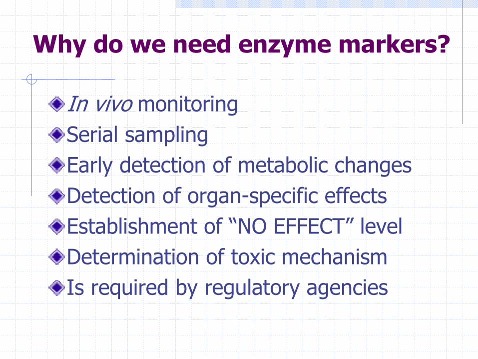
Why do we need enzyme markers?
In vivo monitoringSerial samplingEarly detection of metabolic changesDetection of organ-specific effectsEstablishment of “NO EFFECT” levelDetermination of toxic mechanismIs required by regulatory agencies
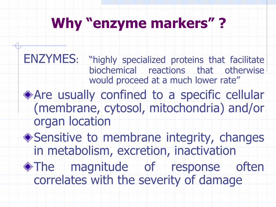
ENZYMES: “highly specialized proteins that facilitate biochemical reactions that otherwise would proceed at a much lower rate”
Are usually confined to a specific cellular (membrane, cytosol, mitochondria) and/or organ locationSensitive to membrane integrity, changes in metabolism, excretion, inactivationThe magnitude of response often correlates with the severity of damage
Why “enzyme markers” ?
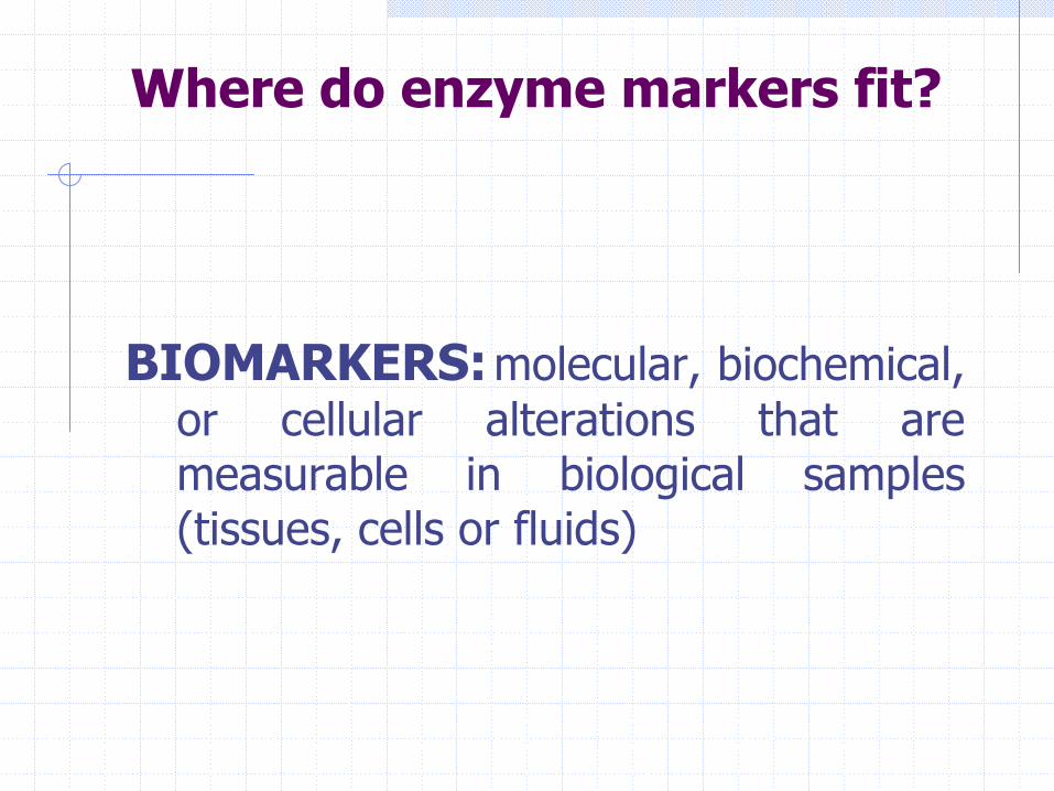
BIOMARKERS:molecular, biochemical, or cellular alterations that are measurable in biological samples (tissues, cells or fluids)
Where do enzyme markers fit?
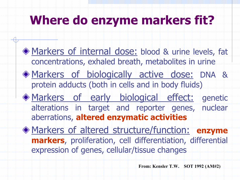
Markers of internal dose: blood & urine levels, fat concentrations, exhaled breath, metabolites in urine
Markers of biologically active dose: DNA & protein adducts (both in cells and in body fluids)
Markers of early biological effect: genetic alterations in target and reporter genes, nuclear aberrations, altered enzymatic activities
Markers of altered structure/function: enzyme markers, proliferation, cell differentiation, differential expression of genes, cellular/tissue changes
Where do enzyme markers fit?
From: Kensler T.W. SOT 1992 (AM#2)
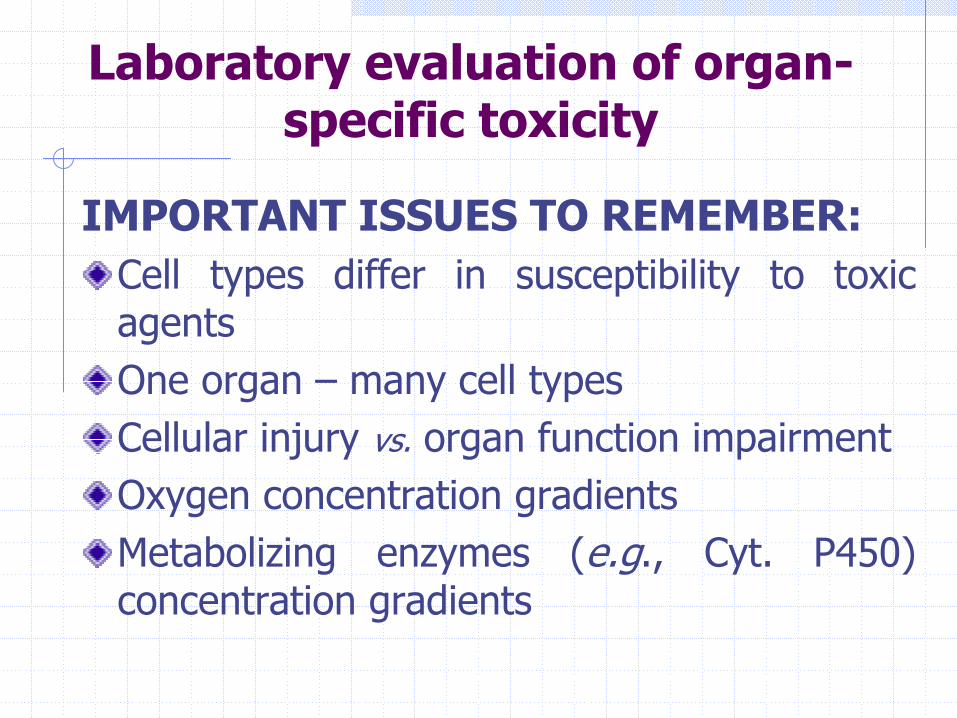
Laboratory evaluation of organ-specific toxicity
IMPORTANT ISSUES TO REMEMBER:Cell types differ in susceptibility to toxic agentsOne organ – many cell typesCellular injury vs. organ function impairmentOxygen concentration gradientsMetabolizing enzymes (e.g., Cyt. P450) concentration gradients
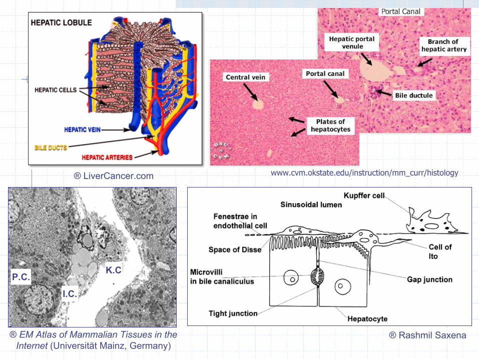
® LiverCancer.com
P.C.K.C.
I.C.
® EM Atlas of Mammalian Tissues in the Internet (Universität Mainz, Germany)
® Rashmil Saxena
www.cvm.okstate.edu/instruction/mm_curr/histology
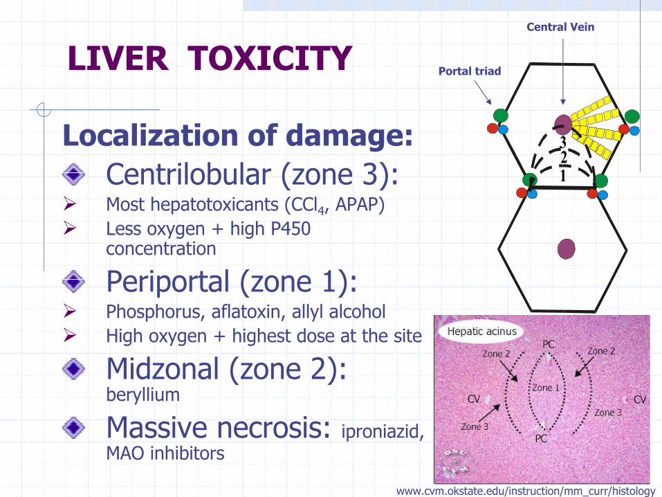
Localization of damage:Centrilobular (zone 3):Most hepatotoxicants (CCl4, APAP)Less oxygen + high P450 concentration
Periportal (zone 1):Phosphorus, aflatoxin, allyl alcoholHigh oxygen + highest dose at the site
Midzonal (zone 2): beryllium
Massive necrosis: iproniazid, MAO inhibitors
LIVER TOXICITY
123
Portal triad
Central Vein
www.cvm.okstate.edu/instruction/mm_curr/histology
Cholestatic Injury
Cytotoxic Injury
Disturbances of hepatic
function/clearance
LIVER TOXICITY

General properties that describe a useful
biomarker of xenobiotic-induced hepatic toxicityAvailability: present in biological fluids in detectable levels
Specificity: is of liver origin (exclusively or predominantly), or its
level is affected by a change in liver function
Prevalence: can be applied across multiple species, including
humans
Sensitivity: can be reliably measured at sub-lethal doses of a
xenobiotic
Persistence: stable to allow studies within days or weeks after
collection
Relevancy: confirmed to be associated with histopathological or
functional changes in the liver
LIVER TOXICITY
Adapted from Amacher DE (2002)
Serum enzyme testsHepatic excretory tests* Alterations in chemical constituents
of the liver* Histological analysis of liver injury
Evaluation of liver toxicity in vivo
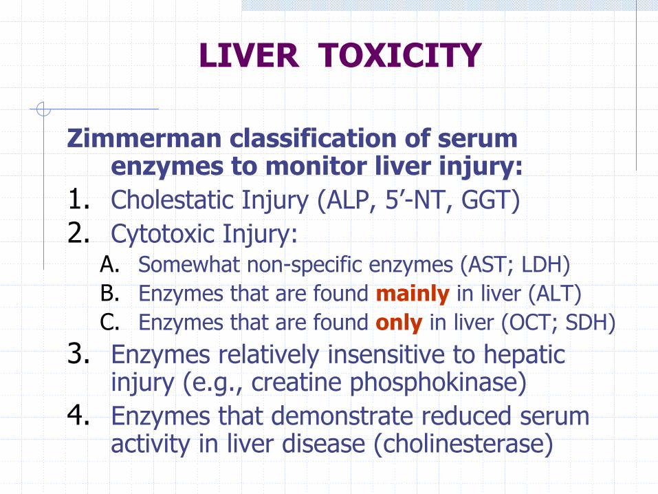
Zimmerman classification of serum enzymes to monitor liver injury:
1. Cholestatic Injury (ALP, 5’-NT, GGT)2. Cytotoxic Injury:
A. Somewhat non-specific enzymes (AST; LDH)B. Enzymes that are found mainly in liver (ALT)C. Enzymes that are found only in liver (OCT; SDH)
3. Enzymes relatively insensitive to hepatic injury (e.g., creatine phosphokinase)
4. Enzymes that demonstrate reduced serum activity in liver disease (cholinesterase)
LIVER TOXICITY
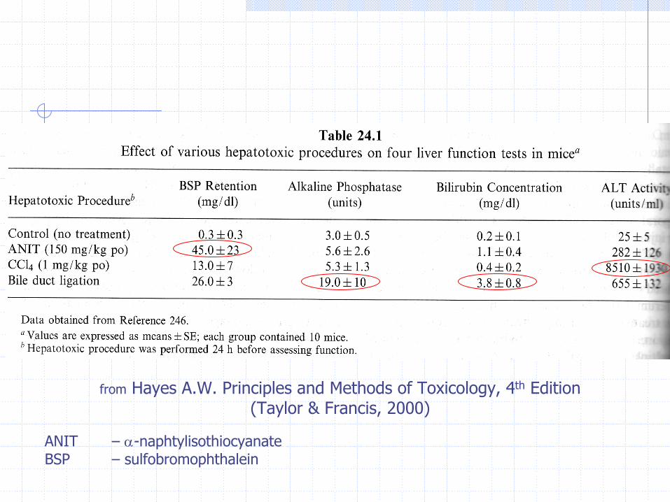
from Hayes A.W. Principles and Methods of Toxicology, 4th Edition (Taylor & Francis, 2000)
ANIT – α-naphtylisothiocyanateBSP – sulfobromophthalein
LIVER TOXICITY
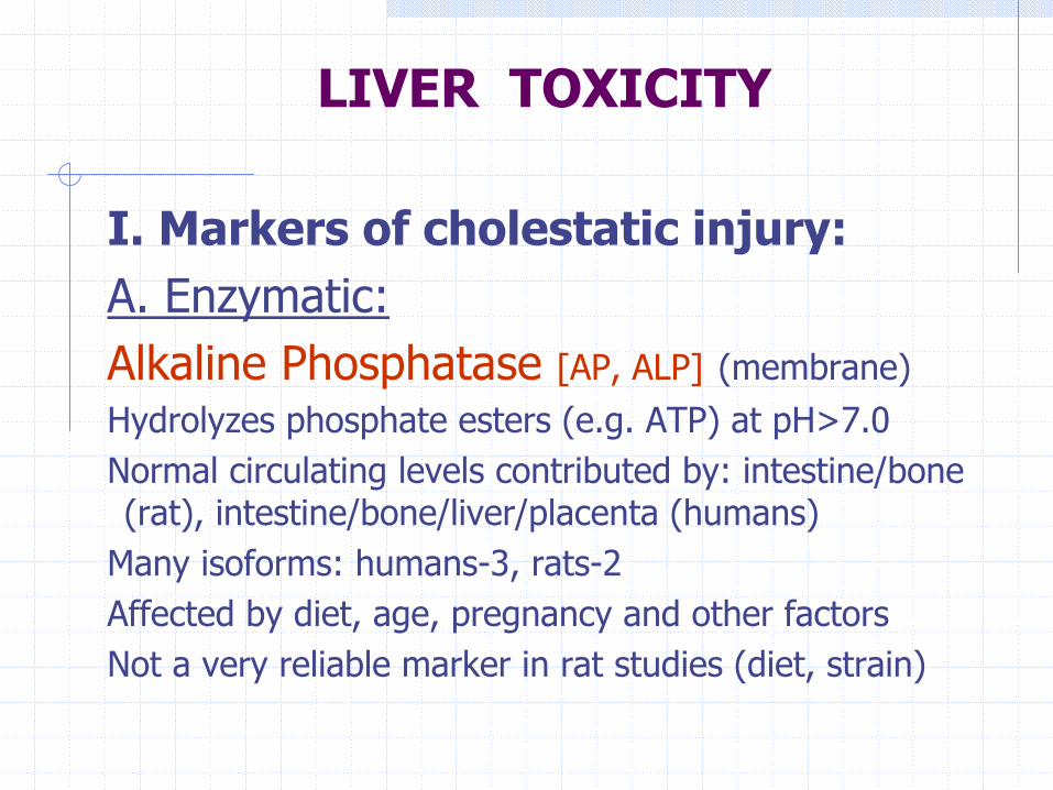
I. Markers of cholestatic injury:A. Enzymatic:Alkaline Phosphatase [AP, ALP] (membrane)Hydrolyzes phosphate esters (e.g. ATP) at pH>7.0Normal circulating levels contributed by: intestine/bone (rat), intestine/bone/liver/placenta (humans)
Many isoforms: humans-3, rats-2Affected by diet, age, pregnancy and other factorsNot a very reliable marker in rat studies (diet, strain)
LIVER TOXICITY
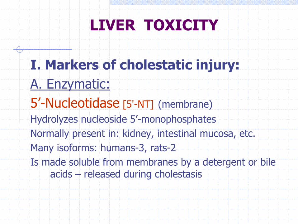
I. Markers of cholestatic injury:A. Enzymatic:5’-Nucleotidase [5'-NT] (membrane)Hydrolyzes nucleoside 5’-monophosphatesNormally present in: kidney, intestinal mucosa, etc.Many isoforms: humans-3, rats-2Is made soluble from membranes by a detergent or bile
acids – released during cholestasis
LIVER TOXICITY
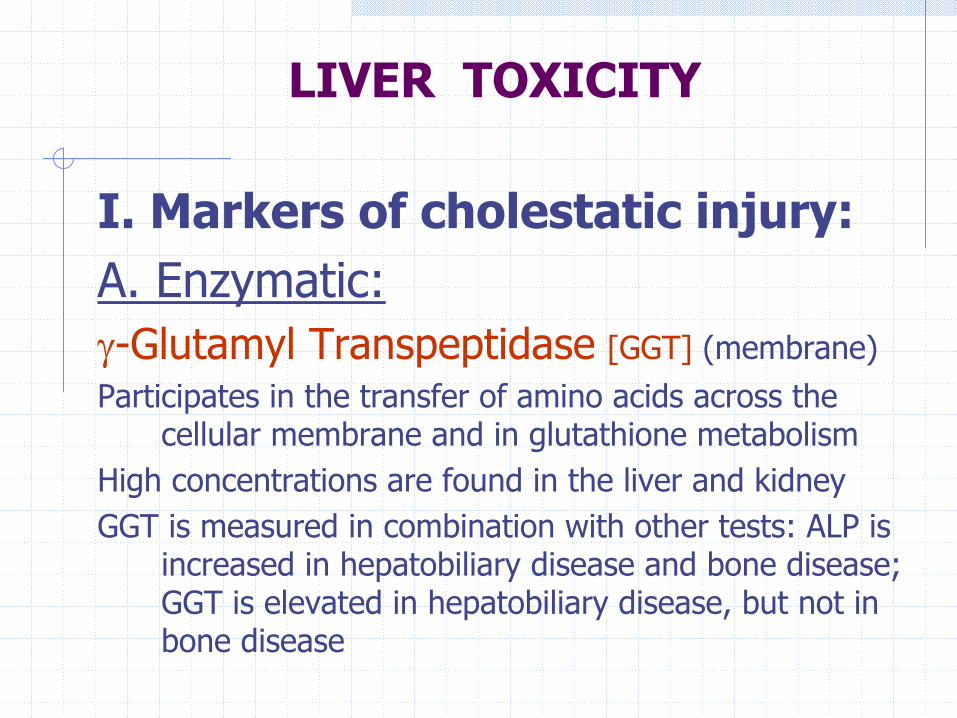
I. Markers of cholestatic injury:A. Enzymatic:γ-Glutamyl Transpeptidase [GGT] (membrane)Participates in the transfer of amino acids across the
cellular membrane and in glutathione metabolismHigh concentrations are found in the liver and kidney GGT is measured in combination with other tests: ALP is
increased in hepatobiliary disease and bone disease; GGT is elevated in hepatobiliary disease, but not in bone disease
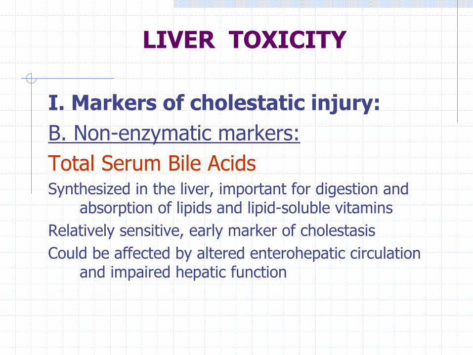
I. Markers of cholestatic injury:B. Non-enzymatic markers:Total Serum Bile AcidsSynthesized in the liver, important for digestion and
absorption of lipids and lipid-soluble vitaminsRelatively sensitive, early marker of cholestasisCould be affected by altered enterohepatic circulation
and impaired hepatic function
LIVER TOXICITY
LIVER TOXICITY
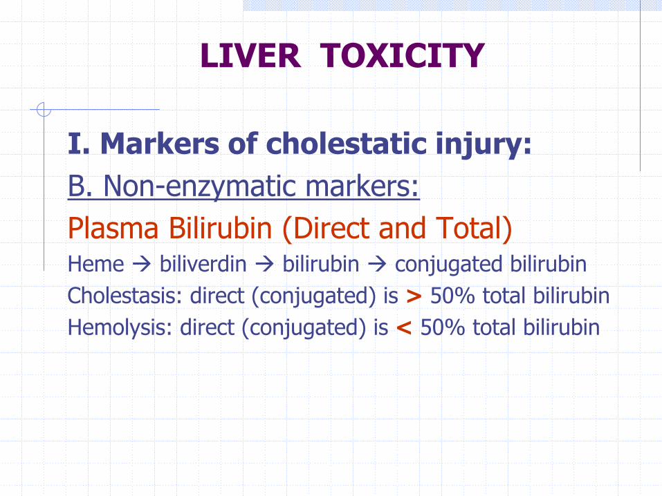
I. Markers of cholestatic injury:B. Non-enzymatic markers:Plasma Bilirubin (Direct and Total)Heme biliverdin bilirubin conjugated bilirubinCholestasis: direct (conjugated) is > 50% total bilirubinHemolysis: direct (conjugated) is < 50% total bilirubin
LIVER TOXICITY
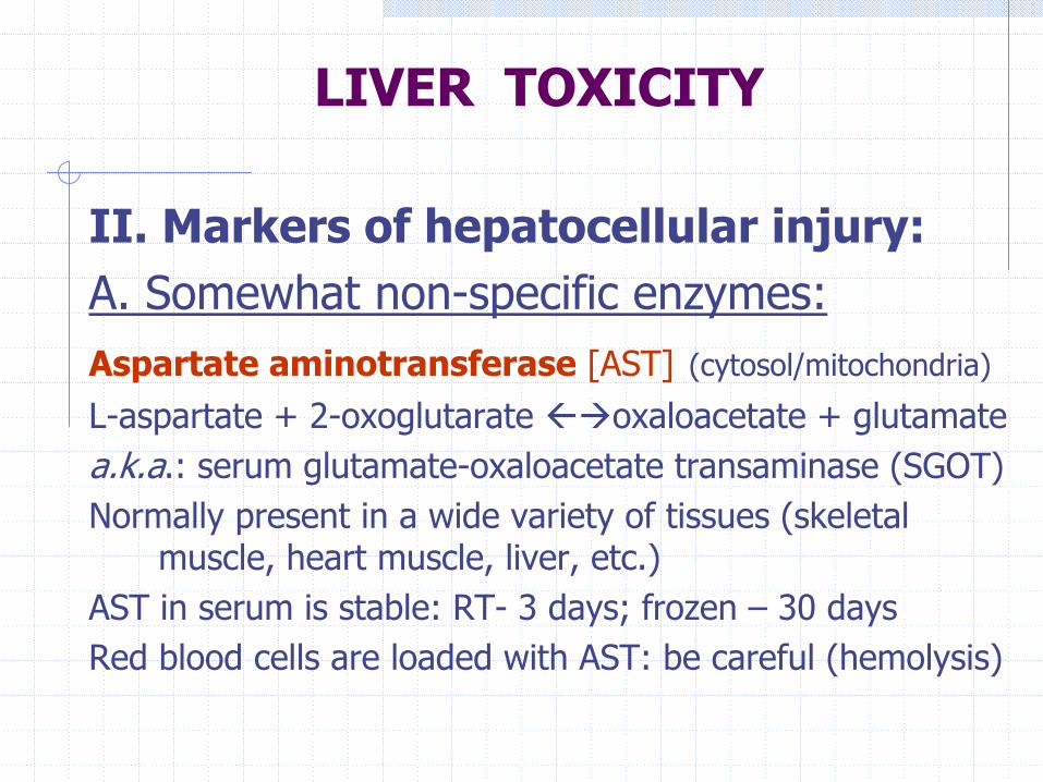
II. Markers of hepatocellular injury:A. Somewhat non-specific enzymes:Aspartate aminotransferase [AST] (cytosol/mitochondria)
L-aspartate + 2-oxoglutarate oxaloacetate + glutamatea.k.a.: serum glutamate-oxaloacetate transaminase (SGOT)Normally present in a wide variety of tissues (skeletal
muscle, heart muscle, liver, etc.)AST in serum is stable: RT- 3 days; frozen – 30 daysRed blood cells are loaded with AST: be careful (hemolysis)

II. Markers of hepatocellular injury:A. Somewhat non-specific enzymes:Lactate Dehydrogenase [LDH] (cytosol)
pyruvate L-lactateNormally present in a wide variety of tissuesFive isoenzymes, isoenzyme profile may help identify
specific tissue origin (LDH-5 liver; LDH-1,-2 kidney)
LIVER TOXICITY
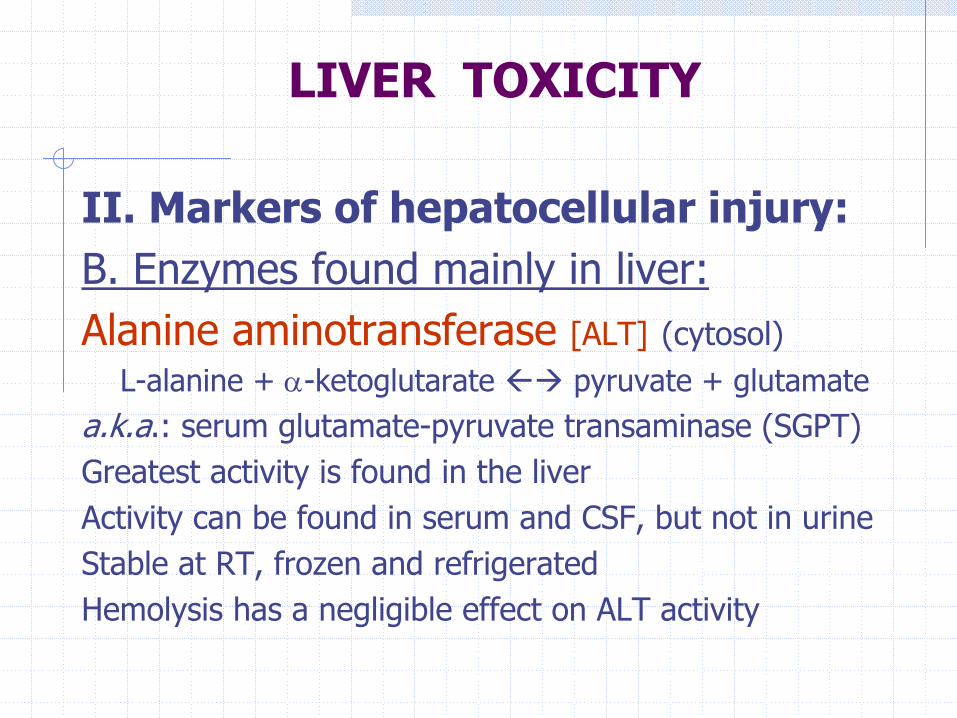
II. Markers of hepatocellular injury:B. Enzymes found mainly in liver:Alanine aminotransferase [ALT] (cytosol)
L-alanine + α-ketoglutarate pyruvate + glutamatea.k.a.: serum glutamate-pyruvate transaminase (SGPT)Greatest activity is found in the liverActivity can be found in serum and CSF, but not in urineStable at RT, frozen and refrigeratedHemolysis has a negligible effect on ALT activity
LIVER TOXICITY
LIVER TOXICITY
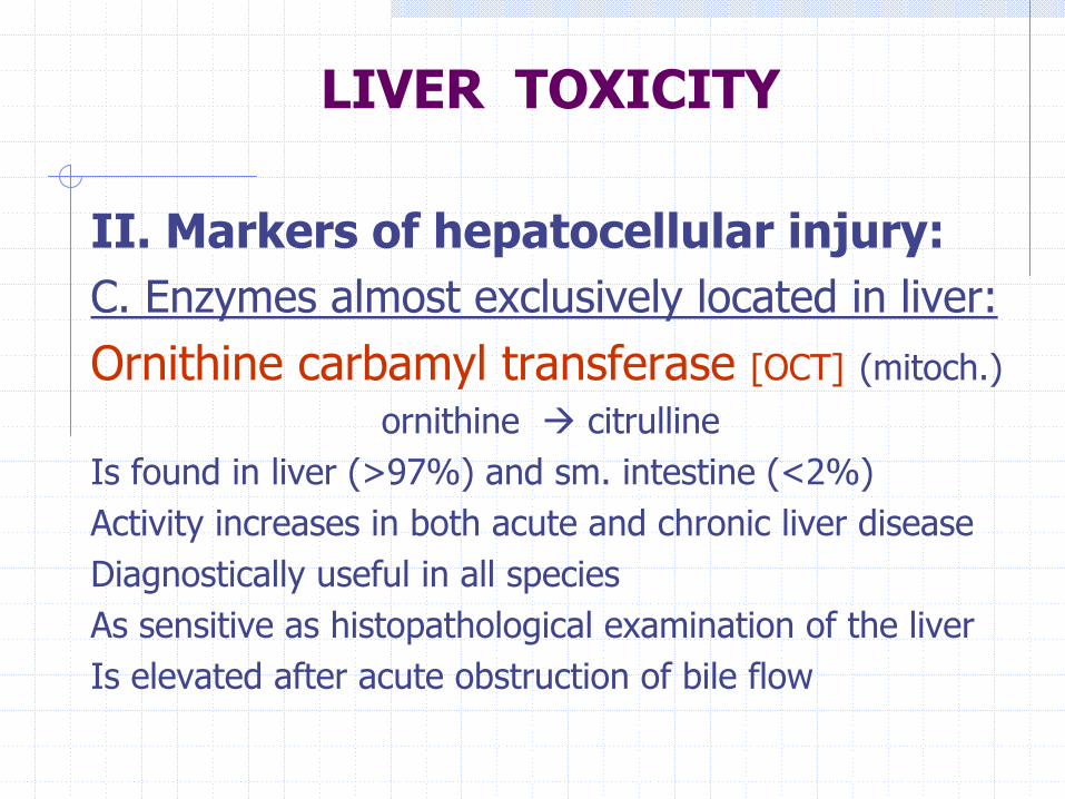
II. Markers of hepatocellular injury:C. Enzymes almost exclusively located in liver:Ornithine carbamyl transferase [OCT] (mitoch.)
ornithine citrullineIs found in liver (>97%) and sm. intestine (<2%)Activity increases in both acute and chronic liver diseaseDiagnostically useful in all speciesAs sensitive as histopathological examination of the liverIs elevated after acute obstruction of bile flow
LIVER TOXICITY
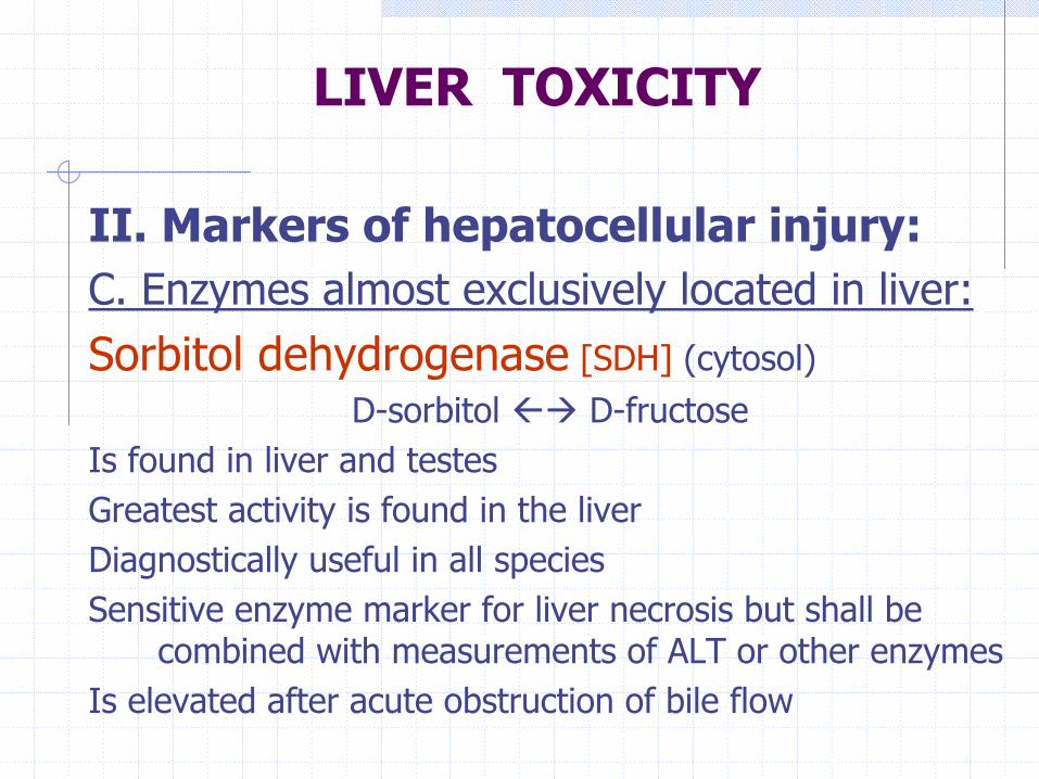
II. Markers of hepatocellular injury:C. Enzymes almost exclusively located in liver:Sorbitol dehydrogenase [SDH] (cytosol)
D-sorbitol D-fructoseIs found in liver and testesGreatest activity is found in the liverDiagnostically useful in all speciesSensitive enzyme marker for liver necrosis but shall be
combined with measurements of ALT or other enzymesIs elevated after acute obstruction of bile flow

III. Enzymes relatively insensitive to hepatic injury:
Creatine phosphokinase [CPK]creatine + ATP creatine phosphate + ADP
Greatest activity is found in skeletal muscleIs used as a marker of muscle injury (clinical use – cardiac
muscle injury)
LIVER TOXICITY

IV.Enzymes that demonstrate reduced serum activity in liver disease:
Choline Esterase [ChE]Acetylcholine esterase and butyrylcholine esterase
Inhibited by organophosphates and carbamatesCan not distinguish between decreased synthesis and
decreased activity
LIVER TOXICITY
LIVER TOXICITY
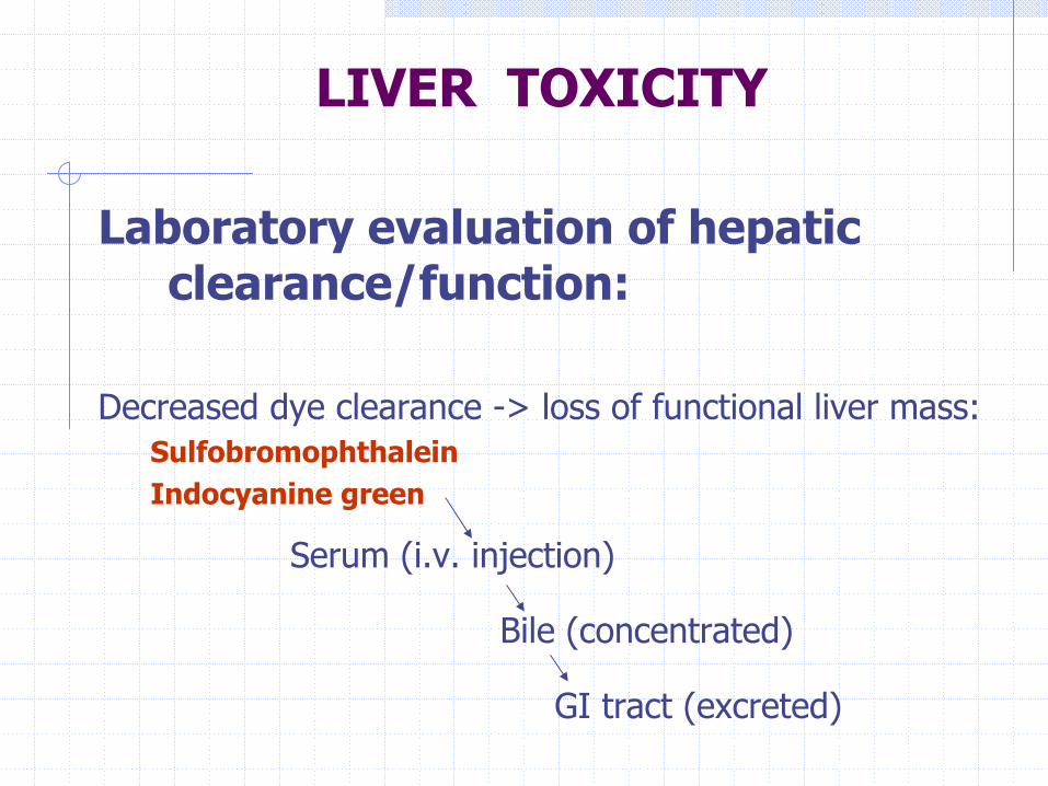
Laboratory evaluation of hepatic clearance/function:
Decreased dye clearance -> loss of functional liver mass:SulfobromophthaleinIndocyanine green
Serum (i.v. injection)
Bile (concentrated)
GI tract (excreted)
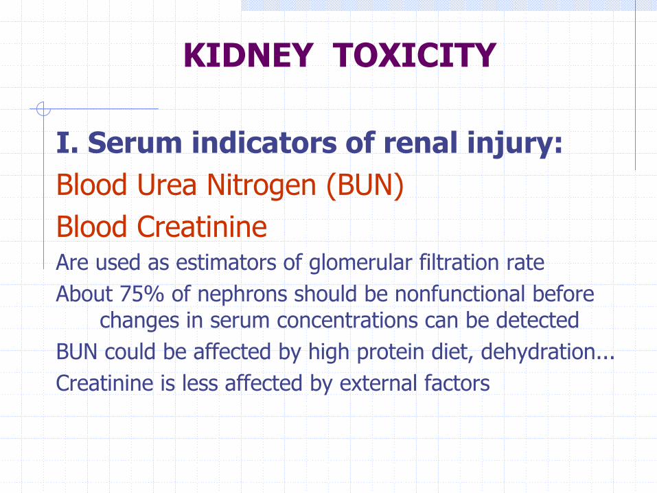
KIDNEY TOXICITY
I. Serum indicators of renal injury:Blood Urea Nitrogen (BUN)Blood CreatinineAre used as estimators of glomerular filtration rateAbout 75% of nephrons should be nonfunctional before
changes in serum concentrations can be detectedBUN could be affected by high protein diet, dehydration...Creatinine is less affected by external factors
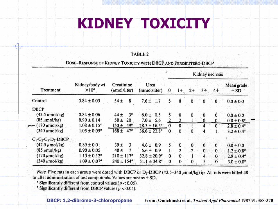
KIDNEY TOXICITY
From: Omichinski et al, Toxicol Appl Pharmacol 1987 91:358-370DBCP: 1,2-dibromo-3-chloropropane
KIDNEY TOXICITY
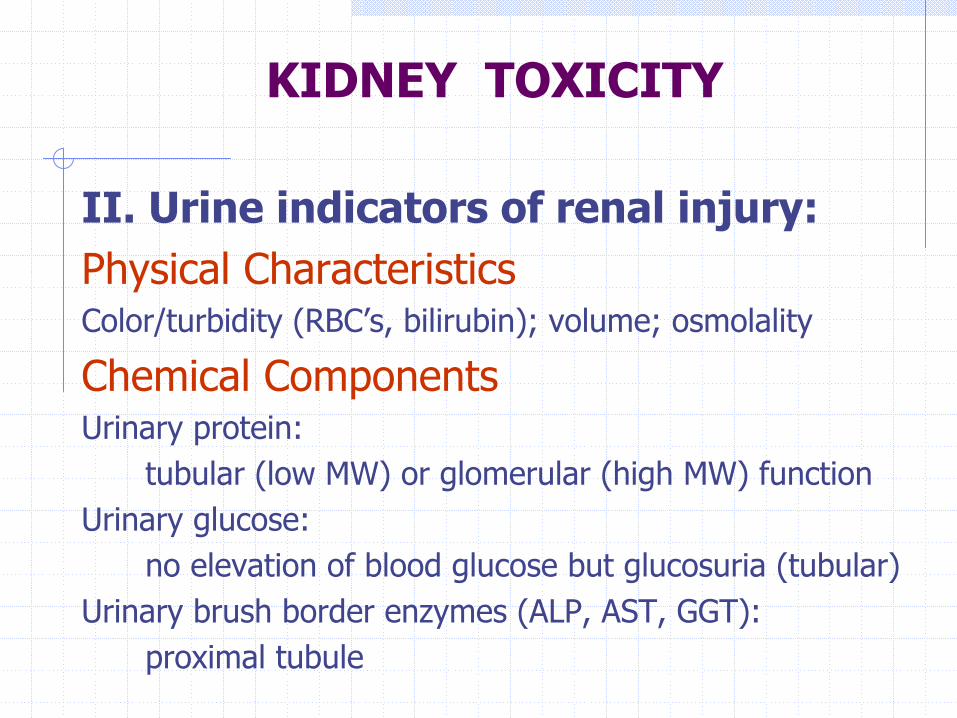
II. Urine indicators of renal injury:Physical CharacteristicsColor/turbidity (RBC’s, bilirubin); volume; osmolality
Chemical ComponentsUrinary protein:
tubular (low MW) or glomerular (high MW) functionUrinary glucose:
no elevation of blood glucose but glucosuria (tubular)Urinary brush border enzymes (ALP, AST, GGT):
proximal tubule
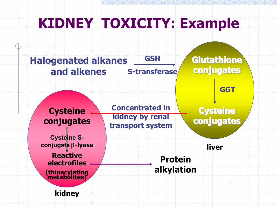
KIDNEY TOXICITY: Example
Halogenated alkanesand alkenes
Glutathione Glutathione Glutathione conjugatesconjugatesconjugates
CysteineCysteineCysteineconjugatesconjugatesconjugates
Cysteineconjugates
Concentrated in kidney by renal
transport system
GSH
S-transferase
Reactive electrofiles
(thioacylatingmetabolites)
Cysteine S-conjugate β-lyase
Protein alkylation
liver
kidney
GGT

KIDNEY TOXICITY: Example