Toxicity Testing
18
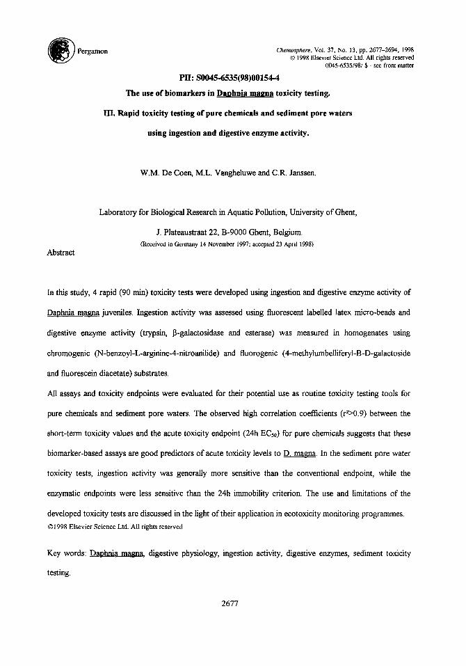
~ JPergamon Chemosphere, Vol.37, No. 13, pp. 2677-2694, 1998 © 1998 Elsevier Science Ltd.All rightsreserved 0045-6535/98/$- see frontmatter PII: S0045-6535(95)00154--4 The use of biomarkers in Daphnia magna toxicity testing. IlL Rapid toxicity testing of pure chemicals and sediment pore waters using ingestion and digestive enzyme activity. W.M. De Coen, M.L. Vangheluwe and C.R, Janssen. Abstract Laboratory for Biological Research in Aquatic Pollution, University of Ghent, J. Plateaustraat 22, B-9000 Ghent, Belgium. (Received in Germany 14 November 1997; accepted 23 April 1998) In this study, 4 rapid (90 min) toxicity tests were developed using ingestion and digestive enzyme activity of Daphnia magna juveniles. Ingestion activity was assessed using fluorescent labelled latex micro-beads and digestive enzyme activity (trypsin, 13-galactosidase and esterase) was measured in homogenates using chromogenic (N-benzoyl-L-arginine-4-nitroanilide) and fiuorogenic (4-methylumbelliferyl-B-D-galactoside and fluorescein diacetate) substrates. All assays and toxicity endpoints were evaluated for their potential use as routine toxicity testing tools for pure chemicals and sediment pore waters. The observed high correlation coefficients (r2>0.9) between the short-term toxicity values and the acute toxicity endpoint (24h ECso) for pure chemicals suggests that these biomarker-based assays are good predictors of acute toxicity levels to D, magna. In the sediment pore water toxicity tests, ingestion activity was generally more sensitive than the conventional endpoint, while the enzymatic endpoints were less sensitive than the 24h immobility criterion. The use and limitations of the developed toxicity tests are discussed in the light of their application in ecotoxicity monitoring programmes. ©1998 Elsevier ScienceLtd. All rights reserved Key words: Daphnia magn_ a, digestive physiology, ingestion activity, digestive enzymes, sediment toxicity testing. 2677
-
Upload
maureen-joy-dagson-galingan -
Category
Documents
-
view
37 -
download
3
description
Toxicity Testing using Daphnia Magna
Transcript of Toxicity Testing
~ J Pergamon Chemosphere, Vol. 37, No. 13, pp. 2677-2694, 1998 © 1998 Elsevier Science Ltd. All rights reserved
0045-6535/98/$ - see front matter
PII: S0045-6535(95)00154--4
The use of biomarkers in Daphnia magna toxicity testing.
IlL Rapid toxicity testing of pure chemicals and sediment pore waters
using ingestion and digestive enzyme activity.
W.M. De Coen, M.L. Vangheluwe and C.R, Janssen.
Abstract
Laboratory for Biological Research in Aquatic Pollution, University of Ghent,
J. Plateaustraat 22, B-9000 Ghent, Belgium. (Received in Germany 14 November 1997; accepted 23 April 1998)
In this study, 4 rapid (90 min) toxicity tests were developed using ingestion and digestive enzyme activity of
Daphnia magna juveniles. Ingestion activity was assessed using fluorescent labelled latex micro-beads and
digestive enzyme activity (trypsin, 13-galactosidase and esterase) was measured in homogenates using
chromogenic (N-benzoyl-L-arginine-4-nitroanilide) and fiuorogenic (4-methylumbelliferyl-B-D-galactoside
and fluorescein diacetate) substrates.
All assays and toxicity endpoints were evaluated for their potential use as routine toxicity testing tools for
pure chemicals and sediment pore waters. The observed high correlation coefficients (r2>0.9) between the
short-term toxicity values and the acute toxicity endpoint (24h ECso) for pure chemicals suggests that these
biomarker-based assays are good predictors of acute toxicity levels to D, magna. In the sediment pore water
toxicity tests, ingestion activity was generally more sensitive than the conventional endpoint, while the
enzymatic endpoints were less sensitive than the 24h immobility criterion. The use and limitations of the
developed toxicity tests are discussed in the light of their application in ecotoxicity monitoring programmes.
© 1998 Elsevier Science Ltd. All rights reserved
Key words: Daphnia magn_ a, digestive physiology, ingestion activity, digestive enzymes, sediment toxicity
testing.
2677
2678
Introduction
The development of water quality criteria for chemicals and the hazard evaluation of waste discharges into
the aquatic environment is usually based on the results of conventional ecotoxicity tests [1, 2]. Due to the
increasing number of chemicals being produced and discharged into the environment, the need for rapid
toxicity screening tests of these chemicals and wastes is growing [3, 4]. Several short-term (mainly bacterial)
toxicity tests have been developed which are based on the assessment of the metabolic status of the organism
such as the measurement of respiration, ATP content and enzyme inhibition [5]. In contrast to this variety of
short-term bacterial toxicity tests, only few rapid screening assays with invertebrates are available.
Several authors have suggested that changes in the physiology and/or behaviour of aquatic invertebrates
could be used as rapid indicators of toxic stress [6, 7]. Changes occurring at the suborganismal level may be
the initial response to toxic insult and might explain subsequent effects occurring at the organismal level such
as reductions in growth, reproduction or survival [8].
Previous studies have shown that ingestion rate is a useful ecotoxicological endpoint in assays with
invertebrates. Day and Kaushik [9] for example pointed out that toxicant-induced ingestion inhibition in
daphnids might explain observed changes in survival, growth and reproduction. However, most of the
techniques used to quantify the ingestion activity of invertebrates are based on algae density measurements
[10,11 ], which is time consuming and therefore less applicable in routine. Recently, fluorescent microspheres
have been used to measure ingestion rates of cladocerans and ciliates [12]. Although the toxicity testing
technique developed by these authors seems to be sensitive, it requires image analysis and the use of
specialised video-equipment which makes it less attractive for routine applications.
Janssen and Persoone [7] developed a lh in vivo fluorescence inhibition test with Danhnia magna. Although
it was originally suggested that in this assay both enzyme inhibition and ingestion activity are measured,
recent studies have shown that the in vivo fluorescence test criterion is mainly based on ingestion inhibition
[13]. The same authors demonstrated that digestive enzyme inhibition alone could also be a useful toxicity
endpoint.
2679
In this study the potential use of both ingestion and digestive enzyme activity of D. magna as rapid toxicity
screening tools was evaluated using pure compounds and environmental samples (sediment pore waters). The
aim of this study was to demonstrate that these rapid toxicity screening tests could be valid alternatives (to
conventional tests) in a battery of tests for routine toxicity assessments.
Materials and methods.
Ingestion measurement using fluorescent micro-beads.
The following test design is described in detail in a previous study [13]: 30 juvenile (<24h) D. magna were
exposed for 90 min to 5 different toxicant concentrations after which the organisms were transferred into a 5
ml fluorescent beads suspension (106 beads.ml "l, Fluoresbrite, Polyscience Inc.). Three replicates per toxicant
concentration were used. Fluorescence was measured at the beginning and end of the 30 min feeding period
using a Perkin Elmer LS50 fluorimeter (ex 458 nm, em. 540 run). Ingestion activity is expressed as number of
beads.daphnid'l.h l .
Enzyme activity measurement.
At the end of each exposure daphnids were collected and shock-frozen in liquid nitrogen and stored at -80°C.
Enzyme activity quantification is described in detail in a previous study [13] and is summarised hereunder.
After homogenisation and centrifugation of the daphnids, the supernatant was collected and used for the
enzymatic activity analysis. The following enzyme specific substrates were used: 4-methylumbelliferyl-13-D-
galactoside for 13-galactosidase [14], fluorescein diacetate for esterase [14], N-benzoyl-L-arginine-4-
nitroanilide for trypsin [ 15]. Protein content of each homogenate was determined according to Bradford [16].
Enzyme activity was expressed as lamol.min'l.mg protein 1.
Sediment sample collection
Samples were collected from seven sedimentation zones in the Bovenschelde (Flanders) using a Van Veen
grab (2 1). The sediments were contaminated with several heavy metals and organic compounds (Table 1).
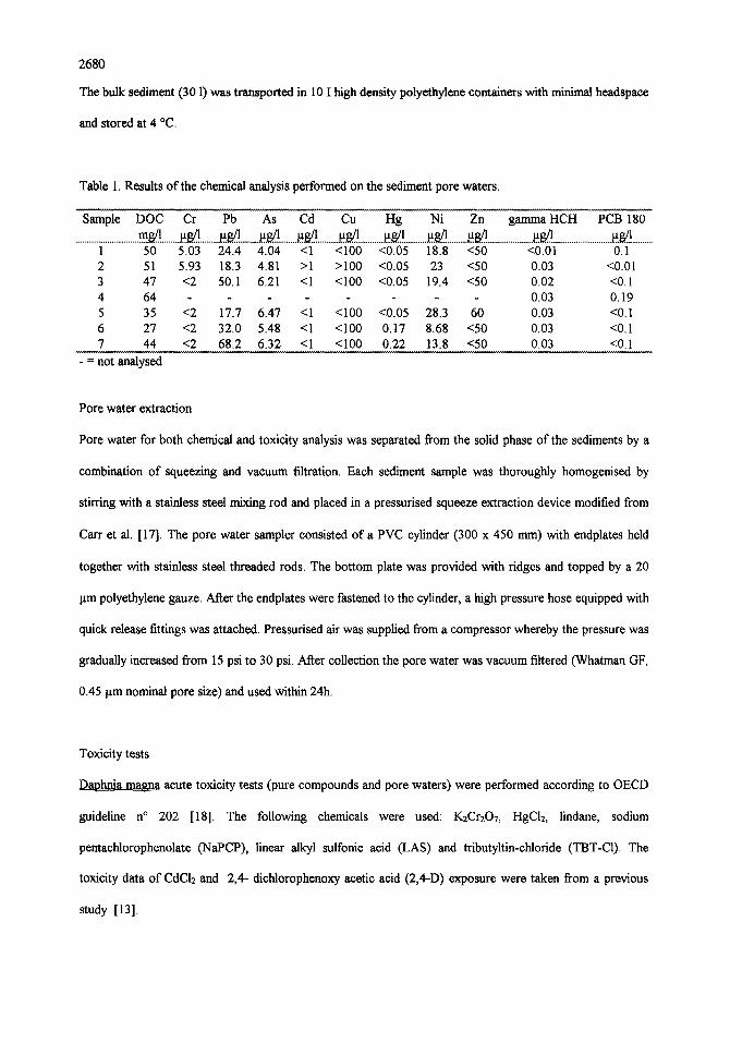
2680
The bulk sediment (30 1) was transported in 10 1 high density polyethylene containers with minimal headspaee
and stored at 4 °C.
Table 1. Results of the chemical analysis performed on the sediment pore waters.
Sample DOC Cr Pb As Cd Cu Fig Ni Zn gamma HCH PCB 180 .............................. m . ~ . . _ . ~ . ~ ......... ~ g . . ! ......... ~ . ~ ........ ~.s/..~ .......... ~.g..1 ............ ~ g ! . .......... ~ . . ~ ......... ~ ...................... a ~ ............................ ~ . . ~ ! ..........
1 50 5.03 24.4 4.04 <1 <100 <0.05 18.8 <50 <0.01 0.1 2 51 5.93 18.3 4.81 >1 >100 <0.05 23 <50 0.03 <0.01 3 47 <2 50.1 6.21 <1 <100 <0.05 19.4 <50 0.02 <0.1 4 64 . . . . 0.03 0.19 5 35 <2 17.7 6.47 <1 <100 <0.05 28.3 60 0.03 <0.1 6 27 <2 32.0 5.48 <1 <100 0.17 8.68 <50 0.03 <0.1 7 44 <2 68.2 6.32 <1 <100 0.22 13.8 <50 0.03 <0.1
- = not analysed
Pore water extraction
Pore water for both chemical and toxicity analysis was separated from the solid phase of the sediments by a
combination of squeezing and vacuum filtration. Each sediment sample was thoroughly homogenised by
stirring with a stainless steel mixing rod and placed in a pressurised squeeze extraction device modified from
Carr et al. [17]. The pore water sampler consisted o f a PVC cylinder (300 x 450 mm) with endplates held
together with stainless steel threaded rods. The bottom plate was provided with ridges and topped by a 20
~tm polyethylene gauze. After the endplates were fastened to the cylinder, a high pressure hose equipped with
quick release fittings was attached. Pressurised air was supplied from a compressor whereby the pressure was
gradually increased from 15 psi to 30 psi. After collection the pore water was vacuum filtered (Whatman GF,
0.45 ~tm nominal pore size) and used within 24h.
Toxicity tests
Daphnia magna acute toxicity tests (pure compounds and pore waters) were performed according to OECD
guideline n ° 202 [18]. The following chemicals were used: K2Cr2OT, HgCI2, lindane, sodium
pentachlorophenolate (NaPCP), linear alkyl sulfonic acid (LAS) and tributyltin-chloride (TBT-C1). The
toxicity data of CdCI2 and 2,4- dichlorophenoxy acetic acid (2,4-D) exposure were taken from a previous
study [13].
2681
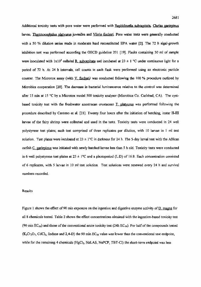
Additional toxicity tests with pore water were performed with Raphidocelis subcapitata Clarias gariepinus
larvae, Thanmocephalus platym'us juveniles and Vibri0 fischerk Pore water tests were generally conducted
with a 50 % dilution series made in moderate hard reconstituted EPA water [2]. The 72 h algal-growth
inhibition test was performed according the OECD guideline 201 [19]. Flasks containing 50 ml of sample
were inoculated with lxl04 cells/ml 1L subcapitata and incubated at 23 ± 1 °C under continuous light for a
period of 72 h. At 24 h intervals, cell counts in each flask were performed using an electronic particle
counter. The Microtox assay (with V. fischeri) was conducted folloWing the 100 % procedure outlined by
Microbics cooperation [20]. The decrease in bacterial luminescence relative to the control was determined
after 15 min at 15 °C by a Microtox model 500 toxicity analyser (Microbics Co. Carlsbad, CA). The cyst-
based toxicity test with the freshwater anostracan crustacean T. platyurus was performed following the
procedure described by Centeno et al. [21]. Twenty four hours after the initiation of hatching, instar II-III
larvae of the fairy shrimp were collected and used in the tests. Toxicity tests were conducted in 24 well
polystyrene test plates; each test comprised of three replicates per dilution, With 10 larvae in 1 ml test
solution. Test plates were incubated at 23 ± I°C in darkness for 24 h. The 5-day larval test With the African
catfish C. gariepinus was initiated With newly hatched larvae less than 5 h old. Toxicity tests were conducted
in 6 well polystyrene test plates at 23 + I°C and a photoperiod (L:D) of 16:8. Each concentration consisted
of 6 replicates, with 5 larvae in 10 ml test solution. Test solutions were renewed every 24 h and survival
numbers recorded.
Results
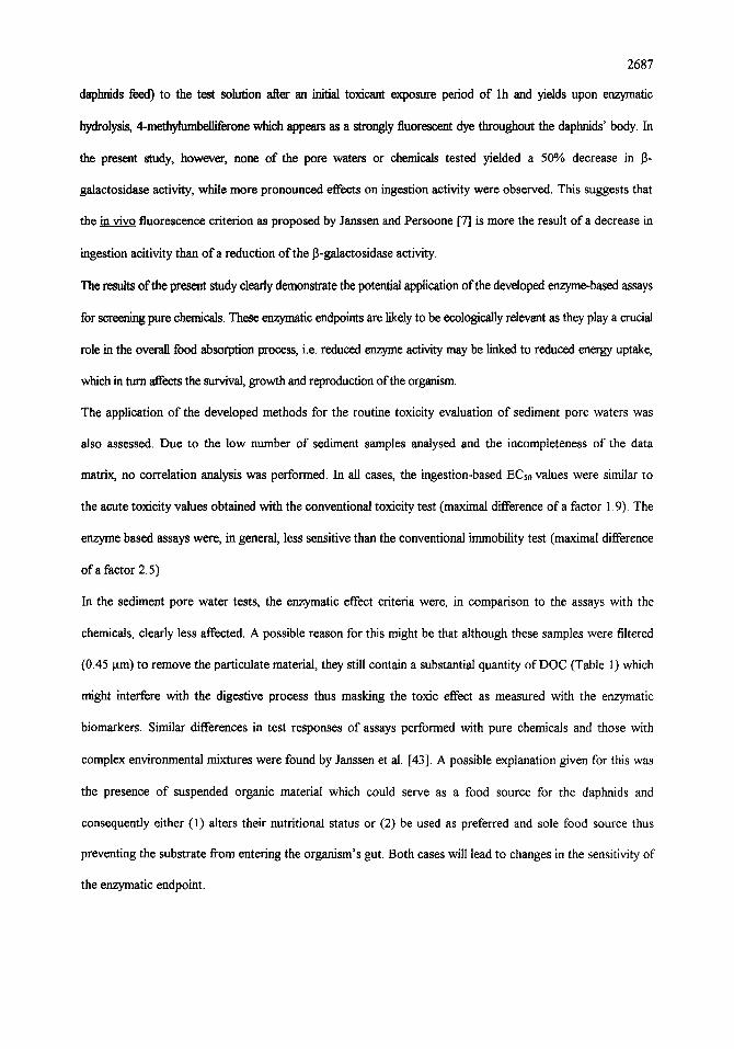
Figure 1 shows the effect of 90 min exposure on the ingestion and digestive enzyme activity ofD. magna for
all 8 chemicals tested. Table 2 shows the effect concentrations obtained With the ingestion-based toxicity test
(90 rain ECs0) and those of the conventional acute toxicity test (24h ECho). For half of the compounds tested
(K2Cr2OT, CdC12, lindane and 2,4-I)) the 90 rain ECso value was lower than the conventional test endpoint,
while for the remaining 4 chemicals (HgC12, NaLAS, NaPCP, TBT-CI) the short-term endpoint was less
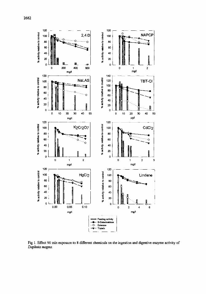
2682
120
loo 8
8o
l ,o 2O
0
120
8O
6O !,o 2O
0
12o ¸
8
.~ , 0
2O
0
120
1~ 80
!°
o
2,4 D .... o .......... o
0 200 4013 600
m~
s
0 10 20 30 40 50 mgn
~ 7
Hi ° .... : Iill , , o 1 2
mg/I
l 1 HgCI2
0.00 0.05 0.10
r, WJ
120
~ 100 8 $1 8 0
20
0
14o T~ 12o
8 loo
~ 60
20 N
0
120
loo 8 fl 80
20
o
120
80
.e
20
0
Feeding activity B-Galactosidase
• -O.. Esterase --qF-- Tryp61n
~ I! !
1 2 m~
TBT-CI
0 10 20 30 40 50 P~
CdCI 2
~"" -.-,.~[ ' . . . . . O
1 2 mN
~ n e
0 2 4 6 m~l
Fig I. Effect 90 min exposure to 8 different chemicals on the ingestion and digestive enzyme activity o f
Daphnia magna.
2683
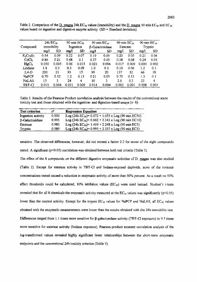
Table 2. Comparison of the D. rnagna 24h ECs0 values (immobility) and the D. magna 90 min ECs0 and ECl0 values based on ingestion and digestive enzyme activity. (SD = Standard deviation).
Compound 24h ECs0 90 min ECs0 90 min EC10 90 min EC1o 90 min ECl0
immobility Ingestion [3-Galactosidase Esterase Trypsin mg/l SD mg/l ..S D ms/1 SD mg/l SD mg/l SD
K2Cr207 0.54 0.09 0.22 0.07 0.19 0.05 0.20 0.05 0.21 0.04 CdCI2 0.86 0.21 0.68 0.I 0.37 0.05 0.38 0.08 0.24 0.03 HgC12 0.030 0.005 0.06 0.015 0.021 0.006 0.017 0.009 0.010 0.002
Lindane 1.8 0.3 0.3 0.09 1.0 0.1 0.19 0.06 1.2 0.1 2,4-D 200 21 93 15 90 20 137 12 66 18
NaPCP 0.70 0.12 1.2 0.15 0.21 0.03 0.70 0.13 1.3 0.1 NaLAS 15 3 24 4 10 3 2.6 0.3 23 4 TBT-CI 0.015 0.004 0.031 0.009 0.014 0.004 0.003 0.001 0.008 0.001
Table 3. Results of the Pearson Product correlation analysis between the results of the conventional acute toxicity test and those obtained with the ingestion- and digestion-based assays (n=8).
Test criterion r 2 Ingestion activity 0.900 13-Galactosidase 0.995 Esterase 0.983 T r yps in 0.989
Regression Equation Log (24h ECs0) = 0.072 + 1.055 x Log (90 min EC50) Log (24h ECs0) = 0.662 + 2.343 x Log (90 min EC10) Log (24h ECs0) = 1.419 + 2.248 x Log (90 min EC5) Log (24h ECs0) = 0.995 + 2.337 x Log (90 min.E(~5) ........
sensitive. The observed differences, however, did not exceed a factor 2.2 for seven of the eight compounds
tested. A significant (p<0.05) correlation was obtained between both test criteria (Table 3).
The effect of the 8 compounds on the different digestive enzymatic activities of D. magna was also studied
(Table 2). Except for esterase activity in TBT-CI and lindane-exposed daphnids, none of the toxicant
concentrations tested caused a reduction in enzymatic activity of more than 50% percent. As a result no 50%
effect thresholds could be calculated; 10% inhibition values (EC~0) were used instead. Student's t-tests
revealed that for all 8 chemicals the enzymatic activity measured at the EC~0 values was significantly (p<0.05)
lower than the control activity. Except for the trypsin EC~0 values for NaPCP and NaLAS, all ECl0 values
obtained with the enzymatic measurements were lower than the results obtained with the 24h immobility test.
Differences ranged from 1.1 times more sensitive for 13-galactosidase activity (TBT-C1 exposure) to 9.5 times
more sensitive for esterase activity (lindane exposure). Pearson-product moment correlation analysis of the
log-transformed values revealed highly significant linear relationships between the short-term enzymatic
endpoints and the conventional 24h toxicity criterion (Table 3).
2684
Seven sediment pore water samples were tested using the short-term toxicity tests. The effect concentrations
of the ingestion- and enzyme-based assays were compared to both the results of the conventional D. magna
toxicity test and the toxicity thresholds of other aquatic organisms (Table 4). Except for one sample (N°6)
the ingestion-based 90 min ECs0's were lower (maximum difference of a factor 1.9) than the 24h EC~0 values.
The enzyme based EC~0 values, on the other hand, were higher than the conventional endpoint with a
maximal observed difference of a factor 2.6. In comparison to the fairy shrimp T. platyurus and the catfish C~
gariepinus tests, all D. magna assays were less sensitive for all pore water samples tested. None of the
samples tested caused acute toxicity towards the luminescent bacteria, V. fisheri and the algae R. subcanitata.
Table 4. Comparison of the results obtained with the D. magna short-term screening assays, with the D~ magna 24 h EC~0's, T. platyurus 24 h ECs0's, and C. gariepinus 5 day ECs0's for seven sediment pore waters (values expressed as % (v/v)).
Sample 24h ECs0 90 rain ECs0 90 rain ECio 90 min 90 rnin EClo 24h ECs0 5 day ECso D. magna Ingestion Galaetosidase ECI0 Trypsin T.plat,curus C. gariepinus
D. magna D.. magna Esterase D. magna ........................................................................................................................................ D L . ~ a .....................................................................................................................
1 35.3 30 66.8 59.6 91.2 10 8.4 2 38.7 20.2 88.5 51.8 91.3 - 3 34.1 20 58.7 82.1 75.9 9.2 8.5 4 22.5 15.7 - 7.5 3.8 5 43.4 67.5 63.8 88.7 8 5.1 6 27 31 - 15.4 7.5 7 70.7 36.9 84.1 > 100 94.2 16.9 8.2
- = not analysed
Discussion
Exposing organisms to xenobiotics induces a cascade of events at the sub-cellular level, starting with
perturbation of sensory receptors, modulation of rate controlling enzymes, eventually leading to
modifications of entire metabolic pathways [22]. For these reasons, biomarkers (physiological and
biochemical endpoints) in general have gained wide spread popularity in toxicity testing since they are
considered as the first indicators of a toxicological interaction [23, 24].
Several authors have demonstrated that physiological endpoints such as feeding behaviour can be used as
sensitive toxicity indicators in various aquatic organisms (e.g. rotifers [25, 26], eopepods [9] and daphnids.
[12, 27]). Determination of the ingestion activity of aquatic invertebrates is, however, considered to be

2685
unpractical for reasons such as: (1) laborious methodologies (e.g. algae counting using a coulter counter
[28]), (2) the need for sophisticated (image analysis) equipment [12] or (3) the use of dichotomous endpoints
(i.e. presence or absence of colored yeast particles; [29]).
In this study, 4 relatively simple methodologies were developed allowing the quantification of the effects of
xenobiotics on the overall digestive physiology ofD. magna. The ingestion assay is based on the fluorimetric
measurement of labelled micro-beads enabling sensitive quantification of the ingestion activity. Additionally,
short-term toxicity tests were developed, measuring the digestive enzyme activity of neonates using
chromogenic and fluorogenic substrates. The results presented in this study indicate that both ingestion and
digestive enzyme activity can be used as sublethal and cost-effective stress indicators in rapid toxicity evaluations.
The high correlation coefficients between the short-term biomarker-based effect concentrations (90 min. ECs0 and
EC~0) and the 24h acute toxicity results for pure chemicals (Table 3) suggest that the former are good predictors of
the conventional 24h ECs0 values.
By analysing simultaneously, two different aspects of the digestion process (ingestion and digestive enzyme
activity) under toxic stress, new insights in the digestive physiology of daphnids have been obtained. The
results show clearly that both physiological and biochemical functions are affected, but each to a different
extend. For all chemicals tested, except for TBT-CI and lindane, ingestion activity was more drastically reduced
(at the same toxicant concentrations) than the enzyme activity. A possible explanation for this phenomenon
might be that the digestive enzyme activity in the organism's gut is persisting longer compared to the feeding
activity which was inhibited immediately. As a result, less drastic effects are noted on the organism's enzyme
system. Based on the results obtained in the present study, we suggest, as indicated in a previous study [13],
that the differential modification in the global digestive physiology ofD. magna exposed to toxic stress could be the
resuk of the organism's energy optimising strategy through metabolic adjustments following toxic exposure.
Various authors have suggested that due to their constant filtration activity, cladocerans have to spend a
significant amount of energy on both the filtering activity and the biochemical transformation of the ingested
food [30, 31]. The cost necessary to perform the latter action is known as specific dynamic action or SDA
[32]. Several studies have examined the relative importance of both processes in the total energy expenditure.
Lampert [33] suggested that SDA is an important component of the total energy expenditure in the feeding
2686
process, while Kersting and van der Leeuwen [34] and Porter [35] concluded that muscular activity for the
filtration process consumes a substantial part of the total energy budget. Little is known about the effect of
toxic stress on the digestive processes in daphnids. From the results obtained in the present study, we believe
that observed inhibitions are indicative of an "energy saving"-strategy of stressed daphnids. Less energy
consuming functions like digestive enzyme synthesis are less affected than feeding activity (e. g. muscle
contraction) which may require a significant fraction of the whole energy budget. This mechanism may enable
the organism to maintain a certain assimilation efficiency despite the drastic reduction of the ingestion rate [9,
36].
Although various enzymatic responses have been shown to be influenced by exposure to xenobiotics [37,38],
until now, little attention has been payed to the use of digestive enzyme activity in ecotoxicity evaluations
with invertebrates. In this study 3 different enzymes, each responsible for the breakdown of one of the 3 macro-
molecular groups (esterase, trypsin and 13-galactosidase), were chosen as toxicity endpoints. Bamard [39] reported
that lipid digestion in invertebrates occurs through simple esterase activity; this observation is confirmed by our
own results [De Coen; unpublished data]. Espiritu [40] was one of the first to measure the in vivo activity of
esterase in toxicant-exposed daphnids using fluorescein diacetate as fluorogenic substrate. The results of the lh
esterase inhibition test were highly correlated (r~).93; n=8) with the 24h ECs0 immobility values. In a similar way,
Snell and co-workers [41, 42] used esterase activity in acute toxicity tests with the marine rotifer Brachionus
plicatilis. The results of these studies confirm the usefidness of esterase acitivity as toxicity endpoint in short-term
assays.
Few studies have examined the effect of toxicants on the protein digestion in invertebrates. Alayse-Danet et al. [37]
studied the influence of copper and zinc on the trypsin activity in Artemia salirta and reported a reduction in
activity after a 4 day exposure. Similar observations were made in the present study: for all chemicals tested, a
decrease in trypsin activity as a function of increasing toxicant concentrations was observed after 90 min exposure.
13-Galactosidase activity was suggested to be (at least partially) the endpoint measured in the lh in vivo
fluorescence test developed by Janssen and Persoone [7]. These authors stated that the assay is based on the in
vivo observation of both the inhibition of (a) certain enzymatic process(es) and the feeding inhibition of the
daphnids. The fluorogenic substrate (4-methylumbelliferyl-13-D-galactoside) is added as a suspension (on which
2687
daphnids feed) to the test solution after an initial toxicant exposure period of lh and yields upon enzymatic
hydrolysis, 4-methylumbelliferone which appears as a strongly fluorescent dye throughout the daphnids' body. In
the present study, however, none of the pore waters or chemicals tested yielded a 50% decrease in 13-
galactosidase activity, while more pronounced effects on ingestion activity were observed. This suggests that
the in vivo fluorescence criterion as proposed by Janssen and Persoone [7] is more the result of a decrease in
ingestion acitivity than of a reduction of the 13-galactosidase activity.
The results of the present study clearly demonstrate the potential application of the developed enzyme-based assays
for screening pure chemicals. These enzymatic endpoints are likely to be ecologically relevant as they play a crucial
role in the overall food absorption process, i.e. reduced enzyme aaivity may be linked to reduced energy uptake,
which in turn affects the survival, growth and reproduction of the organism.
The application of the developed methods for the routine toxicity evaluation of sediment pore waters was
also assessed. Due to the low number of sediment samples analysed and the incompleteness of the data
matrix, no correlation analysis was performed. In all cases, the ingestion-based ECs0 values were similar to
the acute toxicity values obtained with the conventional toxicity test (maximal difference of a factor 1.9). The
enzyme based assays were, in general, less sensitive than the conventional immobility test (maximal difference
of a factor 2.5)
In the sediment pore water tests, the enzymatic effect criteria were, in comparison to the assays with the
chemicals, clearly less affected. A possible reason for this might be that although these samples were filtered
(0.45 ~tm) to remove the particulate material, they still contain a substantial quantity of DOC (Table 1) which
might interfere with the digestive process thus masking the toxic effect as measured with the enzymatic
biomarkers. Similar differences in test responses of assays performed with pure chemicals and those with
complex environmental mixtures were found by Janssen et al. [43]. A possible explanation given for this was
the presence of suspended organic material which could serve as a food source for the daphnids and
consequently either (1) alters their nutritional status or (2) be used as preferred and sole food source thus
preventing the substrate from entering the organism's gut. Both cases will lead to changes in the sensitivity of
the enzymatic endpoint.
2688
These findings indicate that extreme caution has to be taken when newly developed toxicity tests are used to
assess complex waste waters which may contain substances with a toxicity masking effect. Further research is
needed to evaluate the influence of DOC in environmental samples on the sensitivity of the enzyme-based
assays.
In the final part of this study the comparative sensitivity of various toxicity assays was assessed. The catfish
assay was dearly the most sensitive, followed by the T. platyurus and the conventional D. magna toxicity
test. A similar species sensitivity ranking is obtained when biomarker based endpoints are used instead of the
conventional D. magna test. This demonstrates that these short-term toxicity tests may be attractive screening
tools for detecting acute toxicity.
Conclusion
In summary, our results indicate that both ingestion and digestive enzyme activity might be attractive effect
criteria for the rapid toxicity screening of xenobiotics. The observed drastic decrease in ingestion activity
compared to digestive enzyme inhibition suggests an "energy optimising" strategy (preserving global feeding
efficiency by decreasing energy consuming functions) in daphnids exposed for a short period to acutely toxic
concentrations. Although the results obtained with the sediment pore waters suggest that the dissolved
organic carbon in complex waste waters might interfere with the enzymatic effect criteria, these short-term
assays can be an alternative to the conventional D. magna toxicity test in a battery of different assays used for
routine ecotoxicity monitoring purposes.
Acknowledgements
We would like to thank the Provinciaal Instimut voor Hygiene (PIH) Antwerpen for providing the results of
the chemical analysis. This study was supported by a grant of the Flemish Institute for the Promotion of
Scientific and Technological Research in Industry (IWT).
References
2689
1. Development of water quality-based permit limitations for toxic pollutants: National policy. Fed. Reg., 49
(1984.) 9016-9019.
2. US EPA. Methods for measuring the acute toxicity of effluents to freshwater and marine organisms. EPA
600/4-85/013, Cincinatti, 1985.
3. G. Persoone, D. Calamari and P. Wells, Possibilities and limitations of predictions from short-term tests in
the aquatic environment, in: P. Bourdeau (Ed.), Short-term toxicity tests for non-genotoxic effects. Wiley
and Sons, New York, 1990, pp. 301-312.
4. C. Blaise, G. Sergy, P. Wells, N. Bermingham and R. van Coillie, Biological testing - Development n
application and trends in Canadian Environmental Protection Laboratories. Tox. Assess., 3 (1988) 385-406.
5. C. Janssen, Alternative assays for routine toxicity assessments: a review, in: G. Schtifirmann and B.
Markert (Eds.), Ecotoxicology. Wiley and Sons, New York, 1997, in press.
6. J.P. Giesy and R.L. Graney, Recent developments in and intercomparisons of acute and chronic bioassays
and bioindicators. Hydrobiologia, 188/189 (1989) 21-60.
7. C.R. Janssen & G. Persoone, Rapid toxicity screening tests for aquatic biota. 1. Methodology and
experiments with Daphnia magna. Environ. Tox. Chem., 12 (1993) 711-717.
8. D. Peakall, Animal biomarkers as pollution indicators, Chapman and Hall, London, U.K., 1992, p.291.
2690
9. K. Day and N.K. Kaushik, Short-term exposure of zooplankton to the synthetic pyrethroid, fenvalerate,
and its effects on rates of filtration and assimilation of the alga, Chlamydomonas reinhardii. Arch. Environ.
Contarn. Toxicol., 16 (1987) 423-432.
10. A. Flickinger, R. Bruins, R. Winner & J. Skillings, Filtration and phototactic behaviour as indices of
chronic copper stress in Daphnia maena Straus. Arch. Environ. Contarn. Toxicol., 11(1982) 457-463.
11. M.D. Ferrando and E. Andreu, Feeding behaviour as an index of copper stress in ~ and
Brachionus calyciflorus. Comp. Biochem. Physiol., I06C (1993) 327-331.
12. C.M. Juchelka and T.W. Snell, Rapid toxicity assessment using ingestion rate of cladocerans and ciliates.
Arch. Environ. Contam. Toxicol., 28 (1995) 508-512.
13. M.W. De Coen and C.R. Janssen, The use of biomarkers in ~ toxicity testing I. The
digestive physiology of daphnids exposed to toxic stress, Hydrobiologia, In Press.
14. A. Holzapfel-Pschorn, U. Obst & K. Haberer, Sensitive methods for the determination of microbiological
activities in water samples using fluorogenic substrates. Fresenius Z. Anal. Chem., 327 (1987) 521-523.
15. J.F. Samain, J. Daniel & J. Le Coz, Trypsin, amylase et prot6ines du zooplankton: dosage automatique
et manuel. J. exp. mar. Biol. Ecol., 29 (1977) 279-289.
16. M.M. Bradford, A rapid and sensitive method for the quantification of microgram quantities of protein,
utilising the principle of protein-dye landing. Anal.Biochem. 72 (1976) 248-254.
17. R.S. Carr, J.W. Williams & C.T.B. Fragata, Development and
evaluation of a novel marine sediment pore water toxicity test with the
polychaete Dinophilus __ayroeiliatus. Environ. Tox. Water Quality. 8 (1989), 533-543.
2691
18. OECD, OECD guidelines for testing of chemicals. N ° 202. Daphnia sp. Acute immobilisation test and
reproduction test. Paris, France, 1982.
19. OECD, OECD guidelines for testing of chemicals. N ° 201. Alga, growth inhibition test. Paris, France,
1984.
20. Microbics Corporation, Microtox manual, a toxicity testing handbook. Carlsbad, USA, 1992 p. 312.
21. M.D. Centeno, Development of cost-effective toxicity tests with the anostracan crustaceans
Streotocenhalus vroboscideus (Frauenfeld) and Thamnocephalus platyurus (Packard) PhD. Thesis. University
of Ghent, Belgium, 1994, p.331.
22. F.L. Mayer, D.J. Versteeg, M.J. McKee, L.C. Folmar, R.L. Graney, D.C. McCume and B.A. Rattner,
Physiological and nonspecific biomarkers, in R.J. Huggett, R.A. Kimerle, P.M. Mehrle and H.L. Bergman
(Eds.), Biomarkers, biochemical, physiological and histological markers of anthropogenic stress Lewis, Boca
Raton, USA, 1992, pp. 5-86.
23. D.K. Phelps, C.H. Katz, K.J. Scott and B.H. Reynolds, Coastal monitoring: evaluation of monitoring
methods in Narragansett Bay Long Island Sound and New York Bight, and a general monitoring strategy, in
T.B. Boyle (Ed.), New Approaches to monitoring aquatic ecosystems. ASTM STP 940. American society
for testing and materials, Philadelphia, 1987, pp. 107-124.
2692
24. D. Schlenk, Y.S. Zhang and J. Nix, Expression of hepatic metallothionein messenger RNA in feral and
caged fish species correlates with muscle mercury levels. Ecotox. Environ. Saf., 31 (1995) 282-286.
25. A. Femandez-Cassalderrey, M.D. Ferrando and E. Andreu-Moliner, Filtration and ingestion rates of
Brachionus calycifiorus after exposure to endosulfan and diazinon. Comp. Biochem Physiol., 103c (1992)
357-361.
26. Janssen C.R., M.D. Ferrando and G. Persoone, Ecotoxicological studies with the freshwater rotifer
Brachionus calyciflorus: I. Conceptual framework and applications. Hydrobiologia, 255/256 (1993) 21-32.
27. A. Fernandez-Cassalderrey, M.D. Ferrando and E. Andreu-Moliner, Effect of the insecticide
methylparation on filtration and ingestion rates of Brachionus calyciflorus and Daphnia magna, Sci. Total
Environ. Supplement. (1993) 867-876.
28. K~ Kersting and W. van der Leeuwen, The use of the coulter counter for measuring the feeding rates of
Daphnia magna. Hydrobiologia, 40 (1976) 233-237.
29. G. Bitton, K. Rhodes, B. Koopman and M. Comejo, Short-term toxicity assay based on daphnid feeding
behavior. Water Environment Research, 63 (1995) 290-293.
30. T.G. Philippova and A.L. Postnov, The effect of food quantity on feeding and metabolic expenditure in
cladocera, Int. Rev. ges. Hydrobiol., 73 (1988) 601-615.
31. R.G. Hein, D.M. Krueger and S. Richman, Feeding and energy relations in Daphnia galeata mendotae,
Bull. Mar. Sci., 53 (1993) 115-127.
2693
32. M. Kleiber, The fire of life, an introduction to animal energetics, Wiley, New York, 1961, p. 454.
33. W. Lampert, Response of the respiratory rate of Danhnia manna to changing food conditions, Oecologia
(Berlin), 70 (1986) 495-501.
34. K, Kersting and C. van der Leeuwen-Leegwater, Effect of food concentration on the respiration of
Daphnia magna, Hydrobiologia., 49 (1976) 137-142.
35. K.G. Porter, J. Gerritsen and J. Orcutt, The effect of food concentration on swimming patterns, feeding
behaviour, ingestion, assimilation and respiration by Daphnia, Limnol. Oceanogr., 27 (1982) 935-949.
36. C.M.W.Bodar, I. van der Sluis, P.A. Voogt and D.I. Zandee, Effects of cadmium on consumption,
assimilation and biochemical parameters of Daphnia magna: possible implications for reproduction, Comp.
Biochem Physiol., 90C (1988) 341-346.
37. A.M. Alayse-Danet, J.L. Charlou, M. J6z6quel et J.F. Samain, ModUle cle d6tection rapide des effets
subletaux des poUuants: modification des taux d'amylase et de trypsine d'Artemia salina contaminb6s par le
cuivre ou le zinc, Mar. Biol., 51 (1979) 41-46.
38. H.K. Patnaik and M.C. Dash, Activity of gut enzymes in three tropical grassland earthworm species
exposed to sub-lethal malathion suspension, Bull. Environ. Contam. Toxicol., 51 (1993) 780-786.
39. E.A. Barnard, Comparative biochemistry and physiology of digestion, in C.L. Prosser (Ed.),
Comparative Animal Physiology, Vol. I, W.B. Saunders Company, Philadelphia, USA, 1973, pp. 133-164.
40. E.Q. Espiritu, Development of an enzymatic toxiciyt test with selected phyllopod species (Crustacea:
Anostraca and cladocera) Ph. D. thesis. University of Ghent, Belgium, 1995, p.279.
2694
41. S.E. Burbank and T.W. Snell, Rapid toxicity assessment using esterase biomarkers in Brachionus
plicatilis (Rotifera), Environ. Toxieol. Water Qual., 9 (1994) 171-178.
42. B.D. Moffat and T.W. Snell, Rapid toxicity assessment using an in vivo enzyme test for Brachionus
plicatilis (Rotifera), Ecotox. Environ. Saf., 30 (1995) 47-53.
43. C.R. Janssen, E.Q. Espiritu and G. Persoone, Evaluation of the new "enzymatic inhibition" criterion for
rapid toxicity testing with D. magna, in A.M.V.M. Soares and P. Calow (Eds.), Progress in Standardization
of Aquatic Toxicity Tests, Lewis, Boca Raton, USA, 1993, pp.71-80.