Differential effects of hnRNP D/AUF1 isoforms on HIV-1 gene ...

Endotoxic shock in AUF1 knockout micemediated by failure to degradeproinflammatory cytokine mRNAs
Jin-Yu Lu,1,2 Navid Sadri,1 and Robert J. Schneider3
Department of Microbiology, New York University School of Medicine, New York, New York 10016, USA
Excessive production of proinflammatory cytokines, particularly tumor necrosis factor-� (TNF�) andinterleukin-1� (IL-1�), plays a critical role in septic shock induced by bacterial endotoxin (endotoxemia).Precise control of cytokine expression depends on rapid degradation of cytokine mRNAs, mediated by anAU-rich element (ARE) in the 3� noncoding region and by interacting ARE-binding proteins, which control thesystemic inflammatory response. To understand the function of the ARE-binding protein AUF1, we developedan AUF1 knockout mouse. We show that AUF1 normally functions to protect against the lethal progression ofendotoxemia. Upon endotoxin challenge, AUF1 knockout mice display symptoms of severe endotoxic shock,including vascular hemorrhage, intravascular coagulation, and high mortality, resulting from overproductionof TNF� and IL-1�. Overexpression of these two cytokines is specific, and shown to result from an inabilityto rapidly degrade these mRNAs in macrophages following induction. Neutralizing antibodies to TNF� andIL-1� protect AUF1 knockout mice against lethal endotoxic shock. These and other data describe a novelpost-transcriptional mechanism whereby AUF1 acts as a crucial attenuator of the inflammatory response,promoting the rapid decay of selective proinflammatory cytokine mRNAs following endotoxin activation.Defects in the AUF1 post-transcriptionally controlled pathway may be involved in human inflammatorydisease.
[Keywords: AU-rich element; ARE; AUF1; mRNA decay; endotoxic shock; cytokines]
Supplemental material is available at http://www.genesdev.org.
Received July 11, 2006; revised version accepted September 25, 2006.
Septic shock accounts for 9% of U.S. annual deaths andis a leading cause of hospital-related mortality (Tracey etal. 1987; Dinarello 1994; Hotchkiss and Karl 2003; Mar-tin et al. 2003). Typically initiated by a bacteria infec-tion, septic shock results from the uncontrolled expres-sion of proinflammatory cytokines, primarily those pro-duced by macrophages (Tracey et al. 1987; Dinarello1994; Hotchkiss and Karl 2003; Martin et al. 2003),which leads to an overwhelming systemic inflammatoryresponse culminating in multiple organ failure. Lipo-polysaccharide (LPS) endotoxin, a component of the bac-terial cell wall, stimulates macrophages to produce pro-inflammatory cytokines such as tumor necrosis factor-�(TNF�) and interleukin-1� (IL-1�), which are critical me-diators of septic shock (Glauser et al. 1991). The exces-sive production of these proinflammatory cytokinesleads to systemic capillary leakage and vascular hemor-
rhage, tissue destruction, and ultimately lethal organfailure and death (Tracey et al. 1987; Glauser et al. 1991;Dinarello 1994; Hotchkiss and Karl 2003; Martin et al.2003). Thus, the expression of TNF-� and IL-1� cyto-kines needs to be tightly regulated during an inflamma-tory response.
Regulation of cytokines occurs at multiple levels, in-cluding the stability of their encoding mRNAs (Chenand Shyu 1995; Guhaniyogi and Brewer 2001; Wilusz etal. 2001). The mRNAs encoding most inflammatory cy-tokines are short-lived, with instability conferred by anAU-rich element (ARE) in the 3� noncoding region (Chenand Shyu 1995; Guhaniyogi and Brewer 2001). The AREconsists of multiple copies of an AUUUA pentamer, ei-ther adjacent or interrupted by other sequences, or evennoncanonical AU-rich motifs (for review, see Chen andShyu 1995; Guhaniyogi and Brewer 2001). The ARE pro-motes rapid cytoplasmic degradation of mRNAs (Gu-haniyogi and Brewer 2001) and in some cases translationarrest (Grosset et al. 2004). It is thought that the differentarrangements and types of ARE sequences may conferthese different activities and may be responsible for dif-ferent types of control of ARE-mRNA decay (Guhaniyogiand Brewer 2001). ARE-mRNAs can also be rapidly sta-
1These authors contributed equally to this work.2Present address: BCMM 231, Howard Hughes Medical Institute, YaleUniversity School of Medicine, 295 Congress Ave, New Haven, CT06536, USA.3Corresponding author.E-MAIL [email protected]; FAX (212) 263-8276.Article published online ahead of print. Article and publication date areonline at http://www.genesdev.org/cgi/doi/10.1101/gad.1467606.
3174 GENES & DEVELOPMENT 20:3174–3184 © 2006 by Cold Spring Harbor Laboratory Press ISSN 0890-9369/06; www.genesdev.org
Cold Spring Harbor Laboratory Press on February 16, 2019 - Published by genesdev.cshlp.orgDownloaded from

bilized upon exposure to certain signals including im-mune stimulation, ultraviolet (UV) and ionizing radia-tion (Chen and Shyu 1995), and heat shock (Laroia et al.1999). Aberrant stabilization of short-lived ARE-mRNAsmay also be a factor in certain human malignancies (Leb-wohl et al. 1994; Dixon et al. 2001), experimentally in-duced tumors (Nair et al. 1994), and inflammatory dis-orders (Jacob et al. 1996; Kontoyiannis et al. 1999). Forexample, prolonged stabilization of the TNF� mRNA,caused by targeted deletion of the ARE in experimentalmice, leads to TNF� overproduction, resulting inchronic inflammatory arthritis and Crohn’s-like inflam-matory bowel disease (Kontoyiannis et al. 1999).
The ARE controls the stability of ARE-mRNAsthrough interaction with ARE-binding proteins (Guhani-yogi and Brewer 2001). A number of such proteins havebeen identified, and at least five families appear to con-trol ARE-mRNA decay (Guhaniyogi and Brewer 2001).Hu ARE-binding proteins, such as HuR, stabilize ARE-mRNAs and can inhibit mRNA translation in certaincases when experimentally overexpressed in a variety ofphysiological settings (Fan and Steitz 1998; Brennan andSteitz 2001; Katsanou et al. 2005). HuR and HuB mem-bers of the Hu family bind to a variety of ARE sequences(Ma et al. 1996), and gene silencing of HuR destabilizesARE-mRNAs (Levy et al. 1998; Rodriguez-Pascual et al.2000; Wang et al. 2000). In contrast, AUF1 (or hnRNP-D)(Brewer 1991; Zhang et al. 1993), tristetraprolin (TTP)(Carballo et al. 1998), BRF1 (Stoecklin et al. 2002), andKSRP (Chen et al. 2001) promote the rapid decay of vari-ous mRNAs, indicating that cytokine ARE-mRNAs arecontrolled by proteins with opposing stabilizing and de-stabilizing activities, in tissue culture studies. It is notunderstood how the different ARE-binding proteinsfunction at a molecular level to control ARE-mRNA sta-bility, either independently or collectively. None ofthese proteins possess inherent enzymatic activities, in-dicating that they presumably function by interactionwith other proteins as well as ARE sequences.
TTP is the best studied ARE-destabilizing protein,which has been shown to promote destabilization ofGM-CSF and TNF� mRNAs in tissue culture studies(Lai et al. 1999; Carballo et al. 2000) and in knockoutmice (Taylor et al. 1996; Carballo et al. 2000). KSRP isalso involved in mediating ARE-mRNA decay as deter-mined by expression in tissue culture (Chen et al. 2001)and gene silencing, which stabilized IL-2, c-Fos, andTNF� mRNAs (Gherzi et al. 2004). AUF1 is a family ofrelated ARE-binding destabilizing factors, consisting offour isoforms (37, 40, 42, and 45 kDa) generated by al-ternative splicing of one encoding mRNA (Brewer 1991;Zhang et al. 1993). Increased ectopic expression of AUF1has been correlated with more rapid ARE-mRNA degra-dation in various types of cell lines in culture (Buzby etal. 1999; Loflin et al. 1999; Sarkar et al. 2003; He andSchneider 2006), with the p37 AUF1 isoform exhibitingthe highest destabilizing activity and the strongest inter-action with the ARE (Loflin et al. 1999; Wang et al. 2000;Sarkar et al. 2003). The four AUF1 isoforms display dif-ferent affinities for different ARE sequences in vitro
(Kajita et al. 1995; Wagner et al. 1998). Although theAUF1 isoforms are thought to interact with each other,there is little understanding of the stoichiometry orfunction of complex formation (Wilson et al. 1999). Theevidence that AUF1 promotes ARE-mRNA degradationis largely based on binding of specific isoforms to AREsin vitro (Pende et al. 1996; Sirenko et al. 1997) and stud-ies correlating ectopic overexpression of AUF1 isoformsin cell lines with rapid degradation of ARE-mRNA (Lof-lin et al. 1999; Sarkar et al. 2003; He and Schneider 2006).Moreover, several studies have provided conflicting re-sults suggesting that certain AUF1 isoforms might alsobe stabilizers of ARE-mRNAs (Loflin et al. 1999), at leastin certain contexts. This is consistent with studies iden-tifying specific AUF1 isoforms as components of the exo-some, which carries out mRNA decay (Chen et al. 2001),and participants in the �-globin mRNA stabilizationcomplex (Kiledjian et al. 1997). The roles of AUF1 in theregulation of inflammatory cytokine expression and con-trol of cytokine mRNA stability have not been examinedin animals or in a pathophysiological setting.
To examine the role of AUF1 in controlling or promot-ing inflammatory mRNA degradation in vivo, we gener-ated homozygous AUF1-null mutant mice and studiedtheir response to LPS-induced septic-shock-like re-sponses (endotoxemia). We show that AUF1−/− mice areacutely susceptible to endotoxin. At typically sublethaldoses of endotoxin, AUF1 knockout mice demonstratedan fivefold lower survival rate. AUF1 knockout miceoverexpress the proinflammatory cytokines TNF� andIL-1� following LPS treatment, which is shown to resultfrom abnormal stabilization of TNF� and IL-1� mRNAsin macrophages. Importantly, we show that AUF1 ap-pears to have a selective function, targeting TNF� andIL-1� mRNAs for rapid decay, but not IL-6 mRNA,which differs in the arrangement of its ARE and fails tostrongly interact with AUF1 in vivo. Our results providethe first in vivo evidence implicating AUF1 in regulationof the inflammatory response, and they provide a mo-lecular understanding of AUF1 action in selective tar-geted degradation of inflammatory cytokine mRNAs.
Results
Generation of AUF1-null mutant mice
To assess the function of AUF1 in vivo, we generatedAUF1 knockout mice through homologous recombina-tion in mouse embryonic stem cells. The construct tar-geted the third exon of AUF1, which contains two RNA-binding motifs, and disrupted the remainder of the read-ing frame by homologous recombination (Fig. 1A). Thewild-type and targeted AUF1 alleles were identified bySouthern blot DNA hybridization analysis using probes5� and 3� to the homology arm employed in the targetingconstruct (Fig. 1B), and by PCR amplification of genomicDNA (Fig. 1C). Furthermore, a Southern blot analysisusing a probe specific for the neomycin resistance (neor)cassette confirmed the presence of only one copy of theneor cassette in the genome after homologous recombi-
Endotoxic shock in AUF1 knockout mice
GENES & DEVELOPMENT 3175
Cold Spring Harbor Laboratory Press on February 16, 2019 - Published by genesdev.cshlp.orgDownloaded from

nation, ruling out the possibility of a random integration(data not shown). Immunoblot analysis of protein ex-tracts of mouse organs with high levels of AUF1 (Lu andSchneider 2004) demonstrated abrogation of AUF1 ex-pression, while the level of another RNA-binding protein(KSRP) remained unaltered (Fig. 1D), indicating the suc-cessful disruption of the AUF1 locus. Matings ofAUF1+/− mice produced homozygous AUF1 mutant ani-mals in a Mendelian ratio (∼25% of progeny) (Supple-mentary Fig. S1a) with no embryonic lethality detected.AUF1−/− newborns were indistinguishable from wild-type littermates except they were smaller and had re-duced body weight (Supplementary Fig. S1b). The growthretardation phenotype was equally distributed in bothsexes. AUF1−/− mice survived to adulthood and were fer-tile. Other than smaller size and reduced body weight,macroscopic and histological examination did not revealany morphological abnormalities in major organs, ofyoung adult mice. Further analysis of AUF1−/− embryosfound no evidence for major developmental defects
(Supplementary Fig. S2) or significant alteration in fetalhematopoietic organs including thymus, spleen, andliver (data not shown). Therefore, disruption of AUF1 didnot cause any severe defect in mouse development.
Disruption of AUF1 causes a fivefold increasein mortality during LPS-induced endotoxemia
To determine whether AUF1 regulates the inflammatoryresponse, we examined the effect of AUF1 deficiency onthe survival of mice subjected to endotoxin (LPS)-in-duced endotoxemia. AUF1 knockout mice were injectedintraperitoneally with a sublethal dose (20 mg/kg) of bac-terial LPS endotoxin. At this dose, LPS potently stimu-lates proinflammatory cytokine expression and inducesa systemic inflammatory response, but typically withoutprovoking significant mortality. While the majority ofwild-type mice (90%) survived the endotoxin challengeand showed only a slight reduction in activity, AUF1knockout mice displayed severe manifestations of endo-toxemia, including diarrhea, tachypnea, lethargy, and pi-loerection (Fig. 2B). By 72 h post-LPS challenge, AUF1−/−
mice displayed about a fivefold increase in mortalitycompared with wild-type mice (47.4% vs. 10%) (Fig. 2A).Survival analysis showed that the survival rate of AUF1knockout mice is statistically significantly lower thanthat of the wild-type mice after LPS challenge (Fig. 2A,p < 0.01). The surviving AUF1−/− mice required morethan twice as long to recover compared with wild-typeanimals (3–4 d vs. 1–1.5 d, respectively) (data not shown).Thus, AUF1 knockout mice are much more susceptibleto the endotoxin challenge than wild-type mice.
AUF1−/− mice undergo more severe endotoxic shock
One of the essential features of endotoxic shock is dis-seminated intravascular coagulation, characterized bywidespread blood coagulation and vessel hemorrhage,particularly in the kidney and lung (Parrillo 1993). Dis-seminated intravascular coagulation is tightly associatedwith pathological activation of proinflammatory cyto-kines TNF� and IL-1� (Dinarello 1997). Histological ex-amination found widespread hemorrhage in the kidneysof AUF1−/− mice following LPS challenge (Fig. 2C), indi-cating a massive capillary leakage. In contrast, there wasonly mild hemorrhage in the kidney of wild-type controlmice. To assess the intensity of hemorrhage, the meannumber of hemorrhage lesions per field at 10× magni-fication was determined. Compared with wild-typemice, AUF1−/− mice had a significantly greater numberof hemorrhagic lesions in the kidney (mean lesion num-ber/field ± SD: 9.8 ± 2.9 for AUF1−/− and 4.1 ± 1.6 forwild-type mice, p < 0.001), which were more extensivein size. Moreover, sampling of peripheral blood ofAUF1−/− mice demonstrated a fivefold increase in bloodurea nitrogen (BUN) levels and a 2.5-fold increase in theserum levels of released liver enzyme aspartate amino-transferase (AST) (Fig. 2D), markers of tissue destructionthat correlate with a severe loss of renal and liver func-
Figure 1. Generation of AUF1-null mice. (A) The wild-typeAUF1 allele, the targeting vector, and targeted allele are shown.Arrows indicate primers used in PCR genotyping. (B) BglI; (Bm)BamHI; (E) EcoRI; (neo) neomycin-resistant cassette; (TK) thy-midine kinase cassette. (Filled box) coding region; (open box)noncoding region. (B) Southern blot analysis of genomic DNAfrom second-generation (F2) mice. (Top panel) EcoRI-digestedDNA hybridized with the 5� probe. (Bottom panel) BamHI- andBglI-double-digested DNA hybridized with the 3� probe. (C) Thegenomic DNA fragments were amplified from wild-type (340-base-pair [bp]) and targeted (190-bp) alleles by PCR. (M) Marker.(D) Immunoblot confirmation of the absence of AUF1 proteinsin AUF1−/− mice. Four AUF1 isoforms (p37, p42/p40, p45) weredetected in protein extracts from wild-type mouse organs by anAUF1 polyclonal antibody. Note the p40/42 proteins comigrate.All AUF1 protein isoforms were absent in extracts fromAUF1−/− mice. (Bottom panel) The same blot was probed with apolyclonal antibody against KSRP (∼75 kDa) as the control. Theasterisk indicates a nonspecific cross-reactivity of KSRP anti-body to an unknown protein with a molecular weight of ∼100kDa.
Lu et al.
3176 GENES & DEVELOPMENT
Cold Spring Harbor Laboratory Press on February 16, 2019 - Published by genesdev.cshlp.orgDownloaded from
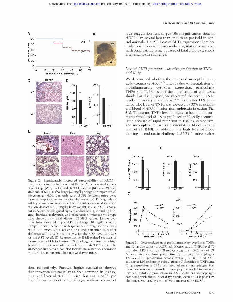
tion, respectively. Further, higher resolution showedthat intravascular coagulation was common in kidney,lung, and liver of AUF1−/− mice, but not in wild-typemice following endotoxin challenge, with an average of
four coagulation lesions per 10× magnification field inAUF1−/− mice and less than one lesion per field in con-trol animals (Fig. 2E). Loss of AUF1 expression thereforeleads to widespread intravascular coagulation associatedwith organ failure, a major cause of fatal endotoxic shockafter endotoxin challenge.
Loss of AUF1 promotes excessive production of TNF�and IL-1�
We determined whether the increased susceptibility toendotoxemia of AUF1−/− mice is due to deregulation ofproinflammatory cytokine expression, particularlyTNF� and IL-1�, two critical mediators of endotoxicshock. For this purpose, we measured the serum TNF�levels in wild-type and AUF1−/− mice after LPS chal-lenge. The level of TNF� was elevated by 30% in periph-eral blood of AUF1−/− mice after endotoxin injection (Fig.3A). The serum TNF� level is likely to be an underesti-mate of the level of TNF� produced and locally accumu-lated because of rapid retention in tissues, catabolism,and incomplete release into circulating blood (Finkel-man et al. 1993). In addition, the high level of bloodclotting in endotoxin-challenged AUF1−/− mice makes
Figure 2. Significantly increased susceptibility of AUF1−/−
mice to endotoxin challenge. (A) Kaplan-Meier survival curvesof wild-type (WT, n = 19) and AUF1 knockout (KO, n = 19) miceafter sublethal LPS challenge (20 mg/kg weight, intraperitonealinjection, p < 0.01, Log-rank test). AUF1-deficient mice weremore susceptible to endotoxin challenge. (B) Photograph ofwild-type and knockout mice 4 h after intraperitoneal injectionof a low dose of LPS (3 mg/kg body weight, n = 3). AUF1 knock-out mice exhibited typical signs of endotoxemia, including leth-argy, diarrhea, tachypnea, and piloerection, whereas wild-typemice showed only mild effects. (C) H&E-stained kidney sec-tions from mice 24 h post-LPS challenge (20 mg/kg weight,intraperitoneal). Note the widespread hemorrhage in the kidneyof AUF1−/− mice. (D) BUN and AST levels in mice 24 h afterchallenge with LPS (n = 3, p < 0.02 for the BUN level, p < 0.18for the AST level). (E) Representative H&E-stained sections ofmouse organs 24 h following LPS challenge to visualize a highdegree of the intravascular coagulation in AUF1−/− mice. Thearrowhead indicates blood clot formation, which was commonin AUF1 knockout mice but not wild-type mice.
Figure 3. Overproduction of proinflammatory cytokines TNF�
and IL-1� due to loss of AUF1. (A) Mouse serum TNF� level 75min after LPS injection (20 mg/kg weight, p < 0.02, n = 4). (B)Accumulated cytokine production by primary macrophages.TNF� and IL-1� secretion were elevated (p < 0.05) in AUF1−/−
cells after LPS endotoxin stimulation. (C) Kinetics of TNF� andIL-1� expression in LPS-stimulated primary macrophages. Sus-tained expression of proinflammatory cytokines led to elevatedlevels of cytokine production in AUF1-deficient macrophagescompared with those in wild-type cells, even at 24 h post-LPSchallenge. Secreted cytokines were measured by ELISA.
Endotoxic shock in AUF1 knockout mice
GENES & DEVELOPMENT 3177
Cold Spring Harbor Laboratory Press on February 16, 2019 - Published by genesdev.cshlp.orgDownloaded from

sampling difficult. In unchallenged wild-type and AUF1knockout mice, there was a barely detectable level ofTNF� (data not shown). These data indicate that there isan increase in the overall production of TNF� inAUF1−/− compared with wild-type mice following LPSchallenge.
To directly examine cytokine production from a ho-mogeneous cell population, primary macrophages,which express AUF1 (Fig. 5E, below), were isolated fromwild-type and AUF1−/− mouse peritoneal cells and placedin primary culture. Levels of ARE-binding and controlproteins were examined by immunoblot analysis ofwhole-cell lysate of primary macrophages derived fromwild-type and AUF1 knockout mice. No significant dif-ferences were found, apart from the absence of AUF1 inknockout mice (Fig. 5E, below). Primary macrophageswere stimulated with LPS and the levels of cytokinessecreted into medium were determined by ELISA. Com-parison of the accumulated levels of cytokine secretionafter LPS stimulation showed that production of TNF�and IL-1� were increased by 40% and 70%, respectively,in AUF1-deficient macrophages (Fig. 3B). Therefore, lossof AUF1 increased TNF� and IL-1� levels in response toendotoxin challenge.
The kinetics of TNF� and IL-1� cytokine expressionwas also examined to monitor the dynamic change ofcytokine levels at time points following LPS challenge.Compared with wild-type cells, TNF� production inLPS-stimulated AUF1−/− macrophages was increased by2.5-fold, and IL-1� by twofold overall (Fig. 3C). Impor-tantly, TNF� and IL-1� expression in AUF1−/− macro-phages remained elevated at 24 h, therefore for a longerduration of time (Fig. 3C) compared with the wild-typemacrophage controls. While the expression level ofTNF� declined by 24 h in wild-type macrophages, it wasstill almost threefold higher in AUF1−/− macrophages.IL-1� expression continued to increase over 24 h, to alevel twofold greater in AUF1−/− macrophages than inwild-type cells. Expression of IL-1� reached a plateau by30 h in wild-type macrophages but continued to increaseslowly in AUF1 knockout mice (data not shown). Thus,there is an increased and more sustained expression ofTNF� and IL-1� in AUF1−/− macrophages compared withwild-type macrophages, which is associated with the se-vere pathophysiological reaction of AUF1−/− animals toendotoxin. These data suggest that AUF1 functions toattenuate the proinflammatory cytokine response afterendotoxin challenge.
Excessive production of proinflammatory cytokinesis not due to an increase in the monocyte/macrophagepopulation in AUF1−/− mice
Hematopoietic cells, especially monocytes and macro-phages, are major sources of proinflammatory cytokinesTNF� and IL-1�. To determine whether lack of AUF1affected the development and circulating levels of hema-topoietic cells, we performed a complete blood count anda differential count of blood cells for wild-type andAUF1−/− mice, and analyzed their macrophage popula-
tion in the peritoneum and spleen by flow cytometricanalysis. Red blood cell parameters, leukocyte, andplatelet counts in peripheral blood were comparable andwithin normal ranges in AUF1−/− and wild-type litter-mates. Our analysis also confirmed the presence of allmajor hematopoietic lineages in AUF1−/− mice withoutsignificant alteration in macrophage numbers (Fig. 4A).Flow cytometry analysis of the macrophage population(F4/80 staining for murine macrophages) in peritoneumand spleen did not reveal significant difference betweenAUF1−/− and wild-type mice (Fig. 4B). Thus, the exces-sive production of proinflammatory cytokines in LPSchallenged AUF1−/− mice is not due to an increase in thenumber of source cells.
Abnormal stabilization of TNF� and IL-1� mRNAsresults from disruption of AUF1
Given the reported function of AUF1 in promoting rapiddegradation of ARE-mRNAs (Guhaniyogi and Brewer2001), we asked whether increased TNF� and IL-1� ex-pression is due to an alteration in mRNA stabilities. Tomonitor the decay of ARE-mRNAs, actinomycin D wasapplied to block transcription after primary macrophageswere stimulated with LPS. The level of remainingmRNA transcripts was determined by real-time quanti-tative RT–PCR at times post-inhibition of transcription.We found that TNF� and IL-1� mRNAs in wild-type
Figure 4. Analysis of peripheral blood and macrophage popu-lation in AUF1−/− mice. (A) Red blood cell parameters and num-bers of leukocyte subpopulations were obtained by completeblood cell count and differential cell count of peripheral blood.No significant difference was detected between AUF1 knockoutand wild-type mice. (B) Numbers of macrophages in peritoneumand spleen were determined by flow cytometry analysis of F4/80surface expression (a cell surface marker specific for murinemacrophages). Representative histograms are shown (n = 5 ineach group). No significant difference was observed.
Lu et al.
3178 GENES & DEVELOPMENT
Cold Spring Harbor Laboratory Press on February 16, 2019 - Published by genesdev.cshlp.orgDownloaded from

macrophages were unstable and rapidly degraded withhalf-lives of 31 and 26 min, respectively (Fig. 5A–B).However, in AUF1−/− macrophages, the half-lives ofTNF� and IL-1� mRNAs were 2.5-fold (60 min for IL-1�mRNA) and twofold greater (63 min for TNF� mRNA)compared with wild-type macrophages. The levels ofTNF� and IL-1� mRNAs in unstimulated wild-type andAUF1 knockout cells were barely detectable (data notshown). There was no change in the stability of ARE-containing IL-6 mRNA in AUF1−/− macrophages (Fig.
5C), or in the stability of cyclophilin A (Cyp A) mRNAs(Fig. 5C, inset; data not shown), which was used as acontrol for normalization. These results indicate thatAUF1 specifically promotes decay of select mRNAs in-cluding TNF� and IL-1�. To confirm the binding ofAUF1 to TNF� and IL-1� mRNAs, immunoprecipita-tion-quantitative RT–PCR (IP qRT–PCR) was performedto determine the intracellular AUF1 protein–RNA inter-action. AUF1 proteins were immunoprecipitated fromcytoplasmic lysates of macrophages, from which mRNAswere isolated. The amount of each target mRNA boundby AUF1 was determined by real-time qRT–PCR usingspecific primers. TNF�, IL-1�, and Cyclooxygenase 2(Cox2) mRNAs were detected in both AUF1 and eIF4Gimmunoprecipitate (Fig. 5D), the latter of which wasconsistent with the interaction of eIF4G with activelytranslated mRNAs. In contrast, IL-6 mRNA and granu-locyte-colony stimulating factor (G-CSF) mRNA, a non-ARE mRNA, were found associated only with eIF4G, butnot with AUF1 (Fig. 5D). These results indicate thatAUF1 proteins selectively bind to TNF�, IL-1�, andCox2 mRNAs, all of which contain overlapping AUUUApentamers. The 3� untranslated regions of TNF� and IL-1� mRNAs differ from that of IL-6 in containing mul-tiple consecutive copies of the AUUUA pentamer,whereas IL-6 contains only two pentamers, and they areinterrupted by intervening sequences. It is likely that ina natural physiological setting in the absence of AUF1ectopic overexpression, AUF1 binding requires the con-secutive placement of multiple AUUUA sequences invivo.
To determine whether disruption of AUF1 affects theexpression of other ARE-binding proteins that have beenimplicated in regulating ARE-mRNA stability, we exam-ined their expression levels in isolated peritoneal mac-rophages by the immunoblot analysis, as describedabove. The expression levels of major ARE-binding pro-teins (TTP, HuR, KSRP, TIA-1) and control protein (�-tubulin) remained unaltered in AUF1−/− macrophages(Fig. 5E). Thus, the increased stability of TNF� and IL-1�mRNAs appears to be a direct consequence of loss ofAUF1. Collectively, these data demonstrate that AUF1governs the inflammatory response and prevents the ex-cessive production of proinflammatory cytokines TNF�and IL-1� by promoting decay of their mRNAs.
Blocking TNF� and IL-1� activity protects AUF1knockout mice from severe endotoxic shock
To confirm that the LPS-induced endotoxic shock phe-notype in AUF1−/− mice is due to overproduction ofTNF� and IL-1�, animals were injected with neutraliz-ing antibodies against TNF� and IL-1� prior to a suble-thal LPS challenge. The general condition of each mousewas monitored, and a condition score was derived basedon numeric manifestations of endotoxemia, includinglethargy, diarrhea, tachypnea, piloerection, and death, asdescribed in the Materials and Methods section. WhereasIgG-injected AUF1−/− mice showed typical signs of en-dotoxic shock observed earlier, animals treated with an-
Figure 5. Abnormal stabilization of TNF� and IL-1� mRNAscaused by loss of AUF1. (A–C) Primary peritoneal macrophagesderived from AUF1 knockout or wild-type mice were stimu-lated with LPS (1 µg/mL) for 4 h followed by addition of acti-nomycin D (2 µg/mL) to block transcription. Cells were col-lected at the indicated time points, and cytoplasmic RNA wasextracted and analyzed by real-time quantitative RT–PCR(Roche Light Cycler) and normalized to the housekeeping Cyp AmRNA. The plots average three independent experiments foreach mRNA to determine mRNA half-lives for TNF� (A), IL-1�
(B), and IL-6 (C), as well as the standard deviation of results. Theinset in C shows the level of Cyp A mRNAs determined byqRT–PCR at different time points (0, 15, 30, 45, and 60 minpost-Actinomycin D). (D) Intracellular AUF1 proteins selec-tively bind to IL-1�, TNF�, and Cox2 mRNAs but not IL-6mRNA or a non-ARE G-CSF mRNA. Cultured macrophageswere lysed after LPS stimulation of cytokine expression andRNA bound by AUF1 was immunoprecipitated by AUF1-spe-cific antibody. Immunoprecipitation with translation initiationfactor eIF4G was used as a control. The amount of target mRNAbound by AUF1 or eIF4G was determined by real-time quanti-tative RT–PCR. (E) Primary macrophages derived from perito-neal cells were subjected to immunoblot analysis using anti-bodies against AUF1 and the other ARE-binding proteins as in-dicated. Disruption of AUF1 did not significantly alter theexpression level of ARE-binding proteins examined here.
Endotoxic shock in AUF1 knockout mice
GENES & DEVELOPMENT 3179
Cold Spring Harbor Laboratory Press on February 16, 2019 - Published by genesdev.cshlp.orgDownloaded from
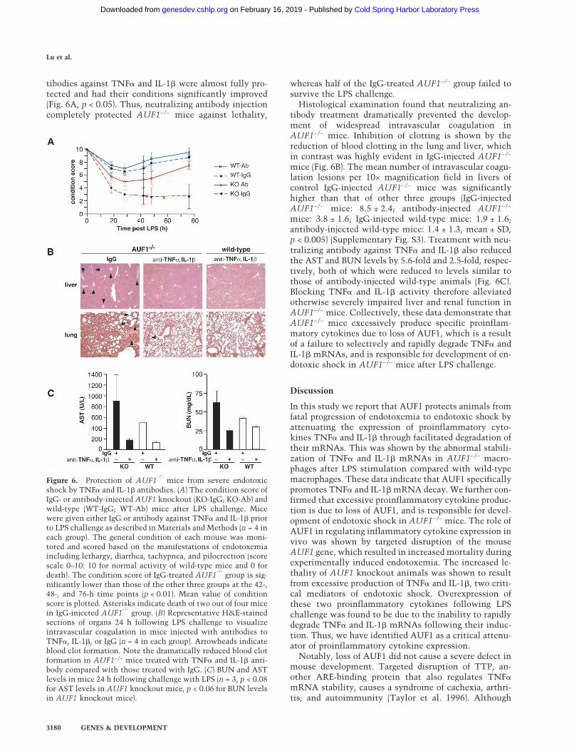
tibodies against TNF� and IL-1� were almost fully pro-tected and had their conditions significantly improved(Fig. 6A, p < 0.05). Thus, neutralizing antibody injectioncompletely protected AUF1−/− mice against lethality,
whereas half of the IgG-treated AUF1−/− group failed tosurvive the LPS challenge.
Histological examination found that neutralizing an-tibody treatment dramatically prevented the develop-ment of widespread intravascular coagulation inAUF1−/− mice. Inhibition of clotting is shown by thereduction of blood clotting in the lung and liver, whichin contrast was highly evident in IgG-injected AUF1−/−
mice (Fig. 6B). The mean number of intravascular coagu-lation lesions per 10× magnification field in livers ofcontrol IgG-injected AUF1−/− mice was significantlyhigher than that of other three groups (IgG-injectedAUF1−/− mice: 8.5 ± 2.4; antibody-injected AUF1−/−
mice: 3.8 ± 1.6; IgG-injected wild-type mice: 1.9 ± 1.6;antibody-injected wild-type mice: 1.4 ± 1.3, mean ± SD,p < 0.005) (Supplementary Fig. S3). Treatment with neu-tralizing antibody against TNF� and IL-1� also reducedthe AST and BUN levels by 5.6-fold and 2.5-fold, respec-tively, both of which were reduced to levels similar tothose of antibody-injected wild-type animals (Fig. 6C).Blocking TNF� and IL-1� activity therefore alleviatedotherwise severely impaired liver and renal function inAUF1−/− mice. Collectively, these data demonstrate thatAUF1−/− mice excessively produce specific proinflam-matory cytokines due to loss of AUF1, which is a resultof a failure to selectively and rapidly degrade TNF� andIL-1� mRNAs, and is responsible for development of en-dotoxic shock in AUF1−/− mice after LPS challenge.
Discussion
In this study we report that AUF1 protects animals fromfatal progression of endotoxemia to endotoxic shock byattenuating the expression of proinflammatory cyto-kines TNF� and IL-1� through facilitated degradation oftheir mRNAs. This was shown by the abnormal stabili-zation of TNF� and IL-1� mRNAs in AUF1−/− macro-phages after LPS stimulation compared with wild-typemacrophages. These data indicate that AUF1 specificallypromotes TNF� and IL-1� mRNA decay. We further con-firmed that excessive proinflammatory cytokine produc-tion is due to loss of AUF1, and is responsible for devel-opment of endotoxic shock in AUF1−/− mice. The role ofAUF1 in regulating inflammatory cytokine expression invivo was shown by targeted disruption of the mouseAUF1 gene, which resulted in increased mortality duringexperimentally induced endotoxemia. The increased le-thality of AUF1 knockout animals was shown to resultfrom excessive production of TNF� and IL-1�, two criti-cal mediators of endotoxic shock. Overexpression ofthese two proinflammatory cytokines following LPSchallenge was found to be due to the inability to rapidlydegrade TNF� and IL-1� mRNAs following their induc-tion. Thus, we have identified AUF1 as a critical attenu-ator of proinflammatory cytokine expression.
Notably, loss of AUF1 did not cause a severe defect inmouse development. Targeted disruption of TTP, an-other ARE-binding protein that also regulates TNF�mRNA stability, causes a syndrome of cachexia, arthri-tis, and autoimmunity (Taylor et al. 1996). Although
Figure 6. Protection of AUF1−/−
mice from severe endotoxicshock by TNF� and IL-1� antibodies. (A) The condition score ofIgG- or antibody-injected AUF1 knockout (KO-IgG; KO-Ab) andwild-type (WT-IgG; WT-Ab) mice after LPS challenge. Micewere given either IgG or antibody against TNF� and IL-1� priorto LPS challenge as described in Materials and Methods (n = 4 ineach group). The general condition of each mouse was moni-tored and scored based on the manifestations of endotoxemiaincluding lethargy, diarrhea, tachypnea, and piloerection (scorescale 0–10: 10 for normal activity of wild-type mice and 0 fordeath). The condition score of IgG-treated AUF1
−/−group is sig-
nificantly lower than those of the other three groups at the 42-,48-, and 76-h time points (p < 0.01). Mean value of conditionscore is plotted. Asterisks indicate death of two out of four micein IgG-injected AUF1
−/−group. (B) Representative H&E-stained
sections of organs 24 h following LPS challenge to visualizeintravascular coagulation in mice injected with antibodies toTNF�, IL-1�, or IgG (n = 4 in each group). Arrowheads indicateblood clot formation. Note the dramatically reduced blood clotformation in AUF1−/− mice treated with TNF� and IL-1� anti-body compared with those treated with IgG. (C) BUN and ASTlevels in mice 24 h following challenge with LPS (n = 3, p < 0.08for AST levels in AUF1 knockout mice, p < 0.06 for BUN levelsin AUF1 knockout mice).
Lu et al.
3180 GENES & DEVELOPMENT
Cold Spring Harbor Laboratory Press on February 16, 2019 - Published by genesdev.cshlp.orgDownloaded from

AUF1 and TTP are both ARE-binding proteins involvedin regulating ARE-mRNA stability and both are found inimmune cells (Carballo et al. 1998; Loflin et al. 1999;Sarkar et al. 2003; Lu and Schneider 2004), they demon-strate different phenotypes in knockout mice. TTPknockout mice develop several abnormalities in hema-topoietic systems, particularly a smaller thymus with-out cortical/medullary organization (Carballo et al.1998). In contrast, thymus development in AUF1-defi-cient mice is normal (data not shown). It has also beenreported that in peripheral blood of TTP−/− mice, thewhite blood cell count is elevated by more than twofoldover that of wild-type mice, and there is a marked in-crease in the number of circulating neutrophils and mac-rophages (Taylor et al. 1996). In contrast, in AUF1−/−
mice, similar numbers of hematopoietic cells are foundin peripheral blood compared with wild-type mice, andno change was observed in macrophages in peritoneumand spleen (Fig. 4). These observations suggest thatAUF1 may have different regulatory effects and differentfunctions than TTP in the ARE-mRNA decay pathway.The phenotype of TTP−/− mice is likely the consequenceof chronic excessive TNF� circulating in the blood (Tay-lor et al. 1996). Thus, TTP may constitutively targetTNF� mRNA for degradation, and loss of TTP leads to achronic and constant accumulation of TNF� mRNA andits overproduction. Our data from AUF1 knockout micesuggests that AUF1 may be a regulatory factor that nor-mally does not execute its function until the activationof proinflammatory cytokine expression. In particular, inthe absence of LPS-challenge, AUF1 knockout mice didnot display elevated levels of TNF� or IL-1�, and AUF1promoted the rapid decay of TNF� and IL-1� mRNAsonly after their induction in response to endotoxin chal-lenge. AUF1-directed mRNA degradation may thereforeserve as a protective means to attenuate the inflamma-tory response post-stimulation, and to protect againstdeleterious effects of excessive inflammation.
The increased susceptibility of AUF1−/− mice to endo-toxin challenge is reminiscent of the phenotype of micelacking the TIA-1 gene (Piecyk et al. 2000), which en-codes an ARE-binding protein that specifically binds theTNF�–ARE and represses TNF� translation. Increasedproduction of TNF� in TIA-1−/− mice is mainly due toderepression of TNF� mRNA translation, as no alter-ation in TNF� mRNA stability is detected. In contrast,we found that abnormal stabilization of TNF� and IL-1�mRNAs contributes to their overexpression in AUF1−/−
mice. The difference in the mechanisms by which AUF1and TIA-1 control TNF� production implies that a pre-cise control of cytokine expression demands regulationat multiple levels.
At this time, we cannot formally rule out a hypotheti-cal role for AUF1 in regulating both the stability andtranslation of TNF� and IL-1� mRNAs. However, previ-ous work on TTP and TIA-1 (Kontoyiannis et al. 1999;Piecyk et al. 2000) indicates that the translation repres-sion and stability of ARE-mRNAs are independentlycontrolled by different ARE-binding proteins. Thesefindings suggest that AUF1 may mainly control the sta-
bility of target mRNAs. Given the overlapping tissuedistribution of decay-promoting AUF1 and stability-pro-moting HuR (Lu and Schneider 2004), as well as the bind-ing of AUF1 and HuR to common target mRNAs (Lal etal. 2004), loss of AUF1 might function in part by allow-ing HuR to play a more active role in stabilizing targetmRNAs and, consequently, causing their overexpression.
Despite previous reports that mitogen-activated pro-tein kinase-activated protein (MAPKAP) kinase 2 signal-ing regulates IL-6 mRNA stability and its biosynthesisvia the AU-rich 3� noncoding region (Winzen et al. 1999;Neininger et al. 2002), we did not detect a significantalteration in IL-6 mRNA stability in AUF1-deficientcells (Fig. 5C). Our results suggest that AUF1 more se-lectively regulates TNF� and IL-1� mRNA stability, andthat IL-6 mRNA may not be controlled by AUF1 undernormal physiological conditions (without AUF1 overex-pression). mRNAs targeted for rapid decay by ARE ele-ments, such as TNF� and IL-1� mRNAs, often containthree to five uninterrupted copies of an AUUUApentamer in the 3� noncoding region (Guhaniyogi andBrewer 2001). IL-6 mRNA, however, contains no con-secutive AUUUA pentamers. Instead, its 3� noncodingregion contains two pentamers interspersed in an AU-rich region with varying U stretches of 2–5 nucleotidesin length. We speculate that multiple consecutiveAUUUA pentamers may be important for AUF1 to in-teract with target ARE-mRNAs and regulate their stabil-ity. Consistent with this hypothesis, Cox2 mRNA, aclass II ARE-mRNA harboring multiple overlappingAUUUA pentamers, was found to associate with AUF1in vivo (Fig. 5D) and its half-life was increased at least50% in AUF1-deficient macrophages derived from AUF1knockout mice (N. Sadri and R. Schneider, unpubl.). It isalso possible that IL-6 mRNA stability is regulated bydifferent AUF1 isoforms with opposing functions so thatinactivation of all four AUF1 isoforms may obscure theeffect on the target mRNA. It has been suggested that thedifferent AUF1 isoforms possess different, or perhapseven opposing functions in regulating ARE-mRNA decay(Laroia et al. 1999; Loflin et al. 1999; Sarkar et al. 2003;Raineri et al. 2004). With the availability AUF1-deficientcells, these questions can now be experimentally ad-dressed.
In summary, our results provide the first in vivo evi-dence for a novel post-transcriptional mechanism criti-cal for controlling proinflammatory cytokine expressionand protecting against fatal endotoxic shock after endo-toxin exposure. Our findings indicate that after proin-flammatory cytokine induction, AUF1 promotes decayof TNF�, IL-1�, and possibly other inflammatory cyto-kine mRNAs as a means for controlling and temperingthe inflammatory response. Defects in this post-tran-scriptional regulation may be involved in human inflam-matory diseases.
Materials and methods
Generation of AUF1−/− mice
A 1.4-kb genomic fragment upstream of mouse AUF1 exon 3,and a 7.9-kb genomic fragment downstream from AUF1 exon 3,
Endotoxic shock in AUF1 knockout mice
GENES & DEVELOPMENT 3181
Cold Spring Harbor Laboratory Press on February 16, 2019 - Published by genesdev.cshlp.orgDownloaded from

were used as short and long recombination arms, respectively,in the construction of the targeting vector. A loxP-flanked neor
expression cassette was inserted between the two regions, re-sulting in a vector designed to delete the two RNA-bindingmotifs in exon 3. W4 embryonic stem (ES) cells were electro-porated with the NotI-linearized targeting construct, selected inG418 (150 µg/mL) and gancyclovir (2 mM), and used for South-ern blot DNA analysis. Five positive clones were identified us-ing Southern blot and PCR analysis. Blastocyst injection wasperformed using two independent targeted ES cell clones. Germ-line transmission was obtained on further crossing of male chi-meras with C57BL/6J females.
Animal studies and histology
Experiments were carried out in accordance with NIH guide-lines for animal treatment, housing, and euthanasia. Mice werechallenged by intraperitoneal LPS injection (low dose: 3 mg/kgweight; sublethal dose: 20 mg/kg) and monitored for generalcondition and survival. At indicated times, mice were eutha-nized and blood serum was collected. Blood BUN and AST lev-els were determined using Infinity BUN Reagent and AST Re-agent (Sigma Diagnostics), respectively. For histological analy-sis, tissues were fixed in 10% buffered formaldehyde overnightat 4°C and paraffin-embedded. Sections (5 µm) were stainedwith hematoxylin and eosin (H&E). To determine the generalcondition of mice after LPS challenge, mice were monitoredblindly (without knowing their genotypes) and individual scoreswere given based on the symptoms of endotoxemia (motor ac-tivity/coordination (0–2, normal being 2), strength (0–2, normalbeing 2), food intake (0–1, normal being 1), ocular exudate (0–2,no exudates denoting 2), diarrhea (0–1, no diarrhea being 1),piloerection/coat appearance (0–1, normal being 1), and breathrate (0–1, 0 denoting slowed rate)). The condition score of aparticular mouse is the summed score of individual symptomscores (10 being normal activity and 1 denoting close to death).
Quantification of cytokine expression
Serum cytokine levels and cytokines secreted from primarymacrophages were determined by ELISA. Equal numbers of peri-toneal macrophages derived from three mice were pooled andseeded in three replicative wells (5 × 10⁵ cells/well). Peritonealcells were incubated at 37°C in an atmosphere of 5% CO2 for 3h to allow peritoneal macrophages to adhere. Nonadherent cellswere removed by washing with PBS twice, and then macro-phages were stimulated with 1 µg/mL LPS (Sigma) for the timesindicated. Levels of TNF� and IL-1� were measured by ELISA(eBioscience) using an equal amount of culture supernatant atindicated time points (Fig. 3C), or at 8 h after LPS treatment forTNF� and 24 h after LPS treatment for IL-1� (Fig. 3B).
Determination of mRNA half-life
For decay studies, peritoneal macrophages were stimulatedwith LPS (1 µg/mL) for 4 h. After 4 h of LPS stimulation, acti-nomycin D (2 µg/mL) was added to block transcription, andtotal RNA was isolated after 0, 20, and 40 min post-actinomycinD treatment for TNF-� measurements, or after 0, 30, and 60 minpost-actinomycin D treatment for IL-1� and IL-6 measure-ments. mRNA levels were determined by quantitative real-timeRT–PCR (qRT–PCR, Roche Light Cycler), and were normalizedto Cyp A mRNA as a control. PCR reactions were performedwith gene-specific primers (primer sequences available upon re-quest). The plots (Fig. 5A–C) average three independent experi-
ments for each mRNA to determine mRNA half-lives and stan-dard deviation of results.
TNF-� and IL-1� antibody administration studies
AUF1−/− and AUF1+/+ male mice each received intraperitonealinjection of 200 µg of hamster monoclonal antibody to mouseTNF� (clone TN3-19.12, BD PharMingen) and 100 µg of ham-ster monoclonal antibody to mouse IL-1� (clone B122, BioLeg-end). Mice of each genotype in the control group received 100 µgof hamster isotype IgG control (clone A19-3, BD PharMingen).Six hours post-antibody injection, mice in both groups receivedan intraperitoneal injection of a sublethal dose of LPS (20 mg/kg). Mice were monitored for general condition and survival, asdescribed in the above animal studies and histology section.Measurement of BUN and AST levels, and histological analyseswere performed as described above.
Immunoblot analysis and IP qRT–PCR
Immunoblot analysis was performed following standard proce-dures. Polyclonal antibodies against AUF1, KSRP (from D.Black), and TIA-1 (from P. Anderson), and specific antibodies forHuR (Santa Cruz Biotechnology), TTP (Santa Cruz Biotechnol-ogy), and �-tubulin (Sigma) were used. ECL Western BlottingDetection Kit (Amersham Pharmacia) was used for detection. IPqRT–PCR was performed using anti-AUF1 and anti-eIF4G poly-clonal antibody according to a protocol previously described(Sarkar et al. 2003).
Analysis of immune cells
Peripheral blood analysis was performed by Anilytics, Inc.Spleen and peritoneal cells were made into single-cell suspen-sions in RPMI 1640 medium by passing through a 70-µm nyloncell strainer. The cell suspensions were depleted of erythrocytesby osmotic lysis, and cells were incubated on ice for 15 minwith anti-CD16/CD32 (BD PharMingen) to block the Fc recep-tor. Subsequently, these cells were incubated with anti-F4/80-biotin (BM8, eBioscience) for 30 min. The cells were thenwashed with PBS and incubated with streptoavidin-APC (BDPharMingen) on ice for 15 min. Cells were washed, fixed, andanalyzed on a FACSCalibur (Becton Dickson) using a live lym-phocyte gate. Data were analyzed using CellQuest software(Becton Dickson).
Statistical analysis
Data are presented as mean ± SD for statistical comparison oftwo samples, the Student t-test was used for evaluation. Sur-vival curves were generated using the Kaplan-Meier method,and significance was evaluated using the Log-rank test.
Acknowledgments
We thank Ralf Kist for critical advice, Tung-Tien Sun for a129/SvJ mouse genomic library, Klaus Rajewsky for the neoplasmid, Douglas Black for anti-KSRP antibody, and PaulAnderson for anti-TIA-1 antibody. We also thank Anna Auer-bach and the staff at New York University (NYU) School ofMedicine Transgenic Mouse/ES Cell Chimera Facility for assis-tance in producing chimeric mice, and Doris Tse and the staff atCenter for AIDS Research at NYU for assistance in FACS analy-sis. This work was supported by grant GM60428 from the
Lu et al.
3182 GENES & DEVELOPMENT
Cold Spring Harbor Laboratory Press on February 16, 2019 - Published by genesdev.cshlp.orgDownloaded from

NIH (R.J.S.). The authors declare that they have no competingfinancial interests.
References
Brennan, C.M. and Steitz, J.A. 2001. HuR and mRNA stability.Cell. Mol. Life Sci. 58: 266–277.
Brewer, G. 1991. An A + U-rich element RNA-binding factorregulates c-myc mRNA stability in vitro. Mol. Cell. Biol. 11:2460–2466.
Buzby, J.S., Brewer, G., and Nugent, D.J. 1999. Developmentalregulation of RNA transcript destabilization by A + U- richelements is AUF1-dependent. J. Biol. Chem. 274:33973–33978.
Carballo, E., Lai, W.S., and Blackshear, P.J. 1998. Feedback in-hibition of macrophage tumor necrosis factor-� productionby tristetraprolin. Science 281: 1001–1005.
Carballo, E., Lai, W.S., and Blackshear, P.J. 2000. Evidence thattristetraprolin is a physiological regulator of granulocyte-macrophage colony-stimulating factor messenger RNAdeadenylation and stability. Blood 95: 1891–1899.
Chen, C.Y. and Shyu, A.B. 1995. AU-rich elements: Character-ization and importance in mRNA degradation. Trends Bio-chem. Sci. 20: 465–470.
Chen, C.Y., Gherzi, R., Ong, S.E., Chan, E.L., Raijmakers, R.,Pruijn, G.J., Stoecklin, G., Moroni, C., Mann, M., and Karin,M. 2001. AU binding proteins recruit the exosome to degradeARE-containing mRNAs. Cell 107: 451–464.
Dinarello, C.A. 1994. The interleukin-1 family: 10 years of dis-covery. FASEB J. 8: 1314–1325.
Dinarello, C.A. 1997. Proinflammatory and anti-inflammatorycytokines as mediators in the pathogenesis of septic shock.Chest (Suppl.) 112: 321S–329S.
Dixon, D.A., Tolley, N.D., King, P.H., Nabors, L.B., McIntyre,T.M., Zimmerman, G.A., and Prescott, S.M. 2001. Alteredexpression of the mRNA stability factor HuR promotes cy-clooxygenase-2 expression in colon cancer cells. J. Clin. In-vest. 108: 1657–1665.
Fan, X.C. and Steitz, J.A. 1998. Overexpression of HuR, anuclear-cytoplasmic shuttling protein, increases the in vivostability of ARE-containing mRNAs. EMBO J. 17:3448–3460.
Finkelman, F.D., Madden, K.B., Morris, S.C., Holmes, J.M.,Boiani, N., Katona, I.M., and Maliszewski, C.R. 1993. Anti-cytokine antibodies as carrier proteins. Prolongation of invivo effects of exogenous cytokines by injection of cytokine–anti-cytokine antibody complexes. J. Immunol. 151: 1235–1244.
Gherzi, R., Lee, K.Y., Briata, P., Wegmuller, D., Moroni, C.,Karin, M., and Chen, C.Y. 2004. A KH domain RNA bindingprotein, KSRP, promotes ARE-directed mRNA turnover byrecruiting the degradation machinery. Mol. Cell 14: 571–583.
Glauser, M.P., Zanetti, G., Baumgartner, J.D., and Cohen, J.1991. Septic shock: Pathogenesis. Lancet 338: 732–736.
Grosset, C., Boniface, R., Duchez, P., Solanilla, A., Cosson, B.,and Ripoche, J. 2004. In vivo studies of translational repres-sion mediated by the granulocyte-macrophage colony-stimulating factor AU-rich element. J. Biol. Chem. 279:13354–13362.
Guhaniyogi, J. and Brewer, G. 2001. Regulation of mRNA sta-bility in mammalian cells. Gene 265: 11–23.
He, C. and Schneider, R. 2006. 14–3–3� is a p37 AUF1-bindingprotein that facilitates AUF1-mediated ARE-mRNA decay.EMBO J. 25: 3823–3831.
Hotchkiss, R.S. and Karl, I.E. 2003. The pathophysiology andtreatment of sepsis. N. Engl. J. Med. 348: 138–150.
Jacob, C.O., Lee, S.K., and Strassmann, G. 1996. Mutationalanalysis of TNF-� gene reveals a regulatory role for the 3�-untranslated region in the genetic predisposition to lupus-like autoimmune disease. J. Immunol. 156: 3043–3050.
Kajita, Y., Nakayama, J., Aizawa, M., and Ishikawa, F. 1995. TheUUAG-specific RNA binding protein, heterogeneousnuclear ribonucleoprotein D0. Common modular structureand binding properties of the 2xRBD-Gly family. J. Biol.Chem. 270: 22167–22175.
Katsanou, V., Papadaki, O., Milatos, S., Blackshear, P.J., Ander-son, P., Kollias, G., and Kontoyiannis, D.L. 2005. HuR as anegative posttranscriptional modulator in inflammation.Mol. Cell 19: 777–789.
Kiledjian, M., DeMaria, C.T., Brewer, G., and Novick, K. 1997.Identification of AUF1 (heterogeneous nuclear ribonucleo-protein D) as a component of the �-globin mRNA stabilitycomplex. Mol. Cell. Biol. 17: 4870–4876.
Kontoyiannis, D., Pasparakis, M., Pizarro, T.T., Cominelli, F.,and Kollias, G. 1999. Impaired on/off regulation of TNF bio-synthesis in mice lacking TNF AU-rich elements: Implica-tions for joint and gut-associated immunopathologies. Im-munity 10: 387–398.
Lai, W.S., Carballo, E., Strum, J.R., Kennington, E.A., Phillips,R.S., and Blackshear, P.J. 1999. Evidence that tristetraprolinbinds to AU-rich elements and promotes the deadenylationand destabilization of tumor necrosis factor � mRNA. Mol.Cell. Biol. 19: 4311–4323.
Lal, A., Mazan-Mamczarz, K., Kawai, T., Yang, X., Martindale,J.L., and Gorospe, M. 2004. Concurrent versus individualbinding of HuR and AUF1 to common labile target mRNAs.EMBO J. 23: 3092–3102.
Laroia, G., Cuesta, R., and Schneider, R.J. 1999. Control ofmRNA decay by heat shock–ubiquitin–proteasome path-way. Science 284: 499–502.
Lebwohl, D.E., Muise-Helmericks, R., Sepp-Lorenzino, L.,Serve, S., Timaul, M., Bol, R., Borgen, P., and Rosen, N. 1994.A truncated cyclin D1 gene encodes a stable mRNA in ahuman breast cancer cell line. Oncogene 9: 1925–1929.
Levy, N.S., Chung, S., Furneaux, H., and Levy, A.P. 1998. Hy-poxic stabilization of vascular endothelial growth factormRNA by the RNA-binding protein HuR. J. Biol. Chem. 273:6417–6423.
Loflin, P., Chen, C.Y., and Shyu, A.B. 1999. Unraveling a cyto-plasmic role for hnRNP D in the in vivo mRNA destabili-zation directed by the AU-rich element. Genes & Dev. 13:1884–1897.
Lu, J.Y. and Schneider, R.J. 2004. Tissue distribution of AU-richmRNA-binding proteins involved in regulation of mRNAdecay. J. Biol. Chem. 279: 12974–12979.
Ma, W.J., Cheng, S., Campbell, C., Wright, A., and Furneaux, H.1996. Cloning and characterization of HuR, a ubiquitouslyexpressed Elav-like protein. J. Biol. Chem. 271: 8144–8151.
Martin, G.S., Mannino, D.M., Eaton, S., and Moss, M. 2003. Theepidemiology of sepsis in the United States from 1979through 2000. N. Engl. J. Med. 348: 1546–1554.
Nair, A.P., Hahn, S., Banholzer, R., Hirsch, H.H., and Moroni, C.1994. Cyclosporin A inhibits growth of autocrine tumourcell lines by destabilizing interleukin-3 mRNA. Nature 369:239–242.
Neininger, A., Kontoyiannis, D., Kotlyarov, A., Winzen, R.,Eckert, R., Volk, H.D., Holtmann, H., Kollias, G., and Gaes-tel, M. 2002. MK2 targets AU-rich elements and regulatesbiosynthesis of tumor necrosis factor and interleukin-6 in-dependently at different post-transcriptional levels. J. Biol.
Endotoxic shock in AUF1 knockout mice
GENES & DEVELOPMENT 3183
Cold Spring Harbor Laboratory Press on February 16, 2019 - Published by genesdev.cshlp.orgDownloaded from

Chem. 277: 3065–3068.Parrillo, J.E. 1993. Pathogenetic mechanisms of septic shock. N.
Engl. J. Med. 328: 1471–1477.Pende, A., Tremmel, K.D., DeMaria, C.T., Blaxall, B.C., Mi-
nobe, W.A., Sherman, J.A., Bisognano, J.D., Bristow, M.R.,Brewer, G., and Port, J. 1996. Regulation of the mRNA-bind-ing protein AUF1 by activation of the �-adrenergic receptorsignal transduction pathway. J. Biol. Chem. 271: 8493–8501.
Piecyk, M., Wax, S., Beck, A.R., Kedersha, N., Gupta, M., Mar-itim, B., Chen, S., Gueydan, C., Kruys, V., Streuli, M., et al.2000. TIA-1 is a translational silencer that selectively regu-lates the expression of TNF-�. EMBO J. 19: 4154–4163.
Raineri, I., Wegmueller, D., Gross, B., Certa, U., and Moroni, C.2004. Roles of AUF1 isoforms, HuR and BRF1 in ARE-de-pendent mRNA turnover studied by RNA interference.Nucleic Acids Res. 32: 1279–1288.
Rodriguez-Pascual, F., Hausding, M., Ihrig-Biedert, I., Furneaux,H., Levy, A.P., Forstermann, U., and Kleinert, H. 2000. Com-plex contribution of the 3�-untranslated region to the expres-sional regulation of the human inducible nitric-oxide syn-thase gene. Involvement of the RNA-binding protein HuR. J.Biol. Chem. 275: 26040–26049.
Sarkar, B., Xi, Q., He, C., and Schneider, R.J. 2003. Selectivedegradation of AU-rich mRNAs promoted by the p37 AUF1protein isoform. Mol. Cell. Biol. 23: 6685–6693.
Sirenko, O.I., Lofquist, A.K., DeMaria, C.T., Morris, J.S.,Brewer, G., and Haskill, J.S. 1997. Adhesion-dependent regu-lation of an A + U-rich element-binding activity associatedwith AUF1. Mol. Cell. Biol. 17: 3898–3906.
Stoecklin, G., Colombi, M., Raineri, I., Leuenberger, S., Mal-laun, M., Schmidlin, M., Gross, B., Lu, M., Kitamura, T., andMoroni, C. 2002. Functional cloning of BRF1, a regulator ofARE-dependent mRNA turnover. EMBO J. 21: 4709–4718.
Taylor, G.A., Carballo, E., Lee, D.M., Lai, W.S., Thompson,M.J., Patel, D.D., Schenkman, D.I., Gilkeson, G.S., Brox-meyer, H.E., Haynes, B.F., et al. 1996. A pathogenetic role forTNF � in the syndrome of cachexia, arthritis, and autoim-munity resulting from tristetraprolin (TTP) deficiency. Im-munity 4: 445–454.
Tracey, K.J., Fong, Y., Hesse, D.G., Manogue, K.R., Lee, A.T.,Kuo, G.C., Lowry, S.F., and Cerami, A. 1987. Anti-cachec-tin/TNF monoclonal antibodies prevent septic shock duringlethal bacteraemia. Nature 330: 662–664.
Wagner, B.J., DeMaria, C.T., Sun, Y., Wilson, G.M., and Brewer,G. 1998. Structure and genomic organization of the humanAUF1 gene: Alternative pre-mRNA splicing generates fourprotein isoforms. Genomics 48: 195–202.
Wang, W., Furneaux, H., Cheng, H., Caldwell, M.C., Hutter, D.,Liu, Y., Holbrook, N., and Gorospe, M. 2000. HuR regulatesp21 mRNA stabilization by UV light. Mol. Cell. Biol. 20:760–769.
Wilson, G.M., Sun, Y., Lu, H., and Brewer, G. 1999. Assembly ofAUF1 oligomers on U-rich RNA targets by sequential dimerassociation. J. Biol. Chem. 274: 33374–33381.
Wilusz, C.J., Wormington, M., and Peltz, S.W. 2001. The cap-to-tail guide to mRNA turnover. Nat. Rev. Mol. Cell Biol. 2:237–246.
Winzen, R., Kracht, M., Ritter, B., Wilhelm, A., Chen, C.Y.,Shyu, A.B., Muller, M., Gaestel, M., Resch, K., and Holt-mann, H. 1999. The p38 MAP kinase pathway signals forcytokine-induced mRNA stabilization via MAP kinase-acti-vated protein kinase 2 and an AU-rich region-targetedmechanism. EMBO J. 18: 4969–4980.
Zhang, W., Wagner, B.J., Ehrenman, K., Schaefer, A.W., De-Maria, C.T., Crater, D., DeHaven, K., Long, L., and Brewer,G. 1993. Purification, characterization, and cDNA cloning of
an AU-rich element RNA-binding protein, AUF1. Mol. Cell.Biol. 13: 7652–7665.
Lu et al.
3184 GENES & DEVELOPMENT
Cold Spring Harbor Laboratory Press on February 16, 2019 - Published by genesdev.cshlp.orgDownloaded from

10.1101/gad.1467606Access the most recent version at doi: originally published online November 3, 200620:2006, Genes Dev.
Jin-Yu Lu, Navid Sadri and Robert J. Schneider degrade proinflammatory cytokine mRNAs
knockout mice mediated by failure toAUF1Endotoxic shock in
Material
Supplemental
http://genesdev.cshlp.org/content/suppl/2006/10/26/gad.1467606.DC1
References
http://genesdev.cshlp.org/content/20/22/3174.full.html#ref-list-1
This article cites 50 articles, 28 of which can be accessed free at:
License
ServiceEmail Alerting
click here.right corner of the article or
Receive free email alerts when new articles cite this article - sign up in the box at the top
Copyright © 2006, Cold Spring Harbor Laboratory Press
Cold Spring Harbor Laboratory Press on February 16, 2019 - Published by genesdev.cshlp.orgDownloaded from