Endogenous nonneuronal modulators of synaptic … nonneuronal modulators of synaptic transmission...
6
Endogenous nonneuronal modulators of synaptic transmission control cortical slow oscillations in vivo Tommaso Fellin a,b,1 , Michael M. Halassa a , Miho Terunuma a,c , Francesca Succol a,b , Hajime Takano a , Marcos Frank a , Stephen J. Moss a,c , and Philip G. Haydon a,c a Silvio Conte Center for Integration at the Tripartite Synapse, Department of Neuroscience, University of Pennsylvania School of Medicine, Philadelphia, PA 19104; b Department of Neuroscience and Brain Technologies, Italian Institute of Technology, 16163 Genova, Italy; and c Department of Neuroscience, Tufts University School of Medicine, 136 Harrison Avenue, Boston, MA 02111 Edited by Tullio Pozzan, University of Padua, Padua, Italy, and approved July 14, 2009 (received for review June 10, 2009) Gliotransmission, the release of molecules from astrocytes, regu- lates neuronal excitability and synaptic transmission in situ. Whether this process affects neuronal network activity in vivo is not known. Using a combination of astrocyte-specific molecular genetics, with in vivo electrophysiology and pharmacology, we determined that gliotransmission modulates cortical slow oscilla- tions, a rhythm characterizing nonrapid eye movement sleep. Inhibition of gliotransmission by the expression of a dominant negative SNARE domain in astrocytes affected cortical slow oscil- lations, reducing the duration of neuronal depolarizations and causing prolonged hyperpolarizations. These network effects re- sult from the astrocytic modulation of intracortical synaptic trans- mission at two sites: a hypofunction of postsynaptic NMDA recep- tors, and by reducing extracellular adenosine, a loss of tonic A1 receptor-mediated inhibition. These results demonstrate that rhythmic brain activity is generated by the coordinated action of the neuronal and glial networks. adenosine astrocytes gliotransmission NMDA receptor N onrapid eye movement (NREM) sleep is characterized by global cortical oscillations of synchronized neuronal activity (1, 2). Major components of this activity are slow oscillations (1 Hz), observed also under some forms of anesthesia (2–5). Slow oscillations are a fundamental network phenomenon that orga- nizes other sleep rhythms (1), and have been suggested to have a role in sleep-dependent memory consolidation (6, 7). Different mechanisms have been proposed to control slow oscillations, including excitatory-inhibitory interactions (8), neu- ronal intrinsic properties (9, 10), and the modulation of synaptic transmission (4, 11). Although pharmacological (4, 11) studies have shown the importance of synaptic modulation on slow oscillations, it is not known whether endogenous cellular systems capable of modulating synaptic transmission have a role in shaping this cortical rhythm. Recently, brain slice experiments showed that a glial cell subtype, the astrocyte, modulates synaptic transmission through the release of different molecules (gliotransmission) including D-serine and ATP (12, 13). D-serine acts as a coagonist of NMDA receptors (14) increasing postsynaptic NMDA currents (15), whereas ATP is rapidly hydrolyzed to adenosine, which acts on A1 receptors to suppress synaptic transmission (16, 17). Because astrocytes are intimately associated with pre and postsynaptic terminals (18, 19), on which they exert modulatory actions, they have the potential to act as endogenous regulators of slow oscillations. Support of this hypothesis comes from the observation that gliotransmission is essential for the normal accumulation of the homeostatic sleep pressure (20). This fundamental process is assessed by measuring the dynamic change in slow wave activity (SWA; 0.5– 4 Hz) of the EEG during NREM sleep. Because slow oscillations underlie low frequency (1 Hz) components of SWA, we hypothesized that gliotransmission may directly impact brain dynamics in the slow oscillation (1 Hz) range. To test this hypothesis, we have used a combination of glial-cell specific molecular genetics, which inhibits the release of chemical trans- mitters from astrocytes, together with patch-clamp, extracellular and EEG recordings in vivo to monitor the impact of gliotrans- mission on the generation of slow oscillations. We show that the attenuation of gliotransmission leads to reduced cortical slow oscillations. These changes in network activity are due to integrated effects of gliotransmission on A1 and NMDA receptors. These data provide a demonstration that the output of a neuronal network relies on the coordinated activity of an electrically excitable neuronal system in concert with an electrically inexcitable glial cell system, in which synaptic connectivity generates the rhythm that is tuned by glial- dependent neuromodulation. Results To determine the role of astrocytes in the regulation of cortical slow oscillations, we used a line of transgenic mice with impaired gliotransmission (17), in which expression of the dominant negative (dn)SNARE domain within astrocytes blocks the re- lease of neuroactive molecules (21), including ATP (17) from these cells. Dominant negative SNARE mice are obtained by crossing two different mouse lines: GFAP.tTA, in which the expression of the tet-off tetracycline transactivator is driven by the astrocyte-specific promoter GFAP, and tetO.dnSNARE, in which the dnSNARE domain of the vesicle protein synaptobre- vin II, as well as the reporter EGFP are coexpressed (20) under the control of a tetO promoter (17). Doxycycline (dox) sup- presses transgene expression in dnSNARE mice (Fig. S1 A). To prevent potential developmental influences of transgenes, ani- mals were maintained on a dox-containg diet until weaning, which prevents transgene expression throughout development (20). Dox was then removed for at least 3 weeks (see Materials and Methods), and transgene expression was assessed by moni- toring the fluorescence of the reporter transgene EGFP (Fig. S1 A, the use of an antibody against GFP did not result in the staining of additional cells compared with monitoring EGFP fluorescence; Fig. S1 K). Transgenes were selectively expressed in astrocytes, because EGFP colocalizes with the astrocytic markers GFAP (Fig. S1 B and E), glutamine synthetase (Fig. S1 C and E), S100 (Fig. S1 D and E), but not with the neuronal marker NeuN (Fig. S1 F and J) nor with NG2, aspartocyclase (ASPA) (22) or Iba1, markers of NG2-positive glia, oligoden- drocytes, and microglia, respectively (Fig. S1 G–J). GFAP staining is similar in WT and dnSNARE animals (Fig. S1 L and M), Author contributions: T.F., M.M.H., and P.G.H. designed research; T.F., M.M.H., M.T., and F.S. performed research; M.F. and S.J.M. contributed new reagents/analytic tools; T.F., M.M.H., M.T., and H.T. analyzed data; and T.F., M.M.H., and P.G.H. wrote the paper. The authors declare no conflict of interest. This article is a PNAS Direct Submission. 1 To whom correspondence should be addressed. E-mail: [email protected]. This article contains supporting information online at www.pnas.org/cgi/content/full/ 0906419106/DCSupplemental. www.pnas.orgcgidoi10.1073pnas.0906419106 PNAS September 1, 2009 vol. 106 no. 35 15037–15042 NEUROSCIENCE
Transcript of Endogenous nonneuronal modulators of synaptic … nonneuronal modulators of synaptic transmission...

Endogenous nonneuronal modulators of synaptictransmission control cortical slow oscillations in vivoTommaso Fellina,b,1, Michael M. Halassaa, Miho Terunumaa,c, Francesca Succola,b, Hajime Takanoa, Marcos Franka,Stephen J. Mossa,c, and Philip G. Haydona,c
aSilvio Conte Center for Integration at the Tripartite Synapse, Department of Neuroscience, University of Pennsylvania School of Medicine, Philadelphia,PA 19104; bDepartment of Neuroscience and Brain Technologies, Italian Institute of Technology, 16163 Genova, Italy; and cDepartment of Neuroscience,Tufts University School of Medicine, 136 Harrison Avenue, Boston, MA 02111
Edited by Tullio Pozzan, University of Padua, Padua, Italy, and approved July 14, 2009 (received for review June 10, 2009)
Gliotransmission, the release of molecules from astrocytes, regu-lates neuronal excitability and synaptic transmission in situ.Whether this process affects neuronal network activity in vivo isnot known. Using a combination of astrocyte-specific moleculargenetics, with in vivo electrophysiology and pharmacology, wedetermined that gliotransmission modulates cortical slow oscilla-tions, a rhythm characterizing nonrapid eye movement sleep.Inhibition of gliotransmission by the expression of a dominantnegative SNARE domain in astrocytes affected cortical slow oscil-lations, reducing the duration of neuronal depolarizations andcausing prolonged hyperpolarizations. These network effects re-sult from the astrocytic modulation of intracortical synaptic trans-mission at two sites: a hypofunction of postsynaptic NMDA recep-tors, and by reducing extracellular adenosine, a loss of tonic A1receptor-mediated inhibition. These results demonstrate thatrhythmic brain activity is generated by the coordinated action ofthe neuronal and glial networks.
adenosine � astrocytes � gliotransmission � NMDA receptor
Nonrapid eye movement (NREM) sleep is characterized byglobal cortical oscillations of synchronized neuronal activity
(1, 2). Major components of this activity are slow oscillations (�1Hz), observed also under some forms of anesthesia (2–5). Slowoscillations are a fundamental network phenomenon that orga-nizes other sleep rhythms (1), and have been suggested to havea role in sleep-dependent memory consolidation (6, 7).
Different mechanisms have been proposed to control slowoscillations, including excitatory-inhibitory interactions (8), neu-ronal intrinsic properties (9, 10), and the modulation of synaptictransmission (4, 11). Although pharmacological (4, 11) studieshave shown the importance of synaptic modulation on slowoscillations, it is not known whether endogenous cellular systemscapable of modulating synaptic transmission have a role inshaping this cortical rhythm.
Recently, brain slice experiments showed that a glial cellsubtype, the astrocyte, modulates synaptic transmission throughthe release of different molecules (gliotransmission) includingD-serine and ATP (12, 13). D-serine acts as a coagonist ofNMDA receptors (14) increasing postsynaptic NMDA currents(15), whereas ATP is rapidly hydrolyzed to adenosine, which actson A1 receptors to suppress synaptic transmission (16, 17).Because astrocytes are intimately associated with pre andpostsynaptic terminals (18, 19), on which they exert modulatoryactions, they have the potential to act as endogenous regulatorsof slow oscillations.
Support of this hypothesis comes from the observation thatgliotransmission is essential for the normal accumulation of thehomeostatic sleep pressure (20). This fundamental process isassessed by measuring the dynamic change in slow wave activity(SWA; 0.5–4 Hz) of the EEG during NREM sleep. Because slowoscillations underlie low frequency (�1 Hz) components ofSWA, we hypothesized that gliotransmission may directly impactbrain dynamics in the slow oscillation (�1 Hz) range. To test this
hypothesis, we have used a combination of glial-cell specificmolecular genetics, which inhibits the release of chemical trans-mitters from astrocytes, together with patch-clamp, extracellularand EEG recordings in vivo to monitor the impact of gliotrans-mission on the generation of slow oscillations.
We show that the attenuation of gliotransmission leads toreduced cortical slow oscillations. These changes in networkactivity are due to integrated effects of gliotransmission on A1and NMDA receptors. These data provide a demonstration thatthe output of a neuronal network relies on the coordinatedactivity of an electrically excitable neuronal system in concertwith an electrically inexcitable glial cell system, in which synapticconnectivity generates the rhythm that is tuned by glial-dependent neuromodulation.
ResultsTo determine the role of astrocytes in the regulation of corticalslow oscillations, we used a line of transgenic mice with impairedgliotransmission (17), in which expression of the dominantnegative (dn)SNARE domain within astrocytes blocks the re-lease of neuroactive molecules (21), including ATP (17) fromthese cells. Dominant negative SNARE mice are obtained bycrossing two different mouse lines: GFAP.tTA, in which theexpression of the tet-off tetracycline transactivator is driven bythe astrocyte-specific promoter GFAP, and tetO.dnSNARE, inwhich the dnSNARE domain of the vesicle protein synaptobre-vin II, as well as the reporter EGFP are coexpressed (20) underthe control of a tetO promoter (17). Doxycycline (dox) sup-presses transgene expression in dnSNARE mice (Fig. S1 A). Toprevent potential developmental influences of transgenes, ani-mals were maintained on a dox-containg diet until weaning,which prevents transgene expression throughout development(20). Dox was then removed for at least 3 weeks (see Materialsand Methods), and transgene expression was assessed by moni-toring the fluorescence of the reporter transgene EGFP (Fig.S1A, the use of an antibody against GFP did not result in thestaining of additional cells compared with monitoring EGFPfluorescence; Fig. S1K). Transgenes were selectively expressedin astrocytes, because EGFP colocalizes with the astrocyticmarkers GFAP (Fig. S1 B and E), glutamine synthetase (Fig. S1C and E), S100� (Fig. S1 D and E), but not with the neuronalmarker NeuN (Fig. S1 F and J) nor with NG2, aspartocyclase(ASPA) (22) or Iba1, markers of NG2-positive glia, oligoden-drocytes, and microglia, respectively (Fig. S1 G–J). GFAPstaining is similar in WT and dnSNARE animals (Fig. S1 L and M),
Author contributions: T.F., M.M.H., and P.G.H. designed research; T.F., M.M.H., M.T., andF.S. performed research; M.F. and S.J.M. contributed new reagents/analytic tools; T.F.,M.M.H., M.T., and H.T. analyzed data; and T.F., M.M.H., and P.G.H. wrote the paper.
The authors declare no conflict of interest.
This article is a PNAS Direct Submission.
1To whom correspondence should be addressed. E-mail: [email protected].
This article contains supporting information online at www.pnas.org/cgi/content/full/0906419106/DCSupplemental.
www.pnas.org�cgi�doi�10.1073�pnas.0906419106 PNAS � September 1, 2009 � vol. 106 � no. 35 � 15037–15042
NEU
ROSC
IEN
CE
suggesting that transgenes expression in astrocytes does notinduce reactive astrocytosis.
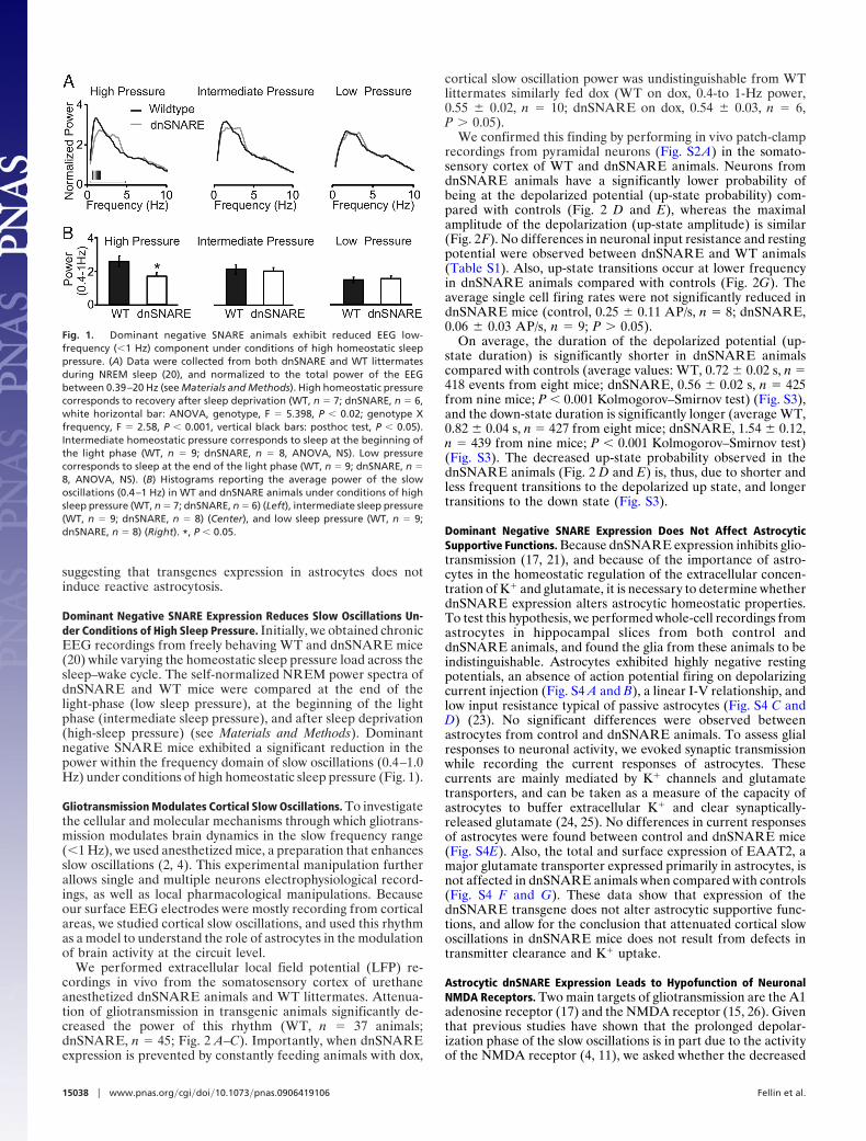
Dominant Negative SNARE Expression Reduces Slow Oscillations Un-der Conditions of High Sleep Pressure. Initially, we obtained chronicEEG recordings from freely behaving WT and dnSNARE mice(20) while varying the homeostatic sleep pressure load across thesleep–wake cycle. The self-normalized NREM power spectra ofdnSNARE and WT mice were compared at the end of thelight-phase (low sleep pressure), at the beginning of the lightphase (intermediate sleep pressure), and after sleep deprivation(high-sleep pressure) (see Materials and Methods). Dominantnegative SNARE mice exhibited a significant reduction in thepower within the frequency domain of slow oscillations (0.4–1.0Hz) under conditions of high homeostatic sleep pressure (Fig. 1).
Gliotransmission Modulates Cortical Slow Oscillations. To investigatethe cellular and molecular mechanisms through which gliotrans-mission modulates brain dynamics in the slow frequency range(�1 Hz), we used anesthetized mice, a preparation that enhancesslow oscillations (2, 4). This experimental manipulation furtherallows single and multiple neurons electrophysiological record-ings, as well as local pharmacological manipulations. Becauseour surface EEG electrodes were mostly recording from corticalareas, we studied cortical slow oscillations, and used this rhythmas a model to understand the role of astrocytes in the modulationof brain activity at the circuit level.
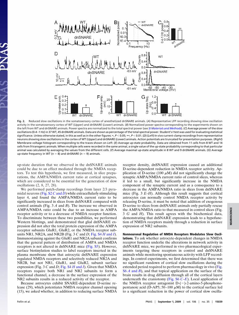
We performed extracellular local field potential (LFP) re-cordings in vivo from the somatosensory cortex of urethaneanesthetized dnSNARE animals and WT littermates. Attenua-tion of gliotransmission in transgenic animals significantly de-creased the power of this rhythm (WT, n � 37 animals;dnSNARE, n � 45; Fig. 2 A–C). Importantly, when dnSNAREexpression is prevented by constantly feeding animals with dox,
cortical slow oscillation power was undistinguishable from WTlittermates similarly fed dox (WT on dox, 0.4-to 1-Hz power,0.55 � 0.02, n � 10; dnSNARE on dox, 0.54 � 0.03, n � 6,P � 0.05).
We confirmed this finding by performing in vivo patch-clamprecordings from pyramidal neurons (Fig. S2 A) in the somato-sensory cortex of WT and dnSNARE animals. Neurons fromdnSNARE animals have a significantly lower probability ofbeing at the depolarized potential (up-state probability) com-pared with controls (Fig. 2 D and E), whereas the maximalamplitude of the depolarization (up-state amplitude) is similar(Fig. 2F). No differences in neuronal input resistance and restingpotential were observed between dnSNARE and WT animals(Table S1). Also, up-state transitions occur at lower frequencyin dnSNARE animals compared with controls (Fig. 2G). Theaverage single cell firing rates were not significantly reduced indnSNARE mice (control, 0.25 � 0.11 AP/s, n � 8; dnSNARE,0.06 � 0.03 AP/s, n � 9; P � 0.05).
On average, the duration of the depolarized potential (up-state duration) is significantly shorter in dnSNARE animalscompared with controls (average values: WT, 0.72 � 0.02 s, n �418 events from eight mice; dnSNARE, 0.56 � 0.02 s, n � 425from nine mice; P � 0.001 Kolmogorov–Smirnov test) (Fig. S3),and the down-state duration is significantly longer (average WT,0.82 � 0.04 s, n � 427 from eight mice; dnSNARE, 1.54 � 0.12,n � 439 from nine mice; P � 0.001 Kolmogorov–Smirnov test)(Fig. S3). The decreased up-state probability observed in thednSNARE animals (Fig. 2 D and E) is, thus, due to shorter andless frequent transitions to the depolarized up state, and longertransitions to the down state (Fig. S3).
Dominant Negative SNARE Expression Does Not Affect AstrocyticSupportive Functions. Because dnSNARE expression inhibits glio-transmission (17, 21), and because of the importance of astro-cytes in the homeostatic regulation of the extracellular concen-tration of K� and glutamate, it is necessary to determine whetherdnSNARE expression alters astrocytic homeostatic properties.To test this hypothesis, we performed whole-cell recordings fromastrocytes in hippocampal slices from both control anddnSNARE animals, and found the glia from these animals to beindistinguishable. Astrocytes exhibited highly negative restingpotentials, an absence of action potential firing on depolarizingcurrent injection (Fig. S4 A and B), a linear I-V relationship, andlow input resistance typical of passive astrocytes (Fig. S4 C andD) (23). No significant differences were observed betweenastrocytes from control and dnSNARE animals. To assess glialresponses to neuronal activity, we evoked synaptic transmissionwhile recording the current responses of astrocytes. Thesecurrents are mainly mediated by K� channels and glutamatetransporters, and can be taken as a measure of the capacity ofastrocytes to buffer extracellular K� and clear synaptically-released glutamate (24, 25). No differences in current responsesof astrocytes were found between control and dnSNARE mice(Fig. S4E). Also, the total and surface expression of EAAT2, amajor glutamate transporter expressed primarily in astrocytes, isnot affected in dnSNARE animals when compared with controls(Fig. S4 F and G). These data show that expression of thednSNARE transgene does not alter astrocytic supportive func-tions, and allow for the conclusion that attenuated cortical slowoscillations in dnSNARE mice does not result from defects intransmitter clearance and K� uptake.
Astrocytic dnSNARE Expression Leads to Hypofunction of NeuronalNMDA Receptors. Two main targets of gliotransmission are the A1adenosine receptor (17) and the NMDA receptor (15, 26). Giventhat previous studies have shown that the prolonged depolar-ization phase of the slow oscillations is in part due to the activityof the NMDA receptor (4, 11), we asked whether the decreased
Fig. 1. Dominant negative SNARE animals exhibit reduced EEG low-frequency (�1 Hz) component under conditions of high homeostatic sleeppressure. (A) Data were collected from both dnSNARE and WT littermatesduring NREM sleep (20), and normalized to the total power of the EEGbetween 0.39–20 Hz (see Materials and Methods). High homeostatic pressurecorresponds to recovery after sleep deprivation (WT, n � 7; dnSNARE, n � 6,white horizontal bar: ANOVA, genotype, F � 5.398, P � 0.02; genotype Xfrequency, F � 2.58, P � 0.001, vertical black bars: posthoc test, P � 0.05).Intermediate homeostatic pressure corresponds to sleep at the beginning ofthe light phase (WT, n � 9; dnSNARE, n � 8, ANOVA, NS). Low pressurecorresponds to sleep at the end of the light phase (WT, n � 9; dnSNARE, n �8, ANOVA, NS). (B) Histograms reporting the average power of the slowoscillations (0.4–1 Hz) in WT and dnSNARE animals under conditions of highsleep pressure (WT, n � 7; dnSNARE, n � 6) (Left), intermediate sleep pressure(WT, n � 9; dnSNARE, n � 8) (Center), and low sleep pressure (WT, n � 9;dnSNARE, n � 8) (Right). *, P � 0.05.
15038 � www.pnas.org�cgi�doi�10.1073�pnas.0906419106 Fellin et al.
up-state duration that we observed in the dnSNARE animalscould be due to an effect mediated through the NMDA recep-tors. To test this hypothesis, we first measured, in slice prepa-rations, the AMPA/NMDA current ratio at cortical synapses,which are considered to be essential for the generation of slowoscillations (2, 8, 27, 28).
We performed patch-clamp recordings from layer 2/3 pyra-midal neurons (Fig. S2 C and D) while extracellularly stimulatinglayer 4, and found the AMPA/NMDA current ratio to besignificantly increased in slices from dnSNARE compared withcontrol animals (Fig. 3 A and B). The increase we observed inAMPA/NMDA ratio could be due to an increase in AMPAreceptor activity or to a decrease of NMDA receptor function.To discriminate between these two possibilities, we performedWestern blottings, and demonstrated that glial dnSNARE ex-pression did not alter the total protein expression of the AMPAreceptor subunits GluR1, GluR2, or the NMDA receptor sub-units NR1, NR2A, and NR2B (Fig. 3 C and D; Fig. S4 H and I).Immunostaining against the GluR1 and NR2A subunit confirmsthat the general pattern of distribution of AMPA and NMDAreceptors is not altered in dnSNARE mice (Fig. S5). However,surface biotinylation studies to label receptors inserted in theplasma membrane show that astrocytic dnSNARE expressionregulated NMDA receptors and selectively reduced NR2A andNR2B, but not NR1, GluR1, and GluR2, subunit surfaceexpression (Fig. 3 C and D; Fig. S4 H and I). Given that NMDAreceptors require both NR1 and NR2 subunits to form afunctional channel, a decrease in the surface expression of theNR2 subunits results in a reduced activity of the receptor.
Because astrocytes exhibit SNARE-dependent D-serine re-lease (29), which potentiates NMDA receptor channel opening(15), we asked whether, in addition to reducing surface NMDA
receptor density, dnSNARE expression caused an additionalD-serine-dependent reduction in NMDA receptor activity. Ap-plication of D-serine (100 �M) did not significantly change thesynaptic AMPA/NMDA current ratio of control slices, whereasit led to a small, but significantly increase in the NMDAcomponent of the synaptic current and as a consequence to adecrease in the AMPA/NMDA ratio in slices from dnSNAREmice (Fig. 3 E–H). Although this result suggests that corticalastrocytes can normally control NMDA receptor activity byreleasing D-serine, it must be noted that addition of exogenousD-serine to slices from dnSNARE animals only partially rescuethe AMPA/NMDA ratio to that measured in control slices (Fig.3 G and H). This result agrees with the biochemical data,demonstrating that dnSNARE expression leads to a hypofunc-tion of NMDA receptors mainly through a reduction in surfaceexpression of NR2 subunits.
Nonneuronal Regulation of NMDA Receptors Modulates Slow Oscil-lations. To ask whether astrocyte-dependent changes in NMDAreceptor function underlie the alterations in network activity indnSNARE mice, we performed in vivo pharmacological exper-iments targeting these receptors in control and dnSNAREanimals while monitoring spontaneous activity with LFP record-ings. In control experiments, we first determined that there wasno significant rundown of cortical slow oscillations during theextended period required to perform pharmacology in vivo (Fig.S6 A and B), and that topical application on the surface of thebrain results in drug diffusion through all of the cortical layersunderneath the craniotomy (Fig. S6 C–E). Local application ofthe NMDA receptor antagonist D-(�)-2-amino-5-phosphono-pentanoic acid (D-AP5; 50–100 �M) to the cortical surface ledto a significant reduction in the power of cortical slow oscilla-
Fig. 2. Reduced slow oscillations in the somatosensory cortex of anesthetized dnSNARE animals. (A) Representative LFP recording showing slow oscillationactivity in the somatosensory cortex of WT (Upper) and dnSNARE (Lower) animals. (B) Normalized power spectra corresponding to the experiments shown onthe left from WT and dnSNARE animals. Power spectra are normalized to the total spectral power (see SI Materials and Methods). (C) Average power of the slowoscillations (0.4–1 Hz) in 37 WT, 45 dnSNARE animals. Data are shown as percentage of the total spectral power. Student’s t test was used for evaluating statisticalsignificance. Unless otherwise stated, in this as well as in the other figures: *, P � 0.05; **, P � 0.01. (D) (Left) In vivo current-clamp recordings from representativeneurons showing slow oscillations in the cortex of WT (Upper) and dnSNARE (Lower) animals. Action potentials are truncated for presentation purposes. (Right)Membrane voltage histogram corresponding to the traces shown on Left. (E) Average up-state probability. Data are obtained from 11 cells from 8 WT and 14cells from 9 transgenic animals. When multiple cells were recorded in the same animal, a single value of the up-state probability corresponding to that particularanimal was calculated by averaging the values from the different cells. (F) Average maximal up-state amplitude in 8 WT and 9 dnSNARE animals. (G) Averageup-state frequency in WT (n � 8) and dnSNARE (n � 9) animals.
Fellin et al. PNAS � September 1, 2009 � vol. 106 � no. 35 � 15039
NEU
ROSC
IEN
CE
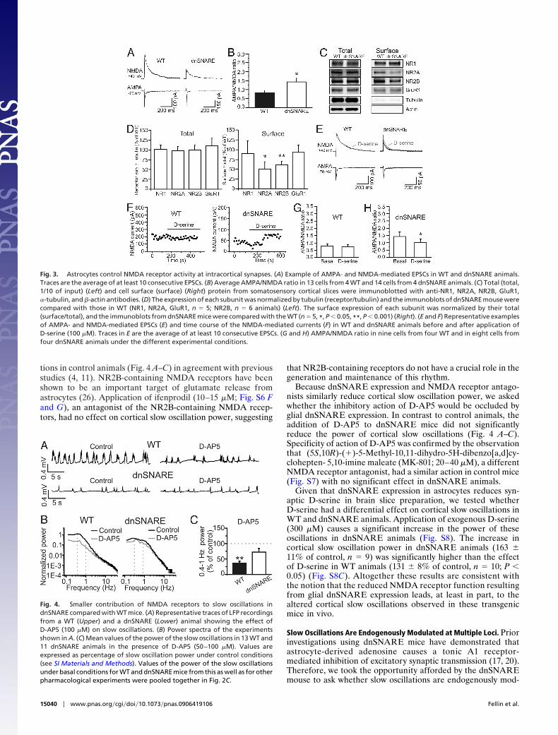
tions in control animals (Fig. 4 A–C) in agreement with previousstudies (4, 11). NR2B-containing NMDA receptors have beenshown to be an important target of glutamate release fromastrocytes (26). Application of ifenprodil (10–15 �M; Fig. S6 Fand G), an antagonist of the NR2B-containing NMDA recep-tors, had no effect on cortical slow oscillation power, suggesting
that NR2B-containing receptors do not have a crucial role in thegeneration and maintenance of this rhythm.
Because dnSNARE expression and NMDA receptor antago-nists similarly reduce cortical slow oscillation power, we askedwhether the inhibitory action of D-AP5 would be occluded byglial dnSNARE expression. In contrast to control animals, theaddition of D-AP5 to dnSNARE mice did not significantlyreduce the power of cortical slow oscillations (Fig. 4 A–C).Specificity of action of D-AP5 was confirmed by the observationthat (5S,10R)-(�)-5-Methyl-10,11-dihydro-5H-dibenzo[a,d]cy-clohepten- 5,10-imine maleate (MK-801; 20–40 �M), a differentNMDA receptor antagonist, had a similar action in control mice(Fig. S7) with no significant effect in dnSNARE animals.
Given that dnSNARE expression in astrocytes reduces syn-aptic D-serine in brain slice preparation, we tested whetherD-serine had a differential effect on cortical slow oscillations inWT and dnSNARE animals. Application of exogenous D-serine(300 �M) causes a significant increase in the power of theseoscillations in dnSNARE animals (Fig. S8). The increase incortical slow oscillation power in dnSNARE animals (163 �11% of control, n � 9) was significantly higher than the effectof D-serine in WT animals (131 � 8% of control, n � 10; P �0.05) (Fig. S8C). Altogether these results are consistent withthe notion that the reduced NMDA receptor function resultingfrom glial dnSNARE expression leads, at least in part, to thealtered cortical slow oscillations observed in these transgenicmice in vivo.
Slow Oscillations Are Endogenously Modulated at Multiple Loci. Priorinvestigations using dnSNARE mice have demonstrated thatastrocyte-derived adenosine causes a tonic A1 receptor-mediated inhibition of excitatory synaptic transmission (17, 20).Therefore, we took the opportunity afforded by the dnSNAREmouse to ask whether slow oscillations are endogenously mod-
Fig. 3. Astrocytes control NMDA receptor activity at intracortical synapses. (A) Example of AMPA- and NMDA-mediated EPSCs in WT and dnSNARE animals.Traces are the average of at least 10 consecutive EPSCs. (B) Average AMPA/NMDA ratio in 13 cells from 4 WT and 14 cells from 4 dnSNARE animals. (C) Total (total,1/10 of input) (Left) and cell surface (surface) (Right) protein from somatosensory cortical slices were immunoblotted with anti-NR1, NR2A, NR2B, GluR1,�-tubulin, and �-actin antibodies. (D) The expression of each subunit was normalized by tubulin (receptor/tubulin) and the immunoblots of dnSNARE mouse werecompared with those in WT (NR1, NR2A, GluR1, n � 5; NR2B, n � 6 animals) (Left). The surface expression of each subunit was normalized by their total(surface/total), and the immunoblots from dnSNARE mice were compared with the WT (n � 5, *, P � 0.05, **, P � 0.001) (Right). (E and F) Representative examplesof AMPA- and NMDA-mediated EPSCs (E) and time course of the NMDA-mediated currents (F) in WT and dnSNARE animals before and after application ofD-serine (100 �M). Traces in E are the average of at least 10 consecutive EPSCs. (G and H) AMPA/NMDA ratio in nine cells from four WT and in eight cells fromfour dnSNARE animals under the different experimental conditions.
Fig. 4. Smaller contribution of NMDA receptors to slow oscillations indnSNARE compared with WT mice. (A) Representative traces of LFP recordingsfrom a WT (Upper) and a dnSNARE (Lower) animal showing the effect ofD-AP5 (100 �M) on slow oscillations. (B) Power spectra of the experimentsshown in A. (C) Mean values of the power of the slow oscillations in 13 WT and11 dnSNARE animals in the presence of D-AP5 (50–100 �M). Values areexpressed as percentage of slow oscillation power under control conditions(see SI Materials and Methods). Values of the power of the slow oscillationsunder basal conditions for WT and dnSNARE mice from this as well as for otherpharmacological experiments were pooled together in Fig. 2C.
15040 � www.pnas.org�cgi�doi�10.1073�pnas.0906419106 Fellin et al.
ulated at multiple loci, the A1 receptor as well as the NMDAreceptor.
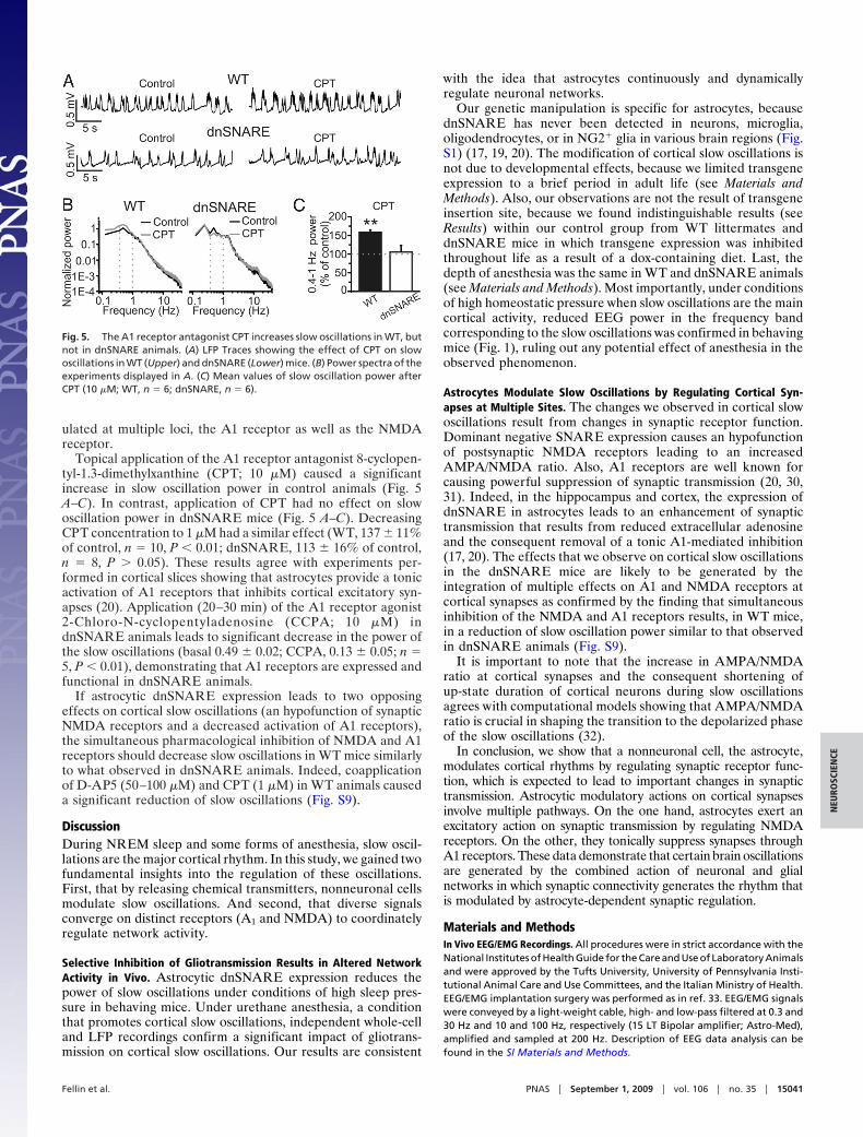
Topical application of the A1 receptor antagonist 8-cyclopen-tyl-1.3-dimethylxanthine (CPT; 10 �M) caused a significantincrease in slow oscillation power in control animals (Fig. 5A–C). In contrast, application of CPT had no effect on slowoscillation power in dnSNARE mice (Fig. 5 A–C). DecreasingCPT concentration to 1 �M had a similar effect (WT, 137 � 11%of control, n � 10, P � 0.01; dnSNARE, 113 � 16% of control,n � 8, P � 0.05). These results agree with experiments per-formed in cortical slices showing that astrocytes provide a tonicactivation of A1 receptors that inhibits cortical excitatory syn-apses (20). Application (20–30 min) of the A1 receptor agonist2-Chloro-N-cyclopentyladenosine (CCPA; 10 �M) indnSNARE animals leads to significant decrease in the power ofthe slow oscillations (basal 0.49 � 0.02; CCPA, 0.13 � 0.05; n �5, P � 0.01), demonstrating that A1 receptors are expressed andfunctional in dnSNARE animals.
If astrocytic dnSNARE expression leads to two opposingeffects on cortical slow oscillations (an hypofunction of synapticNMDA receptors and a decreased activation of A1 receptors),the simultaneous pharmacological inhibition of NMDA and A1receptors should decrease slow oscillations in WT mice similarlyto what observed in dnSNARE animals. Indeed, coapplicationof D-AP5 (50–100 �M) and CPT (1 �M) in WT animals causeda significant reduction of slow oscillations (Fig. S9).
DiscussionDuring NREM sleep and some forms of anesthesia, slow oscil-lations are the major cortical rhythm. In this study, we gained twofundamental insights into the regulation of these oscillations.First, that by releasing chemical transmitters, nonneuronal cellsmodulate slow oscillations. And second, that diverse signalsconverge on distinct receptors (A1 and NMDA) to coordinatelyregulate network activity.
Selective Inhibition of Gliotransmission Results in Altered NetworkActivity in Vivo. Astrocytic dnSNARE expression reduces thepower of slow oscillations under conditions of high sleep pres-sure in behaving mice. Under urethane anesthesia, a conditionthat promotes cortical slow oscillations, independent whole-celland LFP recordings confirm a significant impact of gliotrans-mission on cortical slow oscillations. Our results are consistent
with the idea that astrocytes continuously and dynamicallyregulate neuronal networks.
Our genetic manipulation is specific for astrocytes, becausednSNARE has never been detected in neurons, microglia,oligodendrocytes, or in NG2� glia in various brain regions (Fig.S1) (17, 19, 20). The modification of cortical slow oscillations isnot due to developmental effects, because we limited transgeneexpression to a brief period in adult life (see Materials andMethods). Also, our observations are not the result of transgeneinsertion site, because we found indistinguishable results (seeResults) within our control group from WT littermates anddnSNARE mice in which transgene expression was inhibitedthroughout life as a result of a dox-containing diet. Last, thedepth of anesthesia was the same in WT and dnSNARE animals(see Materials and Methods). Most importantly, under conditionsof high homeostatic pressure when slow oscillations are the maincortical activity, reduced EEG power in the frequency bandcorresponding to the slow oscillations was confirmed in behavingmice (Fig. 1), ruling out any potential effect of anesthesia in theobserved phenomenon.
Astrocytes Modulate Slow Oscillations by Regulating Cortical Syn-apses at Multiple Sites. The changes we observed in cortical slowoscillations result from changes in synaptic receptor function.Dominant negative SNARE expression causes an hypofunctionof postsynaptic NMDA receptors leading to an increasedAMPA/NMDA ratio. Also, A1 receptors are well known forcausing powerful suppression of synaptic transmission (20, 30,31). Indeed, in the hippocampus and cortex, the expression ofdnSNARE in astrocytes leads to an enhancement of synaptictransmission that results from reduced extracellular adenosineand the consequent removal of a tonic A1-mediated inhibition(17, 20). The effects that we observe on cortical slow oscillationsin the dnSNARE mice are likely to be generated by theintegration of multiple effects on A1 and NMDA receptors atcortical synapses as confirmed by the finding that simultaneousinhibition of the NMDA and A1 receptors results, in WT mice,in a reduction of slow oscillation power similar to that observedin dnSNARE animals (Fig. S9).
It is important to note that the increase in AMPA/NMDAratio at cortical synapses and the consequent shortening ofup-state duration of cortical neurons during slow oscillationsagrees with computational models showing that AMPA/NMDAratio is crucial in shaping the transition to the depolarized phaseof the slow oscillations (32).
In conclusion, we show that a nonneuronal cell, the astrocyte,modulates cortical rhythms by regulating synaptic receptor func-tion, which is expected to lead to important changes in synaptictransmission. Astrocytic modulatory actions on cortical synapsesinvolve multiple pathways. On the one hand, astrocytes exert anexcitatory action on synaptic transmission by regulating NMDAreceptors. On the other, they tonically suppress synapses throughA1 receptors. These data demonstrate that certain brain oscillationsare generated by the combined action of neuronal and glialnetworks in which synaptic connectivity generates the rhythm thatis modulated by astrocyte-dependent synaptic regulation.
Materials and MethodsIn Vivo EEG/EMG Recordings. All procedures were in strict accordance with theNational Institutes of Health Guide for the Care and Use of Laboratory Animalsand were approved by the Tufts University, University of Pennsylvania Insti-tutional Animal Care and Use Committees, and the Italian Ministry of Health.EEG/EMG implantation surgery was performed as in ref. 33. EEG/EMG signalswere conveyed by a light-weight cable, high- and low-pass filtered at 0.3 and30 Hz and 10 and 100 Hz, respectively (15 LT Bipolar amplifier; Astro-Med),amplified and sampled at 200 Hz. Description of EEG data analysis can befound in the SI Materials and Methods.
Fig. 5. The A1 receptor antagonist CPT increases slow oscillations in WT, butnot in dnSNARE animals. (A) LFP Traces showing the effect of CPT on slowoscillations in WT (Upper) and dnSNARE (Lower) mice. (B) Power spectra of theexperiments displayed in A. (C) Mean values of slow oscillation power afterCPT (10 �M; WT, n � 6; dnSNARE, n � 6).
Fellin et al. PNAS � September 1, 2009 � vol. 106 � no. 35 � 15041
NEU
ROSC
IEN
CE

In Vivo Patch-Clamp Recordings. Bigenic GFAP.tTA and tetO.dnSNARE trans-genic animals (dnSNARE mice) (17) and their WT littermates aged 6–12 weeksold were anesthetized by an intraperitoneal injection of urethane (2 g/kg).Because these transgenic mice have been backcrossed onto a C57BL6/J geno-type for �10 generations, C57BL6/J mice were used as controls in someexperiments. Because no difference was observed between C57BL6/J and theWT littermates of dnSNARE mice, control data were pooled. Body tempera-ture was measured with a rectal probe and kept at 37 °C with a heating pad.Depth of anesthesia was assured by continuously monitoring, respiration rate,eyelid reflex, vibrissae movements, and monitoring reactions to tail and toepinching. Monitoring all these parameters no difference in anesthesia depthwas observed between WT and dnSNARE animals. After the animal wasanesthetized, an incision was made and the skull exposed. A craniotomy (1.8mm) was drilled in the skull overlaying the somatosensory cortex, and thesurface of the cortex was continuously kept moist with normal Hepes-buffered artificial cerebrospinal fluid (aCSF). The dura was carefully dissected,and a metal plate was glued on the skull for head fixation. Patch-clamprecordings were performed as described in ref. 34; 3- to 4-M� glass pipettewere used as recording electrodes and filled with the following intrapipettesolution (in mM): K-gluconate 140, MgCl2 1, NaCl 8, Na2ATP 2, NaGTP 0.5,Hepes 10, phophocreatine 10 to pH 7.2 with KOH. Biocytin 2 mg/mL wasincluded in the pipette solution for subsequent morphological reconstruction.Signals were amplified by a Multiclamp 700 B, filtered at 2 KHz, digitized at 10KHz with a digidata 1320, and stored with pClamp 9.2 (Axon instruments).
Details on data analysis and slice electrophysiology can be found in SI Mate-rials and Methods.
In Vivo Extracellular Recordings. Mouse preparation and surgery was as de-scribed for patch-clamp experiments. LFPs were recorded with custom-builtelectrodes made of two parallel tungsten electrodes (FHC). Electrodes wereplaced to record from the superficial layers of the somatosensory cortex.Signals were amplified with an AM-amplifier (AM-system), filtered at 0.1Hz-10 KHz, and digitized at 50 KHz. Recordings started 5–10 min after theelectrodes were inserted in the cortex. For the description of data analysis, seeSI Materials and Methods.
Statistics. In in vivo experiments, N values always refer to animal number,whereas in slice experiments, N values refer to the slice number. Student’s ttest was generally used to evaluate statistical significance. In pharmacologicalexperiments when drug effect was assessed within the same animal, paired ttest was used. For Fig. S3C, Kolmogorov–Smirnov test was used. Data arepresented as mean � SEM. A 2-factor ANOVA (frequency, genotype) was usedto compare the power spectra in Fig. 1.
ACKNOWLEDGMENTS. We thank Dr. D. Contreras and Dr. M. B. Dalva forcomments. This work was supported by National Institutes of Health and theItalian Institute of Technology.
1. Steriade M (2006) Grouping of brain rhythms in corticothalamic systems. Neuroscience137:1087–1106.
2. Steriade M, Nunez A, Amzica F (1993) Intracellular analysis of relations between theslow (less-than-1 Hz) neocortical oscillation and other sleep rhythms of the electroen-cephalogram. J Neurosci 13:3266–3283.
3. Steriade M, Contreras D, Dossi RC, Nunez A (1993) The slow (less-than-1 Hz)oscillation in reticular thalamic and thalamocortical neurons: Scenario of sleeprhythm generation in interacting thalamic and neocortical networks. J Neurosci13:3284 –3299.
4. Steriade M, Nunez A, Amzica F (1993) A novel slow (less-than-1 Hz) oscillation ofneocortical neurons in-vivo: Depolarizing and hyperpolarizing components. J Neurosci13:3252–3265.
5. Petersen CCH, Hahn TTG, Mehta M, Grinvald A, Sakmann B (2003) Interaction ofsensory responses with spontaneous depolarization in layer 2/3 barrel cortex. Proc NatlAcad Sci USA 100:13638–13643.
6. Huber R, Ghilardi MF, Massimini M, Tononi G (2004) Local sleep and learning. Nature430:78–81.
7. Marshall L, Helgadottir H, Molle M, Born J (2006) Boosting slow oscillations duringsleep potentiates memory. Nature 444:610–613.
8. Shu YS, Hasenstaub A, McCormick DA (2003) Turning on and off recurrent balancedcortical activity. Nature 423:288–293.
9. Amzica F, Steriade M (1995) Disconnection of intracortical synaptic linkages disruptssynchronization of a slow oscillation. J Neurosci 15:4658–4677.
10. Cunningham MO, et al. (2006) Neuronal metabolism governs cortical network re-sponse state. Proc Natl Acad Sci USA 103:5597–5601.
11. Compte A, Sanchez-Vives MV, McCormick DA, Wang XJ (2003) Cellular and networkmechanisms of slow oscillatory activity (� 1 Hz) and wave propagations in a corticalnetwork model. J Neurophysiol 89:2707–2725.
12. Newman EA (2003) New roles for astrocytes: Regulation of synaptic transmission.Trends Neurosci 26:536–542.
13. Volterra A, Meldolesi J (2005) Astrocytes, from brain glue to communication elements:The revolution continues. Nat Rev Neurosci 6:626–640.
14. Martineau M, Baux G, Mothet JP (2006) D-serine signalling in the brain: Friend and foe.Trends Neurosci 29:481–491.
15. Panatier A, et al. (2006) Glia-derived D-serine controls NMDA receptor activity andsynaptic memory. Cell 125:775–784.
16. Zhang JM, et al. (2003) ATP released by astrocytes mediates glutamatergic activity-dependent heterosynaptic suppression. Neuron 40:971–982.
17. Pascual O, et al. (2005) Astrocytic purinergic signaling coordinates synaptic networks.Science 310:113–116.
18. Ventura R, Harris KM (1999) Three-dimensional relationships between hippocampalsynapses and astrocytes. J Neurosci 19:6897–6906.
19. Halassa MM, Fellin T, Takase H, Dong JH, Haydon PG (2007) Synaptic islands defined bythe territory of a single astrocyte. J Neurosci 27:6473–6477.
20. Halassa MM, et al. (2009) Astrocytic adenosine controls sleep homeostasis and cogni-tive consequences of sleep loss. Neuron 61:213–219.
21. Zhang Q, et al. (2004) Fusion-related release of glutamate from astrocytes. J Biol Chem279:12724–12733.
22. Orthmann-Murphy JL, Enriquez AD, Abrams CK, Scherer SS (2007) Loss-of-functionGJA12/Connexin47 mutations cause Pelizaeus-Merzbacher-like disease. Mol Cell Neu-rosci 34:629–641.
23. Matthias K, et al. (2003) Segregated expression of AMPA-type glutamate receptors andglutamate transporters defines distinct astrocyte populations in the mouse hippocam-pus. J Neurosci 23:1750–1758.
24. Bergles DE, Jahr CE (1997) Synaptic activation of glutamate transporters in hippocam-pal astrocytes. Neuron 19:1297–1308.
25. Saint Jan DD, Westbrook GL (2005) Detecting activity in olfactory bulb glomeruli withastrocyte recording. J Neurosci 25:2917–2924.
26. Fellin T, et al. (2004) Neuronal synchrony mediated by astrocytic glutamate throughactivation of extrasynaptic NMDA receptors. Neuron 43:729–743.
27. Timofeev I, Grenier F, Bazhenov M, Sejnowski TJ, Steriade M (2000) Origin of slowcortical oscillations in deafferented cortical slabs. Cerebral Cortex 10:1185–1199.
28. Sanchez-Vives MV, McCormick DA (2000) Cellular and network mechanisms of rhyth-mic recurrent activity in neocortex. Nat Neurosci 3:1027–1034.
29. Mothet JP, et al. (2005) Glutamate receptor activation triggers a calcium-dependentand SNARE protein-dependent release of the gliotransmitter D-serine. Proc Natl AcadSci USA 102:5606–5611.
30. Brand A, Vissiennon Z, Eschke D, Nieber K (2001) Adenosine A(1) and A(3) receptorsmediate inhibition of synaptic transmission in rat cortical neurons. Neuropharmacol-ogy 40:85–95.
31. Lopes LV, Cunha RA, Ribeiro JA (1999) Cross-talk between A(1)/A(2A) adenosinereceptors in the rat hippocampus and cortex. Br J Pharmacol 127:U20.
32. Wolf JA, et al. (2005) NMDA/AMPA ratio impacts state transitions and entrainment tooscillations in a computational model of the nucleus accumbens medium spiny pro-jection neuron. J Neurosci 25:9080–9095.
33. Frank MG, Stryker MP, Tecott LH (2002) Sleep and sleep homeostasis in mice lacking the5-HT2c receptor. Neuropsychopharmacology 27:869–873.
34. Margrie TW, Brecht M, Sakmann B (2002) In vivo, low-resistance, whole-cell recordingsfrom neurons in the anaesthetized and awake mammalian brain. Pflug Arch Eur J Phy444:491–498.
15042 � www.pnas.org�cgi�doi�10.1073�pnas.0906419106 Fellin et al.