Enas A. Hassan1, Naglaa M. Balabel2 3, 3, El shahat …...then cycled through 30 cycles of 94 C for...
14
International Journal of Scientific & Engineering Research, Volume 8, Issue 1, January-2017 91 ISSN 2229-5518 IJSER © 2017 http://www.ijser.org Relationship between Ralstonia Solanacearum and bioagents recovered from different habitats Enas A. Hassan 1 , Naglaa M. Balabel 2 , Abeer E.Ahmed 3, , Nerhan A. Eid 3 , El shahat M. Ramadan 1 1 Agric. Microbiol. Dept., Fac. Agric., Ain Shams Univ., Cairo, Egypt 2 Bact. Dis. Res. Dept., Plant Pathol. Res. Instit., Agric. Res. Center, Giza, Egypt 3 Plant Protec., Des. Res. Center, Cairo, Egypt Corresponding Author : Naglaa M. Balabel, [email protected] Abstract--- Bacterial wilt caused by Ralstonia solanacearum phylotype II sequevar I (race 3 biovar 2), is considered one of the most destructive bacterial diseases of potato plants. The aim of this study was to evaluate the potential of some bacterial antagonists on suppressing the causative bacterium. In this regard, 420 isolates of R.solanacearum were risolated from different habitats at different Egyptian districts. The variation among these isolates was assessed on the basis of their pathogenic potentialities to tomato seedlings in the greenhouse. The highest potential, as shown by the wilt severity, was obtained by 11 out of 420 isolates. The most aggressiveness isolates were identified by accurate techniques. A collection of 318 isolates of rhizobacteria from rhizosphere of different plants were tested to lay out a biological control protocol against R.solanacearum selected strains. In vitro, 14 bacterial isolates were more effective in suppressing the pathogen at different levels of relative power of antibiosis. These isolates were categorized into three groups according to their gram-staining reaction, cell morphology and cultural characteristics. Further evaluation under the greenhouse condition has shown that six of the antagonistic isolates were effective in suppressing disease development, as expressed by the Area Under Disease Progress Curve (AUDPC). These isolates were identified as: Streptomyces toxytricini, Stenotrophomonas maltophilia, Bacillus pseudomycoides and Brevibacillus brevis. Keywords: Potato, bacterial wilt , Ralstonia solanacearum , biological control, , PCR , PGPR . —————————— —————————— 1. Introduction Ralstonia solanacearum is a soil-borne, rod shaped, gram negative, β-proteobacterium that causes bacterial wilt disease to more than 450 plant species including many economically important crops. Due to its wide geographic distribution and unusually broad host range (over 50 plant families) the pathogen is responsible for severe crop losses worldwide (Hayward, 1991 and Saad , 2016). Although various control measures have been documented, bacterial wilt is still a difficult to control because of wide host range of the pathogen and long survival of the pathogen in soil, especially in deeper layers (Hsu, 1991).Crop rotation with non-host plants, although recommended, is not an efficient method, since R. solanacerum has its disseminating and survival phases in the soil and it remains viable for long periods of time. The race and strain diversity of the pathogen has made breeding for resistant cultivars ineffective or with limited value in the control of bacterial wilt (Farag et al., 1982; Hanson et al., 1996 and Wang et al., 1998 ). The use of soil fumigants is environmentally unacceptable, expensive and largely ineffective against the disease (Saddler, 2005). Chemical and soil treatments such as modification of soil pH, heat treatment by solarization, and application of stable bleaching powder, as well as plant resistance inducers as acibenzolar –S-methyl, plant essential oils as thymol, or phosphoric acid have been shown to reduce bacterial populations and disease severity on a small scale (Norman et al., 2006 and Abo-Elyousr et al., 2012). Biological control strategies may be either used directly as a practice or after being integrated with other practices for effective disease management at the field level (Myint & Ranamukhaarachchi, 2006). A large numbers of bacteria including species of Pseudomonas, Azospirillum, Azotobacter (Ahmed et al., 2008), Klebsiella (Govindarajan et al., 2007), Serratia (Gyaneshwar et al., 2001) have been reported to enhance plant growth and suppress disease development in various crops. Several strains of Pseudomonas fluorescens have been reported to suppress soil- borne diseases (Weller, 1988). On the other hand, numerous of Actinomycetes and bacteria such as Stenotrophomonas maltophilia, P. glumae, Burkholderia cepacia, Bacillus spp., Erwinia spp. have been reported to be active control agents against R. solanacearum (Messiha et al., 2007; Aliye et al.,2008 and Xue et al., 2009). The present study aimed to obtain plant growth promoting rhizobacteria (PGPR) from the rhizosphere of IJSER
Transcript of Enas A. Hassan1, Naglaa M. Balabel2 3, 3, El shahat …...then cycled through 30 cycles of 94 C for...
International Journal of Scientific & Engineering Research, Volume 8, Issue 1, January-2017 91 ISSN 2229-5518
IJSER © 2017 http://www.ijser.org
Relationship between Ralstonia Solanacearum and bioagents recovered
from different habitats
Enas A. Hassan1, Naglaa M. Balabel2, Abeer E.Ahmed3,, Nerhan A. Eid3, El shahat M. Ramadan1
1 Agric. Microbiol. Dept., Fac. Agric., Ain Shams Univ., Cairo, Egypt 2 Bact. Dis. Res. Dept., Plant Pathol. Res. Instit., Agric. Res. Center, Giza, Egypt
3 Plant Protec., Des. Res. Center, Cairo, Egypt
Corresponding Author : Naglaa M. Balabel, [email protected] Abstract--- Bacterial wilt caused by Ralstonia solanacearum phylotype II sequevar I (race 3 biovar 2), is considered one of the most destructive bacterial diseases of potato plants. The aim of this study was to evaluate the potential of some bacterial antagonists on suppressing the causative bacterium. In this regard, 420 isolates of R.solanacearum were risolated from different habitats at different Egyptian districts. The variation among these isolates was assessed on the basis of their pathogenic potentialities to tomato seedlings in the greenhouse. The highest potential, as shown by the wilt severity, was obtained by 11 out of 420 isolates. The most aggressiveness isolates were identified by accurate techniques. A collection of 318 isolates of rhizobacteria from rhizosphere of different plants were tested to lay out a biological control protocol against R.solanacearum selected strains. In vitro, 14 bacterial isolates were more effective in suppressing the pathogen at different levels of relative power of antibiosis. These isolates were categorized into three groups according to their gram-staining reaction, cell morphology and cultural characteristics. Further evaluation under the greenhouse condition has shown that six of the antagonistic isolates were effective in suppressing disease development, as expressed by the Area Under Disease Progress Curve (AUDPC). These isolates were identified as: Streptomyces toxytricini, Stenotrophomonas maltophilia, Bacillus pseudomycoides and Brevibacillus brevis. Keywords: Potato, bacterial wilt , Ralstonia solanacearum , biological control, , PCR , PGPR .
—————————— —————————— 1. Introduction Ralstonia solanacearum is a soil-borne, rod shaped, gram negative, β-proteobacterium that causes bacterial wilt disease to more than 450 plant species including many economically important crops. Due to its wide geographic distribution and unusually broad host range (over 50 plant families) the pathogen is responsible for severe crop losses worldwide (Hayward, 1991 and Saad , 2016). Although various control measures have been documented, bacterial wilt is still a difficult to control because of wide host range of the pathogen and long survival of the pathogen in soil, especially in deeper layers (Hsu, 1991).Crop rotation with non-host plants, although recommended, is not an efficient method, since R. solanacerum has its disseminating and survival phases in the soil and it remains viable for long periods of time. The race and strain diversity of the pathogen has made breeding for resistant cultivars ineffective or with limited value in the control of bacterial wilt (Farag et al., 1982; Hanson et al., 1996 and Wang et al., 1998 ). The use of soil fumigants is environmentally unacceptable, expensive and largely ineffective against the disease (Saddler, 2005). Chemical and soil treatments such as modification of soil pH, heat treatment by solarization, and application of
stable bleaching powder, as well as plant resistance inducers as acibenzolar –S-methyl, plant essential oils as thymol, or phosphoric acid have been shown to reduce bacterial populations and disease severity on a small scale (Norman et al., 2006 and Abo-Elyousr et al., 2012). Biological control strategies may be either used directly as a practice or after being integrated with other practices for effective disease management at the field level (Myint & Ranamukhaarachchi, 2006). A large numbers of bacteria including species of Pseudomonas, Azospirillum, Azotobacter (Ahmed et al., 2008), Klebsiella (Govindarajan et al., 2007), Serratia (Gyaneshwar et al., 2001) have been reported to enhance plant growth and suppress disease development in various crops. Several strains of Pseudomonas fluorescens have been reported to suppress soil- borne diseases (Weller, 1988). On the other hand, numerous of Actinomycetes and bacteria such as Stenotrophomonas maltophilia, P. glumae, Burkholderia cepacia, Bacillus spp., Erwinia spp. have been reported to be active control agents against R. solanacearum (Messiha et al., 2007; Aliye et al.,2008 and Xue et al., 2009). The present study aimed to obtain plant growth promoting rhizobacteria (PGPR) from the rhizosphere of
IJSER
International Journal of Scientific & Engineering Research, Volume 8, Issue 1, January-2017 92 ISSN 2229-5518
IJSER © 2017 http://www.ijser.org
different plants with high biocontrol efficiency against virulent isolates of R. solanacearum , which was isolated and identified from different habitats.
2. Material and Methods 2.1. Isolation and growth conditions of
R.solanacearum: R. solanacearum isolates in this study were isolated from different habitats, i.e. potato tubers, field soil, irrigation water and weeds (Chenopodium album, Portulaca olaraceae, Rumex dentatus and Solanum nigrum) obtained from different areas at Al-Gharbia , Al- Behira and Al- Menofia governorates, in Egypt. Isolation was carried out on SMSA medium (Semi Selective Medium of South Africa) according to Elphinstone et al. (1996) and incubated at 28±1°C. Typical virulent fluidal, slightly irregular, white colonies with pink centers were selected as R. solanacearum (Buddenhagen & Kelman, 1964). The selected isolates were propagated on nutrient agar medium for 48 hours and then were verified serologically using immuno-fluorescent antibody staining (IFAS) (Janse, 1988). Pathogenicity was determined on Super Strain B tomato by stem inoculation .The most virulent pathogenic isolates were selected for further experimentation. The physiological and biochemical tests were made to confirm R. solanacearum identity(Hayward et al.,1990).Selected isolates were maintained in long-term storage as suspensions in sterile tap water and were revived by plating on tetrazolium chloride (TTC) medium (Kelman, 1954). 2.2. Diagnosis and identification of R. solanacearum: Pathogenicity and identification are the first steps for diagnosis of bacterial brown rot. Accurate identification of R.solanacearum race3 biovar2 based on symptomatic or a symptomatic plants along with multiple microbiological and molecular methods were followed. 2.2.a. Pathogenicity test on tomato seedling: Four weeks old healthy tomato seedlings (Solanum lycopersicum cv. Super Strain B) were used as a test plants to determine the virulence of R. solanacearum under greenhouse conditions at 20-30°C. Bacterial inoculum was prepared in nutrient broth for 2 days at 28±1°C, centrifuged and suspended in sterile destilled water, and adjusted to be 107cfu/ml using
spectrophotometer at 620 nm. The seedlings at the three true-leaves stage were inoculated with puncturing the basal part of the stem with the bacterial suspension (Janse, 1988). Five replicates were made for each isolate. Control treatment was prepared by applying few drops of sterile distilled water instead of the bacterium. Disease severity was determined 7 days after inoculation based on the scale of Winstead &Kelman (1952) as follows: 0 = No wilt symptoms; 1 = One or 2 wilted leaves; 2= Three wilted leaves; 3 = All leaves wilted except the tip; 4 = Whole plant wilted and 5= Death (collapse) of whole plant . Wilt severity was calculated by the following formula: Σ (nxv) % Wilt severity = ----------------X100 5 N Where: n =Number of the inspected samples in each category. v = Numerical values of each category. N = Total number of the inspected samples. 5= The highest grade scale. 2.2.b. Identification of R.solancearum biovars: Biovar characterization of the selected pathogenic bacterium (R. solanacearum) was done based on the differential ability of pathogen isolates to oxidize sugar and sugar alcohols using standard procedure described by Hayward et al. (1990). 2.2.c. Genotypic characterization: At the sub-species level, identification of R. solanacearum isolates can be determined by polymerase chain reaction (PCR) amplification with specific probes and primers. Real-time PCR (Taq-Man) was performed on selected highly pathogenic R.solanacearum according to Weller et al. (2000) by using the apparatus of Applied Biosystem7500 . The reaction mixture consisted of 12.5µl of master mix, 1 µl of primer forward, 1 µl of primer reverse, 1 µl of probe and 7 µl of water and 2.5µl of nucleic acid extract. The sequence of primers and probe used is shown in Table (1), which were provided by OPRON, USA. Positive control of DNA extracted was used and water was used as a negative control.
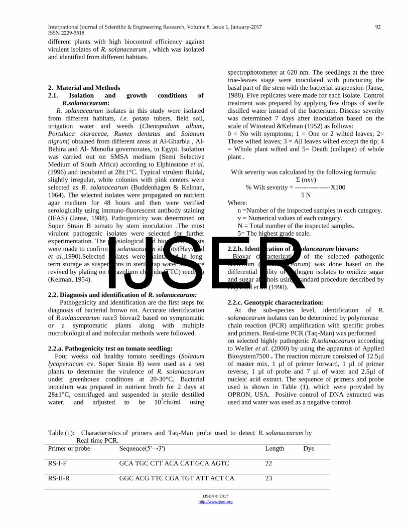
Table (1): Characteristics of primers and Taq-Man probe used to detect R. solanacearum by Real-time PCR. Primer or probe Sequence(5'→3') Length Dye
RS-I-F GCA TGC CTT ACA CAT GCA AGTC 22
RS-II-R GGC ACG TTC CGA TGT ATT ACT CA 23
IJSER

International Journal of Scientific & Engineering Research, Volume 8, Issue 1, January-2017 93 ISSN 2229-5518
IJSER © 2017 http://www.ijser.org
RS-P AGC TTG CTA CCT GCC GGC GAG TG 23 FAM
2.2.d. Phylotype analysis of R. solanacearum by
Multiplex- PCR: Representative isolates from observed PCR clusters were further analyzed to assess the phylotype. Analysis of selected isolates was made by using the Opina primers 759/760 as internal markers specific for the R. solanacearum strains and a set of four phylotype - specific forward primers with a unique and conserved reverse primer targeted in the 16S-23S Intergentic Spacer region (Opina et al.,1997). Table (2) shows the characteristics of the used primers. Suspend one colony of the tested isolates in 100µl of sterile water and heating for 5 min. at 100°C to spin down cell debris. The reaction mixture consisted of 12.5µl of ready master mix, 1 µl from each primer, 3.5µl of water and 2µl of nucleic acid
extract. The following cycling program was used in a thermal cycler (Biometra T personal): 96°C for 5 min. and then cycled through 30 cycles of 94°C for 15s, 59°C for 30s and 72°C for 30s, followed by a final extension period of 10 min. at 72°C. A13 μl aliquot of each amplified PCR products was subjected to electrophoresis on 2 % (w/v) agarose gels, stained with ethidium bromide (0.5% μgL-1) and bands were visualized on a UV- transilluminator. This Pmx-PCR amplifies the 280-bp"Universal" R. solanacearum specific reference band plus following phylotype-specific PCR products: a 144-bp amplicon from phylotype I strains; a 372-bp amplicon from phylotype II strains; a 91-bp amplicon from phylotype III strains and a 213-bp amplicon from phylotype IV strains (Sagar et al.,2014).
Table 2. Characteristics of primers used for Phylotype analysis of R .solanacearum by Multiplex-PCR
2.3. Isolation of potent antagonistic rhizobacteria: Potent antagonists were isolated from the rhizosphere of different healthy plants, grown in El-Adlia farm at Sharkia governorate. Vitis sp.( grape), Triticum sp. (wheat) , Ocimum basilicum (basil), Vicia faba (faba bean), Solanum lycopersicum (tomato), Alliumo mpeloprasum (leek), Zea mays (corn), Solanum lacinistum (solanum), Ltissima glumerulifora (solidago), Cymbogon citrates (lemon grass), Salvia officinalis (salvia), Calendula officinalis (pot marigold), Foeniculum vulgar (fennel), Solanum melongena (eggplant) and Rosmarinus officinali (rosemary) . Root samples were vigorously shaken to remove loosely adhering soil (Leben et al., 1968). Five serial 10-fold dilutions were made in sterile phosphate buffer for each sample and spread on glucose nutrient (Dowson,1957), King’s B (King et al., 1954) and glycerol nitrate agar (Waksman,1961) media to isolate bacilli and yeasts , fluorescent Pseudomonades and Acyinomycetes, respectively. Plates were incubated at 30 ±1º C for 2-4 days then single colonies were picked up and stored at 4±1º C on the appropriate medium.
2.3.a. Inoculum preparation of the antagonistic rhizobacteria :
Selected bacteria were checked for antagonistic potential, after turbidity standardization. The 48 h. old culture on suitable broth medium was centrifuged at 10.000 g for 10 min. Bacterial pellets were washed twice with sterilized distilled water by centrifugation. The optical density (OD) of the solution was adjusted to 0.45 (620 nm) to obtain l07cfu /ml (Mortensen, 1999). 2.3.b. Screening of R. solanacearum antagonistic
rhizobacteria in vitro: Collected isolates of rhizobacteria were tested for their antagonistic potentials against R. solanacearum according to the method described by Li et al. (2008).Ten ml. of R. solanacearum inoculum grown in nutrient broth for 24 h./ 28±1° C and centrifuged were used. The pellets were re-suspended in sterilized saline solution (0.85% NaCl) to a final concentration107cfu /ml. then was mixed with 1 litter of lukewarm melted nutrient agar and pouring into sterilized plates. After solidification, each plate was peripherally inoculated with three agar disks (6mm) of antagonist as a standard inoculum and incubated at 28-30°C for72 h. The relative power (RPA) of antibiosis was
Primer Sequence (5ˋ 3ˋ) Length
759 760 Nmult:21:1F Nmult:21:2F Nmult:23:AF Nmult:22:InF Nmult:22:RR
GTC GCC GTC AAC TCA CTT TCC GTC GCC GTC AGC AAT GCG GAA TCG CGT TGA TGA GGC GCG CAA TTT AAG TTA TGG ACG GTG GAA GTC ATT ACS AGA GCA ATC GAA AGA TT ATT GCC AAG ACG AGA GAA GTA TCG CTT GAC CCT ATA ACG AGT A
21 24 21 21 23 21 22 IJSER

International Journal of Scientific & Engineering Research, Volume 8, Issue 1, January-2017 94 ISSN 2229-5518
IJSER © 2017 http://www.ijser.org
used to measure the inhibitory effect of isolates against R.solanacearum based on the formulation of inhibition zone around that antagonistic organism .The formula which used to measure this parameter was proposed by Ibrahim et al. (1987) as follows: RPA = Diameter of inhibition zone / Diameter of
spotted antagonistic organism. Bacteria that displayed remarkable inhibition activity were considered antagonistic and selected for further investigations. 2.4. Evaluation of antagonistic potential in vivo: 2.4.a. Inoculum preparation of antagonistic
rhizobacteria : The potential of bio-antagonistic rhizobacteria in vitro was carried out. The bacteria were inoculated in sterilized suitable broth media individually and incubated at 28-30°C for 2-4 days favorable for each group. After incubation period, the cell density were adjusted to give 109cfu/ml . 2.4.b. Greenhouse experiment:
The experiment was carried out under greenhouse conditions for verification bio-control potentials of selected rhizobacteria. Three tomato seedlings ( Solanum lycopersicum cv. Super Strain B) of 4 weeks old were transplanted in each pot ( 25 cm. in diameter) filled with sterilized potting mixture (1 soil: 1 peat moss). 50 ml of antagonistic bacterial suspension contained109cfu/ ml were add individually into soil for directly transplanting of tomato seedlings. Then, soil-drenching method was applied to infest the plants by bacterial wilt as follows: The root system of each seedling was wounded with a scalpel and 5 ml. of inoculum (108 cfu / ml) per plant were poured on the wounded root system. The planted pots were maintained under greenhouse conditions with 5 replicates for each treatment. The temperature was ranging between 25 to 30°C and the relative humidity between 70 to 90%. Seedlings without pathogen or/and antagonist bacteria were considered as control treatments . The treated plants were examined for disease development over 7 days period after treatment and disease was recorded as disease incidence and bio-control efficiency according to Song et al.(2004).
(Disease incidence of control - Disease incidence of antagonist treated group) % Bio-control efficiency= ------------------------------------------------------------------------------------------------------- × 100 Disease incidence of control Control treatments without either the pathogen or the tested antagonist isolates were considered as negative controls. Complete randomized block design with five replicates for each treatment was followed. 2.5. Molecular analysis: The most efficient antagonistic isolates were identified by 16SrRNA sequance. Isolation of cellular DNA was performed as described by Ausubell et al. (1987) and amplification of 16S rDNA according to Lane (1991) using the universal 16S primers (F1 5’ AGAGTTT(G/C)ATCCTGGCTCAG 3’ R1 5’ ACGG/C) TACCTTGTTACGACTT 3’). PCR was run on a Gene Amp PCR System 2400 thermal cycler (Perkin Elmer). DNA was amplified over 35 cycles of denaturation for 1 min at 94◦C, annealing at 55◦C for 1.5 min and extension at 72◦C for 2 min. After the last cycle, DNA was extended at 72◦C for 10 min. Amplification was confirmed by analyzing 5μl of PCR reaction mixture on 1 % agarose gel (Promega). The resulting PCR products sizes were ranged from 1450 to 1500 bp. The PCR-product was purified using QI Aquik PCR Purification Kit (Qia gen). The sequencing was performed in two directions using the previously described primers (Lane, 1991) in GATC Company (Germany). Sequencing data was analyzed by two different computer alignment programs, DNAStar (DNASTAR, Inc., USA) and Sequence Navigator (Perkin, Corp., USA). 2.6. Statistical analysis:
Data were statistically analyzed according to Duncan (1955). LSD test at 5 % level of significance was used for comparison between the means of different treatments. 3. Results 3.1. Isolation and characterization of R. solanacearum
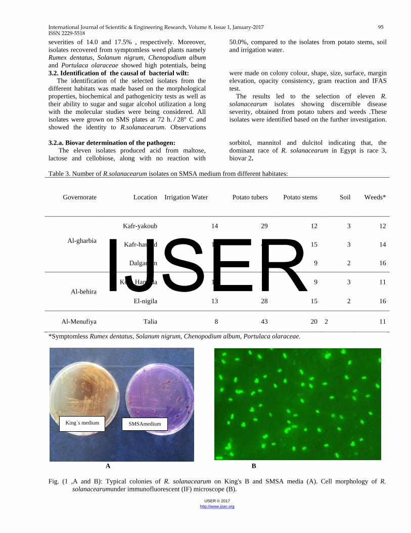
isolates: Isolation of R.solanacearum was made by using SMSA medium, from different habitats collected from Al-gharbia, Al-behira and Al-minufiya governorates. Table (3) shows 420 colonies developed typical colonies of R.solanacearum on the selective medium (SMSA) from irrigation water, potato tubers, potato stems, soil and weeds. Moreover, IFAS testing was carried out on colonies developed to determine the morphological shape. The cells showed short rod shaped morphology and stained evenly as bright green fluorescent were considered (Fig.1, A and B).The greatest numbers of typical colonies were recognized and selected from potato tubers while, the lowest number was selected from soil samples. On the other hand, approximately similar numbers of typical colonies were selected from potato stems and weeds (Table, 4). The pathogenic potential of the collected isolates (420 isolates) was evaluated for producing wilt to tomato seedlings, 7 days after inoculation under greenhouse conditions presented by disease incidence %. Fig.(2) shows that tuber isolates caused 100% infection to tomato with corresponding wilt severity of 60.0% compared to 13.5% for the potato stem isolates. Soil and irrigation water isolates showed disease
IJSER
International Journal of Scientific & Engineering Research, Volume 8, Issue 1, January-2017 95 ISSN 2229-5518
IJSER © 2017 http://www.ijser.org
severities of 14.0 and 17.5% , respectively. Moreover, isolates recovered from symptomless weed plants namely Rumex dentatus, Solanum nigrum, Chenopodium album and Portulaca olaraceae showed high potentials, being
50.0%, compared to the isolates from potato stems, soil and irrigation water.
3.2. Identification of the causal of bacterial wilt: The identification of the selected isolates from the different habitats was made based on the morphological properties, biochemical and pathogenicity tests as well as their ability to sugar and sugar alcohol utilization a long with the molecular studies were being considered. All isolates were grown on SMS plates at 72 h. ̸ 28° C and showed the identity to R.solanacearum. Observations
were made on colony colour, shape, size, surface, margin elevation, opacity consistency, gram reaction and IFAS test. The results led to the selection of eleven R. solanacearum isolates showing discernible disease severity, obtained from potato tubers and weeds .These isolates were identified based on the further investigation.
3.2.a. Biovar determination of the pathogen: The eleven isolates produced acid from maltose, lactose and cellobiose, along with no reaction with
sorbitol, mannitol and dulcitol indicating that, the dominant race of R. solanacearum in Egypt is race 3, biovar 2.
Table 3. Number of R.solanacearum isolates on SMSA medium from different habitates:
Governorate Location
Irrigation Water
Potato tubers Potato stems Soil Weeds*
Al-gharbia
Kafr-yakoub 14 29 12 3 12
Kafr-hashad 10 47 15 3 14
Dalgamon 9 17 9 2 16
Al-behira Kom Hamada 11 16 9 3 11
El-nigila 13 28 15 2 16
Al-Menufiya Talia 8 43 20 2 11
*Symptomless Rumex dentatus, Solanum nigrum, Chenopodium album, Portulaca olaraceae.
A B Fig. (1 ,A and B): Typical colonies of R. solanacearum on King's B and SMSA media (A). Cell morphology of R.
solanacearumunder immunofluorescent (IF) microscope (B).
SMSAmedium King´s medium
IJSER

International Journal of Scientific & Engineering Research, Volume 8, Issue 1, January-2017 96 ISSN 2229-5518
IJSER © 2017 http://www.ijser.org
Table 4. Number of samples, infected samples, selected colonies and number of positive IFAS test recovered from different
collected habitats.
* Symptomless Rumex dentatus, Solanum nigrum, Chenopodium album and Portulaca olaraceae
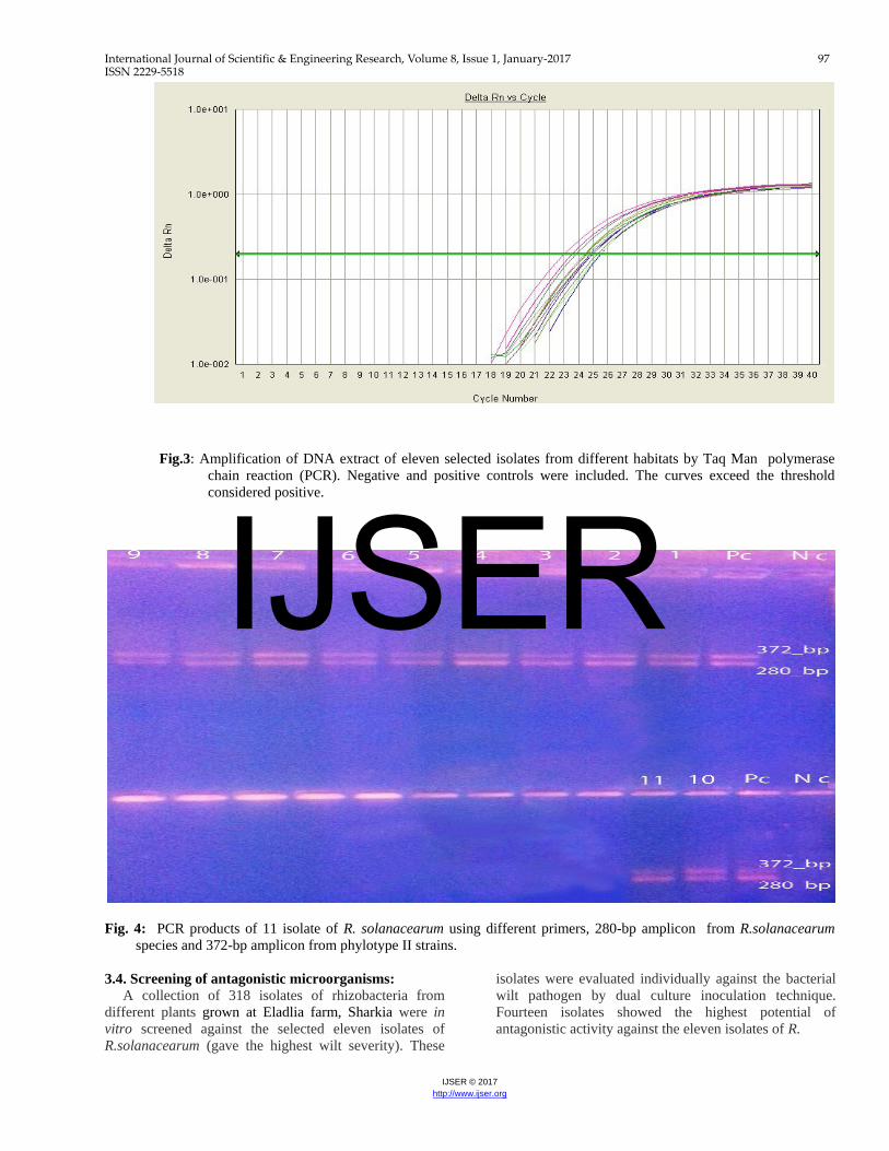
Fig. 2: Percentage of infection and wilt severity on tomato seedling produced by isolates of R. solanacearum collected from different sources. 3.3.b. Real-Time PCR assay: The RS primers and probe were employed to detect all biovars and races of R. solanacearum. Positive results were noticed with all tested isolates indicating that, the eleven strains are R. solanacearum biovar 2 race 3 (Fig. 3), equivalent to phylotype II ,sequevar I. 3.3.c. Phylotype analysis of R.solanacearum by
Multiplex- PCR:
Phylotype specific multiplex PCR revealed that all the eleven isolates of R. solanacearum belonged to phylotype II as 372- bp amplicon was observed with all tested isolates when Pmx-PCR products of these isolates were subjected to electrophoresis on 2% agarose gel (Fig., 4). These results indicating that, the dominant race of R. solanacearum in Egypt is race 3 (phylotype II).
Habitat No. of samples Infected samples No. of selected colonies No. of IF(+)
Potato tubers 60 60 180 180
Potato stems 42 30 80 80
Soil 24 15 15 15
Irrigation water 18 18 65 65
*Weeds 54 35 80 80
Total 198 158 420 420
IJSER
International Journal of Scientific & Engineering Research, Volume 8, Issue 1, January-2017 97 ISSN 2229-5518
IJSER © 2017 http://www.ijser.org
Fig.3: Amplification of DNA extract of eleven selected isolates from different habitats by Taq Man polymerase
chain reaction (PCR). Negative and positive controls were included. The curves exceed the threshold considered positive.
Fig. 4: PCR products of 11 isolate of R. solanacearum using different primers, 280-bp amplicon from R.solanacearum
species and 372-bp amplicon from phylotype II strains. 3.4. Screening of antagonistic microorganisms: A collection of 318 isolates of rhizobacteria from different plants grown at Eladlia farm, Sharkia were in vitro screened against the selected eleven isolates of R.solanacearum (gave the highest wilt severity). These
isolates were evaluated individually against the bacterial wilt pathogen by dual culture inoculation technique. Fourteen isolates showed the highest potential of antagonistic activity against the eleven isolates of R.
IJSER
International Journal of Scientific & Engineering Research, Volume 8, Issue 1, January-2017 98 ISSN 2229-5518
IJSER © 2017 http://www.ijser.org
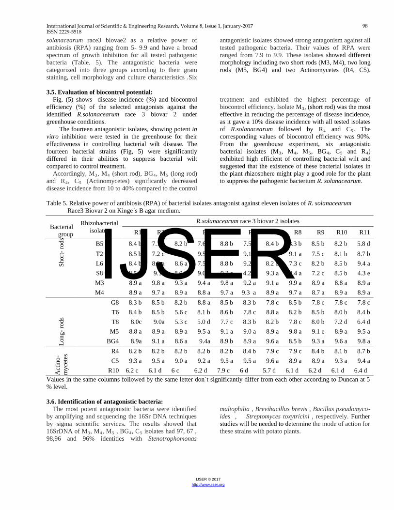
solanacearum race3 biovae2 as a relative power of antibiosis (RPA) ranging from 5- 9.9 and have a broad spectrum of growth inhibition for all tested pathogenic bacteria (Table. 5). The antagonistic bacteria were categorized into three groups according to their gram staining, cell morphology and culture characteristics .Six
antagonistic isolates showed strong antagonism against all tested pathogenic bacteria. Their values of RPA were ranged from 7.9 to 9.9. These isolates showed different morphology including two short rods (M3, M4), two long rods (M5, BG4) and two Actinomycetes (R4, C5).
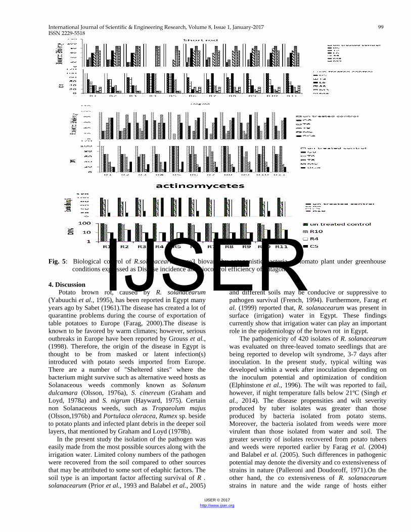
3.5. Evaluation of biocontrol potential: Fig. (5) shows disease incidence (%) and biocontrol efficiency (%) of the selected antagonists against the identified R.solanacearum race 3 biovar 2 under greenhouse conditions. The fourteen antagonistic isolates, showing potent in vitro inhibition were tested in the greenhouse for their effectiveness in controlling bacterial wilt disease. The fourteen bacterial strains (Fig, 5) were significantly differed in their abilities to suppress bacterial wilt compared to control treatment. Accordingly, MR3R, MR4R (short rod), BGR4R, MR5R (long rod) and RR4R, CR5R (Actinomycetes) significantly decreased disease incidence from 10 to 40% compared to the control
treatment and exhibited the highest percentage of biocontrol efficiency. Isolate MR3R, (short rod) was the most effective in reducing the percentage of disease incidence, as it gave a 10% disease incidence with all tested isolates of R.solanacearum followed by RR4R and CR5R. The corresponding values of biocontrol efficiency was 90%. From the greenhouse experiment, six antagonistic bacterial isolates (MR3R, MR4R, MR5R, BGR4R, CR5R and RR4R) exhibited high efficient of controlling bacterial wilt and suggested that the existence of these bacterial isolates in the plant rhizosphere might play a good role for the plant to suppress the pathogenic bacterium R. solanacearum.
Table 5. Relative power of antibiosis (RPA) of bacterial isolates antagonist against eleven isolates of R. solanacearum Race3 Biovar 2 on Kinge´s B agar medium.
Values in the same columns followed by the same letter don´t significantly differ from each other according to Duncan at 5 % level. 3.6. Identification of antagonistic bacteria: The most potent antagonistic bacteria were identified by amplifying and sequencing the 16Sr DNA techniques by sigma scientific services. The results showed that 16SrDNA of MR3R, MR4R, MR5R , BGR4R, CR5R isolates had 97, 67 , 98,96 and 96% identities with Stenotrophomonas
maltophilia , Brevibacillus brevis , Bacillus pseudomyco-ides , Streptomyces toxytricini , respectively. Further studies will be needed to determine the mode of action for these strains with potato plants.
Bacterial
group
Rhizobacterial isolates
R.solanacearum race 3 biovar 2 isolates
R1 R2 R3 R4 R5 R6 R7 R8 R9 R10 R11
Shor
t- ro
ds
B5 8.4 b 7.1 c 8.2 b 7.6 c 8.8 b 7.5 c 8.4 b 8.3 b 8.5 b 8.2 b 5.8 d T2 8.5 b 7.2 c 6.8 c 9.5 a 7.8 c 9.1 a 9.1 a 9.1 a 7.5 c 8.1 b 8.7 b L6 8.4 b 8.9 a 8.6 a 7.5 c 8.8 b 9.2 a 8.2 b 7.3 c 8.2 b 8.5 b 9.4 a S8 8.5 b 9.1a 8.0 b 9.0 a 9.2 a 4.2 d 9.3 a 9.4 a 7.2 c 8.5 b 4.3 e M3 8.9 a 9.8 a 9.3 a 9.4 a 9.8 a 9.2 a 9.1 a 9.9 a 8.9 a 8.8 a 8.9 a M4 8.9 a 9.7 a 8.9 a 8.8 a 9.7 a 9.3 a 8.9 a 9.7 a 8.7 a 8.9 a 8.9 a
Long
- rod
s
G8 8.3 b 8.5 b 8.2 b 8.8 a 8.5 b 8.3 b 7.8 c 8.5 b 7.8 c 7.8 c 7.8 c T6 8.4 b 8.5 b 5.6 c 8.1 b 8.6 b 7.8 c 8.8 a 8.2 b 8.5 b 8.0 b 8.4 b T8 8.0c 9.0a 5.3 c 5.0 d 7.7 c 8.3 b 8.2 b 7.8 c 8.0 b 7.2 d 6.4 d
M5 8.8 a 8.9 a 8.9 a 9.5 a 9.1 a 9.0 a 8.9 a 9.8 a 9.1 e 8.9 a 9.5 a BG4 8.9a 9.1 a 8.6 a 9.4a 8.9 b 8.9 a 9.6 a 8.5 b 9.3 a 9.6 a 9.8 a
Act
ino-
myc
etes
R4 8.2 b 8.2 b 8.2 b 8.2 b 8.2 b 8.4 b 7.9 c 7.9 c 8.4 b 8.1 b 8.7 b C5 9.3 a 9.5 a 9.0 a 9.2 a 9.5 a 9.5 a 9.6 a 8.9 a 8.9 a 9.3 a 9.4 a
R10 6.2 c 6.1 d 6 c 6.2 d 7.9 c 6 d 5.7 d 6.1 d 6.2 d 6.1 d 6.4 d
IJSER
International Journal of Scientific & Engineering Research, Volume 8, Issue 1, January-2017 99 ISSN 2229-5518
IJSER © 2017 http://www.ijser.org
Fig. 5: Biological control of R.solanacearum race3 biovar2 by antagonistic bacteria on tomato plant under greenhouse
conditions expressed as Disease incidence and biocontrol efficiency of antagonist. 4. Discussion Potato brown rot, caused by R. solanacearum (Yabuuchi et al., 1995), has been reported in Egypt many years ago by Sabet (1961).The disease has created a lot of quarantine problems during the course of exportation of table potatoes to Europe (Farag, 2000).The disease is known to be favored by warm climates; however, serious outbreaks in Europe have been reported by Grouss et al., (1998). Therefore, the origin of the disease in Egypt is thought to be from masked or latent infection(s) introduced with potato seeds imported from Europe. There are a number of "Sheltered sites" where the bacterium might survive such as alternative weed hosts as Solanaceous weeds commonly known as Solanum dulcamara (Olsson, 1976a), S. cinereum (Graham and Loyd, 1978a) and S. nigrum (Hayward, 1975). Certain non Solanaceous weeds, such as Tropaeolum majus (Olsson,1976b) and Portulaca oleracea, Rumex sp. beside to potato plants and infected plant debris in the deeper soil layers, that mentioned by Graham and Loyd (1978b). In the present study the isolation of the pathogen was easily made from the most possible sources along with the irrigation water. Limited colony numbers of the pathogen were recovered from the soil compared to other sources that may be attributed to some sort of edaphic factors. The soil type is an important factor affecting survival of R . solanacearum (Prior et al., 1993 and Balabel et al., 2005)
and different soils may be conducive or suppressive to pathogen survival (French, 1994). Furthermore, Farag et al. (1999) reported that, R. solanacearum was present in surface (irrigation) water in Egypt. These findings currently show that irrigation water can play an important role in the epidemiology of the brown rot in Egypt. The pathogenicity of 420 isolates of R. solanacearum was evaluated on three-leaved tomato seedlings that are being reported to develop wilt syndrome, 3-7 days after inoculation. In the present study, typical wilting was developed within a week after inoculation depending on the inoculum potential and optimization of condition (Elphinstone et al., 1996). The wilt was reported to fail, however, if night temperature falls below 21ºC (Singh et al., 2014). The disease propensities and wilt severity produced by tuber isolates was greater than those produced by bacteria isolated from potato stems. Moreover, the bacteria isolated from weeds were more virulent than those isolated from water and soil. The greater severity of isolates recovered from potato tubers and weeds were reported earlier by Farag et al. (2004) and Balabel et al. (2005). Such differences in pathogenic potential may denote the diversity and co extensiveness of strains in nature (Palleroni and Doudoroff, 1971).On the other hand, the co extensiveness of R. solanacearum strains in nature and the wide range of hosts either
IJSER
International Journal of Scientific & Engineering Research, Volume 8, Issue 1, January-2017 100 ISSN 2229-5518
IJSER © 2017 http://www.ijser.org
symptomatic or asymptomatic may be in part attributed to such preferential host effect that generates certain forms with different pathogenic potentials. Such observation may be of value in studying epidemiology of R. solanacearum as mentioned before by He et al., (1983), Farag et al., (2004) and Balabel (2006). The fourteen isolates of the rhizobacteria were evaluated as antagonists against the pathogen that provided an impression on different degrees of antagonism in vitro and in vivo under greenhouse conditions. Six out of these antagonists gave the highest efficient against pathogenic bacteria that appear in reduced disease incidence (%) and high biocontrol efficiency(%) on tomato plants. These antagonists are affiliated to different species identified as Streptomyces toxytricini, Stenotrophomonas maltophilia, Bacillus pseudomycoides and Brevibacillus brevis. With regard, Brevibacillus brevis, is a Gram positive spore-forming bacterium, studies showed that it had significant inhibitory potential on many animal and plant pathogens (Ge et al., 2009) and such as Ralstonia solanacearum (Che et al., 2012). On the other hand, this bacterium can secrete large amounts of secondary metabolites, as tyrocidine, gramicidin, gratisin (Tamaki et al., 1983), and edeine (Czajgucki et al., 2006). Moreover, this bacterium can secrete non ribosome peptides, which were shown to effectively inhibit the growth of R.solanacearum by disturbing cell membrane integrity (Chena et al., 2012). Therefore, B. brevis may be considered as a good biocontrol agent against R. solanacearum. Previous studies had proved the potential
of actinomycetes as effective biocontrol for Ralstonia solanacearum. The inhibition of this pathogen may be due to the emission of volatile secondary metabolites secreted by Actinomycetes (Rado et al., 2015). Stenotrophomonas maltophilia is a gram - negative bacterium, Initially classified as Pseudomonas maltophilia, it was also grouped in the genus Xanthomonas before eventually becoming the type species of the genus Stenotrophomonas (Palleroni and Bradbury 1993), Denton and Kerr1998). It is an effective biocontrol agent for the control of some fungal and oomycetous plant diseases (Dal Bello et al., 2002 and Berg et al., 2005). On the other hand, Messiha et al., (2007) reported that, this bacterium may be useful for control of brown rot bacterium. S. maltophilia produces various antibiotics, for example, maltophilin, a macrocyclic lactam antibiotic, which has antifungal activity, but is inactive against Gram-positive and Gram-negative bacteria (Jakobi et al., 1996). 5. Conclusion: The present study showed that R. solanacearum race3 biovar2 is the dominant pathogenic bacterium under Egyptian conditions causing brown rot .PGPR isolates antagonistic to the causal bacterium could be isolated from local fields and could be used to control the deleterious effect of R.solanacearum .Thus, it is hoped that the obtained inoculants antagonists will manage the bacterial wilt disease.
6. References [1] Abo-Elyousr, K., Ebrahim, Y.E and Balabel, N.M.
(2012). Induction of disease defensive enzymes in response to treatment with acibenzolar-S-methyl (ASM) and Pseudomonas fluorescens PF2 and inoculation with R.solanacearum race 3, biovar2 (phylotype II). J. of Phytopathol., 160: 382-389.
[2] Ahmed, F.; Ahmed, I. and Khan, M.S. (2008). Screening of free-living rhizosphereric bacteria activities. Microbiol. Res., 163:173-181.
[3] Aliye, N.; Fininsa, C. and Hiskias, Y. (2008) Evaluation of rhizosphere bacterial antagonists for their potential to bioprotect potato (Solanum tuberosum) against bacterial wilt (Ralstonia solanacearum). Biol. Cont., 47:282–288.
[4] Ausubell, F.M.; Brent, R.; Kingston, R.E.; Moore, D.D.; Seidman, J.G.; Smith, J.A. and Struhl, K. (1987). Current Protocols in Molecular Biology, Greene Publishing Associates/Wiley Interscience, New York.
[5] Balabel, N., M. (2006). Persistence of R. solanacearum (Syn. Pseudomonas solanacearum) in Different Habitats in Egypt (Ph. D. Thesis).
Department of Microbiology, Fac. Of Agric. Ain Shams Univ.
[6] Balabel, N. M.; Eweda, W. E.; Mostafa, M.I. and Farag, N.S. (2005). Some epidemiology aspects of Ralstonia solanacearum. Egypt. J. Agric. Res., 83(4) 1547.
[7] Berg, G.; Zachow, C.; Lottmann, J.; GÖTZ, M.; COSTA, R. and Smalla, K. (2005). Impact of plant species and site on rhizosphere-associated fungi antagonistic to Verticillium dahlia Kleb. Appl. and Environ. Microbiol., 71 (8): 4203-4213.
[8] Buddenhagen, I.W. and Kelman, A. (1964). Biological and physiological aspects of bacterial wilt caused by Ps. solanacearum. Ann. Rev. Phytopathol., 2: 203-230.
[9] Che, J.M.; Chen, Z.; Shi, H. and Liu, B. (2012). Analysis of functional components from Brevibacillus brevis FJAT-0809-GLX by Gas chromatography/mass spectrometry (GC/MSD). Fujian J. Agric. Sci., 27:1106–1111.
[10] Chena, W.; Wangb, Y.; Lib, D.; Lin, L.; Xiaob, Q. and Zhoua Q. (2012). Draft genome sequence of Brevibacillus brevis strain X23, a biocontrol agent
IJSER
International Journal of Scientific & Engineering Research, Volume 8, Issue 1, January-2017 101 ISSN 2229-5518
IJSER © 2017 http://www.ijser.org
against bacterial wilt. J. Bacteriol., 194:6634–6635. http://dx.doi.org/10.1128/JB.013 12-12.
[11] Czagucki, Z.; Andruszkiewicz, R. and Kamys, Z.W. (2006). Structure activity relationship studies on the antimicrobial activity of novel edin A and D analogues. J. Pept.Sci.,12:653-662.
[12] Dal Bello, G.; Mo´naco, C. and Simo´n, M. (2002). Biological control of seedling blight of wheat caused Fusariumgraminearum with beneficial rhizosphere microorganisms. World J. of Microbiol, & Biotechnol., 18:627-636.
[13] Denton, M. and Kerr, K.G. (1998). Microbiological and clinical aspects of infection associated with stenotrophomonas maltophilia . Clin. Microbiol. Rev., 11:57-80.
[14] Dowson, W.J. (1957). Plant disease due to bacteria. Second edition. Cambridge at the University Press, PP. 232.
[15] Duncan, D.B (1955). Multiple range and F.test. Biometrics, 11:1-24.
[16] Elphinstone, J.G.; Hennessy, J., Wilson, J.K. and Stead, D.E. (1996). Sensitivity of different methods for the detection of Ralstonia solanacearum in potato tuber extracts. Bulletin OEEP/EPPO Bulletin, 26: 663-678.
[17] Farag, N.S. (2000). Spotlights on potato brown rot in Egypt. Proc. 9th Congress of the Egypt. Phytopathol. Soc., May, 405-408.
[18] Farag, N.S.; Stead, D.E. and Janse, J.D. (1999). Ralstonia(Pseudomonas) solanacearum race 3, biovar 2, in surface (irrigation) water in Egypt. J. Phytopathol., 147: 485-487.
[19] Farag, N.S.; Lashin, S.M.; Abdel, R.S.; Shatta, H.M. and Seif-Elyazal A.M. (1982). Antibiotics and control of potato black leg and brown rot diseases. Agricultural Research Review, 60,149-166.
[20] Farag, N.S.; Eweda, W. E.; Mostafa, M.I. and Balabel, N. M. (2004). Preliminary observations on the bacteriology and pathology of Ralstonia solanacearum. Egypt. J. Agric. Res., 82 (4): 1519-1523.
[21] French, E.R. (1994). Strategies for integrated control of bacterial wilt of potatoes. In: A.C. Hayward & G.L. Hartman (Eds.), Bacterial Wilt: The disease and its causative agent. Ps. solanacearum. CAB International, Wallingford, Oxon, U.K, PP. 199-208.
[22] Ge, C.B.; Liu, B.; Lane, J.L.; Huang, S.F. and Zhu,Y.J. (2009). The antibacterial activity research of biocontrol bacteria JK-2 on Fusarium oxysporium., Fujian J.Agric. Sci., 24:29-34.
[23] Govindarajan, M.; Kwon, S.W. and Wean, H.Y. (2007). Isolation, molecular characterization and growth promoting activities of endophytic sugarcane diazotroph klebsiella sp.GR9.W.J. Microbiol. Biotechnol., 23:997-1006.
[24] Graham, J. and Loyd, A.B. (1978 a). Solanum cinereum R.Br., a wild host of Ps. solanacearum biotype II. J. Aust. Inst. Agric. Sci., 44: 124-126.
[25] Graham, J. and Loyd, A.B. (1978 b). An improved indicator plant method for detection of Pseudomonas solanacearum race 3 in soil. Plant Disease Reporter 62: 35-37.
[26] Gyaneshwar, James, P.; Mathan, E.K., Reddy, N., Reihold-Hurek, P.M. and Ladha, V.J.K. (2001).Endophytic colonization of rice by diazotrophic strains of Serratia marcescens. J. of Bacteriol., 183:2634-2645.
[27] Hanson, P.M; Wang, J.F.; Licardo,O.; Hanudin, Mah, S.Y.; Hartman, G.L.; Lin Y.C. and Chen, J.T., (1996).Variable reactions of tomato lines to bacterial wilt evaluated at several location in south east Asia, Hort. Sci., 31: 143-146.
[28] Hayward, A.C. (1975). Biotypes of Psesudomonas solanacearum in Australia. Australian Plant Pathology Society Newsletter, 2: 9-11.
[29] Hayward, A.C., El-Nashaar H.M.; Nydegger, U. and Lindo, L.De (1990). Variation in nitrate metabolism in biovars of Pseudomonas solanacearum. J. of Appl. Bacteriol., 69; 269-280.
[30] Hayward, A.C. (1991). Biology and epidemiology of bacterial wilt caused by Pseudomonas solanacearum. Ann. Rev. of Phytopathol., 29: 65-87.
[31] He, L.Y.; Sequeira, L. and Kelman, A. (1983). Characteristics of Pseudomonas solanacearum from China. Plant Dis., 67: 1357-1361.
[32] Hsu, S.T. (1991). Ecology and control of Pseudomonas solanacearum in Taiwan. Plant Protec. Bullet. Taipei, 33 (1): 72-79; 41 ref.
[33] Ibrahim, M.E.K.; Mehiar F.F. and El-Gremi S.M. (1987). Biologcal control of blackleg, soft-rot and common scab of potato by bacterial antagonists. J. Agric. Res. Tanta Univ., 13 (1): 1 – 15.
[34] Jakobi, R.; Chen, C.J., Tuazon, P.T. and Traugh. J.A. (1996) Molecular cloning and sequencing of the cytostatic G protein-activated protein kinase PAK Int. J. Biol.Chem., 271(11):6206-11. Journal Article.
[35] Janse, J.D. (1988). A detection method for Pseudomonas solanacearum in symptomless potato tubers and some data on its sensitivity and specificity. EPPO Bulletin, 18: 343-351.
[36] Kelman, A. (1954). The relationship of pathogenicity in Pseudomonas solanacearum to colony appearance on a tetrazolium medium. Phytopathology, 44: 693-695.
[37] King, E.O.; Ward, M.K. and Raney, D.E. (1954). Two simple media for the demonstration of pyocyanin and fluorescin. J. Lab. Clin. Med., 44: 301-307.
[38] Lane, D.J. (1991) .16S/23S rRNA Sequencing. in Nucleic Acid Techniques in Bacterial Systematics,
IJSER
International Journal of Scientific & Engineering Research, Volume 8, Issue 1, January-2017 102 ISSN 2229-5518
IJSER © 2017 http://www.ijser.org
eds Stacke brandt E, Good fellow M (Wiley, New York), pp 115–175.
[39] Leben, C.; Daft, G.C.and Schmetthenner, A.F. (1968). Bacterial blight of soybeans: Population levels of Pseudomonas glycinea in relation to symptom development. Phytopathology, 58: 1143-1146.
[40] Li, Q.; Li, Y., Zhu, X. and Cai, B. (2008). Isolation and characterization of atrazine-degrading Arthrobacter sp. Ad26 and use of this strain in bioremediation of contaminated soil. J. Environ. Sci., 20(10): 1226-1230.
[41] Messiha, N.; van Diepeningen, A.; Farag, N.; Abdallah, S.; Janse, J.and van Bruggen, A. (2007). Stenotrophomonas maltophilia: a new potential biocontrol agent of Ralstonia solanacearum, causal agent of potato brown rot. Eur. J. Plant Pathol., 118:211–225.
[42] Mortensen, C.N. (1999).Seed-Borne Bacterial
Diseases. Hellerup , Copenhagen, Danish Government Institute of Seed Pathology for Developing Countries, pp 86.
[43] Myint, L. and Ranamukhaarachchi, S.L. (2006). Biocontrol potential of Pseudomonas fluorescens against bacterial wilt of Brinjal and its possible plant growth promoting effects. Inter. J. of Agric. and Biology., 8( 5): 657 – 660
[44] Norman, D.J.; Chen, J.; Yuen, J.M.F.; Mangravita, A.; Novo Byrne, D. and Walsh, L. (2006). Control of bacterial wilt of geranium with phosphorous acid. Plant Dis., 90, 798-802.
[45] Olsson, K. (1976a). Experience of brown rot caused by Ps. solanacearum (Smith) in Sweden. Bulletin OEPP/EPPO Bulletin, 6: 199-207.
[46] Olsson, K. (1976 b). Over wintering of Ps. solanacearum in Seweden, In: Sequeir, L.; Kelman, A. (eds.), Proceedings of International Planting Conference Workshop on Ecology and Control of Bacterial Wilt. Raleigh, North Carolina, USA, North Carolina State University, 105-109.
[47] Opina, N.F.; Tavner, G.; Halloway, J.F.; Wang, T.H.L.R.; Maghirang, M.; Fegan, A.C.; Hayward, V.; Krishnapillai, W.F.; Hong, B.W. and Holloway, J.N. (1997). A novel method for development of species and strain - specific DNA probes and PCR primers for identifying Burkholderia solanacearum (formerly Ps. solanacearum). As.Pac.J.Mol. Biol. Biotechnol., 5:19-33.
[48] Palleroni, N.J.1. and Bradbury, J.F. (1993). Stenotrophomonas, a new bacterial genus for Xanthomonas maltophilia (Hugh 1980) Swings et al., 1983. Int. J. Syst. Bacteriol. Jul., 43(3):606-9.
[49] Palleroni, N.J. and Doudoroff, M. (1971). Phenotypic characterization and deoxy ribonucleic acid homologies of Psedomonas solanacearum. J. of Bacteriol., 107: 690-696.
[50] Prior, P.; Beramis, M.; Clairon, M.; Quiquampoix, H.; Robert, M. and Schmit, J. (1993). Contribution to integrated control against bacterial wilt in different pedoclimatic situations: Guadeloupe experience. In: G.L. Hartman and A.C. Hayward (Eds.), Bacterial Wilt: Proceedings of an International Conference held at Kaohsiung Taiwan, 28-31 October 1992. ACIAR Proceedings No. 45, Canberra, Australia, PP. 294-304.
[51] Rado, R.; Onja A. and Tokiniaina, R. (2015). Biological Properties of Actinomycetes Isolated from Marine Sponges in Madagascar. J .of Advan. Lab. Res. in Biol., 4,(1)., January.
[52] Saad, Maryan, K.Y.(2016).Different strategies to control brown rot disease . Ph.D. Thesis., Fac. Agric., Cairo Univ.
[53] Sabet, K.A. (1961). The occurrence of bacterial wilt of potatoes caused by Pseudomonas solanacearum (E.F. Smith) in Egypt. Tech. Bul., 1-10, Extension Dept., Min. Agric. Egypt.
[54] Saddler, G.S. (2005). Management of bacterial wilt disease. In: Bacterial wilt disease and the Ralstonia solanacearum species complex. C.Allen, P. Prior and A.C. Hayward (eds.). American Phytopatholocial Society, St. Paul MN.
[55] Sagar,V.; Gurjar, M.S.; Jeevalatha, A.; Bakade, R.R.; Chakrabarti, S.K.; Arora, R.K. and Sharma S., (2014). Phylotype analysis of Ralstonia solanacearum strains causing potato bacterial wilt in Karnataka in India. Afr. J. of Microbiol. Res., 8(12):1277-1281.
[56] Singh, D.; Yadav, D.K.; Sinha, S. and Choudhary, G. (2014). Effect of temperature, cultivars, injury of root and inoculums load of Ralstonia solanacearum to cause bacterial wilt of tomato. Arch. of Phytopathol. and Plant Protec., 47(13):1574-1583.
[57] Song, W.; Zhou L.; Yang C., Cao X.; Zhang L. and Liu, X. (2004). Tomato Fusarium wilt and its chemical control strategies in a hydroponic system. Crop Protec., 23: 243-247.
[58] Tamaki, M.; Takimoto, M.; Sofuku, S. and Muramatsu, I. (1983). Synthetic studies on gratisin. II. J. Antibiot. (Tokyo)., 36:751–752.
[59] Waksman, S.A. (1961).The Actinomyces .Vol. classification and description of genera and species. Williams and Wilkins coman Y, Baltimore,U.S.A., P.430.
[60] Wang,J.F.; Hanson, P. and Barnes J.A. (1998). Worldwide evaluation of an international set of resistance sources of bacterial wilt in tomato. In: Bacterial Wilt disease: Molecular and Ecological Aspects. P. Prior, C. Allen and J. Elphinstone, eds. Springer Verleg, Berlin Germany: 269-275. –92.
[61] Weller, D. (1988). Biological control of soil borne plant pathogens in the Rhizosphere with Bacteria. Ann. Rev. Phytopathol., 26, 379-407.
IJSER
International Journal of Scientific & Engineering Research, Volume 8, Issue 1, January-2017 103 ISSN 2229-5518
IJSER © 2017 http://www.ijser.org
[62] Weller, S.A.; Elphinstone, J.G.; Smith, N.C.; Boonham, N and Stead, D.E. (2000). Detection of Ralstonia solanacearum strains with a Quantitative, Multiplex, Real-Time, Fluorogenic PCR (Taq Man) assay. Appl. and Environ. Microbiol., 66 (7): 2853-2858.
[63] Winstead, N.N. and Kelman, A. (1952). Inoculation techniques for evaluating resistance of Pseudomonas solanacearum. Phytopathology, 42: 628-634.
[64] Xu, J.; Pan, Z.C.; Prior, P.; Xu, J.S., Zhang, Z.; Zhang, H.; Zhang, L.Q.; He, L.Y. and Feng, J.
(2009) Genetic diversity of Ralstonia solanacearum strains from China. Eur .J. Plant Pathol., 125: 641–653.
[65] Yabuuchi, E.; Kosaka, Y.;Yano, I.; Hotta, H. and Nishiuchi,Y. (1995). Transfer of two Burkholderia and Alcaligenes species. To Ralstonia gen. nov.; Proposal of Ralstonia picketii (Ralston, Palleroni and Doudoroff 1973) Comb. nov.; Ralstonia solanacearum (Smith, 1896) Comb. nov. and Ralstonia eutropha(Davis 1969) Comb. nov. Microbial. Immunol., 39 (11): 897-904.
IJSER
International Journal of Scientific & Engineering Research, Volume 8, Issue 1, January-2016 104 ISSN 2229-5518
IJSER © 2017 http://www.ijser.org
IJSER