Emerging Coxsackievirus A6 Causing Hand, Foot and Mouth ... · Louise Thwaites, H. Rogier van...
14
Hand, foot and mouth disease (HFMD) is a major public health issue in Asia and has global pandemic potential. Cox- sackievirus A6 (CV-A6) was detected in 514/2,230 (23%) of HFMD patients admitted to 3 major hospitals in southern Vietnam during 2011–2015. Of these patients, 93 (18%) had severe HFMD. Phylogenetic analysis of 98 genome sequences revealed they belonged to cluster A and had been circulating in Vietnam for 2 years before emergence. CV-A6 movement among localities within Vietnam occurred frequently, whereas viral movement across international borders appeared rare. Skyline plots identified fluctuations in the relative genetic diversity of CV-A6 corresponding to large CV-A6–associated HFMD outbreaks worldwide. These data show that CV-A6 is an emerging pathogen and emphasize the necessity of active surveillance and under- standing the mechanisms that shape the pathogen evolu- tion and emergence, which is essential for development and implementation of intervention strategies. H and, foot and mouth disease (HFMD) is an emerg- ing infection that has overwhelmed countries in the Asia–Pacific region over the past 2 decades. The outbreak in Sarawak, Malaysia, in 1997 caused 2,628 reported cases and 29 deaths and marked the start of explosive regional HFMD outbreaks in subsequent years. On average, >1 million cases have been recorded in China annually since 2008 (1). In Vietnam, the average annual incidence is ≈80,000 cases; an epidemic peak occurred during 2011– 2012, resulting in >200,000 cases and >200 deaths (2). HFMD is caused by enterovirus A (genus Enterovi- rus, family Picornaviridae), but the epidemic patterns un- til now have been punctuated by the frequent replacement of dominant pathogens between enterovirus serotypes over time. Enterovirus A71 (EV-A71) and coxsackievirus A16 (CV-A16) have been regarded as the major causes of HFMD (3). CV-A6 was isolated in the United States in 1949 (4) and has steadily become one of the main viruses causing HFMD outbreaks in Europe, America, and Asia, including China, Japan, Taiwan, and Thailand (3,5–10). Unlike EV-A71, for which the (sub)genogroup designa- tion has been well established (11), but similar to other coxsackieviruses (CV-A16 and CV-A10), CV-A6 is ar- bitrarily divided into several phylogenetic clusters or lin- eages, from cluster A to F (12) or lineage A to E (E1 and E2) (3). Cluster A/lineage E2 is distributed worldwide and has frequently been detected in recent outbreaks. We use the term cluster in this article. Despite the public health burden of HFMD, no antivi- ral drug has been clinically proven effective. A vaccine for EV-A71 has recently been licensed in China only (13), and CV-A16 vaccines (either monovalent or EV-A71/CV-A16 bivalent forms) are under development (14,15). The emergence of CV-A6 has further challenged the development of intervention strategies, including vaccines, to reduce the burden of HFMD (13). It also emphasizes the need to better understand the molecular evolution of this emerging pathogen, which is essential for development of an effective CV-A6 vaccine in the future (16). However, few studies from endemic countries have documented the longitudinal evolution of CV-A6 (5,17–19). In this study, we applied next-generation sequencing to obtain whole-genome sequences for CV-A6 strains sampled from Emerging Coxsackievirus A6 Causing Hand, Foot and Mouth Disease, Vietnam Nguyen To Anh, Le Nguyen Truc Nhu, Hoang Minh Tu Van, Nguyen Thi Thu Hong, Tran Tan Thanh, Vu Thi Ty Hang, Nguyen Thi Han Ny, Lam Anh Nguyet, Tran Thi Lan Phuong, Le Nguyen Thanh Nhan, Nguyen Thanh Hung, Truong Huu Khanh, Ha Manh Tuan, Ho Lu Viet, Nguyen Tran Nam, Do Chau Viet, Phan Tu Qui, Bridget Wills, Sarawathy Sabanathan, Nguyen Van Vinh Chau, Louise Thwaites, H. Rogier van Doorn, Guy Thwaites, Maia A. Rabaa, Le Van Tan 654 Emerging Infectious Diseases • www.cdc.gov/eid • Vol. 24, No. 4, April 2018 Author affiliations: Oxford University Clinical Research Unit, Ho Chi Minh City, Vietnam (N.T. Anh, L.N.T. Nhu, H.M.T. Van, N.T.T. Hong, T.T. Thanh, V.T.T. Hang, N.T.H. Ny, L.A. Nguyet, B. Wills, S. Sabanathan, L. Thwaites, H.R. van Doorn, G. Thwaites, M.A. Rabaa, L.V. Tan); Hospital for Tropical Diseases, Ho Chi Minh City (T.T.L. Phuong, P.T. Qui, N.V.V. Chau); Children’s Hospital 1, Ho Chi Minh City (L.N.T. Nhan, N.T. Hung, T.H. Khanh); Children’s Hospital 2, Ho Chi Minh City (H.M. Tuan, H.L. Viet, N.T. Nam, D.C. Viet); University of Oxford, Oxford, United Kingdom (L. Thwaites, H.R. van Doorn, G. Thwaites, M.A. Rabaa) DOI: https://doi.org/10.3201/eid2404.171298
-
Upload
nguyenthuy -
Category
Documents
-
view
215 -
download
0
Transcript of Emerging Coxsackievirus A6 Causing Hand, Foot and Mouth ... · Louise Thwaites, H. Rogier van...

Hand, foot and mouth disease (HFMD) is a major public health issue in Asia and has global pandemic potential. Cox-sackievirus A6 (CV-A6) was detected in 514/2,230 (23%) of HFMD patients admitted to 3 major hospitals in southern Vietnam during 2011–2015. Of these patients, 93 (18%) had severe HFMD. Phylogenetic analysis of 98 genome sequences revealed they belonged to cluster A and had been circulating in Vietnam for 2 years before emergence. CV-A6 movement among localities within Vietnam occurred frequently, whereas viral movement across international borders appeared rare. Skyline plots identified fluctuations in the relative genetic diversity of CV-A6 corresponding to large CV-A6–associated HFMD outbreaks worldwide. These data show that CV-A6 is an emerging pathogen and emphasize the necessity of active surveillance and under-standing the mechanisms that shape the pathogen evolu-tion and emergence, which is essential for development and implementation of intervention strategies.
Hand, foot and mouth disease (HFMD) is an emerg-ing infection that has overwhelmed countries in the
Asia–Pacific region over the past 2 decades. The outbreak in Sarawak, Malaysia, in 1997 caused 2,628 reported cases and 29 deaths and marked the start of explosive regional HFMD outbreaks in subsequent years. On average, >1
million cases have been recorded in China annually since 2008 (1). In Vietnam, the average annual incidence is ≈80,000 cases; an epidemic peak occurred during 2011–2012, resulting in >200,000 cases and >200 deaths (2).
HFMD is caused by enterovirus A (genus Enterovi-rus, family Picornaviridae), but the epidemic patterns un-til now have been punctuated by the frequent replacement of dominant pathogens between enterovirus serotypes over time. Enterovirus A71 (EV-A71) and coxsackievirus A16 (CV-A16) have been regarded as the major causes of HFMD (3). CV-A6 was isolated in the United States in 1949 (4) and has steadily become one of the main viruses causing HFMD outbreaks in Europe, America, and Asia, including China, Japan, Taiwan, and Thailand (3,5–10). Unlike EV-A71, for which the (sub)genogroup designa-tion has been well established (11), but similar to other coxsackieviruses (CV-A16 and CV-A10), CV-A6 is ar-bitrarily divided into several phylogenetic clusters or lin-eages, from cluster A to F (12) or lineage A to E (E1 and E2) (3). Cluster A/lineage E2 is distributed worldwide and has frequently been detected in recent outbreaks. We use the term cluster in this article.
Despite the public health burden of HFMD, no antivi-ral drug has been clinically proven effective. A vaccine for EV-A71 has recently been licensed in China only (13), and CV-A16 vaccines (either monovalent or EV-A71/CV-A16 bivalent forms) are under development (14,15).
The emergence of CV-A6 has further challenged the development of intervention strategies, including vaccines, to reduce the burden of HFMD (13). It also emphasizes the need to better understand the molecular evolution of this emerging pathogen, which is essential for development of an effective CV-A6 vaccine in the future (16). However, few studies from endemic countries have documented the longitudinal evolution of CV-A6 (5,17–19). In this study, we applied next-generation sequencing to obtain whole-genome sequences for CV-A6 strains sampled from
Emerging Coxsackievirus A6 Causing Hand, Foot and Mouth
Disease, VietnamNguyen To Anh, Le Nguyen Truc Nhu, Hoang Minh Tu Van, Nguyen Thi Thu Hong, Tran Tan Thanh,
Vu Thi Ty Hang, Nguyen Thi Han Ny, Lam Anh Nguyet, Tran Thi Lan Phuong, Le Nguyen Thanh Nhan, Nguyen Thanh Hung, Truong Huu Khanh, Ha Manh Tuan, Ho Lu Viet, Nguyen Tran Nam,
Do Chau Viet, Phan Tu Qui, Bridget Wills, Sarawathy Sabanathan, Nguyen Van Vinh Chau, Louise Thwaites, H. Rogier van Doorn, Guy Thwaites, Maia A. Rabaa, Le Van Tan
654 Emerging Infectious Diseases • www.cdc.gov/eid • Vol. 24, No. 4, April 2018
Author affiliations: Oxford University Clinical Research Unit, Ho Chi Minh City, Vietnam (N.T. Anh, L.N.T. Nhu, H.M.T. Van, N.T.T. Hong, T.T. Thanh, V.T.T. Hang, N.T.H. Ny, L.A. Nguyet, B. Wills, S. Sabanathan, L. Thwaites, H.R. van Doorn, G. Thwaites, M.A. Rabaa, L.V. Tan); Hospital for Tropical Diseases, Ho Chi Minh City (T.T.L. Phuong, P.T. Qui, N.V.V. Chau); Children’s Hospital 1, Ho Chi Minh City (L.N.T. Nhan, N.T. Hung, T.H. Khanh); Children’s Hospital 2, Ho Chi Minh City (H.M. Tuan, H.L. Viet, N.T. Nam, D.C. Viet); University of Oxford, Oxford, United Kingdom (L. Thwaites, H.R. van Doorn, G. Thwaites, M.A. Rabaa)
DOI: https://doi.org/10.3201/eid2404.171298

Emerging Coxsackievirus A6, Vietnam
primary, secondary, and tertiary referral hospitals in Ho Chi Minh City, Vietnam, during 2011–2015. To investi-gate the molecular evolution and recent spread of CV-A6, we performed phylogenetic and phylogeographic analysis on both a global scale and within the southern provinces of Vietnam.
Materials and Methods
Patients and Clinical SamplesWe obtained the clinical samples used in this study from patients enrolled in an HFMD research program in which outpatients and inpatients with all severities of disease were recruited (20). During August 2011–June 2013, we carried out the research program at the pediatric intensive care unit (PICU) of the Hospital for Tropical Diseases in Ho Chi Minh City, Vietnam. This PICU admitted only patients with severe HFMD (the clinical grading system is described in the online Technical Appendix, https://wwwnc.cdc.gov/EID/article/24/4/17-1298-Techapp1.pdf). In the subse-quent phase (July 2013–December 2015), we expanded patient enrollment to outpatient clinics, infectious disease wards, and PICUs in 3 major referral hospitals in Ho Chi Minh City (Children’s Hospital 1, Children’s Hospital 2, and Hospital for Tropical Diseases). We selected for analy-sis CV-A6–positive throat and rectal swab specimens with sufficient viral load (samples with real-time PCR crossing point values of <30 [21]) collected in viral transport me-dium from study participants. The real-time reverse tran-scription PCR (RT-PCR) methods used are described in the online Technical Appendix.
CV-A6 Whole-Genome Sequencing and Sequence AssemblyWe performed whole-genome sequencing of CV-A6 on the selected swabs with sufficient viral load using a previously described MiSeq-based approach (21). In brief, we pre-treated 110 µL of selected swab specimens in viral trans-port medium by a centrifugation step at 13,000 rpm for 10 min to remove host cells or large cellular components and followed this step with DNase treatment of the obtained supernatants. We then isolated viral nucleic acid (NA) us-ing a QIAamp viral RNA kit (QIAGEN, Hilden, Germany) and recovered it in 50 µL of the elution buffer (provided with the kit). We subjected 10 µL of the isolated NA to cDNA synthesis using a Super Script III kit (Invitrogen, Carlsbad, CA, USA) and FR26RV-Endoh primer (21). We then converted the cDNA to double-stranded DNA using exo-Klenow (Invitrogen) and preamplified the cDNA using Platinum PCR supermix (Invitrogen) and FR20RV primer (21). We then purified the PCR product and subjected it to library preparation using a Nextera XT DNA sample prep-aration kit (Illumina, San Diego, CA, USA). Finally, we
sequenced the product using MiSeq reagent kits (Illumina) in a MiSeq platform (Illumina) (21).
We performed whole-genome sequence assembly using the Geneious 8.1.5 software package (Biomatters, Auckland, New Zealand) with a reference-based mapping approach. This method involves the mapping of individual reads of each sample to a reference sequence and manual editing of the consensus.
Multiple Sequence Alignment, Recombination Detection, and Phylogenetic AnalysisWe performed multiple sequence alignment using MUS-CLE (multiple sequence comparison by log-expectation) (22), available in Geneious. For Vietnam sequences, we then calculated the percentages of sequence identities among them from the resulting multiple sequence align-ment files using Geneious.
We inferred recombination using a combination of methods (Chimera, GENECONV, Maxchi, Bootscan, and Siscan) within RDP4 (Recombination Detection Program version 4) (22) using the default settings with recombina-tion supported if >3 methods showed significant values (p<0.05) and reconfirmed findings by phylogenetic analy-sis. We then removed identified recombinants from further phylogenetic analysis.
To investigate the relationship between Vietnam CV-A6 strains and global strains downloaded from GenBank, we constructed maximum-likelihood trees for viral capsid protein 1 (VP1) and complete coding sequences (CDS) using IQ-TREE version 1.4.3 (23). The maximum likeli-hood phylogenetic analysis used the general time reversible (for CDS dataset) and Tamura-Nei 93 (for VP1 dataset) nucleotide substitution models with a gamma distributed among site rate variation (4 rate categories). We assessed support for individual nodes using a bootstrap procedure (10,000 replicates).
We analyzed the phylogeographic history of CV-A6 in Vietnam and worldwide using BEAST version 1.8.3 (https://github.com/beast-dev/beast-mcmc/releases/tag/v1.8.3). We performed this analysis for both complete CDS and VP1 sequences downloaded from GenBank (October 2016) and the sequences obtained from the present study. For GenBank sequences, we excluded all the partial se-quences, identical sequences, sequences with internal gaps, recombinant sequences, and sequences without sampling dates or locations. We then performed regression analysis implemented in TempEst (https://academic.oup.com/ve/article/2/1/vew007/1753488) to further exclude sequences with insufficient temporal signals. For global strains, 170 VP1 and 52 complete CDS sequences from China, Finland, France, India, Japan, Spain, Taiwan, and the United King-dom were included for analysis. For Vietnam sequences, we used 98 sequences. Southern provinces in Vietnam
Emerging Infectious Diseases • www.cdc.gov/eid • Vol. 24, No. 4, April 2018 655

RESEARCH
from where the viruses were sampled were grouped into 3 discrete locations: Ho Chi Minh City (from where about half of the HFMD cases from Vietnam have been reported), southeast provinces (Long An, Can Tho, Tien Giang, Kien Giang, Dong Thap, and Hau Giang provinces), and Mekong Delta provinces (Tay Ninh, Dong Nai, Binh Duong, Binh Phuoc, Ba Ria, and Vung Tau provinces). Small sample sizes from individual provinces precluded phylogeographic analyses at a finer spatial scale.
For all analyses, we used the general time reversible (24) (for the CDS dataset) and Tamura-Nei 93 (25) (for the VP1 dataset) nucleotide substitution models with a gamma distributed among site rate variation (4 rate categories) (as indicated by IQ-TREE), the strict molecular clock model, and a Bayesian skyline plot (10 groups). We employed a Bayesian Markov chain Monte Carlo framework (avail-able in BEAST) with 800 million steps and sampling ev-ery 80,000 steps. We assessed convergence using Tracer version 1.5 (http://tree.bio.ed.ac.uk/software/tracer/). We selected a burn-in threshold of 10% and accepted effective sample size values above 200. Maximum-clade credibility (MCC) trees were then summarized with TreeAnnotator (available in the BEAST package) and visualized in Fig-tree version 1.4.2 (http://tree.bio.ed.ac.uk/software/figtree).
To estimate the relative genetic diversity of CV-A6 over time, we analyzed CDS and VP1 sequences separately using the same Bayesian skyline method. We submitted the sequences of CV-A6 obtained in this study to the National Center for Biotechnology Information (GenBank accession nos. MF578282–MF578381).
Ethics ConsiderationsThe study was approved by the corresponding institutional review board of the local hospitals in Vietnam where pa-tients were enrolled: Children’s Hospital 1, Ho Chi Minh City; Children’s Hospital 2, Ho Chi Minh City; and Hos-pital for Tropical Diseases, Ho Chi Minh City. The study was also approved by the Oxford Tropical Research Ethics Committee and was performed in accordance with the ethics
standards noted in the 1964 Declaration of Helsinki and its later amendments, or comparable ethics standards.
Results
Baseline Characteristics of Patients with CV-A6 InfectionsDuring August 2011–December 2015, a total of 514 pa-tients with HFMD had specimens that tested positive for CV-A6, accounting for 23% of the HFMD study partici-pants who were enterovirus PCR positive (n = 2,230). We detected EV-A71 in 36% (812) of the patients, CV-A16 in 10% (240), and CV-A10 in 7% (164). Temporally, the detection rate of CV-A6 in HFMD patients increased from 6% in 2011 to 13% in 2012, 18% in 2013, 32% in 2014, and 29% in 2015 (Figure 1). Complete data on demograph-ics and clinical grades were available from 510/514 CV-A6 infected patients (Table). Although CV-A6–associated HFMD was mostly mild, 93 (18%) patients had grade 2b1, 2b2, or 3 HFMD (i.e., severe HFMD), accounting for 76/76 (100%) of patients with CV-A6 who were enrolled in the first phase of the study and 17/434 (4%) of patients with CV-A6 who were enrolled in the second phase of the study.
CV-A6 Whole-Genome SequencesFrom the 514 patients who had CV-A6, we subjected 131 swabs (97 throat swabs and 34 rectal swabs) with suffi-cient viral load to whole-genome sequencing. Of these, we successfully recovered 100 nearly complete or com-plete coding sequences (80%). We identified evidence of recombination in 2 CV-A6 sequences (data not shown) and removed these 2 sequences from subsequent phylo-genetic analyses.
Phylogeny and PhylogeographyPhylogenetic analyses of 282 VP1 sequences of global strains, including 98 from Vietnam, showed that CV-A6 was grouped into 6 genetic clusters, in agreement with a previous report (12). All of the Vietnam CV-A6 isolates
656 Emerging Infectious Diseases • www.cdc.gov/eid • Vol. 24, No. 4, April 2018
Figure 1. Temporal distribution of PCR-positive hand, foot and mouth disease cases and detection rates of CV-A6 during 2011–2015, Vietnam. CV, coxsackievirus.

Emerging Coxsackievirus A6, Vietnam
belonged to cluster A (Figure 2), showing nucleotide iden-tity of 91.8%–100% and amino acid identity of 98.6%–100%. This genogroup consists of viruses sampled from various geographic locations worldwide, whereas the CV-A6 strains from Vietnam fell within a viral lineage consist-ing of CV-A6 strains from China, India, Japan, Taiwan, and the United Kingdom.
Delineating the dispersal of an emerging pathogen be-tween geographic locations and within endemic countries is critical for outbreak control. In-depth phylogeographic analysis for all 13 discrete provinces from where the pa-tients came was uninformative because of the small sample sizes from some provinces. When we conducted the anal-ysis for 3 main discrete geographic locations, the results revealed that CV-A6 spread widely within southern Viet-nam during the sampling period (Figure 3, panel A; online Technical Appendix Figure 1, panel A). This finding is in contrast with what has been observed at the international level, at which movement of CV-A6 between endemic countries appears rare (Figure 3, panel B; online Technical Appendix Figure 1, panel B).
Estimation of the time to the most recent common an-cestor in the phylogeny including only strains from Viet-nam suggested that the CV-A6 lineage that circulated in Vietnam during this time period began circulating within the country by 2010. This estimation is consistent between VP1-based and complete CDS-based analyses, which show time to most recent common ancestor estimates of November 2010 (95% CI May 2010–March 2011) and May 2010 (95% CI February 2010–August 2010), respectively. These estimates suggest that CV-A6 was being transmit-ted in Vietnam for at least 2 years before becoming the
dominant cause of HFMD in 2012. The nucleotide substitu-tion rate of VP1 sequences was estimated to be 7.42 × 10⁻³ (95% CI 6.1126 × 10⁻³ to 8.722 × 10⁻³) and the nucleo-tide substitution rate of complete CDS was estimated to be 4.556 × 10⁻³ (95% CI 4.209 × 10⁻³ to 4.913 × 10⁻³) substitu-tions per site per year.
CV-A6 DemographicsBecause of the relatively small number of CV-A6 whole genome sequences available in GenBank, we first assessed the global demographic history of the lineage using skyline plot analysis on VP1 sequences. The Bayesian VP1-based skyline plot of genogroup A viruses sampled across the world revealed fluctuations in the relative genetic diversity of CV-A6 from 2008 onward, especially during 2010–2012 (Figure 4, panel A), highlighting notable changes in viral diversity. This phenomenon coincided with CV-A6 out-breaks reported worldwide, including the 2008 Finland outbreak and major outbreaks affecting Asia, Europe, and the United States in subsequent years.
Bayesian skyline plot analysis for the CDS of the Viet-namese viruses alone did not suggest any major changes in relative genetic diversity during 2011–2015 compared with a global scale (Figure 4, panel B). Similar results were obtained when the analyses were done for VP1 sequences of the Vietnam strains and CDS of global strains (online Technical Appendix Figure 2).
DiscussionWe report the evolutionary process of CV-A6 in Vietnam during 2011–2015. In addition, we summarize its detection rate in HFMD patients and associated demographics and
Emerging Infectious Diseases • www.cdc.gov/eid • Vol. 24, No. 4, April 2018 657
Table. Demographics and clinical severity grades for patients with CV-A6–associated HFMD, Vietnam, 2011–2015*
Demographics Total, n = 510 2011–2012, n = 76 2013–2015, n = 434
Group 1, patients with CDS included in analyses, n = 98
Group 2, patients excluded from phylogenetic analyses,
n = 412 Sex M 330 (64.7) 50 (65.8) 280 (64.5) 68 (69.4) 262 (63.6) F 180 (35.3) 26 (34.2) 154 (35.5) 30 (30.6) 150 (36.4) Age, mo Median 16.07 15.23 16.18 15.52 16.17 IQR 11.57–22.46 9.83–24.74 11.85–22.43 10.68–24.48 11.71–22.41 Highest grade 1 188 (36.8) 0 188 (43.3) 39 (39.8) 149 (36.1) 2a 229 (44.9) 0 229 (52.8) 26 (26.5) 203 (49.3) 2b1 81 (15.9) 68 (89.5) 13 (3.0) 30 (30.6) 51 (12.4) 2b2 5 (1.0) 2 (2.6) 3 (0.7) 1 (1.0) 4 (1.0) 3 7 (1.4) 6 (7.9) 1 (0.2) 2 (2.0) 5 (1.2) Location HCMC 327 (64.1) 54 (71.1) 273 (62.9) 57 (58.2) 270 (65.5) Mekong Delta† 89 (17.5) 17 (22.4) 72 (16.6) 21 (21.4) 68 (16.5) Southeast‡ 88 (17.3) 5 (6.6) 83 (19.1) 20 (20.4) 68 (16.5) Others§ 6 (1.1) 0 6 (1.4) 0 6 (1.5) *Values are no. (%) patients except as indicated. CV, coxsackievirus; HCMC, Ho Chi Minh City; HFMD, hand, foot and mouth disease; IQR, interquartile range. †Long An, Can Tho, Tien Giang, Kien Giang, Dong Thap, and Hau Giang provinces. ‡Tay Ninh, Dong Nai, Binh Duong, Binh Phuoc, Ba Ria, and Vung Tau provinces. §Binh Thuan (n = 2), Hai Duong (n = 1), Quang Ngai (n = 1), Khanh Hoa (n = 1), and Thanh Hoa (n = 1).

RESEARCH
clinical outcomes. Of 510 patients with CV-A6 infection, 93 (18%) had severe HFMD (grade 2b1 or above). However, 18% should not be interpreted as the proportion of CV-A6 infections associated with severe disease because most se-vere cases (76/93 [82%]; Table) came from the first phase of the study, during which only patients with severe HFMD were enrolled. However, when patients with all severities of HFMD were enrolled in the second phase of the study, CV-A6–associated severe HFMD accounted for 4% (17/434; Table) of the total number of CV-A6 patients. This result is in accordance with previous reports showing that HFMD is a mild disease with sporadic severe cases and demonstrates the potential association of CV-A6 with severe HFMD (26).
Our analysis placed the Vietnam CV-A6 strains within cluster A of CV-A6. This cluster A includes viruses sampled from various countries worldwide, and has emerged only re-cently: in Finland in 2008, followed by France, the United Kingdom, and subsequently the United States and countries in Asia in subsequent years (3,5,6,8–10,18). Although this
migration highlights the global dispersal of cluster A viruses, and their potential to cause outbreaks worldwide, phylogeo-graphic analysis of global strains did not reveal a high frequen-cy of CV-A6 movement between endemic countries. In con-trast, viral transmission between geographic locations within southern Vietnam appeared frequently; Ho Chi Minh City is a likely source of viral circulation given the observed phylogeo-graphic patterns, and transmission is highly connected with other endemic countries in the region and southern provinces in Vietnam through international and domestic transport. This observation is in agreement with previously observed pat-terns of EV-A71 transmission within and between endemic countries (20,27).
HFMD affects mostly young children, especially those <5 years of age, and humans are the only known natural host of HFMD-causing enteroviruses. It is therefore likely that human movement is critical to the transmission and spread of HFMD at both global and local scales. Although young children likely play a major role in local transmission
658 Emerging Infectious Diseases • www.cdc.gov/eid • Vol. 24, No. 4, April 2018
Figure 2. Maximum-likelihood tree of viral capsid protein 1 sequences of coxsackievirus A6 strains from Vietnam and worldwide. Branches are colored by cluster; cluster A, which includes the Vietnam strains, is indicated. Scale bar indicates nucleotide substitutions per site.

Emerging Coxsackievirus A6, Vietnam
Emerging Infectious Diseases • www.cdc.gov/eid • Vol. 24, No. 4, April 2018 659
Figure 3. Maximum clade credibility trees illustrating the phylogeography of coxsackievirus A6. A) Complete coding sequence–based tree of Vietnam strains; B) viral capsid protein 1–based tree of global strains. Branches are color-coded according to location of sampling. Posterior probabilities >85% and state probabilities >75% (black circles) are indicated at all nodes. Map in panel A obtained from https://mapchart.net.

RESEARCH
of these pathogens, asymptomatic adults carrying HFMD-causing viruses have previously been reported (28); be-cause adults are more likely to travel longer distances, they may play an integral role in the movement of these viruses across long distances.
Dating analysis based on VP1 and CDS consistently showed that CV-A6 had been circulating in Vietnam for 2 years before it emerged as a cause of illness in 2012 and subsequently become a dominant pathogen of HFMD. This finding parallels previous studies showing that emerg-ing EV-A71 subgenogroups circulated cryptically for 2–3 years before emergence (20,27). Likewise, although our estimated evolutionary rates of CV-A6 were slightly differ-ent between VP1 and complete CDS, this finding is likely because CV-A6 VP1 is the main target of neutralizing an-tibody (29) and is therefore subjected to a higher selection
pressure than other viral proteins. Still, our findings were within the ranges of previous estimations for emerging en-teroviruses such as EV-A71 (27).
Skyline plots generated using global strains illustrate that CV-A6 cluster A maintained a relatively constant population size until 2010–2011, when a sharp increase in relative genetic diversity was detected along with out-breaks in several countries. Notably, although the most re-cent common ancestor for the Vietnam CV-A6 lineage is inferred to have existed during the first half of 2010, the first CV-A6 infections found in this study were in patients enrolled in September 2011. The skyline plot did not reveal major changes in terms of genetic diversity of the Vietnam CV-A6 strains during the study period, which may suggest that large-scale transmission occurred in the community for some months before its detection in hospitals.
660 Emerging Infectious Diseases • www.cdc.gov/eid • Vol. 24, No. 4, April 2018
Figure 4. Skyline plots depicting the relative genetic diversity of CV-A6 over time. A) Result obtained from the analysis of viral capsid protein 1 sequences of global strains; B) result obtained from the analysis of complete coding sequences of Vietnam strains. Blue shading indicates 95% highest posterior density interlude. Arrows in panel A indicate worldwide CV-A6 outbreaks and associated fluctuations in relative genetic diversity; map (obtained from https://mapchart.net) illustrates the countries in which CV-A6–associated HFMD outbreaks have been recorded to date (3). No sequences from Cuba, Singapore, or the United States fulfilled the selection criteria for the skyline plot and phylogenetic analyses (see Methods section). CV, coxsackievirus.

Emerging Coxsackievirus A6, Vietnam
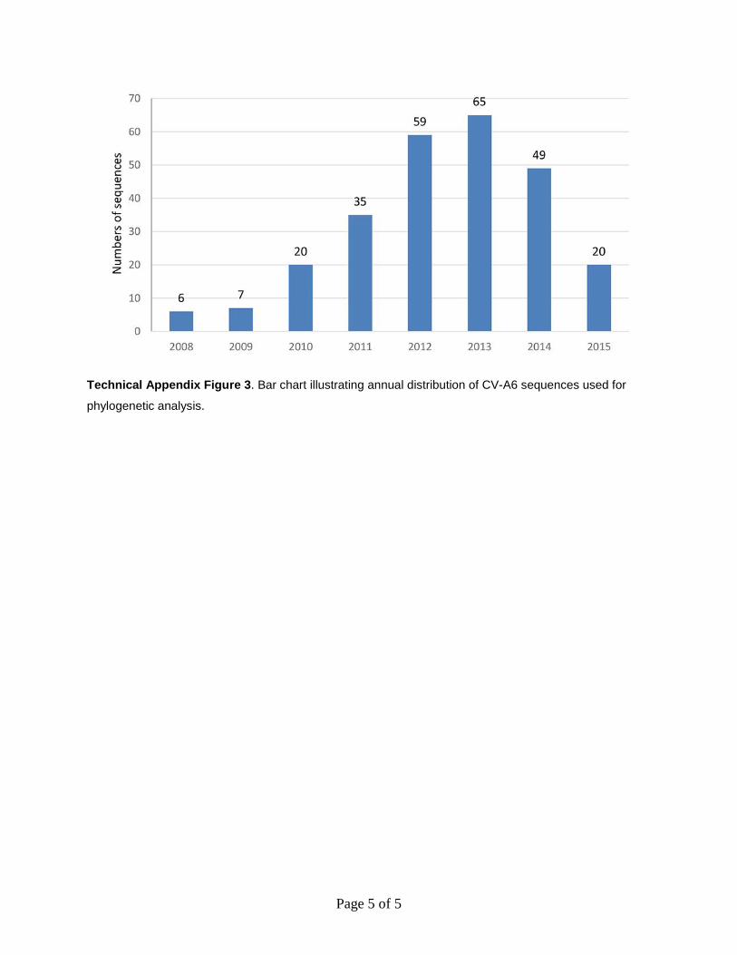
The observed demographic features in this study should be interpreted with caution. For the Vietnam CV-A6 strains, the discordance between the fluctuating numbers of CV-A6 infections detected per year and the relatively constant de-mographic picture illustrated by the skyline plot may be the result of sampling bias inherent to the study, the initial fo-cus of which was enrollment of patients with severe enough illness to go to the pediatric ICU; only later expansion in-cluded patients at outpatient facilities. Viruses of the family Picornaviridae (including CV-A6) are ubiquitous, and most infections are asymptomatic. It is therefore possible that the proportion of CV-A6 viruses detected during the first half of this study may have underestimated the overall epidemio-logic burden of CV-A6 relative to other HFMD-causing en-teroviruses during this time period because of the focus on severe cases, given that CV-A6 is proportionally less likely to cause severe disease than EV-A71. Likewise, for global strains, the skyline plot analysis in this study was in part based on publicly available CV-A6 sequences derived from studies across the world, of which most were from recent epidemic years (online Technical Appendix Figure 3); as such, the dramatic fluctuations in relative genetic diversity shown on the skyline plot in highly sampled epidemic years may be partly attributed to this sampling bias. However, the general trend toward increasing genetic diversity shown in the skyline plot during 2002–2015 would not be strongly af-fected by this bias and is likely to reflect a true increase in CV-A6 infections and genetic diversity during this period.
Together, these data emphasize the importance of ac-tive surveillance for molecular epidemiology of HFMD in disease-endemic countries. It is also critical to identify the underlying mechanisms that shape the evolutionary process and the emergence of new HFMD-causing entero-virus lineages in countries with high HFMD endemicity. Further research in these key areas would have profound implications for the development and implementation of HFMD vaccines. We hypothesize that population immu-nity and antigenic differences between circulating strains and emergent lineages are key drivers of the transmission dynamics and epidemiology of HFMD; therefore, studies to characterize cross-neutralization titers in serum samples of patients infected with common serotypes, including EV-A71, CV-A6, CV-A10, and CV-A16, to inform vaccine development are needed and ongoing.
AcknowledgmentsWe thank Le Kim Thanh for her logistic support. We are indebted to the patients and their parents for their participation in this study, and all the nursing and medical staff at the pediatric ICU, infectious disease wards, and outpatient clinics at Children’s Hospital 1, Children’s Hospital 2, and the Hospital for Tropical Diseases who provided care for the patients and helped collect clinical data.
This work was supported by the Wellcome Trust, UK (101104/Z/13/Z, 106680/B/14/Z, and 204904/Z/16/Z). The funding body did not have any influence on the study design, study conduct, preparation of the manuscript, or decision to publish.
About the AuthorMs. To Anh is a PhD candidate in life science at Open University, Milton Keynes, UK. Her research interests are virus discovery and evolution of emerging pathogens such as enteroviruses.
References 1. Xing W, Liao Q, Viboud C, Zhang J, Sun J, Wu JT, et al. Hand, foot,
and mouth disease in China, 2008–12: an epidemiological study. Lancet Infect Dis. 2014;14:308–18. http://dx.doi.org/10.1016/ S1473-3099(13)70342-6
2. Khanh TH, Sabanathan S, Thanh TT, Thoa PK, Thuong TC, Hang V, et al. Enterovirus 71-associated hand, foot, and mouth disease, southern Vietnam, 2011. Emerg Infect Dis. 2012;18:2002–5. http://dx.doi.org/10.3201/eid1812.120929
3. Bian L, Wang Y, Yao X, Mao Q, Xu M, Liang Z. Coxsackievirus A6: a new emerging pathogen causing hand, foot and mouth disease outbreaks worldwide. Expert Rev Anti Infect Ther. 2015;13: 1061–71. http://dx.doi.org/10.1586/14787210.2015.1058156
4. Oberste MS, Peñaranda S, Maher K, Pallansch MA. Complete genome sequences of all members of the species human enterovirus A. J Gen Virol. 2004;85:1597–607. http://dx.doi.org/10.1099/vir.0.79789-0
5. Ogi M, Yano Y, Chikahira M, Takai D, Oshibe T, Arashiro T, et al. Characterization of genome sequences and clinical features of coxsackievirus A6 strains collected in Hyogo, Japan in 1999–2013. J Med Virol. 2017;89:1395–403. http://dx.doi.org/10.1002/jmv.24798
6. Puenpa J, Chieochansin T, Linsuwanon P, Korkong S, Thongkomplew S, Vichaiwattana P, et al. Hand, foot, and mouth disease caused by coxsackievirus A6, Thailand, 2012. Emerg Infect Dis. 2013;19:641–3. http://dx.doi.org/10.3201/eid1904.121666
7. He SZ, Chen MY, Xu XR, Yan Q, Niu JJ, Wu WH, et al. Epidemics and aetiology of hand, foot and mouth disease in Xiamen, China, from 2008 to 2015. Epidemiol Infect. 2017;145:1865–74. http://dx.doi.org/10.1017/S0950268817000309
8. Montes M, Artieda J, Piñeiro LD, Gastesi M, Diez-Nieves I, Cilla G. Hand, foot, and mouth disease outbreak and coxsackievirus A6, northern Spain, 2011. Emerg Infect Dis. 2013;19. http://dx.doi.org/10.3201/eid1904.121589
9. Fujimoto T, Iizuka S, Enomoto M, Abe K, Yamashita K, Hanaoka N, et al. Hand, foot, and mouth disease caused by coxsackievirus A6, Japan, 2011. Emerg Infect Dis. 2012;18:337–9. http://dx.doi.org/10.3201/eid1802.111147
10. Österback R, Vuorinen T, Linna M, Susi P, Hyypiä T, Waris M. Coxsackievirus A6 and hand, foot, and mouth disease, Finland. Emerg Infect Dis. 2009;15:1485–8. http://dx.doi.org/10.3201/eid1509.090438
11. Brown BA, Oberste MS, Alexander JP Jr, Kennett ML, Pallansch MA. Molecular epidemiology and evolution of enterovirus 71 strains isolated from 1970 to 1998. J Virol. 1999;73:9969–75.
12. Lu J, Zeng H, Zheng H, Yi L, Guo X, Liu L, et al. Hand, foot and mouth disease in Guangdong, China, in 2013: new trends in the continuing epidemic. Clin Microbiol Infect. 2014;20:O442–5. http://dx.doi.org/10.1111/1469-0691.12468
13. Mao Q, Wang Y, Bian L, Xu M, Liang Z. EV-A71 vaccine licensure: a first step for multivalent enterovirus vaccine to control HFMD and other severe diseases. Emerg Microbes Infect. 2016;5:e75. http://dx.doi.org/10.1038/emi.2016.73
14. Ku Z, Liu Q, Ye X, Cai Y, Wang X, Shi J, et al. A virus-like particle based bivalent vaccine confers dual protection against
Emerging Infectious Diseases • www.cdc.gov/eid • Vol. 24, No. 4, April 2018 661
RESEARCH
enterovirus 71 and coxsackievirus A16 infections in mice. Vaccine. 2014;32:4296–303. http://dx.doi.org/10.1016/j.vaccine.2014.06.025
15. Li J, Liu G, Liu X, Yang J, Chang J, Zhang W, et al. Optimization and characterization of candidate strain for Coxsackievirus A16 inactivated vaccine. Viruses. 2015;7:3891–909. http://dx.doi.org/ 10.3390/v7072803
16. Caine EA, Fuchs J, Das SC, Partidos CD, Osorio JE. Efficacy of a trivalent hand, foot, and mouth disease vaccine against enterovirus 71 and coxsackieviruses A16 and A6 in mice. Viruses. 2015;7:5919–32. http://dx.doi.org/10.3390/v7112916
17. Puenpa J, Vongpunsawad S, Österback R, Waris M, Eriksson E, Albert J, et al. Molecular epidemiology and the evolution of human coxsackievirus A6. J Gen Virol. 2016;97:3225–31. http://dx.doi.org/10.1099/jgv.0.000619
18. Mirand A, le Sage FV, Pereira B, Cohen R, Levy C, Archimbaud C, et al. Ambulatory pediatric surveillance of hand, foot and mouth disease as signal of an outbreak of coxsackievirus A6 infections, France, 2014–2015. Emerg Infect Dis. 2016;22:1884–93. http://dx.doi.org/10.3201/eid2211.160590
19. Tan X, Li L, Zhang B, Jorba J, Su X, Ji T, et al. Molecular epidemiology of coxsackievirus A6 associated with outbreaks of hand, foot, and mouth disease in Tianjin, China, in 2013. Arch Virol. 2015;160:1097–104. http://dx.doi.org/10.1007/ s00705-015-2340-3
20. Geoghegan JL, Tan V, Kühnert D, Halpin RA, Lin X, Simenauer A, et al. Phylodynamics of enterovirus A71-associated hand, foot, and mouth disease in Viet Nam. J Virol. 2015;89:8871–9. http://dx.doi.org/10.1128/JVI.00706-15
21. Nguyen AT, Tran TT, Hoang VM, Nghiem NM, Le NN, Le TT, et al. Development and evaluation of a non-ribosomal random PCR and next-generation sequencing based assay for detection and sequencing of hand, foot and mouth disease pathogens. Virol J. 2016;13:125. http://dx.doi.org/10.1186/s12985-016-0580-9
22. Edgar RC. MUSCLE: multiple sequence alignment with high accuracy and high throughput. Nucleic Acids Res. 2004;32:1792–7. http://dx.doi.org/10.1093/nar/gkh340
23. Nguyen LT, Schmidt HA, von Haeseler A, Minh BQ. IQ-TREE: a fast and effective stochastic algorithm for estimating maximum-likelihood phylogenies. Mol Biol Evol. 2015;32:268–74. http://dx.doi.org/10.1093/molbev/msu300
24. Lanave C, Preparata G, Sacone C, Serio G. A new method for calculating evolutionary substitution rates. J Mol Evol. 1984;20:86–93. http://dx.doi.org/10.1007/BF02101990
25. Tamura K, Nei M. Estimation of the number of nucleotide substitutions in the control region of mitochondrial DNA in humans and chimpanzees. Mol Biol Evol. 1993;10:512–26.
26. Yang F, Yuan J, Wang X, Li J, Du J, Su H, et al. Severe hand, foot, and mouth disease and coxsackievirus A6—Shenzhen, China. Clin Infect Dis. 2014;59:1504–5. http://dx.doi.org/10.1093/cid/ciu624
27. Tee KK, Lam TT, Chan YF, Bible JM, Kamarulzaman A, Tong CY, et al. Evolutionary genetics of human enterovirus 71: origin, population dynamics, natural selection, and seasonal periodicity of the VP1 gene. J Virol. 2010;84:3339–50. http://dx.doi.org/10.1128/JVI.01019-09
28. Chang LY, Tsao KC, Hsia SH, Shih SR, Huang CG, Chan WK, et al. Transmission and clinical features of enterovirus 71 infections in household contacts in Taiwan. JAMA. 2004;291: 222–7. http://dx.doi.org/10.1001/jama.291.2.222
29. Xu L, Zheng Q, Li S, He M, Wu Y, Li Y, et al. Atomic structures of coxsackievirus A6 and its complex with a neutralizing antibody. Nat Commun. 2017;8:505. http://dx.doi.org/10.1038/s41467-017-00477-9
Address for correspondence: Le Van Tan, Oxford University Clinical Research Unit, Ho Chi Minh City, Vietnam; email: [email protected]
662 Emerging Infectious Diseases • www.cdc.gov/eid • Vol. 24, No. 4, April 2018
Byron Breedlove, managing editor ofthe journal, elaborates on aestheticconsiderations and historical factors,
as well as the complexities of obtainingartwork for Emerging Infectious Diseases.
EID Podcast:Emerging InfectiousDiseases Cover Art
Visit our website to listen:https://www2c.cdc.gov/
podcasts/player.asp?f=8646224
Page 1 of 5
Article DOI: https://doi.org/10.3201/eid2404.171298
Emerging Coxsackievirus A6 Causing Hand, Foot and Mouth Disease, Vietnam
Technical Appendix
Clinical Grading System for HFMD (1)
• Grade 1: mouth ulcers or vesicles/papules on hands, feet, or buttocks, with or without
mild fever (<39°C)
• Grade 2a: central nervous system (CNS) involvement (myoclonus reported by parents
or caregivers only, fever >39°C or ataxia)
• Grade 2b1: myoclonus observed by medical staff or history of myoclonus and lethargy
or pulse higher than 130 bpm
• Grade 2b2: ataxia, nystagmus, limb weakness, cranial nerve palsies, persistent high
fever, or pulse higher than 150 bpm
• Grade 3: autonomic dysfunction with sweating, hypertension, tachycardia, and
tachypnea
• Grade 4: additional cardiopulmonary compromise with pulmonary edema or shock
syndrome
Enterovirus Real-Time RT-PCR
The procedure for detection of enteroviruses in clinical samples was carried out as
previously described (2), and is detailed as follows.
We extracted viral RNA from throat and rectal swabs using the QIAamp Viral RNA Mini
kit (QIAGEN, Hilden, Germany). In brief, we first mixed 140 L of throat/rectal swabs in viral
transport medium with 20 L of equine arteritis virus and then extracted total nucleic acid

Page 2 of 5
according to the manufacturer’s instructions, eluted it in 100 L elution buffer and stored it at
80°C until used.
We performed real-time RT-PCR using the SuperScript III One-Step qRT-PCR System
with Platinum Taq DNA Polymerase (Invitrogen, Carlsbad, CA, USA) in a LightCycler 480 II
machine (Roche Diagnostics, Mannheim, Germany). The reaction was performed in a final
volume of 25 L containing 12.5 L 2X RT-PCR reaction Mix (Invitrogen), primers and probes
at appropriate concentrations (Technical Appendix Table), 0.5 L enzyme mix, and 2 L viral
RNA. The cycling conditions included 1 cycle of 60°C for 3 minutes, followed by 15 minutes at
53°C and 2 minutes at 95°C, and 45 cycles of 15 seconds at 95°C, 1 min at 53°C (including
fluorescence acquisition), and 15 seconds at 72°C.
References
1. Khanh TH, Sabanathan S, Thanh TT, Thoa PK, Thuong TC, Hang V, et al. Enterovirus 71-associated
hand, foot, and mouth disease, Southern Vietnam, 2011. Emerg Infect Dis. 2012;18:2002–5.
http://dx.doi.org/10.3201/eid1812.120929
2. Thanh TT, Anh NT, Tham NT, Van HM, Sabanathan S, Qui PT, et al. Validation and utilization of an
internally controlled multiplex real-time RT-PCR assay for simultaneous detection of
enteroviruses and enterovirus A71 associated with hand foot and mouth disease. Virol J.
2015;12:85. http://dx.doi.org/10.1186/s12985-015-0316-2
Technical Appendix Table. Primer and probe sequences and final concentrations used in 1 reaction for detecting enterovirus in clinical samples*
Name Sequence (53) Final
concentration Notes EAV-F primer CATCTCTTGCTTTGCTCCTTA G 400 nM Internal control EAV-R primer AGCCGCACCTTCACATTG 400 nM EAV-probe FAM-CGCTGTCAGAACAACATTATTGCCCAC-BHQ1 100 nM ENT-F CCCTGAATGCGGCTAAT 400 nM Enterovirus
specific primers and probe
ENT-R ATTGTCACCATAAGCAGCC 400 nM ENTr-probe Cy5-ACCCAAAGTAGTCGGTTCCG -BHQ3 200 nM EV-A71–634F GGAGAACACAARCARGAGAAAGA 400 nM Enterovirus 71
specific primers and probe
EV-A71–743R ACYAAAGGGTACTTGGAYTTVGA 400 nM EV-A71-probe Cyan500-TGATGGGCACDTTCTCRGTGCG-BHQ1 40 nM *BHQ = black hole quencher; Cy5 = cyanine 5; Cyan500 = cyan 500 NHS ester; FAM = Carboxyfluorescein; D = A, G, and T; R = A and G; V = A, C, and G; Y = T and C.

Page 3 of 5
Technical Appendix Figure 1. Maximum clade credibility (MCC) trees illustrating the phylogeography of
CV-A6: A) MCC tree of viral capsid protein (VP1) sequences of Vietnamese strains; B) MCC tree of
complete coding sequences (CDS) of global strains. Branches are color-coded according to location of
sampling.

Page 4 of 5
Technical Appendix Figure 2. Skyline plots depicting the relative genetic diversity of CV-A6 over time.
A) Results obtained from the analysis of CDS of global strains; B) results obtained from the analysis of
VP1 sequences of Vietnamese strains.
Page 5 of 5
Technical Appendix Figure 3. Bar chart illustrating annual distribution of CV-A6 sequences used for
phylogenetic analysis.