Electronic Fetal Monitoring: Guidelines for Interpretation · •Definitions •Limitations...
16
7/13/2018 1 Electronic Fetal Monitoring: Guidelines for Interpretation Kathryn Welch, MD Objectives • History of fetal monitoring • Definitions • Limitations • Practice Introduction • Goal of electronic fetal monitoring (EFM) is to detect fetal hypoxia and signal to the clinician that an intervention is needed to correct the oxygen deficiency • EFM is the most common obstetric procedure • Use common language to communicate and document findings A brief history • Fetal heart sounds were first reported in the 1600’s • In the 1800’s, again described and used to determine viability and fetal lie. Gabbe 2012 Fetoscope • 1917 David Hillis in Chicago described the fetoscope, but in 1922 Joseph DeLee took the credit! – Eventually known as DeLee-Hillis fetoscope • Concept of intermittent monitoring evolved and became standard of care well into the 1970s Gabbe 2012 “Father of EFM” • In 1958, Dr. Edward Hon reported fetal ECG from the maternal abdomen. • Similar achievements were made around the globe • 1972, Hon invented the fetal scalp electrode Gabbe 2012
Transcript of Electronic Fetal Monitoring: Guidelines for Interpretation · •Definitions •Limitations...

7/13/2018
1
Electronic Fetal Monitoring:Guidelines for Interpretation
Kathryn Welch, MD
Objectives
• History of fetal monitoring
• Definitions
• Limitations
• Practice
Introduction
• Goal of electronic fetal monitoring (EFM) is
to detect fetal hypoxia and signal to the
clinician that an intervention is needed to
correct the oxygen deficiency
• EFM is the most common obstetric
procedure
• Use common language to communicate and
document findings
A brief history
• Fetal heart sounds were first reported in the 1600’s
• In the 1800’s, again described and used to determine viability and fetal lie.
Gabbe 2012
Fetoscope
• 1917 David Hillis in Chicago described the fetoscope, but in 1922 Joseph DeLee took the credit!– Eventually known as
DeLee-Hillis fetoscope
• Concept of intermittent monitoring evolved and became standard of care well into the 1970s
Gabbe 2012
“Father of EFM”
• In 1958, Dr. Edward Hon reported fetal ECG from the maternal abdomen.
• Similar achievements were made around the globe
• 1972, Hon invented the fetal scalp electrode
Gabbe 2012

7/13/2018
2
How does it work?
• “FHR results from the signal processor, which counts every R-R interval of the ECG from the scalp electrode, converts this interval to rate, and displays every interval (bpm)”
Gabbe 2012
EFM Guidelines
NICHD Research Planning Workshop. Am J Obstet Gynecol.1997
EFM Guidelines
• NICHD Workshop Research Planning Workshop 1997
– Objective: to propose a standardized (and
unambiguous) set of definitions
• Goals:
– more precise interpretation of FHR patterns
– more evidence-based approach to the
management of labor
NICHD Research Planning Workshop. Am J Obstet Gynecol.1997
NICHD Guidelines
•
Macones G. et al. Obstet Gynecol.2008
EFM Guidelines
• 2008 National Institute of Child Health and Human
Development (NICHD) partnered with ACOG & SMFM
• Goals:
– Review and update the definitions for FHR pattern
categorization from the prior 1997 workshop
– Assess existing classifications systems for
interpreting FHR patterns
– Recommendations for research priorities for EFM
NICHD EFM Guidelines
• Assumptions:
– Definitions are for visual interpretation of FHR patterns
• Primarily for intrapartum events, but also applicable to antepartum observations
ACOG PB #106
7/13/2018
3
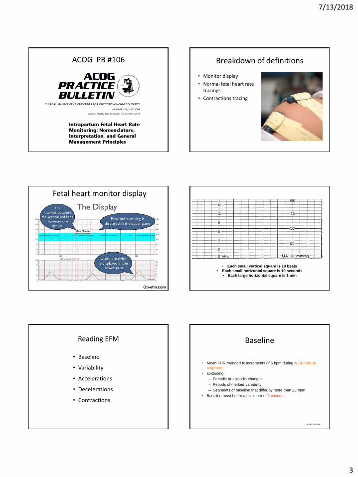
ACOG PB #106
•
Breakdown of definitions
• Monitor display
• Normal fetal heart rate tracings
• Contractions tracing
Fetal heart monitor display
Ob-efm.com
• Each small vertical square is 10 beats
• Each small horizontal square is 10 seconds
• Each large horizontal square is 1 min
Reading EFM
• Baseline
• Variability
• Accelerations
• Decelerations
• Contractions
Baseline
• Mean FHR rounded to increments of 5 bpm during a 10-minute
segment
• Excluding:
– Periodic or episodic changes
– Periods of marked variability
– Segments of baseline that differ by more than 25 bpm
• Baseline must be for a minimum of 2 minutes
ACOG PB #106
7/13/2018
4
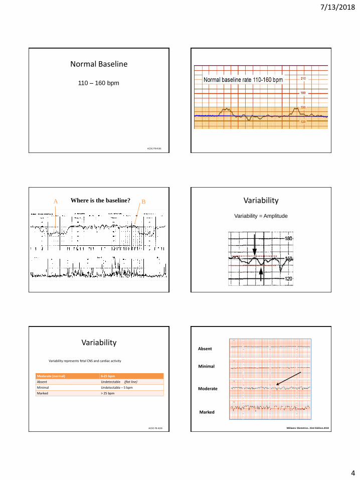
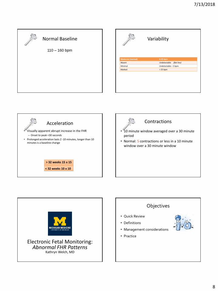
Normal Baseline
110 – 160 bpm
ACOG PB #106
Where is the baseline? BA Variability
Variability = Amplitude
Variability
Moderate (normal) 6-25 bpm
Absent Undetectable (flat line)
Minimal Undetectable – 5 bpm
Marked > 25 bpm
Variability represents fetal CNS and cardiac activity
ACOG PB #106
Absent
Minimal
Moderate
Marked
Williams Obstetrics. 23rd Edition.2010

7/13/2018
5
Variability
As a rule, moderate
variability provides
reassurance about
fetal status and the
absence of
metabolic acidemia
ACOG PB #106
Acceleration
• Visually apparent abrupt increase in the FHR
– Onset to peak <30 seconds
• Prolonged acceleration lasts 2 -10 minutes, longer than 10 minutes is a baseline change
> 32 weeks 15 x 15
< 32 weeks 10 x 10
ACOG PB #106
Reading EFM
• Baseline
• Variability
• Accelerations
• Decelerations (next lecture)
• Contractions
Uterine Contractions
• External tocodynamometer
– Frequency and duration of contractions
– Noninvasive but uncomfortable, difficult to monitor obese patients
• Intrauterine pressure catheter
– Frequency, duration & adequacy of contractions
– Resting tone between contractions
– Only when membranes are ruptured; invasive
– Uncomfortable and limits patient mobility
– Can be used for intrauterine resuscitation
Gabbe 2012
Contractions
• 10 minute window averaged over a 30 minute period
• Normal: 5 contractions or less in a 10 minute window over a 30 minute window
ACOG PB #106

7/13/2018
6
Uterine contractions Uterine contractions
External tocodynamometer
Ob-efm.com
Montevideo Units
• Adequacy of contractions
• IUPC ONLY
– number of ctx in 10 mins X mean amplitude (mm Hg)
ACOG PB #106
Montevideo Units
Williams Obstetrics. 23rd Edition.2010
Montevideo Units
Ob-efm.com

7/13/2018
7
How often do I have to do this??Intrapartum
Uncomplicated patient First stage of labor: 30 minSecond stage: 15 min
Complicated Patient First stage of labor: 15 minSecond stage: 5 min
*Don’t forget to document your findings!
ACOG PB #106
Limitations of EFM
• Poor interobserver and intraobserverreliability
• Uncertain efficacy
• High false-positive rate
ACOG PB #106
Limitations of EFM
• EFM reduces risk of neonatal seizures
• Increased cesarean and operative vaginal delivery rate
• EFM does not reduce perinatal mortality
• EFM does not reduce the risk of cerebral palsy
ACOG PB #106
Electronic Fetal Monitoring vsIntermittent ausculation
• There are no RCT to document that EFM is superior therefore it is acceptable that an uncomplicated patient could opt for IA
• However, this is hospital and staff dependent, as IA is very “labor intensive”
– ACOG recommends: q15 min in active phase of latent labor and at least q5 min in second stage
ACOG PB #106
Reading EFM
• Baseline
• Variability
• Accelerations
• Decelerations Contractions
Baseline
• Mean FHR rounded to increments of 5 bpm during a 10-minute
segment
• Excluding:
– Periodic or episodic changes
– Periods of marked variability
– Segments of baseline that differ by more than 25 bpm
• Baseline must be for a minimum of 2 minutes
7/13/2018
8
Normal Baseline
110 – 160 bpm
Variability
Moderate (normal) 6-25 bpm
Absent Undetectable (flat line)
Minimal Undetectable – 5 bpm
Marked > 25 bpm
Acceleration
• Visually apparent abrupt increase in the FHR
– Onset to peak <30 seconds
• Prolonged acceleration lasts 2 -10 minutes, longer than 10 minutes is a baseline change
> 32 weeks 15 x 15
< 32 weeks 10 x 10
Contractions
• 10 minute window averaged over a 30 minute period
• Normal: 5 contractions or less in a 10 minute window over a 30 minute window
Electronic Fetal Monitoring:Abnormal FHR Patterns
Kathryn Welch, MD
Objectives
• Quick Review
• Definitions
• Management considerations
• Practice

7/13/2018
9
Abnormal baseline
• Tachycardia > 160 bpm
– Maternal fever & drugs
• Bradycardia < 110 bpm
– Maternal drugs, hypothyroidism, SLE
– Fetal heart block
Gabbe 2012
Bradycardia
Williams Obstetrics. 23rd Edition.2010
Variability
Moderate (normal) 6-25 bpm
Absent Undetectable (flat line)
Minimal Undetectable – 5 bpm
Marked > 25 bpm
Absent
Minimal
Moderate
Marked
Williams Obstetrics. 23rd Edition.2010
Changes in Variability
• Hypoxic causes
– Tachysystole
– Abruption
– Maternal hypotension
• Non-hypoxic causes
– Sleep cycle
– Prematurity
– Cardiac arrhythmias
– Medications (narcotics)
Acceleration
• Visually apparent abrupt increase in the FHR
– Onset to peak <30 seconds
• Prolonged acceleration lasts 2 -10 minutes, longer than 10 minutes is a baseline change
> 32 weeks 15 x 15
< 32 weeks 10 x 10
7/13/2018
10
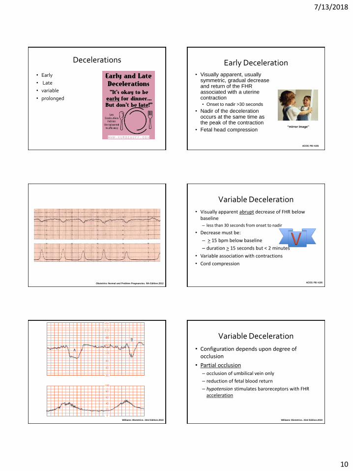
Decelerations
• Early
• Late
• variable
• prolonged
Early Deceleration
• Visually apparent, usually symmetric, gradual decrease and return of the FHR associated with a uterine contraction
• Onset to nadir >30 seconds
• Nadir of the deceleration occurs at the same time as the peak of the contraction
• Fetal head compression“mirror image”
ACOG PB #106
Obstetrics Normal and Problem Pregnancies. 6th Edition.2012
Variable Deceleration
• Visually apparent abrupt decrease of FHR below baseline
– less than 30 seconds from onset to nadir
• Decrease must be:
– > 15 bpm below baseline
– duration > 15 seconds but < 2 minutes
• Variable association with contractions
• Cord compression
ACOG PB #106
V
Williams Obstetrics. 23rd Edition.2010
Variable Deceleration
• Configuration depends upon degree of occlusion
• Partial occlusion
– occlusion of umbilical vein only
– reduction of fetal blood return
– hypotension stimulates baroreceptors with FHR acceleration
Williams Obstetrics. 23rd Edition.2010
7/13/2018
11
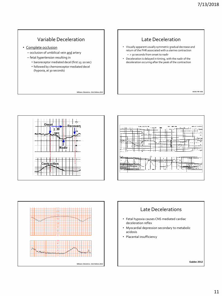
Variable Deceleration
• Complete occlusion
– occlusion of umbilical vein and artery
– fetal hypertension resulting in
• baroreceptor mediated decel (first 15-20 sec)
• followed by chemoreceptor mediated decel (hypoxia, at 30 seconds)
Williams Obstetrics. 23rd Edition.2010
Late Deceleration
• Visually apparent usually symmetric gradual decrease and return of the FHR associated with a uterine contraction
– > 30 seconds from onset to nadir
• Deceleration is delayed in timing, with the nadir of the deceleration occuring after the peak of the contraction
ACOG PB #106
Williams Obstetrics. 23rd Edition.2010
Late Decelerations
• Fetal hypoxia causes CNS mediated cardiac deceleration reflex
• Myocardial depression secondary to metabolic acidosis
• Placental insufficiency
Gabbe 2012

7/13/2018
12
• Deceleration pattern
–Defines the nature of the insult
• Variability
–Characterizes the ability of fetus
to tolerate the insult
Prolonged decelerations
• Visually apparent decrease in the FHR below the baseline
• 15 bpm or more, lasting 2 minutes or more but less than 10.
• If a deceleration lasts 10 minutes or longer, it is a baseline change
FHR Decelerations
• Depth and duration should be quantitated
• Recurrent: > 50% of ctx
• Intermittent: < 50% of ctx
Macones G. et al. Obstet Gynecol.2008
Sinusoidal pattern
• Visually apparent, smooth, sine wave-like undulating pattern in FHR baseline with a cycle frequency of 3-5 per minute which persists for 20 minutes of more
ACOG PB #106
Sinusoidal pattern

7/13/2018
13
Contractions
• 10 minute window averaged over a 30 minute period
• Normal: 5 contractions or less in a 10 minute window over a 30 minute window
Tachysystole
• > 5 contractions in 10 minutes averaged
over 30 minute window
• Should also describe the presence or
absence of associated decelerations
• Can be used for spontaneous or induced
contractions
Macones G. et al. Obstet Gynecol.2008
Tachysystole
Uterine hyperstimulation
• Uterus does not relax between contractions
• Resting uterine tone > 25 mm Hg
• Perfusion of intervillus space is compromised
• FHR decelerations secondary to lack of oxygen
Gabbe 2012

7/13/2018
14
Fetal Metabolic Acidemia
Accelerations present
and/or
moderate variability
Unlikely risk of acidemia
Interpretation of FHR Patterns
• Patterns reflect the current acid-base status of the fetus
• Tracing patterns will change over time
• Cannot predict the development of cerebral palsy
• Three tier system
– Category I, II, III
Macones G. et al. Obstet Gynecol.2008
Category I = NORMAL
Macones G. et al. Obstet Gynecol.2008
Category I
• Baseline rate: 110 – 160 bpm
• Baseline variability: Moderate
• Late or variable decels: Absent
• Early decels: Present or absent
• Accelerations: Present or absent
• Strongly predictive of normal acid-base status
• No action needed
Macones G. et al. Obstet Gynecol.2008
Category III = BAD!!!!
Macones G. et al. Obstet Gynecol.2008

7/13/2018
15
Category III
• Recurrent late decels with absent variability
• Recurrent variable decels with absent variability
• Bradycardia with absent variability
• Sinusoidal pattern
Macones G. et al. Obstet Gynecol.2008
Category II = everything else
Macones G. et al. Obstet Gynecol.2008
Category II
• Baseline rate:– Bradycardia with mod/min variability
– Tachycardia
• Baseline variability:– Minimal variability
– Absent not accompanied by recurrent decels
– Marked variability
• Accelerations:– Absent of induced accels after fetal stimulation
Macones G. et al. Obstet Gynecol.2008
Category II
• Requires evaluation and continue surveillance and reevaluation
Macones G. et al. Obstet Gynecol.2008
Categories
• Category I– Normal/strongly predictive of normal acid-base status– No action needed
• Category II– Indeterminate– Not predictive of abnormal acid-base status– Requires evaluation, increased surveillance
• Category III– Predictive of abnormal acid-base status– Requires prompt evaluation and intervention
Macones G. et al. Obstet Gynecol.2008AJOG 2013

7/13/2018
16
ACOG PB #116ACOG PB #116