Engineering elastic sealants based on gelatin and elastin ...

RESEARCH Open Access
Elastin degradation products in acute lunginjury induced by gastric contentsaspirationPedro Ayala1, Raúl Vivar1, Rebeca Montalva1, Pablo Olmos2, Manuel Meneses3 and Gisella R. Borzone1*
Abstract
Background: Gastric contents aspiration is a high-risk condition for acute lung injury (ALI). Consequences range fromsubclinical pneumonitis to respiratory failure, depending on the volume of aspirate. A large increment in inflammatorycells, an important source of elastase, potentially capable of damaging lung tissue, has been described in experimentalmodels of aspiration. We hypothesized that in early stages of aspiration-induced ALI, there is proteolytic degradation ofelastin, preceding collagen deposition. Our aim was to evaluate whether after a single orotracheal instillation of gastricfluid, there is evidence of elastin degradation.
Methods: Anesthesized Sprague-Dawley rats received a single orotracheal instillation of gastric fluid and wereeuthanized 4, 12 and 24 h and at day 4 after instillation (n = 6/group). We used immunodetection of soluble elastin inlung tissue and BALF and correlated BALF levels of elastin degradation products with markers of ALI. We investigatedpossible factors involved in elastin degradation and evaluated whether a similar pattern of elastin degradation can befound in BALF samples of patients with interstitial lung diseases known to have aspirated. Non-parametric ANOVA(Kruskall-Wallis) and linear regression analysis were used.
Results: We found evidence of early proteolytic degradation of lung elastin. Elastin degradation products are detectedboth in lung tissue and BALF in the first 24 h and are significantly reduced at day 4. They correlate significantly with ALImarkers, particularly PMN cell count, are independent of acidity and have a similar molecular weight as those obtainedusing pancreatic elastase. Evaluation of BALF from patients revealed the presence of elastin degradation products notpresent in controls that are similar to those found in BALF of rats treated with gastric fluid.
Conclusions: A single instillation of gastric fluid into the lungs induces early proteolytic degradation of elastin, inrelation to the magnitude of alveolar-capillary barrier derangement. PMN-derived proteases released during ALI aremostly responsible for this damage. BALF from patients showed elastin degradation products similar to those found inrats treated with gastric fluid. Long-lasting effects on lung elastic properties could be expected under conditions ofrepeated instillations of gastric fluid in experimental animals or repeated aspiration events in humans.
Keywords: Elastinolysis, Elastin degradation, Gastric fluid aspiration, Acute lung injury, Extracellular matrix
* Correspondence: [email protected] of Respiratory Diseases and Medical Research Center, PontificiaUniversidad Católica de Chile, Marcoleta 350, piso 1, Santiago, ChileFull list of author information is available at the end of the article
© The Author(s). 2018 Open Access This article is distributed under the terms of the Creative Commons Attribution 4.0International License (http://creativecommons.org/licenses/by/4.0/), which permits unrestricted use, distribution, andreproduction in any medium, provided you give appropriate credit to the original author(s) and the source, provide a link tothe Creative Commons license, and indicate if changes were made. The Creative Commons Public Domain Dedication waiver(http://creativecommons.org/publicdomain/zero/1.0/) applies to the data made available in this article, unless otherwise stated.
Ayala et al. Respiratory Research (2018) 19:165 https://doi.org/10.1186/s12931-018-0873-1

BackgroundGastric contents aspiration is a high-risk condition forlung injury. Consequences range from subclinical pneu-monitis to diffuse alveolar damage and progressive re-spiratory failure, depending on the volume of aspirate,with fibrosis development in some patients [1, 2].Various experimental approaches have been used to
gain insight into the pathogenesis and pathophysiologyof aspiration-induced lung injury. Instillation of individ-ual components of gastric fluid has contributed to theunderstanding of their relative roles in lung injury [3].Whereas hydrochloric acid instillation results in de-rangement of the alveolar-capillary barrier with edemaand an intense inflammatory reaction [4–14], instillationof acid-free gastric food particles induces a delayed in-flammatory reaction, followed by granuloma formationwithout significant edema [15–17]. Synergistic effectshave been reported when acid and gastric food particlesare instilled in combination [4, 9]. Few studies have usedthe whole gastric fluid to study the pathogenesis of as-piration. Those studies have used small volumes of gas-tric fluid instilled into small areas of the lung with theaim of answering questions regarding lung transplant re-jection [15–17].Our group has addressed the study of the continuum
of changes after a single event of bilateral aspiration ofwhole gastric contents and has shown that a single oro-tracheal instillation of gastric fluid in the rat lung resultsin severe acute lung injury with several histological simi-larities to diffuse alveolar damage (DAD), that evolves toan organization process involving intraluminal plugs ofmyofibroblasts and collagen fibers, affecting small bron-chioles, alveolar ducts and peribronchiolar alveolarspaces, associated with particle-containing foreign-bodygiant cells either isolated or forming granulomas thatlater resolves [18]. This sequence of events reflects im-portant remodeling of lung extracellular matrix (ECM)involving deposition and degradation of its components.Most studies regarding mechanisms involved in ECM re-modeling after acute insults to the lung have focused ondeposition of new ECM components, mainly collagendeposition [8, 15] but very few have evaluated ECM deg-radation [19–21]. Elastin, a polymer of tropoelastin is amajor component of the lung ECM providing the lungwith elasticity, tensile strength, and stability [22]. Increasedcatabolism of elastin can be detected by a reduction in ma-ture elastin content or by the release of elastin-degradationproducts after mature elastin breakdown. In this regard,evidence for a reduction in mature elastin content has beenunexpectedly obtained in fibrotic diseases such as usualinterstitial pneumonia (UIP) and cryptogenic organizingpneumonia (COP) using modern non-invasive microscopytechnology [23], whereas elastin-degradation products havebeen documented in animal models of acute lung injury
ending in fibrosis [19, 20] and in human diseases as diverseas chronic obstructive pulmonary disease (COPD) [24],acute respiratory distress syndrome (ARDS) [25] and, idio-pathic pulmonary fibrosis (IPF) [26]. Interestingly, all thesedisease conditions have also been associated with gastriccontents aspiration [4, 27].In our model, a 15- to 20-fold increase in bronchoal-
veolar lavage fluid (BALF) total cell count was found inthe first 24 h after a single instillation of gastric fluid,with polymorphonuclear (PMN) cell predominance [18],an important source of elastase and free radicals, withthe potential of damaging lung elastic tissue [28]. Inaddition to the inflammatory reaction induced by aspir-ation, elastic tissue damage could be produced by the directeffect of gastric fluid or could be part of the changes thattake place in the remodeling of lung ECM after aspiration.We hypothesized that in gastric fluid aspiration-induced
ALI there is proteolytic degradation of elastin precedingcollagen deposition.Our aim was to evaluate at different time points dur-
ing the course of ALI induced by a single instillation ofgastric fluid whether there is evidence of elastin degrad-ation in lung tissue and BALF. We used Western blotanalysis to detect lung elastin degradation products andcorrelated the presence of these products in BALF andlung tissue with markers of acute lung injury. Inaddition, we studied if these degradation products arepresent in BALF samples of human patients with inter-stitial lung diseases (ILDs) that have evidence ofaspiration.We found that a single orotracheal instillation of gastric
fluid into the rat lung is associated with early degradationof lung elastin. The significant positive correlation foundwith PMN cell count in BALF suggests that neutrophilelastase could be involved, since exogenous elastase pro-duces a similar pattern of elastin degradation products.Evaluation of BALF from patients with ILD who have evi-dence of aspiration revealed the presence of elastin deg-radation products similar to those found in BALF of ratstreated with gastric fluid.These results are important to be considered, since re-
petitive aspirations of gastric contents could result inlong-lasting alterations of lung elastic properties.
MethodsThe study was performed according to a protocol submit-ted to and approved by the Animal Research Ethics Com-mittee of the Pontificia Universidad Católica de Chile inadult male Sprague-Dawley rats (270–300 g).
Rat model of single orotracheal instillation of gastric fluidGastric contents poolAdult male Sprague-Dawley rats fasted overnight were anes-thetized i.p. with xylazine-ketamine (5.1 and 55.1 mg/kg,
Ayala et al. Respiratory Research (2018) 19:165 Page 2 of 13

respectively) to obtain gastric fluid through a gastrotomy.Gastric fluid samples were pooled, filtered through a 100um mesh, and kept at -80 °C. Animals were euthanizedthereafter by exsanguination under anesthesia.
Orotracheal instillation of gastric fluidUnder the same anesthetic protocol, another set of ani-mals was orotracheally intubated with a 22 gauge wire-fedcatheter. A modified human otoscope (Welch Allyn,Skaneateles Falls, NY) was used to visualize the glottis. Avolume of gastric fluid previously determined by the au-thors (data not shown) to distribute evenly (1.5 mL/kg,pH 1.69) was instilled, and animals were allowed to re-cover spontaneously from anesthesia.
Study groupsHistological and biochemical studies were performed at4, 12 and 24 h and at day 4 after instillation (n = 6 pergroup). Animals without intervention (n = 6) served ascontrols since they did not differ significantly from salinetreated animals. Diagram in Fig. 1 shows animal groups,tissue sampling and analysis.
Sample collectionLungs were excised en bloc, and the left main bronchuswas cannulated for bronchoalveolar lavage (BAL). Foreach animal, three aliquots of 0.15 M saline (1 mL each)were instilled, immediately aspirated and pooled. Totaland differential cell count was obtained using a Neubauerchamber and a cytospin slide centrifuge (StatSpin Cyto-fuge 2; Iris, Westwood, MA). Cytoslides were stained withDiffQuik (QCA, Tarragona, Spain). After centrifugation,
BALF was stored at -80 °C until used for the measurementof hemoglobin concentration, total protein content, andwestern blot analysis of soluble elastin. The right middlelobe was excised, frozen and later homogenized for west-ern blot analysis of soluble elastin and for matrixmetalloproteinase-9 (MMP-9) and − 2 (MMP-2) activitiesby zymography. The right lower lobe was fixed at 20 cmH2O with 10% buffered formaldehyde solution and paraf-fin embedded for histological studies. The caudate lobewas used to obtain the wet/dry weight ratio.
Histologic evidence of tissue injuryFor each animal, four right lower lobe longitudinal sec-tions were embedded in paraffin, sectioned at 5 μm, andstained with hematoxylin-and-eosin. A board-certifiedpathologist (M.M.) scored samples according to ATSstatement [29]. Scores for PMN cells in alveolar spaces,PMN cells in the interstitium, proteinaceous debris andalveolar septal thickening were used to correlate withsoluble elastin. In addition, sections underwent specificstaining of the elastic system using the Unna-Taenzeracid orcein stain [30].
Western blot analysis of soluble elastinEqual amounts of protein extracts from lung homogenatesor BALF were heat-denatured in Laemmli sample buffer with2-mercaptoethanol (5%), resolved in 10% SDS-PAGE gel andtransferred to nitrocellulose membranes (Thermoscientific,Rockford, IL, USA). Next, blots were blocked with 5%PBS-nonfat dry milk for 1 h at room temperature and thenincubated with a goat polyclonal anti-elastin primary detec-tion antibody (1:1000) (sc-17,580 Santa Cruz Biotechnology,
Fig. 1 Diagram showing animal groups, timing of tissue sampling and analysis
Ayala et al. Respiratory Research (2018) 19:165 Page 3 of 13

Dallas, Texas, USA) overnight at 4 °C. After thoroughlywashing with PBS 0.05% Tween-20, membranes were incu-bated for 2 h at room temperature with a rabbit anti-goatHRP-conjugated secondary antibody (1:5000) (ThermoScientific, Rockford, IL, USA). Elastin fragment immunoreac-tivity was visualized by enhanced chemiluminescence (Super-Signal™ Pico Chemiluminescent Substrate kit; ThermoScientific, Rockford, IL, USA). C-DiGit Blot Scanner (Li-Cor,Lincoln, NE, USA) was used to image chemiluminescent sig-nals by scanning. Densitometric analysis was performedusing the ImageJ software version 1.46 m (NIH, Bethesda,MD). β-tubulin was used to control for equal loading.Animal and human samples were studied using this
method. In addition, samples from control rats treated “invitro” with HCl (pH: 1.69, 37 °C, for 2,4,6 or 8 h) or pan-creatic elastase (0.5 μM, pH:8 and 10 min incubation)were also studied.
Acute lung injury markersLung wet/dry weight ratio of the caudate lobe was ob-tained using an oven at 60 °C until stable dry weight wasachieved.Total protein concentration in BALF was measured
using the Bradford assay.Hemoglobin concentration in BALF was measured
by light absorbance at 510- to 650-nm wavelength usinga spectrophotometer (Shimadzu, Kyoto, Japan).Lung tissue MMP-9 and MMP-2 activities: Gelati-
nolytic activity of these lung tissue MMPs was studiedusing zymography [31]. Equal amount of lung tissuehomogenate total protein (30 μg) were loaded into agelatin-containing electrophoresis gel (10% polyacrylamideand 1% gelatin under non-reducing conditions). After elec-trophoresis, gels were washed in 2.5% TritonX-100(Sigma-Aldrich, St. Louis, MO) to remove SDS, incubatedovernight at 37 °C in a calcium containing developing buffer,stained with 0.1% Coomassie Brilliant Blue and destaineduntil areas of gelatinolytic activity became evident. Densito-metric analysis was performed using ImageJ software ver-sion 1.46 m (NIH, Bethesda, MD).
Analysis of BALF samples from patients with exacerbationof interstitial lung diseasesBALF samples obtained from six patients with an acuteexacerbation of their ILD as part of their routine clinicalevaluation were studied in a similar way as the rat sam-ples. They all exhibited evidence of gastric contents as-piration, since they all had high levels of BALF pepsin.As controls for this part of the study, we used six BALFsamples from patients without interstitial lung diseasewho required bronchoscopy for the study of a pulmon-ary nodule and had no evidence of aspiration, since theyall had negative BALF pepsin levels.
Statistical analysisNon-parametric analysis of variance (Kruskall-Wallis) wasused because of the small sample size. Linear regression ana-lysis and Spearman’s rank correlation were also used [32].Unless otherwise noted, the results are expressed as medianvalues, interquartile range and range. A p value < 0.05 wasconsidered statistically significant. Analyses were performedusing GraphPad Prism 5.0 software.
ResultsHistological evaluation of acute lung injury in the first4 days after a single orotracheal instillation of gastricfluidHistological changes in the first 4 days after instillationare shown in Fig. 2. Figure 2a shows the time course ofchanges with H-E staining. At 4 h there is increased alveo-lar thickening by interstitial edema and inflammatory cellinfiltration, along with abundant protein-rich intra-alveolarexudate containing neutrophils and red blood cells, adopt-ing a peri-bronchiolar distribution. These changes becomemore intense at 12 and 24 h, with patchy consolidation,due to coalescence of affected areas. At day 4, markers ofALI, as those described in the first 24 h are no longer ob-served. Instead, intra-alveolar buds of granulation tissue,characteristic of organizing pneumonia (OP) are seen,sometimes containing granulomas and giant cells.Figure 2b shows alpha-SMA immunostaining of both,
control lung and lung of animals studied at 24 h and atday 4 after gastric contents instillation. The control and24-h samples exhibit alpha-SMA (brown) staining local-ized only to the wall of bronchioles and blood vessels,without intra-alveolar alpha-SMA-positive structures,which are only seen at day 4.
Evidence of damage to the lung elastic fiber system inanimals treated with a single instillation of gastric fluid andstudied at 4, 12 and 24 h and at day 4 after instillationFigure 3 shows the elastic fiber system distribution inlung samples from a control animal and from animalswith acute lung injury induced by gastric fluid. The con-trol sample shows preserved architectural pattern of theelastic system. Samples in the first 24 h after instillationshow sparce and fragmented bundles of elastic systemfibers.At each of the time points studied, we observed elastic
fiber fragmentation in areas with inflammatory reactionand not in preserved areas.At day 4, with significantly less inflammatory cells,
elastic fiber fragmentation was less evident and localizedonly to the alveolar septa adjacent to intra-alveolar fibro-sis. Interestingly, elastic fiber fragmentation was not ob-served inside Masson bodies.
Ayala et al. Respiratory Research (2018) 19:165 Page 4 of 13

BALF total and differential cell count in the first 4 daysafter a single orotracheal instillation of gastric fluidChanges in total and differential cell count in BALF areshown in Table 1. A 15- to 20-fold increase in total cellcount was seen in the first 24 h, with PMN cell predom-inance. By day 4, there was a return to mononuclear cellpredominance.
Soluble elastin in lung tissue homogenate and BALF aftergastric fluid instillationFigure 4 shows the results of soluble elastin immunode-tection in lung tissue homogenate. In Fig. 4a, the immu-noblot of the control sample shows a 70 kDa band,likely corresponding to tropoelastin, whereas smallermolecular weight bands in the 35–50 kDa range corre-sponding to elastin degradation products are barely de-tectable. In treated animals, the 70 kDa band exhibits
variable size and is accompanied by bands in the 35–50 kDa range, with variable densities depending on timeafter instillation. Figure 4b and c show the densitometricanalysis of these bands. In Fig. 4b, the 70 kDa band at 4 hexhibits a peak increment in density of 2.2 times the con-trol band (p < 0.01). Later on, this band decreases progres-sively to become similar to the control band at day 4 (rS: −0.6515; p < 0.01). In Fig. 4c, the 35–50 kDa bands show aprogressive increment up to 24 h (rs: + 0.7376; p < 0.001)and although these bands decrease in size at day 4, they arestill detectable.Figure 5 shows the results of soluble elastin in BALF. In
Fig. 5a, the immunoblot of the control sample shows asingle band of soluble elastin with a molecular weight of70 kDa. Bands in the 35–50 kDa range are non-detectable.As in lung tissue homogenate, in treated animals, the sizeof the 70 kDa band is variable depending on time after
Fig. 2 Histological evaluation of acute lung injury in the first 4 days after a single orotracheal instillation of gastric fluid. a Light microscopy (hematoxylinand eosin stain) of lung from a control animal and from animals studied 4, 12 and 24 h and at day 4, after gastric fluid instillation. Polymorphonuclearneutrophils and red blood cells with abundant intra-alveolar proteinaceous material are seen at 4 h. A more intense reaction is seen at 12 and 24 h. Atday 4, markers of ALI, as seen in the first 24 h, are no longer observed. Instead, intra-alveolar buds of granulation tissue, characteristic of OP containinggiant-cell granulomas are seen. Arrow: giant-cell granuloma inside a Masson body. Original magnification: 200X. b Light microscopy (alpha-SMAimmunostaining) of control lung and lung of animals studied at 24 h and at day 4 after gastric fluid instillation. The control and 24-h samples exhibitalpha-SMA (brown) staining localized to the wall of bronchioles and blood vessels only. Intra-alveolar alpha-SMA-positive structures (myofibroblasts) areobserved only at day 4. Original magnification: 200X
Fig. 3 Evidence of damage to the lung elastic fiber system in animals treated with a single instillation of gastric fluid and studied at 4, 12 and24 h and at day 4 after instillation. Representative fields illustrating elastic fiber system distribution in lung samples from control and acute lunginjury induced by gastric fluid. Elastic fibers are stained in deep violet within alveolar walls (arrows). Photographs were taken at an originalmagnification of 600X from slides stained with orcein
Ayala et al. Respiratory Research (2018) 19:165 Page 5 of 13

instillation and is accompanied by small molecular weightbands in the 35–50 kDa range. Figure 5b and c show thedensitometric analysis of these bands. In Fig. 5b, the70 kDa band at 4 h exhibits a peak increment in density of10 times the control band (p < 0.001). Later on, it de-creases progressively to become similar to the controlband at day 4 (rS: − 0.5599; p < 0.01). In Fig. 5c, band dens-ities in the 35–50 kDa range are visible 4 h after instilla-tion. Later on, these bands decrease progressively indensity to become similar to the median density of thecontrol samples at day 4 (rS: − 0.6676; p < 0.001).The time course of changes in band densities shows
that changes in tropoelastin slightly precede changes insmall molecular weight elastin-derived peptides, mainlyin lung tissue homogenates. Whereas the peak incre-ment for the 70 kDa band in lung tissue homogenates isobserved at 4 h, the peak increment for the 35-50 kDabands is seen between 12 and 24 h after instillation.
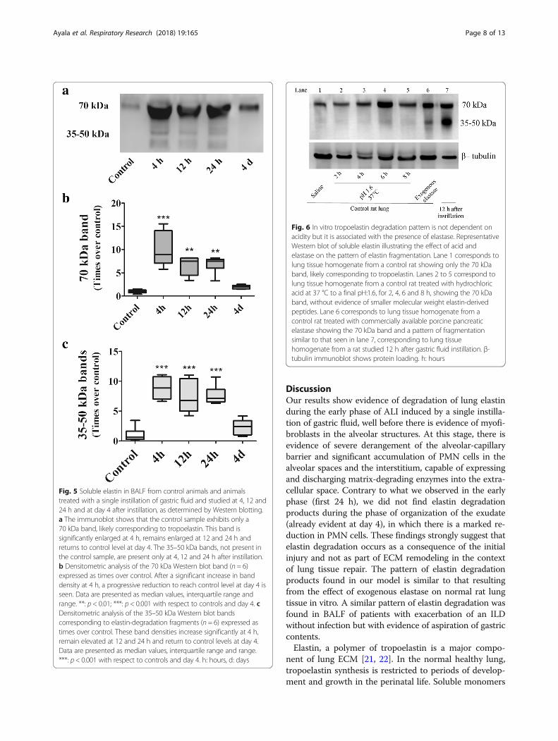
In vitro effect of acid and exogenous elastase on thepattern of lung tissue elastin degradationFigure 6 shows a representative Western blot of soluble elas-tin illustrating the effects of acid and exogenous elastase onthe pattern of elastin degradation in samples of control lungin vitro. Lane 1 corresponds to lung tissue homogenate froma control rat sample exposed to saline, showing only the70 kDa band, likely corresponding to tropoelastin. Lanes 2to 5 correspond to lung tissue homogenate from a controlrat sample treated with hydrochloric acid at 37 °C to a finalpH:1.6, for 2, 4, 6 and 8 h, showing only the 70 kDa band,without evidence of elastin degradation over time. Lane 6corresponds to lung tissue homogenate from a control ratsample treated with porcine pancreatic elastase showing the70 kDa band and a pattern of elastin degradation that issimilar to that seen in lane 7, corresponding to lung tissuehomogenate from a rat studied 12 h after gastric fluid instil-lation. The small molecular weight elastin-derived fragments(35–50 kDa) observed in this lane are similar to those ob-tained when using exogenous elastase in normal rat lung.
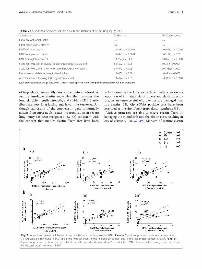
Correlations between soluble elastin and markers ofacute lung injuryTable 2 and Fig. 7 illustrate the correlations between sol-uble elastin as determined by Western blotting and
several markers of ALI . Table 2 shows no correlationbetween any of the bands corresponding to soluble elas-tin and both the wet/dry weight ratio and MMP-9 activ-ity. Only MMP-9 data was used for correlations sinceMMP-2 activity did not change in the study period.Figure 7a shows significant positive correlations betweenthe 70 kDa band density found in BALF and: a) thePMN cell count (r = 0.8181, p < 0.0001) b) the hemoglobincontent (r = 0.777, p < 0.0001) and c) the total proteincontent (r = 0.6445, p < 0.0001) in BALF. Figure 7b showssignificant positive correlations between the 35–50 kDaband densities found in BALF and: a) the PMN cell count(r = 0.8026, p < 0.0001), b) the hemoglobin content(r = 0.8673, p < 0.0001) and c) the total protein con-tent (r = 0.5120, p < 0.001) in BALF. In addition, sig-nificant positive correlations were found between the70 kDa band and several histological markers of acutelung injury and between the 35–50 kDa bands andthe same histological markers (Table 2).
Elastin degradation products in BALF of patients withexacerbated interstitial lung diseases and evidence ofaspirationTo assess the possibility of lung elastin degradation inhumans with high probability of gastric contents aspir-ation, we studied BALF samples obtained from patientswith an exacerbation of interstitial lung disease and highlevels of pepsin (n = 6) Fig. 8 shows a representativeWestern blot of soluble elastin in BALF samples fromthese patients and from patients without interstitial lungdisease and no evidence of aspiration who requiredbronchoscopy for the study of a lung nodule) (n = 6) andserved as controls for this evaluation. BALF samplesfrom subjects without interstitial lung disease and nega-tive pepsin) (lanes 1 to 6) show a single band in the70 kDa range, without evidence of smaller molecularweight elastin-derived peptides.All six BALF samples from subjects with an exacerba-
tion of an interstitial lung disease, with no evidence ofinfection and with high levels of pepsin (lanes 7 to 12)show the 70 kDa band seen in control samples, and adiffuse pattern of bands in the 50–70 kDa range. Five ofthe samples (lanes 7 to 12) show small molecular weightbands in the 35–50 kDa range as those seen in the ratBALF obtained 12 h after gastric fluid instillation (lane13). We had access to the total cell count of these hu-man BALF samples. Whereas in control BALFs, total cellcount was homogeneously low (0.48 ± 0.2 × 10⁶ cells xml− 1; range: 0.2 to 0.7), samples from exacerbated ILDpatients showed variable cell counts (2,1 ± 1.3 × 10⁶ cellsx ml− 1; range: 0.8 to 4.0). Interestingly the sample withthe highest total cell count showed the greatest elastindegradation.
Table 1 BALF total and differential cell count
Total cell count (mean ± SD) % PMN % MN
Control 8.2 ± 3.1 × 10⁴ 0.8 99.2
4 h 1.7 ± 0.9 × 10⁶ 82 18
12 h 7.8 ± 3.9 × 10⁵ 82 18
24 h 1.2 ± 0.5 × 10⁶ 77 13
4d 1.5 ± 0.7 × 10⁵ 12 88
SD standard deviation, PMN polymorphonuclear, MN mononuclear
Ayala et al. Respiratory Research (2018) 19:165 Page 6 of 13

Fig. 4 Soluble elastin in lung tissue homogenate of control animals and animals treated with a single instillation of gastric fluid and studied at 4, 12and 24 h and at day 4 after instillation, as determined by Western blotting. a The immunoblot shows that the control sample exhibits a single band ofsoluble elastin with a molecular weight of 70 kDa, likely corresponding to tropoelastin. In samples from treated animals, this band is accompanied bysmall molecular weight bands in the 35–50 kDa range. The 70 kDa band density increases at 4 h and shows a slight progressive reduction thereafter.Band densities in the 35-50 kDa range are largely increased at 4 h, exhibit a further increase at 12 and 24 h and are significantly smaller at day 4. β-tubulin immunoblot shows equal protein loading. b Densitometric analysis of the 70 kDa Western blot band (n = 6) normalized to beta-tubulin andexpressed as times over control. After a significant increase in band density at 4 h, a progressive reduction to reach control level at day 4 is seen. Dataare presented as median values, interquartile range and range. **: p < 0.01; *: p < 0.05 with respect to controls and day 4. c Densitometric analysis ofthe 35–50 kDa Western blot bands corresponding to elastin-degradation fragments (n = 6) normalized to beta-tubulin and expressed as times overcontrol. These band densities increase significantly at 4 h, remain elevated at 12 and 24 h and decrease without reaching control levels at day 4. Dataare presented as median values, interquartile range and range. **: p < 0.01; ***:p < 0.001 with respect to controls. h: hours, d: days
Ayala et al. Respiratory Research (2018) 19:165 Page 7 of 13
DiscussionOur results show evidence of degradation of lung elastinduring the early phase of ALI induced by a single instilla-tion of gastric fluid, well before there is evidence of myofi-broblasts in the alveolar structures. At this stage, there isevidence of severe derangement of the alveolar-capillarybarrier and significant accumulation of PMN cells in thealveolar spaces and the interstitium, capable of expressingand discharging matrix-degrading enzymes into the extra-cellular space. Contrary to what we observed in the earlyphase (first 24 h), we did not find elastin degradationproducts during the phase of organization of the exudate(already evident at day 4), in which there is a marked re-duction in PMN cells. These findings strongly suggest thatelastin degradation occurs as a consequence of the initialinjury and not as part of ECM remodeling in the contextof lung tissue repair. The pattern of elastin degradationproducts found in our model is similar to that resultingfrom the effect of exogenous elastase on normal rat lungtissue in vitro. A similar pattern of elastin degradation wasfound in BALF of patients with exacerbation of an ILDwithout infection but with evidence of aspiration of gastriccontents.Elastin, a polymer of tropoelastin is a major compo-
nent of lung ECM [21, 22]. In the normal healthy lung,tropoelastin synthesis is restricted to periods of develop-ment and growth in the perinatal life. Soluble monomers
Fig. 6 In vitro tropoelastin degradation pattern is not dependent onacidity but it is associated with the presence of elastase. RepresentativeWestern blot of soluble elastin illustrating the effect of acid andelastase on the pattern of elastin fragmentation. Lane 1 corresponds tolung tissue homogenate from a control rat showing only the 70 kDaband, likely corresponding to tropoelastin. Lanes 2 to 5 correspond tolung tissue homogenate from a control rat treated with hydrochloricacid at 37 °C to a final pH:1.6, for 2, 4, 6 and 8 h, showing the 70 kDaband, without evidence of smaller molecular weight elastin-derivedpeptides. Lane 6 corresponds to lung tissue homogenate from acontrol rat treated with commercially available porcine pancreaticelastase showing the 70 kDa band and a pattern of fragmentationsimilar to that seen in lane 7, corresponding to lung tissuehomogenate from a rat studied 12 h after gastric fluid instillation. β-tubulin immunoblot shows protein loading. h: hours
Fig. 5 Soluble elastin in BALF from control animals and animalstreated with a single instillation of gastric fluid and studied at 4, 12 and24 h and at day 4 after instillation, as determined by Western blotting.a The immunoblot shows that the control sample exhibits only a70 kDa band, likely corresponding to tropoelastin. This band issignificantly enlarged at 4 h, remains enlarged at 12 and 24 h andreturns to control level at day 4. The 35–50 kDa bands, not present inthe control sample, are present only at 4, 12 and 24 h after instillation.b Densitometric analysis of the 70 kDa Western blot band (n = 6)expressed as times over control. After a significant increase in banddensity at 4 h, a progressive reduction to reach control level at day 4 isseen. Data are presented as median values, interquartile range andrange. **: p < 0.01; ***: p < 0.001 with respect to controls and day 4. cDensitometric analysis of the 35–50 kDa Western blot bandscorresponding to elastin-degradation fragments (n = 6) expressed astimes over control. These band densities increase significantly at 4 h,remain elevated at 12 and 24 h and return to control levels at day 4.Data are presented as median values, interquartile range and range.***: p < 0.001 with respect to controls and day 4. h: hours, d: days
Ayala et al. Respiratory Research (2018) 19:165 Page 8 of 13
of tropoelastin are rapidly cross-linked into a network ofmature insoluble elastin molecules that provides thelung elasticity, tensile strength, and stability [21]. Elasticfibers are very long-lasting and have little turnover. Al-though expression of the tropoelastin gene is normallyabsent from most adult tissues, its reactivation in severelung injury has been recognized [33–36] consistent withthe concept that mature elastin fibers that have been
broken down in the lung are replaced with often excessdeposition of immature elastin fibers and elastin precur-sors, in an unsuccessful effort to restore damaged ma-ture elastin [23]. Alpha-SMA positive cells have beendescribed as the site of new tropoelastin synthesis [33].Various proteases are able to cleave elastin fibers by
damaging the microfibrils and the elastin core, resulting inloss of elasticity [28, 37–39]. Markers of mature elastin
Fig. 7 Correlations between soluble elastin and markers of acute lung injury in BALF. Panel a: Significant positive correlations between the70 kDa band density found in BALF and: i) the PMN cell count, ii) the hemoglobin content and iii) the total protein content in BALF. Panel b:Significant positive correlations between the 35–50 kDa band densities found in BALF and: i) the PMN cell count, ii) the hemoglobin content andiii) the total protein content in BALF
Table 2 Correlations between soluble elastin and markers of acute lung injury (ALI)
ALI marker 70 kDa band 35–50 kDa bands
Lung dry/wet weight ratio N.S. N.S.
Lung tissue MMP-9 activity N.S. N.S
BALF PMN cell count r: 0.8181; p < 0.0001 r: 0.8026; p < 0.0001
BALF total protein content r: 0.6445; p < 0.0001 r: 0.5120; p < 0.001
BALF hemoglobin content r: 0.777; p < 0.0001 r: 0.8673; p < 0.0001
Score for PMN cells in alveolar spaces (histological evaluation) r: 0.4572; p < 0.05 r: 0.78; p < 0.0001
Score for PMN cells in the interstitium (histological evaluation) r: 0.4572; p < 0.05 r: 0.785; p < 0.0001
Proteinaceous debris (histological evaluation) r: 0.6153; p < 0.001 r: 0.65; p < 0.0001
Alveolar septal thickening (histological evaluation) r: 0.4074; p < 0.05 r: 0.7892; p < 0.0001
BALF bronchoalveolar lavage fluid, MMP-9 matrix metalloproteinase-9, PMN polymorphonuclear, N.S. non-significant
Ayala et al. Respiratory Research (2018) 19:165 Page 9 of 13

degradation, mainly desmosine and isodesmosine havebeen commonly used and found to be present in chronicconditions such as aging, COPD [24, 40] and idiopathicpulmonary fibrosis [26]. Little is known about the role ofelastic tissue destruction in acute lung injuries. There is evi-dence that elastin degradation can occur in association withacute lung injury characterized by fibrotic repair in experi-mental animals. In this regard, in the bleomycin-inducedlung injury, we have described proteolytic fragmentation ofthe alveolar septa and enlargement of the peribronchiolarair spaces, changes that become apparent only after reso-lution of DAD [41]. In the same line, high levels of desmo-sine in BALF of animals treated with bleomycin have beendocumented [19, 42]. With regard to human studies, McClintoch et al. [25] showed that elevated levels of urine des-mosine, early in the course of ALI are associated with highermortality rates. They also showed that ventilator-inducedextracellular matrix breakdown relates to the type of ventila-tion used. Among patients with acute lung injury, those ven-tilated with less injurious ventilatory modalities had lowerurine desmosine levels than those ventilated with more in-jurious forms of ventilation [25].The use of antibodies to identify tropoelastin and its
degradation products according to their molecular weightis very recent and most of the investigations refer to or-gans other than the lung [37, 43, 44]. These investigationshave shown small amounts of soluble elastin with a mo-lecular weight of 70 kDa corresponding to the tropoelastinmonomer present in normal tissues [37, 43, 44] withoutevidence of elastin degradation. With a similar approach
we were able to detect lung soluble tropoelastin (70 kDaband) in lung tissue and BALF of control animals in thepresent study.After gastric fluid instillation, the density of the
70 kDa band changes with a similar pattern both in lungtissue and in BALF, with a peak at 4 h after instillation,and a progressive decline thereafter, in parallel with theprogressive reduction in PMN cells. The few studies thatuse SDS-PAGE in other tissues in more chronic conditions,interpret the increment in the 70 kDa band density as sec-ondary to an increment in tropoelastin synthesis [43, 44].However, in our model it is likely that the very early incre-ment in this protein content, that takes place well beforethere is evidence of myofibroblasts in the alveolar struc-tures may represent a large initial breakdown of matureelastin, rather than proof of an early increase in tropoelastinsynthesis. Myofibroblasts are considered to be the source ofreactivation of tropoelastin gene expression [33] and theyare seen at day 4. Thus, the very early increment in the70 kDa band density can be seen in itself as a degradationproduct of mature elastin. Further support to this interpret-ation is provided by our finding of elastic fiber system dam-age when using histochemical staining.With regard to elastin degradation products of smaller
molecular weight (50 and 35 kDa), in some disease condi-tions not affecting the lungs, these have been interpretedas the result of tropoelastin degradation [37, 43, 44],mainly by elastolytic enzymes produced by neutrophilsand macrophages. Differences in the time course ofchanges of both tropoelastin and small molecular weightdegradation products suggest that indeed in our model,the small molecular weight degradation products resultfrom tropoelastin degradation.Possible mechanisms involved in elastin degradation in
our model include a number of enzymes and acidic hy-drolysis, among others [45–47]. However, according toour results, the acidic pH of gastric fluid is likely notresponsible for elastin degradation. Instead, the destruc-tive effects of inflammatory cells on the ECM seem tobe the main factor responsible for elastin degradation.Although several enzymes are capable of elastin degrad-ation, several pieces of evidence support a major role forneutrophil elastase in lung elastolysis in our model: a)the significant positive correlation found between elastindegradation products and the PMN cell count in BALFand in histological sections during initial ALI, b) the lackof elastin degradation products during the organizationphase of the exudate, a period of time in which the num-ber of PMN cells is significantly reduced, c) the lack ofcorrelation between soluble elastin and lung tissueMMP-9 activity, and d) the pattern of degradation prod-ucts found in our model, which is similar to that result-ing from the effect of exogenous elastase on normal ratlung tissue in vitro. These associations will require
Fig. 8 Elastin degradation products in patients with interstitial lungdiseases and evidence of aspiration. Representative Western blot ofsoluble elastin in BALF samples obtained from patients with interstitiallung diseases and controls. Lanes 1 to 6 correspond to BALF samplesfrom subjects without interstitial lung disease and no evidence ofaspiration (negative pepsin). A single band in the 70 kDa range isdetected in all samples, without evidence of smaller molecular weightelastin-derived peptides. Lanes 7 to 12 correspond to BALF samplesfrom subjects with an exacerbation of an interstitial lung disease, withno evidence of infection and with high levels of pepsin. The 70 kDaband corresponding to tropoelastin is present in all samples, whereasthe 35–50 kDa bands are detected in 5 out of 6 samples. In addition, adiffuse pattern of bands in the 50–70 kDa range is seen in all samples.Lane 13 corresponds to a BALF sample from a rat that received asingle instillation of gastric fluid and was studied 12 h later (a timepoint at which elastin degradation products exhibit maximum levels).ILD: interstitial lung disease; h: hours
Ayala et al. Respiratory Research (2018) 19:165 Page 10 of 13

further experiments like PMN depletion in order to con-firm the role of these cells in the elastinolytic processthat we have described.Studies have revealed that elastin is not only a struc-
tural protein influencing the architecture and biomech-anical properties of the ECM but also plays an activerole in various physiological processes [48]. In fact,elastin-derived peptides are not only degradation prod-ucts, but also bioactive moieties evoking reactions in thesurrounding tissues. Thus, it has been shown that theyparticipate in the regulation of cell adhesion, chemo-taxis, migration, proliferation, protease activation, andapoptosis [48]. Although we did not study the bioactiveproperties of the elastin degradation products generatedin our model, the molecular weight of these products issimilar to that of elastase-derived peptides described ascapable of inducing chemotaxis, migration, etc. in othermodels [49, 50]. In this sense, the elastin degradationproducts detected in our model are not just end prod-ucts of elastin damage, but could be regarded also as im-portant contributors to the cascade of events in thepathogenesis of lung tissue injury after gastric contentsaspiration.The present investigation provides evidence that a
similar but distinct pattern of elastin degradation ispresent in BALF samples from patients with exacerba-tion of ILD with evidence of aspiration.We speculate that in disease states in which gastro-
esophageal reflux is prevalent [27, 51] and possibilitiesof aspiration into the lung are high, the cascade ofevents triggered by gastric contents aspiration may bean important mechanism contributing to the elastin deg-radation reported in these conditions. Thus, protectionof lung elastic tissue from the effects of inflammatorycell-derived proteases could be an important therapeutictarget to modulate injury severity induced by gastriccontents aspiration.
ConclusionsA single instillation of gastric fluid into the rat lung in-duces early proteolytic degradation of elastin, in relationto the magnitude of alveolar-capillary barrier derange-ment. Our data suggests that PMN-derived proteases re-leased during ALI and not the acid component of gastricfluid, are mostly responsible for this damage. Evaluationof BALF from patients with ILDs who have evidence ofaspiration showed elastin degradation products similarto those found in BALF of rats treated with gastric fluid.Based on our findings and since there is consensus in thatchronic damage to the elastic system of the lung can pro-duce irreversible damage to lung architecture and loss oflung function, repeated instillations of gastric fluid in ex-perimental animals or repeated aspiration events inhumans, could contribute to long-lasting effects on lung
elastic properties. We propose that protection of lungelastic tissue from the effects of inflammatory cell-derivedproteases could be an important therapeutic target tomodulate injury severity induced by gastric contentsaspiration.
AbbreviationsALI: Acute lung injury; ARDS: Acute respiratory distress syndrome;BALF: Bronchoalveolar lavage fluid; COP: Cryptogenic organizing pneumonia;COPD: Chronic obstructive pulmonary diseases; DAD: Diffuse alveolardamage; ECM: Extracellular matrix; ILD: Interstitial lung diseases;IPF: Idiopathic pulmonary fibrosis; MMP-2: Matrix metalloproteinase-2; MMP-9: Matrix metalloproteinase-9; OP: Organizing pneumonia;PMN: Polymorphonuclear; SDS-PAGE: Sodium dodecyl sulphate-polyacrylamide gel electrophoresis; UIP: Usual interstitial pneumonia; α-SMA: Alpha-smooth muscle actin
FundingThis research was supported by Grants 1120943 and 11140913 fromFONDECYT (Fondo Nacional de Desarrollo Científico y Tecnológico) Chile.
Availability of data and materialsThe datasets used and/or analyzed during the current study are availablefrom the corresponding author on reasonable request.
Authors’ contributionsPA Involvement in conception, hypothesis delineation, acquisition of data, dataanalysis and interpretation, writing, substantial involvement in revision. RV andRM Standardization of methods and techniques, acquisition of data, data analysisand interpretation. MM Acquisition of data, data analysis and interpretation. POData analysis and interpretation, writing, substantial involvement in revision. GRBInvolvement in conception, hypothesis delineation, design of the study,acquisition of data, data analysis and interpretation, writing, substantialinvolvement in revision. All authors read and approved the final manuscript.
Ethics approvalThe study was performed according to a protocol submitted to andapproved by the Animal Research Ethics Committee of the PontificiaUniversidad Católica de Chile (protocol number: CEBA UC: 14–056).Human BALF samples (n = 12) were obtained from an anonymous samplebank that maintains samples previously used for routine clinical evaluationobtained with informed consent. The Research Ethics Committee of thePontificia Universidad Católica de Chile approved publication of the resultsof these samples.
Consent for publicationNot applicable
Competing interestsThe authors declare that they have no competing interests.
Publisher’s NoteSpringer Nature remains neutral with regard to jurisdictional claims inpublished maps and institutional affiliations.
Author details1Department of Respiratory Diseases and Medical Research Center, PontificiaUniversidad Católica de Chile, Marcoleta 350, piso 1, Santiago, Chile.2Department of Diabetes and Nutrition, Pontificia Universidad Católica deChile, Santiago, Chile. 3Pathology Unit, Instituto Nacional del Tórax, Santiago,Chile.
Received: 5 June 2018 Accepted: 23 August 2018
References1. Hu X, Lee JS, Pianosi PT, Ryu JH. Aspiration-related pulmonary syndromes.
Chest. 2015;14:815–23.
Ayala et al. Respiratory Research (2018) 19:165 Page 11 of 13

2. Cabrera-Benitez NE, Laffey JG, Parotto M, Spieth PM, Villar J, Zhang H,Slutsky AS. Mechanical ventilation-associated lung fibrosis in acuterespiratory distress syndrome: a significant contributor to poor outcome.Anesthesiology. 2014;121:189–98.
3. Matthay MA, Mednick G, Matthay ZA. Aspiration-induced lung injury;experimental and clinical studies. In: Vincent JL, editor. Intensive CareMedicine. New York: Springer Science and Business Media Inc; 2006. p.359–65.
4. Raghavendran K, Nemzek J, Napolitano LM, Knight PR. Aspiration-inducedlung injury. Crit Care Med. 2011;39:818–26.
5. Amigoni M, Bellani G, Scanziani M, Masson S, Bertoli E, Radaelli E, PatronitiN, Di Lelio A, Pesenti A, Latini R. Lung injury and recovery in a murinemodel of unilateral acid aspiration: functional, biochemical, andmorphologic characterization. Anesthesiology. 2008;108:1037–46.
6. Kennedy TP, Johnson KJ, Kunkel RG, Ward PA, Knight PR, Finch JS. Acuteacid aspiration lung injury in the rat: biphasic pathogenesis. Anesth Analg.1989;69:87–92.
7. Knight PR, Druskovich G, Tait AR, Johnson KJ. The role of neutrophils,oxidants, and proteases in the pathogenesis of acid pulmonary injury.Anesthesiology. 1992;77:772–8.
8. Patel BV, Wilson MR, Takata M. Resolution of acute lung injury andinflammation: a translational mouse model. Eur Respir J. 2012;39:1162–70.
9. Knight PR, Rutter T, Tait AR, Coleman E, Johnson K. Pathogenesis of gastricparticulate lung injury: a comparison and interaction with acidicpneumonitis. Anesth Analg. 1993;77:754–60.
10. Teabeaut JR 2nd. Aspiration of gastric contents: an experimental study. AmJ Pathol. 1952;28:51–67.
11. Knight PR, Davidson BA, Nader ND, Helinski JD, Marschke CJ, Russo TA,Hutson AD, Notter RH, Holm BA. Progressive, severe lung injurysecondary to the interaction of insults in gastric aspiration. Exp LungRes. 2004;30:535–57.
12. Raghavendran K, Davidson BA, Mullan BA, Hutson AD, Russo TA,Manderscheid PA, Woytash JA, Holm BA, Notter RH, Knight PR. Acid andparticulate-induced aspiration lung injury in mice: importance of MCP-1. AmJ Physiol Lung Cell Mol Physiol. 2005;289:134–43.
13. Mendelson CL. The aspiration of stomach contents into the lungs duringobstetric anesthesia. Am J Obstet Gynecol. 1946;52:191–205.
14. Davidson BA, Knight PR, Wang Z, Chess PR, Holm BA, Russo TA, Hutson A,Notter RH. Surfactant alterations in acute inflammatory lung injury fromaspiration of acid and gastric particulates. Am J Physiol Lung Cell MolPhysiol. 2005;288:699–708.
15. Appel JZ 3rd, Lee SM, Hartwig MG, Li B, Hsieh CC, Cantu E 3rd, Yoon Y, LinSS, Parker W, Davis RD. Characterization of the innate immune response tochronic aspiration in a novel rodent model. Respir Res. 2007;8:87.
16. Downing TE, Sporn TA, Bollinger RR, Davis RD, Parker W, Lin SS. Pulmonaryhistopathology in an experimental model of chronic aspiration isindependent of acidity. Exp Biol Med (Maywood). 2008;233:1202–12.
17. Hartwig MG, Appel JZ, Li B, Hsieh CC, Yoon YH, Lin SS, Irish W, Parker W,Davis RD. Chronic aspiration of gastric fluid accelerates pulmonary allograftdysfunction in a rat model of lung transplantation. J Thorac CardiovascSurg. 2006;131:209–17.
18. Araos J, Ayala P, Meneses M, Contreras R, Cutiño A, Montalva R, Tazelaar H,Borzone G. Resolution of lung injury after a single event of aspiration: amodel of bilateral instillation of whole gastric fluid. Am J Pathol. 2015;185:2698–708.
19. Mecham RP. Elastin in lung development and disease pathogenesis. MatrixBiol. 2018; https://doi.org/10.1016/j.matbio.2018.01.005.
20. Liu X, Ma S, Turino G, Cantor J. The pattern of elastic Fiber breakdown inBleomycin-induced pulmonary fibrosis may reflect microarchitecturalchanges. Lung. 2017;195:93–9.
21. Fukuda Y, Ferrans VJ. Pulmonary elastic fiber degradation in paraquattoxicity. An electron microscopic immunohistochemical study. J SubmicroscCytol Pathol. 1988;20:15–23.
22. Davidson JM. Biochemistry and turnover of lung interstitium. Eur Respir J.1990;3:1048–68.
23. Kottmann RM, Sharp J, Owens K, Salzman P, Xiao GQ, Phipps RP, Sime PJ,Brown EB, Perry SW. Second harmonic generation microscopy revealsaltered collagen microstructure in usual interstitial pneumonia versushealthy lung. Respir Res. 2015;16:61.
24. Deslee G, Woods JC, Moore CM, Liu L, Conradi SH, Milne M, GieradaDS, Pierce J, Patterson A, Lewit RA, Battaile JT, Holtzman MJ, Hogge JC,
Pierce RA. Elastin expression in very severe human COPD. Eur Respir J.2009;34:324–31.
25. McClintock DE, Starcher B, Eisner MD, Thompson BT, Hayden DL, ChurchGD, Matthay MA. Higher urine desmosine levels are associated withmortality in patients with acute lung injury, Am J Physiol Lung Cell MolPhysiol. 2006;29:566–71.
26. de Brouwer B, Drent M, van den Ouweland JMW, Wijnen PA, van MoorselCHM, Bekers O, Grutters JC, White ES, Janssen R. Increased circulatingdesmosine and age-dependent elastinolysis in idiopathic pulmonaryfibrosis. Respir Res. 2018;19:45.
27. Ghebre YT, Raghu G. Idiopathic pulmonary fibrosis: novel concepts ofproton pump inhibitors as Antifibrotic drugs. Am J Respir Crit Care Med.2016;193:1345–52.
28. Blázquez-Prieto J, López-Alonso I, Huidobro C, Albaiceta GM. The emergingrole of neutrophils in repair after acute lung injury. Am J Respir Cell MolBiol. 2018; https://doi.org/10.1165/rcmb.2018-0101PS.
29. Matute-Bello G, Downey G, Moore BB, Groshong SD, Matthay MA, SlutskyAS, Kuebler WM. An official American Thoracic Society workshop report:features and measurements of experimental acute lung injury in animals.Am J Respir Cell Mol Biol. 2011;44:725–38.
30. Lillie RD, Fullmer HM. Histopathological technic and practical histochemistry.New York: Mc Graw Hill; 1976. p. 714.
31. Toth M, Sohail A, Fridman R. Assessment of gelatinases (MMP-2 and MMP-9)by gelatin zymography. Methods Mol Biol. 2012;878:121–35.
32. Sokal RR, Rohlf FJ. Biometry. New York: WH Freeman; 1981.33. Mariani TJ, Crouch E, Roby JD, Starcher B, Pierce RA. Increased elastin
production in experimental granulomatous lung disease. Am J Pathol. 1995;147:988–1000.
34. Starcher BC, Kuhn C, Overton JE. Increased elastin and collagen content inthe lungs of hamsters receiving an intratracheal injection of bleomycin. AmRev Respir Dis. 1978;117:299–305.
35. Raghow R, Lurie S, Seyer JM, Kang AH. Profiles of steady state levels ofmessenger RNAs coding for type I procollagen, elastin, and fibronectin inhamster lungs undergoing bleomycin-induced interstitial pulmonaryfibrosis. J Clin Invest. 1985;76:1733–9.
36. Rocco PR, Negri EM, Kurtz PM, Vasconcellos FP, Silva GH, Capelozzi VL, RomeroPV, Zin WA. Lung tissue mechanics and extracellular matrix remodeling inacute lung injury. Am J Respir Crit Care Med. 2001;164:1067–71.
37. Wei PC, Tsai CH, Chiu PS, Lai SC. Matrix metalloproteinase-12 leads to elastindegradation in BALB/c mice with eosinophilic meningitis caused byAngiostrongylus cantonensis. Int J Parasitol. 2011;41:1175–83.
38. Campbell EJ, Senior RM, Welgus HG. Extracellular matrix injury during lunginflammation. Chest. 1987;92:161–7.
39. Heinz A, Jung MC, Duca L, Sippl W, Taddese S, Ihling C, Rusciani A, JahreisG, Weiss AS, Neubert RH, Schmelzer CE. Degradation of tropoelastin bymatrix metalloproteinases -cleavage site specificities and release ofmatrikines. FEBS J. 2010;277:1939–56.
40. He J, Turino GM, Lin YY. Characterization of peptide fragments from lungelastin degradation in chronic obstructive pulmonary disease. Exp Lung Res.2010;36:548–57.
41. Borzone G, Moreno R, Urrea R, Meneses M, Oyarzún M, Lisboa C. Bleomycin-induced chronic lung damage does not resemble human idiopathicpulmonary fibrosis. Am J Respir Crit Care Med. 2001;163:1648–53.
42. Idell S, Thrall RS, Maunder R, Martin TR, McLarty J, Scott M, Starcher BC.Bronchoalveolar lavage desmosine in bleomycin-induced lung injury inmarmosets and patients with adult respiratory distress syndrome. Exp LungRes. 1989;15:739–53.
43. Chou PH, Lai SC. Elevated concentrations of matrix metalloproteinase-12and elastin degradation products in the sera of pregnant women infectedwith toxoplasma gondii. Ann Trop Med Parasitol. 2011;105:225–31.
44. Akima T, Nakanishi K, Suzuki K, Katayama M, Ohsuzu F, Kawai T. Solubleelastin decreases in the progress of atheroma formation in human aorta.Circ J. 2009;73:2154–62.
45. Collins JF, Fine R. The enzymatic digestion of elastin at acidic pH. BiochimBiophys Acta. 1981;657:295–303.
46. Umeda H, Nakamura F, Suyama K. Oxodesmosine and isooxodesmosine,candidates of oxidative metabolic intermediates of pyridinium cross-links inelastin. Arch Biochem Biophys. 2001;385:209–19.
47. Hayashi A, Ryu A, Suzuki T, Kawada A, Tajima SH. In vitro degradationof tropoelastin by reactive oxygen species. Arch Dermatol Res. 1998;290:497–500.
Ayala et al. Respiratory Research (2018) 19:165 Page 12 of 13

48. Gaggar A, Weathington N. Bioactive extracellular matrix fragments in lunghealth and disease. J Clin Invest. 2016;126:3176–84.
49. Senior RM, Griffin GL, Mecham RP. Chemotactic activity of elastin-derivedpeptides. J Clin Invest. 1980;66:859–62.
50. Hunninghake GW, Davidson JM, Rennard S, Szapiel S, Gadek JE, Crystal RG.Elastin fragments attract macrophage precursors to diseased sites inpulmonary emphysema. Science. 1981;212:925–7.
51. Cardasis JJ, MacMahon H, Husain AN. The spectrum of lung disease due tochronic occult aspiration. Ann Am Thorac Soc. 2014;11:865–73.
Ayala et al. Respiratory Research (2018) 19:165 Page 13 of 13