EKG Technician Certification (ETC) Study...
24
American Medical Certification Association, EKG Technician Study Guide 1/2011 American Medical Certification Association ‐A division of the American School of Business‐ EKG Technician Certification (ETC) Study Guide
-
Upload
nguyenquynh -
Category
Documents
-
view
215 -
download
0
Transcript of EKG Technician Certification (ETC) Study...

American Medical Certification Association, EKG Technician Study Guide 1/2011
American Medical Certification Association
‐A division of the American School of Business‐
EKG Technician Certification (ETC)
Study Guide
American Medical Certification Association, EKG Technician Study Guide 1/2011
Dear Student,
The exam consists of 100 multiple choice questions and you will have 2 hours in which to complete the exam. When taking the test, always apply these test taking strategies:
Look for distracters in the question such as the words, not, always, exactly, first, next, etc.
Read all the answers Eliminate the ones that you know are incorrect Narrow it down to 2 possible answers Choose the BEST possible answer
ON TEST DAY
1. Please bring a picture ID with you. A valid driver’s license, county ID, and passport are all acceptable forms of ID.
2. Please bring a #2 pencil with you. 3. Fill out all registration and test answer sheets in their entirety. Your full name as
you would like it to appear on your certification card, your complete SSN and mailing address are necessary. Failure to provide this information, will delay the processing of your exam.
4. DO NOT WRITE IN THE TEST BOOKLET! All of your answers must be recorded on the answer sheet.
5. Cheating of any kind will not be tolerated. If someone is suspected of cheating, they will be removed from the classroom. They will forfeit their right to retake the exam.
6. In order to be successful on the exam, you must achieve a 70% or better on the exam.
7. Once the exam begins, you will not be allowed to access your cell phone or any other electronic device. Please turn them to silent prior to entering the classroom.
8. Once the exam begins, you will not be allowed to use the restroom. Please use the restroom before the exam begins.

American Medical Certification Association, EKG Technician Study Guide 1/2011
Special Accommodations
AMCA and the American School of Business pledge to comply with the provisions of the Americans with Disabilities Act. as amended (42 USCG Section 12101, et. seq.), and with Title VII of the Civil Rights Act, as amended (42 U.S.C. 2000e, et seq.), to the best of their ability.
If you need special accommodations because of a disabling condition, you may ask for special testing services. This request must be submitted in writing and included with your registration. All requests are handled on an individual basis.
If you are requesting special accommodations you must submit a letter from an appropriate healthcare professional that is licensed to evaluate the disability. The letter must be written on the healthcare professional’s letterhead and include the professional’s title, address and telephone number and date. The letter must also include a diagnosis of the disabling condition and explain why special testing accommodations are necessary. The letter must have an original signature from the professional and be dated no more than 2 years prior to registration of the exam
Exam Challenges
If you have a question or believe any part of the exam was unfair or misleading, you can email customer service and your concerns will be forwarded to the appropriate department. When emailing, please include “Exam Challenge” in the subject line and email to: [email protected].
Good luck on your exam!
American Medical Certification Association, EKG Technician Study Guide 1/2011
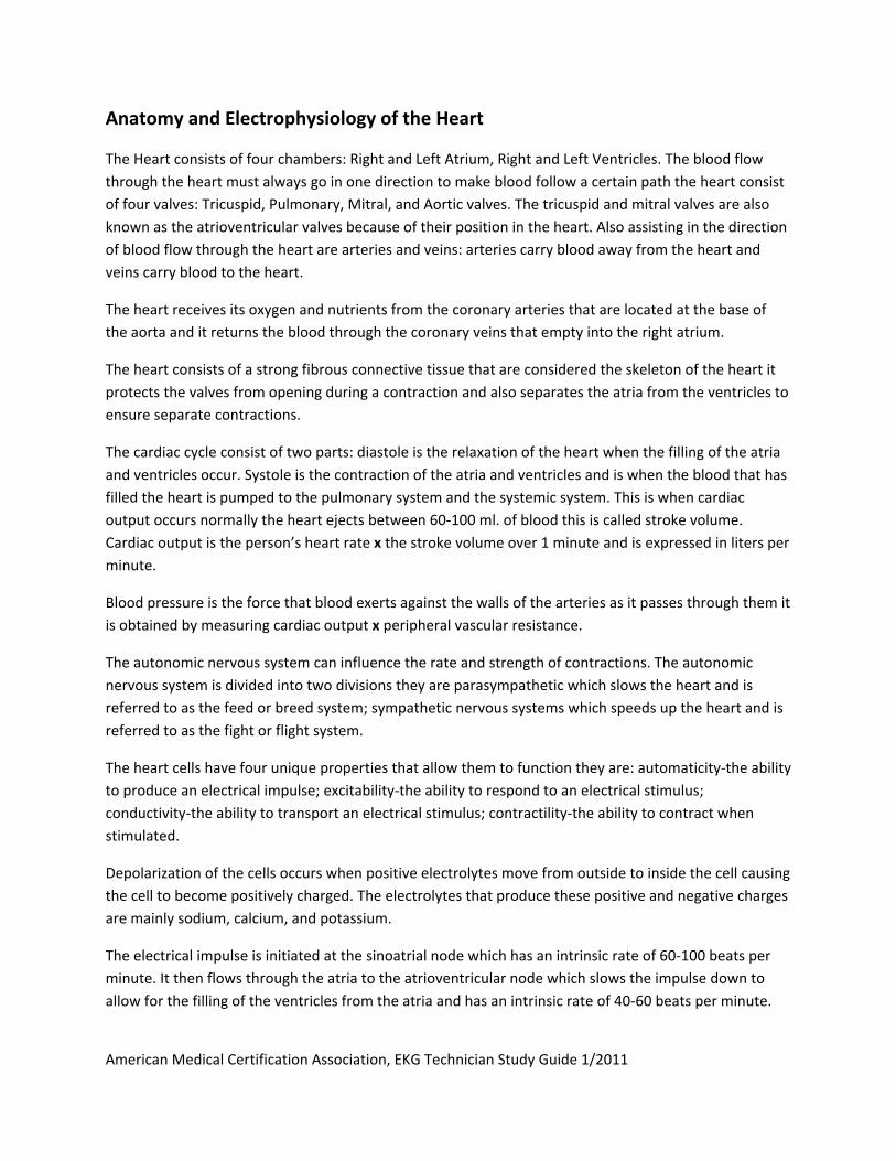
Anatomy and Electrophysiology of the Heart
The Heart consists of four chambers: Right and Left Atrium, Right and Left Ventricles. The blood flow
through the heart must always go in one direction to make blood follow a certain path the heart consist
of four valves: Tricuspid, Pulmonary, Mitral, and Aortic valves. The tricuspid and mitral valves are also
known as the atrioventricular valves because of their position in the heart. Also assisting in the direction
of blood flow through the heart are arteries and veins: arteries carry blood away from the heart and
veins carry blood to the heart.
The heart receives its oxygen and nutrients from the coronary arteries that are located at the base of
the aorta and it returns the blood through the coronary veins that empty into the right atrium.
The heart consists of a strong fibrous connective tissue that are considered the skeleton of the heart it
protects the valves from opening during a contraction and also separates the atria from the ventricles to
ensure separate contractions.
The cardiac cycle consist of two parts: diastole is the relaxation of the heart when the filling of the atria
and ventricles occur. Systole is the contraction of the atria and ventricles and is when the blood that has
filled the heart is pumped to the pulmonary system and the systemic system. This is when cardiac
output occurs normally the heart ejects between 60‐100 ml. of blood this is called stroke volume.
Cardiac output is the person’s heart rate x the stroke volume over 1 minute and is expressed in liters per
minute.
Blood pressure is the force that blood exerts against the walls of the arteries as it passes through them it
is obtained by measuring cardiac output x peripheral vascular resistance.
The autonomic nervous system can influence the rate and strength of contractions. The autonomic
nervous system is divided into two divisions they are parasympathetic which slows the heart and is
referred to as the feed or breed system; sympathetic nervous systems which speeds up the heart and is
referred to as the fight or flight system.
The heart cells have four unique properties that allow them to function they are: automaticity‐the ability
to produce an electrical impulse; excitability‐the ability to respond to an electrical stimulus;
conductivity‐the ability to transport an electrical stimulus; contractility‐the ability to contract when
stimulated.
Depolarization of the cells occurs when positive electrolytes move from outside to inside the cell causing
the cell to become positively charged. The electrolytes that produce these positive and negative charges
are mainly sodium, calcium, and potassium.
The electrical impulse is initiated at the sinoatrial node which has an intrinsic rate of 60‐100 beats per
minute. It then flows through the atria to the atrioventricular node which slows the impulse down to
allow for the filling of the ventricles from the atria and has an intrinsic rate of 40‐60 beats per minute.

American Medical Certification Association, EKG Technician Study Guide 1/2011
The impulse then goes into the purkinje fibers system which has an intrinsic rate of 20‐40 beats per
minute, to be distributed throughout the ventricles.
The Electrocardiogram
The electrocardiogram is the graphic record or tracing of the heart’s electrical activity; the
electrocardiograph is the machine with color coded or numbered lead wires that are placed on the
electrodes that are attached to the patient’s skin.
ECG rhythms may be shown on a display called an oscilloscope or printed on graph paper with static
tracing. The graph paper used to record ECG’s consists of horizontal and vertical lines that form grids the
smallest of the squares represents .04 seconds in duration horizontally or .1 millivolt vertically. The
larger squares equal five small squares which read as .20 seconds horizontally or .5 mV vertically. You
can use the horizontal figures to get distance and rate of the waveforms. You can use the vertical figures
to measure amplitude of the waveforms.
Any abnormal rhythm is called a dysrhythmia. Artifact on a rhythm has no relationship the electrical
activity of the heart and can be caused by patient movement, shivering, muscle tremors, or loose
electrodes.
Each lead provides a different view of the heart. Impulses that travel toward a positive electrode are
recorded as upward deflections. Impulses that travel toward a negative electrode are recorded as a
downward deflection.
Reading an ECG
The ECG technician should read and ECG in a systematic manner. Always remember to compare your
ECG findings to the patient: some dysrhythmias can be life threatening and some can be no problem to
the patient.
The five steps in analyzing an ECG are: heart rate, regularity, P‐waves, QRS complex, and P‐R interval.
Heart rate‐ normal rate for an adult is between 60‐100 bpm. Heart rates above 100 are called
Tachycardia, heart rates below 60 are called bradycardia. There are four methods for obtaining a heart
rate: 6‐second interval X 10 method‐ involves counting the number of QRS complexes on a 6 second
rhythm and multiplying that number by 10, this is the only method that can be used on an irregular
rhythm. The 300,150,100,75,60,50 method‐ involves locating an R‐wave on a bold line on the ECG paper
then finding the next consecutive R wave and counting down from 300 on the subsequent bold lines to
determine the rate. 1500 method‐ you count number of small squares between two consecutive R‐
waves and divide that number by 1500, it is the most accurate method of obtaining the heart rate but
can only be used on regular rhythms. Rate calculators are devices that you use to measure between R‐
waves and it gives you the rate.
American Medical Certification Association, EKG Technician Study Guide 1/2011
You count the QRS complexes to get the Ventricular rate and count the P‐waves to determine the atrial
rate.
Regularity is the second step in analyzing and ECG. To find this you measure the distance between all
the R‐R waves on the strip and if they are all the same the rhythm is called regular if not the rhythm is
called irregular and irregular rhythms are considered abnormal. You can also measure the P‐waves and
this is called the P‐P interval to see if they are regular.
Methods used to measure these intervals are calipers‐ which involves using a device called a caliper to
measure the intervals, which is the quickest and easiest method. Paper and pen method‐ which involves
using a piece of paper and marking an R‐wave then marking the next R‐wave and using that to judge the
R‐waves of the rest of the rhythm. The last method is to count the small squares between each R‐R
interval and see if they are all the same, this method takes longer but does not require any equipment
to use.
Types of irregularities that you may find are irregularly (totally) irregular‐ which means there is no
consistency to the rhythm such as atrial fibrillation, patterned irregularity‐ is where the irregularity
repeats over and over such as AV‐heart blocks, slightly irregular‐ is where there is and abnormality that
does not occur very often such as one PVC in a rhythm strip, and very irregular rhythms‐which means
there are multiple occurrences of an abnormality such as a PVC or PAC in one rhythm strip.
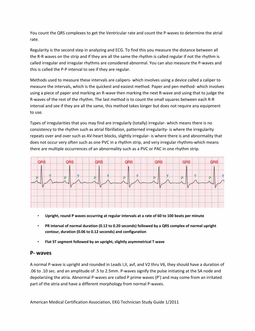
• Upright, round P waves occurring at regular intervals at a rate of 60 to 100 beats per minute
• PR interval of normal duration (0.12 to 0.20 seconds) followed by a QRS complex of normal upright
contour, duration (0.06 to 0.12 seconds) and configuration
• Flat ST segment followed by an upright, slightly asymmetrical T wave
P‐ waves
A normal P‐wave is upright and rounded in Leads I,II, avf, and V2 thru V6, they should have a duration of
.06 to .10 sec. and an amplitude of .5 to 2.5mm. P‐waves signify the pulse initiating at the SA node and
depolarizing the atria. Abnormal P‐waves are called P prime waves (P’) and may come from an irritated
part of the atria and have a different morphology from normal P‐waves.
American Medical Certification Association, EKG Technician Study Guide 1/2011
You can separate the P‐wave into two parts the first half of the P‐wave is the depolarization of the right
atrium and the second half is the depolarization to the left atrium. If there is a dilation of the right
atrium then the P‐wave will have higher amplitude and be tall and rounded, if there is a dilation of the
left atrium then the P‐wave will be notched or elongated.
Inverted P‐waves indicate that the impulse started in or around the AV node. If you have more P‐waves
than ORS complexes then the impulse was blocked at the AV junction and you have a AV heart block.
QRS complexes
Normal ORS complex should be .06 to .12 sec. in duration and 5mm to 30mm in amplitude. A normal
QRS complex indicates the impulse originated above the ventricles and traveled through the ventricles
in a normal fashion.
The Q‐wave is the first negative deflection from the base line after the P‐wave. The R‐wave is the first
positive deflection from the base line after the P‐wave. The S‐wave is the first negative deflection that
extends below the base line after the R‐wave. These definitions are literal and if one wave is missing
then you drop that wave from the complex name (i.e. no negative deflection before the R‐wave then
you have a RS complex).
Abnormal QRS complexes are produced by abnormal depolarization of the ventricles. The duration of
abnormal complexes is usually greater than .12 sec. abnormal QRS complex varies widely and can have
strange, wide, and bizarre shapes. Very tall QRS complexes are usually caused by hypertrophy of one or
both ventricles. Abnormally small or low‐voltage complexes are seen in obese patients, hyper‐thyroid
patients, and pleural effusion.
P‐R intervals
The P‐R interval is the distance from the beginning of the P‐wave to the beginning of the Q‐wave or R‐
wave. It signifies the depolarization of the heart from the SA node through the atria and AV node. The
duration of the P‐R interval should be .12 to .20sec.
P‐R intervals are considered abnormal if they are shorter, longer, or absent. Shorter P‐R intervals
indicate the impulse originated in or around the AV node. Longer P‐R intervals indicate the impulse was
slowed down more than it should have been usually in the AV node. Absent P‐R intervals indicate that
there are too many impulses in the atria to signify a P‐R interval (i.e. atrial fibrillation or flutter).
American Medical Certification Association, EKG Technician Study Guide 1/2011
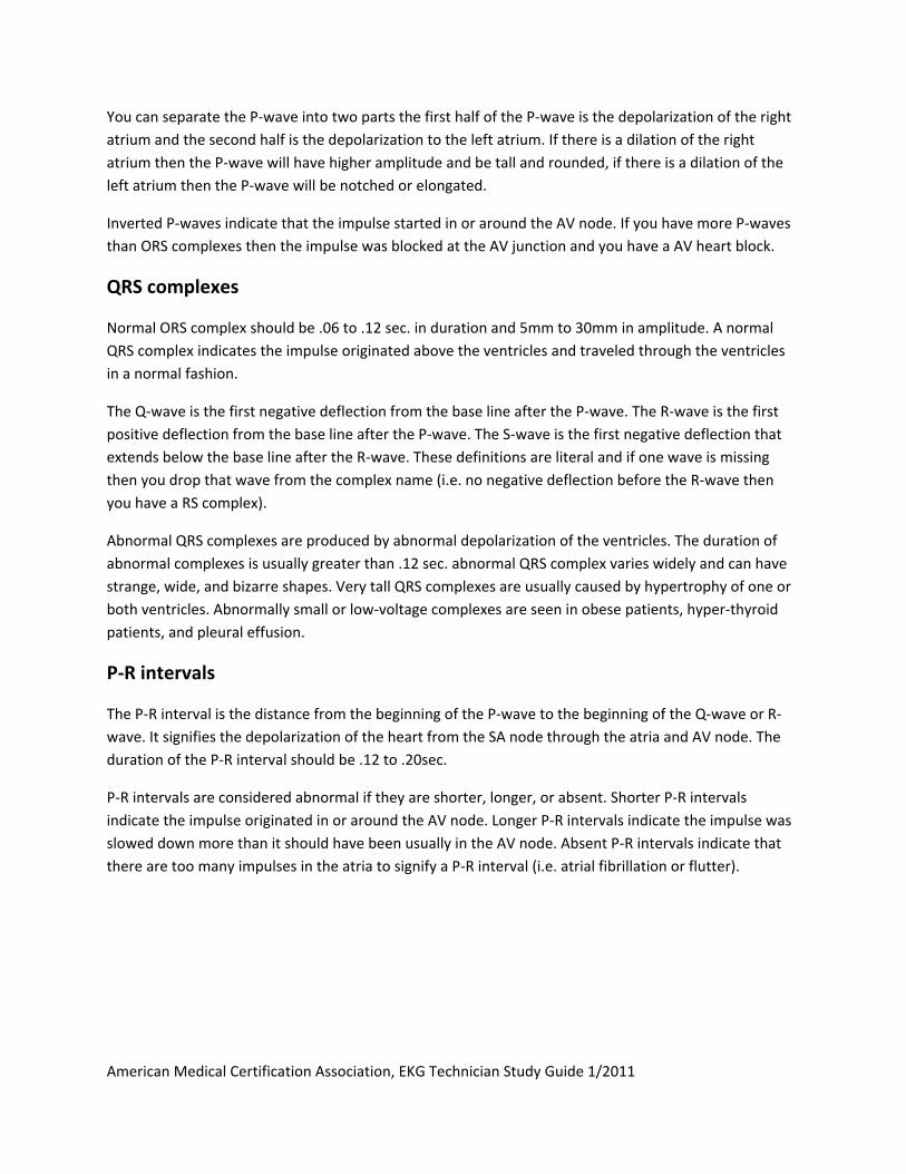
Sinus Dysrhythmia
Sinus dysrhythmias originate from the SA node with that being the case there are only four
dysrhythmias that are associated with the SA node: Tachycardia (heart rate over 100bpm),
Bradycardia(heart rate under 60bpm), sinus arrest (the total absence of the p‐wave, QRS‐complex, and
T‐wave), sinus dysrhythmia(is the same as sinus rhythm except there is the presence of a patterned
irregularity that can be described as slowing the speeding up then slowing again) this will usually happen
with the inspiration and exhalation cycle(normal finding in children, athletes, and older patients)
Atrial Dysrhythmia
Atrial dysrhythmias originate outside the SA node in the atrial tissue or in the intermodal pathways.
The three mechanism responsible for atrial dysrhymias are:
Increased automaticity‐the atrial cells spontaneously depolarize and initiate impulses before the SA
node can generate its pulse.
Triggered activity‐ injured cells sometimes only partially repolarize and this can sometimes lead to
repetitive ectopic firing that may lead to atrial or ventricular tachycardia.
Reentry‐ occurs when an impulse is delayed along a slow conduction pathway and the impulse is able to
remain active long enough to produce another impulse during myocardial repolarization.
Key characteristics of atrial dysrhythmia are shortened or prolonged P‐R intervals p‐waves that differ in
appearance from normal p‐waves, and normal QRS‐complexes.
American Medical Certification Association, EKG Technician Study Guide 1/2011
Types of atrial dysrhythmias and characteristics:
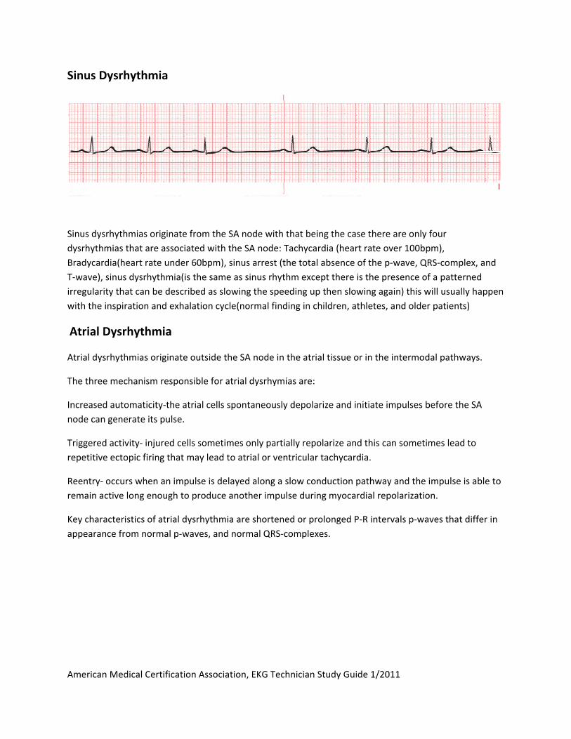
Premature Atrial Complexes
Rate: Depends on the underlying rhythm
Regularity: depends on the number of PAC’s present
P‐waves: may be upright or inverted, will appear different than those of the underlying rhythm
QRS complexs: Normal
PR interval: may be normal, shortened, or prolonged
Atrial tachycardia
Rate: 150‐250 beats per minute
Regularity: regular
P‐waves: may be upright or inverted will appear different from underlying rhythm
QRS‐complex: Normal
PR interval‐ may be normal, shortened, or prolonged
American Medical Certification Association, EKG Technician Study Guide 1/2011
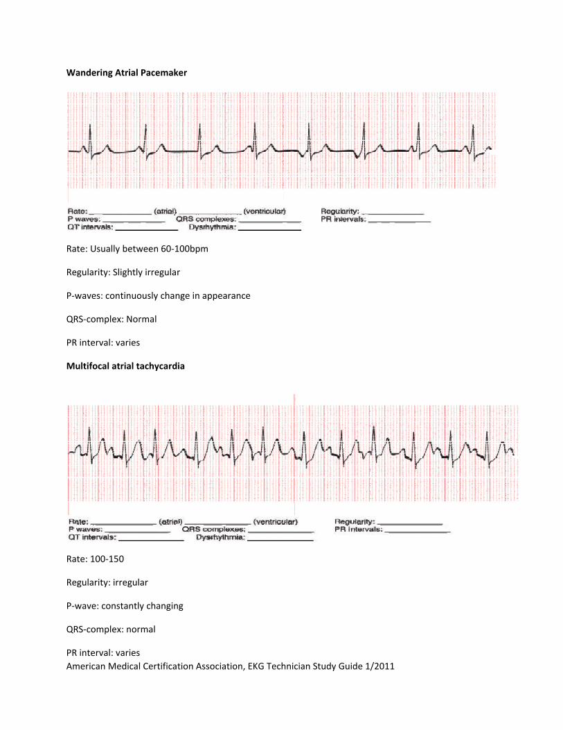
Wandering Atrial Pacemaker
Rate: Usually between 60‐100bpm
Regularity: Slightly irregular
P‐waves: continuously change in appearance
QRS‐complex: Normal
PR interval: varies
Multifocal atrial tachycardia
Rate: 100‐150
Regularity: irregular
P‐wave: constantly changing
QRS‐complex: normal
PR interval: varies

American Medical Certification Association, EKG Technician Study Guide 1/2011
Supraventricular Tachycardia
Rate: usually above 250bpm
Regularity: regular
P‐wave: cannot be identified
QRS‐complex: normal
PR‐interval: absent
Atrial flutter
Rate: atrial between 250‐350, ventricular rate can vary
Regularity: may be regular or irregular
P‐wave: absent (flutter waves)
QRS‐complex: normal
PR‐interval: absent
American Medical Certification Association, EKG Technician Study Guide 1/2011
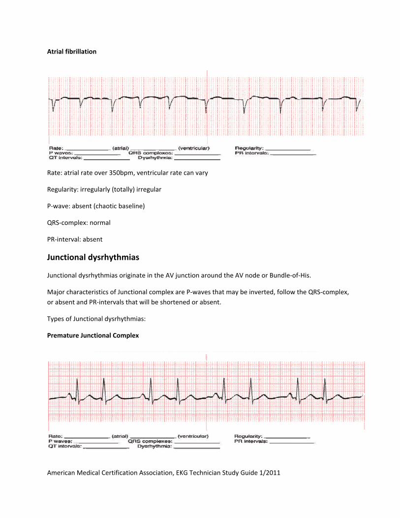
Atrial fibrillation
Rate: atrial rate over 350bpm, ventricular rate can vary
Regularity: irregularly (totally) irregular
P‐wave: absent (chaotic baseline)
QRS‐complex: normal
PR‐interval: absent
Junctional dysrhythmias
Junctional dysrhythmias originate in the AV junction around the AV node or Bundle‐of‐His.
Major characteristics of Junctional complex are P‐waves that may be inverted, follow the QRS‐complex,
or absent and PR‐intervals that will be shortened or absent.
Types of Junctional dysrhythmias:
Premature Junctional Complex
American Medical Certification Association, EKG Technician Study Guide 1/2011
Rate: depends on the underlying rhythm
Regularity: occasional or frequently irregular depends on the number of PJCs
ORS‐complex: Normal
PR Interval: short or absent
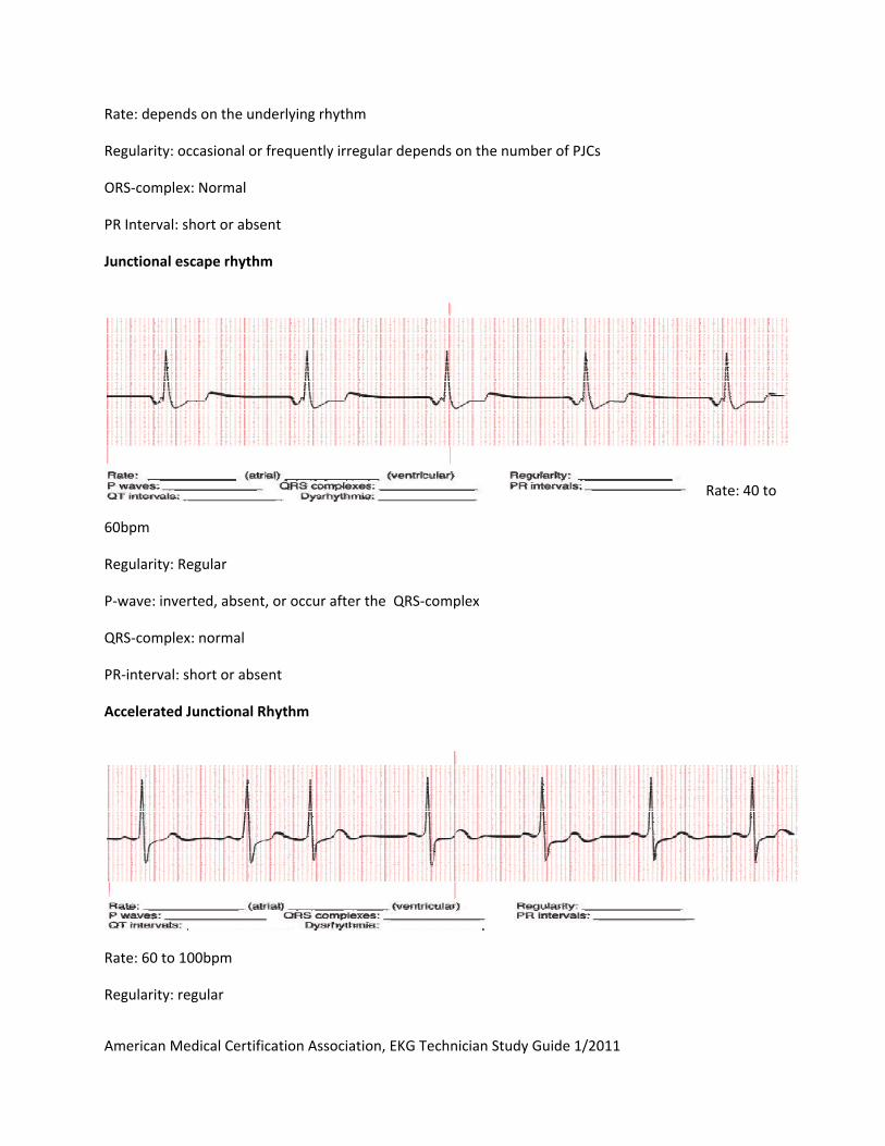
Junctional escape rhythm
Rate: 40 to
60bpm
Regularity: Regular
P‐wave: inverted, absent, or occur after the QRS‐complex
QRS‐complex: normal
PR‐interval: short or absent
Accelerated Junctional Rhythm
Rate: 60 to 100bpm
Regularity: regular
American Medical Certification Association, EKG Technician Study Guide 1/2011
P‐wave: inverted, absent, or occur after the QRS‐complex
QRS‐complex: normal
PR‐interval: short or absent
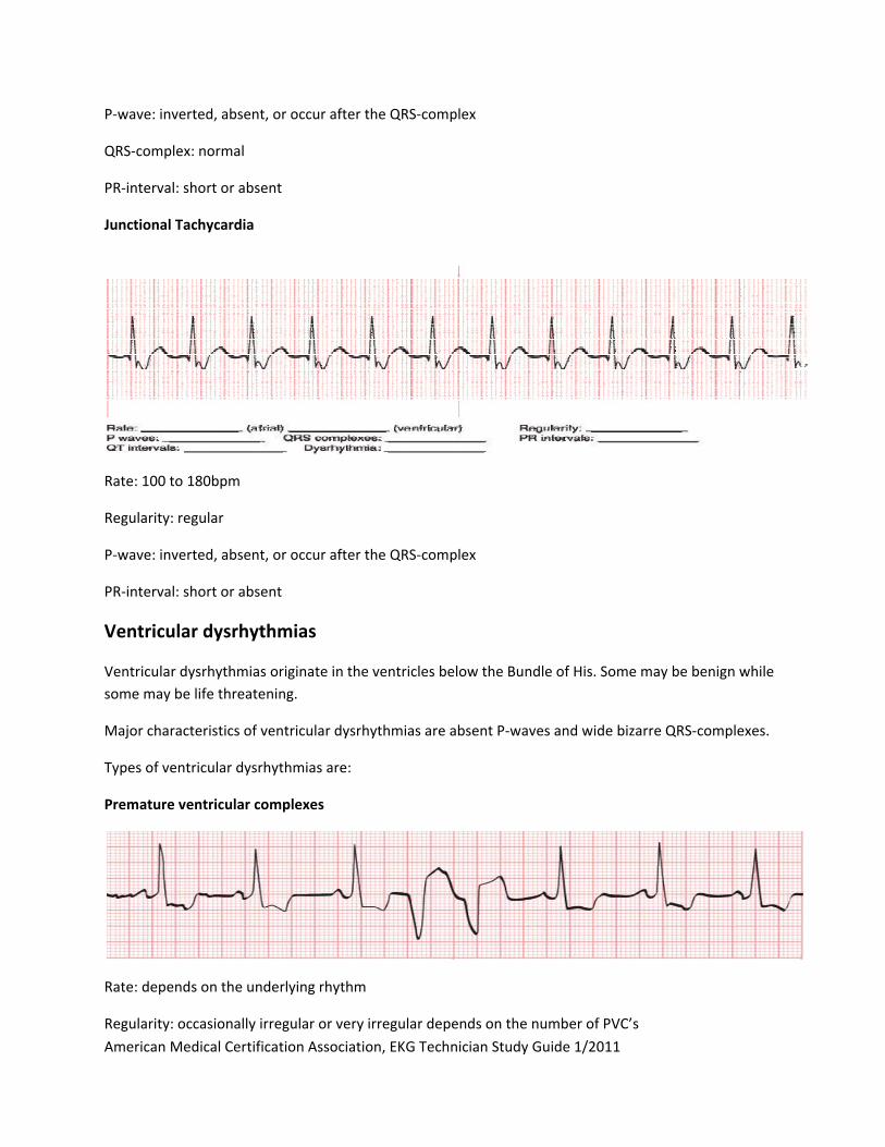
Junctional Tachycardia
Rate: 100 to 180bpm
Regularity: regular
P‐wave: inverted, absent, or occur after the QRS‐complex
PR‐interval: short or absent
Ventricular dysrhythmias
Ventricular dysrhythmias originate in the ventricles below the Bundle of His. Some may be benign while
some may be life threatening.
Major characteristics of ventricular dysrhythmias are absent P‐waves and wide bizarre QRS‐complexes.
Types of ventricular dysrhythmias are:
Premature ventricular complexes
Rate: depends on the underlying rhythm
Regularity: occasionally irregular or very irregular depends on the number of PVC’s

American Medical Certification Association, EKG Technician Study Guide 1/2011
P‐wave: absent at the PVC
QRS‐complex: the PVC will have a wide bizarre looking QRS
PR‐interval: absent
Two PVC’s in a row are called a couplet
Three PVC’s in a row are called a salvo, run, or burst and are considered ventricular tachycardia.
PVC’s occurring on or near the previous T wave (R on T) are likely to precipitate ventricular tachycardia
or fibrillation.
Idioventricular rhythm
Rate: 20‐40bpm
Regularity: regular
P‐wave: absent
QRS‐complex: wide and bizarre
PR‐interval: absent
Accelerated Idioventricular rhythm
Rate:
40‐100bpm
American Medical Certification Association, EKG Technician Study Guide 1/2011
Regularity: regular
P‐wave: absent
QRS‐complex: wide and bizarre
PR‐interval: absent
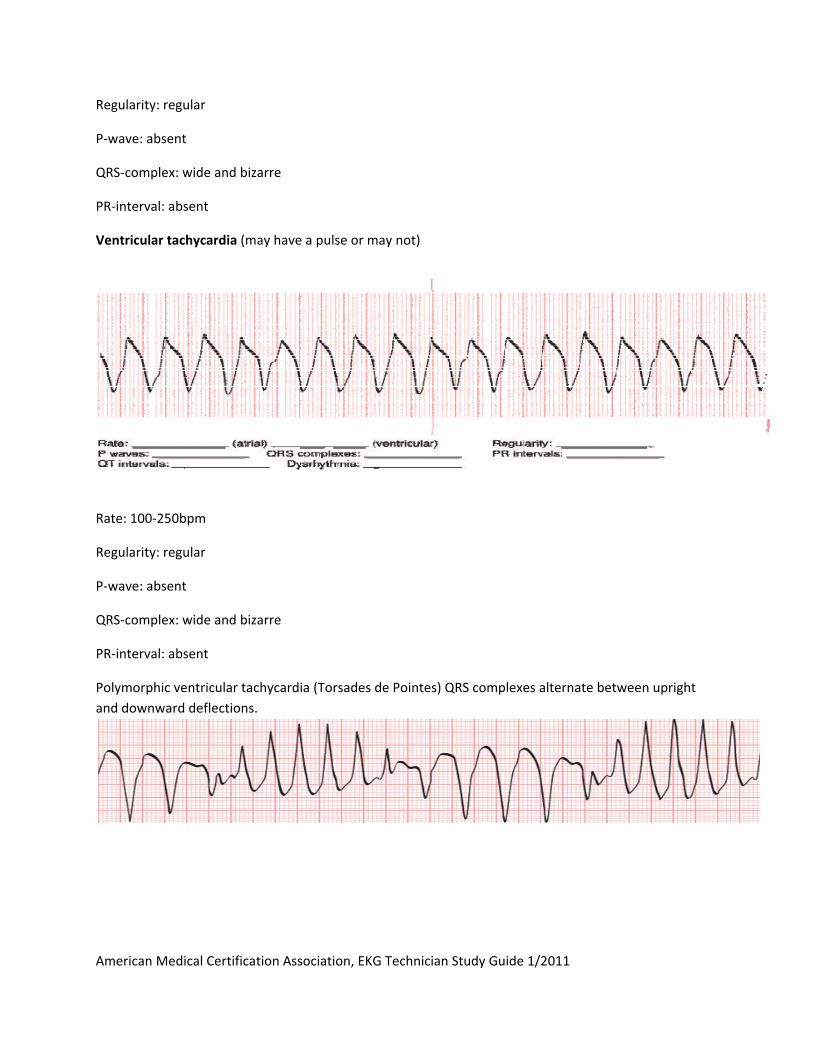
Ventricular tachycardia (may have a pulse or may not)
Rate: 100‐250bpm
Regularity: regular
P‐wave: absent
QRS‐complex: wide and bizarre
PR‐interval: absent
Polymorphic ventricular tachycardia (Torsades de Pointes) QRS complexes alternate between upright
and downward deflections.
American Medical Certification Association, EKG Technician Study Guide 1/2011
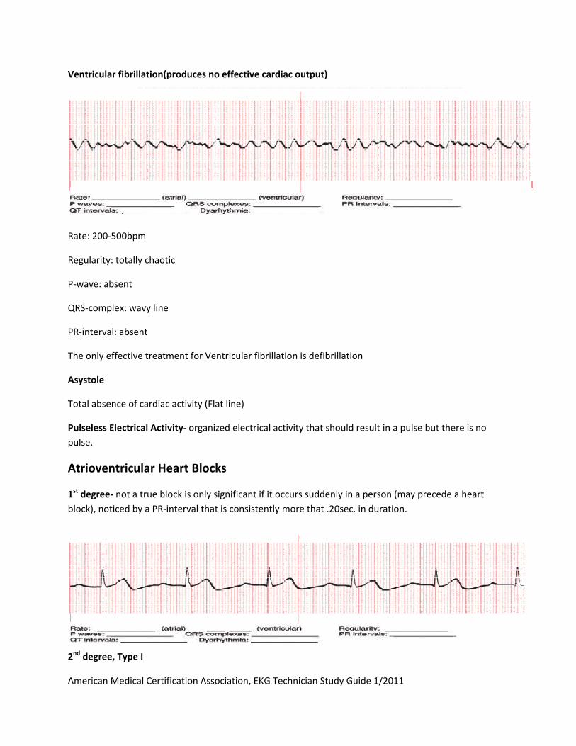
Ventricular fibrillation(produces no effective cardiac output)
Rate: 200‐500bpm
Regularity: totally chaotic
P‐wave: absent
QRS‐complex: wavy line
PR‐interval: absent
The only effective treatment for Ventricular fibrillation is defibrillation
Asystole
Total absence of cardiac activity (Flat line)
Pulseless Electrical Activity‐ organized electrical activity that should result in a pulse but there is no
pulse.
Atrioventricular Heart Blocks
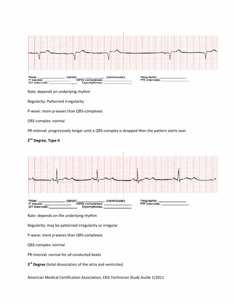
1st degree‐ not a true block is only significant if it occurs suddenly in a person (may precede a heart
block), noticed by a PR‐interval that is consistently more that .20sec. in duration.
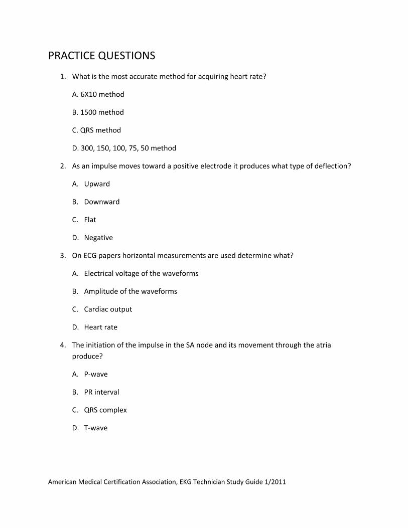
2nd degree, Type I
American Medical Certification Association, EKG Technician Study Guide 1/2011
Rate: depends on underlying rhythm
Regularity: Patterned irregularity
P‐wave: more p‐waves than QRS‐complexes
ORS‐complex: normal
PR‐interval: progressively longer until a QRS‐complex is dropped then the pattern starts over
2nd Degree, Type II
Rate: depends on the underlying rhythm
Regularity: may be patterned irregularity or irregular
P‐wave: more p‐waves than QRS‐complexes
QRS‐complex: normal
PR‐interval: normal for all conducted beats
3rd Degree (total dissociation of the atria and ventricles)

American Medical Certification Association, EKG Technician Study Guide 1/2011
Rate: normal atrial rate, slow ventricular rate
Regularity: atrial and ventricular rate are regular but not working together
P‐wave: more p‐waves than QRS‐complexes
QRS‐complex: normal or wide and bizarre depends on where the impulse is from
PR‐interval: absent
Hypotrophy‐ condition, in which the muscular wall enlarges to increase the strength of the contraction,
usually happens in the ventricles.
Dilation‐ condition, in which the muscular wall thins and expands to accommodate more blood, usually
happens in the atria.
Preexcitation Syndromes
Wolff‐Parkinson‐White Syndrome (WPW)‐ condition in which the impulse takes a different path than
the AV‐node (bundle of Kent), and is characterized by the slurring of the R‐wave called the Delta wave.
Patients with this are prone to SVT.
Lown‐Ganong‐Levine Syndrome (LGL)‐ impulse travels through the James fibers rather than the AV‐
node. Only indication of this in the ECG will be a shortened PR‐interval.
American Medical Certification Association, EKG Technician Study Guide 1/2011
PRACTICE QUESTIONS
1. What is the most accurate method for acquiring heart rate?
A. 6X10 method
B. 1500 method
C. QRS method
D. 300, 150, 100, 75, 50 method
2. As an impulse moves toward a positive electrode it produces what type of deflection?
A. Upward
B. Downward
C. Flat
D. Negative
3. On ECG papers horizontal measurements are used determine what?
A. Electrical voltage of the waveforms
B. Amplitude of the waveforms
C. Cardiac output
D. Heart rate
4. The initiation of the impulse in the SA node and its movement through the atria
produce?
A. P‐wave
B. PR interval
C. QRS complex
D. T‐wave
American Medical Certification Association, EKG Technician Study Guide 1/2011
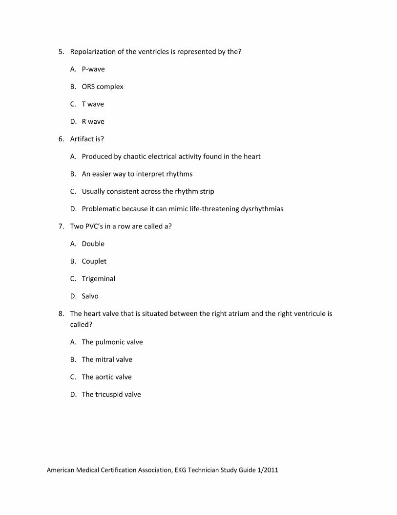
5. Repolarization of the ventricles is represented by the?
A. P‐wave
B. ORS complex
C. T wave
D. R wave
6. Artifact is?
A. Produced by chaotic electrical activity found in the heart
B. An easier way to interpret rhythms
C. Usually consistent across the rhythm strip
D. Problematic because it can mimic life‐threatening dysrhythmias
7. Two PVC’s in a row are called a?
A. Double
B. Couplet
C. Trigeminal
D. Salvo
8. The heart valve that is situated between the right atrium and the right ventricule is
called?
A. The pulmonic valve
B. The mitral valve
C. The aortic valve
D. The tricuspid valve
American Medical Certification Association, EKG Technician Study Guide 1/2011
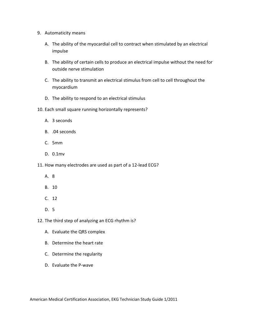
9. Automaticity means
A. The ability of the myocardial cell to contract when stimulated by an electrical
impulse
B. The ability of certain cells to produce an electrical impulse without the need for
outside nerve stimulation
C. The ability to transmit an electrical stimulus from cell to cell throughout the
myocardium
D. The ability to respond to an electrical stimulus
10. Each small square running horizontally represents?
A. 3 seconds
B. .04 seconds
C. 5mm
D. 0.1mv
11. How many electrodes are used as part of a 12‐lead ECG?
A. 8
B. 10
C. 12
D. 5
12. The third step of analyzing an ECG rhythm is?
A. Evaluate the QRS complex
B. Determine the heart rate
C. Determine the regularity
D. Evaluate the P‐wave
American Medical Certification Association, EKG Technician Study Guide 1/2011
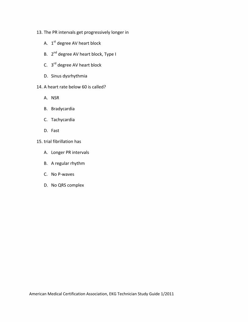
13. The PR intervals get progressively longer in
A. 1st degree AV heart block
B. 2nd degree AV heart block, Type I
C. 3rd degree AV heart block
D. Sinus dysrhythmia
14. A heart rate below 60 is called?
A. NSR
B. Bradycardia
C. Tachycardia
D. Fast
15. trial fibrillation has
A. Longer PR intervals
B. A regular rhythm
C. No P‐waves
D. No QRS complex
American Medical Certification Association, EKG Technician Study Guide 1/2011
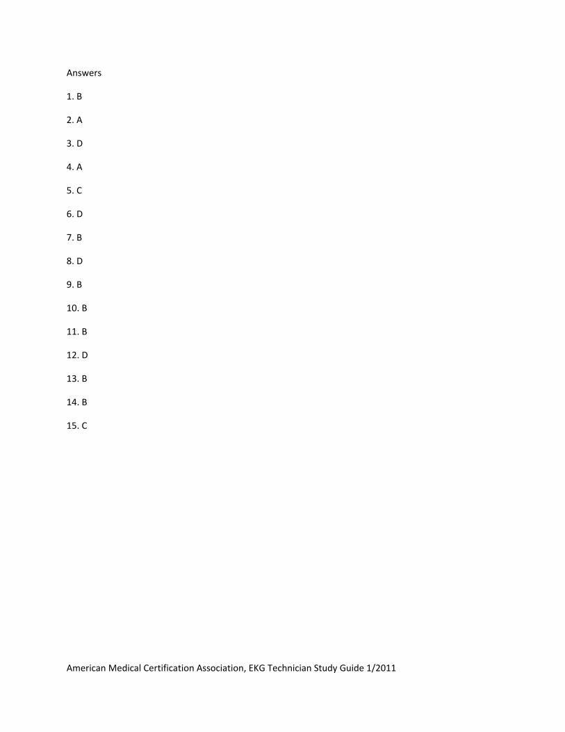
Answers
1. B
2. A
3. D
4. A
5. C
6. D
7. B
8. D
9. B
10. B
11. B
12. D
13. B
14. B
15. C