Effects of rolipram and diazepam on the adaptive changes induced by morphine withdrawal in the...
8
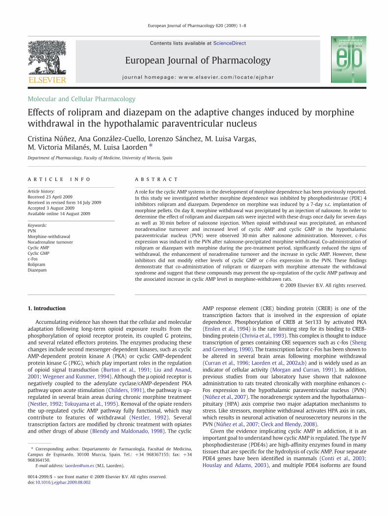
Molecular and Cellular Pharmacology Effects of rolipram and diazepam on the adaptive changes induced by morphine withdrawal in the hypothalamic paraventricular nucleus Cristina Núñez, Ana González-Cuello, Lorenzo Sánchez, M. Luisa Vargas, M. Victoria Milanés, M. Luisa Laorden ⁎ Department of Pharmacology, Faculty of Medicine, University of Murcia, Spain abstract article info Article history: Received 23 April 2009 Received in revised form 14 July 2009 Accepted 3 August 2009 Available online 14 August 2009 Keywords: PVN Morphine-withdrawal Noradrenaline turnover Cyclic AMP Cyclic GMP c-Fos Rolipram Diazepam A role for the cyclic AMP systems in the development of morphine dependence has been previously reported. In this study we investigated whether morphine dependence was inhibited by phosphodiesterase (PDE) 4 inhibitors rolipram and diazepam. Dependence on morphine was induced by a 7-day s.c. implantation of morphine pellets. On day 8, morphine withdrawal was precipitated by an injection of naloxone. In order to determine the effect of rolipram and diazepam rats were injected with these drugs once daily for seven days as well as 30 min before of naloxone injection. When opioid withdrawal was precipitated, an enhanced noradrenaline turnover and increased level of cyclic AMP and cyclic GMP in the hypothalamic paraventricular nucleus (PVN) were observed 30 min after naloxone administration. Moreover, c-Fos expression was induced in the PVN after naloxone-precipitated morphine withdrawal. Co-administration of rolipram or diazepam with morphine during the pre-treatment period, significantly reduced the signs of withdrawal, the enhancement of noradrenaline turnover and the increase in cyclic AMP. However, these inhibitors did not modify either levels of cyclic GMP or c-Fos expression in the PVN. These findings demonstrate that co-administration of rolipram or diazepam with morphine attenuate the withdrawal syndrome and suggest that these compounds may prevent the up-regulation of the cyclic AMP pathway and the associated increase in cyclic AMP level in morphine-withdrawn rats. © 2009 Elsevier B.V. All rights reserved. 1. Introduction Accumulating evidence has shown that the cellular and molecular adaptation following long-term opioid exposure results from the phosphorylation of opioid receptor protein, its coupled G proteins, and several related effectors proteins. The enzymes producing these changes include second messenger-dependent kinases, such as cyclic AMP-dependent protein kinase A (PKA) or cyclic GMP-dependent protein kinase G (PKG), which play important roles in the regulation of opioid signal transduction (Burton et al., 1991; Liu and Anand, 2001; Wegener and Kunmer, 1994). Although the μ opioid receptor is negatively coupled to the adenylate cyclase/cAMP-dependent PKA pathway upon acute stimulation (Childers, 1991), the pathway is up- regulated in several brain areas during chronic morphine treatment (Nestler, 1992; Tokuyama et al., 1995). Removal of the opiate renders the up-regulated cyclic AMP pathway fully functional, which may contribute to features of withdrawal (Nestler, 1992). Several transcription factors are modified by chronic treatment with opiates and other drugs of abuse (Blendy and Maldonado, 1998). The cyclic AMP response element (CRE) binding protein (CREB) is one of the transcription factors that is involved in the expression of opiate dependence. Phosphorylation of CREB at Ser133 by activated PKA (Enslen et al., 1994) is the rate limiting step for its binding to CREB- binding protein (Chrivia et al., 1993). This complex is thought to induce transcription of genes containing CRE sequences such as c-fos (Sheng and Greenberg, 1990). The transcription factor c-Fos has been shown to be altered in several brain areas following morphine withdrawal (Curran et al., 1996; Laorden et al., 2002a,b) and is widely used as an indicator of cellular activity (Morgan and Curran, 1991). In addition, previous studies from our laboratory have shown that naloxone administration to rats treated chronically with morphine enhances c- Fos expression in the hypothalamic paraventricular nucleus (PVN) (Núñez et al., 2007). The noradrenergic system and the hypothalamus– pituitary (HPA) axis comprise two major adaptation mechanisms to stress. Like stressors, morphine withdrawal activates HPA axis in rats, which results in neuronal activation of neurosecretory neurons in the PVN (Núñez et al., 2007; Cleck and Blendy, 2008). Given the evidence implicating cyclic AMP in addiction, it is an important goal to understand how cyclic AMP is regulated. The type IV phosphodiesterase (PDE4s) are high-affinity enzymes found in many tissues that are specific for the hydrolysis of cyclic AMP. Four separate PDE4 genes have been identified in mammals (Conti et al., 2003; Houslay and Adams, 2003), and multiple PDE4 isoforms are found European Journal of Pharmacology 620 (2009) 1–8 ⁎ Corresponding author. Departamento de Farmacología, Facultad de Medicina, Campus de Espinardo, 30100 Murcia, Spain. Tel.: +34 968367155; fax: +34 968364150. E-mail address: [email protected] (M.L. Laorden). 0014-2999/$ – see front matter © 2009 Elsevier B.V. All rights reserved. doi:10.1016/j.ejphar.2009.08.002 Contents lists available at ScienceDirect European Journal of Pharmacology journal homepage: www.elsevier.com/locate/ejphar
-
Upload
cristina-nunez -
Category
Documents
-
view
213 -
download
0
Transcript of Effects of rolipram and diazepam on the adaptive changes induced by morphine withdrawal in the...
European Journal of Pharmacology 620 (2009) 1–8
Contents lists available at ScienceDirect
European Journal of Pharmacology
j ourna l homepage: www.e lsev ie r.com/ locate /e jphar
Molecular and Cellular Pharmacology
Effects of rolipram and diazepam on the adaptive changes induced by morphinewithdrawal in the hypothalamic paraventricular nucleus
Cristina Núñez, Ana González-Cuello, Lorenzo Sánchez, M. Luisa Vargas,M. Victoria Milanés, M. Luisa Laorden ⁎Department of Pharmacology, Faculty of Medicine, University of Murcia, Spain
⁎ Corresponding author. Departamento de FarmacoCampus de Espinardo, 30100 Murcia, Spain. Tel.:968364150.
E-mail address: [email protected] (M.L. Laorden).
0014-2999/$ – see front matter © 2009 Elsevier B.V. Aldoi:10.1016/j.ejphar.2009.08.002
a b s t r a c t
a r t i c l e i n f oArticle history:Received 23 April 2009Received in revised form 14 July 2009Accepted 3 August 2009Available online 14 August 2009
Keywords:PVNMorphine-withdrawalNoradrenaline turnoverCyclic AMPCyclic GMPc-FosRolipramDiazepam
A role for the cyclic AMP systems in the development of morphine dependence has been previously reported.In this study we investigated whether morphine dependence was inhibited by phosphodiesterase (PDE) 4inhibitors rolipram and diazepam. Dependence on morphine was induced by a 7-day s.c. implantation ofmorphine pellets. On day 8, morphine withdrawal was precipitated by an injection of naloxone. In order todetermine the effect of rolipram and diazepam rats were injected with these drugs once daily for seven daysas well as 30 min before of naloxone injection. When opioid withdrawal was precipitated, an enhancednoradrenaline turnover and increased level of cyclic AMP and cyclic GMP in the hypothalamicparaventricular nucleus (PVN) were observed 30 min after naloxone administration. Moreover, c-Fosexpression was induced in the PVN after naloxone-precipitated morphine withdrawal. Co-administration ofrolipram or diazepam with morphine during the pre-treatment period, significantly reduced the signs ofwithdrawal, the enhancement of noradrenaline turnover and the increase in cyclic AMP. However, theseinhibitors did not modify either levels of cyclic GMP or c-Fos expression in the PVN. These findingsdemonstrate that co-administration of rolipram or diazepam with morphine attenuate the withdrawalsyndrome and suggest that these compounds may prevent the up-regulation of the cyclic AMP pathway andthe associated increase in cyclic AMP level in morphine-withdrawn rats.
© 2009 Elsevier B.V. All rights reserved.
1. Introduction
Accumulating evidence has shown that the cellular and molecularadaptation following long-term opioid exposure results from thephosphorylation of opioid receptor protein, its coupled G proteins,and several related effectors proteins. The enzymes producing thesechanges include second messenger-dependent kinases, such as cyclicAMP-dependent protein kinase A (PKA) or cyclic GMP-dependentprotein kinase G (PKG), which play important roles in the regulationof opioid signal transduction (Burton et al., 1991; Liu and Anand,2001; Wegener and Kunmer, 1994). Although the μ opioid receptor isnegatively coupled to the adenylate cyclase/cAMP-dependent PKApathway upon acute stimulation (Childers, 1991), the pathway is up-regulated in several brain areas during chronic morphine treatment(Nestler, 1992; Tokuyama et al., 1995). Removal of the opiate rendersthe up-regulated cyclic AMP pathway fully functional, which maycontribute to features of withdrawal (Nestler, 1992). Severaltranscription factors are modified by chronic treatment with opiatesand other drugs of abuse (Blendy and Maldonado, 1998). The cyclic
logía, Facultad de Medicina,+34 968367155; fax: +34
l rights reserved.
AMP response element (CRE) binding protein (CREB) is one of thetranscription factors that is involved in the expression of opiatedependence. Phosphorylation of CREB at Ser133 by activated PKA(Enslen et al., 1994) is the rate limiting step for its binding to CREB-binding protein (Chrivia et al., 1993). This complex is thought to inducetranscription of genes containing CRE sequences such as c-fos (Shengand Greenberg, 1990). The transcription factor c-Fos has been shown tobe altered in several brain areas following morphine withdrawal(Curran et al., 1996; Laorden et al., 2002a,b) and is widely used as anindicator of cellular activity (Morgan and Curran, 1991). In addition,previous studies from our laboratory have shown that naloxoneadministration to rats treated chronically with morphine enhances c-Fos expression in the hypothalamic paraventricular nucleus (PVN)(Núñez et al., 2007). The noradrenergic system and the hypothalamus–pituitary (HPA) axis comprise two major adaptation mechanisms tostress. Like stressors, morphine withdrawal activates HPA axis in rats,which results in neuronal activation of neurosecretory neurons in thePVN (Núñez et al., 2007; Cleck and Blendy, 2008).
Given the evidence implicating cyclic AMP in addiction, it is animportant goal to understand how cyclic AMP is regulated. The type IVphosphodiesterase (PDE4s) are high-affinity enzymes found in manytissues that are specific for the hydrolysis of cyclic AMP. Four separatePDE4 genes have been identified in mammals (Conti et al., 2003;Houslay and Adams, 2003), and multiple PDE4 isoforms are found

2 C. Núñez et al. / European Journal of Pharmacology 620 (2009) 1–8
throughout the brain, including the PVN (Cherry and Davis, 1999;Perez Torres et al., 2000). Studies of PDE4 function using the selectiveinhibitor rolipram indicate that PDE4 is involved in physicaldependence and tolerance to morphine (Itoh et al., 1998; Hamdyet al., 2001; Mamiya et al., 2001). However, there are no studiesevaluating the role of PDE4 selective inhibitor rolipram (Beavo, 1995)and diazepam, a benzodiazepine receptor agonist, which suppressPDE4 activity (Collado et al., 1998) to abolish the adaptive changesobserved in PVN during morphine withdrawal. Therefore, the presentwork was designed to investigate the role of both drugs, which act onthe cyclic AMP system in the changes of catecholaminergic activityand gene expression observed during morphine withdrawal in thePVN, which is involved in the neuroendocrine control of the HPA axis.
2. Materials and methods
Male Sprague–Dawley rats (220–240 g at the beginning of theexperiments) were housed four-to-five per cage under a 12-h light/dark cycle (light: 8:00–20:00 h) in a room with controlled temper-ature (22±2 ºC), humidity (50±10%), food and water available adlibitum and prehandled for several days preceding the experiment tominimize stress, as previously described (Laorden et al., 2000). Allsurgical and experimental procedures were performed in accordancewith the European Communities Council Directive of 24 November1986 (86/609/EEC) and the local Committee. The drug and moleculartarget nomenclature used in this study conforms to the BJP's Guide toReceptors and Channels (Alexander et al., 2008).
2.1. Animals and treatments
Rats were rendered tolerant/dependent on morphine by s.c.implantation of morphine base pellets (75 mg), one on day 1, twoon day 3 and three on day 5, under light ether anaesthesia. Controlanimals were implanted with placebo pellets containing lactoseinstead of morphine, on the same time schedule. These procedureshave repeatedly been shown to induce both tolerance and depen-dence as measured behaviourally and biochemically (Fuertes et al.,2000; Laorden et al., 2000). In order to determine the effect ofrolipram and diazepam, the animals were injected with rolipram(1 mg/kg i.p.), diazepam (0.25 mg/kg, i.p.) or vehicle (dymethylsulf-oxide 20%, i.p.) once daily for seven days.
On day 8, the animals pre-treated with morphine or placebopellets were injected with rolipram, diazepam or vehicle 30 minbefore the administration of saline s.c. or naloxone (5 mg/kg s.c.).Thirty min after saline s.c. or naloxone (5 mg/kg s.c.) rats weredecapitated and the brains were removed rapidly, fresh–frozen, andstored immediately at −80 ºC until use. The hypothalamic tissuecontaining PVN was dissected according to the technique of Palkovits(Palkovits, 1973; Palkovits and Brownstein, 1988) and the PVNcorresponds to those in Plates 25 and 26 in the atlas of Paxinos andWatson (1998).
In order to determine the effect of inhibiting protein phosphor-ylation on the morphine withdrawal-induced changes in c-Fosexpression at the PVN and brainstem catecholaminergic areas, c-Foswas determined in morphine dependent and control rats treated withSL327 (a selective inhibitor of MEK; Atkins et al., 1998) 1 h before theadministration of naloxone or saline. This inhibitor was dissolved indymethylsulfoxide (DMSO) 100%. On the basis of our previousexperiments (Almela et al., 2007; Núñez et al., 2008) SL327 wasinjected intraperitoneally (ip) at doses of 100 mg/kg.
2.2. Estimation of noradrenaline and its metabolite in the PVN
Noradrenaline and its metabolite 3-metoxy-4-hydroxyphenyl-lethylen glycol (MHPG) in the central nervous system (CNS) weredetermined by high-performance liquid chromatography (HPLC) with
electrochemical detection. Each tissue was weighed, placed in a dry-cooled propylene vial and homogenized with a Polytron-Typehomogenizer in 600 μl perchloric acid (0.1 M). The homogenateswere then centrifuged (27,167 g, 4 ºC, 15 min), the supernatant layerwas removed into a 1-ml syringe and filtered through a 0.22 μm filter(Millipore, Bedford, USA). Two aliquots of the supernatant from thesame tissue sample were used, the first for analysis of noradrenalineand the second for analysis of MHPG. Ten μl of the first aliquot of eachsample was injected into a 5-μm C18 reverse phase column (Waters,Milford, MA, USA) through a Rheodyne syringe-loading injector 200 μlloop. Electrochemical detection was accomplished with a glassycarbon electrode set at a potential of +0.65 with respect to the Ag/AgCl reference electrode (Waters). The mobile phase consisted of a95:5 (v/v) mixture of water and methanol with sodium acetate(50 mM), citric acid (20 mM), L-octyl-sodium sulfonate (3.75 mM),di-n-butylamine (1 mM) and EDTA (0.135 mM), adjusted to pH 4.3.The flow rate was 0.9 ml/min, and chromatographic data wereanalysed with Millenium 2010 Chromatography Manager Equipment(Millipore). Under these conditions, MHPG was not detected. Since inthe rat CNSmost of MHPG is conjugatedwith sulphate, themethod forthe determination of total MHPG in the PVN was based on the acid-catalysed hydrolysis of MHPG-sulphate (Artigas et al., 1986; Looking-land et al., 1991). The aliquots for MHPG analysis were kept inpolypropylene screw-capped tubes for 5 min in a water bath at 100 ºC.The tubes were then cooled on ice and centrifuged (7245 g, 4 ºC,10 min). An aliquot of the supernatant of the hydrolysed samples wasinjected into the HPLC. Noradrenaline and MHPG were simultaneous-ly detected and were quantified by reference to calibration curves runat the beginning and the end of each series of assays. Linear rela-tionships were observed between the amount of standard injectedand the peak height measured. The content of noradrenaline andMHPG in the PVN was expressed as ng/g of tissue.
2.3. Measurement of cyclic AMP
The levels of cyclic AMP were measured by radioimmunoassay[I125] TME-S-cAMP; Diagnostic Pasteur, France), according to manu-facture instructions. The tissue was weighed and homogenized(setting 4 for 30 s) and centrifuged (10,000 g, 4 ºC, 15 min). Thesupernatants were treated with potassium hydroxide until pH 6.2 wasreached. The sensitivity of the assay was 2 pmol/ml. Intra and inter-assay coefficients of variation were 7.7 and 8.2%, respectively. Theantibody cross-reacted 100% with 3′,5′-cyclic AMP and <0.3% withother nucleotides. Concentrations of cyclic AMP were expressed aspmol/g of tissue.
2.4. Measurement of cyclic GMP
The levels of cyclic GMP were measured by radioimmunoassay [I125]TME-S-cGMP; Immunotech, France), according to the manufacturer'sinstructions. The tissue wasweighed and homogenized in perchloric acidand centrifuged (10,000 g, 4 ºC, 15 min). The supernatants weresuccinylated until the succinic anhydride was completely dissolved. Thesensitivity of the assay was 10 pM. The antibody cross-reacted 100%withsuccinyl cyclic GMP, 0.003% with succinyl cyclic AMP, 1.9% with CyclicGMP and 0.0001% with cyclic AMP. Concentrations of cyclic GMP wereexpressed as pmol/g of tissue.
2.5. Tissue preparation for immunohistochemistry
Ninety min after administration of naloxone or saline, rats weredeeply anaesthetized with an overdose of pentobarbital (100 mg/kgip) and perfused transcardially with 300 ml of phosphate-bufferedsaline (PBS pH 7.4; 1 mM NaF, that was included in all buffers andincubation solutions) followed by 500 ml of fixative containing 4%paraformaldehyde in PBS. After removal of the perfused brains, they
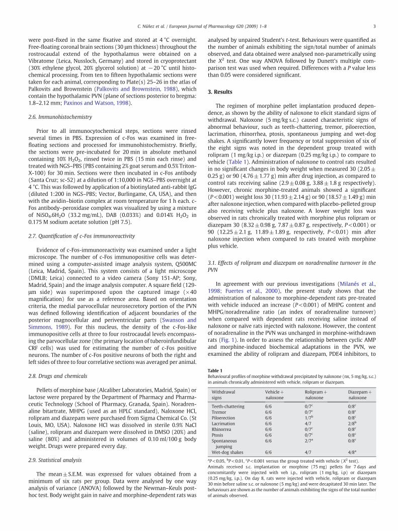
Table 1Behavioural profiles of morphine withdrawal precipitated by naloxone (nx, 5 mg/kg, s.c.)in animals chronically administered with vehicle, rolipram or diazepam.
Withdrawalsigns
Vehicle+naloxone
Rolipram+naloxone
Diazepam+naloxone
Teeth-chattering 6/6 0/7c 0/8c
Tremor 6/6 0/7c 0/8c
Piloerection 6/6 1/7b 0/8c
Lacrimation 6/6 4/7 2/8b
Rhinorrea 6/6 0/7c 0/8c
Ptosis 6/6 0/7c 0/8c
Spontaneousjumping
6/6 2/7a 0/8c
Wet-dog shakes 6/6 4/7 4/8a
aP<0.05, bP<0.01, cP<0.001 versus the group treated with vehicle (Х2 test).Animals received s.c. implantation or morphine (75 mg) pellets for 7 days andconcomitantly were injected with veh i.p., rolipram (1 mg/kg, i.p) or diazepam(0.25 mg/kg, i.p.). On day 8, rats were injected with vehicle, rolipram or diazepam30 min before saline s.c. or naloxone (5 mg/kg) and were decapitated 30 min later. Thebehaviours are shown as the number of animals exhibiting the signs of the total numberof animals observed.
3C. Núñez et al. / European Journal of Pharmacology 620 (2009) 1–8
were post-fixed in the same fixative and stored at 4 °C overnight.Free-floating coronal brain sections (30 μm thickness) throughout therostrocaudal extend of the hypothalamus were obtained on aVibratome (Leica, Nussloch, Germany) and stored in cryoprotectant(30% ethylene glycol, 20% glycerol solution) at −20 °C until histo-chemical processing. From ten to fifteen hypothalamic sections weretaken for each animal, corresponding to Plate(s) 25–26 in the atlas ofPalkovits and Brownstein (Palkovits and Brownstein, 1988), whichcontain the hypothalamic PVN (plane of sections posterior to bregma:1.8–2.12 mm; Paxinos and Watson, 1998).
2.6. Immunohistochemistry
Prior to all immunocytochemical steps, sections were rinsedseveral times in PBS. Expression of c-Fos was examined in free-floating sections and processed for immunohistochemistry. Briefly,the sections were pre-incubated for 20 min in absolute methanolcontaining 10% H2O2, rinsed twice in PBS (15 min each rinse) andtreatedwith NGS–PBS (PBS containing 2% goat serum and 0.5% Triton-X-100) for 30 min. Sections were then incubated in c-Fos antibody(Santa Cruz; sc-52) at a dilution of 1:10,000 in NGS–PBS overnight at4 °C. This was followed by application of a biotinylated anti-rabbit IgG(diluted 1:200 in NGS–PBS; Vector, Burlingame, CA, USA), and thenwith the avidin–biotin complex at room temperature for 1 h each. c-Fos antibody–peroxidase complex was visualized by using a mixtureof NiSO4.6H2O (33.2 mg/mL), DAB (0.033%) and 0.014% H2O2 in0.175 M sodium acetate solution (pH 7.5).
2.7. Quantification of c-Fos immunoreactivity
Evidence of c-Fos-immunoreactivity was examined under a lightmicroscope. The number of c-Fos immunopositive cells was deter-mined using a computer-assisted image analysis system, Q500MC(Leica, Madrid, Spain). This system consists of a light microscope(DMLB; Leica) connected to a video camera (Sony 151-AP; Sony,Madrid, Spain) and the image analysis computer. A square field (129-μm side) was superimposed upon the captured image (×40magnification) for use as a reference area. Based on orientationcriteria, the medial parvocellular neurosecretory portion of the PVNwas defined following identification of adjacent boundaries of theposterior magnocellular and periventricular parts (Swanson andSimmons, 1989). For this nucleus, the density of the c-Fos-likeimmunopositive cells at three to four rostrocaudal levels encompass-ing the parvocellular zone (the primary location of tuberoinfundibularCRF cells) was used for estimating the number of c-Fos positiveneurons. The number of c-Fos positive neurons of both the right andleft sides of three to four correlative sectionswas averaged per animal.
2.8. Drugs and chemicals
Pellets of morphine base (Alcaliber Laboratories, Madrid, Spain) orlactose were prepared by the Department of Pharmacy and Pharma-ceutic Technology (School of Pharmacy, Granada, Spain). Noradren-aline bitartrate, MHPG (used as an HPLC standard), Naloxone HCl,rolipram and diazepam were purchased from Sigma Chemical Co. (StLouis, MO, USA). Naloxone HCl was dissolved in sterile 0.9% NaCl(saline), rolipram and diazepam were dissolved in DMSO (20%) andsaline (80%) and administered in volumes of 0.10 ml/100 g bodyweight. Drugs were prepared every day.
2.9. Statistical analysis
The mean±S.E.M. was expressed for values obtained from aminimum of six rats per group. Data were analysed by one wayanalysis of variance (ANOVA) followed by the Newman–Keuls post-hoc test. Body weight gain in naive andmorphine-dependent rats was
analysed by unpaired Student's t-test. Behaviours were quantified asthe number of animals exhibiting the sign/total number of animalsobserved, and data obtained were analysed non-parametrically usingthe Х2 test. One way ANOVA followed by Dunett's multiple com-parison test was used when required. Differences with a P value lessthan 0.05 were considered significant.
3. Results
The regimen of morphine pellet implantation produced depen-dence, as shown by the ability of naloxone to elicit standard signs ofwithdrawal. Naloxone (5 mg/kg s.c.) caused characteristic signs ofabnormal behaviour, such as teeth-chattering, tremor, piloerection,lacrimation, rhinorrhea, ptosis, spontaneous jumping and wet-dogshakes. A significantly lower frequency or total suppression of six ofthe eight signs was noted in the dependent group treated withrolipram (1 mg/kg i.p.) or diazepam (0.25 mg/kg i.p.) to compare tovehicle (Table 1). Administration of naloxone to control rats resultedin no significant changes in body weight when measured 30 (2.05±0.25 g) or 90 (4.76±1.77 g) min after drug injection, as compared tocontrol rats receiving saline (2.9±0.08 g, 3.88±1.8 g respectively).However, chronic morphine-treated animals showed a significant(P<0.001) weight loss 30 (11.93±2.14 g) or 90 (18.57±1.49 g) minafter naloxone injection, when comparedwith placebo-pelleted groupalso receiving vehicle plus naloxone. A lower weight loss wasobserved in rats chronically treated with morphine plus rolipram ordiazepam 30 (8.32±0.98 g, 7.87±0.87 g, respectively, P<0.001) or90 (12.25±2.1 g, 11.89±1.89 g, respectively, P<0.01) min afternaloxone injection when compared to rats treated with morphineplus vehicle.
3.1. Effects of rolipram and diazepam on noradrenaline turnover in thePVN
In agreement with our previous investigations (Milanés et al.,1998; Fuertes et al., 2000), the present study shows that theadministration of naloxone to morphine-dependent rats pre-treatedwith vehicle induced an increase (P<0.001) of MHPG content andMHPG/noradrenaline ratio (an index of noradrenaline turnover)when compared with dependent rats receiving saline instead ofnaloxone or naïve rats injected with naloxone. However, the contentof noradrenaline in the PVN was unchanged in morphine-withdrawnrats (Fig. 1). In order to assess the relationship between cyclic AMPand morphine-induced biochemical adaptations in the PVN, weexamined the ability of rolipram and diazepam, PDE4 inhibitors, to

Fig. 1. Noradrenaline (NA), MHPG and MHPG/NA ratio in the PVN after naloxone-precipitated withdrawal in animals chronically administered with vehicle (veh),rolipram (ro) or diazepam (dz). Animals received s.c. implantation of placebo (pla) ormorphine (mor, 75 mg) pellets for 7 days and concomitantly were injected with veh i.p., ro (1 mg/kg, i.p) or dz (0.25 mg/kg, i.p.). On day 8, rats were treated with veh, dz orro 30 min before saline s.c. or naloxone (5 mg/kg) and were decapitated 30 min later.Data are the means±S.E.M. (n=6–8). *P<0.05, **P<0.01, ***P<0.001 versus the groupreceiving saline instead of naloxone; +P<0.05, ++P<0.01, +++P<0.001 versus thegroup treated with placebo instead morphine; &&P<0.01 versus the group treated withveh+mor.
Fig. 2. Cyclic AMP levels in the PVN after naloxone-precipitated withdrawal in animalschronically administered with vehicle (veh), rolipram (ro) or diazepam (dz). Animalsreceived s.c. implantation of placebo (pla) or morphine (mor, 75 mg) pellets for 7 daysand concomitantly were treated with veh i.p., ro (1 mg/kg, i.p) or dz (0.25 mg/kg, i.p.).On day 8, rats were injected with veh, ro or dz 30 min before saline s.c. or naloxone(5 mg/kg) and were decapitated 30 min later. Data are the means±S.E.M. (n=6–7).⁎⁎P<0.01 versus the morphine dependent group receiving saline instead of naloxone;++P<0.01 versus the group treated with placebo instead morphine; &&P<0.01,&&&P<0.001 versus the group treated with veh+mor.
Fig. 3. Cyclic GMP levels in the PVN after naloxone-precipitated withdrawal in animalschronically administered with vehicle (veh), rolipram (ro) or diazepam (dz). Animalsreceived s.c. implantation of placebo (pla) ormorphine (mor, 75 mg) pellets for 7 days andconcomitantly were treated with veh i.p., ro (1 mg/kg, i.p) or dz (0.25 mg/kg, i.p.). On day8, rats were injected with veh or ro 30 min before saline s.c. or naloxone (5 mg/kg) andwere decapitated 30 min later. Data are themeans±SEM (n=6–7). **P<0.01, ***P<0.001versus the morphine dependent group receiving saline instead of naloxone; ++P<0.01versus the group treated with placebo instead morphine.
4 C. Núñez et al. / European Journal of Pharmacology 620 (2009) 1–8
modify noradrenaline turnover in the PVN. Rolipram and diazepamsuppressed the increase in MHPG content and the enhancement ofnoradrenaline turnover (P<0.001, Dunett's test) induced by naloxoneadministration to morphine dependent rats in the PVN (Fig. 1).
3.2. Effects of rolipram and diazepam on cyclic AMP and cyclic GMPcontent in the PVN
The levels of cyclic AMP in the PVN obtained from rats implantedwith placebo or morphine and treated with vehicle rolipram ordiazepam are shown in Fig. 2. Naloxone administration to naïve ratsdid not significantly change the cyclic AMP levels compared to thecorresponding control group. In morphine dependent rats the levelsof cyclic AMP were unchanged 30 min after saline (s.c.). However, theadministration of naloxone to rats pre-treated with morphine plusvehicle significantly (P<0.01, P<0,001) increased the cyclic AMPlevels in the PVN compared with naïve rats injected with naloxone ormorphine-dependent rats injected with saline. Chronic treatmentwith rolipram and diazepam abolished (Dunett's test) the ability ofnaloxone-precipitated morphine withdrawal to increase the levels ofcyclic AMP (Fig. 2).
To elucidate whether the cyclic GMP system is involved in thedevelopment of tolerance/dependence tomorphine, wemeasured thelevels of cyclic GMP after naloxone administration to rats implantedwith placebo or morphine pellets. As can be seen in the Fig. 3,naloxone induced a significant increase (P<0.01) of cyclic GMP levelsin rats dependent to morphine when compared with the morphinedependent rats injected with saline or control group injected with
naloxone. The co-administration of rolipram or diazepam withmorphine did not suppress (Dunett's t test) the increase of cyclicGMP levels observed after naloxone administration to morphine-dependent rats (Fig. 3).
3.3. Effects of rolipram and diazepam on the c-Fos expression
Immunohistochemical analysis showed a strong induction of c-Fosexpression in the PVN after naloxone administration to morphine-dependent rats pre-treated with vehicle versus the dependent grouptreated with saline or the control group injected with naloxone(Fig. 4). When rolipram was administered concomitantly with mor-phine, the number of c-Fos positive neurons in the PVN was similar tothat obtained in rats treated with vehicle instead rolipram. Similarresults were obtained with diazepam (Fig. 4).
Since the transcriptional regulation of c-Fos is critically controlledby extracellular signal-regulated kinase (ERK) (Monje et al., 2005), weanalyzed c-Fos expression after inhibition of ERK by SL327, a drug thatprevents the activation of ERK by inhibiting ERK kinase (MEK), theupstream kinase of ERK (Atkins et al., 1998). As shown in Fig. 5, SL327(100 mg/kg i.p.) significantly (P<0.05) attenuated the number of c-Fos positive neurons observed after naloxone administration tomorphine dependent rats injected with vehicle instead SL327.

Fig. 4. Number of c-Fos immunopositive neurons in the PVN after saline or naloxone administration to placebo (pla) or morphine (mor) treated rats in animals chronically treatedwith vehicle (veh), rolipram (ro) or diazepam (dz). Animals received s.c. implantation of mor (75 mg) pellets for 7 days and concomitantly were treated with veh i.p., ro (1 mg/kg, i.p) or dz (0.25 mg/kg, i.p.). On day 8, rats were injected with veh, ro or dz 30 min before naloxone (5 mg/kg) and were decapitated 90 min later. A–F: Photographs representingimmunohistochemical detection of c-Fos in the PVN neurons from control andmorphine dependent rats injected with naloxone and treated chronically with veh (A, B), ro (C, D) anddz (E, F). Scale bar, 100 μm. G: Quantitative analysis of the number of c-Fos immunopositive neurons in the PVN. Data are the means±S.E.M. (n=3–5). ***P<0.001 versus morphineplus saline; +++P<0.001 versus placebo plus naloxone.
5C. Núñez et al. / European Journal of Pharmacology 620 (2009) 1–8
4. Discussion
With our experimental schedule, rats that received vehicle withmorphine showed behavioural signs of opiate withdrawal, such asteeth-chattering, tremor, piloerection, lacrimation, rhinorrea, ptosis,spontaneous jumping and wet-dog shakes together with an increasein weight loss. These results indicate that morphine dependence isdevelopedwith this schedule. Repeated co-administration of rolipramor diazepam with morphine reduced or suppressed the withdrawal
behaviour signs and decreased the weight loss observed afternaloxone administration to rats dependent on morphine. Thesefindings are in agreement with previous studies performed withrolipram (Hamdy et al., 2001; Mamiya et al., 2001; González-Cuelloet al., 2007) or diazepam (Maldonado et al., 1991; Suzuki et al., 1995;González-Cuello et al., 2007) and suggest that naloxone-precipitatedmorphine withdrawal mediated by the cyclic AMP pathway.
On the other hand, it is well established that benzodiazepinesexert their effect through GABAergic mechanisms. Activation of the

Fig. 5. Number of c-Fos immunopositive neurons in the PVN from placebo ormorphine treated rats injectedwith DMSO (1ml/kg, i.p.) or SL327 (100mg/kg, i.p.) 1 h before naloxone. Animalsreceived s.c. implantation ofmorphine (75mg) or placebopellets for 7 days. Onday 8, ratswere injectedwithDMSOor SL327 1 hbefore naloxone (5 mg/kg) andwere decapitated 90min later.A–D: Photographs represent immunohistochemical detection of c-Fos in the PVN neurons from control and morphine dependent rats injected with DMSO (A, B) or SL327 (C, D) 1 h beforenaloxone injection. Scale bar, 100 μm. E: Quantitative analysis of the number of c-Fos immunopositive neurons in the PVN. Data are the means±S.E.M. (n=3–5) +P<0.05 vs morphine plusDMSO plus naloxone; **P<0.01 vs placebo plus SL327 plus naloxone. ##P<0.01 vs placebo plus DMSO plus naloxone.
6 C. Núñez et al. / European Journal of Pharmacology 620 (2009) 1–8
central benzodiazepine binding site facilitates the inhibitory action ofGABAA. Both GABAA and GABAB receptor subtypes may have aninhibitory influence on naloxone induced withdrawal signs. Forexample i.p. or i.c.v. injection of muscimol, a GABAA receptor agonist,and baclofen, a GABAB receptor agonist, reduced naloxone inducedjumping in morphine-dependent mice (Zarrindast and Mousa-Ahmadi, 1999; Bexis and Ong, 2001; Kemmling et al., 2002). However,the effect of muscimol on withdrawal signs was reversed by a GABAB
receptor antagonist, but not by a GABAA receptor antagonist (Mirzaii-Dizgah et al., 2008), suggesting that muscinol effect is accomplishedby way of GABAB receptors. In addition, a recent study emphasizes thecapacity of GABAB receptors in abatement of morphine withdrawalsyndrome (Riahi et al., 2009). Taken together, it seems that the GABAB
receptors are involved in the behavioural signs of morphinewithdrawal. Since benzodiazepines activate GABAA but not GABAB
receptors it is possible that the decrease in the withdrawal signsobserved in the present study after diazepam treatment is mediatedby cyclic AMP pathways. However, present results do not rule out theimplication of GABAergic mechanisms in the sings of morphinewithdrawal.
To stress-related neurochemical, corticotropin-releasing factor(CRF) and noradrenaline, have been strongly implicated in variousaspects of drug dependence, including craving and long-term relapse
to drug use (Goeders, 2002). Acute opioid withdrawal increases theHPA axis activity which results in increased activation of stress-related neurosecretory neurons in the PVN (Núñez et al., 2007; Cleckand Blendy, 2008). The involvement of the HPA axis in developmentof addiction has been a subject or recent investigation (Koob and LeMoal, 2008).
As expected, the present results show that naloxone-inducedwithdrawal produced an increase in the noradrenaline turnover in thePVN which confirms previous results obtained in the PVN (Milanéset al., 1998; Fuertes et al., 2000; Laorden et al., 2000). In order toassess the relationships between cyclic AMP and morphine-inducedbiochemical adaptations in the PVN, we examined the ability ofrolipram and diazepam to modify noradrenaline turnover in the PVN.Chronic pre-treatment with rolipram or diazepam concomitantlywith morphine significantly blocked the morphine withdrawal-induced increased noradrenaline turnover in the PVN.
Opioid receptors are coupled by Gi/Go to intracellular signallingresponses by acting on effectors molecules such as adenylate cyclase,phospholipase or regulating ion channel function. Opioids acutelyinhibit adenylate cyclase activity, whereas chronic opioid treatmentleads to an up-regulation of the cyclic AMP system in several brainregions (Nestler, 1992; Selley et al., 1997). The mechanism of up-regulation of cyclic AMP levels during morphine withdrawal has been

7C. Núñez et al. / European Journal of Pharmacology 620 (2009) 1–8
considered to be due to an increase adenylate cyclase activity.Intracellular cyclic AMP levels are mainly regulated by PDE4 indifferent tissues (Houslay, 2001; Mehats et al., 2002). However, in ratstreated chronically with morphine, naloxone challenge produced nosignificant effects on PDE4 activity in several brain regions althoughcyclic AMP was significantly increased (Kimura et al., 2006). Presentdata demonstrate that naloxone administration to morphine-depen-dent rats leads to an enhancement of cyclic AMP levels in the PVN.This up-regulation of cyclic AMP increases neurotransmitters releaseby activation of PKA (Williams et al., 2001). However, elevation ofcyclic AMP levels was not observed in rats treated with selective PDE4inhibitors rolipram or diazepam. It is known that the transitionaldecrease in cyclic AMP levels evoked by acute morphine is one of theinitial and most important molecular steps in the development ofmorphine dependence. Rolipram and diazepam could avoid thetransitional decrease of cyclic AMP necessary to induce the up-regulation of adenylate cyclase observed during morphine withdraw-al. Our results are in agreement with different studies performed inthe CNS with different phosphodiesterase inhibitors (nifiracetam,rolipram and diazepam) (Itoh et al., 1998; 2000; Mamiya et al., 2001;Sheu et al., 1995) and indicate that chronic rolipram or diazepamtreatment in combinationwithmorphine attenuates the developmentof morphine dependence by abolishing the up-regulation of cyclicAMP pathway. In addition, the decrease in cyclic AMP levels inducedby rolipram or diazepam could be involved in the inhibition of theenhancement of noradrenaline turnover induced by naloxoneadministration to morphine-dependent rats.
Previous studies of morphine withdrawal have been focused onthe cyclic AMP system, however, the role of cyclic GMP has receivedlittle attention. The present study examined whether cyclic GMP isinvolved in the adaptive changes that occur after naloxone-precipitated morphine withdrawal. Present results demonstratethat the administration of naloxone to rats treated chronically withmorphine produces an increase in the PVN levels of cyclic GMP. Theincrease in cyclic GMP induces the activation of PKG, which regulatesseveral genes expression, but compared with PKA, PKG play only aminor role in the regulation of CRE-dependent transcription (Collinsand Uhler, 1999). These data agree with previous studies performedwith a guanylyl cyclase inhibitor in the CNS (Sullivan et al., 2000) andsuggest that both cyclic nucleotides could have a role in morphinewithdrawal. It is known that cyclic AMP is metabolized by severalphosphodiesterase isoforms. The activity of one of this isoforms,PDE3, is inhibited by cyclic GMP (Sonnenburg and Beavo, 1994).Thus, the increase in cyclic GMP observed during morphine with-drawal could inhibit this enzyme isoform and thereby, elevate thelevels of cyclic AMP. The chronic treatment with rolipram or diaz-epam did not block the increase in cyclic GMP observed duringmorphine withdrawal.
Intracellular levels of cyclic AMP and cyclic GMP are regulated bythe activity of PDE, which break down these cyclic nucleotides intotheir chemically inactive products 5′AMP and 5′GMP. On the basis oftheir structure, kinetic properties, substrate specificity and regulation,these PDEs can be grouped into different families. Whereas PDE1 andPDE2 can hydrolyze both cyclic AMP and cyclic GMP, PDE3preferentially hydrolyzes cAMP, PDE4 is specific for cyclic AMP andPDE5 is specific for cyclic GMP (Lucas et al., 2000; Bender and Beavo,2006). Since cyclic GMP is not hydrolyzed by PDE4, both rolipram anddiazepam, selective PDE4 inhibitors, fail to antagonize the increase ofcyclic GMP observed after naloxone-induced withdrawal.
Gene expression is thought to play an important role in morphinetolerance and dependence. In support of our previous observations(Benavides et al., 2003; Laorden et al., 2002a,b; Núñez et al., 2007),present experiments also show thatmorphine gives rise to an increasein c-Fos expression in the PVN. However, both rolipram and diazepamfailed to alter the c-Fos expression observed after naloxone inducedmorphine withdrawal in the PVN, suggesting that PDE4 regulation of
cyclic AMP might not interact with the c-Fos activating pathwayduring morphine withdrawal.
Since c-Fos is a prime marker of neuronal activation duringmorphine withdrawal, and its transcriptional regulation is criticallycontrolled by ERK (Monje et al., 2005) we analyzed c-Fos expressionafter inhibition of ERK. ERK is a family of serine/treonine proteinkinases that have been functionally linked to addiction throughphosphorylation of transcription factors, leading to changes in targetgene expression (Liu and Anand, 2001). In support of our previousfindings (Laorden et al., 2002b), present experiments also show thatmorphine withdrawal gives rise to an increase in c-Fos expression inthe PVN. To assess the relative contribution of ERK to the regulation ofc-Fos, we have examined c-Fos expression during morphine with-drawal in animals receiving the selective inhibitor of MEK, SL327. Inagreement with a previous study (Núñez et al., 2008), attenuatedmorphine withdrawal-induced c-Fos expression was found aftertreatment with SL327 in the PVN, which suggests that activation ofERK pathway might be implicated in morphine withdrawal-inducedneuronal activation in the parvocellular part of the PVN.
In conclusion, our findings demonstrate that the co-administrationof rolipram or diazepam with morphine attenuated the withdrawalsyndrome and suggest that these compounds prevent the upregulation of the cyclic AMP pathway and the associate increase incyclic AMP levels after naloxone administration.
Conflict of interest
The authors state no conflict of interest.
Acknowledgements
This work was supported by Ministerio de Educación y Ciencia(SAF/FEDER 2006-00331, 2007-62758) and Fundación Séneca, Comu-nidad Autónoma de Murcia (05679/PI/07).
References
Alexander, S.P.H., Mathie, E., Peters, J.A., 2008. Guide to receptors and channels (GRAC),3rd edn (2008 revision). Br. J. Pharmacol. 150 (Suppl. 1), S1–S168.
Almela, P., Milanés, M.V., Laorden, M.L., 2007. Activation of ERK signalling pathwaycontribuyes to the adaptive changes in rat hearts during naloxone-inducedmorphine withdrawal. Br. J. Pharmacol. 151, 787–797.
Artigas, F., Sarrias, M.J., Adell, A., Gelpí, E., 1986. Quantification of total MHPG in the ratbrain using a non enzymatic hydrolysis procedure. Effects of drugs. Life Sci. 39,1571–1578.
Atkins, C.M., Selcher, J.C., Petraitis, J.J., Trzaskos, J.M., Sweatt, J.D., 1998. The MAPKcascade is required for mammalian associative learning. Nat. Neurosci. 1, 602–609.
Beavo, J.A., 1995. Cyclic nucleotide phosphodiesterases: functional implications ofmultiple isoforms. Physiol. Rev. 75, 725–748.
Benavides, M., Laorden, M.L., Milanés, M.V., 2003. Regulation of tyrosine hydroxylaselevels and activity and Fos expression during opioid withdrawal in thehypothalamic PNV and medulla oblongata catecholaminergic cell groups inner-vating the PVN. Eur. J. Neurosci. 17, 103–112.
Bender, A.T., Beavo, J.A., 2006. Cyclic nucleotide phosphodiesterases: molecularregulation to clinical use. Pharmacol. Rev. 58, 488–520.
Bexis, S., Ong, J., 2001. Attenuation of morphine withdrawal signs by the GABAB
receptor agonist baclofen. Life Sci. 70, 395–401.Blendy, J.A., Maldonado, R., 1998. Genetic analysis of drug addiction: the role of cAMP
response element binding proteins. J. Mol. Med. 76, 104–110.Burton, C.K., Ho, I.K., Hoskins, B., 1991. Effect of cyclo (Leu-Gly) on cyclic GMP-
phosphodiesterase activity changes associated with development of tolerance tomorphine-induced actinociception, catalepsy, respiratory depression and mydri-asis. J. Pharmacol. Exp. Ther. 258, 871–876.
Childers, S.R., 1991. Opioid receptor-coupled second messengers systems. Life Sci. 48,1991–2003.
Chrivia, J.C., Kwok, R.P., Lamb, N., Hagiwara, M., Montminy, M.R., Goodman, R.H., 1993.Phosphorylated CREB finds specifically to the nuclear protein CBP. Nature 365,855–859.
Cherry, J.A., Davis, R.L., 1999. Cyclic AMP phosphodiesterase are localizad in regions ofthe mouse brain associated with reinforcement, movement and affect. J. Comp.Neurol. 407, 287–301.
Cleck, J.N., Blendy, J.A., 2008. Making a bad thingworse: adverse effects of stress on drugaddiction. J. Clin. Invest. 118, 454–461.

8 C. Núñez et al. / European Journal of Pharmacology 620 (2009) 1–8
Collado, M.C., Beleta, J., Martínez, E., Miralpeix, M., Doménech, T., Palacios, J.M.,Hernández, J., 1998. Functional and biochemical evidence for diazepam as a cyclicnucleotide phosphodiesterase type 4 inhibitor. Br. J. Pharmacol. 123, 1047–1054.
Collins, S.P., Uhler, M.D., 1999. Cyclic AMP- and cyclic GMP-dependent protein kinasesdiffer in their regulation of cyclic AMP response element-dependent genetranscription. J. Biol. Chem. 274, 8391–8404.
Conti, M., Richter, W., Mehats, C., Livera, G., Park, J.Y., Jin, C., 2003. Cyclic AMP-specificPDE4 phosphodiesterases as critical components of cyclic AMP signalling. J. Biol.Chem. 278, 5493–5496.
Curran, E., Akil, H., Watson, S., 1996. Psychomotor stimulant- and opiate-induced c-fosmRNA expression patterns in the rat forebrain: comparisons between acute drugtreatment and adrug challenge in sensitisedanimals.Neurochem.Res. 21, 1425–1435.
Enslen, H., Sun, P., Brickey, D., Soderling, S.H., Klamo, E., Soderling, T.R., 1994.Characterization of Ca+2/calmodulin-dependent protein kinase IV. Role intranscription regulation. J. Biol. Chem. 22, 15520–15527.
Fuertes, G., Laorden, M.L., Milanés, M.V., 2000. Noradrenergic and dopaminergic activityin the hypothalamic paraventricular nucleus after naloxone-induced morphinewithdrawal. Neuroendocrinology 71, 60–67.
Goeders, N.E., 2002. Stress and cocaine addiciton. J. Pharmacol. Exp. Ther. 301, 785–789.González-Cuello, A., Sánchez, L., Hernández, J., Castells, M.T., Milanés, M.V., Laorden, M.L.,
2007. Phosphodiesterase 4 inhibitors, rolipram and diazepam block the adaptivechanges observed during morphine withdrawal in the heart. Eur. J. Pharmacol. 570,1–9.
Hamdy, M.M., Mamiya, T., Noda, Y., Sayed, M., Assi, A.A., Gomaa, A., Yamada, K.,Nabeshima, T., 2001. A selective phosphodiesterase IV inhibitor, rolipram blocksbody withdrawal behavioural manifestations, and c-Fos protein expression inmorphine dependent mice. Behav. Brain. Res. 118, 85–93.
Houslay, M.D., 2001. PDE4-cAMP-specific phosphodiesterase. Prog. Nuclei Acid Res.Mol. Biol. 69, 249–315.
Houslay, M.D., Adams, D.R., 2003. PDE4 cAMP phosphodiesterase: modular enzymesthat orchestrate signalling cross-talk, desensitization and compartmentalization.Biochemistry 370, 1–18.
Itoh, A., Noda, Y., Mamiya, T., Hasegawa, T., Nabeshima, T., 1998. A therapeutic strategyto prevent morphine dependence and tolerance by co-administration of cAMP-related reagents with morphine. Methods Find. Exp. Clin. Pharmacol. 20, 619–625.
Itoh, A., Shiotani, T., Nakayama, S., Mamiya, T., Hasegawa, T., Noda, Y., Nabeshima, T.,2000. Attenuation of the development of morphine dependence/tolerance bynefiracetam: involvement of adenosine 3′:5′-cyclic monophosphate system. Beh.Brain Res. 115, 65–74.
Kemmling, A.K., Rubio, M.C., Balerio, G.M., 2002. Baclofen prevents morphinewithdrawal irrespective of seasonal variation. Behav. Pharmacol. 13, 87–92.
Kimura, A., Tokumura, M., Itoh, T., Inoue, O., Abe, K., 2006. Lack of cyclic AMP-specificphosphodiesterase 4 activation during naloxone-precipitated morphine withdraw-al in rats. Neurosci. Letters 404, 107–111.
Koob, G.F., Le Moal, M., 2008. Neurobiological mechanisms for opponent motivationalprocesses in addiction. Phil Trans. R. Soc. B. 363, 313–3123.
Laorden, M.L., Castells, M.T., Martínez, M.D., Martínez, P.J., Milanés, M.V., 2000.Activation of c-fos expression in hypothalamic nuclei by mu- and kappa-receptoragonists. Correlation with catecholaminergic activity in the hypothalamic para-ventricular nucleus. Endocrinology 141, 1366–1376.
Laorden, M.L., Castells, M.T., Milanés, M.V., 2002a. Effects of morphine and morphinewithdrawal on brainstem neurons innervating hypothalamic nuclei that controlthe pituitary–adrenocortical axis in rats. Br. J. Pharmacol. 136, 67–75.
Laorden, M.L., Núñez, C., Almela, P., Milanés, M.V., 2002b. Morphine withdrawal-induced c-fos expression in the hypothalamic paraventricular nucleus is dependenton the activation of catecholaminergic neurones. J. Neurochem. 83, 132–140.
Liu, J.G., Anand, K.J., 2001. Protein kinase modulates the cellular adaptations associatedwith opioid tolerance and dependence. Brain Res. Brain Res. Rev. 38, 1–19.
Lookingland, K.J., Ireland, L.M., Gunnet, J.W., Manzanares, J., Tian, Y., Moore, K.E., 1991.3-Methoxy-4-hydroxyphenylethyleneglycol concentrations in discrete hypotha-lamic nuclei reflect the activity of noradrenergic neurons. Brain Res. 559, 82–88.
Lucas, K.A., Pitari, G.M., Kazerounian, S., Ruiz-Stewart, I., Park, J., Schulz, S., Chepenik, K.P.,Waldman, S.A., 2000. Guanyl cyclases and signalling by cyclic GMP. Pharmacol. Rev.52, 375–413.
Maldonado, R., Mico, J.A., Valverde, O., Saavedra, M.C., Leonsegui, I., Gibert-Rahola, J.,1991. Influence of different benzodiazepines on the experimental morphineabstinence syndrome. Psychopharmacology 105, 197–203.
Mamiya, T., Noda, Y., Ren, X., Hamdy, M., Furukawa, S., Kameyama, T., Yamada, K.,Nabeshima, T., 2001. Involvement of cyclic AMP systems in morphine physical
dependence in mice: prevention of development of morphine dependence byrolipram a phosphodiesterase 4 inhibitor. Br. J. Pharmacol. 132, 1111–1117.
Mehats, C., Andersen, C.B., Filopanti, M., Jin, S.L., Conti, M., 2002. Cyclic nucleotidephopsphodiesterases and their role in endocrine cell signalling. Trends Endocri.Metab. 13, 29–35.
Milanés, M.V., Laorden, M.L., Chapleur-Chateau, M., Burlet, A., 1998. Alterations incorticotropin-releasing factor and vasopressin content in rat brain duringmorphine withdrawal: correlation with hypothalamic noradrenergic activity andpituitary–adrenal response. J. Pharmacol. Exp. Ther. 285, 700–706.
Mirzaii-Dizgah, I., Karimian, S.M., Hajimashhadi, Z., Riahi, E., Ghasemi, T., 2008.Attenuation of morphine withdrawal signs by muscimol in the locus coeruleus ofrats. Behav. Pharmacol. 19, 171–175.
Monje, P., Hernández-Losa, J., Lyons, R.J., Castellone, M.D., Gutkind, J.S., 2005. Regulationof the transcriptional activity of c-Fos by ERK. A novel role for the prolyl isomerasePIN1. J. Biol. Chem. 280, 35081–35084.
Morgan, J.I., Curran, T., 1991. Stimulus transcription coupling in the nervous system:involvement of the inducible proto-oncogenes fos and jun. Ann. Rev. Neurosci. 14,421–451.
Nestler, E.J., 1992. Molecular mechanism of drug addiction. J. Neurosci. 278, 58–63.Núñez, C., Földes, A., Laorden, M.L., Milanés, M.V., Kovács, K.J., 2007. Activation of stress-
related hypothalamic neuropeptide gene expression during morphine withdrawal.J. Neurochem. 101, 1060–1071.
Núñez, C., Castells, M.T., Laorden, M.L., Milanés, M.V., 2008. Regulation of extracellularsignal-regulated kinases (ERKs) by naloxone-induced morphine withdrawal in thebrain stress system. Naunyn Schmiedebergs Arch. Pharmacol. 378, 407–420.
Palkovits, M., 1973. Isolated removal of hypothalamic or other brain nuclei of the rat.Brain Res. 59, 449–450.
Palkovits, M., Brownstein, M.J., 1988. Maps and guide to microdissection of the ratbrain. New York, Elsevier.
Paxinos, G., Watson, C., 1998. The rat brain in stereotaxic coordinates, fourth ed.Academic Press, San Diego.
Perez Torres, S., Miró, X., Palacios, J.M., Cortes, R., Puigdomenech, P., Mengod, G., 2000.Phosphodiesterase type 4 isoenzyme expression in human brain examined by insitu hibridization histochemistry and (3 h) rolipram binding autoradiography.Comparison with monkey and rat brain. J. Chem. Neuroanat. 20, 349–374.
Riahi, E., Mirzaii-Dizgah, I., Karimiarn, S.M., Roodsari, H.R.S., Dehpour, A.R., 2009.Attenuation of morphine withdrawal signs by a GABAB receptor agonist in the locuscoeruleus of rats. Behav. Brain Res. 196, 11–14.
Selley, D.E., Nestler, E.J., Breivogel, C.S., Childers, S.R., 1997. Opioid receptor-coupled G-proteins in rat locus coeruleus membranes: decrease in activity after chronicmorphine treatment. Brain Res. 746, 10–18.
Sheng, M., Greenberg, M.E., 1990. The regulation and function of c-fos and otherimmediate early genes in the nervous system. Neuron 4, 477–485.
Sheu, M.J., Sribanditmongkol, P., Santosa, D.N., Tejwani, G.A., 1995. Inhibition ofmorphine tolerance and dependence by diazepam and its relation to cyclic AMPlevels in discrete rat brain regions and spinal cord. Brain Res. 675, 31–37.
Sonnenburg, J.A., Beavo, J.A., 1994. Cyclic GMP and regulation of cyclic nucleotidehydrolysis. Adv. Pharmacol. 26, 87–114.
Sullivan, M.E., Hall, S.R., Milne, B., Jhamandas, K., 2000. Suppression of acute and chronicopioid withdrawal by a selective soluble guanylyl cyclase inhibitor. Brain Res. 859,45–56.
Suzuki, T., Tsuda, M., Narita, M., Funada, M., Mizoguchi, H., Misawa, M., 1995. Diazepampretreatment suppresses morphine withdrawal signs in the mouse. Life Sci. 58,349–357.
Swanson, L.W., Simmons, D.M., 1989. Differential steroid hormone and neuralinfluences on peptide mRNA levels in CRH cells of the paraventricular nucleus: ahybridization histochemical study in the rat. J. Comp. Neurol. 285, 413–435.
Tokuyama, S., Feng, Y., Wakabayashi, H., Ho, I.K., 1995. Possible involvement ofproteinkinases in physical dependence on opioids: studies using protein kinaseinhibitors, H-7 and H-8. Eur. J. Pharmacol. 284, 101–107.
Wegener, A., Kunmer, W., 1994. Sympathetic noradrenergic fibbers as the source ofimmunoreactivity alpha-neoendorphin and dynorphin in the guinea-pig heart.Acta Anat. 151, 112–119.
Williams, J.T., Christie, J., Manzoni, O., 2001. Cellular and synaptic adaptationsmediating opioide dependence. Phisiol. Rev. 81, 299–343.
Zarrindast, M.R., Mousa-Ahmadi, E., 1999. Effects of GABAergic system on naloxone-induced jumping in morphine-dependent mice. Eur. J. Pharmacol. 381, 129–133.