Toll-like receptor 4 inhibition within the paraventricular ...
15
Toll-like receptor 4 inhibition within the paraventricular nucleus attenuates blood pressure and inflammatory response in a genetic model of hypertension Dange et al. Dange et al. Journal of Neuroinflammation (2015) 12:31 DOI 10.1186/s12974-015-0242-7
Transcript of Toll-like receptor 4 inhibition within the paraventricular ...

Toll-like receptor 4 inhibition within theparaventricular nucleus attenuates blood pressureand inflammatory response in a genetic model ofhypertensionDange et al.
Dange et al. Journal of Neuroinflammation (2015) 12:31 DOI 10.1186/s12974-015-0242-7

JOURNAL OF NEUROINFLAMMATION
Dange et al. Journal of Neuroinflammation (2015) 12:31 DOI 10.1186/s12974-015-0242-7
RESEARCH Open Access
Toll-like receptor 4 inhibition within theparaventricular nucleus attenuates blood pressureand inflammatory response in a genetic model ofhypertensionRahul B Dange1, Deepmala Agarwal2, Ryoichi Teruyama3 and Joseph Francis1*
Abstract
Background: Despite the availability of several antihypertensive medications, the morbidity and mortality causedby hypertension is on the rise, suggesting the need for investigation of novel signaling pathways involved in itspathogenesis. Recent evidence suggests the role of toll-like receptor (TLR) 4 in various inflammatory diseases,including hypertension. The role of the brain in the initiation and progression of all forms of hypertension is wellestablished, but the role of brain TLR4 in progression of hypertension has never been explored. Therefore, weinvestigated the role of TLR4 within the paraventricular nucleus (PVN; an important cardioregulatory center in thebrain) in an animal model of human essential hypertension. We hypothesized that a TLR4 blockade within the PVNcauses a reduction in mean arterial blood pressure (MAP), inflammatory cytokines and sympathetic drive inhypertensive animals.
Methods: Spontaneously hypertensive rats (SHR) and normotensive Wistar Kyoto (WKY) rats were administeredeither a specific TLR4 blocker, viral inhibitory peptide (VIPER), or control peptide in their PVN for 14 days. MAP wasrecorded continuously by radiotelemetry. PVN and blood were collected for the measurement of pro-inflammatorycytokines (Tumor Necrosis Factor (TNF)-α, interleukin (IL)-1β), anti-inflammatory cytokine IL-10, inducible nitric oxidesynthase (iNOS), TLR4, nuclear factor (NF) κB activity and plasma norepinephrine (NE) and high mobility group box(HMGB)1 expression, respectively.
Results: Hypertensive rats exhibited significantly higher levels of TLR4 in the PVN. TLR4 inhibition within the PVNattenuated MAP, improved cardiac hypertrophy, reduced TNF-α, IL-1β, iNOS levels, and NFκB activity in SHR but notin WKY rats. These results were associated with a reduction in plasma NE and HMGB1 levels and an increase in IL-10levels in SHR.
Conclusions: This study demonstrates that TLR4 upregulation in PVN plays an important role in hypertensiveresponse. Our results provide mechanistic evidence that hypertensive response in SHR are mediated, at least in part,by TLR4 in the PVN and that inhibition of TLR4 within the PVN attenuates blood pressure and improvesinflammation, possibly via reduction in sympathetic activity.
Keywords: Paraventricular nucleus, Hypertension, TLR4, Cytokines, IL-10, NFκB
* Correspondence: [email protected] Biomedical Sciences, School of Veterinary Medicine, LouisianaState University, 1909 Skip Bertman Drive, Baton Rouge, LA 70803, USAFull list of author information is available at the end of the article
© 2015 Dange et al.; licensee BioMed Central. This is an Open Access article distributed under the terms of the CreativeCommons Attribution License (http://creativecommons.org/licenses/by/4.0), which permits unrestricted use, distribution, andreproduction in any medium, provided the original work is properly credited. The Creative Commons Public DomainDedication waiver (http://creativecommons.org/publicdomain/zero/1.0/) applies to the data made available in this article,unless otherwise stated.

Dange et al. Journal of Neuroinflammation (2015) 12:31 Page 2 of 14
BackgroundAccording to current statistics, hypertension affectsmore than 33% of US adults [1] and it has emerged asone of the most common chronic disease in the UnitedStates. Hypertension is characterized by elevated bloodpressure, cardiac hypertrophy and chronic low-grade in-flammation. Several mechanisms that have been shownto exist in hypertension include excessive vasoconstric-tion, commonly involving the endogenous peptides,angiotensin II and endothelin, or deficient vasodilatation,often involving nitric oxide (NO), dysregulation of ad-renergic mechanisms [2], increase in the total peripheralresistance either due to increase in the contractile re-sponses or a decrease in relaxant responses in resistancearteries [3]. Nevertheless, uncontrolled hypertensionmay lead to various end organ injuries including, butnot limited to, cardiac remodeling [4-6], renal dysfunc-tion [5,6] , retinopathy [7] coronary heart disease, heartfailure and stroke. A critical player leading all theseevents is the immune system, which does so by partici-pating in an inflammatory cascade in the cardiovascular,renal and central nervous system (CNS) [8-10]. In theCNS, the brain is the central organ playing an importantin fluid homeostasis and sympathetic regulation of bloodpressure (BP). Unlike previous studies indicating the roleplayed by the brain in late stage hypertensive disease, agrowing body of evidence suggests the role of the brainin the initiation and progression of all forms of hyper-tension including essential hypertension [11].Innate immunity is the first line of defense against
pathogens, which is initiated by infection or tissueinsult. This critical response is initiated by identifica-tion of pathogen-associated molecular patterns bypathogen-recognition receptors present on cell mem-brane [12]. One such pathogen-recognition receptorincludes toll-like receptors (TLRs), which are majorcomponents of the innate immune system. TLR is anevolutionarily conserved receptor family that was ini-tially discovered to be responsible for determining thedorsal-ventral axis in Drosophila. Later, TLRs werefound controlling host defense against microbes inplants, vertebrates, and mammals. A total of 13 iso-forms of TLRs (TLR1, 2, 3, etcetera) have been identi-fied in mammalian cells, including leukocytes,cardiomyocytes and endothelial cells [13,14], eachof which recognizes specific pathogen-associatedmolecular patterns (PAMPs) or damage-associatedmolecular patterns (DAMPs). Of several DAMPs, highmobility group box-1 (HMGB1) is the most importantDAMP that has been implicated in various inflamma-tory conditions. In response to injury or infection,HMGB1 is released actively by immune cells and pas-sively by insulted cells into the extracellular space andinitiates an inflammatory response [15-17].
Hypothalamic paraventricular nucleus (PVN) is themost important cardiovascular regulatory center withinthe brain, playing a major role in sympathetic control ofBP. The PVN regulates sympathetic output, baroreflexfunction and salt appetite [4,18], thus acting as an im-portant endocrine-autonomic control area in the brain.Evidence suggests that the increased pro-inflammatorycytokines(PICs) within the PVN of the brain play an im-portant role in the development of hypertension [19,20].It has been observed that, administration of PICs intothe PVN causes increased renal sympathetic output andmean arterial pressure [21]. A cytokine-induced increasein inducible nitric oxide synthase (iNOS) has also beenfound to assist in the development of hypertension[22,23]. It is now well established that an increase inPICs causes increased production of reactive oxygenspecies (ROS), leading to increased activity of down-stream transcription factor, nuclear factor-kappa B(NFκB), which in turn further upregulates PICs. This vi-cious cycle ultimately contributes to an altered patho-physiologic phenotype and high blood pressure.A growing body of evidence suggests the innate im-
mune system plays a role in several inflammatory pro-cesses affecting the cardiovascular system, includinghypertension. Although TLR is a critical component ofthe innate immune system, the role of TLR in the eti-ology of hypertension is not well understood. Based onprevious reports from our laboratory and others, out of13 TLRs, TLR4 can be implicated in the pathogenesis ofhypertension. Given the role of PVN in initiation of highblood pressure, it is imperative to investigate the role ofTLR4 within the PVN in modulating inflammatory re-sponse in hypertension. In addition, the underlying mo-lecular mechanisms in TLR4 mediated inflammatoryresponse in hypertension have never been studied.Therefore, we hypothesize that TLR4 signaling in thePVN of the brain might be one of the main factors forthe establishment of hypertension and cardiac remodel-ing observed in the SHR rats. In this study, we aim to in-vestigate the role of TLR4 in the PVN of SHR rats, amodel of human essential hypertension, and evaluate ifthe TLR4 blockade within the PVN has any protectiverole in hypertension, cardiac function, inflammatory cy-tokines in the brain and plasma, and sympathetic activ-ity. The outcomes of this study will help us tounderstand pathogenesis of hypertension and developnewer therapeutics for its effective control.
MethodsEthics statementAll of the animal procedures in this study were reviewedand approved by the Louisiana State University InstitutionalAnimal Care and Use Committee (IACUC) in accordance

Dange et al. Journal of Neuroinflammation (2015) 12:31 Page 3 of 14
with the National Institutes of Health Guide for the Careand Use of Laboratory Animals.
Experimental designMale spontaneously hypertensive (SHR) rats and theirnormotensive control Wistar Kyoto (WKY) rats (10 to12 weeks old, 230 to 340 g) were used in this study.They were housed in temperature-controlled (23 ± 2°C)and light-controlled (lights on between 7 AM and 7 PM)animal quarters and were provided with chow ad libi-tum. The rats were surgically implanted with radiotelem-etry transmitters for continuous monitoring of meanarterial pressure (MAP). Bilateral canulae were im-planted into the PVN of these animals for infusion ofviral inhibitory peptide of TLR4 (VIPER, a specific TLR4receptor blocker; Imagenex Corp, San Diego, CA, USA)or control peptide (CP) (40 μg/kg/day; Imagenex Corp,San Diego, CA, USA) dissolved in aCSF (artificial CSF)through osmotic minipump (14 days, infusion rate0.11 μl/h; Alzet, model 1004) for two weeks. VIPER spe-cifically inhibit TLR4 signaling by binding its adaptorproteins Mal/TIRAP and TRAM [24,25].The dose of theVIPER used was assessed from a pilot study in ratswhere three different doses of 10, 40 and 160 μg/kg/daywere used. The 40 μg/kg/day was found to be optimal,whereas the highest dose caused mortality and lowestdose did not produce complete inhibition of TLR4 re-ceptors as measured by immunofluorescence stainingand western blot results (data not shown). The rats weredivided into four groups (n =20/group): (1) WKY + CP;(2) WKY + VIPER; (3) SHR + CP; (4) SHR + VIPER. Atthe end of the study, rats were euthanized using CO2 in-halation; the brains and heart tissues (left ventricle) werecollected, and immediately frozen on dry ice until fur-ther analysis.
Blood pressure measurementsMAP was recorded continuously in conscious rats(n = 14/group) using surgically implanted radio-telemetrytransmitters (Model TA11PA-C40, Data SciencesInternational, St. Paul, MN, USA), as described previously[26,27]. The rats were anesthetized with a ketamine(90 mg/kg) and xylazine (10 mg/kg) mixture (i.p.) andplaced dorsally on a heated surgical table. Limb with-drawal response to toe pinching was used to ascertain theadequacy of anesthesia. Through a small incision on theventral surface of left leg, femoral artery and vein were ex-posed and dissected apart. The femoral artery was ligateddistally, and a small clamp was applied to temporarily stopthe blood flow. The catheter tip was introduced through asmall incision in the femoral artery, advanced into the ab-dominal aorta such that the catheter tip was distal to therenal arteries, and secured in place. The transmitter bodywas placed in to the abdominal cavity and secured to the
abdominal wall. Following implantation, abdominal mus-culature and skin layers were sutured and closed. Aftersurgery, all rats received benzathine penicillin (30,000 U, i.m.) and buprenorphine (0.1 mg/kg, s.c.), which wasrepeated 12 h postoperatively. The rats were allowed torecover for 7 days from surgical trauma.
Intra-paraventricular nucleus cannula implantationThe rats were implanted with intra-PVN cannula for in-fusion of VIPER or CP, as described previously [19].Briefly, the rats were anesthetized with a ketamine(90 mg/kg) and xylazine (10 mg/kg) mixture (i.p.),and then placed in a stereotaxic instrument (Kopfinstruments; Tujunga, CA). The adequacy of anesthesiawas monitored by limb withdrawal response to toepinching. Bregma was identified and according toPaxinos and Watson [28] rat atlas, coordinates for thePVN were obtained at 1.8 mm posterior and 7.9 mmventral to the zero level. A custom-made bilateral can-nulae (Plastic One; Roanoke, VA, USA) was implantedin to the PVN of animals, and fixed to the cranium usingsmall screws and dental cement. A 14-day miniosmoticpump was implanted subcutaneously and connected tothe infusion cannula through a sterile vinyl tubing to de-liver VIPER or CP in the PVN of the brain. Cannulaplacement within the PVN was examined during cryo-stat slicing with crystal violet staining prior to PVNpunching and immunofluorescence sectioning. Rats thathad malfunctioning pumps (such as tube detachmentfrom pump or cannula, based on postmortem analysis)or received unilateral treatment into the PVN wereremoved from the final analysis (success rate: bilateralcannulation, 80%; n = 25).
Paraventricular nucleus collection, RNA isolation, andreal-time RT-PCRFreshly frozen brains were cut on a cryostat to get cor-onal brain sections, which were mounted on slides forpunch microdissection. The PVN punches were madefrom the frozen brain sections using Stoelting brainpunch (Stoelting, IL, USA) as described earlier [29,30].Semi-quantitative real-time RT-PCR was used to deter-mine the mRNA levels of TNF-α, IL-1β, IL-10, iNOS,and TLR4 in the PVN tissues (n = 8/group) and ANP(Atrial Natriuretic Peptide- a marker of cardiac hyper-trophy) in left ventricular tissues by using specificprimers (Table 1). Total RNA isolation, cDNA synthesisand RT-PCR were performed as previously described[31]. DNAase- treatment was performed to preventcDNA contamination from genomic DNA. Semi-logamplification curves were evaluated by the comparativequantification method (2-ΔΔCT), and GAPDH was usedfor normalization of all reported gene expression levels.
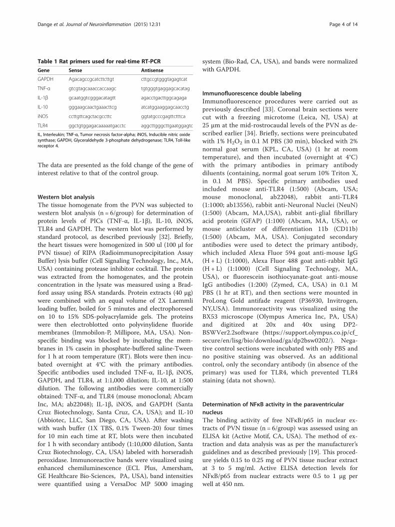
Table 1 Rat primers used for real-time RT-PCR
Gene Sense Antisense
GAPDH Agacagccgcatcttcttgt cttgccgtgggtagagtcat
TNF-α gtcgtagcaaaccaccaagc tgtgggtgaggagcacatag
IL-1β gcaatggtcgggacatagtt agacctgacttggcagaga
IL-10 gggaagcaactgaaacttcg atcatggaaggagcaacctg
iNOS ccttgttcagctacgccttc ggtatgcccgagttctttca
TLR4 ggctgtggagacaaaaatgacctc aggcttgggcttgaatggagtc
IL, Interleukin; TNF-α, Tumor necrosis factor-alpha; iNOS, Inducible nitric oxidesynthase; GAPDH, Glyceraldehyde 3-phosphate dehydrogenase; TLR4, Toll-likereceptor 4.
Dange et al. Journal of Neuroinflammation (2015) 12:31 Page 4 of 14
The data are presented as the fold change of the gene ofinterest relative to that of the control group.
Western blot analysisThe tissue homogenate from the PVN was subjected towestern blot analysis (n = 6/group) for determination ofprotein levels of PICs (TNF-α, IL-1β), IL-10, iNOS,TLR4 and GAPDH. The western blot was performed bystandard protocol, as described previously [32]. Briefly,the heart tissues were homogenized in 500 ul (100 μl forPVN tissue) of RIPA (Radioimmunoprecipitation AssayBuffer) lysis buffer (Cell Signaling Technology, Inc., MA,USA) containing protease inhibitor cocktail. The proteinwas extracted from the homogenates, and the proteinconcentration in the lysate was measured using a Brad-ford assay using BSA standards. Protein extracts (40 μg)were combined with an equal volume of 2X Laemmliloading buffer, boiled for 5 minutes and electrophoresedon 10 to 15% SDS-polyacrylamide gels. The proteinswere then electroblotted onto polyvinylidene fluoridemembranes (Immobilon-P, Millipore, MA, USA). Non-specific binding was blocked by incubating the mem-branes in 1% casein in phosphate-buffered saline-Tweenfor 1 h at room temperature (RT). Blots were then incu-bated overnight at 4°C with the primary antibodies.Specific antibodies used included TNF-α, IL-1β, iNOS,GAPDH, and TLR4, at 1:1,000 dilution; IL-10, at 1:500dilution. The following antibodies were commerciallyobtained: TNF-α, and TLR4 (mouse monoclonal; AbcamInc, MA; ab22048); IL-1β, iNOS, and GAPDH (SantaCruz Biotechnology, Santa Cruz, CA, USA); and IL-10(Abbiotec, LLC, San Diego, CA, USA). After washingwith wash buffer (1X TBS, 0.1% Tween-20) four timesfor 10 min each time at RT, blots were then incubatedfor 1 h with secondary antibody (1:10,000 dilution, SantaCruz Biotechnology, CA, USA) labeled with horseradishperoxidase. Immunoreactive bands were visualized usingenhanced chemiluminescence (ECL Plus, Amersham,GE Healthcare Bio-Sciences, PA, USA), band intensitieswere quantified using a VersaDoc MP 5000 imaging
system (Bio-Rad, CA, USA), and bands were normalizedwith GAPDH.
Immunofluorescence double labelingImmunofluorescence procedures were carried out aspreviously described [33]. Coronal brain sections werecut with a freezing microtome (Leica, NJ, USA) at25 μm at the mid-rostrocaudal levels of the PVN as de-scribed earlier [34]. Briefly, sections were preincubatedwith 1% H2O2 in 0.1 M PBS (30 min), blocked with 2%normal goat serum (KPL, CA, USA) (1 hr at roomtemperature), and then incubated (overnight at 4°C)with the primary antibodies in primary antibodydiluents (containing, normal goat serum 10% Triton X,in 0.1 M PBS). Specific primary antibodies usedincluded mouse anti-TLR4 (1:500) (Abcam, USA;mouse monoclonal, ab22048), rabbit anti-TLR4(1:1000; ab13556), rabbit anti-Neuronal Nuclei (NeuN)(1:500) (Abcam, MA,USA), rabbit anti-glial fibrillaryacid protein (GFAP) (1:100) (Abcam, MA, USA), ormouse anticluster of differentiation 11b (CD11b)(1:500) (Abcam, MA, USA). Conjugated secondaryantibodies were used to detect the primary antibody,which included Alexa Fluor 594 goat anti-mouse IgG(H + L) (1:1000), Alexa Fluor 488 goat anti-rabbit IgG(H + L) (1:1000) (Cell Signaling Technology, MA,USA), or fluorescein isothiocyanate-goat anti-mouseIgG antibodies (1:200) (Zymed, CA, USA) in 0.1 MPBS (1 hr at RT), and then sections were mounted inProLong Gold antifade reagent (P36930, Invitrogen,NY,USA). Immunoreactivity was visualized using theBX53 microscope (Olympus America Inc, PA, USA)and digitized at 20x and 40x using DP2-BSWVer2.2software (https://support.olympus.co.jp/cf_secure/en/lisg/bio/download/ga/dp2bsw0202/). Nega-tive control sections were incubated with only PBS andno positive staining was observed. As an additionalcontrol, only the secondary antibody (in absence of theprimary) was used for TLR4, which prevented TLR4staining (data not shown).
Determination of NFκB activity in the paraventricularnucleusThe binding activity of free NFκB/p65 in nuclear ex-tracts of PVN tissue (n = 6/group) was assessed using anELISA kit (Active Motif, CA, USA). The method of ex-traction and data analysis was as per the manufacturer’sguidelines and as described previously [19]. This proced-ure yields 0.15 to 0.25 mg of PVN tissue nuclear extractat 3 to 5 mg/ml. Active ELISA detection levels forNFκB/p65 from nuclear extracts were 0.5 to 1 μg perwell at 450 nm.

Dange et al. Journal of Neuroinflammation (2015) 12:31 Page 5 of 14
Detection of plasma norepinephrine and HMGB1Norepinephrine (NE) and HMGB1 were measured inplasma samples (n =8/group) using a norepinephrineELISA kit (Abnova, Taiwan) and a HMGB1 ELISA kit(MyBioSource, CA, USA), respectively as per the manu-facturer’s guidelines.
Statistical analysisAll results are expressed as mean ± SEM. Statistical analysiswas done by either a two-way ANOVA or a one-wayANOVA with a Bonferroni post hoc test using Graph PadPrism software (version 5.0; GraphPad Software, San DiegoCalifornia, USA). MAP data were analyzed by repeated-measures ANOVA to examine with-in group changes overtime. A value of P <0.05 was considered statisticallysignificant.
ResultsToll-like receptor 4 is highly expressed in the neuronsand microglia of paraventricular nucleus in hypertensiveratsImmunofluorescence staining of the PVN sections showedthat TLR4 is highly expressed in SHR+CP groups whencompared to WKY+CP (Figures 1, 2 and 3). Cell-type dis-tribution of TLR4 was further investigated in thePVN of all four groups using a double-labeling
Figure 1 An immunofluorescence double labeling image (x 20) showingTLR4 and NeuN in the PVN of WKY and SHR rats. n = 5/group. SHR + CPneurons of PVN, whereas, VIPER infusion in these rats caused significant redinfusion in saline-infused rats did not have any effects. Scale bar 20 μm: CP, cospontaneously hypertensive rat; TLR4, Toll-like receptor 4; VIPER, viral inhibitory
immunofluorescence technique. The frozen floating sec-tions were labeled with TLR4 antibody and one of the fol-lowing: neuronal nuclei (NeuN), glial fibrillary acidicprotein (GFAP) or anti-CD11b antibodies. NeuN, GFAPand anti-CD11b were used to identify neurons, astrocytesand microglia, respectively. An overwhelming majority ofTLR4 (red) was co-localized with NeuN-positive neurons(green) (Figure 1) in SHR + CP rats. Some of the TLR4-positive cells (green) were also labeled with CD11b-positive microglia/macrophage cells (red) (Figure 2);whereas, almost none of the TLR4-positive cells (red) wereco-localized with GFAP-positive astrocytes (green) in thePVN of SHR +CP rats (Figure 3). These results indicatedthat TLR4 is mainly expressed in the neurons and micro-glia of the PVN. Furthermore, chronic intra-PVN infusionof VIPER in SHR caused an apparent reduction in TLR4fluorescent staining in the PVN. These results corrobo-rated with RT-PCR and western blot analysis confirmingthe efficacy of VIPER in inhibiting TLR4 expression withinthe PVN (Figure 4A-C).
Toll-like receptor 4 blockade reduced HMGB1 levels incirculation and in the paraventricular nucleus ofhypertensive ratsSHR + CP group had increased gene and protein expres-sion of HMGB1 when compared to WKY + CP rats
the effects of intra-PVN infusion of VIPER on protein expression ofrats showed higher levels of immunofluorescence for TLR4 within theuction in TLR4 expression. Arrow indicates double- labeled cells.VIPERntrol peptide; NeuN, neuronal nuclei; PVN, paraventricular nucleus; SHR,peptide of TLR4; WKY, wistar-Kyoto.

Figure 2 An immunofluorescence double labeling image (x 40) showing the effects of intra-PVN infusion of VIPER on proteinexpression of TLR4 and CD11B in the PVN of WKY and SHR rats. SHR + CP rats showed modest expression of TLR4 within the microgliaof PVN, whereas, VIPER infusion in these rats caused significant reduction in TLR4 expression. Arrow indicates double-labeled cells.VIPERinfusion in saline-infused rats did not have any effects. n = 5/group. Scale bar 20 μm : CD11B, cluster of differentiation molecule 11B; CP, controlpeptide; PVN, paraventricular nucleus; SHR, spontaneously hypertensive rat; TLR4, Toll-like receptor 4; VIPER, viral inhibitory peptide of TLR4;WKY, wistar-Kyoto.
Figure 3 An immunofluorescence double labeling image (x 20) showing the effects of intra-PVN infusion of VIPER on protein expression ofTLR4 and GFAP in the PVN of WKY and SHR rats. n = 5/group. Scale bar 20 μm : GFAP, glial fibrillary acidic protein; PVN, paraventricularnucleus; SHR, spontaneously hypertensive rat; TLR4, Toll-like receptor 4; VIPER, viral inhibitory peptide of TLR4; WKY, wistar-Kyoto.
Dange et al. Journal of Neuroinflammation (2015) 12:31 Page 6 of 14
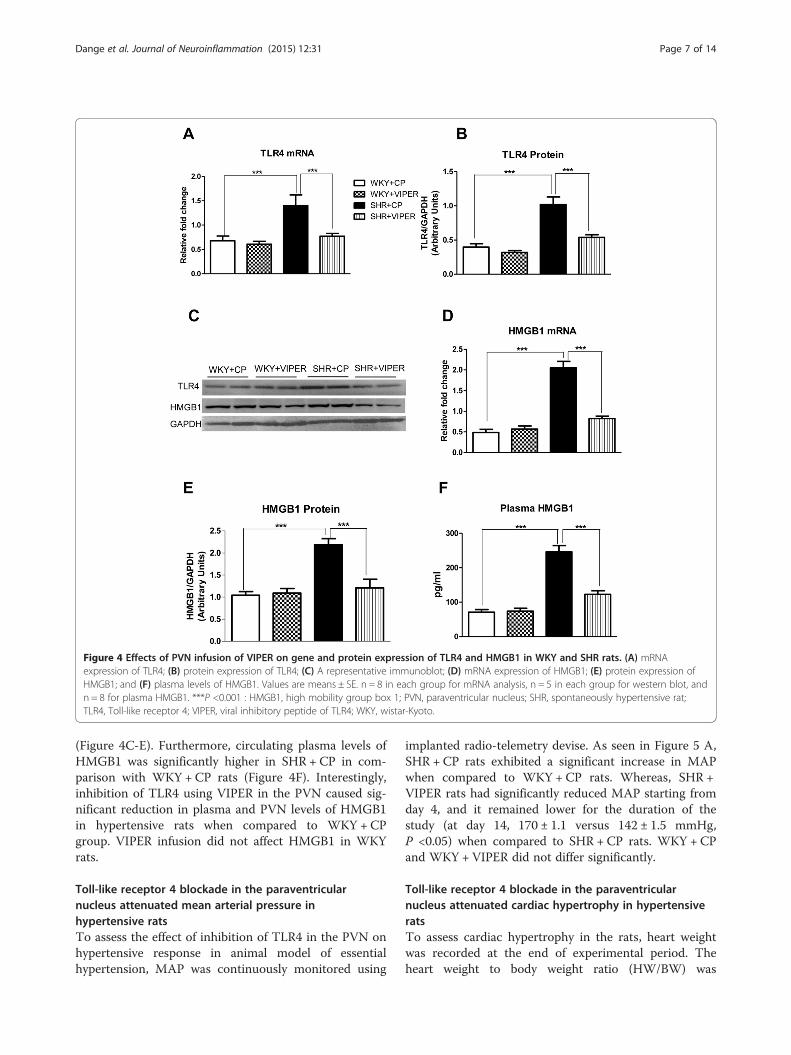
Figure 4 Effects of PVN infusion of VIPER on gene and protein expression of TLR4 and HMGB1 in WKY and SHR rats. (A) mRNAexpression of TLR4; (B) protein expression of TLR4; (C) A representative immunoblot; (D) mRNA expression of HMGB1; (E) protein expression ofHMGB1; and (F) plasma levels of HMGB1. Values are means ± SE. n = 8 in each group for mRNA analysis, n = 5 in each group for western blot, andn = 8 for plasma HMGB1. ***P <0.001 : HMGB1, high mobility group box 1; PVN, paraventricular nucleus; SHR, spontaneously hypertensive rat;TLR4, Toll-like receptor 4; VIPER, viral inhibitory peptide of TLR4; WKY, wistar-Kyoto.
Dange et al. Journal of Neuroinflammation (2015) 12:31 Page 7 of 14
(Figure 4C-E). Furthermore, circulating plasma levels ofHMGB1 was significantly higher in SHR + CP in com-parison with WKY + CP rats (Figure 4F). Interestingly,inhibition of TLR4 using VIPER in the PVN caused sig-nificant reduction in plasma and PVN levels of HMGB1in hypertensive rats when compared to WKY + CPgroup. VIPER infusion did not affect HMGB1 in WKYrats.
Toll-like receptor 4 blockade in the paraventricularnucleus attenuated mean arterial pressure inhypertensive ratsTo assess the effect of inhibition of TLR4 in the PVN onhypertensive response in animal model of essentialhypertension, MAP was continuously monitored using
implanted radio-telemetry devise. As seen in Figure 5 A,SHR + CP rats exhibited a significant increase in MAPwhen compared to WKY + CP rats. Whereas, SHR +VIPER rats had significantly reduced MAP starting fromday 4, and it remained lower for the duration of thestudy (at day 14, 170 ± 1.1 versus 142 ± 1.5 mmHg,P <0.05) when compared to SHR + CP rats. WKY + CPand WKY + VIPER did not differ significantly.
Toll-like receptor 4 blockade in the paraventricularnucleus attenuated cardiac hypertrophy in hypertensiveratsTo assess cardiac hypertrophy in the rats, heart weightwas recorded at the end of experimental period. Theheart weight to body weight ratio (HW/BW) was

Figure 5 Effect of bilateral intra-PVN infusion of VIPER on mean arterial blood pressure (MAP, in millimeters of mercury) and cardiachypertrophy in WKY and SHR rats. (A) SHR + CP group had significantly increased MAP when compared to WKY + CP rats. Interestingly,infusion of VIPER in PVN of SHR rats for 14 days resulted in significant decrease in MAP, starting from day 4 of VIPER infusion; (B) heart weight tobody weight ratio; and (C) mRNA expression of atrial natriuretic peptide (ANP) in tissue obtained from left ventricle. Values are mean ± SE; n = 8/group for mRNA analysis and n = 14 for MAP. *P <0.05 SHR + CP versus WKY + CP; #P < 0.05 SHR + VIPER versus SHR + CP; ***P <0.001 : ANP, AtrialNatriuretic Peptide; CP, control peptide; MAP, mean arterial pressure; PVN, paraventricular nucleus; SHR, spontaneously hypertensive rat; TLR4,Toll-like receptor 4; VIPER, viral inhibitory peptide of TLR4; WKY, wistar-Kyoto.
Dange et al. Journal of Neuroinflammation (2015) 12:31 Page 8 of 14
calculated as a predictor of cardiac hypertrophy.Additionally, ANP, a molecular marker of cardiachypertrophy was estimated in heart tissue using realtime RT-PCR. Increased cardiac hypertrophy was ob-served in SHR + CP rats compared with WKY + CPrats, as indicated by a significantly increased HW/BWratio and ANP levels in SHR + CP group (Figure 5Band C). VIPER treatment in SHR rats caused a signifi-cant decrease in the HW/BW ratio as well as ANPlevels in comparison with SHR + CP, suggestive ofreduced cardiac hypertrophy by blockade of TLR4within the PVN of SHR. These data suggest arole for TLR4 in the PVN of the brain on MAPregulation and cardiac hypertrophy in the essentialhypertension.
Toll-life receptor 4 blockade altered expression of pro- andanti-inflammatory cytokines in hypertensive ratsTo determine the effect of TLR4 blockade within thePVN on inflammatoy response in hypertension, we
examined the levels of pro-inflammatory cytokines(PICs), TNF-α and IL-1β mRNA (Figure 6A-B) andprotein (Figure 6 C-D) levels in the PVN by real timeRT PCR and western immunoblot, respectively. Weobserved that SHR + CP rats exhibited marked in-crease in TNF-α and IL-1β expression compared toWKY + CP rats in their PVN. Interestingly, thisupregulation of TNF-α and IL-1β was significantly at-tenuated in SHR + VIPER group. However, VIPERinfusion did not affect PICs levels in WKY rats.Furthermore, IL-10 gene and protein expression in
the PVN tissue was measured to assess the effect ofTLR4 blockade on anti-inflammatory axis. A signifi-cant reduction in IL-10 gene and protein expressionin the SHR + CP compared with the WKY + CP ratswas evident (Figure 6 C and E). Interestingly, SHR +VIPER group had significantly higher levels of IL-10in comparison with SHR + CP rats; whereas there wasno difference between WKY + CP and WKY + VIPERgroups.

Figure 6 Effects of bilateral intra-PVN infusion of VIPER on gene and protein expression of pro- and anti-inflammatory cytokines andiNOS in WKY and SHR rats. (A) mRNA expression of TNF-α; (B) mRNA expression of IL1-β; (C) A representative immunoblot; (D) densitometricanalysis of protein expression of TNF-α, IL1-β, IL10 and iNOS; (E) mRNA expression of IL10; and (F) mRNA expression of iNOS. Values are means ±SE. n = 8 in each group for mRNA analysis and n = 5 in each group for western blot. ***P <0.001; *P <0.05 SHR + CP versus WKY + CP; #P <0.05SHR + VIPER versus SHR + CP: CP, control peptide; IL-1β, interleukin-1 beta; IL10, interleukin10; iNOS, inducible nitric oxide; PVN, paraventricular nu-cleus; SHR, spontaneously hypertensive rat; TLR4, Toll-like receptor 4; TNF-α, tumor necrosis factor-alpha; VIPER, viral inhibitory peptide of TLR4;WKY, wistar-Kyoto. Furthermore, IL-10 gene and protein expression in the PVN tissue was measured to assess the effect of TLR4 blockade on anti-inflammatory axis. A significant reduction in IL-10 gene and protein expression in the SHR + CP compared with the WKY + CP rats was evident(Figure 6 C and E). Interestingly, the SHR + VIPER group had significantly higher levels of IL-10 in comparison with the SHR + CP rats, whereasthere was no difference between the WKY + CP and WKY + VIPER groups.
Dange et al. Journal of Neuroinflammation (2015) 12:31 Page 9 of 14
Toll-like receptor 4 blockade alters inducible nitric oxidesynthase levels in the hypertensive ratsHypertension is found to modulate iNOS expression.iNOS is induced primarily by PICs and is another markerof inflammation. We found that SHR +CP rats hadmarked increase in mRNA and protein levels of iNOS(Figure 6 C and F) in comparison with WKY +CP rats.Importantly, chronic infusion of VIPER in the PVN causeda significant decrease in mRNA and protein expression ofiNOS in the SHR rats but not in the WKY rats.
Toll-like receptor 4 blockade attenuates NFκB activity inhypertensive ratsWe performed NFκB binding activity assay using thePVN tissues of all rat groups as illustrated in Figure 7A.As expected, we observed that SHR + CP rats had sig-nificantly higher NFκB activity in the PVN homogenatesthan WKY + CP rats. Interestingly, this response wasabolished by chronic VIPER infusion within the PVN ofSHR rats. However, VIPER infusion did not affect NFκBactivity in the WKY rats.

Figure 7 Effects of bilateral intra-PVN infusion of VIPER on (A)NFκB activity of PVN tissue and (B) plasma levels of norepinephrine(NE) in WKY and SHR rats. NFκB activity assay showing increasedactivity in PVN of SHR + CP rats when compared to WKY + CP rats,whereas, VIPER infusion in SHR rats resulted in significant reduction inthe NFκB activity. Similar trends were observed with regard to plasmanorepinephrine levels as well. Values are means ± SE. n = 6/group forNFκB activity; n = 8 for plasma NE analysis. ***P <0.001. CP, controlpeptide; NE, norepinephrine; NFĸB, nuclear factor-kappa B; PVN, paraventricularnucleus; SHR, spontaneously hypertensive rat; TLR4, Toll-like receptor 4; TNF-α,tumor necrosis factor-alpha; VIPER, viral inhibitory peptide of TLR4;WKY, wistar-Kyoto.
Dange et al. Journal of Neuroinflammation (2015) 12:31 Page 10 of 14
Toll-like receptor 4 blockade reduced circulatingnorepinephrine levels in hypertensive ratsSHR + CP group had an increased circulating plasma NE(Figure 7B) concentration when compared to WKY + CPrats. Interestingly, inhibition of TLR4 using VIPER inthe PVN caused a significant reduction in the plasmalevels of NE in SHR but not in WKY.
DiscussionIn the present study, we investigated the effects of bilat-eral inhibition of TLR4 within the hypothalamic PVN ofthe brain of SHR rats, which are well established as agenetic model of human essential hypertension. The sali-ent findings of this study are as follows:
1. SHR rats had robust increase in TLR4 levels in thePVN, which was mostly localized in the neurons andmicroglia. These results were associated withincrease in HMGB1 levels within the PVN as well asin the circulation.
2. Blockade of TLR4 within the PVN prevented, atleast in part, the increase in blood pressure in SHRand attenuated cardiac hypertrophy in SHR.
3. TLR4 inhibition in the PVN attenuates pro-inflammatory cytokines, iNOS, and transcriptionfactor, NFκB activity; whereas, TLR4 inhibitioncauses an increase in anti-inflammatory IL-10 in thePVN of hypertensive SHR rats.
4. TLR4 blockade resulted in significant reduction incirculating plasma NE in SHR rats. In the presentstudy, we observed a significant increase in theTLR4 gene and protein expression within the PVNof hypertensive rats. More importantly, our double-labeling immunofluorescence staining of the frozenfloating PVN sections demonstrated that TLR4protein is present mainly in neurons and microglia(albeit at much lower level) and not so much in theastrocytes, indicating that TLR4 upregulation inneurons and microglia of the PVN could be one ofthe characteristics of hypertensive response observedin these animals. To the best of our knowledge, thepresent study is the first to demonstrate thatincreased TLR4 expression in the PVN contributesto a hypertensive response in a genetic model ofhypertension. Furthermore, a corresponding increasein the HMGB1 levels in the PVN and in circulationindicates that TLR4 acts via the HMGB1. Theseresults provide mechanistic evidence that detrimentaleffects seen in genetic hypertension are mediated, atleast in part, by TLR4 in the PVN and that inhibitionof TLR4 attenuates hypertensive response, possibly viadownregulation of inflammatory components andupregulation of anti-inflammatory mediators in thePVN. Taken together, the results of this study suggestTLR4 as a newer therapeutic target for effectivecontrol of blood pressure. The proposed mechanismby which TLR4 exerts its role in hypertension isdepicted in Figure 8.
Hypertension is a chronic inflammatory condition. Inthis context, immune system works as a first line ofdefense against an insult or tissue injury and acts viamechanism of inflammation. This critical response is ini-tiated by identification of pathogen-associated molecularpatterns by pathogen recognition receptors present oncell membrane [12]. TLRs are one such pathogen recog-nition receptors, which are considered a major compo-nent of the innate immune system. To date, 13 TLRshave been cloned in mammals and each TLR have a dis-tinct function in innate immune recognition [35]. TLRsare type I transmembrane glycoproteins. They can beclassified into two groups according to their subcellularlocalization: TLR1, TLR2, TLR4-6 and TLR11 areexpressed on the plasma membrane, whereas TLR3,TLR7 and TLR9 are found in the endosomal compart-ment [36]. Human TLR4 was the first characterizedmammalian TLR [37]. In addition to immune cells,

Figure 8 Schematic illustrating proposed mechanism of TLR4activation and downstream signaling in the PVN hypertension.One of the mechanisms of sustained elevation of blood pressure inessential hypertension could be due to increased activation of TLR4in the PVN and subsequent activation of NFκB pathway to produceinflammatory alterations, sympathoexcitation, and cardiachypertrophy. Blockade of TLR4 in the PVN attenuates hypertensiveresponse and prevents detrimental inflammatory changes associatedwith hypertension. Besides reduction in MAP in VIPER treated SHRrats, we also observed reduction in plasma NE levels. Thereforethese beneficial effects might be also due to reduction in plasma NEand HMGB1 levels as observed in this study: HMGB1, high mobilitygroup box 1; MAP, mean arterial pressure; NE, norepinephrine; NFĸB,nuclear factor-kappa B; PVN, paraventricular nucleus; SHR,spontaneously hypertensive rat; TLR4, Toll-like receptor 4; VIPER,viral inhibitory peptide of TLR4.
Dange et al. Journal of Neuroinflammation (2015) 12:31 Page 11 of 14
TLR4 has also been found to be expressed in nonim-mune cells of the cardiovascular system [38], brain[39,40], neuronal culture stimulated with LPS [12], andvascular smooth muscle cells [41], indicating that bodytissues have control over their own immune response.Our current results together with previous reports sug-gest role of TLR4 in hypertension. Although hyperten-sion is a chronic inflammatory condition, the role playedby TLR4 , which responds to endogenous DAMPs, inpathogenesis of hypertension is largely unclear. More-over, it is well known that the PVN is central in initi-ation and development of hypertension, the role ofTLR4 within the PVN of genetic model of human essen-tial hypertension has never been investigated before.In this study, to test our hypothesis that hypertensive
response observed in SHR rats could be mediated by ac-tivation of TLR4 in the PVN, we chronically infused aspecific TLR4 blocker (VIPER) or control peptide (CP)by intra-PVN route in the SHR rats and their normoten-sive control, the WKY rats. We observed a significantreduction in MAP in the hypertensive rats that receivedVIPER (SHR + VIPER) when compared to the
hypertensive rats receiving the control peptide (SHR +CP), and saw no comparable changes in WKY rats re-ceiving VIPER. Additionally, our telemetric recordingsshowed a significant reduction in MAP in the SHR +VIPER group starting from day 4 of the VIPER infusion,which remained lower until the end of the experiment.In keeping with our results, a previous report showedthat a neutralizing TLR4 antibody reduces blood pres-sure in the SHR rats [3]. The present study provided fur-ther evidence that inhibition of TLR4 in the PVNpartially restored blood pressure to normal levels, sup-porting that TLR4, along with other central mechanisms,plays an important role in BP regulation [42-44].Hypertension is characterized by increased cardiac
hypertrophy and cardiac dysfunction. [45]. Therefore, weinvestigated whether TLR4 blockade in the PVN is car-dioprotective in hypertensive rats. SHR + CP rats had in-creased ANP levels and HW:BW when compared withWKY + CP rats, indicating presence of cardiac hyper-trophy in SHR as also reported in previous studies[30,46]. More importantly, TLR4 blockade within thePVN resulted in a significant reduction in ANP and theHW:BW ratio in hypertensive rats, suggesting reducedcardiac hypertrophy by TLR4 blockade. A handful ofprevious studies have reported that TLR4 deficiency pro-tects the myocardium in Angiotensin II-induced hyper-tension in rat [40] and from ischemia/reperfusion injuryin mice [47]. Taken together, the current findings sug-gest the role of brain TLR4 in cardiac hypertrophy inhypertension.Each TLR recognizes specific pathogen-associated mo-
lecular patterns (PAMPs) in microbial injury. TLRs, es-pecially TLR4, can also be stimulated by host-derivedmolecules, known as damage-associated molecular pat-tern molecules (DAMPs) [16,17]. Of several DAMPs,high mobility group box-1 (HMGB1) is the most import-ant DAMP that has been implicated in various inflam-matory conditions. In response to injury or infection,HMGB1 is released actively by immune cells and pas-sively by insulted cells into the extracellular space andinitiates an inflammatory response [15-17]. Therefore,one possible mechanism by which TLR4 is activated inhypertension could be via upregulation of HMGB1. Re-inforcing this hypothesis, in the present study, hyperten-sive rats were found to have significantly increased levelsof HMGB1 in the PVN as well as in circulation. Similarsecretion of HMGB1 and expression of TLR4 was ob-served in ischemic brain injury [48]. However, the novelfinding of the present study is that inhibition of TLR4within the PVN resulted in a significant decrease inHMGB1 levels in the PVN and circulation. Given thatthe TLR4 blockade reduces MAP, it can be postulatedthat TLR4 plays role in hypertension, possibly viaHMGB1. Whether other known ligands of TLR4, such
Dange et al. Journal of Neuroinflammation (2015) 12:31 Page 12 of 14
as heat shock proteins, have any roles within the PVN ofhypertensive animals is not clear at this time and needsto be investigated in detail. Nevertheless, the presentfindings provide strong evidence that TLR4 within thePVN plays a critical role in the pathogenesis of hyper-tension, at least in part, due to increased binding withits specific DAMP, HMGB1.Once a TLR recognizes an endogenous DAMP, activa-
tion of inflammatory signaling begins [16,17]. Moreover,an inflammatory response in hypertension is character-ized by a peripheral and central increase in various PICs,in particular, TNF-α and IL-1β [20,49,50]. In addition,PICs are known to induce iNOS expression in hyperten-sion [22]. Therefore, to investigate whether TLR4 signal-ing within the PVN contributes to inflammatoryresponse seen in hypertension, we measured the geneand protein expression of TNF-α, IL-1β and iNOS in thePVN tissue. We found that chronic TLR4 inhibition inhypertensive rats causes a significant reduction in TNF-α and IL-1β levels, as well as iNOS levels, in the PVN.Additionally, anti-inflammatory cytokine IL-10 wasfound to be significantly increased in VIPER-infusedhypertensive rats. These results clearly suggest the roleof TLR4, specifically within the PVN in inflammatory re-sponse in hypertension. Although not within the PVN, ahandful of previous reports showed that systemic injec-tion of anti-TLR4 antibody decreases serum levels of IL-6 in SHR [3] and systemically infused TLR4 antagonistreduces serum levels of inflammatory markers in heartfailure animals [51]. Similarly, another study has shownthat the TLR4 and PICs were increased in the heart tis-sues of the SHR rats [45]. It is noteworthy that althoughthese studies show suppression of inflammation in theend-organs, none of the studies were able to point outthe role of the brain in hypertension Given our currentfindings that TLR4 is dramatically upregulated withinthe PVN and that the TLR4 blockade attenuates MAP,our results suggest a vital role played by hypothalamicTLR4 in hypertensive response. Hence, an improvementin MAP and cardiac function by central inhibition ofTLR4, as observed in the present study, could be attrib-utable to a reduction in iNOS and PICs. Supporting ourresults, a recent study has demonstrated that ablation ofiNOS delays cardiac hypertrophy and contractile dys-function in mice with aortic banding-induced hyperten-sion [52]. Binding of free HMGB1 to TLRs leads to theproduction and release of PICs [53]. Our findings thatHMGB1 levels were increased both in the circulationand in the PVN together with increase in PICs ,iNOSand plasma NE suggest that TLR4 participates in the in-flammatory process associated with hypertension leadingto sustained activation of NFκB pathway to produce in-flammatory alterations, sympathoexcitation, and cardiachypertrophy in SHR rats.
NFκB is one of the most important downstream tran-scription factors responsible for the transcription of PICsand iNOS. It is also well established that TLR4 sharesthe same NFκB signaling pathways. Given that VIPERinjection into the PVN of hypertensive rats reduces PICsand iNOS levels, one possible mechanism by whichTLR4 inhibition in PVN exerts its beneficial effectscould be via downregulation of NFκB. Reinforcing thishypothesis, the present study showed that the TLR4blockade causes downregulation of NFκB activity inSHR rats. To the best of our knowledge, the presentstudy is the first to investigate the downstream signalingmechanism of TLR4 within the PVN in regulation ofMAP. Nevertheless, these results clearly suggest that at-tenuation of NFκB activity might be attributable to re-duced inflammation, which in turn leads to disruption ofa detrimental positive feedback cycle involved in cardiacremodeling and the progression of hypertension.It has been well established that sympathetic hyper-
activity contributes to cardiac remodeling in hyperten-sion via an increase in TNF-α production [54] and NFκBactivation leading to end organ damage [55]. In accord-ance with these previous reports, in the present study,the SHR rats showed higher circulating plasma levels ofNE (an indirect indicator of sympathetic activity), indi-cating increased sympathetic outflow compared to WKYrats. However, a novel finding of this study is that inhib-ition of TLR4 in the PVN with VIPER resulted in adramatic reduction in plasma levels of NE in the hyper-tensive rats, suggesting that upregulation of brain TLR4plays a key role in sympathoexcitation as observed inhypertensive animals. The mechanism by which TLR4increases sympathetic output in SHR is not clear at thistime. However, given that inhibition of TNF-α or NFκBin the PVN decreases arterial pressure [30] and sympa-thetic activity [56], it is plausible to suggest that activa-tion of TLR4 signaling induces sympathetic hyperactivitypossibly via increased PICs. As evidence, Ogawa et al.demonstrated that siRNA-mediated inhibition of brainTLR4 decreases urinary norepinephrine excretion in ratswith myocardial infarction [44]. Nonetheless, the resultsof the present study suggest that activation of TLR4 inthe PVN of the brain leads to increased sympatheticactivation in an animal model of essential hypertension.
LimitationsThere are two common pathways shared by TLRs,namely the myeloid differentiation factor 88 (MyD88)-dependent and the MyD88-independent pathways[57,58]. An increase in PICs expression induced throughNFκB activation is the main feature of the MyD88-dependent pathway, whereas activation of interferon-γregulatory factor 3 is characteristic of the MyD88-independent pathway [59,60]. Given our findings that
Dange et al. Journal of Neuroinflammation (2015) 12:31 Page 13 of 14
the TLR4 blockade attenuates PICs and NFκB, we be-lieve that the MyD88-dependent pathway is involved inthis process. Specific adaptors and possible ligands,other than HMGB1 involved in TLR4 signaling, need tobe investigated and could be a focus for future studies.Furthermore, here we explored the PVN region, al-though there are multiple cardiorelevant sites in thebrain that can play a role in modulating the hypertensiveresponse. However, we feel that the PVN is of import-ance because of its recognized integrative functions.Nonetheless, the current data undermines the key roleplayed by brain TLR4 signaling in the pathogenesis ofhypertension and suggest that TLR4 as a novel thera-peutic target for the treatment of essential hypertension.
ConclusionsTo summarize, TLR4 regulates various diseases in anNFκB-dependent manner. It has been shown to increasecardiac hypertrophy and dysfunction in animal modelsof heart failure and hypertension. Here, we show thatthe SHR rats exhibit dramatic upregulation of TLR4within the cellular components of the hypothalamicPVN. More importantly, inhibition of TLR4 by bilateralmicroinjection into the PVN of the SHR rats delays theprogression of hypertension; reduces cardiac hyper-trophy; attenuates PICs, iNOS, and NFκB, as well as NElevels; and improves anti-inflammatory IL-10 levelswithin the PVN. Additionally, our results identify TLR4as a specific TLR, and HMGB1 as one of its endogenousligands that plays a vital role in the hypertensive re-sponse. Taken together, the present study shows activa-tion of HMGB1 as an upstream phenomenon andactivation of inflammatory signals as a downstreampathway in TLR4 signaling within the PVN of hyperten-sive rats. Overall, our data for the first time demonstratethat an excessive increase in TLR4 within the PVN playsan important role in initiation of the inflammatoryprocess observed in essential hypertension.
AbbreviationsaCSF: Artificial cerebrospinal fluid; ANOVA: Analysis of variance; ANP: Atrialnatriuretic peptide; BW: Body weight; CD11B: Cluster of differentiationmolecule 11B; CP: Control peptide; ELISA: Enzyme-linked immunosorbentassay; GAPDH: Glyceraldehyde 3-phosphate dehydrogenase; GFAP: Glialfibrillary acidic protein; HMGB1: High mobility group box 1; HW: Heartweight; HW/BW: Heart weight/body weight; IACUC: Institutional Animal Careand Use Committee; IL-1β: Interleukin-1 beta; IL-10: Interleukin-10;iNOS: Inducible nitric oxide; MAP: Mean arterial pressure; MyD88: Myeloiddifferentiation factor 88; mmHg: Millimeters of mercury; NE: Norepinephrine;NeuN: Neuronal nuclei; NFĸB: Nuclear factor-kappa B; NO: Nitric oxide;PIC: Proinflammatory cytokine; PVN: Paraventricular nucleus;SHR: Spontaneously hypertensive rat; TLR: Toll-like receptor; TNF-α: Tumornecrosis factor-alpha; VIPER: Viral inhibitory peptide of TLR4; WKY:Wistar-Kyoto.
Competing interestThe authors declare that they have no competing interests.
Authors’ contributionsRBD participated in the study design, carried out the animal experiments,performed molecular analysis of tissues and drafted the manuscript. DAcarried out the analysis and interpretation of RT-PCR and western blot dataand revised the manuscript critically for important intellectual content. RTworked on tissue processing and revised the manuscript. JF conceived ofand designed the study and critically revised the manuscript. All authors readand approved the final manuscript.
FundingThis work was supported by Louisiana State University CORP Grant to JosephFrancis.
Author details1Comparative Biomedical Sciences, School of Veterinary Medicine, LouisianaState University, 1909 Skip Bertman Drive, Baton Rouge, LA 70803, USA.2William Hansel Cancer Prevention Laboratory, Pennington BiomedicalResearch Center, 6400 Perkins Road, Baton Rouge, LA 70808, USA.3Department of Biological Sciences, College of Science, Louisiana StateUniversity, 202 Life Sciences Building, Baton Rouge, LA 70803, USA.
Received: 8 August 2014 Accepted: 10 January 2015
References1. Lloyd-Jones D, Adams RJ, Brown TM, Carnethon M, Dai S, De Simone G,
et al. Heart disease and stroke statistics–2010 update: a report from theAmerican Heart Association. Circulation. 2010;121:e46–215.
2. Michel MC, Brodde OE, Insel PA. Peripheral adrenergic receptors inhypertension. Hypertension. 1990;16:107–20.
3. Bomfim GF, Dos Santos RA, Oliveira MA, Giachini FR, Akamine EH, Tostes RC,et al. Toll-like receptor 4 contributes to blood pressure regulation andvascular contraction in spontaneously hypertensive rats. Clin Sci.2012;122:535–43.
4. Sriramula S, Haque M, Majid DS, Francis J. Involvement of tumor necrosisfactor-alpha in angiotensin II-mediated effects on salt appetite, hypertension,and cardiac hypertrophy. Hypertension. 2008;51:1345–51.
5. Theuer J, Dechend R, Muller DN, Park JK, Fiebeler A, Barta P, et al.Angiotensin II induced inflammation in the kidney and in the heart ofdouble transgenic rats. BMC Cardiovasc Disord. 2002;2:3.
6. Muller DN, Dechend R, Mervaala EM, Park JK, Schmidt F, Fiebeler A, et al.NF-kappaB inhibition ameliorates angiotensin II-induced inflammatorydamage in rats. Hypertension. 2000;35:193–201.
7. Coban E, Nizam I, Topal C, Akar Y. The association of low-grade systemicinflammation with hypertensive retinopathy. Clin Exp Hypertens.2010;32:528–31.
8. Abboud FM, Harwani SC, Chapleau MW. Autonomic neural regulation of theimmune system: implications for hypertension and cardiovascular disease.Hypertension. 2012;59:755–62.
9. Ryan MJ. An update on immune system activation in the pathogenesis ofhypertension. Hypertension. 2013;62:226–30.
10. Zubcevic J, Waki H, Raizada MK, Paton JF. Autonomic-immune-vascularinteraction: an emerging concept for neurogenic hypertension.Hypertension. 2011;57:1026–33.
11. Jennings JR, Zanstra Y. Is the brain the essential in hypertension?Neuroimage. 2009;47:914–21.
12. Leow-Dyke S, Allen C, Denes A, Nilsson O, Maysami S, Bowie AG, et al.Neuronal Toll-like receptor 4 signaling induces brain endothelial activationand neutrophil transmigration in vitro. J Neuroinflammation. 2012;9:230.
13. Medzhitov R, Preston-Hurlburt P, Janeway Jr CA. A human homologue ofthe Drosophila Toll protein signals activation of adaptive immunity. Nature.1997;388:394–7.
14. Rock FL, Hardiman G, Timans JC, Kastelein RA, Bazan JF. A family of humanreceptors structurally related to Drosophila Toll. Proc Natl Acad Sci U S A.1998;95:588–93.
15. Lotze MT, Tracey KJ. High-mobility group box 1 protein (HMGB1): nuclearweapon in the immune arsenal. Nat Rev Immunol. 2005;5:331–42.
16. Piccinini AM, Midwood KS. DAMPening inflammation by modulating TLRsignalling. Mediators Inflamm. 2010; Volume (2010), article ID 672395:1-21.
17. Bianchi ME. DAMPs, PAMPs and alarmins: all we need to know aboutdanger. J Leukoc Biol. 2007;81:1–5.

Dange et al. Journal of Neuroinflammation (2015) 12:31 Page 14 of 14
18. Davisson RL, Oliverio MI, Coffman TM, Sigmund CD. Divergent functions ofangiotensin II receptor isoforms in the brain. J Clin Invest. 2000;106:103–6.
19. Cardinale JP, Sriramula S, Mariappan N, Agarwal D, Francis J. Angiotensin II-induced hypertension is modulated by nuclear factor-kappaBin the paraventricularnucleus. Hypertension. 2012;59:113–21.
20. Shi P, Diez-Freire C, Jun JY, Qi Y, Katovich MJ, Li Q, et al. Brain microglialcytokines in neurogenic hypertension. Hypertension. 2010;56:297–303.
21. Shi Z, Gan XB, Fan ZD, Zhang F, Zhou YB, Gao XY, et al. Inflammatorycytokines in paraventricular nucleus modulate sympathetic activity andcardiac sympathetic afferent reflex in rats. Acta Physiol (Oxf).2011;203:289–97.
22. Hong HJ, Loh SH, Yen MH. Suppression of the development ofhypertension by the inhibitor of inducible nitric oxide synthase. Br JPharmacol. 2000;131:631–7.
23. Agarwal D, Elks CM, Reed SD, Mariappan N, Majid DSA, Francis J. Chronicexercise preserves renal structure and hemodynamics in spontaneouslyhypertensive rats. Antioxid Redox Signal. 2012;16:139–52.
24. Stack J, Bowie AG. Poxviral protein A46 antagonizes Toll-like receptor 4signaling by targeting BB loop motifs in Toll-IL-1 receptor adaptor proteinsto disrupt receptor:adaptor interactions. J Biol Chem. 2012;287:22672–82.
25. Oda S, Franklin E, Khan AR. Poxvirus A46 protein binds to TIR domain-containing Mal/TIRAP via an alpha-helical sub-domain. Mol Immunol.2011;48:2144–50.
26. Dhandapani S, Dhandapani M, Agarwal M, Chutani AM, Subbiah V, SharmaBS, et al. The prognostic significance of the timing of total enteral feedingin traumatic brain injury. Surg Neurol Int. 2012;3:31.
27. Agarwal D, Dange RB, Vila J, Otamendi AJ, Francis J. Detraining differentiallypreserved beneficial effects of exercise on hypertension: effects on bloodpressure, cardiac function, brain inflammatory cytokines and oxidative stress.PLoS One. 2012;7(12):e52569.
28. Paxinos G, Watson C. The rat brain in stereotaxic coordinates. 6th ed.Amsterdam ; Boston: Academic Press/Elsevier; 2007.
29. Palkovits M. Punch sampling biopsy technique. Methods Enzymol.1983;103:368–76.
30. Sriramula S, Cardinale JP, Francis J. Inhibition of TNF in the brain reversesalterations in RAS components and attenuates angiotensin II-inducedhypertension. PLoS One. 2013;8:e63847.
31. Agarwal D, Haque M, Sriramula S, Mariappan N, Pariaut R, Francis J. Role ofproinflammatory cytokines and redox homeostasis in exercise-induceddelayed progression of hypertension in spontaneously hypertensive rats.Hypertension. 2009;54:1393–400.
32. Agarwal D, Welsch MA, Keller JN, Francis J. Chronic exercise modulates RAScomponents and improves balance between pro- and anti-inflammatorycytokines in the brain of SHR. Basic Res Cardiol. 2011;106:1069–85.
33. Lin S, Yin Q, Zhong Q, Lv FL, Zhou Y, Li JQ, et al. Heme activates TLR4-mediated inflammatory injury via MyD88/TRIF signaling pathway inintracerebral hemorrhage. J Neuroinflammation. 2012;9:46.
34. Swanson LW, Kuypers HG. The paraventricular nucleus of the hypothalamus:cytoarchitectonic subdivisions and organization of projections to thepituitary, dorsal vagal complex, and spinal cord as demonstrated byretrograde fluorescence double-labeling methods. J Comp Neurol.1980;194:555–70.
35. Medzhitov R. Toll-like receptors and innate immunity. Nat Rev Immunol.2001;1:135–45.
36. Akira S. Toll receptor families: structure and function. Semin Immunol.2004;16:1–2.
37. Vallejo JG. Role of toll-like receptors in cardiovascular diseases. Clin Sci(Lond). 2011;121:1–10.
38. Frantz S, Kobzik L, Kim YD, Fukazawa R, Medzhitov R, Lee RT, et al. Toll4(TLR4) expression in cardiac myocytes in normal and failing myocardium.J Clin Invest. 1999;104:271–80.
39. Tang SC, Arumugam TV, Xu X, Cheng A, Mughal MR, Jo DG, et al. Pivotalrole for neuronal Toll-like receptors in ischemic brain injury and functionaldeficits. Proc Natl Acad Sci U S A. 2007;104:13798–803.
40. Dange RB, Agarwal D, Masson GS, Vila J, Wilson B, Nair A, et al. Centralblockade of TLR4 improves cardiac function and attenuates myocardialinflammation in angiotensin II-induced hypertension. Cardiovasc Res.2014;103:17–27.
41. Ji Y, Liu J, Wang Z, Liu N. Angiotensin II induces inflammatory responsepartly via toll-like receptor 4-dependent signaling pathway in vascularsmooth muscle cells. Cell Physiol Biochem. 2009;23:265–76.
42. Chen QH, Andrade MA, Calderon AS, Toney GM. Hypertension induced byangiotensin II and a high salt diet involves reduced SK current andincreased excitability of RVLM projecting PVN neurons. J Neurophysiol.2010;104:2329–37.
43. Sonner PM, Lee S, Ryu PD, Lee SY, Stern JE. Imbalanced K+ and Ca2+subthreshold interactions contribute to increased hypothalamicpresympathetic neuronal excitability in hypertensive rats. J Physiol.2011;589:667–83.
44. Ogawa K, Hirooka Y, Kishi T, Ide T, Sunagawa K. Partially silencing brain toll-like receptor 4 prevents in part left ventricular remodeling withsympathoinhibition in rats with myocardial infarction-induced heartfailure. PLoS One. 2013;8:e69053.
45. Eissler R, Schmaderer C, Rusai K, Kuhne L, Sollinger D, Lahmer T, et al.Hypertension augments cardiac Toll-like receptor 4 expression and activity.Hypertens Res. 2011;34:551–8.
46. Boissiere J, Eder V, Machet MC, Courteix D, Bonnet P. Moderate exercisetraining does not worsen left ventricle remodeling and function inuntreated severe hypertensive rats. J Appl Physiol. 2008;104:321–7.
47. Chong AJ, Shimamoto A, Hampton CR, Takayama H, Spring DJ, Rothnie CL,et al. Toll-like receptor 4 mediates ischemia/reperfusion injury of the heart.J Thorac Cardiovasc Surg. 2004;128:170–9.
48. Qiu J, Nishimura M, Wang Y, Sims JR, Qiu S, Savitz SI, et al. Early release ofHMGB-1 from neurons after the onset of brain ischemia. J Cereb Blood FlowMetab. 2008;28:927–38.
49. Kang YM, Ma Y, Zheng JP, Elks C, Sriramula S, Yang ZM, et al. Brain nuclearfactor-kappa B activation contributes to neurohumoral excitation inangiotensin II-induced hypertension. Cardiovasc Res. 2009;82:503–12.
50. Lu Y, Chen J, Yin X, Zhao H. Angiotensin II receptor 1 involved in thecentral pressor response induced by interleukin-1 beta in the paraventricularnucleus. Neurol Res. 2009;31:420–4.
51. Shimamoto A, Chong AJ, Yada M, Shomura S, Takayama H, Fleisig AJ, et al.Inhibition of Toll-like receptor 4 with eritoran attenuates myocardialischemia-reperfusion injury. Circulation. 2006;114:I270–4.
52. Dias FA, Urboniene D, Yuzhakova MA, Biesiadecki BJ, Pena JR, Goldspink PH,et al. Ablation of iNOS delays cardiac contractile dysfunction in chronichypertension. Front Biosci (Elite Ed). 2010;2:312–24.
53. Andersson U, Wang H, Palmblad K, Aveberger AC, Bloom O, Erlandsson-Harris H, et al. High mobility group 1 protein (HMG-1) stimulatesproinflammatory cytokine synthesis in human monocytes. J Exp Med.2000;192:565–70.
54. Fu YC, Chi CS, Yin SC, Hwang B, Chiu YT, Hsu SL. Norepinephrine inducesapoptosis in neonatal rat cardiomyocytes through a reactive oxygenspecies-TNF alpha-caspase signaling pathway. Cardiovasc Res.2004;62:558–67.
55. Gupta MK, Neelakantan TV, Sanghamitra M, Tyagi RK, Dinda A, Maulik S,et al. An assessment of the role of reactive oxygen species and redoxsignaling in norepinephrine-induced apoptosis and hypertrophy of H9c2cardiac myoblasts. Antioxid Redox Signal. 2006;8:1081–93.
56. Kang YM, Gao F, Li HH, Cardinale JP, Elks C, Zang WJ, et al. NF-kappaB inthe paraventricular nucleus modulates neurotransmitters and contributes tosympathoexcitation in heart failure. Basic Res Cardiol. 2011;106:1087–97.
57. Hedayat M, Netea MG, Rezaei N. Targeting of Toll-like receptors: a decade ofprogress in combating infectious diseases. Lancet Infect Dis. 2011;11:702–12.
58. Akira S. Toll-like receptor signaling. J Biol Chem. 2003;278:38105–8.59. Salomao R, Brunialti MK, Rapozo MM, Baggio-Zappia GL, Galanos C,
Freudenberg M. Bacterial sensing, cell signaling, and modulation of theimmune response during sepsis. Shock. 2012;38:227–42.
60. Kawai T, Takeuchi O, Fujita T, Inoue J, Muhlradt PF, Sato S, et al.Lipopolysaccharide stimulates the MyD88-independent pathway and resultsin activation of IFN-regulatory factor 3 and the expression of a subset oflipopolysaccharide-inducible genes. J Immunol. 2001;167:5887–94.