
The Effect Of Environmental Factors Of Antioxidant Enzyme ...

Cell Biology 2015; 3(1): 1-13
Published online January 12, 2015 (http://www.sciencepublishinggroup.com/j/cb)
doi: 10.11648/j.cb.20150301.11
ISSN: 2330-0175 (Print); ISSN: 2330-0183 (Online)
Effects of lycopene on kidney antioxidant enzyme activities and functions in streptozotocin-induced diabetic Wistar rats
Eze Ejike Daniel1, *
, Aliyu Mohammed2, Yusuf Tanko
2, Abubakar Ahmed
3
1Department of Physiology, Faculty of Basic Medical Sciences, Bingham University, Karu, Nasarawa State, Nigeria 2Department of Human Physiology, Faculty of Medicine, Ahmadu Bello University, Zaria, Nigeria 3Department of Pharmacognosy and Drug Development, Ahmadu Bello University, Zaria, Nigeria
Email address: [email protected] (E. E. Daniel)
To cite this article: Eze Ejike Daniel, Aliyu Mohammed, Yusuf Tanko, Abubakar Ahmed. Effect of Lycopene on Altered Kidney Antioxidant Enzymes Activity
and Functions in Streptozotocin-Induced Diabetic Wistar Rats. Cell Biology. Vol. 3, No. 1, 2015, pp.1-13.
doi: 10.11648/j.cb.20150301.11
Abstract: The present study assessed the effects of lycopene on kidney antioxidant enzymes activities and functions in
streptozotocin-induced diabetic Wistar rats.Diabetes was induced in animals by single intra-peritoneal injection of
streptozotocin. Thereafter the animals were randomly assigned into the following groups: Group I and II (Normal control +
olive oil and Diabetic control + olive oil)while Group III to VI were treated with (10, 20 and 40 mg/kg of lycopene and 2
mg/kg glibenclamide) respectively. Alltreatments were givenonce daily orally for four weeks. Results obtained showed that
blood glucose was significantly (P < 0.05) reduced. MDAconcentration was reduced in kidney tissue, with increased activities
of antioxidant enzymes (superoxide dismutase, catalase, glutathione peroxidase) in diabetic animals administered with
lycopene when compared with diabetic control group.There was significant (P < 0.05) increase in the level of serum sodium
ion and reduction in serum urea level in diabetic rats treated with lycopene when compared with the diabetic control group.
Histological findings showed improved renal architecture as reflected by reduced glomerular and tubular necrosisin all treated
groups when compared with control group. It can be concluded that lycopene protects against diabetes-induced kidney damage
through elevation of endogenous antioxidant enzymes and improved renal dysfunction in diabetic animals.
Keywords: Diabetes Mellitus, Lycopene, Streptozotocin, Electrolytes, Antioxidants, Kidney
1. Introduction
Diabetic nephropathy (DN) has become a worldwide
epidemic, accounting for approximately one third of all cases
of end stage renal disease[1]. DN is classically defined as the
increase in protein excretion in the urine. The early stage of
DN is characterized by a small increase in urinary albumin
excretion (microalbuminuria), while overt diabetic
nephropathy is defined as the presence of macroalbuminuria
or proteinuria [2]. Pathophysiological changes associated
with diabetic nephropathy include renal and glomerular
hypertrophy, mesangial cell hypertrophy and matrix accretion,
glomerular basal membrane thickening and functional
alterations in glomerular filtration barriers [3], leading to a
progressive reduction in thefiltration surface of the
glomerulus, a process known as glomerulosclerosis[4].
Furthermore, there is now compelling evidence to suggest
that disruption of the tubulointerstitial architecture is as
important, if not more important in contributing to kidney
injury as glomerular damage [5]. An upregulation of reactive
oxygen species in diabetes has been implicated in the
pathogenesis of kidney injury [6]. Forbes et al.[6] have
shown that in diabetic kidney, there is enhanced glucose
uptake in many of the cell populations including glomerular
epithelial cells, mesangial cells and proximal tubular
epithelial cells, leading to the excessive production of
intracellular reactive oxygen species, making these cells
particularly susceptible to the diabetic milieu. Reports from
Cerielloet al. [7] suggest that glucose alters antioxidant
defenses in endothelial cells as well as in patients with
diabetic complications such as diabetic nephropathy [8].
From example, the concentration of the antioxidant

2 Eze Ejike Daniel et al.: Effect of Lycopene on Kidney Antioxidant Enzyme Activities and Functions in
Streptozotocin-Induced Diabetic Wistar Rats
(glutathione) has been found to decrease in a range of organs
including the kidney, liver, pancreas, plasma, and red blood
cells of chemically induced diabetic animals [9]. Thus,
increased reactive oxygen species in diabetes is not only the
result of their increased production, but also a consequence
of impaired antioxidant defenses[10]. Furthermore, reports
exist from both clinical and pre-clinical studies, to suggest
that oxidative stress accompanies the progression of diabetic
nephropathy. Hyperglycaemia has been shown to increase 8-
hydroxy-2’-deoxyguanosine (8-OHdG), a marker of
oxidative mitochondrial DNA damage in diabetic rat kidneys
[11]. Therefore, development of targeted therapeutics is
needed in order to ameliorate or eliminate diabetes associated
kidney damage. In both experimental and clinical models of
diabetes, antioxidants have been reported to reduce markers
of oxidative stress [12, 13]. Besides, some studies have
showed that antioxidants are effective and cheaper than
conventional therapy in management of some diseases [14].
Carotenoids have extensive applications as anti-oxidants in
dietary supplements and have extensively studied because of
their potential health benefits. More recently, protective
effects of carotenoids against serious disorders such as cancer
[15], heart disease [16] and degenerative eye disease [17]
have been recognized, and have stimulated intensive research
into the role of carotenoids as antioxidants. The carotenoids
used as food ingredients include astaxanthin, beta-apo-
carotenal, canthaxanthin, beta-carotene, lutein, zeaxanthin
and lycopene [18].Lycopene has been reported as one of the
strongest antioxidants among dietary carotenoids found in
foods. The higher antioxidant capacity has been linked to the
fact that the β-cycle in its structure is opened[19].Lycopene,
the predominant carotenoid in tomatoes,has the highest
antioxidant activity among all dietary carotenoids[20].
Tomato fruits are virtually the sole dietary source of lycopene
[21]. Other sources of lycopene include: Gac
(MomordicacochinchinensisSpreng) fruit, tomatoes and
tomato products, including ketchup, tomato juice, pizza sauce,
watermelon, papaya, pink grapefruit, and pink guava [22].
Several studies have been carried out into lycopene’s
antioxidant properties andan increasing body of evidence
based on laboratory and population-based research has
demonstrated the role of lycopene in health and disease.
However, there are limited studies on the nephroprotective
activity of lycopene in diabetes. Therefore, the present
investigation was aimed at evaluating the effect of lycopene
on changes in antioxidant enzymes activity and functions in
streptozotocin-induced diabetic Wistar rats.
2. Materials and Methods
2.1. Materials
2.1.1. Animals
Adult Wistar rats of both sexes weighing 150 to 200 g
were obtained from the Animal House of the Department of
Human Physiology, Ahmadu Bello University Zaria Kaduna
State. The animals were kept and maintained under
laboratory condition of temperature, humidity and light. The
animals were housed five animals per cage. They were fed on
standard commercial feeds with water ad libitum.
2.1.2. Chemicals and Lycopene
Streptozotocin was purchased from Sigma chemicals (St
Louis U.S.A), while Lycopene (30 mg capsule, General
Nutrition Corporation, Pittsburgh, U.S.A.). It was
reconstituted in olive oil (Goya en espana, S.A.U., Savilla,
Spain) to appropriate working dosage as earlier described by
Ogundejiet al.[23] with modifications. All other chemicals
andsolvents used were of analytical grade.
2.2. Methods
2.2.1. Induction of Diabetes Mellitus
Experimental diabetes mellitus was induced by single
intra-peritoneal injection of 60 mg/kg body weight dose of
streptozotocin (STZ) dissolved in fresh 0.1M cold citrate
buffer of pH 4.5 into 18 h-fasted Wistar rats. Seventy two (72)
hours after STZ injection, blood was taken from tail vein of
the rats. Animals having blood glucose levels ≥200mg/dl
were considered diabetic and included in the study. The
diabetic animals were randomly divided into different
groups[24].
2.2.2. Experimental Design
In the present study, a total of 30 Wistar rats were used.
They comprised of twenty five (25) diabetic and five (5)
normal animals and were randomly divided into six groups of
five rats each as follows:
Group 1:Normal control (NC) and administered (0.5 ml/kg
body weight) olive oil
Group 2:(DC) Diabetic control and administered (0.5
ml/kg body weight) olive oil
Group 3:(D + LYC 10 mg/kg) Diabetic and treated with 10
mg/kg b w of lycopene
Group 4:(D + 20 mg/kg) Diabetic and treated with 20
mg/kg b w of lycopene
Group 5:(D + 40 mg/kg) Diabetic and received 40 mg/kg b
w of lycopene
Group 6:(D + GLB 2 mg/kg) Diabetic and received
Glibenclamide 2 mg/kg b w
All administration was given orally once daily for four
weeks. The study was carried out according to the
specification of the Ahmadu Bello University Animal
Research Committee.
2.2.3. Determination of Fasting Blood Glucose Level
Fasting blood glucose level was estimated at interval of 0
week, 1stweek, 2
ndweek, 3
rdweek and 4
thweek of the treatment
period respectively by glucose-oxidase principle as described
by Beach and Turner [25] using digital glucometer (Accu-
chek Advantage) and was expressed as mg/dL.
2.2.4. Blood Sample Collection and Serum Preparation
Twenty four hours after last administration animals from
each group were euthanized by exposure to light chloroform
soaked in cotton wool placed in anesthetic box. Blood was

Cell Biology 2015; 3(1): 1-13 3
withdrawn by cardiac puncture into specimen bottles and was
allowed to clot. The serum was separated by centrifugation at
2,000 g for 10 minutes using Centrifuge Hettich (Universal
32, Made in Germany), after which the supernatant obtained
and used for the determination of serum electrolytes.
2.2.5. Tissue Sample Collection and Homogenates
Preparation
Kidney tissue (1.0 g) was dissected out and immediately
washed with ice-cold normal saline, weighed and
homogenized immediately in equivalent volume of ice-cold
phosphate buffer of 0.1 M of pH 7.4.The homogenates were
centrifuged at 2,000 × g for 5 min to remove debris and
supernatant collected for evaluation of some of the kidney
tissue antioxidant enzyme activities and malondialdehyde
(MDA) concentration.
2.3. Assessment of Some Kidney Tissue Oxidative Stress
Bio-makers
2.3.1. Estimation of Kidney Tissue Lipid Peroxidative
Changes (MDA) Concentration
Kidney tissue Malondialdehyde Concentration (MDA)
levels were measured by the double heating method as
described by Placer et al[26]. The principle of the method
was based on the spectrophotometric measurement of the
colour during the reaction to thiobarbituric acid with MDA.
Concentration of thiobarbituric acid reactive substance was
calculated by the absorbance coefficient of
malondialdehydethiobarbituric acid complex and expressed
in nmol/mg protein.
2.3.2. Determination of Kidney Tissue Superoxide
Dismutase Activity (SOD)
Total superoxide dismutase activity was measured
spectrophotometrically using xanthine/xanthine oxidase as an
O2- generating system and nitro-bluetetrazolium (NBT) as a
detector as described by Suzuki [27]. Briefly, each sample
was diluted 1:10 with phosphate buffer (50 mM, pH 7.5,
EDTA 1 mM). Sodium-carbonate working solution (50 mM,
xanthine 0.1 mM, NBT 0.025 mM, EDTA 0.1 mM), xanthine
oxidase (0.1 U/mL in ammonium sulfate 2 M) and sample or
blank (phosphate buffer 50 mM, pH 7.5; EDTA 1 mM) were
mixed in a cuvette. The change in absorbance per minute at
560 nm (DA560) was calculated. Enzymatic activity was
expressed in units of SOD activity per mg of protein (U/mg
protein). One unit of SOD activity is defined as the amount
of enzyme needed to inhibit the reaction of O2- with NBT by
50%.
2.3.3. Determination of Kidney Tissue Catalase Activity
(CAT)
Catalase activity in the kidney tissue was evaluated by
measuring the decrease in H2O2 concentration at 240 nm as
described by Aebi[28]. Briefly, working solution (phosphate
buffer 100 mM; H2O2 10 mM) and sample were mixed in a
cuvette. The change in absorbance per minute at 240 nm
(DA240) was calculated. Enzyme activity was expressed in
units of CAT activity per milligram of protein (U/mg protein).
One unit of CAT activity is defined as the amount of enzyme
needed to reduce 1 µmol H2O2/min.
2.3.4. Determination of Kidney Tissue Glutathione
Peroxidase Activity (GPx)
Glutathione Peroxidase activity in the kidney tissue was
measured by monitoring the continuous decrease in NADPH
concentration using H2O2 as a substrate as described by
Flohe´andGünzler[29]. Briefly, in a cuvette, potassium
phosphate buffer (500 mM), EDTA (50 mM), sodium azide
(20 mM), glutathione reductase (15 U/mL), NADPH (1.5
mM), reduced glutathione (250 mM), sample and H2O2 (10
mM) were mixed; the absorbance was followed at 340 nm,
and the change in absorbance per minute (DA340) was
calculated. Two blanks, one without H2O2 and another
without sample, were run simultaneously. Enzyme activity
was expressed in units of GPx activity per mg of protein (U/
mg protein). One unit of GPx activity is defined as the
amount of enzyme that oxidizes 1 Amol of NADPH per min.
2.4. Evaluation of Renal Function
The level of serum urea was determined using the method
of Tietzet al.[30]. Serum sodium and potassium ions were
measured by the flame photometry method of Vogel [31] and
bicarbonate ion was determined using the titration method of
Segal [32]. Chloride ion was analyzed using the method of
Schales and Schales [33].
2.5. Histological Preparation of Kidney Tissues
At the end of four weeks of lycopene and drug treatment,
all animals from each group were euthanized and kidney
tissues dissected out and fixed immediately in 10% neutral
formal-saline fixative solution for histological studies with
Haematoxylin and Eosin. The slides were viewed at the
magnification of X 250 and photomicrographs taken.
2.6. Statistical Analysis
Data obtained from each group were expressed as mean ±
SEM. The data was statistically analyzed using ANOVA with
Tukey’sPost hoc test to compare the levels of significant
between the control and experimental groups. All statistical
analysis was evaluated using SPSS version 17.0 software and
Microsoft Excel (2007). The values of p ≤ 0.05 were
considered as significant.
3. Results
Results obtained showed that after STZ injection there was
significant (P < 0.05) increase inthe fasting blood glucose
level in diabetic control animals when compared with the
normal control group. These values represent the fasting
blood glucose levels at week 0 that is, before treatment
commenced. Following treatment with lycopene and
glibenclamide, there was significant (P < 0.05) steady
decease on blood glucose levels especially after week 4 when
compared with corresponding diabetic control group (Table

4 Eze Ejike Daniel et al.: Effect of Lycopene on Kidney Antioxidant Enzyme Activities and Functions in
Streptozotocin-Induced Diabetic Wistar Rats
1).
The concentration of MDA, a maker of lipid per oxidation
was significantly (P < 0.05) higher in the kidney of diabetic
control rats (2.45 ± 0.10) when compared with those obtained
in normal control animals (1.60 ± 0.15). However, lycopene
treatment at all doses and glibenclamide lowered the level of
kidney MDA to (1.80 ± 0.19, 1.36 ± 0.18, 1.42 ± 0.12) and
(1.44 ± 0.11) when compared to diabetic control animals
(Figure 1).
The activity of SOD was significantly (P < 0.05) depleted
in the kidney of diabetic untreated rats (1.66 ± 0.05) as
compared to the animals in the normal control group.
Decreased activity of SOD was elevated to (1.80 ± 0.08, 2.08
± 0.21, 2.32 ± 0.12) and (2.22 ± 0.06) when the diabetic
animals was treated with (10, 20 and 40 mg/kg) of lycopene
and glibenclamide (2 mg/kg) in comparison with diabetic
control group (Figure 2).
Result obtained also showed a significantly (P < 0.05)
decreased catalase activity in kidney of diabetic untreated
group (43.20 ± 1.16) when compared with that obtained in
the normal control group (52.20 ± 1.39). Administration of
lycopene (10, 20 and 40 mg/kg) and glibenclamide (2 mg/kg)
caused a significant (P < 0.05) increase on the level of CAT
to (44.80 ± 0.90, 47.80 ± 0.90, 52.60 ± 1.86) and (52.00 ±
0.95) when compared with the diabetic control group (Figure
3).
The results obtained showed that kidney GPx activity was
significantly (P < 0.05) lowered in diabetic control animals
(36.00 ± 0.84) when compared with those obtained from the
normal control animals (46.62 ± 0.66). Lycopene and
glibenclamide administration at dose concentrations of (10,
20 40 mg/kg) and (2 mg/kg) to diabetic animals, significantly
(P < 0.05) increase the activity of GPx to (41.20 ± 0.50,
46.00 ± 1.79, 49.20 ± 1.80) and 46.80 ± 0.97 when compared
with the diabetic control group (Figure 4).
Table 2: showed the results of changes in some kidney
serum electrolytes of both control and experimental groups.
There was a significant (P < 0.05) depleted serum sodium ion
concentration with a concomitant elevated serum urea level
in diabetic untreated group (128.60 ± 0.60 and 5.04 ± 0.18),
when compared with animals in the normal control animals
(139.00 ± 0.84 and 2.68 ± 0.16). However, oral
administration of (10, 20 and 40 mg/kg) of lycopene and (2
mg/kg) glibenclamide produced a significant (P < 0.05)
increase of (138.80 ± 0.97, 135.20 ± 3.93, 130.40 ± 4.69) and
(139.80 ±1.20) in the serum sodium ion level and reduction
of (3.14 ± 0.12, 2.20 ± 0.31, 2.40 ± 0.18) and (2.84 ± 0.24) in
the serum urea level when compared with the diabetic control
group.No significant (P > 0.05) difference was recorded in
the serum potassium, chloride and bicarbonate ions
concentrations of all diabetic animals treated various doses of
lycopene when compared with the diabetic control group.
The histological changes of kidney tissues of control and
experimental groups treated with graded doses of lycopene
and glibenclamide are shown in (Plates I to VI). The
microscopic findings revealed intact and normal architecture
of glomeruli, renal tubules and epithelium in kidney tissue of
normal control rats treated with olive oil (Plate I). However,
the histological finding of kidney tissue of streptozotocin-
induced diabetic untreated group showed severe glomerular
necrosis with lymphocyte hyperplasia when compared (Plate
II). Administration of lycopene and glibenclamide to diabetic
animals showed improved renal damage as reflected by
reduce glomerular and tubular necrosis (Plate III to VI).
Table 1. Effects of Lycopene Administration on Blood Glucose Levels in Streptozotocin-induced Diabetic Wistar Rats.
Fasting blood glucose level (mg/dL)
Treatment Groups(n=5) Week 0 Week1 Week 2 Week 3 Week4
NC + OL (0.5 ml) 91.0 ± 5.74a 90.8 ± 7.50a 73.4 ± 5.87a 76.0 ± 3.86a 79.8 ± 2.94a
DC + OL (0.5 ml) 364.4 ± 44.50b 392.6 ± 33.52b 465.2 ± 39.81b 487.0 ± 25.64b 431.4 ± 48.84b
D+ LYC10 mg/kg 373.8 ±36.45 b 278.2 ± 26.40 c 216.4 ±19.55 c 186.2 ±9.20 c 171.1 ± 7.65 c
D+ LYC 20 mg/kg 371.2 ± 40.82 b 277.43 ±24.33c 240.2 ±21.60 c 183.0 ± 10.57 c 118.4 ± 1.97c
D+ LYC 40 mg/kg 370.6 ± 26.54 b 279.8 ±38.47 c 216.0 ±28.51 c 164.4 ± 21.19c 100.8 ± 6.89c
D+ GLB 2 mg/kg 374.2 ± 58.65 b 260.3 ± 29.744 c 188.0 ± 10.06d 150.2 ± 20.28c 108.8 ± 16.74c
a,b,c,d = Means on the same column with different superscript letters differ significantly (P < 0.05) compared with the control groups
a b c P < 0.05 Vs NC ; a b c P < 0.05 Vs DC
Table 2. Effects of Lycopene Administration on Some Serum Electrolytes in Streptozotocin- induced Diabetic Wistar Rats.
Treatment Groups Na+(mmol/L) K+ (mmol/L) Cl- (mmol/L) HCO3- (mmol/L) Urea(mmol/L)
NC+OL (0.5 ml) 139.00 ± 0.84a 4.14 ± 0.14a 97.60 ± 1.47a 22.80 ± 1.36a 2.68 ± 0.16a
DC+OL (0.5 ml) 128.60 ± 0.60 b 4.26 ± 0.13a 98.40 ± 0.93a 24.60 ± 0.87a 5.04 ± 0.18b
D+LYC10 mg/kg 138.80 ± 0.97c 4.08 ± 0.17a 96.40 ± 1.17a 20.80 ± 1.02a 3.14 ± 0.12c
D+ LYC 20 mg/kg 135.20 ± 3.93c 4.06 ± 0.51a 97.40 ± 3.40a 22.80 ± 1.02a 2.20 ± 0.31c
D+ LYC 40 mg/kg 130.40 ± 4.69c 3.28 ± 0.49a 92.00 ± 3.74a 21.40 ± 2.44a 2.40 ± 0.18c
D+ GLB 2 mg/kg 139.80 ± 1.20c 4.08 ± 0.13a 98.80 ± 1.24a 24.00 ± 0.63a 2.84 ± 0.24c
a, b, c = Means on the same column with different superscript letters differ significantly (P < 0.05) compared with the control groups ; a b c P < 0.05Vs NC ; a b c
P < 0.05Vs DC

DC+OL = Diabetic rats treated with olive oil (0.5 ml),
NC+ OL = Normal (Non-diabetic) rats treated with olive oil
(0.5 mL) D+ LYC10 mg/kg = Diabetic rats treated with 10
mg/kg of lycopene, D+ LYC 20 mg/kg = Diabetic rats treated
Each bar represent mean of five animals
Figure 1. Effects of lycopene on kidney tissue malondialdehyde concentration in streptozotocin
Vs DC.
Bars with different superscripts letters (a, b, c, d) differ
significantly (P < 0.05) compared with the control groups
DC+OL = Diabetic rats treated with olive oil (0.5 ml),
NC+ OL = Normal (Non-diabetic) rats treated with olive oil
(0.5 ml) D+ LYC10 mg/kg = Diabetic rats treated with 10
Each bar represent mean of five animals
Figure 2. Effects of lycopene on kidney tissue superoxide dismutase activity in streptozotoci
DC.
Bars with different superscripts letters (a, b, c
Cell Biology 2015; 3(1): 1-13
DC+OL = Diabetic rats treated with olive oil (0.5 ml),
diabetic) rats treated with olive oil
(0.5 mL) D+ LYC10 mg/kg = Diabetic rats treated with 10
mg/kg of lycopene, D+ LYC 20 mg/kg = Diabetic rats treated
with 20 mg/kg of lycopene,D+ LYC 40 mg/kg =
treated with 40 mg/kg of lycopene and D+ GLB 2 mg/k
Diabetic rats treated with gilbenclamide 2 mg/kg
malondialdehyde concentration in streptozotocin-induced diabetic Wistar rats
Bars with different superscripts letters (a, b, c, d) differ
control groups
DC+OL = Diabetic rats treated with olive oil (0.5 ml),
diabetic) rats treated with olive oil
(0.5 ml) D+ LYC10 mg/kg = Diabetic rats treated with 10
mg/kg of lycopene, D+ LYC 20 mg/kg = Diabetic rats treated
with 20 mg/kg of lycopene,D+ LYC 40 mg/kg =
treated with 40 mg/kg of lycopene and D+ GLB 2 mg/k
Diabetic rats treated with glibenclamide 2 mg/kg
kidney tissue superoxide dismutase activity in streptozotocin-induced diabetic Wistar rats;
superscripts letters (a, b, c) differ significantly (P < 0.05) compared
5
+ LYC 40 mg/kg = Diabetic rats
treated with 40 mg/kg of lycopene and D+ GLB 2 mg/kg =
ilbenclamide 2 mg/kg
induced diabetic Wistar rats; a b c P < 0.05 Vs NC;a b c P < 0.05
mg/kg of lycopene, D+ LYC 20 mg/kg = Diabetic rats treated
g of lycopene,D+ LYC 40 mg/kg = Diabetic rats
treated with 40 mg/kg of lycopene and D+ GLB 2 mg/kg =
libenclamide 2 mg/kg
istar rats; a b c P < 0.05 Vs NCa b c P < 0.05 Vs
significantly (P < 0.05) compared with the control groups

6 Eze Ejike Daniel et al.: Effect of Lycopene on Kidney Antioxidant Enzyme
DC+OL = Diabetic rats treated with olive oil (0.5 ml),
NC+ OL = Normal (Non-diabetic) rats treated with olive oil
(0.5 ml) D+ LYC10 mg/kg = Diabetic rats treated with 10
mg/kg of lycopene, D+ LYC 20 mg/kg = Diabetic rats treated
Each bar represent mean of five animals
Figure 3. Effects of lycopene on kidney tissue catalase activity in streptozotocin
Bars with different superscripts letters (a, b, c, d) differ
significantly (P < 0.05) compared with the control groups
DC+OL = Diabetic rats treated with olive oil (0.5 ml),
NC+ OL = Normal (Non-diabetic) rats treated with olive oil
(0.5 ml) D+ LYC10 mg/kg = Diabetic rats treated with 10
Each bar represent mean of five animals
Figure 4. Effects of lycopene on kidney tissue glutathione peroxidase activity in streptozotoc
0.05Vs DC.
Bars with different superscripts letters (a, b, c
significantly (P < 0.05) compared with the control groups
DC+OL = Diabetic rats treated with olive oil (0.5 ml),
Effect of Lycopene on Kidney Antioxidant Enzyme Activities and Function
Streptozotocin-Induced Diabetic Wistar Rats
DC+OL = Diabetic rats treated with olive oil (0.5 ml),
diabetic) rats treated with olive oil
(0.5 ml) D+ LYC10 mg/kg = Diabetic rats treated with 10
mg/kg of lycopene, D+ LYC 20 mg/kg = Diabetic rats treated
with 20 mg/kg of lycopene,D+ LYC 40 mg/kg =
treated with 40 mg/kg of lycopene and D+ GLB 2 mg/kg =
Diabetic rats treated with glibenclamide 2 mg/kg
Effects of lycopene on kidney tissue catalase activity in streptozotocin-induced diabetic Wistar rats; a b c P < 0.05
Bars with different superscripts letters (a, b, c, d) differ
y (P < 0.05) compared with the control groups
DC+OL = Diabetic rats treated with olive oil (0.5 ml),
diabetic) rats treated with olive oil
(0.5 ml) D+ LYC10 mg/kg = Diabetic rats treated with 10
mg/kg of lycopene, D+ LYC 20 mg/kg = Diabetic rats treated
with 20 mg/kg of lycopene,D+ LYC 40 mg/kg =
treated with 40 mg/kg of lycopene and D+ GLB 2 mg/k
Diabetic rats treated with glibenclamide 2 mg/kg
Effects of lycopene on kidney tissue glutathione peroxidase activity in streptozotocin-induced diabetic Wistar rats;
ters (a, b, c) differ
significantly (P < 0.05) compared with the control groups
DC+OL = Diabetic rats treated with olive oil (0.5 ml),
NC+ OL = Normal (Non-diabetic) rats treated with olive oil
(0.5 ml) D+ LYC10 mg/kg = Diabetic rats
mg/kg of lycopene, D+ LYC 20 mg/kg = Diabetic rats treated
and Functions in
th 20 mg/kg of lycopene,D+ LYC 40 mg/kg = Diabetic rats
treated with 40 mg/kg of lycopene and D+ GLB 2 mg/kg =
libenclamide 2 mg/kg
P < 0.05 Vs NC while a b c P < 0.05 Vs DC.
mg/kg of lycopene, D+ LYC 20 mg/kg = Diabetic rats treated
with 20 mg/kg of lycopene,D+ LYC 40 mg/kg = Diabetic rats
treated with 40 mg/kg of lycopene and D+ GLB 2 mg/kg =
libenclamide 2 mg/kg
induced diabetic Wistar rats;a b c P < 0.05Vs NC while a b c P <
diabetic) rats treated with olive oil
(0.5 ml) D+ LYC10 mg/kg = Diabetic rats treated with 10
mg/kg of lycopene, D+ LYC 20 mg/kg = Diabetic rats treated
Cell Biology 2015; 3(1): 1-13 7
with 20 mg/kg of lycopene,D+ LYC 40 mg/kg = Diabetic rats
treated with 40 mg/kg of lycopene and D+ GLB 2 mg/kg =
Diabetic rats treated with glibenclamide 2 mg/kg
DC+OL = Diabetic rats treated with olive oil (0.5 ml),
NC+ OL = Normal (Non-diabetic) rats treated with olive oil
(0.5 ml) D+ LYC10 mg/kg = Diabetic rats treated with 10
mg/kg of lycopene, D+ LYC 20 mg/kg = Diabetic rats treated
with 20 mg/kg of lycopene,D+ LYC 40 mg/kg = Diabetic rats
treated with 40 mg/kg of lycopene and D+ GLB 2 mg/kg =
Diabetic rats treated with glibenclamide 2 mg/kg.
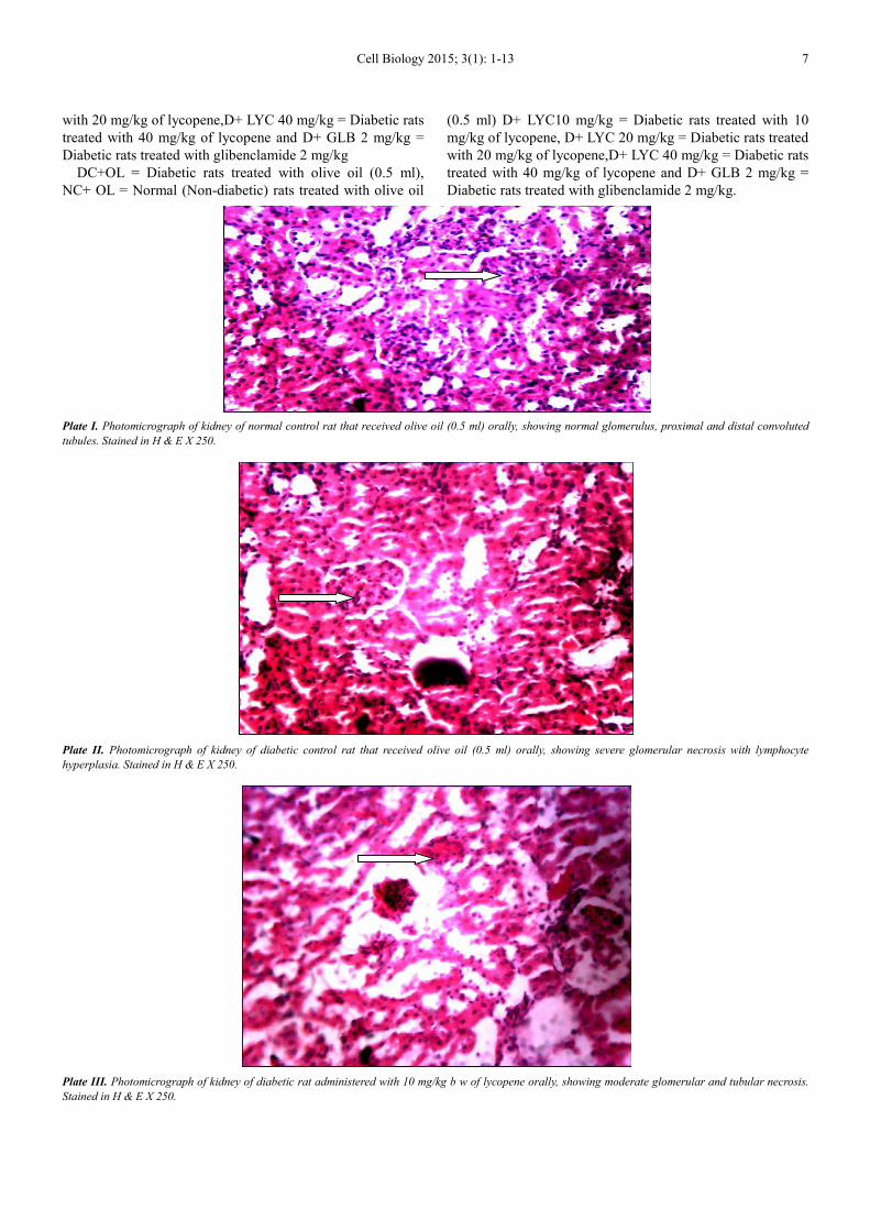
Plate I. Photomicrograph of kidney of normal control rat that received olive oil (0.5 ml) orally, showing normal glomerulus, proximal and distal convoluted
tubules. Stained in H & E X 250.
Plate II. Photomicrograph of kidney of diabetic control rat that received olive oil (0.5 ml) orally, showing severe glomerular necrosis with lymphocyte
hyperplasia. Stained in H & E X 250.
Plate III. Photomicrograph of kidney of diabetic rat administered with 10 mg/kg b w of lycopene orally, showing moderate glomerular and tubular necrosis.
Stained in H & E X 250.

8 Eze Ejike Daniel et al.: Effect of Lycopene on Kidney Antioxidant Enzyme Activities and Functions in
Streptozotocin-Induced Diabetic Wistar Rats
Plate IV. Photomicrograph of kidney of diabetic rat administered with 20 mg/kg b w of lycopene orally, showing moderate glomerular and tubular necrosis.
Stained in H & E X 250.
Plate V. Photomicrograph of kidney of diabetic rat administered with 40 mg/kg b w of lycopene orally, showing mild glomerular and tubular necrosis. Stained
inH & E X 250.
Plate VI. Photomicrograph of kidney of diabetic rat administered with 2 mg/kg b w of glibenclamide orally, showing mild glomerular and tubular
necrosis.Stained in H & E X 250.

Cell Biology 2015; 3(1): 1-13 9
4. Discussion
In this study, the intra-peritoneal administration of
streptozotocin (STZ) effectively induced diabetes mellitus in
rats which was confirmed by elevated levels of fasting blood
glucose, 72 hours after STZ injection. This agrees with the
reports of Mohammed et al.[23] and Krishna et al.[34] who
demonstrated that blood glucose level was increased
significantly after 72 hours of STZ injection to Wistarrats.
Streptozotocin has been shown to induce diabetes which is
similarto human hyperglycaemic non-ketotic diabetes mellitus
in animal models. STZ selectively destroys the insulin
producing β-cells which is accompanied by characteristic
alterations in blood insulin and glucose concentrations [35].
Oral administration lycopene significantly decreased the blood
glucose concentration in diabetic animals with highest
hypoglycemic activity of lycopene observed after week 3 and
week 4 respectively when compared with corresponding
diabetic untreated animals. This finding agrees with the reports
of previous investigators [36, 37, 38,
39].Furthermore,oxidative stress induced by reactive oxygen
species generated due to sustained hyperglycaemia has been
implicated in the onset and progression of diabetes mellitus
and its related complications [40]. Hyperglycemia in diabetes
mellitus causes a depletion of the cellular antioxidant defenses
and increases the levels of free radicals [41]. Lycopene which
is one of the potent antioxidants have been shown to have
good free radical scavenging capacity because of its unique
structure (high number of conjugated double bonds) [42].
Therefore, hypoglycaemic effect of lycopene may also be
attributed to its strong antioxidant property [43]. Bose and
Agrawal [42] reported that lycopene have the ability to quench
the superoxide and other free radical anions which are released
in diabetes due to abnormal glucose metabolism, hence
resulting to decreased blood glucose concentration in diabetic
animals as observed in the present study.
The biochemical indices monitored in the kidney are
useful ‘markers’ for assessment of tissue damage. The
measurement of activities of various enzymes in the tissues
and body fluids plays a significant role in disease
investigation and diagnosis [44] as well as assault on the
organs/tissues and to a reasonable extent the toxicity of the
drug [45]. And one of the most important among numerous
diseases in which changes in antioxidant defense systems are
detected is diabetes mellitus [19]. Tissue enzymes can also
indicate tissue cellular damage caused by chemical
compounds long before structural damage that can be picked
by conventional histological techniques [46].The present
investigation indicated that the concentration of MDA, a
maker of lipid peroxidation was significantly higher in the
kidney tissue of diabetic untreated rats when compared with
the animals in the normal control group. This result
corroborates other investigators [47, 48, 49] who have
reported a significantly increased kidney MDA in
experimentally induced diabetes in animals. This increase in
the kidney MDA indicated enhanced lipid peroxidation
which could cause injury to the cells. Increased levels of lipid
peroxides in the plasma are usually considered to be the
consequence of high production and liberation of tissue lipid
peroxides into circulation due to pathological changes
[50].Oxidative stress causes a bimolecular damage as a result
of the attack of reactive species on components of living
organisms and is known as oxidative damage [51]. This is
caused by increased production and or reduction in the
removal of reactive species by the antioxidant defenses[51].
Hyperglycaemia leads to generation of free radicals due to
auto-oxidation of glucose and glycosylation of proteins [52]
and induces oxidative stress which becomes the chief factor
that leads to diabetic complications [53]. Abnormal elevated
levels of free radicals and the simultaneous reduction of
antioxidant defense can result in damage of cellular
organelles and enzymes, increased lipid peroxidation and
development of insulin resistance [54].The elevated level of
lipid peroxidation causes oxidative damage by increasing
peroxy radicals and hydroxyl radicals [55] and is usually
measured through the catabolite, malonaldehyde (MDA), in
terms of TBARS as a maker of lipid peroxidation [56]. The
decrease in MDA of kidney tissues upon administration of
lycopene to diabetic animals in the present study, clearly
demonstrated the antioxidant property of lycopene. These
findings suggest that the lycopene may exert antioxidant
activity and protect the tissue from lipid peroxidation. In the
same manner, the kidney antioxidant enzymes (SOD, CAT
and GPx) of diabetic control animals were significantly
decreased in comparison with the animals in the normal
control group. These observations are in consonance with the
findings of Kinalskiet al.[47] and Bukanet al. [48] who
demonstrated a significant decrease in kidney antioxidant
enzymes in diabetes. SOD, a superoxide radical scavenging
enzyme is considered the firstline of defense against the
deleterious effect of oxygen radicals in the cells and it
scavenges reactive oxygen radical species by catalyzing the
dismutation of O2- radical to H2O2 and O2
-[57].SOD has been
reported to be involved in the conversion of superoxide anion
radicals produced in the body to hydrogen peroxide, thereby
reducing the likelihood of superoxide anion interacting with
nitric oxide to form reactive peroxynitrite. As a result,
reduction in SOD activity in diabetic animals observed in the
present study may be as a results of an increased influx of O2-
radical and hence may reflects the cause of tissue injury [57].
This finding is consistent with the report of Kedziora-
Kornatowskaet al.[58] who reported decreased renal activity
of SOD three to six weeks after STZ administration. In
contrast, treatment of diabetic rats with lycopene and
glibenclamide enhanced the activity of SOD in comparison
with the diabetic control group. On the other hand, catalase
(CAT) is an enzymatic antioxidant widely distributed in all
animal tissues. CAT decomposes hydrogen peroxide and
protects the tissues from highly reactive hydroxyl radicals
[59]. The present investigation showed that STZ injection
caused a significant decrease in the activity of kidney
catalase level.Thisagrees with the finding of Rauscher et

10 Eze Ejike Daniel et al.: Effect of Lycopene on Kidney Antioxidant Enzyme Activities and Functions in
Streptozotocin-Induced Diabetic Wistar Rats
al.[60] who have reported similar observation. In the current
study, the decreased CAT activity in kidney of diabetic
animals was significantly improved following oral
administration of lycopene and glibenclamide. This
observation is in consonance with the reports of Kuhadet
al.[37] and Ali and Agha [38] who showed that
administration of lycopene (90 mg/kg body weight) to
streptozotocin-induced hyperglycemic rats caused increased
antioxidant enzyme activities (i.e., superoxide dismutase and
catalase). In addition, glutathione peroxidase (GPx) is a
potent endogenous antioxidant that helps to protect cells from
a number of noxious stimuli including oxygen derived free
radicals [61, 62].GPx is an antioxidant enzyme involved in
the detoxification ofhydrogen and lipid peroxidesand also
acts as a peroxynitritereductase[63]. A significant decrease in
GPx activity might be accompanied by a significant increase
in lipid peroxidation as evidenced by increased kidney MDA
concentration observed in the present study. The results of
the current finding agree with the reports of other researchers
[64].However, administration of graded doses of lycopene
and glibenclamide to diabetic animals to significantly
increased level of the activity of GPx. These findings have
been substantiated by other investigators [37, 38]. Therefore,
the increased activity of GPx might be a protective
mechanism in response to increased concentrations of H2O2
and other lipid peroxides in kidney of diabetic animals
treated with lycopene.These observations also indicate that
lycopene has nephroprotectiveeffects.Lycopene’s
configuration has been reported to be responsible for its
ability to inactivate free radicals and to interfere with free-
radical-initiated reactions, particularly lipid peroxidation,
thereby preventing tissue injury [65].The antioxidant
enzymes are very good biochemical markers of stress and
their elevated activity may confirm a potential for
remediation [66]. In addition, it has been reported that
lycopene has high efficient antioxidant and free radical
scavenging capacity [43].Lycopene has been shown to be one
of the best biological suppressants of free radicals, especially
those derived from oxygen. It has the highest singlet oxygen-
quenching rate of all carotenoids in biological systems
[67].The findings in the present study denoted the ability of
lycopene to protect the kidney tissue from oxidative damage
through elevation of endogenous antioxidant enzymes (SOD,
CAT and GPx). The attenuation of kidney tissue in diabetic
animals treated with lycopene, which also positively
correlated with the significantly reduced kidney MDA level a
marker of oxidative stress, further strengthens the notion and
also suggests that lycopene may have the ability to protect
the kidneys from oxidative injury.
Glycosuria, which is a pertinent diagnostic feature of
diabetes, imposes dehydration via glucose osmotic dieresis
[68, 69,70]. In the present study, our findings showed that
there was a significant decrease in the serum sodium ion
concentration when compared with the normal control
animals. Kidney function has been reported to be
compromised in uncontrolled diabetes mellitus.Hence the
depleted serum sodium ion may be attributed to dehydration
which is accompanied with severe loss of electrolytes
including sodium, potassium, calcium, chloride and
phosphates. However, oral administration of lycopene to
diabetic rats restored the serum sodium ion level almost close
to normal. On other hand, the study also showed a significant
increased serum urea level in diabetic control rats in
comparison with the normal control animals. Plasma urea are
recognized markers of glomerular filtration rate (GFR) and in
nephropathy [71]. This result is an agreement with the report
of Sapnaet al.[72] that has showed increased serum urea level
in diabetic patients. An elevation of serum urea usually
signifies decreased renal function [72]. More so, in diabetes
there is increased catabolism of amino acids resulting in high
urea formation from the urea cycle [72]. Hence in this study,
the elevation of serum urea with hyperglycemia can be
suggested as indicator for renal dysfunction [73], and
reduced filtering capacity of kidneys which leads to
accumulation of waste products within the system of diabetic
animals. Treatment diabetic rats with lycopene and
glibenclamide produced a significantly reduced serum urea
level, suggesting its ability to protect against diabetes-
induced kidney damage, by preventing altered protein
metabolism and/or impaired renal function that often exist in
diabetes mellitus. Diabetes is characterized by increased
volume and metabolites excretions via the kidneys, usually in
excess of normal thresholds. These usually give rise to
derangements in homeostatic balance with respect to
electrolytes [69, 70]. Although no significant difference was
recorded in the serum potassium, chloride and bicarbonate
ions concentrations in diabetic control rats when compared
with normal control animals. Following treatment with
lycopene the serum level of potassium, chloride and
bicarbonate ions did not differ significantly with those of
diabetic control animals when compared.
5. Conclusion
Available evidence obtained in the present study
demonstrated lycopene produced a significant improvement
in blood glucose level and attenuated kidney oxidative
damage by significant reduction in kidney MDA
concentration and increased kidney SOD, CAT and GPx
activities in diabetic animals. There was also improved renal
function as indicated by restored depleted serum sodium ion
and significantly reduced serum urea level in diabetic
animals.Thus, this finding suggests the ability of lycopene to
protect against diabetes-induced kidney injury.
References
[1] Rossing P, Diabetic nephropathy: Worldwide epidemic and effects of currenttreatment on natural history. Current Diabetes Reports2006;6:479-83
[2] Zelmanovitz T, Gerchman F, Balthazar A, Thomazelli F, Matos J, and Canani L,Diabetic Nephropathy. Diabetology and Metabolic Syndrome2009;1:10-26

Cell Biology 2015; 3(1): 1-13 11
[3] Djordjević V, Hypertension and nephropathy in diabetes mellitus: What is inherited and what is acquired? Nephrology Dialysis Transplantation2001;16(suppl 6):92-93.
[4] Kalant N,Diabetic glomerulosclerosis: Current status. Canadian Medical AssociationJournal1978;119:146-53
[5] Nangaku M, Mechanisms of Tubulointerstitial Injury in the Kidney: Final Common Pathways to End-stage Renal Failure. Internal Medicine2004;43:9-17.
[6] Forbes JM, Coughlan MT,and Cooper ME, Oxidative Stress as a Major Culprit in Kidney Disease in Diabetes. Diabetes2008;57:1446-54.
[7] Ceriello A, Dello- Russo P, Amstad P,and Cerutti P, High glucose inducesantioxidant enzymes in human endothelial cells in culture; Evidence linkinghyperglycemia and oxidative stress. Diabetes1996;45:471-477.
[8] Hodgkinson AD, Bartlett T, Oates PJ, Millward BA, andDemaine AG. The Response of Antioxidant Genes to Hyperglycemia Is Abnormal in Patients With Type 1Diabetes and Diabetic Nephropathy. Diabetes2003;52:846-51.
[9] Maritim AC, Sanders RA Watkins JB, Diabetes, oxidative stress, andantioxidants: A review. Journal of Biochemical and Molecular Toxicology 2003b;17:24-38.
[10] Sih MT, Arpeeta S, Judy BD, Oxidative Stress and Novel AntioxidantApproachesto Reduce Diabetic Complications, Oxidative Stress and Diseases, Dr. VolodymyrLushchak (Ed.), InTech, Available from: http://www.intechopen.com/books/oxidativestress-anddiseases/oxidative-stress-and-novel-antioxidant-approaches-to-reduce-diabetic complications, 2012.
[11] Kakimoto M, Inoguchi T, Sonta T, Yu HY, Imamura M,Etoh T. et al.Accumulation of 8- hydroxy-2'-deoxyguanosine and mitochondrial DNA deletion inkidney of diabetic rats. Diabetes2002;51:1588-95
[12] Fenercioglu AK, Saler T, Genc E, Sabuncu H, andAltuntas Y, The effects of polyphenol-containing antioxidants on oxidative stress and lipid peroxidation in Type 2 diabetes mellitus without complications. Journal of Endocrinology andInvestigation2010; 33(2):118-124.
[13] Neri S, Calvagno S, Mauceri B, Misseri M, Tsami A, Vecchio C, Mastrosimone G, Di Pino, A, Maiorca D, Judica A, Romano G, Rizzotto A, and Signorelli S,Effects of antioxidants on postprandial oxidative stress and endothelial dysfunction in subjects with impaired glucose tolerance and type 2 diabetes. European journal of Nutrition2010; 49 (7):409-416.
[14] TrevithickJ, Massel D, Robertson JM, Tomany S, and Wall R. Model study of AREDS antioxidant supplementation of AMD compared to Visudyne: a dominant strategy? Ophthalmic Epidemiology2004; 11(5):337-346.
[15] KantoffP, Prevention, Complementary Therapies, and New Scientific Developments in the Field of Prostate Cancer. Rev. Urol.2006; 8(Suppl 2), S9-S14.
[16] Sesso HD, Liu S, Gaziano JM, and Buring JE, Dietary Lycopene, Tomato-Based Food Products and Cardiovascular Disease in Women. Journal ofNutrition2003; 133, 2336- 2341.
[17] Mozaffarieh M, Sacu S andWedrich A, The Role of the Carotenoids, Lutein and Zeaxanthin, in Protecting Against Age-related Macular Degeneration: A Review Basedon Controversial Evidence.Nutr. J. 2003; 2: 20.
[18] Carlier C, Coste J, Etchepare M, Périquet B, and Amédée-Manesme O,A Randomised Controlled Trial to Test Equivalence Between RetinylPalmitate and BetaCarotene for Vitamin A Deficiency. BMJ 1993; 307(6912):1106-1110.
[19] Sevim ÇY,Fatmagül Y,and Ebubekir C,Effect of Lycopene Application in Rats with Experimental Diabetes Using Lipoprotein, Paraoxonase and Cytokines Journal of MembraneBiology 2013; 246: 621–626
[20] Rao AV, and Agrawal S,Role of lycopene as antioxidant carotenoid in the preventionof chronic diseases, a review. Nutrition Research1999; 19: 305-323.
[21] Bramley P M, Regulation of carotenoid formation during tomato fruit ripening and development. Journal of Experimental Botany2002; 53 (377): 2107-2113.
[22] Betty K, Charlotta T, Mary CH,and McKeon T A, Fatty acid and carotenoidcomposition of Gac (MomordicacochinchinensisSpreng) fruit. Journal of Agricultureand Food Chemistry2004; 52:274-279.
[23] Ogundeji T, Ayo J O, Aluwong T, and Mohammed A,Behavioural and haematological studies on effects of lycopene in Wistar rats subjected to psychological stress. Journal of Neuroscience and Behavioral Health2013;5(2):30-35.
[24] MohammedA, Tanko Y, Okasha M A, Magaji R A, and Yaro AH, Effects ofaqueous leaves extract of Ocimumgratissimum on bloodglucose levels of streptozocininduced diabetic wistar rats. African Journal of Biotechnology 2007;6:2087-2090.
[25] Beach EF, and Turner JJ. An enzymatic method for glucose determination uptake in bodyfluids. Clinical Chemistry1958; 4:462-468.
[26] Placer ZA,Cushmann L, and Johnson BC,Estimation of products of lipid peroxidation in biochemical systems. Analytical Biochemistry 1996; 16: 359-364.
[27] Suzuki K, Measurement of Mn-SOD and Cu, Zn-SOD. In: Taniguchi, N., Gutteridge, J. (Eds.), Experimental Protocols for Reactive Oxygen and Nitrogen Species. Oxford University Press, U.K. 2000; Pp. 91–95.
[28] Aebi H, Catalase in vitro.Methods in Enzymology 1984; 105, 121– 126.
[29] Flohe LA,and Gunzler WA, Assays for glutathione peroxidase. Methods inEnzymology 1984;105:114– 120.
[30] Tietz NW, Prude EL, and Sirgard-Anderson O,Textbook of clinical chemistry. Edited by Burtis C.A. AshwoodE.R.andBruns DE. WB Saunders Company, Philadelphia, London 1994; Pp 1354 – 1374.
[31] Vogel A I, A Textbook of Quantitative Inorganic Analysis. LongmanGroup Ltd. London. 3rd Edition, 1960; Pp 882-885.
[32] Segal M A, A rapid electrotitimetricmethod for determining CO2 combining power in plasma or serum. American Journal of Clinical Pathology 1955;25(10): 1212-1216.
[33] Schales O, Schales S, A simple and accurate method for the determination of chloride in biological fluids.Journal of Biochemistry1941; 140, 879-884.
[34] Krishna D, Rao S, Satyanarayana ML, Serum insulin levels and lipid profiles of streptozotocin induced diabetic wistar rats. Journal of Indian Veterinary Association, Kerala2012; 10 (2):22-26.

12 Eze Ejike Daniel et al.: Effect of Lycopene on Kidney Antioxidant Enzyme Activities and Functions in
Streptozotocin-Induced Diabetic Wistar Rats
[35] Szkudelski T, The mechanism of alloxan and streptozotocin action in β-cells of the rat pancreas. Physiological Research 2001;50(6):537-546.
[36] Duzguner V, Kucukgul A, Erdogan S, Celik S, andSahinK, Effect of lycopene administration on plasma glucose, oxidative stress and body weight in streptozotocin diabetic rats. Journal of Applied Animal Research 2008; 33: 17–20.
[37] Kuhad A, Sethi R, Chopra S,Lycopene attenuates diabetes-associated cognitive decline in rats. Life Science 2008; 83(3-4):128-134.
[38] Ali MM,and Agha FC, Amelioration of streptozotocin-induced diabetes mellitus, oxidative stress and dyslipidemia in rats by tomato extract lycopene. Scandinavian Journal of Clinical and Laboratory Investigation 2009; 69(3): 371-379.
[39] Aydin M, and Celik S,Effects of lycopene on plasma glucose, insulin levels, oxidative stress, and body weights of streptozotocin-induced diabetic rats. Turkish Journal of Medical Sciences2012; 42 (Sup.2): 1406-1413.
[40] Giacco F, and Brownlee M, Oxidative stressand diabetic complications. Circulation Research2010; 107(9):1058- 1070.
[41] Tsuruta R, Fujita M, Ono T, Koda Y, Koga Y, Yamamoto T, Nanba M, Shitara M, Kasaoka S, Maruyama I, Yuasa M, and Maekawa T, Hyperglycemia enhances excessive superoxide anion radical generation, oxidative stress, early inflammation, and endothelial injury in forebrain ischemia/reperfusion rats. Brain research2010; 1309:155-163.
[42] Bose KSC, and Agrawal B K,Effect of Long Term Supplementation of Tomatoes (Cooked) on Levels of Antioxidant Enzymes, Lipid Peroxidation Rate, Lipid Profile and GlycatedHaemoglobin in Type 2 Diabetes Mellitus. West Indian Medical Journal 2006; 55(4): 274-278.
[43] Atessahin A, Yilmaz S, Karahan I, Ceribas A O, and Karauglu A, Effects oflycopene against cisplant-induced nephrotoxicity and oxidative stress in rats. Toxicology2005; 212: 116-123.
[44] Malomo SO, Toxicological implication of ceftriaxone administration in rats. Nigerian JournalBiochemistry and Molecular Biology2000; 15(1): 33-38.
[45] Yakubu MT, Salau I O and Muhammad N O, Phosphatase activities in selected rat tissues following repeated administration of ranitidine. Nigerian Journal of Biochemistry and Molecular Biology2003b;18(1): 21-24.
[46] Akanji M A andYakubu M T,α- Tocopherol protects against metabisulphite –induced tissue damage in rats. Nigerian Journal of Biochemistry and Molecular Biology2000; 15: 179-183.
[47] Bukan N, Sancak B, Yavuz O, Tutkin CF, Ozcelikay T, andAltan N, Lipid peroxidation and Scavenging Levels in the Liver of streptozotocin-induced Diabetic rats.Indian Journal of Biochemistry and Biophysics2003; 40: 447-450.
[48] Siddiqui MH, Al-Whaibi MH, and Basalah MO, Role of nitric oxide in tolerance ofplants to abiotic stress.Protoplasma2011; 248: 447-455
[49] Erejuwa OO, Oxidative Stress in diabetes mellitus: is there a role for hypoglycaemicdrugs and/or antioxidants? Oxidative stress and diseases, Volodymyr I. Lushchak and Dmytro V. Gospodaryov (Ed.), ISBN: 978-953-51-0552-7, 2012; InTech, Available from:http://www.intechopen.com/books/oxidative-stress-and-diseases/oxidative-stress in diabetes
[50] Al-Faris NA, Al-Sawadi AD, and Alokail MS, Effect of Samh seedssupplementation (MesembryanthemumforsskaleiHochst) on liver enzymes and lipid profiles of streptozotocin (STZ)-induced diabetic Wistar rats. Saudi Journal of Biological Sciences 2010; 17:23-28.
[51] Kumawat M, Pahwa MB, Gahlant VS, and Singh N, Status of antioxidant enzymes and lipid peroxidation in type 2 diabetes mellitus with microvascular complications. The Open Endocrinology Journal2009; 3:12-15. Asian Pacific Journal of Tropical Disease,
[52] Tirgar P, Jadav P, Sheth D, Desai T, Tirgar PR, Jadav PD, and Sheth MDB,Therapeutic role of anti-oxidant properties of Emblicaofficinalis(amla) in streptozotocin induced type 1 diabetic rats. Pharmacologyonline2010; 1:728-743.
[53] ShuklaK, Dikshit P, Tyagi MK, Shukla R,and Gambhir JK, Ameliorative effect of Withaniacoagulans on dyslipidaemia and oxidative stress in nicotinamide-streptozotocin induced diabetes mellitus. Food and Chemical Toxicology, 2012.50: 3595-3599.
[54] Kumar R, Kar B, Dolai N, Bala A, and Haldar PK, Evaluation of antihyperglycaemic andantioxidant properties of StreblusasperLour against streptozotocin–induced diabetes in rats. Asian Pacific Journal of Tropical Disease 2012; 2(2): 139-143.
[55] Singh U, Singh S, and Kochhar A, Therapeutic potential of antidiabeticnutraceuticals.Phytopharmacology, 2012;2(2):144-169.
[56] Kedziora-Kornatowska KZ, Luciak M, and Paszkowski J, Lipid peroxidation and activities of antioxidant enzymes in the diabetic kidney: Effect of treatment with angiotensin convertase inhibitors. International Union of Biochemistry and Molecular Biology Life2000; 49: 303–307.
[57] Onyeka CA, Aligwekwe AU, Nwakanma AA, Bakare AA, and Ofoego UC,Effects of Ethanolic Root Bark Extract of Chrysophyllumalbidumon Serum Superoxide Dismutase, Catalase and Malondialdehyde in Rat. International Journal of PharmaSciences and Research2012; 3(3):347-351.
[58] Rauscher FM, Sanders RA, and Watkins JB,Effects of coenzyme Q10 treatment on antioxidant pathways in normal and streptozotocin-induced diabetic rats. Journal of Biochemistry and Molecular Toxicology2001; 15: 41–46
[59] Kim BJ, Hood BL, Aragon RA, Hardwick JP, Conrads TP, Veenstra TD, Song BJ. Increased oxidation and degradation of cytosolic proteins in alcohol-exposed mouse liver and hepatoma cells. Proteomics2006; 6: 1250-1260.
[60] Onyema OO, Farombi EO, Emerole GO, Ukoha AI andOnyeze GO, Effect ofvitamin E on monosodium glutamate induced hepatotoxicity and oxidative stress in rats.Indian Journal of Biochemistry Biophysics2006; 43: 20-24.
[61] Erejuwa OO, Sulaiman SA, Wahab MS, Sirajudeen KNS, Salleh MS andGurtu S, Effect of Glibenclamide alone versus Glibenclamide and Honey on Oxidative Stress in Pancreas of Streptozotocin-Induced Diabetic Rats. International Journal of Applied Research in Natural Products2011;4(2): 1-10.
[62] Moussa SA. Oxidative Stress in Diabetes Mellitus. Romanian Journal of Biophysics2008; 18(3): 225–236.
[63] Seren S, Leberman R, Bayraktar UD, Health EH, Sahin K, Andic F, andKucuk, O,Lycopene in cancer prevention and treatment. American Journal of Therpeutics 2008; 15:66-81.

Cell Biology 2015; 3(1): 1-13 13
[64] Dauqan E,Sani H A, Abdullah A, and Kasim ZM, effect of four different vegetable oils (red palm olein, palm olein, corn oil, coconut oil) on antioxidant enzymes activity of rat liver. Pakistan Journal of Biological Sciences 2011; 14: 399-403.
[65] Bhoomika A D andVaghela NR, Supercritical fluid extraction of lycopene from tomatos by using CO2 as a solvent: A review. Journal of Chemical and Pharmaceutical Research2013; 5(4):188-191
[66] Eteng MU, Ibekwe HA, Essien AD, and Onyeama HP, Effects of Catharanthusroseus on Electrolyte Derangement induced by Chlopropamide (Diabinese)R on Normoglycemic Albino Wistar Rat. Bio- Research2008;6(2): 364-366.
[67] Item JA, Patrick EE, Godwin EE, andIme FA, Effects of Co-administration of Extracts of Vernonia Amygdalina and AzadirachtaIndicaon Serum Electrolyte Profile of Diabetic and non Diabetic Rats. Australian Journal of Basic and Applied Sciences 2009; 3(3):2974-2978.
[68] Tanko Y, Ismail AS,Mohammed KA, Eze ED, Jimoh A, Sada
NM, Muhammad A, and Mohammed A,Ameliorative Effects of Magnesium and Copper Sulphateson Blood glucose and Serum Electrolytes Levels in Fructose-induced Diabetic Wistar Rats. Journal of Applied Pharmaceutical Science2013; 3(07): 160-163.
[69] Madsen TE, Muhlestein JB, Carlquist JF, Horne BD, Bair TL, Jackson JD, Lappe, JM, Pearson RR, and Anderson JL, Serum uric acid independently predicts mortality in patients with significant, angiographically defined coronary disease. American Journal of Nephrology2005; 25: 45-49.
[70] Sapna SL, Yogesh S, Amit S, Ekta AA, and Alok ML,Hyperuricemia, High Serum Urea and Hypoproteinemia are the Risk Factor for Diabetes. Asian Journal of Medical Sciences 2009; 1(2): 33-34.
[71] Trujillo, J., Chirino, Y.I, Molina-Jijon, E., Anderica-Romero, A.C., Tapia, E. and PedrazaChaverri, J,Renoprotective effect of the antioxidant curcumin: Recent findings.Redox Biology2013; 1(1):448–456.


![Welcome! [] · 2019-09-16 · Infographic #DYK tomatoes contain lycopene, an antioxidant that is good for your heart and helps prevent some cancers. ... antioxidants, and dietary](https://static.fdocuments.us/doc/165x107/5f48eb97d39abd31d5409e5c/welcome-2019-09-16-infographic-dyk-tomatoes-contain-lycopene-an-antioxidant.jpg)


