
Effect of bore fluid flow rate on formation and properties ...
12
ORIGINAL ARTICLE Effect of bore fluid flow rate on formation and properties of hollow fibers Asrar A. Alobaidy 1 • Bashir Y. Sherhan 2 • Areej D. Barood 2 • Qusay F. Alsalhy 2 Received: 25 April 2017 / Accepted: 20 June 2017 / Published online: 28 June 2017 Ó The Author(s) 2017. This article is an open access publication Abstract In this work, for high performance and wide range of ultrafiltration applications, the effects of the most widely used values of internal coagulant flow rates (ICFR) (i.e., 2.6, 3.6, 4, 5, 7, 9, 11, and 13 ml/min) on the different features of the polyvinylchloride hollow fiber have been investigated. Both the idealized straight and the cylindrical pore with small effect of tortuosity were approximately obtained through the effect of ICFR. Atomic force micro- scope (AFM), scanning electron microscope (SEM), and ultrafiltration measurements were utilized to characterize the hollow fibers. The SEM and AFM results indicated that the cross-sectional morphology of the fibers is changed significantly with various ICFR. The structure of the inner surface was also changed from an open cellular structure to a porous structure by means of high pore density and small pore diameter. In addition, the membrane thickness was reduced by 314% with an increase in the ICFR from 2.6 to 13 ml/min. The pure water permeation flux was improved 17 times when ICFR was increased to 13 ml/min, while the BSA rejection remained within the acceptable range (from 93.4 to 90.4) when the ICFR was increased from 2.6 to 9 ml/min. Keywords Hollow fiber Ultrafiltration PVC Internal coagulant Separation performance Introduction One of the main goals of membrane technology is to control both membrane pore size and distribution during the con- figuration, as they have a large impact on the separation performance of any membrane. Several factors affect membrane pore size and distribution such as composition of the polymer, composition and type of the additives, air-gap length, type and temperatures of inner and outer coagulants, flow rate of the polymer solution, flow rate of the internal coagulant, and the design of the spinneret. In fact, the flow rate of the internal clotting is the most significant factor influencing the membrane pore size and distribution (Jack et al. 2006). Therefore, one of the main goals of this effort is to investigate this hypothesis. The literature review indicated that the effect of the flow rate of the internal coagulant on the characteristics and performance of the fiber has not been extensively studied (Jack et al. 2006; Chung et al. 1977; Aptel et al. 1985; Mok et al. 1995; Miao et al. 1996; Qin and Chung 2004; Wan and Chung 2015; Cheng et al. 2017; Peng et al. 2012). For example, Chung et al. (1977) studied the fabrication of polyethersulfone (PES) membrane with ultrathin skin layer membranes of 50 nm skin layer thickness controlled by the lumen liquid flow rate and lumen liquid chemistry. Aptel et al. (1985) investigated the influence of the internal coagulant flow rate on characteristics of poly- sulfone (PSF) ultrafiltration. They reported that, as the non- solvent flux in the lumen side of the nascent fiber increased from 4 9 10 -2 to 6 9 10 -2 cm 3 /s, the hydraulic perme- ability also increased from 2 9 10 -8 to 6 9 10 -8 cm/s-Pa. Whereas, rejection for polyvinylpyrrolidone [PVP, & Bashir Y. Sherhan [email protected]; [email protected] & Qusay F. Alsalhy [email protected]; [email protected] 1 Chemical Engineering Department, College of Engineering, University of Baghdad, Baghdad, Iraq 2 Membrane Technology Research Unit, Chemical Engineering Department, University of Technology, Alsinaa Street 52, P.B.O 35010, Baghdad, Iraq 123 Appl Water Sci (2017) 7:4387–4398 https://doi.org/10.1007/s13201-017-0584-7
Transcript of Effect of bore fluid flow rate on formation and properties ...

ORIGINAL ARTICLE
Effect of bore fluid flow rate on formation and propertiesof hollow fibers
Asrar A. Alobaidy1 • Bashir Y. Sherhan2 • Areej D. Barood2 • Qusay F. Alsalhy2
Received: 25 April 2017 / Accepted: 20 June 2017 / Published online: 28 June 2017
� The Author(s) 2017. This article is an open access publication
Abstract In this work, for high performance and wide
range of ultrafiltration applications, the effects of the most
widely used values of internal coagulant flow rates (ICFR)
(i.e., 2.6, 3.6, 4, 5, 7, 9, 11, and 13 ml/min) on the different
features of the polyvinylchloride hollow fiber have been
investigated. Both the idealized straight and the cylindrical
pore with small effect of tortuosity were approximately
obtained through the effect of ICFR. Atomic force micro-
scope (AFM), scanning electron microscope (SEM), and
ultrafiltration measurements were utilized to characterize
the hollow fibers. The SEM and AFM results indicated that
the cross-sectional morphology of the fibers is changed
significantly with various ICFR. The structure of the inner
surface was also changed from an open cellular structure to
a porous structure by means of high pore density and small
pore diameter. In addition, the membrane thickness was
reduced by 314% with an increase in the ICFR from 2.6 to
13 ml/min. The pure water permeation flux was improved
17 times when ICFR was increased to 13 ml/min, while the
BSA rejection remained within the acceptable range (from
93.4 to 90.4) when the ICFR was increased from 2.6 to
9 ml/min.
Keywords Hollow fiber � Ultrafiltration � PVC � Internalcoagulant � Separation performance
Introduction
One of the main goals of membrane technology is to control
both membrane pore size and distribution during the con-
figuration, as they have a large impact on the separation
performance of any membrane. Several factors affect
membrane pore size and distribution such as composition of
the polymer, composition and type of the additives, air-gap
length, type and temperatures of inner and outer coagulants,
flow rate of the polymer solution, flow rate of the internal
coagulant, and the design of the spinneret. In fact, the flow
rate of the internal clotting is the most significant factor
influencing the membrane pore size and distribution (Jack
et al. 2006). Therefore, one of the main goals of this effort is
to investigate this hypothesis. The literature review indicated
that the effect of the flow rate of the internal coagulant on the
characteristics and performance of the fiber has not been
extensively studied (Jack et al. 2006; Chung et al. 1977;
Aptel et al. 1985; Mok et al. 1995; Miao et al. 1996; Qin and
Chung 2004; Wan and Chung 2015; Cheng et al. 2017; Peng
et al. 2012). For example, Chung et al. (1977) studied the
fabrication of polyethersulfone (PES) membrane with
ultrathin skin layermembranes of 50 nm skin layer thickness
controlled by the lumen liquid flow rate and lumen liquid
chemistry. Aptel et al. (1985) investigated the influence of
the internal coagulant flow rate on characteristics of poly-
sulfone (PSF) ultrafiltration. They reported that, as the non-
solvent flux in the lumen side of the nascent fiber increased
from 4 9 10-2 to 6 9 10-2 cm3/s, the hydraulic perme-
ability also increased from 2 9 10-8 to 6 9 10-8 cm/s-Pa.
Whereas, rejection for polyvinylpyrrolidone [PVP,
& Bashir Y. Sherhan
& Qusay F. Alsalhy
[email protected]; [email protected]
1 Chemical Engineering Department, College of Engineering,
University of Baghdad, Baghdad, Iraq
2 Membrane Technology Research Unit, Chemical
Engineering Department, University of Technology, Alsinaa
Street 52, P.B.O 35010, Baghdad, Iraq
123
Appl Water Sci (2017) 7:4387–4398
https://doi.org/10.1007/s13201-017-0584-7

molecular weight (MW) = 10,000] decreased from 80 to
40%. The influence of the internal coagulant flow rate, the
temperature, and the composition of casting solution on the
separation performance of PESmembranes was investigated
by Mok et al. (1995). These authors found that the outer to
inner radius of the hollow fiber seems to decrease with the
internal coagulant flow rate.
The influence of internal coagulant flow rates (7.5 and
5 ml/min) on the properties of PES membranes made using
dope spinning method was investigated by Miao et al.
(1996). They reported that, at a constant air-gap length, by
increasing the flow rate of the internal coagulant, both inner
diameter (ID) and outer diameter (OD) can be enhancedwith
the reduction of the thickness of the wall for the formed
membranes. The separation factor and permeability of UF
membrane for polyethylene glycol (PEG) solutes reduced
with the increase in the flow rate of the lumen-side coagulant
due to the decrease in the skin layer thickness and the size of
the skin layer pores, and narrow the pore size distribution on
the skin layer. Moreover, Qin and Chung (2004) found that,
as the flow rate of the internal coagulant diminished, the
diameter of the inner PESmembrane surface declined, while
the diameter of the outer surface remained the same. Besides,
as internal coagulant flow rate increases, the mass transport
at the outer surface increases,while that at the inner surface is
minimized, resulting in an increase in themacro-void length.
In fact, extensive work has been dedicated to the fabri-
cation of PVC hollow fiber membranes for different sepa-
ration applications. However, the effect of spinning
parameters on either the fabrication of PVC fibers or the
properties of the PVC fibers was not studied extensively.
Membrane properties, especially permeability, can be
affected by different resistances, which are a function of
pore-size distribution, porosity, membrane barrier thickness,
and the solvent properties, whilst both the idealized straight
and the cylindrical pore with small effect of tortuosity are
relevant to the influence of ICFR. Consequently, this study
was focused on the impact of ICFR on the structural prop-
erties and membrane performance, as it exerts the greatest
influence on the membrane thickness, size, and distribution
of the pores. Inner and outer surface structures, as well as the
fiber cross-section were characterized by SEM, and the
ultrafiltration measurements were carried out using bovine
serum albumin (BSA) with MW = 67 kg/mol as a solute.
Experimental part
Materials
The PVC resins and N, N-dimethylacetamide (DMAc)
solvent were supplied by Sigma-Aldrich, while Chemical
Co was used for membrane preparation. PVC with Mw of
65 kg/mol was obtained from Georgia Gulf Company
(Georgia, USA). It was used as a membrane material in this
study due to its excellent physical and chemical properties,
stiffness, low cost, superior mechanical properties, and
good solvent resistance.
Polymer spinning solution
PVC dope solution was prepared from 16 wt% PVC
polymer and 84 wt% DMAc solvent. Dried PVC resin was
gradually added into the covered container containing
DMAc solvent and then mixed by a magnetic stirrer until
the solution became clear and homogeneous.
PVC/DMAc hollow fiber spinning procedure
The PVC/DMAc homogenous solution was kept for at least
24 h to eliminate air bubbles. The PVC/DMAc solution
was then transferred to a vertical column with 6 cm inner
diameter. Throughout the entire spinning process, the
temperature was maintained at 26 �C. Hollow fibers were
prepared using the dry/wet spinning method with various
internal coagulant flow rates, as described elsewhere (Al-
salhy et al. 2011, 2013; Alsalhy 2013). The dope polymer
solution was forced via pressurized nitrogen to the spin-
neret of 0.5 and 0.9 mm inner and outer diameter,
respectively. The PVC/DMAc ratio was 16/84 wt%, and
the fabrication conditions of the prepared PVC membranes
were as follows: 1.75 bar extrusion pressure, 3 cm air-gap
length, 19 �C external coagulation bath temperature with
tap water, and bore fluid with various flow rates (i.e., 2.6,
3.6, 4, 5, 7, 9, 11, and 13 ml/min).
The experimental apparatus and the process procedure
were described elsewhere (Alsalhy et al. 2011, 2013, 2014;
Alsalhy 2013). It is well known that water is always used as
an internal and external coagulant in hollow fiber prepa-
ration and other membrane configurations due to the low
cost of solvent and its good affinity and solubility with all
of the polymer solvents. Therefore, in this work, water was
used as the internal and external coagulant liquid. The
prepared fibers were wetted and kept in a tap water bath at
ambient temperature for 24 h to eliminate the residual
DMAc solvent. PVC fibers were kept for 72 h in a con-
tainer containing 40 wt% glycerol aqueous solution to
avoid the fiber structure collapse. Prior to testing, the PVC
fibers were dried in air at room temperature.
SEM and AFM observations
The internal and the external membrane structures were
tested by means of scanning electron microscopy (SEM) at
University of Technology/Nanotechnology and Advanced
Material Research Center. The hollow fiber (HF)
4388 Appl Water Sci (2017) 7:4387–4398
123
membranes were dried and then immersed in liquid nitro-
gen for 15 s and thereafter cut to reveal the cross-sectional
structure.
AFM device (Angstrom Advanced Inc., Braintree,
Boston, USA) model AA3000 was used to obtain 2D and
3D images of the membrane surface. The fiber was sub-
jected to a wide surface analysis using an AFM in contact
mode with a tip made from silicon. A statistical distribution
of the pore size was estimated for the PVC surfaces of each
membrane using IMAGER 4.31 software.
Diameter, thickness, and porosity of PVC
membranes
The diameter and thickness of HF membranes were
determined using optical microscope Model B600.POL-I
S/N: 0016243, Italy.
Membrane porosity (e) is the ratio between the pore
volume and the total membrane volume. Fiber porosity was
measured using the procedure described elsewhere (Al-
salhy et al. 2011). The fiber sample was dried in a vacuum
oven for approximately 6 h at 80 �C to obtain constant
hollow fiber weight. Then, the sample was weighed using a
digital balance. According to the fiber weight and volume,
fiber density (qm) was estimated, yielding the density of the
polymer material (qp) of 1.4 g/cm3 (Alsalhy et al. 2011).
The total porosity was estimated using the following
equation:
eð%Þ ¼ 1� qmqp
!� 100 ð1Þ
UF experiments
Figure 1 shows the schematic drawing of performance rig.
PVC membrane was examined in pack of five fibers, each
of 20 cm length. The fibers were fixed at the ends of the
module using epoxy resin. Pressure and feed solution flow
rate of the performance test were 1 bar and 1 m/s,
respectively. Permeate was collected from the lumen side,
while feed solution was moved to the shell side of the
module. The BSA composition in the aqueous solution was
800 ppm. The BSA concentration was measured by a UV-
spectrophotometer (Shimadzu-UV160 A, Japan) at
k = 280 nm. The BSA rejection factor (F) was estimated
according to the equation below (Alsalhy et al.
2011, 2013):
F ¼ Cfeed � Cpermeate
Cfeed
ð2Þ
where Cfeed and Cpermeate are defined as the BSA concen-
tration in the feed and permeate side of the hollow fiber,
respectively.
Pure water permeation rate (PWP) can be estimated by
applying the following equation:
PWP ¼ V
nAtDPð3Þ
where V denotes the collected permeate volume, n is the
number of fibers in the bundle, A is the membrane surface
area, t is the collected time, and DP is the transmembrane
pressure.
Results and discussion
SEM test
Figures 2 and 3 provide the SEM images of the PVC
hollow fibers prepared at various flow rates for the internal
coagulant (distilled water). It can be seen that the inner
structures and the cross-section are highly dependent on the
internal coagulant flow rate on the lumen side of the fiber.
The hollow fiber prepared from 16 wt% PVC dope under
2.6 ml/min internal coagulant flow rate comprises of two
small layers forming a finger-like structure. These corre-
spond to the inner and the outer surface of the cross-section
depicted in Fig. 2a. Similarly, two large layers in the shape
of finger and sphere-like structure can clearly be observed
at the center of the cross-section of the membrane, as
depicted in Fig. 2a. It can be seen in Fig. 2b that the two
small layers at the inner and outer edges are reduced with
increment of the internal coagulant flow rate to 3.6 ml/min.
On the other hand, the two fingers and the sphere-like
structure layers at the center of the cross-section appear to
be visibly changed because the layer towards the outer
surface is transformed to a wide finger-like structure, while
the layer towards the inner surface has become larger
within the same spherical shape.
Under lumen liquid rate of 5 ml/min, the layer in the
center of the cross-section toward the external surface is
seen to be merged with the layer located near the edge of
the outer surface, and their final shape is transformed into a
finger-like structure shown in Fig. 2c. However, the layer
in the center of the cross-section toward the inner surface
has been combined with the layer located near the inner
edge of the fiber, whereby the final structure takes ellip-
soidal shape, as shown in Fig. 2c. Further increase in the
internal coagulant flow rate to 7 ml/min results in the two
layers adopting the finger-like structure depicted in Fig. 2d.
Moreover, due to the increase in the internal coagulant flow
rate to 9 ml/min, two layers with different structures are
formed. The first small one is located near the outer surface
and takes a finger-like form, while the second layer is
found near the inner surface and forms an ellipsoidal-like
structure, as exhibited in Fig. 2e. Further, increasing the
Appl Water Sci (2017) 7:4387–4398 4389
123

flow rate of the internal coagulant can be guided to create
two layers of large finger-like structure with the same
thickness such as that shown in Fig. 2f, even as the
increase of the flow rate may be resulted to construct two
layers of the finger-like structure, as shown in Fig. 2g.
Figure 3 confirms the effect of internal coagulant flow rate
on the inner surface structure of the hollow fiber mem-
branes. It can be noticed that, as the internal coagulant flow
rate increases, the obtained inner surface seems to be a
skinless porous structure of the fiber membrane made under
lumen liquid rate of internal coagulant of 3.6 ml/min, as
depicted in Fig. 3a. Increasing the flow rate of the internal
coagulant to 5 and 7 ml/min has also led to the formation
of the same structure, along with diminishing in the pore
size at the surfaces, as shown in Fig. 3b, c.
Actually, this phenomenon is due to the speed of the
demixing process of water in internal coagulant with the
solvent of polymer solution, followed by domain growth
that reaches the point of domain (droplet) coalescence. In
this case, both phases keep their liquid character until
coalescence occurs. This should prevent the formation of a
skin layer at the fiber inner surface, resulting in the for-
mation of a microporous surface (Zeman and Zydney 1996;
Alsalhy et al. 2011). This phenomenon occurs when the
demixing process of the solvent in the dope solution and
water in the internal coagulant is delayed.
Additional increase in the internal coagulant flow rate to
9 ml/min can result in the emergence of a porous structure
with small pore size, as shown in Fig. 3d. Increasing the
internal coagulant flow rate to 13 ml/min might produce a
surface with porous structure and high pore density, as
observed in Fig. 3e, in comparison with that given in
Fig. 3d. This observation can be attributed to the instan-
taneous liquid–liquid demixing process between DMAC in
polymer solution and the water in internal coagulant due to
the short contact time between high amount of water and
DMAC in dope solution. The short contact time between
bore fluid and dope solution can be explained by the high
exchange rate between water in internal coagulant and
DMAC solvent in polymer solution. This results in an
instantaneous liquid–liquid demixing process, which in
turn leads to the removal of high amount of solvent from
the dope solution during the formation of the hollow fiber.
Consistent with the last finding, the solidification occurred
very rapidly within the small porous structure of high pore
density. Porter (1990) reported that very high rates of
precipitation (corresponding to short precipitation time)
result in the formation of a finger-like micro-void structure.
The use of a larger volume of bore fluid causes complete
polymer precipitation as the non-solvent penetrates from
the bore towards the outer surface of the fiber. Fast
demixing process (higher precipitation rates) forms finer
pores while delayed demixing process (slow precipitation
rates) results in rough structures.
It can thus be concluded that internal coagulant flow rate
is the main factor in controlling the pore size and density
(Jack et al. 2006; Miao et al. 1996). As increasing the
internal coagulant rate results in a 17-fold increase in the
permeation flux, this parameter has a significant impact on
the membrane structure and/or membrane performance.
Membrane thickness and porosity
The most important factor affecting the barrier perme-
ability is the membrane thickness owing to the direct
relationship between the membrane thickness and both the
shape and the size of pores. For this reason, our work
focused on the effect of ICFR on the membrane properties.
Fig. 1 Schematic diagram of
the UF experimental rig
4390 Appl Water Sci (2017) 7:4387–4398
123

Fig. 2 Cross-section SEM
images of the PVC hollow fiber
membranes at various ICFR
Appl Water Sci (2017) 7:4387–4398 4391
123

Figure 4 shows the effect of various ICFR on the mem-
brane thickness. It can be noted that the membrane thick-
ness decreases with increasing the ICFR from 2.6 to 13 ml/
min, and in this range of the ICFR, the membrane thickness
reduces by 314%. This observation is attributed to the
effect of the internal pressure increase as a consequence of
Fig. 3 SEM images of the inner surfaces of the PVC hollow fiber membranes at various ICFR
4392 Appl Water Sci (2017) 7:4387–4398
123

the increase in the amount of ICFR on the inner surface of
the hollow fiber for the period of the spinning process,
which ultimately leads to a reduction in the membrane
thickness.
Table 1 summarizes the porosity of the hollow fibers
with various ICFR. It can be noted that the effect of the
ICFR on the porosity of the hollow fibers is insignificant.
AFM analysis
Three dimensions of AFM pictures of the inner surface for
an area of 4000 9 4000 nm for PVC fibers made using
various bore fluid flow rates are presented in Fig. 5. A
nodular structure can be observed in the inner surface of
PVC membranes. The interconnected sinus trajectory
between the conglomerated nodules on the inner skin of the
membrane is also visible. After increasing the bore flow
rate, the height of the nodules is reduced due to an increase
in the density at the inner surface, as depicted in Fig. 5.
This finding is supported by the earlier observation from
the SEM images, which revealed a reduction in pore size
on the surface and the elevation in pore density in con-
junction with the increase of the bore flow rate at the lumen
side of the hollow fiber. The effect of bore flow rate on the
pore size and the pore density will be discussed at the end
of this section. Both nucleation and growth (i.e., delayed
liquid–liquid demixing process) of phase separation
mechanism have taken place throughout the formation of
the fiber skin as the lumen liquid rate is raised from 2.6 to
9 ml/min. Conversely, the spinodal decomposition (i.e.,
fast liquid–liquid demixing process) of phase separation
mechanism has occurred through the formation of the fiber
skin when the lumen liquid rate was increased to 11 and
13 ml/min. The variance in liquid–liquid demixing velocity
at the inner surface of the PVC fibers is influenced by the
lumen liquid rate, which can lead to the alteration of pore
diameter and pore diameter distribution.
The inner surface of the hollow fiber prepared from low
bore flow rate has large-sized pores, as shown in Fig. 5.
Increasing the bore flow rate has resulted in a decrease in
the pore size at the inner surface, as demonstrated in Fig. 5.
The rate of the reciprocity of solvent and water can play a
significant role in the formation of the skin of the PVC
inner surface. Thus, a higher rate of reciprocity of solvent
and water leads to the formation of skin with a porous
structure because of the influence of speed of bore fluid at
the lumen side of the fiber.
The average pore size, and the distribution of the pore
size of inner surfaces and the pore size of outer surface of
the PVC hollow fiber membranes, are estimated over an
area of 4000 9 4000 nm, as shown in Figs. 6, 7, 8, and 9.
It could be seen that the average pore size of the fibers
decreases gradually as the lumen liquid rate increases. For
instance, the mean pore size of the hollow fibers is reduced
from 146.27 nm at bore flow rate of 2.6 ml/min–104.7 nm
at bore fluid flow rate of 13 ml/min, as depicted in Fig. 6.
As discussed in the SEM analysis section, this occurrence
is attributed to either fast mass transfer or fast reciprocity
rate between the water in lumen liquid and the solvent in
PVC solution, where the short contact time between them
can result in high amount of solvent being taken out from
the dope solution owing to the high amount of water
flowing at the lumen side of the hollow fiber. For this
reason, solidification occurs rapidly, and might result in the
formation of small-sized pores. Figure 7 shows the effect
of ICFR on the mean pore size of the outer fiber surface. It
could be seen that the average pore size of the fibers is
approximately the same despite increasing the lumen liquid
rate, while increasing the lumen liquid rate from 9 to
13 ml/min results in increasing the mean pore size of the
hollow fibers, as depicted in Fig. 7. Jack et al. (2006) found
that, when the ICFR is high, the exchange rate of solvent–
non-solvent is expected to be high; thus, high pore size
maybe observed.
The distribution of pore size of the PVC hollow fiber
was studied according to the cumulative and the volume
0
50
100
150
200
250
300
2.6 3.6 5 7 9 11 13
Thic
knes
s (μm
)
Internal Coagulant Flow Rate (ml/min)
Fig. 4 Effect of ICFR on the hollow fiber thickness
Table 1 Effect of ICFR on porosity of the hollow fibers
Membrane no. Internal coagulant flow rate (ml/min) Porosity (%)
1 2.6 81.8
2 3.6 81.9
3 5 80.1
4 7 80.8
5 9 79.6
6 11 81.8
7 13 79.0
Appl Water Sci (2017) 7:4387–4398 4393
123

4394 Appl Water Sci (2017) 7:4387–4398
123

percentage of pore size, as illustrated in Figs. 8 and 9. As
shown in Fig. 8, the cumulative percentage of the pore size
at the inner surface at different bore flow rates is shifted to
the left due to an increase in the lumen liquid rate. This
means that the pore size at the inner surface follows a
narrow distribution. Figure 8 also reveals that the volume
percent of the inner diameter is shifted to the left as the
bore flow rate increases. This observation corroborates the
narrow range of the pore size on the inner surface. These
results support the findings yielded by the SEM images of
the inner surface.
Influence of internal coagulant rate on hollow fiber
performance
The hollow fibers synthesized under various internal
coagulant flow rates can be described by the pure water
permeation and protein (i.e., Bovine serum albumin, BSA)
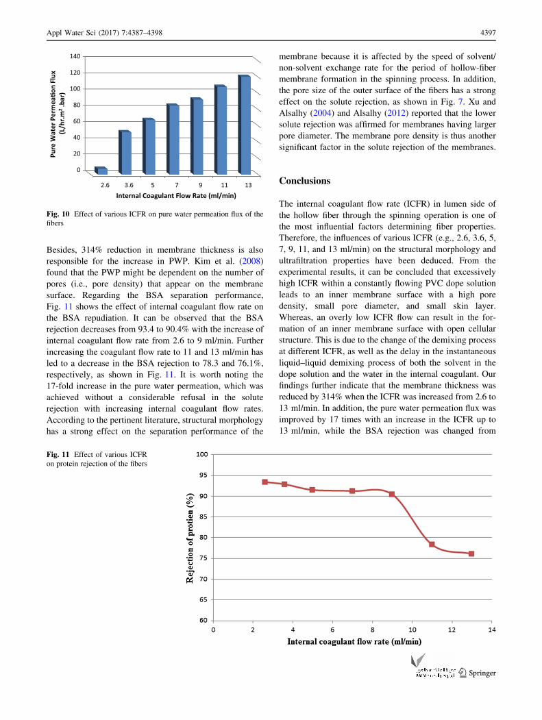
rejection. Figures 10 and 11 depict the measured values of
PWP and BSA retention of various hollow fibers synthe-
sized under various internal coagulant flow rates. It can be
seen from Fig. 10 that the PWP of the membranes is
improved from 6.97 to 119.3 (l/m2 h bar) due to the
increase in the internal coagulant flow rate from 2.6 to 13
(ml/min). In other words, the PWP is enhanced by
approximately 17 times. This observable fact is attributed
to the change in the morphology of the synthesized fiber as
discussed in the preceding section and explained in Figs. 2
and 3. The most significant factor can be noted in Fig. 3,
depicting the reduction of average pore diameter of
membrane with high pore density, along with the increase
of internal coagulant flow rates. The results of AFM
analysis shown in Fig. 5 confirm this phenomenon.
bFig. 5 3D AFM images of the hollow fibers prepared from different
effect of ICFR. a ICFR 2.6 ml/min; b ICFR 3.6 ml/min; c ICFR 5 ml/
min; d ICFR 7 ml/min; e ICFR 9 ml/min; f ICFR 11 ml/min; g ICFR
13 ml/min
0
20
40
60
80
100
120
140
160
2.6 3.6 5 7 9 11 13
Mea
n Po
re S
ize
(nm
)
Internal Coagulant Flow Rate (ml/min)
Fig. 6 Effect of ICFR on the mean pore size of the inner surface
Fig. 7 Effect of ICFR on the mean pore size of the outer surface
0
20
40
60
80
100
120
0 50 100 150 200 250 300
Cum
ula�
on (%
)
Diameter (nm)
inner
BFR= 2.6 ml/min
BFR= 3.6 ml/min
BFR= 5 ml/min
BFR= 7 ml/min
BFR= 9ml/min
BFR= 11 ml/min
BFR= 13 ml/min
Fig. 8 Cumulative distribution
of the pore size of the inner
surface
Appl Water Sci (2017) 7:4387–4398 4395
123

Fig. 9 Pore size distribution of the inner surface
4396 Appl Water Sci (2017) 7:4387–4398
123
Besides, 314% reduction in membrane thickness is also
responsible for the increase in PWP. Kim et al. (2008)
found that the PWP might be dependent on the number of
pores (i.e., pore density) that appear on the membrane
surface. Regarding the BSA separation performance,
Fig. 11 shows the effect of internal coagulant flow rate on
the BSA repudiation. It can be observed that the BSA
rejection decreases from 93.4 to 90.4% with the increase of
internal coagulant flow rate from 2.6 to 9 ml/min. Further
increasing the coagulant flow rate to 11 and 13 ml/min has
led to a decrease in the BSA rejection to 78.3 and 76.1%,
respectively, as shown in Fig. 11. It is worth noting the
17-fold increase in the pure water permeation, which was
achieved without a considerable refusal in the solute
rejection with increasing internal coagulant flow rates.
According to the pertinent literature, structural morphology
has a strong effect on the separation performance of the
membrane because it is affected by the speed of solvent/
non-solvent exchange rate for the period of hollow-fiber
membrane formation in the spinning process. In addition,
the pore size of the outer surface of the fibers has a strong
effect on the solute rejection, as shown in Fig. 7. Xu and
Alsalhy (2004) and Alsalhy (2012) reported that the lower
solute rejection was affirmed for membranes having larger
pore diameter. The membrane pore density is thus another
significant factor in the solute rejection of the membranes.
Conclusions
The internal coagulant flow rate (ICFR) in lumen side of
the hollow fiber through the spinning operation is one of
the most influential factors determining fiber properties.
Therefore, the influences of various ICFR (e.g., 2.6, 3.6, 5,
7, 9, 11, and 13 ml/min) on the structural morphology and
ultrafiltration properties have been deduced. From the
experimental results, it can be concluded that excessively
high ICFR within a constantly flowing PVC dope solution
leads to an inner membrane surface with a high pore
density, small pore diameter, and small skin layer.
Whereas, an overly low ICFR flow can result in the for-
mation of an inner membrane surface with open cellular
structure. This is due to the change of the demixing process
at different ICFR, as well as the delay in the instantaneous
liquid–liquid demixing process of both the solvent in the
dope solution and the water in the internal coagulant. Our
findings further indicate that the membrane thickness was
reduced by 314% when the ICFR was increased from 2.6 to
13 ml/min. In addition, the pure water permeation flux was
improved by 17 times with an increase in the ICFR up to
13 ml/min, while the BSA rejection was changed from
0
20
40
60
80
100
120
140
2.6 3.6 5 7 9 11 13
Pure
Wat
er P
erm
ea�o
n Fl
ux(L
/hr.m
2.b
ar)
Internal Coagulant Flow Rate (ml/min)
Fig. 10 Effect of various ICFR on pure water permeation flux of the
fibers
Fig. 11 Effect of various ICFR
on protein rejection of the fibers
Appl Water Sci (2017) 7:4387–4398 4397
123

93.4 to 90.4 as the ICFR increased from 2.6 and 9 ml/min
as a result of high pore density, reduced membrane thick-
ness, and small pore size.
Open Access This article is distributed under the terms of the
Creative Commons Attribution 4.0 International License (http://
creativecommons.org/licenses/by/4.0/), which permits unrestricted
use, distribution, and reproduction in any medium, provided you give
appropriate credit to the original author(s) and the source, provide a
link to the Creative Commons license, and indicate if changes were
made.
References
Alsalhy QF (2012) Hollow fiber ultrafiltration membranes prepared
from blends of poly (vinyl chloride) and polystyrene. Desalina-
tion 294:44–52
Alsalhy QF (2013) Influence of spinning conditions on the morphol-
ogy, pore size, pore size distribution, mechanical properties and
performance of PVC hollow fiber membranes. Sep Sci Technol
48:234–245
Alsalhy QF, Algebory S, Alwan GM, Figoli A, Simone S, Drioli E
(2011a) Hollow fiber ultrafiltration membranes from poly(vinyl
chloride): preparation, morphologies and properties. Sep Sci
Technol 46(14):1–12
Alsalhy QF, Algebory S, Alwan GM, Simone S, Figoli A, Drioli E
(2011b) Hollow fiber ultrafiltration membranes from poly(vinyl
chloride): preparation, morphologies, and properties. Sep Sci
Technol 46:2199–2210
Alsalhy QF, Rashid KT, Ibrahim SS, Ghanim AH, Van der Bruggen
B, Luis P, Zablouk M (2013) Poly(vinylidene fluoride-co-
hexafluropropylene) (PVDF-co-HFP) hollow fiber membranes
prepared from PVDF-co-HFP/PEG-600Mw/DMAC solution for
membrane distillation. J Appl Polym Sci 129:3304–3313
Alsalhy QF, Salih HA, Simone S, Figoli A, Zablouk M, Drioli E
(2014) Poly (ether sulfone) (PES) hollow-fiber membranes
prepared from various spinning parameters. Desalination
345:21–35
Aptel P, Abidine N, Ivaldi F, Lafaille JP (1985) Polysulfone hollow
fibers-effect of spinning conditions on ultrafiltration properties.
J Membr Sci 22:199–215
Cheng ZL, Li X, Feng Y, Wan CF, Chung TS (2017) Tuning water
content in polymer dopes to boost the performance of outer
selective thin-film composite (TFC) hollow fiber membranes for
osmotic power generation. J Membr Sci 524:97–107
Chung TS, Teoh SK, Hu X (1977) Formation of ultrathin high-
permeance polyethersulfone hollow fiber membrane. J Membr
Sci 133:161–175
Jack U, Hendry BA, Jacobs EP (2006) Fabrication of wet phase
inversion capillary membrane, dimension and diffusion effects,
Ph.D. thesis, Cape Peninsula University of Technology
Kim N, Kim CS, Leeb YT (2008) Preparation and characterization of
polyethersulfone membranes with p-toluenesulfonic acid and
polyvinylpyrrolidone additives. Desalination 233:226–236
Miao X, Sourirajan S, Zhang H, Lau WWY (1996) Production of
polyethersulfone hollow fiber ultrafiltration membranes. Part I.
Effects of water (internal coagulant) flow rate and length of air
gap. Sep Sci Technol 31:141
Mok S, Worsfold DJ, Fouda AE, Matsuura T, Wang S, Chan K (1995)
Study on the effect of spinning conditions and surface treatment
on the geometry and performance of polymeric hollow fiber
membranes. J Membr Sci 100:183–192
Peng N, Widjojo N, Sukitpaneenit P, Teoh MM, Lipscomb GG,
Chung TS, Lai JY (2012) Molecular design of polymeric hollow
fibers as sustainable technologies: past, present, and future. Prog
Polym Sci 37:1401–1424
Porter M (1990) Synthetic membranes and their preparation. In:
Porter MC (ed) Handbook of industrial membrane technology,
1st edn. Elsevier, Amsterdam
Qin JJ, Chung TS (2004) Effects of orientation relaxation and bore
fluid chemistry on morphology and performance of polyether-
sulfone hollow fibers for gas separation. J Membr Sci 229:1–9
Wan CF, Chung TS (2015) Osmotic power generation by pressure
retarded osmosis using seawater brine as the draw solution and
wastewater brine as the feed. J Membr Sci 479:148–158
Xu ZL, Alsalhy QF (2004) Polyethersulfone (PES) hollow fiber
ultrafiltration membrane prepared by PES/non-solvent/NMP
solution. J Membr Sci 233:101–111
Zeman LJ, Zydney AL (1996) Microfiltration and ultrafiltration:
principles and applications. Marcel Dekker, Inc., New York,
p 91 (chapter 2)
4398 Appl Water Sci (2017) 7:4387–4398
123


