
Eells slides combined - mimhtraining.com · 4/4/2019 1 Janis T. Eells, Ph.D. BiomedicalSciences...
14
4/4/2019 1 Janis T. Eells, Ph.D. Biomedical Sciences University of Wisconsin-Milwaukee Ancient Times • Ancient Egyptians are said to have built special temples for healing with sunlight and colored light. • Pythagorus used color for healing five hundred years before the birth of Christ. • Reference to color healing can be found in ancient Chinese and Indian texts. Ancient Times • Virtually all the major civilizations recognized the importance of light in healing. • The Assyrians, Babylonians and Egyptians all practiced therapeutic sun-bathing. • The Greek city of Heliopolis (which means 'City of the Sun') was renowned for its healing temples and light rooms. 1700 – 1800s
-
Upload
duongkhuong -
Category
Documents
-
view
214 -
download
0
Transcript of Eells slides combined - mimhtraining.com · 4/4/2019 1 Janis T. Eells, Ph.D. BiomedicalSciences...

4/4/2019
1
Janis T. Eells, Ph.D. Biomedical Sciences
University of Wisconsin-Milwaukee
Ancient Times
• Ancient Egyptians are said to have built special temples for healing with sunlight and colored light.
• Pythagorus used color for healing five hundred years before the birth of Christ.
• Reference to color healing can be found in ancient Chinese and Indian texts.
Ancient Times
• Virtually all the major civilizations recognized the importance of light in healing.
• The Assyrians, Babylonians and Egyptians all practiced therapeutic sun-bathing.
• The Greek city of Heliopolis (which means 'City of the Sun') was renowned for its healing temples and light rooms.
1700 – 1800s

4/4/2019
2
Niels Ryberg Finsen
Red and blue light to treat Lupus Vulgaris
1903 Nobel Prize in Medicine and Physiology
Photodynamic Therapy PDT
• Oscar Raab and Herman von Tappeiner in Germany noted that phototoxic effects on cells could be enhanced with exogenous dyes.
• This field of antimicrobial and, eventually, antitumor phototherapy is termed photodynamic therapy (PDT).
1930s
• (1933) Russian scientist Gurwitsch hypothesised that all cells emitted light. It took until the late seventies before German biophysicists (Popp et al.) proved that every cell emits at least 100.000 light impulses per second at a variety of frequencies.
• Charing Cross Hospital in London used 'sun-lamps' to treat circulatory diseases, anaemia, varicose veins, heart disease and degenerative disorders.
Post – World War II
• Antibiotics
• The Pharmaceutical Industry
• The grip of the American Medical Association and the age of the 'clinical trial' had arrived.
Invention of the LASER
• Einstein postulated stimulated emission of photons in 1917.
• Theodore Maiman built first LASER in 1960
• LASER as precision surgical tool

4/4/2019
3
Endre Mester
• Low-Dose Laser Treatments stimulated wound healing.
• Low-Level Laser Therapy LLLT
NASA: LED Development for Plant Growth and Wound Healing
John Ott
• John Ott demonstrated that different wavelengths of light have specific influences on cellular function in both plants and animals.
• He coined the term 'mal-illumination'
• He helped develop the first 'full-spectrum' fluorescent tube and in the early 1970s and undertook a study on the effects of 'full-spectrum' light on school children. Behaviour and academic performance improved markedly.
Thank You !
Janis T. Eells, Ph.D. Biomedical Sciences
University of Wisconsin-Milwaukee
• PBM has been known for nearly 50 years
• Tiina Karu– Head of Laboratory of Laser Biology and
Medicine, Moscow Russia– Medical Uses of Lasers
• Endre Mester– Semmelweis Medical University– Ruby laser‐induced hair growth
Photobiomodulation(aka Low-Level Laser Therapy)

4/4/2019
4
Why has PBM not gained widespread acceptance?
• Too much like magic
• Incomplete understanding of mechanism(s) of action
• Pharmaceutical Model of Medicine
The Solar Spectrum
• Photobiomodulation is the process by which a chain of biochemical reactions is triggered by exposure to light
• Photons must be absorbed by achromophore or photoacceptormolecule
• Photoacceptor molecules include chlorophylls, Vitamin D, rhodopsin, cytochrome c oxidase
PBM in All Life Forms Effect on all Life FormsFR/NIR Photons Stimulate Mitochondrial
Cytochrome c Oxidase and Activate Protective Intracellular Pathways
4/4/2019
5
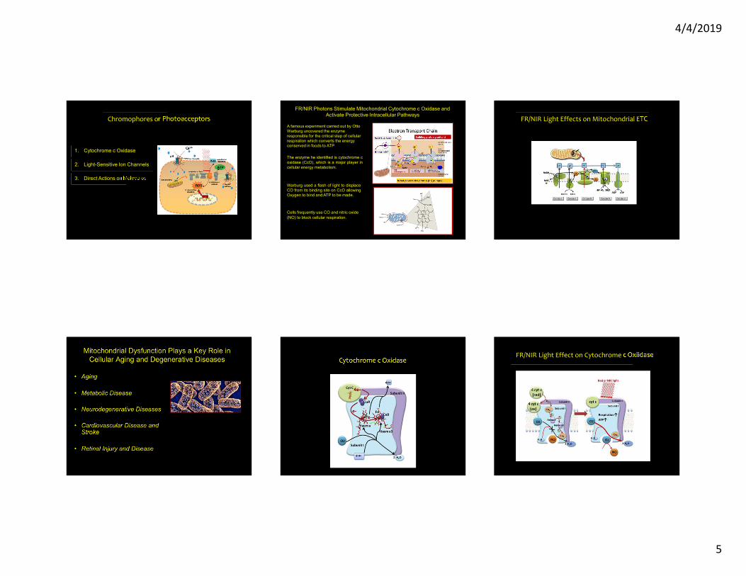
Chromophores or Photoacceptors
1. Cytochrome c Oxidase
2. Light-Sensitive Ion Channels
3. Direct Actions on Molecules
FR/NIR Photons Stimulate Mitochondrial Cytochrome c Oxidase and Activate Protective Intracellular Pathways
A famous experiment carried out by Otto Warburg uncovered the enzyme responsible for the critical step of cellular respiration which converts the energy conserved in foods to ATP
The enzyme he identified is cytochrome coxidase (CcO), which is a major player incellular energy metabolism.
Warburg used a flash of light to displaceCO from its binding site on CcO allowingOxygen to bind and ATP to be made.
Cells frequently use CO and nitric oxide (NO) to block cellular respiration.
FR/NIR Light Effects on Mitochondrial ETC
Cytochrome c OxidaseFR/NIR Light Effect on Cytochrome c Oxiidase

4/4/2019
6
SignalingMechanismsParameters Thank You !
Janis T. Eells, Ph.D. Biomedical Sciences
University of Wisconsin-Milwaukee
• Photobiomodulation is the process by which a chain of biochemical reactions is triggered by exposure to light
• Photons must be absorbed by achromophore or photoacceptormolecule
• Photoacceptor molecules include chlorophylls, Vitamin D, rhodopsin, cytochrome c oxidase
FR/NIR Photobiomodulation From Space Shuttle to Cancer Patients
• NASA funded the development of LEDs for use in plant growth experiments on the space shuttle and international space station
• PBM Improves healing of chemotherapy or radiation-induced mucositis.– FrontlineEpisode

4/4/2019
7
FR/NIR Photons Stimulate Mitochondrial Cytochrome c Oxidase and Activate Protective
Intracellular Pathways
Mitochondrial Dysfunction Plays an Important Role in Retinal Injury and Disease
The Retina
.
Photoreceptors -Vulnerable to Metabolic Inhibition and
Oxidative StressRodent Models of Retinal Disease
1. Methanol Toxicity
2. Retinitis Pigmentosa
3. Age-Related Macular Degeneration (AMD)

4/4/2019
8
Methanol Intoxication Disrupts Retinal Function
0 . 3- 0 . 9 - 0 . 3- 3 . 3
10
20
30
40
50
60
0- 3 . 3 - 2 . 7 - 2 . 1 - 1 . 5
Log Relative Retinal Illumination
ERG
Amplitude
(µV)
Control
24 hr
72 hr
*
*48 hr
*
Photobiomodulation Attenuates Methanol
Toxicity
670 nm TreatmentAt 5 hr, 25hr, 50 hr25 mW/cm2 – 160sec4 joules/cm2 20
40
60
80
Rod and M-Cone Mediated ERG Response
0- 3 -2 .7 -2 .4 -2 .1 -1 .8 -1 .5 -1 .2 -0 .9 -0 .6 -0 .3 0
Log Relative Retinal Illumination
Ro
d a
nd
M-C
on
e E
RG
Am
pli
tud
e(µ
V)
Untreated Control
LE D - Co ntro l
Methanol- Intoxicated
LED- Tre ated
PBM Attenuates Retinal Degeneration in Retinitis Pigmentosa
•Severe retinal degeneration• Affects 1:4000•Common cause: Rhodopsin mutations
830 nm PBM Ameliorates RP in the P23H Rat
Treatment Protocol Critical Period From p10 ‐ p25
830nm LED Array 180 sec
25mW/cm2 4.5J/cm2
Outcomes at P30
Retinal Metabolic State Retinal Function
Retinal Morphology
P23HRat Model of RPMutation in Rhodopsin Gene - same ashuman mutation. – P23H
Rod photoreceptors begin to die duringearly in development. Death slows intoadulthood
830nm PBM Normalizes Retinal Mitochondrial Function
Assessed by NADH/FAD Mitochondrial Redox CryoImaging
Detects changes in the oxidation state of the mitochondrial respiratory chain

4/4/2019
9
830 nm PBM Protects Retinal Function Assessed by Scotopic ERG
a‐wave amplitude b‐wave amplitude
830 PBM Attentuates Photoreceptor Cell DeathP23H
P23H PBM
Effect of PBM in Nrf2 knockout Mouse Model of AMD
• Nrf2 knockout mouse
• Nrf2 is a transcription factor that plays a key role in retinal antioxidant and detoxification responses
• Nrf2 ko Mouse Exhibits AMD-like pathology
– RPE degeneration– ERG reductions– Drusen-like deposits
• PBM daily (4.5 J/cm2) for 12 weeks
670nm PBM Attenuates Retinal Dysfunction in the Nrf2 ko
Mouse Model of AMDPBM Attenuates Diabetic Macular Edema
•Complex Pathophysiology of DME• Oxidative Stress• Elevated VEGF• BRB breakdown
Normal Macula
•Resulting in extracellular fluid accumulation in macula and decreased vision.
•Treatment : Anti-VEGF injectionsDME
20/200 vision
670 nm Photobiomodulation as a Therapy for Diabetic Macular Edema
• Treatment Resistant Diabetic patients with clinically significant DME
Control:Standard of Care (n = 4)Treated: Standard of Care plus PBM (n = 6)
• PBM Treatment Protocol:– LED Array given to patient for treatment– Treatment ‐ 90 sec 3 x per week for 8 weeks
• Assessments at Baseline, 8 weeks and 24 weeks– Visual Acuity– OCT

4/4/2019
10
VAsc
ore
Control (n=4) NIR (n=6)
Percentage Change in Visual Acuity(BCVA-ETDRS) score
15
10
5
0
-5
-10
-15
670 nm PBM Improves Visual Acuity in DME
P e r c e n t C h a n g e in O C T: Cent ra l Subf ield Thickness
% Ch
ange
in O
CT C
entra
l Sub
field
Thick
ness
-20
0
20
40
60
80
Cont ro l (n=4) N I R (n-6)
670 nm PBM Decreases Retina Edema in DME PBM Ameliorates Dry AMD Age-Related Macular Degeneration
• Leading cause of age-related vision loss• Complex Pathogenesis
– Mitochondrial Dysfunction– Immune Dysregulation– Oxidative Stress
PBM Ameliorates Dry AMD PBM Improves Visual Acuity in AMD PBM Decreases Drusen Volume in AMD

4/4/2019
11
FR/NIR Photons Stimulate Mitochondrial Cytochrome c Oxidase and Activate Protective Intracellular Pathways Photobiomodulation is Therapeutically Effective in
Retinal Disease
Janis T. Eells, Ph.D. Biomedical Sciences
University of Wisconsin-Milwaukee
The Pivotal Role of Mitochondrial Dysfunction in
Aging and Disease
Population Differences of Mitochondria in a Single Neuron Multiple Roles of Mitochondria
• Energy production
• Generation of ROS
• Metabolic Pathways
• Cell Signaling
• Role in Cell Death
OXPHOS major endogenous source of ROS
ROS are toxic byproducts of respiration
When ETC inhibited electrons accumulate in early stages of ETC and generate superoxide
Mitochondrial detoxification systemsMnSODGSH and GPxNo catalase except in cardiac mito
Chronic ROS exposure can result in oxidative damage to mito and cellular proteins, lipids and nucleic acids
Acute ROS exposure can inactivate the Fe-S centers in ETC complexes I, II and III and aconitase resulting in a shutdown of mito energy production.

4/4/2019
12
Oxidative phosphorylation, superoxide production, and scavenging pathways in mitochondria H202 and other ROS are
Fundamental Signaling Molecules
Dose determines the effect
Martindale and Holbrook. J Cell Phys 192:1-15, 2002
Mitochondrial H2O2 – ROLE IN SIGNALING
• Stimulates Ca2+ release– ER, Mitochondria
• Activates Transcription Factors– AP1 (fos, jun), NFB
• Activates Inflammatory Mediators– MCP-1, TNF-, IL1- , IL6, IL8.STAT1-
• Activates Adhesion Molecules– ICAM-1, VCAM-1, E-Selectin
• Changes Cell Shape--Mechanotransduction
Mitochondrial Dysfunction1. Mitochondrial dysfunction may be inherited,
spontaneous, age-acquired, physiologicallyregulated, or drug-induced
2. Mitochondrial dysfunction may be transient,fixed, or progressive
3. Mitochondrial dysfunction can cause “any disease, in any organ, at any age”
Mitochondria – major switch for initiation of apoptosis
• switch involves opening of mtPTP• mito inner membrane contains death promoting factors
– cyt c– AIF – a flavoprotein– Caspases
• opening of mtPTP causes collapse of mito membrane potential, swelling of inner membrane and release of death factors
• cyt c activates cytosolic caspase cascade• AIF – translocates to nucleus - chromatin destruction• Initiation signals for mtPTP
– excessive uptake of Ca– increased exposure to ROS
• decline in energetic capacity – loss of membrane potential
Mito PTP and Apoptosis

4/4/2019
13
Multiple Roles of Mitochondria• Energy production
• Generation of ROS
• Metabolic Pathways
• Cell Signaling
• Role in Cell Death
Mitochondrial Disease
• Mitochondrial defects occur in wide variety of degenerative diseases, aging and cancer
• Commonly involve tissues with high energy requirements
• Genetic and molecular complexity – bewildering array of inheritance patterns
Unique Mitochondrial Features Contributing to Disease
• Polyplasmy - cells contain multiple mitochondria with multiple genomes
• Heteroplasmy – wt mtDNA and mutant mtDNA
• Threshold Effect – critical number of mutant mtDNAs for tissue to become dysfunctional
• Mitotic Segregation – at cell division proportion of mutant mtDNA in daughter cells may vary
•• Maternal Inheritance – at fertilization all mtDNA derives
from oocyte
Maternal Inheritance of Mitochondrial DNA Mutations
Mitochondrial Mutations Known to Cause Disease
Mitochondrial Theory of Neurodegenerative Disease
During respiration a small fraction of the oxygen is incompletely reduced by the ETC to form reactive oxygen species (ROS)
ROS causes oxidative damage to mitochondrial DNA, lipids and proteins.
These damaged mitochondria with defective respiratory enzymes produce less energy (ATP) and also generate more ROS.
This vicious cycle operates in an age-dependent manner.

4/4/2019
14
Mitochondrial Theory of Age-Related Degenerative Diseases
The electron transport chain (ETC) in the inner membrane is actively involved in ATP synthesis coupled respiration.
During respiration a small fraction of the oxygen is incompletely reduced by the ETC to form reactive oxygen species (ROS) which may cause oxidative damage to mitochondrial DNA, lipids and proteins.
These damaged mitochondria with defective respiratory enzymes produce less energy (ATP) and also generate more ROS.
This vicious cycle operates in an age-dependent manner.
Environmental toxins also contribute to mitochondrial damage.
Parkinson’s Disease• clinical characteristics – bradykinesia, rigidity, tremor• pathology – degeneration of Dopaminergic neurons in SN• Genetic component• Mitochondrial involvement
– complex I deficiency in SN– not known if neurons more affected than glia– GSH depletion– Complex I deficiency
• Environmental Factors– MPTP story– TIQ– Rotenone
Pathogenesis of PD• Genetic Mutations
• Alpha-synuclein
• Environmental ToxinsMPTPPesticides
• Common theme:– Protein mishandling– Oxidative stress– Mitochondrial
dysfunctionGreenamyre et al., (2004) Science 304, 1120-1122.
The end


![[PPT]What's New in z/OS z0S 1.13 whats new_0.ppt · Web viewTitle What's New in z/OS Subject Share 117 Session 1191 Author John Eells Last modified by Jan Tits Created Date 2/2/2004](https://static.fdocuments.us/doc/165x107/5af2138b7f8b9ac246902add/pptwhats-new-in-zos-z0s-113-whats-new0pptweb-viewtitle-whats-new-in-zos.jpg)


