ECG tutorial and arrhythmia management March 2018
12
3/24/18 1 Providing the best quality care and service for the patient, the client, and the referring veterinarian. Carley Saelinger, VMD, DACVIM cardiology ECGs and arrhythmia management GOAL: Reduce anxiety about ECGs Overview l Identify different waves of the ECG and what they represent l Measure heart rate on the ECG l Recognize a normal ECG l Interpret common abnormalities on the ECG l Electrical and mechanical coupled heart l Treatment of common arrhythmias – Supraventricular – SVT, AF – Ventricular – VPCs, VT Back to the basics! What is an ECG?? Electrocardiogram = EKG = ECG = A recording of the electrical activity of the heart from electrodes placed on the surface of skin. The 1st Rule of ECG – A current of depolarization traveling towards the + electrode is recorded as a positive deflection – A current of depolarization traveling away from the + electrode is seen as a negative deflection – A current of repolarization traveling away from the + electrode is seen as a positive deflection The 2 nd Rule of ECG Fibrous = scar tissue Rapid depolarization/repolarization = narrow tracing slow depolarization/repolarization = wide tracing TIME Horizontal axis represents time. If slower variations of electrical potentials, complexes becomes wider (more tissue to depolarize, fibrosis) = = = = + + + + + + + + + + - - - + + + + + + + + + + - - - + + - _ +
Transcript of ECG tutorial and arrhythmia management March 2018

3/24/18
1
Providing the best quality care and service for the patient, the client, and the referring veterinarian.
Carley Saelinger, VMD, DACVIM cardiology
ECGs and arrhythmia management
GOAL: Reduce anxiety about ECGs
Overview
l IdentifydifferentwavesoftheECGandwhattheyrepresent
l MeasureheartrateontheECGl RecognizeanormalECGl InterpretcommonabnormalitiesontheECGl Electricalandmechanicalcoupledheartl Treatmentofcommonarrhythmias– Supraventricular–SVT,AF– Ventricular–VPCs,VT
Back to the basics!
What is an ECG?? Electrocardiogram = EKG = ECG = A recording of the electrical activity of the heart from electrodes placed on the surface of skin.
The 1st Rule of ECG
– A current of depolarization traveling towards the + electrode is recorded as a positive deflection
– A current of depolarization traveling away from the + electrode is seen as a negative deflection
– A current of repolarization traveling away from the + electrode is seen as a positive deflection
The 2nd Rule of ECG
Fibrous = scar tissue
Rapid depolarization/repolarization = narrow tracing
slow depolarization/repolarization = wide tracing TIME
Horizontal axis represents time. If slower variations of electrical potentials, complexes becomes wider (more tissue to depolarize, fibrosis)
= = = =
+ + + + + + + + + + - - -
+ + + + + + + + + + - - - ++
- _ +
3/24/18
2
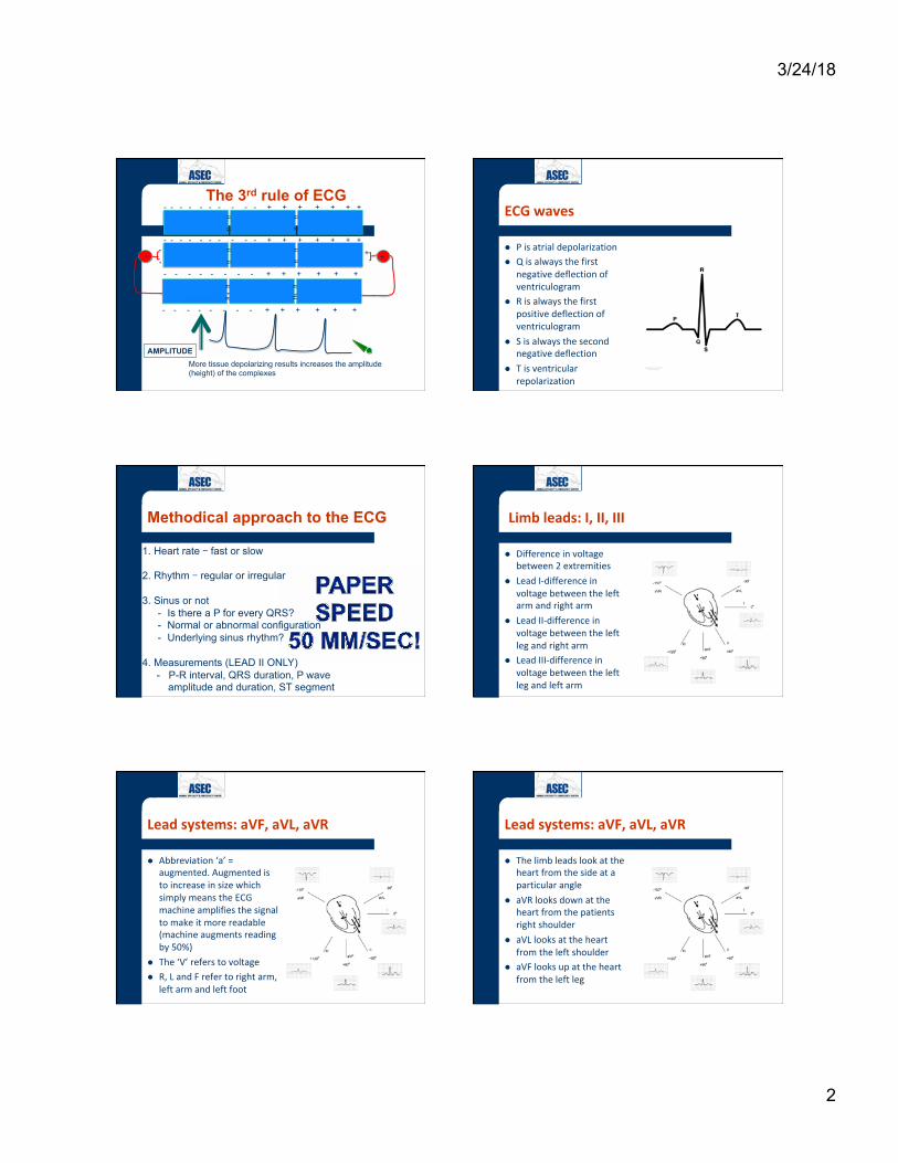
The 3rd rule of ECG
= = = =
- - - - - - - - - - + + + + + + +
- - - - - - - - + + + + + + - - +_ +
= = = =
- - - - - - - - + + + + + +
More tissue depolarizing results increases the amplitude (height) of the complexes
AMPLITUDE
= = = =
- - - - - - - - - - + + + + + + + ECGwaves
l Pisatrialdepolarizationl Qisalwaysthefirst
negativedeflectionofventriculogram
l Risalwaysthefirstpositivedeflectionofventriculogram
l Sisalwaysthesecondnegativedeflection
l Tisventricularrepolarization
Methodical approach to the ECG
1. Heart rate – fast or slow
2. Rhythm – regular or irregular
3. Sinus or not - Is there a P for every QRS? - Normal or abnormal configuration - Underlying sinus rhythm?
4. Measurements (LEAD II ONLY) - P-R interval, QRS duration, P wave amplitude and duration, ST segment
Limbleads:I,II,III
l Differenceinvoltagebetween2extremities
l LeadI-differenceinvoltagebetweentheleftarmandrightarm
l LeadII-differenceinvoltagebetweentheleftlegandrightarm
l LeadIII-differenceinvoltagebetweentheleftlegandleftarm
Leadsystems:aVF,aVL,aVR
l Abbreviation‘a’=augmented.AugmentedistoincreaseinsizewhichsimplymeanstheECGmachineamplifiesthesignaltomakeitmorereadable(machineaugmentsreadingby50%)
l The‘V’referstovoltagel R,LandFrefertorightarm,
leftarmandleftfoot
Leadsystems:aVF,aVL,aVR
l Thelimbleadslookattheheartfromthesideataparticularangle
l aVRlooksdownattheheartfromthepatientsrightshoulder
l aVLlooksattheheartfromtheleftshoulder
l aVFlooksupattheheartfromtheleftleg

3/24/18
3
Otherleadsystem:Precordialleads
l Precordialleadsorchestleads
l RecordECGinaplanthatis90degreesfromlimbleads
l Usedinpeoplebutrarelyinveterinarymedicine
CalibrationoftheECG
l Amplitude– Verticalaxis– Unit:milliVolt(mV)– 10mm/mV– CalibrationonECG(B)
l Speed– Horizontalaxis– Unit:mm/sec– 25mm/sec=>1mm=0.04sec– 50mm/sec=>1mm=0.02sec
25 mm/sec
50 mm/sec
BasicsoftheECG:Rate(HR)inbeats/minutes
AverageHR:– At25mm/sec
l Startat1QRScomplexl CountthenumberofQRScomplexesduring6sec=15cm=1penl Multiplyby10
– At50mm/secl Startat1QRScomplexl CountthenumberofQRScomplexesduring3sec=15cm=1penl Multiplyby20
50 mm/sec 10 mm/mv
15 cm = 3 seconds = bic pen 6 QRS complexes = contractions of the ventricle during 3 s 6 X 20 = 120 beats/min
Instantaneous HR: 26 little boxes between R-R interval, at paper speed of 50 mm/sec: 3000 / # little boxes, so 3000/26 = 115 bpm OR Recording at 50 mm/sec => 1 mm is 0.02 sec 26 mm between 2 QRS complexes => 26 X 0.02 = 0.52 sec 1 minute is 60 sec Number of beats in 1 minute is => 60 /0.52 = 115 bpm
Canineandfelineheartrate
l Dog– Normalheartrate:80to150beats/minute– Bradycardia:HR<60beats/minute– Tachycardia:HR>150beats/minute– Usuallynoclinicalsignsifratebetween40and200beats/
minute
l Cat– Normalheartrate:140to200beats/minute– Bradycardia:HR<100beats/minute– Tachycardia:HR>200beats/minute

3/24/18
4
Meanelectricalaxis
l Directionoftheventriculardepolarization=QRS
l Toptobottom,righttoleft
l Normalimpulsedirectedtowardsleftfootbecauseleftventricleisbiggerthanrightventricle
l QRSisthetallestinleadII
Normalmeanelectricalaxis
l Dog:40to100degreesl Cats:0to160degrees
CalculationMEA
ONLY CALCULATE MEA IF: • Lead II negative or low voltage QRS complex The vector method: • Calculate the algebraic sum of QRS deflections in any 2 leads – USE leads I and AVF The isoelectric method: • Find the most isoelectric lead • MEA is perpendicular to this lead
What is the electrical axis?
Negative overall QRS In lead II
Negative QRS wave Directed away from lead AVF By 7 little boxes
- The axis is between -180 and -120 degrees -> RIGHT AXIS DEVIATION - Normal is 40 to 100 degrees - IF NEG QRS IN LEAD II AND DOG -
> RIGHT AXIS SHIFT - IF NEG QRS IN LEAD II AND CAT ->
LEFT AXIS SHIFT
Negative lead I by 9 little boxes
Referencevalues(leadII)Wave Duration Amplitude
P2x4
40ms(2mmat50mm/sec,1mmat25mm/sec):2littleboxeswide
0.4mV(4mmat10mm/mV)4littleboxestall
QRS 60ms(3mmat50mm/sec,1.5mmat25mm/sec):3littleboxeswide
2.5–3mV(25-30mmat10mm/mV)25littleboxestall
T Lessthan¼QRS
Dog
CatWave Duration Amplitude P 40 ms (2 mm at 50mm/sec, 1 mm
at 25 mm/sec 0.2 mV (2mm at 10 mm/mV)
QRS 40 ms (2 mm at 50 mm/sec, 1 mm at 25 mm/sec)
1 mV (10 mm at 10mm/mV)
T 0.3 mV (3 mm at 10 mm/mV)
Pwave
l Atrialdepolarizationl PositiveinleadIIl Rightatrial
enlargement– Tallpwave
l Leftatrialenlargement
– Widepwave

3/24/18
5
What is the average HR? Look at the p waves
What is the instantaneous HR between the first 2 beats? What is the difference with the first ECG?
What if you don’t know paper speed?? If ST segment closer to one big box -> 50 mm/sec
Interval Dog Cat
P-R <130ms=6.5mmat50mm/sec<6.5littleboxes
<90ms=4.5mmat50mm/sec<4.5littleboxes
Q-T 150–200ms 70–200msec
P-Rinterval
l ReflectsconductionthroughtheAVnode
l LongerP-RintervalmeansconductionissloweddownintheAVnode
l ProlongedP-Rinterval=firstdegreeAVblock
9 mm
PR interval = 9 mm (9 little boxes) If recorded at 50 mm/sec => 1 mm = 0.02 sec = 20 msec 9 X 20 = 180 msec > 130 mm = First degree AV block
QRScomplex
l TallRwaveinleadIIsuggestsleftventricularhypertrophy(moretissuetodepolarize)
l Hypertrophy– Concentric:thickerwalls– Eccentric:dilationof
ventriclel 90%ofdogswithtallRwave
havehypertrophyofleftventricle
l Normally,<25littleboxes
3.7 mV
QRScomplex:leftbundlebranchblock
l QRSduration>60ms=3mm=3smallboxesat50mm/sec
l Indicatesmoretimetodepolarize
l Suggestssevereleftmyocardialdz

3/24/18
6
QRScomplex:rightventricularenlargement
l DeepSwaveinleadIIl Meanaxisshiftedto
therightl Suggestseccentricor
concentricrightventricularhypertrophy
QRScomplex:rightbundlebranchblock
l DeepSwaveinleadIIl WideQRScomplex
– Indicatesmoretimetodepolarize
l TheQRSreflectstheslowestportionofdepolarization
l Incidentalorreflectrightsideddisease
QRS Complex: left anterior fascicular block (LAFB) - CATS
l LAFB (CATS): § Typically
pathologic § Tall R wave
in I and aVL § Deep S in II,
III, aVF l RBBB (DOGS):
§ Deep S wave in lead I, II, III, and aVF
§ Positive R wave in aVR
RBBB LAFB
- IF NEG QRS IN LEAD II AND DOG -> RIGHT AXIS SHIFT
- IF NEG QRS IN LEAD II AND CAT -> LEFT AXIS SHIFT
Twave
l Reflectsventricularrepolarization
l Canbepositiveornegative
l Ifdepolarizationisabnormal,repolarizationisabnormalaswell
QTinterval
l MeasuredfrombeginningofQwavetoendofTwave=refractoryperiod
l QTintervalvarieswithheartrate:
– Shorterwhenheartrateisfaster
– Longerwhenheartrateisslower
l Reflectsshorteningofactionpotentialwithincreasedadrenergictone
S-T segment and T wave abn S-T Segment Changes:
l Depressed S-T: – myocardial ischemia – acute infarction – electrolyte abnormalities – digitalis toxicity – cardiac trauma
l Elevated S-T: – myocardial infarction – pericarditis – myocardial hypoxia
T Wave Abnormalities
l Myocardial hypoxia l Anesthetic complications l Hyperventilation l Heart failure l Bradycardia l Hyperkalemia (large, spiked) l Hypokalemia (small, biphasic) l Anemia l Shock l Uremia l Hypothyroidism l Fever

3/24/18
7
Normalsinusrhythm
l PositivePwaveinleadII->impulseinitiatedinsinusnode
l EachQRSisinitiatedbyapwave
l P-Rintervalisnormall R-Rintervalis
constant
Sinusbradycardia
l PositivePwaveinleadII->impulseinitiatedinsinusnode
l EachQRSisinitiatedbyaPwave
l NormalP-Rintervall HR<60bpml Usuallycausedby
increasedvagaltone
Sinustachycardia
l PositivePwaveinleadII-impulseinitiatedinsinusnode
l EachQRSinitiatedbyaPwave
l NormalP-Rintervall HR>150bpml Usuallycausedby
increasedadrenergictone
(respiratory)sinusarrhythmia
l Sinusrhythml R-Rintervalvariesl Heartrate:
– Increasesduringinspiration
– Decreasesduringexpiration
l Effectofvagaltoneonsinusnode
l NORMALindogs
RSA
7 little boxes x 20 = 140 msec = 1st degree AV block
FirstdegreeAVblock
l ProlongationofPRinterval
l SlowconductionintheAVnode
l UsuallycausedbyfibrosisintheAVnode
l Canbecausedbyincreasedvagaltone

3/24/18
8
SeconddegreeAVblock
l Impulseoriginatesfromsinusnode->positivePwaveinleadII
l SomeofPwavesblockedinAVnode&don’tpropagatetotheventricles
l SecondarytofibrosisintheAVnode
2nd degree AV block Mobitz Type I - Wenchebach
l P-R interval progressively prolongs before “dropping”
Mobitz Type II
l P-R constant with a regular rhythm then dropped “P”
ThirddegreeAVblock
l Impulseoriginatesinthesinusnode->Pwave
l ImpulsealwaysblockedinAVnode
l Secondarypacemakertakesover:
– AVnode(junction):40-60beats/min
– Purkinjefibers:20-40beats/min
3rd degree AV block
l 12yearoldsmallbreeddog
l Historyofsyncope
l HR=30bpml Thirddegree
AVblockl Treatment:
Pacemakerimplantation
Atrialprematurecontraction(APC)
l Originatesfromatrialmyocyte
l Occurssoonerthanexpected(premature)
l ImpulsepropagatestoventriclethroughAVnodeandnormalconductionpathways(narrowunlessBBB)

3/24/18
9
APCs
Instantaneous HR = 3000 / 15 little boxes = 200 bpm
Supraventriculartachycardia(SVT)
l Rate>150bpmindogsl Rate>220bpmincatsl Regularrhythml OriginatesabovethebundleofHis:
– Sinusnode– Atrium– AVnode
l NormalQRScomplex(unlessBBB)
Atrialfibrillation
l Mostcommonarrhythmiaindogsl Fastl Irregularl NoPwaves
– Noorganizedelectricalactivity– Disorganizeddepolarizationofall
atrialcells=fibrillation– (fibrillationwaves)
l Supraventricular-NormalQRScomplex(unlessBBB)
Atrial fibrillation
Ventricularprematurecontraction
l Originatefromventricularmyocytes
l Occurssoonerthanexpectedl Widecomplex:travelsslowly
frommyocytetomyocytel LargeTwavewithdirection
oppositetoQRScomplexl Canhavenormalpwavesthat
arenotassociatedwiththeectopicQRS.
VPCs

3/24/18
10
Ventriculartachycardia
l Tachycardial Originatesfrom
ventricularmyocytel WideandbizarreQRS
complexes:– Noconductionthrough
normalconductionsystem
l Widebecausecelltocellconductionisslow
Ventricular tachycardia - 3 or more consecutive VPCs Intermittent, paroxysmal, or sustained - May cause serious, life-threatening hemodynamic impairment
- Differential diagnoses: Supraventricular tachycardia with aberration Bundle branch blocks (Look for “p” waves!)
Ventricularfibrillation
l Rapiddisorganizeddepolarizationofallventricularcells
l NoQRScomplexl Noeffectivemechanical
contractionl Nocardiacoutputl Deathunlesselectrical
defibrillation
TREATMENT OF ARRHYTHMIAS
l It’s all about ATP, lytes and sympathetic tone l Coupled electrical and mechanical activity of heart l Effective drugs & side effects l Importance of blood work
Sympathetic tone
β stim
ATP
Electrolytes Ca+2 Na+ K+
Supraventricular tachycardia (SVT) treatment depends upon…
l Cause – Myocardial disease – Systemic disease
l Clinical signs – Duration dependent – Rate dependent
l Slow SVT l Fast SVT 250 - 300 bpm
– Weakness, collapse – Poor perfusion
Maneuvers & drugs of acute SVT
l If weakness/collapse à MEDICAL EMERGENCY!!
l Acute therapy: – Physical maneuvers (vagal or thump) – Drugs = BCP
l B = Beta blockers l C = Calcium channel blockers l P = Procainamide

3/24/18
11
Physical maneuvers
l Vagal maneuvers – Slow conduction through AV node (break re-
entry) or slow ventricular response rate – Ocular pressure
l Controlled digital pressure to both globes
– Carotid sinus massage l Gentle, sustained digital pressure to one or both carotid
sinus (caudal to dorsal aspect of the larynx)
l Precordial thump – induce a VPC – 5 J shock to myocardium
Drugs – acute SVT therapy
l Slow conduction through AV node (break re-entry) or slow ventricular response rate (atrial tachycardia)
l Beta blockers – Esmolol: 0.05 - 0.1 mg/kg IV boluses every 5 minutes up to
max dose of 0.5 mg/kg; then CRI (10 - 200 µg/kg/min) (**short acting)
– Propanolol: 0.02 mg/kg IV slowly (**longer acting)
l Calcium channel blockers – Diltiazem: 0.05 - 0.15 mg/kg IV over 5 - 10 minutes up to max
dose of 0.3 mg/kg; then CRI (0.12 - 0.24 mg/kg/h)
*All doses are canine only unless indicated otherwise
Drugs – acute SVT therapy
l To convert focal atrial tachycardia and some re-entrant SVT
l Procainamide – IV boluses of 2 - 5 mg/kg up to 20 mg/kg total; each
dose over 5 minutes l Lidocaine
– IV boluses of 1 - 2 mg/kg (up to 6 - 8 mg/kg total)
l Sotalol – 1 - 2 mg/kg PO q 12 h
*All doses are canine only unless indicated otherwise
Chronic management = BCDs
l B OR C with D for AV nodal dependent SVT l Beta blockers
– Atenolol: 0.25 - 1 mg/kg PO q 12 - 24 h – TITRATION – when in doubt, start low – Caution with myocardial failure
l Calcium channel blockers – Diltiazem: 0.5 - 2 mg/kg PO q 8 h – Diltiazem ER (Dilacor): 2 - 3 mg/kg PO q 12 h
l Digoxin: Increases vagal tone, weak positive inotrope – Dose: 0.0045 - 0.005 mg/kg PO q 12 h
*All doses are canine only unless indicated otherwise
Other chronic management
l Conversion of focal atrial tachycardias (non-reentrant) – Class III anti-arrhythmics
l Sotalol: 1 - 2 mg/kg PO q 12 h
– Class I anti-arrhythmics l Procainamide, quinidine
l Or slow ventricular response rate with BCDs – B OR C with D
*All doses are canine only unless indicated otherwise
Atrial fibrillation (AF)
l BCDs l Preference:
– Diltiazem ER: 2 - 3 mg/kg PO q 12 h – Digoxin: 0.0045 mg/kg PO q 12 h
l Holter monitor l Lone AF, AF 2nd to vagal/sympathetic tone
*All doses are canine only unless indicated otherwise

3/24/18
12
Ventricular ectopy
l VPCs vs. ventricular tachycardia (VT) – Treatment same as VT if needed
l Underlying cardiac disease – Holter monitor, echocardiogram – Breeds – Boxers, Dobermans, GSD – Disease – AC, DCM, SAS, inherited VT
l Accelerated idioventricular rhythm
VT – when to treat
l Hemodynamic compromise – Cardiac disease – Poor pulse quality, weak, hypotensive
l Risk of degenerating into ventricular fibrillation – Cardiac disease – AC, DCM, SAS – Faster rates – Polymorphic > monomorphic – Repetitive forms > single forms
Acute therapy of VT
l Lidocaine – IV bolus of 1 - 2 mg/kg (up to 6 - 8 mg/kg total);
then CRI (35 - 80 mcg/kg/min) – Positive response = rate slowed or abolished – Toxicity – neurologic; cats > dogs – May not be effective if:
l Hypokalemia l Incorrect diagnosis (actual rhythm is SVT) l Slower rates
*All doses are canine only unless indicated otherwise
Acute therapy of VT
l Procainamide – IV boluses of 2 - 5 mg/kg (up to 20 mg/kg total);
each dose over 3 - 5 minutes – CRI dose 10 - 50 mcg/kg/min IV – Side effects – neurologic, esp. if lidocaine prior – SVT and VT
l Magnesium l Amiodarone l Anesthesia and cardioversion
*All doses are canine only unless indicated otherwise
Chronic therapy
l Sotalol – 1 - 2 mg/kg PO q 12 h – Boxers with AC – Some beta blocking properties
l Mexiletine – 3 - 8 mg/kg PO q 8 h – Give with FOOD (GIT upset)
l (Atenolol) – Mildly effective, used in combination – Negative inotrope *All doses are canine only unless indicated otherwise
Questions?
www.cardiacvet.com


![Automatic Detection of Cardiac Arrhythmia through ECG ...€¦ · Cardiac Arrhythmia [3], also known as irregular heartbeat, is a group of conditions in which the heartbeat is irregular,](https://static.fdocuments.us/doc/165x107/607210056cc22557db7f5efb/automatic-detection-of-cardiac-arrhythmia-through-ecg-cardiac-arrhythmia-3.jpg)