
EARLY RECOVERY PROFILES OF LANGUAGE AND EXECUTIVE …
162
EARLY RECOVERY PROFILES OF LANGUAGE AND EXECUTIVE FUNCTION IN BILINGUAL PERSONS DURING THE FIRST TWELVE WEEKS POST BRAIN INJURY Nancy Barber A dissertation submitted to the Faculty of Humanities, School of Human and Community Development, University of the Witwatersrand, Johannesburg in fulfilment of the requirements for the degree of Master in Speech Pathology November 2015
Transcript of EARLY RECOVERY PROFILES OF LANGUAGE AND EXECUTIVE …

i
EARLY RECOVERY PROFILES OF LANGUAGE AND EXECUTIVE FUNCTION IN
BILINGUAL PERSONS DURING THE FIRST TWELVE WEEKS POST BRAIN INJURY
Nancy Barber
A dissertation submitted to the Faculty of Humanities,
School of Human and Community Development,
University of the Witwatersrand, Johannesburg
in fulfilment of the requirements for
the degree of Master in Speech Pathology
November 2015

i
DECLARATION
I, Nancy Barber, hereby declare that this is entirely my own work and that it has not been
submitted as an exercise for the award of a degree at this or any other University. I
acknowledge that I am responsible for the text of this study and all conclusions reached.
________________________________
Nancy Barber (November 2015)

ii
ACKNOWLEDGEMENTS
Participants and family members- I am very thankful to all the participants and their family
members for participating in the study and for warmly welcoming me into their homes when
I did re assessments.
Prof Claire Penn- I am thankful for her insights, her guidance, and her wisdom. Her interest
in this research topic and her ability to look at things with a different perspective assisted me
with the passion I needed to complete this project. Her support, supervision and
encouragement have been invaluable throughout this process.
Prof Peter Fridjhon- His guidance with regards to the methodology and statistical analysis
were greatly appreciated. His approach to the results and his ability to fit the puzzle pieces
together helped shape this research project.
Dean- I could not have completed this research without the love, support and prayers of my
husband. I thank him for the allowing me to resign from a full time job so I could focus on
my research, for all the hours he left me working in our study, and for being considerate
when I had deadlines.
My parents, Cedric and Yvonne, and my sister, Dawn- They have been a never ending well
of support and encouragement. A special thank you to my mom who read and edited
numerous versions of this dissertation.
I am very appreciative of friends who supported me in prayer. I am especially grateful to
Kerri who continuously encouraged me and edited my thesis for me. I am also thankful for
Megan who supported me with prayers as well as a listening ear when times were tough and
when times were fruitful.
I also give glory to God. Throughout this Master’s degree, God has given me a grace to enjoy
each stage of the research process. I thank God for His grace, wisdom, provision and
strength.
“My grace is sufficient for you, for my power is made perfect in weakness.”
2 Corinthians 12:9

iii
ABSTRACT
Background: The nature, rate and pattern of recovery in bilingual persons following brain
damage has been investigated over many years but several controversies remain. Recent
evidence suggests that the relationship between executive function (EF) processes and
language recovery may be distinct in bilinguals. An improved understanding of such
underlying linguistic and cognitive processes may enhance assessment and treatment
particularly in the acute phase. There is limited knowledge regarding how these processes
interact in the acute phase and there remains little guidance as to the choice of an appropriate
assessment battery for bilinguals. In the South African context, bilingual persons with a brain
injury are often treated as monolinguals due to the language challenges and the lack of
standardised assessments. Thus there is a need to develop a simple, effective battery which is
able to differentiate aetiologies, is sensitive to recovery processes, and in a multicultural and
multilingual context is able to distinguish normal from pathological profiles.
Aims: The research study aimed to identify an assessment battery for language and EF that is
sensitive to etiology and the recovery process for South African bilingual persons who have
had a neuronal insult. It also aimed to evaluate the linguistic and executive function skills of
bilingual patients with acquired neurological communication disorders (ANCD) at two time
periods within the first 12 weeks post injury. A further aim was to profile the recovery of
bilingual persons with ANCD in the acute recovery phase according to etiology (Right CVA,
left CVA and TBI).
Method: A multivalent comparison study with a longitudinal component was conducted at
two acute rehabilitation centres. A convenience sample of 29 bilingual, second language
English speaking participants (19 with a cerebral vascular accident (CVA) and 10 with a
traumatic brain injury (TBI)) were assessed at two time periods within the first 12 weeks post
injury. They were assessed using the Comprehensive Aphasia Test (CAT) and a nonverbal
EF battery. The nonverbal battery comprised tasks to assess updating (n-back task), mental
shifting (number-letter task; Wisconsin Card Sorting test), and inhibition (Victoria Stroop;
Tower of Hanoi). A control group of 19 neurologically intact bilingual, second language
English speakers who were matched according to age and education level were assessed
employing the same battery. The control group completed an initial assessment and then were
reassessed six weeks later.
Results: The CAT was found to be a suitable assessment measure when assessing bilingual,
second language English speakers in the South African context. A between- group analysis
identified statistically significant differences between etiologies (including the control group)
for language assessment as well as the EF assessment, indicating the battery was able to
differentiate normal from pathological individuals. While most of the test battery was found
to be suitable for the participants, the Tower of Hanoi and the number-letter task were
deemed inappropriate for the population and the cultural context. Overall the battery of tests
distinguished between aetiologies, testing period (first and second) and pathological from
normal individuals. It was found that this battery was appropriate for a variety of cultural
groups. A within- group analysis determined that there were unique profiles of language and
EF skills according to etiology and that different profiles of change emerged according to
each etiology for both language and EF subtests.
Discussion: The streamlined battery that was found to be beneficial and sensitive to the
multicultural and multilingual nature of South Africa comprised the CAT as the language
assessment and the n-back task (updating), Victoria Stroop (inhibition) and WCST (shifting)
comprised the EF assessment battery in the acute phase.This study confirms prior research on
recovery processses in language across the three aetiologies but also highlights changes in
iv
excutive functioning which may offer some explanations for differential recovery profiles.
The results highlighted that inhibition may be a preserved bilingual advantage in participants
with a right CVA or TBI. However, it was a deficit in participants with a left CVA. The role
of inhibition may support the decision making process with regards to the language for
therapy. Thus the EF profiles may also assist a clincian to determine whether to undertake
monolingual or bilingual therapy There were also distinct relationships between language
skills and EF skills for each etiology according to time frame. This provided insight into the
interactions between language and EF during the acute phase of recovery. Knowledge of the
specific EFs that interact with language recovery per etiology can assist a clinician in
providing effective therapy in the acute phase that complies with neuroplasticity principles.
Conclusion: Language assessment and treatment in the acute phase needs to be provided in
combination with an understanding of recovery patterns, what is driving that pattern, and
which cognitive deficits are contributing to the language behaviour. In addition clinicians
need to be aware of the impact of updating, shifting and inhibition in a bilingual person as
well as the role bilingual advantage may have in decision making for therapy, the recovery
process and as a possible tool to support the therapeutic process.
v
TABLE OF CONTENTS
List of Abbreviations Page vii
List of Tables Page viii
List of Appendices Page ix
Chapter 1 Introduction Page 1
Chapter 2 Literature Review
Introduction Page 7
Cerebral Vascular Accident (CVA) Page 7
Traumatic Brain Injury (TBI) Page 10
The South African Context- historical
socio-political factors that impact aphasia
therapy service delivery Page 12
Executive Function Page 16
Bilingualism Page 25
Chapter 3 Methodology
Research Aims Page 40
Research Design Page 41
Setting Page 41
Participants Page 42
Control group Page 44
Materials Page 45
Procedures Page 55
Ethics Page 56asz
Reliability and Validity Measures Page 56
Data Analysis Page 58
Chapter 4 Results
Introduction Page 60
Control group performance at 6 and 12 weeks Page 60
Clinical group performance Page 63
Differences between the clinical and control group Page 74
Correlations between the CAT and EF subtests Page 77

vi
Summary of results Page 84
Chapter 5 Discussion
Introduction Page 86
Assessment of South African bilinguals
using the CAT and nonverbal EF battery Page 86
The importance of linguistic and EF profiling
of bilinguals in the acute phase Page 90
Insights into bilingualism Page 93
Chapter 6 Implications and Conclusions
Implications for Assessment and Therapy Page 96
Further research Page 100
Conclusions Page 101
Reference List Page 102
Appendices Page 125
vii
LIST OF ABBREVIATIONS
AAC- Alternative and augmentative communication
AIDS- Acquired immune deficiency syndrome
ANCD- Acquired neurological communication disorders
BAT- Bilingual Aphasia Test
CAT- Comprehensive Aphasia Test
CHI- Closed head injury
CVA- Cerebral vascular accident
EF- Executive function
HIV- Human Immunodeficiency Virus
L2- Second language
RM ANOVA- Repeated measure analysis of variance
SLP- Speech-language pathologist
TBI- Traumatic brain injury
ToH- Tower of Hanoi
VicStroop- Victoria Stroop
WCST- Wisconsin Card Sorting Test

viii
LIST OF TABLES
Table 1. Model of Executive functions (Miyake et al., 2000).
Table 2. Recovery patterns observed in bilingual persons with aphasia.
Table 3. Participant demographics regarding age, number of languages spoken, years of
schooling and age of L2 acquisition.
Table 4. Executive function assessment battery.
Table 5. Mean, Standard Deviation (SD) and t-scores for the Control group on the CAT
subtests and EF battery subtests at the initial assessment and six weeks later.
Table 6. Mean, Standard Deviation (SD) and t-scores for the participants with a left CVA on
the CAT subtests and EF battery subtests at the initial assessment and six weeks later.
Table 7. Mean, Standard Deviation (SD) and t-scores for the participants with a right CVA on
the CAT subtests and EF battery subtests at the initial assessment and six weeks later.
Table 8. Mean, Standard Deviation (SD) and t-scores for the participants with a TBI on the
CAT subtests and EF battery subtests at the initial assessment and six weeks later.
Table 9. Statistical significance of left CVA, right CVA, TBI and control group on the CAT
and EF assessment battery.
Table 10. Correlations between CAT subtests and EF subtests at 6 weeks post injury
according to etiology employing Pearson analysis (r).
Table 11. Correlations between CAT language subtests and EF subtests at 12 weeks post
injury according to etiology employing Pearson analysis (r).
Table 12. The different patterns of CAT subtests which correlate with EF subtests according
to time of assessment.
Table 13. Correlations between CAT language subtests and EF subtests of control group at
the initial assessment and at the reassessment 6 weeks later employing Pearson
analysis (r).

ix
LIST OF APPENDICES
Appendix A. University of the Witwatersrand Clearance Certificate
Appendix B. Permission letter from Life Health Care Group
Appendix C. Information pack for participant and their family
Appendix D. Informed Consent Form for Patients with a CVA/TBI
Appendix E. Table of detailed participant demographics
Appendix F. Participant information sheet for the control group.
Appendix G. Informed consent letter for the control group
Appendix H. Language Proficiency Questionnaire
Appendix I. Paragraphs for Comprehension of Spoken Language Subtest
Appendix J. Detailed description of executive function assessment battery
Appendix K. Tables detailing the CAT and EF subtest scores for the control group at initial
and follow up assessment
Appendix L. Table detailing the CAT and EF subtest scores for the participants with a left
CVA at the 6 week and 12 week assessment
Appendix M. Table detailing the CAT and EF subtest scores for the participants with a right
CVA at the 6 week and 12 week assessment
Appendix N. Table detailing the CAT and EF subtest scores for the participants with a TBI at
the 6 week and 12 week assessment

1
Chapter 1
Introduction
This study investigated the relationship between executive function and language in South
African bilingual persons in the first twelve weeks subsequent to a brain injury. This study
arose as the researcher was a speech-language clinician assessing and providing therapy to
bilingual patients in an acute rehabilitation hospital in Johannesburg, South Africa. Concerns
surfaced regarding the nature of linguistic and non-linguistic assessment methods of bilingual
second language English-speaking patients in the acute phase. Patterns of recovery in
linguistic and non-linguistic skills in bilinguals in the acute phase subsequent to injury were
also of interest as there appeared to be limited knowledge pertaining to bilinguals in this area
for both speech-language therapists and other team members such as neuropsychologists.
Thus the researcher wanted to determine an effective and economical battery to use on South
African bilingual patients in order to assess their linguistic and non-linguistic skills. Further
investigation into the recovery pattern in the acute phase, as well as the interactions between
the linguistic and executive functions in the acute phase, was deemed necessary. This
investigation was necessary in order to facilitate the complex therapeutic decision-making
process required for providing language therapy to South African bilinguals with an acquired
neurological communication disorder in the acute phase.
A larger percentage of the world is bilingual (Kecskes, 2010) and South Africa is a largely
multilingual and multicultural nation (Penn, 2014). In South Africa there are eleven official
languages including: Afrikaans, English, isiNdebele, Northern Sotho (Sesotho sa Leboa),
Sesotho, SiSwati, Setswana, Xitsonga, Tshivenda, isiXhosa and isiZulu. A significant
percentage of South African children speak at least two languages from birth and this use of
multiple languages makes a large majority of the South African population bilingual or
multilingual (Raidt, 1999). A further contribution to the multilingual status of the South
African population is that a large majority of South Africans who are competent in one (or
more) of the other official eleven languages, and learn English and Afrikaans as additional
languages for educational, political and economic reasons (Mukhuba, 2005).
Cerebral vascular accidents (CVA) and traumatic brain injuries (TBI) are common within the
South African context (Conner & Bryer, 2006; Schneider, Claassens, Kimmie, Morgan,
2
Sigamoney, Roberts & McLaren, 1999). Acquired neurological communication disorders
(ANCD) arise from CVA and TBI and in addition to these communication disorders,
executive functions are often compromised (Murray, 2012; Zinn et al., 2007; Purdy, 2002;
Boelen et al., 2009; McDowell, Whyte & D’esposito, 1997; Tate, 1999). These
communication deficits have specific presentations and symptoms that occur in bilingual
persons. The nature of these deficits will be delineated in Chapter 2.
Historically in South Africa, there was racial discrimination and separation, leading to
inequities for housing, education, economic employment and health (Penn, 2014). When
apartheid ended there was a political change in South Africa, which focused on human rights
instead of racial and sexual discrimination (Chopra, Lawn, Sanders, Barron, et al., 2009). In
the health sector, political change aimed to decrease the inequalities in health and healthcare
services. However, despite high health care expenditure and many supportive policies, South
Africa continues to have poor health output and outcomes (Chopra et al., 2009). The impact
of apartheid in conjunction with the current difficulties in the healthcare system has led to
unequal allocation of resources.
Availability of and access to healthcare services continues to be unequal for various
individuals and population groups (Penn, 2014). Kathard and Pillay (2013) postulated that in
South Africa, the speech-language pathologist (SLP) to population ratio is 1: 25 000. Whilst
in other countries like the US, UK, Canada and Australia, the SLP to population ratio ranges
from 1: 2 500 to 1: 4 700 (Wylie, McAllister, Davidson & Marshall, 2013). This ratio
therefore highlights that there are limited professional resources with serious under
resourcing in South Africa. Due to earlier disparities in education and healthcare systems,
most healthcare professionals are not fluent in local African languages (Penn, 2014). In the
healthcare sector, English and Afrikaans are the two most prominent languages spoken (Penn,
2014). Even when a patient and a healthcare professional are linguistically and culturally
matched, the interaction does not necessarily occur in the first language of the patient (Penn,
2014). It was identified that 95% of SLPs in the South African context speak English as a
first language (Kathard & Pillay, 2013). In South Africa, it is not unusual for speech-
language therapy to occur in a patient’s second or third language (Penn, 2014). Pillay (2013)
identified that SLPs in the South African context need to develop skills to manage the

3
cultural and linguistic diversity of South Africa. Therefore it is important to identify methods
for SLPs to assess and manage bilingual patients incorporating the SLPs language limitations.
Furthermore, the acute phase is of interest in the South African context as limited patients
have access to post-acute rehabilitation subsequent to a stroke or TBI (Connor & Bryer,
2006). Holland and Fridriksson (2001) define the acute phase of recovery in patients
subsequent to brain injury as the first three months subsequent to the injury. Meyer et al
(2010) hypothesise that the first 90 days subsequent to a stroke is an essential period for
neuronal changes to occur as part of the neuroplasticity inherent to spontaneous recovery.
The acute phase of recovery post injury is of interest within the South African context due to
limited services and limited access to these services. This limited post-acute rehabilitation is
due to difficulties accessing services and is also often due to significant travelling distances
required in order to receive rehabilitation (Connor & Bryer, 2006). Thus many patients are
lost after discharge from the acute hospital and cannot access rehabilitation services.
Therefore it would be helpful to have increased knowledge about the acute phase and how to
assess and treat patients in this phase. Increased knowledge of the recovery pattern would
assist with therapeutic interventions. Internationally there is a trend towards very early
intervention (see Godecke, Ciccone, Granger, Rai et al., 2014; Foster, Worrall, Rose &
O’Halloran, 2013) so SLPs need to understand the processes and the underlying nature of
recovery in order to provide effective therapy.
The relationship between language and executive functions in bilinguals in the acute phase
post brain injury is of interest. Executive functions (EF) are essential in everyday
communicative environments. During communication, it is necessary for communicative
success that individuals attend to their communication partner, communicate information in
an appropriately sequenced manner, monitor the conversation and shift strategies as the
conversation requires (Ramsberger, 1994). EF is often impaired in persons with neuronal
lesions and therefore assessment and treatment of these deficits is vital (Martin, Kohen,
Kalinyak-Fliszar, Soveri & Laine, 2012). Inclusion of EF tasks during an assessment enables
a clinician to have a more detailed description of linguistic and cognitive deficits that are
influencing language function (Martin et al., 2012).
4
Even individuals with mild impairments who may not have linguistic deficits on formal tests,
may have EF deficits that are observed in conversational discourse breakdowns (Hunting-
Pompon, Kendall & Moore, 2011) and thus it is important to assess for these potential
deficits. In individuals with severe forms of aphasia, a clinician may need to take into account
EF skills for successful intervention using alternative and augmentative communication
(Nicholas, Sinotte & Helm-Estabrooks, 2005; Purdy & Koch, 2006). EF skills have been
linked to treatment predictions as well as the consideration of treatment materials and
methods (Ralph et al., 2010). When assessing the EF of persons with significant language
comprehension deficits, severe expressive aphasia or apraxia of speech, the clinician needs to
be mindful that performance may reflect the person’s linguistic and speech deficits as
opposed to their EF skills. It is therefore important to consider methods to assess EF that are
not completely skewed by the speech and/or language deficits of the patient (Purdy, 2002).
Research indicates that the lifelong experience of a bilingual in controlling attention to two
languages may be influential in the reorganisation of specific brain networks as well as a
possible basis for effective executive control (Bialystok, Craik & Luk, 2012). This control
may promote improved cognitive performance sustained throughout one’s lifespan
(Bialystok, Craik & Luk, 2012). There is documented evidence that throughout the lifespan,
bilingualism may have a positive effect on executive functioning (Bialystok, Craik &
Freedman, 2007; Bialystok, Craik & Luk, 2012; Bialystok & Feng, 2009; Costa, Hernandez
& Sebastian-Golles, 2008). There is also evidence that not only is there possible bilingual
advantage throughout the normal life span but also when there is a brain insult. For example
research by Penn, Frankel, Watermeyer and Russell (2010) indicated that there may be some
cognitive reserve in bilingual patients who have had a cerebral vascular accident, thus
altering the effect of a stroke on their communication skills and positively influencing their
communication skills at a conversational discourse level. A case studied completed by Davis
and Harrington (2012) also showed some evidence for bilingual advantage in aphasia.
There is a paucity of literature with regard to the relationship between executive functions
and language as well as the recovery patterns of language and executive functions in the
acute phase of a bilingual person who has sustained a CVA or TBI. The research that has
been completed has been in a first world setting and not in a linguistically unique setting like

5
South Africa. In addition the research in the acute phase tended to investigate either language
recovery (Godecke et al., 2014; Foster et al., 2013) or executive function (Zinn et al., 2007).
Aerts et al. (2015) evaluated the changes in language and neurophysiology in the acute phase
of a monolingual patient with aphasia. They observed general improvements in language
marked by behavioural and neurophysiological outcomes when intensive therapy was
provided to the patient as opposed to conventional therapy. There are no known studies
investigating acute recovery patterns in both language and EF in bilinguals with acquired
neurological communication disorders (ANCD).
Within the South African context, there are concerns with regard to neuropsychological
testing. The concerns include the use of outdated tests that are culturally and linguistically
inappropriate, as well as the need to consider how to accommodate diversity in terms of
language, educational background and socioeconomic status when developing and
administering psychological tests (Laher & Cockcroft, 2014). These concerns highlight that
SLPs require an assessment battery for EF and language that is appropriate for the
multicultural and multilingual nature of South Africa.
Hence considering:
(1) the political history of South Africa and its impact on current healthcare service
delivery in speech-language therapy;
(2) the importance of the acute phase in the South African context for providing effective
speech-language therapy;
(3) the role of bilingualism in ANCD;
(4) the need for culturally and linguistically appropriate assessment battery;
(5) the need for a test battery that can differentiate normal from pathological in English
second language speakers in the South African context;
This study aimed to determine the validity and effectiveness of a language assessment and a
non-verbal EF battery for bilingual second language English speakers in the South African
context. The sensitivity of the battery to the recovery process, etiology and distinguishing
normal from pathological was investigated. This research also aimed to explore the
relationship between language skills of bilingual persons with acquired neurological

6
communication disorders (ANCD) and executive functions (EF) and investigate the recovery
patterns observed in the acute phase in the bilingual South African population.
In addition this exploratory research aimed to evaluate the use of the Comprehensive Aphasia
Test (CAT, Swinburn, Porter & Howard, 2005) in the bilingual South African population.
This assessment has been developed to assess the language capabilities of a person with
aphasia, to screen for associated cognitive deficits, and to assess the impact of the aphasia on
the person’s lifestyle and emotional state (Howard, Swinburn & Porter, 2010). The authors of
the CAT state that it is a standardised assessment measure which is based on current
psychological and linguistic theory (Howard, Swinburn & Porter, 2010). The reasons for
selection of the CAT will be delineated further in Chapter 2. Determining the practical
application of this assessment in the bilingual second language English-speakers of the South
African population would have clinical benefits for SLPs. This would be a useful tool to aid
the assessment of bilingual patients in a standardised way and thus provide a platform from
which to formulate a therapy plan.

7
Chapter 2
Literature review
The literature review will define cerebral vascular accident (CVA) and traumatic brain injury
(TBI) within the South Africa context. The role of past and current inequalities in the South
African health care system on service delivery for those with an acquired neurological
communication disorder will then be delineated. The acute phase subsequent to brain injury
will be discussed with regard to the South African and the international context. Executive
functions will be defined and neuropsychological testing of EF in the South African context
will be explored. The EF deficits observed in CVA and TBI will be delineated. Bilingualism
will be defined and the recent literature regarding the controversy of the bilingual advantage
in executive functions will be explored. The language skills of a bilingual person who has an
ANCD will be described as well as the assessment and treatment controversies thereof. The
selection of the Comprehensive Aphasia Test (CAT, Swinburn, Porter & Howard, 2005) as
the language assessment measure will be discussed. The discussion of these points will
provide a rationale for this research study.
1. Cerebral Vascular Accident
The Southern African Stroke Prevention Initiative (SASPI) published the first stroke
prevalence study in South Africa. A crude prevalence of 300/100 000 was established with a
higher prevalence in females (348/100 000) than males (246/100 000) (Connor & Bryer,
2006). CVA has also been established at the fourth most common cause of death in South
Africa with a rate of 124.9/100 000 (Bradshaw et al., 2003).
CVA is a heterogeneous condition which consists of two different types- haemorrhagic stroke
and ischemic stroke. A haemorrhagic stroke occurs due to a blood vessel rupturing within the
skull (Mloch & Metter, 2001). The haemorrhage can occur in the parenchyma of the brain,
the subarachnoid space or the subdural space (Mloch & Metter, 2001). Symptoms of an
intraparenchymal haemorrhage are a result of the mass displacement of the brain, increased
intracranial pressure and tissue destruction at the site of the lesion (Mloch & Metter, 2001).
Clinical features of a haemorrhagic stroke are dependent on the type and location of the
haemorrhage (Mloch & Metter, 2001). An ischemic stroke occurs when there is complete or
partial occlusion of the arteries. Early after an ischemic stroke the deficits are due to damaged

8
focal neural areas as well as low blood flow to surrounding neural regions (Lee, Kannan &
Hillis, 2006). The clinical deficits observed subsequent to an ischemic CVA are due to
infarcted tissue (that will never recover) and tissue of the ischemic penumbra (that has the
potential to recover) (Lee, Kannan & Hillis, 2006).
Causes of stroke and aphasia within the South African context include hypertension, diabetes
mellitus and human immunodeficiency virus / acquired immune deficiency syndrome
(HIV/AIDS) (Connor & Bryer, 2006). If a person has HIV/AIDS then they have an increased
risk for a stroke. Tipping, de Villiers, Wainwright, Candy and Bryer (2007) studied a group
of stroke patients in Cape Town, South Africa. Six percent of the stroke patients were HIV
positive with the majority of these patients being less than 46 years old and they presented
with an ischemic stroke. Mochan and Modi (2003) also identified that there was a high
incidence of cerebral infarcts in persons who were HIV positive. The mean age for their study
was 32.1 years, indicating that strokes are occurring in younger populations as a result of
their HIV status. Due to the high prevalence of HIV/AIDS and its associated conditions in
South Africa it is important that research is conducted regarding this condition as clinicians in
the South African context will be required to provide therapy for those who have had a CVA.
Research has revealed that recovery subsequent to a CVA varies considerably between
persons and some spontaneous recovery is seen in the first few weeks post CVA (Maas et al.,
2012). Recovery in the acute phase subsequent to a CVA is highly variable and may be
dependent on re-absorption of perilesional oedema, inter-individual variability in perfusion
patterns and the presence of collateral blood supply (Rossini & Dal Forno, 2004). Individual
differences in recovery subsequent to a CVA may also be impacted by the site and extent of
lesion which may cause different language effects because individuals may have different or
more/less effective repair processes (Green, 2005). Further factors that influence recovery
from a CVA include age, premorbid IQ/ education levels and integrity of the frontal lobes
(Green, 2005). The impact of bilingualism has also been suggested (Penn et al., 2010; Davis
& Harrington, 2012; Sebastian, Kiran & Sandberg, 2012).
A CVA which occurs in the left hemisphere of the brain generally causes aphasia. Aphasia is
an impairment in language due to an acquired brain injury that affects speech,

9
comprehension, reading and writing. There are different types of aphasias which can occur
subsequent to a stroke and are dependent on the site and extent of lesion as well as the
individual’s neural organisation (see Chapey & Hallowell, 2001 for an extensive explanation
of the different types of aphasia).
In addition to aphasia, motor speech disorders can also be present post stroke and TBI.
Apraxia of speech as well as dysarthria can occur. These motor speech disorders will affect
the quality and intelligibility of the speech a person produces. Apraxia of speech is a
neurologic speech disorder that is a result of an impaired ability to plan and programme
sensorimotor commands necessary to produce speech (Duffy, 2005). In cases of severe
apraxia of speech, a person is unable to produce speech or will produce a very limited amount
of speech. Dysarthria refers to a group of speech disorders which result from a disturbance in
the muscular control of the speech system due to central or peripheral nervous system
damage. Speech is impacted by dysarthria due to paralysis, weakness or incoordination of the
speech musculature (Duffy, 2005). Dysarthria can impact speech intelligibility and in severe
cases the impact can be significant with limited intelligibility.
A stroke which affects the right hemisphere of the brain presents with different language
deficits as opposed to a stroke affecting the left hemisphere of the brain. Persons with a right
CVA may not have deficits in basic language skills. In general, a person with a right
hemisphere stroke is able to structure sentences and paragraphs according to the syntax rules
of their language. They do not have significant difficulties with word retrieval and rarely
make paraphasic errors (Myers, 2001). Deficits are often observed in conversational
discourse which requires processing of contextual verbal and non-verbal cues in order to
comprehend the speakers intensions (Myers, 2001). 50-80% of persons post right CVA have
communication deficits due to lexical-semantic processing difficulties or deficits in
discourse, prosody or pragmatics (Côté et al., 2007).
Discourse comprehension can be impaired if the person with a right CVA is required to
reconcile multiple, incongruent inferences and understand a complete discourse unit
(Tompkins et al., 2002a). A person may have difficulty understanding the implied meaning of
discourse and they may not recognise the relationships between characters, their emotional

10
states and/or motives behind their actions (Myers, 2001). Subsequent to a right CVA, persons
may not comprehend humour or irony in conversational speech (Myers, 2001). Discourse
produced is often inefficient as it can either be verbose or it is brief and superficial (Myer,
1999). Reduced communication participation and pragmatic deficits can also occur in a
person with a right CVA (Rousseaux, Davely & Kalowski, 2010). The communication
deficits observed in a person subsequent to a right CVA may be due to an interruption in the
complex interactions between linguistic, affective and cognitive domains and that may be a
reason as to why there is a social impact in the communication of persons with right
hemisphere damage (Tompkins et al., 2002b).
2. Traumatic Brain Injury
Brown and Nel (1991) reported an average incidence of 316 brain injuries per 100 000
persons per year in South Africa. There have been no recent incidence values for South
Africa but it is expected that the incidence of TBI is now higher than this previously recorded
incidence (Naidoo, 2013). In the South African context the leading causes of TBI includes
motor vehicle accidents (MVA), pedestrian vehicle accidents (PVA), and interpersonal
violence (Naidoo, 2013). Thus ANCD resulting from high rates of interpersonal violence,
MVA and PVA is also prevalent in South Africa. This highlights the need for SLPs to have
the necessary knowledge of assessment and therapeutic interventions for this population.
Subtle communication difficulties have been observed in discourse of persons with a TBI
(Coelho, Ylvisaker & Turkstra, 2005). Often a person with a TBI will display minimal
deficits on standardised language assessments, whilst presenting with significant
communication difficulties at a discourse level and in everyday life (Mozeiko et al., 2011;
Hinchliffe, Murdoch & Theodoros, 2001). Discourse deficits in persons with a TBI have been
well researched and it has been identified that focal and diffuse lesions disrupt discourse
(Coelho, Lê, Mozeiko, Hamilton, Tyler, Krueger & Grafman, 2013; Coelho, 2007). Research
investigating cognitive-linguistic deficits of persons post TBI (presumed monolingual)
highlighted deficits in verbal fluency, verbal memory, anomaly detection, story recall,
narrative discourse production, complex lexical-semantic manipulation, high level language
processing, organisation and monitoring of responses (Goldstein et al., 2001; Hanten et al.,
2004; Whelan & Murdoch, 2006; Whelan, Murdoch & Bellamy, 2007; Wong, Murdoch &

11
Whelan, 2010). A TBI may alter frontal lobe functioning with regard to formulation and use
of high level, complex language (Wong, Murdoch & Whelan, 2010). It has been suggested by
Whelan and Murdoch (2006) as well as Whelan et al (2007) that the cognitive-linguistic
deficits observed may be due to frontal lobe disconnection caused by diffuse axonal injury
that involved the cerebral white matter. Marini, Galetto, Zampieri, Vorano, Zettin and
Carlomagno (2011) identified that persons with a TBI produce less lexical information units
and less thematic units in narratives indicating a difficulty at the macro- and micro-linguistic
levels of discourse. These symptoms were hypothesised to reflect a deficit in the interface
between cognitive and linguistic processing.
Generally a closed head traumatic brain injury (CHI) results in more diffuse neuronal injury
as a result of shearing of white-matter tracts, focal contusions, haematomas and diffuse
swelling (Maas, Stocchetti & Bullock, 2008). The pattern and extent of brain damage due to a
CHI is due to the nature, intensity, direction and duration of the force, hence the
heterogeneity of the TBI population (Maas, Stocchetti & Bullock, 2008). A typical hallmark
of a closed head injury is diffuse axonal injury (Ylvisaker & Feeney, 1998). This is damage
created by the rotational inertia as a result of acceleration and deceleration forces that occur
during the insult and the widespread stretching and tearing of brain tissue causes the
disruption of neuronal pathways (Ylvisaker & Feeney, 1998). Secondary events such as a
haemorrhage, oedema with resulting increased intracranial pressure, hypoxia and cortical
vasospasm also impact the severity of the injury as well as the recovery (Ylvisaker & Feeney,
1998). Recovery in the acute phase subsequent to a TBI relies on management of brain
oedema and raised intracranial pressure. It is essential that these two elements are decreased
in order to support the natural brain recovery processes (Maas, Stocchetti & Bullock, 2008).
A TBI may also initiate different pathophysiological mechanisms with variable extent and
duration which thus augment the variable recovery patterns particularly in the acute phase
(Maas, Stocchetti & Bullock, 2008).
Neurocognitive functioning and brain injury due to sports in the South African context has
been researched extensively (Shuttleworth-Edwards & Whitefield-Alexander, 2012;
Shuttleworth-Edwards, Radloff, Whitefield-Alexander, Smith & Horsman, 2014;
Shuttleworth-Edwards & Whitefield, 2007). This research has not taken into account

12
monolinguals versus bilinguals as this was not the aim of the research. Studies regarding
cognition and TBI due to other causes are sparse in the South African literature. Research has
been conducted to determine return to work predictors and indicators in South African
persons with a TBI (Watt & Penn, 2000). Based on this research a relationship between
communication, cognition and emotional symptoms and return to work was identified in the
chronic phase. There is a lack of research investigating relationships between cognition and
language in the acute phase post TBI in South Africa. Frankel and Penn (2007) investigated
perseveration in persons with TBI in the South African context. They also investigated
whether pharmacological interventions impacted perseveration. Two participants in the
chronic phase post TBI were assessed. Prior to pharmacological treatment, it was identified
that topic management was disturbed due to verbal perseveration and that there were unique
disruptions in EF especially in behavioural inhibition. Further information is required about
the acute phase and the role of bilingualism in the South African population with a TBI.
3. The South African Context- historical socio-political factors that impact aphasia therapy
service delivery
As mentioned in the introduction there has been a change in the political focus and
atmosphere of South Africa. This change will be discussed because it has had a significant
impact on the healthcare system of South Africa and the service delivery by speech-language
pathologists. Since the abolishment of apartheid, there has been a focus on allocating
resources more equally. The adoption of the South African Constitution and Bill of Rights of
South Africa in 1996 has resulted in the government prioritising equal resource allocation.
The constitution and bill of rights has placed a significant emphasis on human rights which
include the right to access education, healthcare and social services (Republic of South
Africa, 2006).
Health projects and initiatives were initiated to assist disparities in health care service
delivery systems (Penn, 2014). However there continues to be a large scale pervasive
problem in the South African health care system (Kathard & Pillay, 2013). Availability of
and access to health care services continues to be unequal for various individuals and
population groups (Penn, 2014). This is due to the past inequalities and the quadruple burden
of disease which includes (1) maternal, new-born and child health illnesses; (2) HIV/AIDS
13
and Tuberculosis; (3) chronic, non-communicable diseases (cancer, hypertension, diabetes
mellitus) and (4) violence and injury (Kathard & Pillay, 2013). This disease burden is causing
many hospitals and clinics to experience a human resource crisis (Coovadia, Jewkes, Barron,
Sanders & McIntyre, 2009).
As discussed in Chapter 1, the speech-language pathology profession is an under-resourced
profession in South Africa and due to the past inequalities there are an insufficient number of
speech-language therapists who are able to speak a local African language as a first language.
Thus in South Africa, therapy provided to a person with aphasia does not necessarily occur in
the person’s first language (Penn, 2014).
When assessing patients with aphasia the clinician needs to be aware of the impact of not
assessing or treating in the first language of the patient. There is evidence in some bilingual
cases that if treatment occurs in the non-native language, recovery is not necessarily impeded
in the native language (Kohnert, 2009; Faroqi-Shah, Frymark, Mullen & Wang, 2010). It may
be helpful if a clinician is able to assess the patient in English but using an assessment tool
that is culturally appropriate and the results of employing that particular assessment on
bilingual second language English speakers are known. This may assist in identifying
whether the results are due to language disorder as opposed to language difference.
Many South Africans who speak one of the other South African languages, have learnt
English as a mode of communication for education and economic reasons. This language
learning is due to the history of English and Afrikaans being dominant languages of the
country (Mukhuba, 2005). The use of English in assessment and treatment, even if it is a
second or third language, may be appropriate based on the communication community
(language used at home, socially and/or for employment) and the patient’s main language of
communication (Lorenzen & Murray, 2008). Research has tentatively revealed that treatment
in the bilingual person’s weaker language may still result in within-language and between-
language generalisation (Kiran, Sandberg, Gray, Ascenso & Kester, 2013). It is
recommended that therapy should even be based on pre- and post-morbid proficiency and
patterns of use of language (Roberts, 2001), reflecting that if English was used substantially
premorbidly then it may be appropriate for assessment and treatment.

14
In addition to past inequities, disparity in healthcare service delivery is perpetuated by the
current two-tier health care financing system comprising private health care and public health
care (Coovadia et al., 2009). Private healthcare is financed predominantly through medical
aids schemes and is for those who are economically well off (McIntyre, Doherty, & Gilson,
2003). Whilst the public healthcare system provides services to the unemployed or those with
less economic wealth (Seekings, 2013). Therefore this system maintains access to health care
based on socioeconomic status which perpetuates the inequalities in the health care system
(Nevondwe & Odeku, 2014). This financing system causes a significant disparity in service
delivery to persons with aphasia (Penn, 2014). Those who have access to medical aid
schemes will generally have access to advanced neurodiagnostic techniques and rehabilitation
in the acute and chronic phase of the disease (Penn, 2014). However, many people with
aphasia who live in poverty have little/no access to formal therapeutic services (Penn, 2014).
Wasserman, de Villiers and Bryer (2009) established that the majority of persons with
aphasia who live in rural areas or in poverty receive no speech-language therapy in the acute
or chronic phase.
Furthermore, the hospital stay in the public healthcare system is generally short for persons
who have had a stroke, whilst in private hospitals it is generally longer with access to
inpatient rehabilitation units (Penn, 2014). However, generally for both populations that make
use of public and private healthcare systems, patients have limited access to post-acute
rehabilitation (Connor & Bryer, 2006). This limited access is due to difficulties accessing
services as well as the large travelling distances often required to receive the rehabilitation
(Connor & Bryer, 2006). This is important to consider when evaluating speech-language
services provided to patients. The acute phase may be the only phase subsequent to an
acquired brain injury that a person may have access to therapy. Therefore knowing the
recovery rate, pattern and process of language and EF skills in the acute phase may provide
clinicians with insights as to the way in which treatment could be maximised in this phase as
this may be the only phase a patient receives speech therapy. To determine a patient’s profile
of strengths and weakness in both linguistic and non-linguistic skills, an economic and
efficient assessment battery that is appropriate for the bilingual South African population is
required. Accurate profiling in the acute phase is necessary so that a clinician can plan an
effective treatment programme (Helm-Estabrooks, 2002; Murray, 2012).

15
Within the international context questions have been raised in aphasiology as to whether the
therapy techniques provided in the chronic phase remain appropriate for the acute phase.
Historically management of the acute phase of aphasia focused on providing support,
prevention and education rather than structured language therapy (Holland & Fridriksson,
2001). Very early aphasia therapy has been thought to harness the effect of spontaneous
recovery when principles supporting neuroplasticity are incorporated in the treatment plan
(Raymer et al., 2008). Kleim and Jones (2008) identified several fundamental experience-
dependent training principles that influence neural plasticity and successful recovery from
neural lesions. These principles include timing of treatment delivery, use it or lose it,
generalisation and influence of repetition and intensity of treatment. Kleim and Jones (2008)
cautioned that in animal research it has been observed that intense intervention early after an
injury may negatively impact recovery due to the opposing processes of neural compensation
and secondary neurodegenerative processes induced by the injury. It has been hypothesised
that behaviour (even neurological testing) may affect neural events which could possibly alter
the recovery process (Kleim and Jones, 2008). Therefore timing of intervention may be
critical as well as the tasks used during the intervention to ensure maladaptive processes do
not occur (Kleim & Jones, 2008). Therapy that incorporates high levels of repetition and
intensity, task-specific practice and therapy saliency have been identified as factors which
may support spontaneous recovery and neuroplasticity in the acute and chronic phases of
recovery (Raymer et al., 2008).
Based on current research by Godecke et al. (2014), Godecke et al. (2012), Laska et al.
(2011), and Aerts et al. (2015), there is evidence to support efficacy of very early and early
aphasia therapy that is impairment-focused and makes use of structured language tasks for
patients who are medically stable. Of the studies completed, two randomized control trials
were completed and they identified that very early aphasia therapy may be feasible (Godecke
et al., 2012 and Laska et al., 2011). Godecke et al. (2014) determined that very early,
impairment-based therapy resulted in improved communication outcomes which were
sustained at 6 months post stroke. This result may add evidence that intensive aphasia therapy
in the very early and the early recovery phase may be important for augmenting effects of
spontaneous recovery. Foster et al. (2015) highlighted that often clinicians focus on
dysphagia but not on aphasia in the acute phase of recovery. Their study revealed that there is
a need for clinicians to incorporate evidence based practice into acute aphasia rehabilitation.

16
However due to the fact that in the early recovery phase maladaptive processes may occur
based on the treatment provided (Kleim & Jones, 2008), it is important that a clinician has a
good understanding of recovery processes and patterns to provide effective therapy that will
enhance spontaneous recovery and not cause maladaptive behaviours. Hence this study
wanted to identify recovery rate and pattern in the acute phase for bilingual second language
English speakers, who comprise a large percentage of a South African clinician’s clinical
population.
4. Executive function
Executive function (EF) refers to the abilities a person requires in order to have successful
engagement in independent, purposive, self-serving behaviour (Lezak, Howieson & Loring,
2004). EFs are the higher level functions that are involved with integration and control of
basic cognitive processes (Jodzio & Biechowska, 2010). EFs are the control/supervisory/self-
regulation system which organises and directs cognitive activity, emotional responses and
overt behaviour (Gioia & Isquith, 2004). EFs enable a person to be successful in goal directed
activities in a flexible manner and therefore perform tasks of daily living (Helm-Estabrooks,
2002). A loss of executive functions impacts a person’s ability to maintain normal social
relationships, perform useful work independently and engage in satisfactory self-care (Lezak
et al., 2004). Deficits in EF are associated with impaired attention, poor response inhibition,
distractibility, decreased initiation and difficulty benefiting from prior experience or
background knowledge (Busch et al., 2005).
As mentioned in the introduction there are concerns with regard to neuropsychological
assessments in the South African context. It is difficult to identify tests suitable for the South
African context to measure EF deficits. Tests need to consider the socioeconomic, cultural
and racial disparities as well as the differing educational opportunities (Cavé & Grieve, 2009;
Shuttleworth-Edwards, 2012). There has been some research to determine if certain
neuropsychological tests are appropriate for the South African population.
Research has been completed to provide guidelines for clinicians using the Wechsler Adult
Intelligence Scale- fourth edition in the South African context, because there are no local
norms (Shuttleworth-Edwards, 2012). Gadd and Phipps (2012) assessed 93 subjects using a

17
computerised version of the WCST in an attempt to standardise the WCST on Setswana-
speaking university students. Regression analysis revealed that gender, age and level of
education had no influence on the WCST score. The “trials to complete the first category”
was influenced by age. Skuy, Schutte, Fridjhon and O’Carroll (2001) investigated the use of a
neuropsychological test battery on 100 urban African high school students in Soweto. A
significant difference in test performance as a function of educational grade was observed.
Mosdell, Balchin and Ameen (2010) adapted the Cookie Theft Test and Boston Naming Test
to see if it would be suitable for Afrikaans, English and isiXhosa speaking persons in the
Western Cape. Some positive results were obtained because the assessment measures were
adapted to accommodate the cultural diversity in those population groups. However these
adapted tests were only trialled on three persons with aphasia and further use of these adapted
tests in persons with aphasia is required. Lucas and Buchanan (2012) assessed a group of
South African persons with a TBI in the chronic phase using the Tinker Toy Test, the Iowa
Gambling Test and the Wisconsin Card Sorting Test (WCST) in order to determine if any of
these tests were sensitive to socioeconomic status and thus not applicable in the South
African context. A positive result from this study was that the WCST was not sensitive to
differences in socioeconomic status. Thus in selecting an EF model, the researcher needed to
consider the role of socioeconomic, cultural and racial disparities as well as differing
educational opportunities when selecting assessment measures.
There are a number of different models and descriptions of EF (Packwood, Hodgetts &
Tremblay, 2011). EF models have been developed to describe the interactions among the
processes within the executive system, but no single model can explain the entire range of
EFs (Busch et al., 2005). Due to the large amount of research in EF, there are extensive lists
of EFs and inconsistencies regarding the core structure of EF. Many EF theories overlap and
cause redundancy in the EFs defined. Common themes between models that are used to
explain EF include the fact that executive function is overarching in nature and that EF is
comprised of subordinate skills (Hunt et al., 2013). There is a great variability in these
subordinate cognitive skills but the trend is towards those skills associated with task setting
and task monitoring such as goal selection, cognitive flexibility, impulse control, planning,
organising, problem solving and decision making (Hunt et al., 2013).

18
Some models focus on specific aspects of EF such as the model developed by Baddeley and
Hitch (1974) which explains working memory. Moscovitch and Winocur (2002) emphasise
the role of the frontal cortex in “working-with memory” by initiating encoding and retrieval
strategies that assist memory performance. These models were not selected for this research
study as they focused on limited aspects of EF and this study wanted to assess more than one
aspect of EF and the relationship to language.
A model by Cavada and Goldman-Rakic (1989) hypothesised that within the prefrontal
cortex, topographically organised EF domain specific networks are found and each network
has a role in storage and processing functions (Goldman-Rakic & Leung, 2002). Norman and
Shallice (1986) hypothesised a supervisory system in the prefrontal cortex which supports
non-routine operations that are both cognitive and motor. Gioia and Isquith (2004) developed
a model of EF based on a basic set of EF subdomains which are behaviourally based. These
subdomains included initiation, inhibition of competing responses, selection of task goals,
planning and organisation to solve a complex problem, to flexibly shift strategies in order to
problem solve as well as monitor and evaluate and individual’s own behaviour. The emphasis
of this model was on the operation of these subdomains with EF being a supervisory/self-
regulatory system. These aforementioned models were models of complex EF skills and
resulted in a variety of subdomains of EF. A large test battery comprising numerous EF
assessments would not be appropriate for the population in the acute phase because there are
challenges in assessing during the acute phase. These challenges relate to the severity of
injury, the unstable status of the patient, and the attentional demands of the testing (Rossini &
Del Forno, 2004). Therefore these models were not chosen and a model of EF with less, more
easily definable subdomains or functions was required for assessment in the acute phase.
The model chosen to explain EF for the purpose of this study is Miyake et al (2000). Miyake
et al (2000) noted in the literature regarding EF, there were three most frequently postulated
executive functions. These EFs included (1) shifting of mental sets, (2) monitoring and
updating working memory and (3) inhibition of prepotent responses. These three EFs were
chosen as they were relatively lower level functions of EF as opposed to reasoning or
problem solving and thus operational definitions were more precise (Miyake et al., 2000).
19
The assessment tasks used to assess these three functions have been studied extensively in the
literature (Miyake et al., 2000).
Miyake et al (2000) employed structural equation modelling to determine the degree to which
mental shifting, updating and inhibition of prepotent responses were separate. In their
research it was postulated that these skills of updating, inhibition and mental shifting would
be necessary for more complex EF assessments. It was determined that these three EFs seem
to be able to be assessed in isolation as they were separable but moderately correlated
indicating the diversity and unity of EF (Miyake et al., 2000). In addition they appeared to be
involved in the ability to perform more complex EF tasks. Miyake et al (2000) did not
stipulate that these are the only EFs but they are three easily definable and assessable EFs.
Thus this model was chosen for this current research project as it provided the researcher
with lower level EFs that are separable and could be assessed in relative isolation and impact
more complex EFs. Since assessment in this research project occurred in the acute stage, the
assessment of a fewer number of EFs that are possibly the underlying EFs for more complex
EF seemed appropriate. As mentioned previously, the state of the patient in the acute phase
needs to be considered and a long neuropsychological battery that assesses a multitude of EFs
at this stage may not be appropriate.
Mental shifting is defined as the ability to shift back and forth between multiple tasks,
operations or mental sets (Monsell, 1996). It is also referred to as “attention switching” or
“task switching”. Monsell (1996) proposed the use of shifting as an executive function and it
appeared to be important in understanding cognitive control in persons with brain damage.
Norman and Shallice (1986) also assumed that the ability to shift between tasks or mental
sets was an important component of EF. Shifting has been hypothesised to be an EF as it
requires a person to switch between two tasks in order to determine how long the processes
take and what influences the processes (Roger & Monsell, 1995). Switch costs are a result of
the reconfiguration which occurs when switching between tasks. When switch costs are low,
it reflects that an individual was able to initiate an endogenous control process. Task
switching may also require an individual to be able to suppress irrelevant and interfering
information (Rogers & Monsell, 1995).

20
Activation of regions in the parietal lobes and left dorsolateral prefrontal cortex is preferential
during shifting with activation also occurring in the anterior cingulate and the basal ganglia
(Hedden & Gabrieli, 2010). The Wisconsin Card Sorting Test has been identified as a
complex assessment of shifting (Miyake et al., 2000) and has been linked to activation of the
fronto-parietal network, particularly the supramarginal gyrus and the dorsolateral frontal
region (Wang, Kakigi & Hoshiyama, 2001). Activation of the inferior frontal gyrus, the
anterior cingulate cortex and the inferior parietal lobe has been observed in neuroimaging
research of complex shifting tasks such as the Wisconsin Card Sorting Test (Buchsbaum et
al., 2005).
Updating is closely linked to working memory. Updating initially requires an individual to be
able to monitor and code incoming information that is relevant for the task at hand. This
information held in working memory is then revised and old irrelevant information is
replaced with new relevant information (Morris & Jones, 1990). Updating is not only the
maintenance of task-relevant information, but it is also essential for dynamically
manipulating the contents of working memory (Morris & Jones, 1990). Neuroimaging studies
have found that the left dorsolateral areas, left posterior/ventral areas, bilateral
posterior/dorsal areas, as well as hippocampal and parahippocampal regions are activated
during updating (Cabeza, Dolcos, Graham & Nyberg, 2002).
Inhibition is the ability of an individual to purposively inhibit a dominant, automatic or
prepotent response when necessary (Miyake et al., 2000). Nigg’s (2000) taxonomy
differentiated behavioural inhibition from cognitive inhibition. Cognitive inhibition refers to
the control of mental processes such as memory and attention and is reflected in the ability to
supress unwanted or irrelevant thoughts, suppress the inappropriate meanings of ambiguous
words and gate any irrelevant information from working memory. Prepotent response
inhibition has been closely linked to active suppression and executive function (Friedman &
Miyake, 2004). An important brain structure activated during inhibition tasks, such as the
Victoria Stroop task, is the anterior cingulate cortex and the neural networks that arise from
this structure and communicate with the prefrontal regions, the motor cortex, and the basal
ganglia (Wang et al., 2009). Inhibition tasks also activate regions of the dorsolateral and

21
ventrolateral prefrontal cortex, the parietal lobes and the temporal-parietal junction (Hedden
& Gabrieli, 2010).
Table 1 defines the EFs of (1) shifting, (2) updating and monitoring and (3) inhibition of
dominant or prepotent responses, the tasks Miyake et al. (2000) employed to assess these
skills and the neuroanatomical region represented by each EF.
Table 1.
Miyake et al (2000) model of executive functions
Executive Function Shifting Updating and
monitoring
Inhibition of prepotent
responses
Definition The ability to shift
between different mental
sets, multiple tasks or
operations. It is the ability
to perform a new task if
there is interference or
negative priming.
Incoming information
needs to be monitored and
coded according to the
task at hand and when
items are no longer
required, old information
should be discarded and
new, relevant information
stored.
The ability to actively and
deliberately inhibit an
automatic or dominant
response.
Assessment tasks Plus-minus task
Number-letter task
Wisconsin card sorting
test
Keep track task
Tone monitoring task
Letter memory task
N-back task
Antisaccade task
Stroop task
Tower of Hanoi
Neural correlates Anterior cingulate
Basal ganglia
(Hedden & Gabrieli,
2010).
Left dorsolateral and
posterior/ventral
areas
Bilateral
posterior/dorsal areas
Hippocampal and
parahippocampal
(Cabeza, Dolcos,
Graham & Nyberg,
2002)
Anterior cingulate
cortex
Prefrontal, motor and
basal ganglia
networks
Dorsolateral and
ventrolateral
prefrontal cortex
Parietal lobes
(Wang et al., 2009;
Hedden & Gabrieli,
2010)

22
4.1 Executive functions and CVA
In monolinguals with aphasia it has been highlighted that language impairment alone is not a
clear indicator of functional communication (Irwin, Wertz & Avent, 2002). Factors such as
EF may influence communicative success. Research with regard to executive functioning of
persons with aphasia indicates that executive functioning influences the severity of language
impairment as well as recovery (Purdy, 2002; Green, Grogan, Crinion, Ali, Sutton & Price,
2010). The majority of studies investigating the relationship between executive functions and
language deficits in left CVA and right CVA have been conducted on monolingual persons in
the chronic phase (Mecklinger et al., 1999; Fucetola et al., 2006; Harris Wright et al., 2007;
Fucetola et al., 2009; Martin et al., 2012; Neto & Santos, 2012; Murteira & Santos, 2013;
Pettigrew & Hillis, 2014). There have been studies which assess the role of EF in bilinguals
who have had a CVA but this will be discussed further on in the chapter.
When conversing, a person is required to retain what the interlocutor said, plan a response
and inhibit inappropriate responses (Fridriksson et al., 2006). In order to successfully
complete those three tasks, a person relies on working memory, planning and inhibition. In
the bilingual population there is also the added requirement of selecting the correct language
for the communication environment and the interlocutor as well as inhibiting the language/s
not required for the conversation. Thus the relationship between language and EF is complex
in monolinguals and bilinguals (Fridriksson et al., 2006).
EF deficits have been observed in about 50% of persons who have had a first time stroke
(Jodzio, Biechowska & Gasecki, 2008). EF deficits appear to be the most persistent deficit
subsequent to an acquired brain injury. Ramsberger (2005) and Fridriksson et al. (2006)
suggested that there is an important relationship between executive function and functional
communication in persons with aphasia. A person who has an ischemic stroke which affects
the middle and/or anterior cerebral arteries is more likely to have more EF deficits (Jodzio,
2008). Primary EF deficits appear to be in cognitive flexibility and planning (Purdy, 2002).
Processing speed is another cognitive skill which is affected post stroke and it may impact
functional outcome after the stroke (Cumming, Marshall & Lazar, 2013). Murray (2012)
hypothesised that attention, memory and EF are impacted by a stroke.

23
Hula and McNeil (2008) determined that individuals with aphasia have impaired attention,
control processes and inhibition. Tatemichi et al (1994) and Seniów, Litwin and Leśniak
(2009) highlighted that cognitive deficits subsequent to a stroke include attention, memory,
orientation, visuospatial skills and abstract reasoning. Sachdev et al. (2004) reported marked
deficits in abstraction, EF and processing speed. Task switching (Pohl et al., 2007), automatic
processing and impaired selective attention (Hunting-Pompon, Kendall & Moore, 2011) were
other skills where deficits occurred in persons subsequent to a stroke. Frankel, Penn and
Ormond Brown (2007) explained that conversation symptoms observed in aphasia were
based on the EF deficits present. Deficits in shifting attention, verbal and non-verbal working
memory, as well as generation and concept formation seemed to impact conversational repair.
Cognitive disorders such as visual-spatial processing deficits, memory, attention and
orientation deficits have also been identified in persons with a right CVA (Murteira & Santos,
2013). These cognitive deficits can impact communication directly or indirectly. Murteira
and Santos (2013) identified that persons with a right CVA tended to act impulsively and thus
provided faster verbal responses when constructing elaborate verbal productions. Mecklinger
et al. (1999) determined that persons with right CVA were more vulnerable to interference. It
is possible that there is a decreased ability of persons with right CVA to suppress irrelevant
information and this may be linked to deficits in attention.
Tompkins et al (2002a) suggested that persons with a right CVA present with integration
deficits which occur in literal and nonliteral activities. However, integration and discourse
deficits subsequent to a right CVA are not absolute. Deficits in a variety of cognitive and
language domains tend to be impacted by the individual’s processing abilities and demands.
Deficits are more significant when attention and/or working memory is taxed (Tompkins et
al., 2002a). Research revealed that difficulties suppressing mental activation may occur in
persons with right CVA (Tompkins et al., 2000; Tompkins, Lehman Blake, Baumgaertner,
&Fassbinder, 2001). In persons with a right CVA the lack of suppression may cause
cognitive resources to be diverted from comprehension, causing integration difficulties.
Research reveals that persons, who have significant EF impairments subsequent to a stroke,
have poor functional outcomes in activities of daily living (Ownsworth & Shum, 2008;

24
Godefroy & Stuss, 2007). Cognitive impairments post CVA impact rehabilitation outcomes
(Hoffmann & McKenna, 2001); return to independent living (Hofgren et al., 2007); return to
work (Hommel et al., 2009) and a decreased quality of life with increased burden of care on
caregivers (Rigby et al., 2009). It is important to decrease the impact of cognitive deficits
subsequent to a stroke as these skills have a direct influence on quality of life for both the
patient and the caregiver (Cumming et al., 2013). Cognition also plays a role in the recovery
of other domains. Research by Heruti et al. (2002) suggested that patients with higher levels
of cognition on admission to rehabilitation centres achieved better functional outcomes. In an
inpatient rehabilitation centre, EF was identified to have an impact on levels of participation
(Skidmore et al., 2010).
4.2 Executive functions and TBI
Persons subsequent to a TBI present with a wide range of cognitive and EF disorders
associated with impaired attention, poor response inhibition, distractibility, decrease in
initiation and difficulty benefiting from prior experiences or background knowledge (Busch
et al., 2005). Deficits are present in processing speed, attention, memory, language and social
communication as well as higher-order thinking, judgement and reasoning (Arciniegas et al.,
2010). Self-generative behaviour, memory and cognitive flexibility are EFs that also appear
to be affected by a TBI (Busch, McBride, Curtiss & Vanderploeg, 2005). Impaired cognitive
flexibility is thought to impair social functioning that requires a person to be able to behave
flexibly according to social rules and norms (Godfrey & Shum, 2000).
Channon and Watts (2003) determined that persons with a TBI have impaired social
judgement as well as working memory, inhibition and multitasking. Inhibition deficits were
linked most strongly to deficits on social judgement tasks. Inhibition deficits were also linked
to decreased comprehension of indirect and inferential meanings and associations as well as
difficulty suppressing the more readily available concrete literal meanings (Meteyard et al.,
2015). Inferencing deficits which occur at a spoken and written level have also been observed
in persons with TBI (Meteyard et al., 2015).
Research suggests that deficits in EF can lead to difficulties in an individual’s ability to
perform daily life skills and these deficits can disrupt personal and social experiences
25
(Hewitt, Evans & Drischel, 2006). McDonald et al (2005) suggest that individuals with brain
injury may have unproductive routines instead of formulating new, effective problem solving
strategies. They often have low levels of awareness and thus fail to recognise cues, make
unrecognizable errors and display rigidity with an inflexible mind set (McDonald et al.,
2005). The ability to flexibly adapt and change behaviour is thought to be controlled by EF
(Godfrey & Shum, 2000).
5. Bilingualism
A person is defined as bilingual by their use of two or more languages in everyday life
(Grosjean, 1996). The Saussurean view of language incorporates psychological and social
aspects into defining bilingualism (Alptekin, 2010). Therefore bilingualism is not only about
the complete knowledge of a language but also how a person is able to use language at a
specific moment in a specific context. Therefore multicompetence is not “static” but a more
dynamic construct (Alptekin, 2010). A bilingual is not a double monolingual speaker
(Jessner, 1999). It is suggested that bilinguals are able to switch between languages, reflect
on language usage and develop different language learning strategies (Jessner, 1999). Hence,
for the purpose of this proposal, whenever bilingualism is referred to, the term refers to a
person who is able to communicate in two or more languages in different contexts for
different purposes with different people.
When a bilingual person wishes to communicate, not only do they need to determine the
message they wish to convey, but they are also required to select the correct language
appropriate to the communication situation and the interlocutors (Garbin, Sanjuan, Forn,
Bustamante, Rodriguez-Pujadas, Belloch, Hernandez, Costa & Avila, 2010). This ability to
control receptive and expressive language use in a specific context at a specific time is
deemed as a fundamental feature of the human bilingual brain (Abutalebi, Annoni, Zimine,
Pegna, Seghier, Lee-Jahnke, Lazeyras, Cappa & Khateb, 2008). This cognitive mechanism is
termed “language control”. It enables a bilingual person to communicate in one language
over another, and switch between languages in a conversation depending on the language of
the interlocutor. It also allows a bilingual person to identify the language heard and to
produce words in the target language whilst decreasing the interference from the non- target
language (Abutalebi et al., 2008).Therefore it is postulated that whenever a bilingual person
26
wants to communicate, language control processes are activated as well as general cognitive
processes (Abutalebi & Green, 2008). Thus the frequent utilization of general cognitive
processes may be reason bilingual persons have improved cognitive mechanisms (Garbin et
al., 2010).
In order to achieve successful communication, a person who is bilingual may need to ensure
that the lexical representation of the language required for the discourse is selected and
produced. According to a model by Costa, Hernandez and Sebatian-Galles (2008), when
selecting the correct lexical representation, the activation of the lexical representation in the
other language needs to be suppressed. Therefore during conversational discourse, bilinguals
may employ an inhibitory control mechanism which monolinguals do not (Costa, Hernandez
& Sebatian-Galles, 2008). However, controversy does remain as to the precise mechanisms
of bilingual control and multiple theories prevail with regards to brain function and the
control patterns (see Abutalebi et al., 2008; Grogan, Green, Ali, Crinion & Price, 2009;
Wartenburger, Heekeren, Abutalebi, Cappa, Villringer & Perani, 2003; van Heuven,
Schriefers, Dijkstra & Hagoort, 2008; Wang, Kuhl, Chen & Dong, 2009; ).
5.1. The impact of bilingualism on executive functions
A large amount of research has been completed investigating the relationship between
executive functions and the impact of processing more than one language in bilinguals. These
studies, which have explored the executive functioning of bilingual persons, have revealed
interesting results. Bilingualism seems to have an impact on non-linguistic processing skills
whilst a negative impact on linguistic processing skills (Bialystok, 2009). In bilingual persons
there appears to be a decreased rate of speech production during semantic fluency tasks as
there may be competition between executive functioning tasks such as control, attention and
switching (Bialystok, 2009). In addition, bilingual children and adults appear to have a lower
average vocabulary than monolinguals (Bialystok & Feng, 2009). Other disadvantages of
bilingualism include slower confrontational naming with increased error responses, and
increased tip-of-tongue responses (Bialystok & Craik, 2010). However, research has also
revealed that due to executive functions being continually utilised, the executive functions of
bilingual persons are possibly enhanced (Bialystok, 2009).
27
During the non-verbal assessment of executive function, a bilingual advantage has been
observed (Bialystok, 2009). It may be possible that the bilingual brain has increased
flexibility; improved attention during tasks (Bialystok & Feng, 2009); reduced switching
costs (Costa, Hernandez & Sebastian-Galles, 2008) as well as improved ability to monitor
conflicting sensory information and to attend to relevant stimulus in the midst of irrelevant
information utilising inhibitory control (Bialystok, 2011). Inhibitory control is linked with the
ability to maintain attention (Reck & Hund, 2011). Bialystok (2009) and Bialystok, Luk and
Craik (2008) explain that for bilingual individuals both languages are activated during
language tasks and both languages are available during the use of one language. Therefore
attention control and inhibition could be important in language comprehension and
production and these skills may differentiate bilinguals from monolinguals.
Studies also reveal that bilinguals respond faster during conflict resolution than monolinguals
(Bialystok, Luk and Craik, 2008; Costa et al., 2009). A study by Bialystok and Viswanathan
(2009) determined that response times during tasks requiring inhibitory control and
switching, were faster in bilingual children as opposed to monolingual children. However,
during conditions that required response suppression or a control condition not involved with
executive control, there was no significant difference between monolinguals and bilinguals
(Bialystok & Viswanathan, 2009). Thus this research provides insight into the executive
control components that are possibly impacted by bilingualism and those components that are
possibly not impacted by bilingualism.
Non-linguistic working memory (Hernandez, Costa & Humphreys, 2012; Morales, Calvo &
Bialystok, 2013); novel word learning (Bradley, King & Hernandez, 2013); processing of
sentence level linguistic stimuli in conditions that elicited different levels of executive control
(Moreno, Bialystok, Wodniecka & Alain, 2010) and auditory processing skills (Krizman et
al., 2012) are areas of language and EF that have been researched and evidence for a possible
bilingual advantage was observed.
In later life, there appears to be evidence for continued bilingual advantage. There was a
delay of 4.1 years in the onset of symptoms of dementia in participants who were bilingual
(Bialystok, Craik & Freedman, 2007). The underlying neurological mechanisms that may

28
cause this delay are not yet clear. Valenzuela and Sachdev (2006) suggest some explanations
for this mechanism may include increased resting phosphocreatine levels, increased
generation of neurons, synapses and arborized dendrites as well as the functional
reorganisation of brain networks. To provide further support of the effect of bilingualism on
cognitive reserve in age related cognitive diseases, computed tomography (CT) scans of
monolingual and bilingual patients diagnosed with probable Alzheimer’s disease, were
analysed with a number of linear measurements. Patients were matched based on the level of
cognitive performance and years of education (Schweizer, Ware, Fischer, Craik & Bialystok,
2012). Bilingual patients had increased brain atrophy and although the bilingual patients had
increased brain atrophy, their cognitive functioning was at the same level as monolinguals.
Therefore this result supports the assumption that bilinguals may have had increased
cognitive reserve. Bilingualism seems to be an external factor that possibly produces
cognitive reserve due to the mechanism of attention and switching between languages
(Schweizer et al., 2012).
Parker Jones et al. (2011) provided evidence through neuroimaging that bilinguals had
increased patterns of brain activation during language tasks; and further evidence from
neuroimaging studies by Abutalebi et al. (2008) hypothesised that language control in
bilinguals is intimately linked to cognitive control and that general cognitive processes are
activated and this frequent utilisation of general cognitive process is what may lead to
improved cognitive mechanisms in the bilingual population. The processes which occur in
bilingualism are likely to be dependent on a network of connections between the prefrontal
cortex, anterior cingulate, inferior parietal lobe and basal ganglia (Abutalebi & Green, 2007).
These areas have also been attributed to executive functions (Koechlin et al., 2003;
Middleton & Strick, 2000)
The way in which bilingualism impacts an adult’s executive functions, emerges from the
experience of the individual in a bilingual environment. Outcomes in executive function due
to bilingualism are based on both the language proficiency and the experience of using more
than one language over a sufficient period of time (Bialystok & Barac, 2012). Early, intensive
exposure to and mastery of more than one language may be necessary for a bilingual
advantage (Carlson & Meltzoff, 2008).
29
However there is some controversy in the research regarding bilingual advantage. A study by
Kousaie and Phillips (2012) reported that results of behavioural measures such as the Stroop,
Simon and Eriksson flanker tasks revealed no behavioural differences between monolinguals
and bilinguals. There were however, differences in processing but these differences were not
uniform across the tasks. Further research by Paap and Greenberg (2013), was in agreement
with Kousaie and Phillips (2012), reporting no bilingual advantage in tests which assessed
inhibitory control.
5.2. Bilingualism and ANCD
With the increase in the number of bilinguals, there will be an increase in the number of
bilingual persons who have ANCD due to a CVA, TBI or neurodegenerative disease (Green,
2005). A large body of research has studied the way in which aphasia impacts the language
skills of bilingual persons. Weekes and Raman (2008) suggested that bilingual people with
aphasia have selective language differences such as processing in one language may be
affected whilst not in the other language. This difference in bilingual persons with aphasia is
further supported by Paradis (2008) who states that bilingual patients do not necessarily
present with the same language disorders with the same degree of severity in each language.
The most common impairment in bilinguals who have aphasia is a similar impairment in both
languages. The second most common impairment in bilinguals, who have aphasia, is a larger
impairment in their second language as opposed to their first language. A small percentage of
persons with aphasia have a greater impairment in their first language than in their second
language (Paradis, 2008). No empirical research has explained the difference in patterns of
language deficit in bilinguals with aphasia. Several factors have been proposed such as the
social usefulness of a language, language environment, type of aphasia, type of lesion and
site of lesion (Paradis, 2008). However, research does not provide conclusive evidence
(Paradis, 2008).
Miozzo, Costa, Hernandez and Rapp (2010) stated that difficulties in language use or
recovery after injury in bilingual patients may be due to damage of the neural substrates that
control language switching. Pathological mixing and switching of languages can occur in
bilingual patients (Ansaldo, Marcotte, Scherer & Raboyeau, 2008). This symptom may occur

30
due to the damage of brain structures which underlie language control (Ansaldo et al., 2008).
The pathological mixing and switching can impact the pragmatics of language and impede
communication with monolinguals (Ansaldo et al., 2008).
In the acute recovery phase, language recovery may occur in either or both languages with
differences between languages or in both languages. There is agreement that recovery
patterns are diverse, but the variables that contribute to the recovery patterns are debated
(Lorenzen & Murray, 2008). Table 2 details the different language recovery patterns which
can occur in bilinguals with aphasia.
Table 2.
Recovery patterns observed in bilingual persons with aphasia
Recovery Pattern Language characteristics
Parallel recovery Language recovery parallels the premorbid language abilities. So if one
language was more dominant prior to the stroke, it would be the language
to recover better post stroke.
Differential recovery Language recovery is much better in one language than compared to the
premorbid skills of that language.
Blending recovery During recovery, there is uncontrolled mixing of languages with regards to
semantic and syntax when attempting to speak one language. This must not
be confused with code switching which is a common language practice in
bilinguals.
Successive recovery One language recovers before the other language/s.
Antagonistic recovery The person is able to use one language initially, but as the other language/s
recovers, the initially available language decreases.
Alternating antagonism Similar pattern to antagonistic recovery but the pattern alternates language
availability. The length of language cycle may range from 24 hours to
several months.
Selective aphasia The deficit occurs only in one language with no measurable deficit in the
other language/s.
Note. Adapted from Paradis (2004); Fabbro (2001); Lorenzen & Murray (2008).

31
The patterns observed query the role of cognitive skills that are present in normal bilingual
processing and how they are impaired due to brain injury. According to a functional
perspective, this has the potential to enhance assessment and treatment (Weekes, 2010).
There continues to be a lack of causal explanations for the many different recovery patterns
seen in bilinguals with aphasia (Abutalebi et al., 2009). There is also continued controversy
with regards to which factors impact language recovery in bilinguals. Age of language
acquisition and pre-morbid language proficiency and familiarity with a particular language
may impact recovery of languages in bilinguals with aphasia (Paradis, 2004). Site of lesion
and extent of lesion may also impact language recovery patterns and the integrity of the
frontal lobes has also been hypothesised to play a role (Green, 2005).
There has been research into bilingual aphasia and executive functions. However this too has
predominantly occurred in the chronic phase. Green et al. (2010) suggested that the executive
processes of updating working memory and switching between tasks were important in
understanding bilingual aphasia. The study by Ansaldo et al (2010) provided some evidence
for inhibitory mechanisms as evidence for internal mechanisms which allow translation and
external suppression mechanisms in naming, that were observed in bilingual persons with
aphasia. Green and Abutalebi (2008) suggest cognitive patterns and control may be linked to
symptoms seen in bilingual persons with aphasia. Lorenzen and Murray (2008) suggest
different cognitive profiles in bilinguals with aphasia which may result in differing recovery
patterns.
A study completed by Penn, Frankel, Watermeyer and Russell (2010) in South Africa,
indicated that bilingual speakers possibly have enhanced cognitive reserve and therefore may
be more resistant to damage from a stroke. They observed that shifting strategies in
conversations of bilingual persons with aphasia may correlate with cognitive flexibility.
Frankel (2008) developed a test battery to assess executive functioning in persons with
aphasia in order to obtain an indication of executive functioning that was not reliant on the
person’s verbal abilities. Based on the research utilising this test battery, intriguing results
were observed between the conversational abilities of bilingual persons with aphasia as
opposed to monolinguals with aphasia and the correlation of conversational analysis to their
results on the executive functioning battery (Penn et al., 2010). However, this study had a

32
relatively small population size as it consisted of ten participants, two of whom were
bilingual.
Kohnert (2004) completed a case study on a bilingual man in the chronic phase of aphasia.
Cognitive therapy was provided and its impact on language was evaluated. Results revealed
that cognitive therapy for bilingual aphasia which focused on non-linguistic information
processing, improved basic level cognition significantly and there were modest
improvements in each language (even though no language therapy was provided). Kohnert
(2004) suggests that the results may reflect generalisation of skills from non-linguistic to
linguistic domains. This study also highlights that cognition may be an important and
necessary aspect to consider in bilingual therapy. Another case studied completed by Davis
and Harrington (2012) also showed some evidence for bilingual advantage in aphasia.
Research by Sebastian, Kiran and Sandberg (2012) identified that during language processing
of the weaker language of bilinguals with aphasia, there is increased activation of the left
frontal cortex and anterior cingulate gyrus. These areas are associated with executive
functioning and cognition (Wang et al., 2009; Buchsbaum et al., 2005). The engagement of
these additional areas during language processing, which are not engaged during language
processing of monolinguals, may reflect compensatory networks which subserve language
processing in bilinguals. This may also reflect a possible bilingual advantage in recovery
process.
There are few studies that have researched the relationship between bilingualism, stroke and
EF. Many studies have either investigated the relationship between aphasia and EF,
bilingualism and aphasia or bilingualism and EF. Therefore there is paucity in the literature
as to the possible bilingual advantage seen in studies researching the relationship between
bilingualism, language skills and EF.
The relationship between EF and language of a bilingual person who has had a TBI should be
explored because the nature of the insult of a TBI as compared to a CVA is different. The
damage that occurs in the brain as a result of a TBI is a result of more than just the magnitude

33
of the force that is applied to the skull (Ylvisaker & Feeney, 1998). The pathophysiology is
described in more detail in the section about TBI. In monolinguals who have sustained a TBI
it has been highlighted that there is a significant relationship between communication skills
and return to work and ability to maintain a job (Watt & Penn, 2000; Isaki & Turkstra, 2000).
Hence it is important that clinicians obtain a comprehensive profile of linguistic and non-
linguistic skills. In order to develop a treatment programme, a clinician needs to have a good
knowledge of the nature and severity of cognitive-linguistic impairment (Wong, Murdoch &
Whelan, 2010).
There seems to be a lack of research regarding the recovery rate and patterns in the EF and
language skills of bilingual persons in the acute phase subsequent to a TBI. The majority of
research completed on brain injury and bilingualism has been in the realm of CVA and not
TBI and can therefore not be generalised.
5.3. Assessment and treatment of bilinguals with ANCD
Within the realm of bilingual aphasia there continue to be controversies regarding assessment
and treatment. In the assessment and treatment of bilinguals it is important to determine the
way in which the damaged brain impacts the patterns of aphasia and the recovery in the
bilingual speaker (Weekes, 2010). Variables which impact patterns of bilingual aphasia
include word frequency, word imageability, age of acquisition and cognate status similarity
between word forms and meanings across languages) (Paradis, 2008). An assessment that
only focuses on the assessment of language may fail to detect additional factors that may be
critical to recovery (Green, 2005). It has been deemed important to profile patients according
to linguistic, cognitive and communicative strengths and weaknesses (Kohnert, 2004).
Thorough assessment describing preserved and disrupted processes and the underlying neural
networks may aid clinical decisions and optimise intervention (Ansaldo et al., 2008).
Rehabilitation is dependent on the involvement of cognitive, executive and emotional
functioning and thus assessment needs to incorporate these skills (Purdy, 2002).
It is also important to consider EF in patients as this can assist in determining which therapy
approaches would be suitable and beneficial for the patient as discussed in Chapter 1. It has
been noted by researchers that it is difficult to assess EF in persons with aphasia as the

34
linguistic and memory impairments may interfere with the EF assessment (Fridriksson et al.,
2006; Purdy, 2002; Keil & Kaszniak, 2002; Fucetola et al., 2006). A study by Nicholas,
Sinotte and Helm-Estabrooks (2005) as well as Purdy and Koch (2006) highlight that in
severe aphasia, a clinician may need to consider EF or non-linguistic skills when deciding to
use alternative and augmentative communication (AAC). If cognitive skills are severely
affected then utilising AAC may not be appropriate as the patient may not have the cognitive
skills to implement the use of an AAC system. Hunting-Pompon, Kendall and Moore (2011)
identified that participants with mild anomia (word finding deficits), showed some deficits in
automatic processing. There was impaired selective attention during a task when interference
was present and this may have occurred due to insufficient resources required to process
primary and interfering tasks. Slowed responses may also have been present due to disrupted
neuronal networks subsequent to a stroke. The results highlight the importance of assessing
non-linguistic skills even in a person with a mild form of aphasia in order to provide
treatment for the non-linguistic skills affected.
Martin et al. (2012) suggested that having a detailed description of linguistic and non-
linguistic profiles of patients would assist clinicians in identifying which cognitive skills are
influencing language functions. In the study by Martin et al. (2012), it was suggested that
verbal working memory load may be used as a part of a treatment protocol to improve
language function in contexts that required increased verbal working memory capacity. It
may be possible that having detailed information regarding other cognitive skills could assist
a clinician in not only deciding on a therapeutic protocol but also as to how manipulation of
that cognitive skill could be used as part of a therapeutic protocol in order to improve
language function (Martin et al., 2012).
That assessment of language and cognition subsequent to brain injury should occur not only
in the chronic phase but also in the acute phase. There is limited research investigating the
relationship between aphasia and EF in the acute phase subsequent to a brain injury. Zinn et
al. (2007) assessed the EF skills of patients with a stroke in the acute phase. The participants
were presumed monolingual. Working memory, cognitive flexibility and processing speed
were assessed. It was determined that EF deficits were present. This research did not profile

35
the recovery pattern and did not establish whether there were any relationships between the
language skills and the EF tasks.
Rasquin et al. (2013) also assessed cognition in the acute phase of stroke. Attention, memory,
visual attention and EF were assessed and the recovery process was monitored through
multiple assessments. This study revealed good improvements in attention, visual attention
and memory within the first four months subsequent to a stroke. There were no changes
observed in EF skills. However only the Tower of London task was employed to assess EF
and this assessment measure does not provide a comprehensive assessment of EF. Language
was not assessed in this study so inferences regarding the relationship between recovery
process of cognition and language could not be identified.
Both of the aforementioned studies were presumably completed with monolinguals, as the
research did not stipulate whether the participants assessed were monolingual or bilingual.
Further research is required regarding the acute phase post stroke particularly with regard to
the assessment of linguistic and non-linguistic skills.
The Comprehensive Aphasia Test (CAT; Swinburn, Porter & Howard, 2004) was selected for
this research as it is a comprehensive test that includes the assessment of a wide range of
language functions. The CAT is divided into three sections: cognitive screen, language
battery and the disability questionnaire. The language battery comprises language
comprehension, repetition, spoken output, reading aloud, and writing. Scores were obtained
for the following categories of language: spoken language comprehension, written language
comprehension, repetition, oral reading, spoken production and written production. The
disability questionaire comprises a disability total and an impact total, however this section
was not employed as the information was not required for the research and the researcher
wanted to ensure that the length of the assessment was not excessive for patients in the acute
phase of recovery. For each section of the CAT, a raw score was obtained and converted to a
T-score.

36
The tasks and stimulus materials used to assess the language domains were chosen in order to
take into account the neuropsychological and psycholinguistic parameters which are known
to impact the performance of persons with aphasia (Springer & Mantey, 2010). This assists
clinicians in determining a profile of strengths and weakness of a patient as well as the
variables which influence performance (Howard, Swinburn & Porter, 2010). In addition an
important aspect of the CAT is that it screens for neuropsychological deficits. The cognitive
screener includes an assessment of visual neglect, semantic and non-verbal episodic memory
deficits, acalculia and ideomotor/ideational apraxia. Inclusion of a cognitive screen assists
clinicians in having a brief but helpful assessment of cognitive skills that could impact a
person’s ability to perform on a language battery (Howard, Swinburn & Porter, 2010). Due to
the fact that this research wanted to determine EF skills in the acute phase, the presence of a
cognitive screening battery in this test was thought to be useful for comparison with the EF
assessment.
A further reason for selection of the CAT in this research of bilinguals was based on research
by Green, Ruffle, Grogan, Ali, Ramsden, Schofield, Leff, Crinion, and Price (2011). In their
research, they used the CAT in conjunction with the Bilingual Aphasia Test (BAT; Paradis &
Libben, 1987; Paradis, 2011) to assess parallel recovery in a trilingual speaker (German,
Spanish and English) (Green et al., 2011). The BAT revealed a deficit in all three languages
and complemented the assessment of English using the CAT. When comparing scores of
English version of the BAT with the English CAT scores of their trilingual participant, there
was some internal validation of the use of the CAT on bilinguals even though it has been
standardised on monolinguals (Green et al., 2011).
The CAT was also selected as it is a relatively brief assessment whilst being maximally
informative (Swinburn, Porter & Howard, 2004). It can also be used to assess change over
time (Swinburn, Porter & Howard, 2004) and this was useful as the researcher wanted to
employ an assessment battery that could be administered repeatedly. Lastly it is relatively
simple to administer and score (Swinburn, Porter & Howard, 2004). The CAT manual
contains detailed administration and scoring details. Patient examples are also provided to
help the clinician score and interpret the results. This assists in making the CAT fairly easy to
administer and score (Bruce & Edmundson, 2010). The CAT was standardised on 27 people
37
without aphasia and 266 sets of test results from people with aphasia. Fifty-six people were
reassessed at 1, 3, 6, and 12 months post stroke. The control participants for the normative
sampling were only assessed once. This was done to produce normative data and assess
validity of the test to distinguish normal from pathological individuals. Thus it remains
unclear as to whether a practice effect impacts on the test. Results did reveal that the CAT is
able to distinguish between people with aphasia and people without a language deficit
(Swinburn, Porter & Howard, 2004). This is particularly true for moderate to severe aphasia
and it has been noted that it may not be as sensitive in assessing persons with a mild aphasia
(Bruce & Edmundson, 2010). The CAT assists profiling of strength and weaknesses in order
to facilitate effective treatment protocols based on a patient’s strengths and weaknesses. This
is necessary for speech-language therapy provided to monolingual and bilingual persons with
aphasia.
It must be noted that there continue to be controversies with regard to treatment of bilinguals
with aphasia. There are three established constraints in the pattern of bilingual aphasia and
impact of therapy: (1) language type (Nilipour & Paradis, 1995); (2) language status- which
language was acquired first or later (Goral et al., 2012); and (3) language dominance- which
is the most familiar language (Paradis, 2008; Goral et al., 2012). In determining the treatment
protocol, clinicians have to consider (1) whether rehabilitation must be provided in one
language or all the languages; (2) how to choose the language to be used for rehabilitation if
it decided that treatment can only occur in one language; and (3) whether the rehabilitation in
the one language will impact the other language/s (Fabbro, 2001).
Generalisation patterns of therapy for semantic based naming treatment were investigated by
Kiran et al. (2013). Participants were bilingual, English second language speakers with
aphasia in the chronic phase. There were three patterns of generalisation observed in this
study: (1) within- and between-language generalisation; (2) only within-language
generalisation; (3) only between- language generalisation. The generalisation patterns were
influenced by language use, language dominance and language impairment. The between-
language only generalisation occurred when participants were provided therapy in the
stronger language premorbidly. However, within-language only generalisation occurred for
some participants when therapy was provided in the weaker language and for other

38
participants, when therapy was provided in the stronger language. It was hypothesised that
the age of acquisition and the language of the environment may have influenced the
generalisation pattern for the two participants who received therapy in the stronger language
but only presented with within-language generalisation. Furthermore 4 out of the 7
participants who had both within- and between-language generalisation, were treated in the
weaker language. The results provided tentative insights into the variety of generalisation
patterns that occur based on the language used in therapy.
Further research is required regarding the recovery process post injury in bilinguals in order
to provide appropriate therapeutic treatments plans. The provision of effective treatment for
bilinguals with aphasia needs to be consistent with the course of recovery (Green &
Abutalebi, 2008) and further information is required about the acute phase of recovery and
the relationship between language and EF.
5.4. South African Research and Bilingualism
Most of the bilingual research that has been completed in South Africa has been completed
with school age children (see Jordaan, 2011; Meirim, Jordaan, Kallenbach, & Rijhumal,
2010; Cockcroft & Alloway, 2012). As mentioned previously in this Chapter, there has been
some neuropsychological research regarding the use of assessment measures on persons who
are second language English speakers in the South African context (Lucas & Buchanan,
2012; Shuttleworth, 2012; Skuy et al., 2001; Mosdell et al., 2010; Gadd & Phipps, 2012).
However, these research studies have not delved into the mechanisms of bilingualism and EF
in the South African context.
Studies regarding the narrative discourse of bilinguals with aphasia have been completed
(Archer, 2006; Kalmek, 2001; Penn, Venter & Ogilvy, 2001; Ogilvy, von Bentheim, Venter,
Ulatowska & Penn, 2000). These research projects examined the conversation and narrative
discourse of bilingual people with aphasia living in a multilingual context in South Africa. As
mentioned earlier in the literature review, there have also been studies in the South African
context regarding the role of EF in language symptoms and conversational strategies of
bilinguals with aphasia (Frankel, Penn & Ormond Brown, 2007; Frankel & Penn, 2007; Penn,
39
Frankel, Watermeyer, & Russell, 2010). These studies of both discourse and the role of EF
and conversational discourse occurred in the chronic phase post brain injury.
To summarize, this study arose from the recognition that there is a paucity of research and a
clinical need regarding language skills and EF of bilingual adults in the acute phase
subsequent to brain injury in the South African context. The controversy of the bilingual
advantage in recovery patterns, a bilingual person’s EF subsequent to a brain injury as well as
the possible relationship between language and EF in bilingual persons with a brain injury in
the acute phase of recovery, highlights the need for further research to investigate these areas
within the South African context where bilingualism is the norm within the population. This
information would be essential to assist in current speech-language therapy and
neuropsychology trends with regard to assessment and treatment. Hence this research study
aimed to determine whether there was a relationship between language and executive
functions in bilinguals with an ANCD when assessed at two time periods within the first 12
weeks post injury. This research also aimed to establish the recovery profiles for this
population. Due to the lack of standardised assessments in the South African context for
second language English speakers as well as assessments which consider the bilingual nature
of a patient, this research also aimed to evaluate whether the Comprehensive Aphasia Test
(Swinburn, Porter & Howard, 2005) and the non-verbal EF battery was an effective and
economical assessment tool that can be employed in the South African population.

40
Chapter 3
Methodology
A non-experimental, correlational and comparative, as well as ex post facto design was
employed. Participants were assessed at two time points in the acute phase of recovery, six
weeks apart, introducing a longitudinal element. The following sections will detail the aims
of the research as well as the methods which have been adopted to achieve the aims.
1. Research Question:
In a South African acute rehabilitation setting, is there a relationship between
language and executive functioning skills of bilingual persons with ANCD when
assessed at two time periods within the first 12 weeks post injury?
1.1 Primary Research Aims:
To identify an assessment battery for language and EF that is sensitive to etiology and
the recovery process for South African bilingual persons who have had a neuronal
insult.
To evaluate the linguistic and executive function skills of bilingual patients with
ANCD at two time periods within the first 12 weeks post injury.
To profile the recovery of bilingual persons with ANCD in the acute recovery phase
according to etiology (Right CVA, left CVA and TBI).
1.2 Sub Research Aims:
To determine the whether the assessment battery (Comprehensive Aphasia Test
(Swinburn, Porter & Howard, 2005) and the non-verbal EF battery can differentiate
the normal control group from the pathological group.
To determine if the assessment battery is able to provide valid information when
assessing second language English-speakers.
To determine if the assessment battery is sensitive to the recovery process.
To determine if the assessment battery is sensitive to etiology.
To evaluate whether the assessment battery is an effective and economical assessment
tool that can be employed in the South African population.

41
To determine the profile of change in the EF and language skills of bilingual persons
who have had a left CVA, right CVA or TBI over a 6 week period in the first 12
weeks post injury.
To determine the relationship between language and EF skills in bilingual persons at 6
weeks and at 12 weeks subsequent to a left CVA, right CVA or TBI.
2. Research Design
A multivalent comparison research design with a longitudinal component was employed. A
multivalent comparison design was employed as four groups of participants were compared
in order to determine the similarities and difference between them. The attribute independent
variables included the type of brain injury (left CVA / right CVA / TBI); and it was those
parameters whose influence on EF and language were assessed. A mixed design was also
employed for the research. The within-subject design compared the EF with the language
skills within each group (left CVA / right CVA / TBI / neurologically intact) to determine the
linguistic and cognitive profiles of each group. The research was longitudinal as participants
were initially assessed at 6 weeks post injury and then again at 12 weeks post injury. The
results of the initial assessment were then compared with the results at 12 weeks post injury
in order to determine the profile of change which occurred. The between-subject design was
utilised to compare bilingual persons who have sustained a TBI, left CVA, or right CVA as
well as those who are neurologically intact to determine if the battery could distinguish
etiology.
3. Setting
Participants were recruited at two acute rehabilitation inpatient hospitals based in
Johannesburg, South Africa. These two hospitals are part of the private health care system of
South Africa. They service people who belong to a medical aid scheme and the medical aid
scheme funds aspects of their rehabilitation. The participants reflected the typical
demographic of cases in an urban rehabilitation setting in Johannesburg. Many of the
participants’ homes were in outlying areas of Johannesburg and many of the participants did
not have easy access to therapy subsequent to discharge from the inpatient rehabilitation
hospital. All participants were provided with daily speech-language therapy whilst at the
inpatient rehabilitation hospital. Therapy would range from 30-60 minutes per day. It is a

42
limitation that this study could not control for the intensity of therapy, type of therapy as well
as the amount of time spent at the rehabilitation hospital. These aspects of a patient’s
rehabilitation were unique per participant.
4. Participants
A convenience sample of 29 bilingual participants who had sustained a neuronal injury
(CVA/TBI) were assessed. Participants were defined as bilingual for the purpose of this study
if they spoke two or more languages in different settings (i.e. different language at home as
compared to the work setting) and if they had functional fluency in both languages.
Functional fluency in each language did not have to be identical but it was required that they
should be able to converse and engage in similar activities using their languages (Bialystok,
2001). Early acquisition of both the languages (before the age of 12 years) was a selection
criterion. Both female and male participants were included in the study. There could be a
potential difference between bilingual participants who were Afrikaans and those who were
African due an educational advantage in the bilingual Afrikaans participants. This could
occur due to the differential nature of education systems previously provided in the South
African context. Chapter 1 and 2 provide in depth details of this inequality.
Participants were recruited through discussion with the speech-language pathologists working
at each site. Regular reviews of the patient files at each hospital were also conducted so that
the researcher could identify potential participants. If a patient met the criteria for
participating in the research study, the patient’s family was telephoned and the study was
explained verbally with written support to the patient. An information pack and informed
consent letter was provided to each participant and family. They were allowed to consider
involvement in the study for one week and then the researcher contacted them again to
determine whether they wished to be involved or not. Appendix C contains the information
pack provided to patients and their families. Appendix D comprises the informed consent
letter that the patient signed prior to commencing the research.
Nineteen participants who had sustained a CVA (10 left CVA and 9 right CVA) and 10
participants who had sustained a TBI were included in the study. The participants were
required to be in the acute phase post injury so that the initial assessment could occur at 6

43
weeks post injury and then again at 12 weeks post injury. Appendix E delineates the specific
demographics of the individual participants. Table 3 summarises the demographics of the
participants and control group.
Table 3.
Participant demographics regarding age, gender, number of languages spoken, years of
schooling and age of L2 acquisition.
Age
Number of
languages
spoken
Years of
education
Age of L2
acquisition
Total
participants
(n=29)
M, n=20
F, n= 9
46.13
(14.56)
2.6
(0.72)
14.4
(2.89)
5.2
(2.78)
TBI
(n=10)
M, n=9
F, n= 1
32.1
(6.62)
2.5
(0.52)
14.8
(3.45)
6.3
(2.8)
Left CVA
(n=10)
M, n=6
F, n= 4
49.5
(11.21)
2.6
(0.67)
13.5
(2.17)
3.5
(3.0)
Right CVA
(n=9)
M, n=4
F, n= 5
57.4
(12.91)
2.7
(0.97)
15.2
(2.99)
6
(1.4)
Control group
(n=19)
M, n=6
F, n=13
47.84
(17.22)
2.21
(0.42)
16.1
(2.62)
6.4
(3.0)
Note. Mean (Standard deviations); L2 refers to second language; M= male; F= Female
Participant Inclusion Criteria
Participant inclusion criteria for participants who had sustained a brain injury:
The participant had to be in the acute phase subsequent to their injury. Preferably less
than 6 weeks post injury so that informed consent could be provided to participate in
the research and so that the assessment could commence at the 6 week mark.
Cli
nic
al
Pa
rtic
ipa
nts
C
on
tro
l

44
Proficiency in any other official South African language as a first language.
Proficiency in English as a second or third language was required. English as a second
language was utilised so as to remove confounding implications of only assessing in
English.
Early acquisition of spoken languages as research reveals that there may be some
differences in the bilingual advantage present in early versus late bilinguals (Paradis,
2004).
Participants were required to have a minimum of twelve years of schooling.
Neuropsychological testing may be dependent on level of schooling and thus results
should not be skewed by level of education (Lezak et al., 2004).
Participants were not older than 70 years of age due to the fact that research has
shown a natural decline in executive functioning with increased age (Lezak et al.,
2004).
Participants were medically stable and able to participate in formal assessment.
Participants did not present with any other neurological conditions such as previous
CVA/TBI, dementia, multiple sclerosis or motor neuron disease as well as any other
degenerative diseases.
Participants did not have a history of alcohol or substance abuse as prior substance
abuse can affect neurological functioning and this may impact assessment results
(Lezak et al., 2004).
Participants did not present with significant visual impairments that could not be
corrected by visual aids. Visual impairment was established by reviewing the
occupational therapist’s report.
5. Control group
A control group comprising of 19 neurologically intact bilingual individuals were assessed.
They were matched to the participants in terms of age and level of education. They were also
people who made use of the private healthcare system of South Africa. The control group was
recruited through the researcher’s family, friends and colleagues. The control group was
initially contacted in person or telephonically, then an information pack was given to them
and they were allowed to consider participation prior to signing the informed consent. The
information pack given to the control group is in Appendix F. Appendix G contains the

45
informed consent letter that was signed by the control group prior to the assessment. The
control group had an initial assessment and then a follow up assessment was completed at 6
weeks subsequent to the initial assessment. The assessment at six weeks subsequent to the
initial assessment was completed to see if any statistically significant change occurred on the
tests that could possibly be due to practice effect and the impact of test-retest reliability.
Practice effect refers to the improvement in cognitive test performance due to repeated
exposure to the test (Duff, Beglinger, Schultz, Moser et al., 2007).
Control Group Inclusion Criteria
Participant inclusion criteria for participants of the control group who were neurologically
intact:
Participants were required to match the above criteria for the participants with CVA/
TBI. However no history of CVA, TBI or any neurological conditions was essential.
T-tests were completed in order to determine if there was a statistically significant difference
when comparing age, years of education and number of languages spoken between the
participants and the control group. There was no statistically significant difference when
comparing age of participants (M=46.13, SD=14.56) with age of control group (M=47.84,
SD=17.22); (t (18) = 1.29, p= 0.21). There was no statistically significant difference when
comparing years of education of participants (M=14.44, SD=2.89) with years of education of
the control group (M=16.1, SD=2.62); (t (18) = -1.35, p= 0.19). There was also no significant
difference when comparing number of languages spoken of participants (M=2.60, SD=0.72)
with the control group (M=2.21, SD=0.42); (t (18) = 1.68, p= 0.11).
6. Materials
6.1 Language proficiency questionnaire
The participants and the control group were required to complete a questionnaire detailing
languages spoken, age of acquisition of each language, and manner of acquisition
(informal/formal). This information was needed in order to gain insight into the participants’
language history (Luo, Luk, Bialystok, 2010). The questionnaire was completed by the
participant with the support of either the researcher and/or a family member. Appendix H

46
contains the language proficiency questionnaire. Age of acquisition which can be a factor in
recovery patterns (Paradis, 2004) was accounted for and all participants had to have a pattern
of early language acquisition for the primary language spoken. The mean age for second
language acquisition according to etiology is detailed in Table 4.
6.2 Language assessment
Assessment of the participants’ language skills was completed utilising the Comprehensive
Aphasia Test (CAT) (Swinburn, Porter & Howard, 2005). This assessment was completed
only in English. It is recognised that ideally when assessing bilinguals who have aphasia, all
languages spoken should be assessed in order to understand the language dysfunction and
thus enhance therapy (Weekes, 2010). However, in South Africa the common language
accepted and used for administration, education, economic and commercial communication is
English (Mukhuba, 2005). In addition, language assessments for aphasia have not been
standardised in all of the official languages of South Africa. Standardising language
assessments for the eleven official languages was beyond the scope of this research.
The researcher wanted to ensure ecological validity of the study and that was a further reason
to only use English as it reflects what is occurring clinically. As discussed by Kathard and
Pillay (2013) in Chapter 1, 95% of speech-language therapists are first language English
speakers and there is a lack of clinicians who speak a local African language as a first
language. There are also difficulties with the use of interpreters and translated versions of
tests as discussed by Penn (2014). In South Africa there are limited resources for professional
interpreters and there is a shortage of trained interpreters (Penn, 2014). Thus clinicians rely
on assistants, nurses and family members to assist with translation which can lead to some
challenges in assessment validity (Penn, 2014). These facts reflect the socio-political history
of South Africa and its current influence on health care provision as discussed in Chapter 2.
As described in detail in Chapter 2, the CAT was selected for this study. The research wanted
to determine the use of the CAT on second language English speakers in the South African
context so that it could possibly be used as a tool for clinicians who are only proficient in
English and who assess a population of predominantly second language English speakers.
Therefore, all participants and the control group were second language English speakers to

47
remove confounding implications of assessing in English. Scores of the participants and the
control group were compared to the norms provided by the assessment. The CAT has only
been validated in English in the United Kingdom, though in clinical use, it is being used in
English in South Africa. As discussed in the literature review, research has determined when
comparing English BAT results with English CAT scores, that there is some internal
validation of the use of the CAT on bilinguals (Green et al., 2011).
The CAT was adapted in the Comprehension of Spoken Paragraphs subtest and the Spoken
Naming subtest. Names of cities and units of measure were altered to suit the South African
context. Syllable length of words altered was maintained. See Appendix I for the paragraphs
with the changes to ensure the paragraphs were culturally appropriate for the South African
context. In the spoken naming subtest “dog”, “jackal”, “fox” and “wolf” were accepted for
the picture depicting a “fox”. The words “boot”, “shoe” and “ski” were accepted for the
picture depicting a “ski”.
6.3 Executive function assessment
The assessment of executive functioning skills was conducted employing a non-verbal EF
battery developed based on Miyake et al (2000). The three executive functions assessed were:
1. shifting (shifting between mental set or tasks)
2. updating (updating and monitoring working memory) and;
3. inhibition (inhibiting prepotent responses).
As discussed in Chapter 2, these EF skills were assessed because they are skills that are
clearly distinguishable (Miyake et al., 2000). Furthermore, they are baseline EFs which are
clearly linked to more complex EF assessment tasks (Miyake et al., 2000). Assessment of
patients at the acute phase requires assessment of more baseline EF skills and the clinician
needs to consider the impact of language deficits on the EF tasks (Keil & Kaszniak, 2002).
Language components of EF testing may impact results in patients with aphasia. The
execution of EF tasks may be affected by comprehension deficits when a task has multiple
steps with complex instructions (Keil & Kaszniak, 2002). EF for this research was explored
by using non-verbal tasks as Martin et al. (2012) did, in order to avoid the effects of language
processing on the EF tasks.

48
The assessments were selected from the Miyake et al (2000) battery and were chosen because
they have instructions that were simple and thus decreased the linguistic load and no/minimal
speech production was required in tasks. Ease of motoric response was also considered. The
motoric responses were simple and could be completed with a non-dominant hand if the
participant’s dominant hand function had been affected by the brain injury. All tasks were
structured so that if a participant was non-verbal, they could complete the tasks without
speech output. A decrease in processing speed is a common symptom subsequent to neuronal
injury and processing speed has a significant influence on cognitive performance, however,
its influence is not consistent across domains (Cumming, Marshall & Lazar, 2013).
Therefore, assessments with time limits can be skewed, as the test may be more of an
assessment of processing speed than what it is actually attempting to assess (Cumming,
Marshall & Lazar, 2013). Thus in this study timed versions of the tests were not utilised other
than the number – letter task. The number-letter task was employed as it was an assessment
of simple shifting which did not rely on verbal responses and was shown to be a reliable
assessment of mental shifting (Rogers & Monsell, 1995; Miyake et al., 2000).
Table 4 summarises each assessment measure regarding rationale, administration, scoring and
adaptations. Further details regarding the specific test procedures are presented in Appendix
J.
6.3.1 Assessment of mental shifting
In order to assess shifting the number-letter task (Rogers & Monsell, 1995) and the
Wisconsin Card Sorting Test (WCST) (Grant & Berg, 1948) were selected. The number-letter
task requires a participant to shift between mental sets and has been found to be a good
assessment of simple mental shifting skills as determined by Miyake et al (2000) and Rogers
and Monsell (1995). Task switching requires a person to switch back and forth between two
tasks in order to determine how long these processes take and what influences them
(Mecklinger et al., 1999). Switch costs were the dependent measure for the number-letter
task. Switch costs occur due to reconfiguration that occurs when switching between tasks. If
switch costs are low, it reflects that the person was able to initiate and endogenous control

49
process. Task switching also requires an individual to suppress irrelevant and interfering
information (Mecklinger et al., 1999).
The WCST was used as the complex EF assessment to assess shifting (Miyake et al., 2000).
The WCST has been widely used as an assessment of shifting attention. This assessment tool
requires sustained attention, set maintenance, concept formation, working memory, problem
solving and set switching (Jodzio & Biechowska, 2010). Despite the complexity of the
WCST, analysis by Miyake et al (2000) revealed that shifting skills contribute significantly to
performance on the WCST. The WCST has been found to be of high usefulness in patients
with aphasia however the impact of comprehension of instructions needs to be considered
when interpreting results (Keil & Kaszniak, 2002).
6.3.2 Assessment of inhibition of prepotent responses
The Victoria Stroop (VicStroop) test was selected as the simple assessment of inhibition and
the Tower of Hanoi (ToH) was selected to assess complex inhibition skills. The VicStroop
was selected because it can be analysed independently of cognitive speed by using the error
score and the interference ratio which does not require time measures and therefore corrects
for the slowed processing speed and allows one to examine inhibition (Strauss, Sherman &
Spreen, 2006). The Stroop Colour-Word Interference Test (Golden, 1978) was not employed
as it has been found to have moderate usefulness in persons with aphasia due to the time
constraints (Keil & Kaszniak, 2002). The VicStroop is also brief and has reduced
administration time. It may also be more preferable for identifying response inhibition
difficulties due to the fact that the participant does not get extended practice on the task
(Strauss, Sherman & Spreen, 2006).
In addition, the VicStroop was utilised as conflict-monitoring operations are hypothesised to
manage the task performances during this test and these trigger inhibitory processes
(Botvinick, Cohen & Carter, 2004). If conflict is determined then the executive and
supervisory control systems are activated in order to decrease or slow performance so that
more careful processing can occur, goals are updated and irrelevant information is deleted
(Hasher, Zacks & May, 1999). The VicStroop is useful in assessing this process.

50
The Tower of Hanoi (ToH) was selected as the complex EF assessment to assess inhibition.
Miyake et al (2000) determined that inhibition plays an important role in performance on the
ToH. Novel planning, problem solving and rule adherence are skills also assessed by ToH
(Glosser & Goodglass, 1990). Short term memory deficits and goal-subgoal conflict
resolution deficits may also be identified by this assessment measure (Goel & Grafman,
1995). The ToH has been identified to have high usefulness with patients with aphasia as
there is limited language load (Keil & Kaszniak, 2002). In addition, the time element does not
need to be used when employing the ToH in patients with a CVA/TBI as they may be slower
due to general brain damage (Keil & Kaszniak, 2002).
6.3.3 Assessment of updating
The n-back task was selected as the assessment for updating as Miyake et al. (2000)
identified that it was a useful tool for assessing updating. It was the only updating task from
Miyake et al. (2000) EF battery that did not require a verbal response. N-back tasks require a
participant to monitor stimulus input and update information in working memory in a flexible
manner in order to produce an appropriate response (Elliot, 2003). The n-back task requires
temporary storage and manipulation of information while updating contents in working
memory (Wright & Fergadiotis, 2012). There are several processes that are involved with the
n-back task that make it an appropriate task to evaluate updating (Oberaurer, 2005):
1. Elements are encoded and interpreted.
2. The elements to the value of n (2 for this battery) are remembered and remain
available for intentional processing.
3. Performance is dependent on the ability to supress activation of irrelevant elements.
4. In order to be successful in the task, some mechanisms are required to allow elements
to be bound in a temporal context.
Harris Wright et al. (2007) suggest that the n-back task may be a useful assessment tool to
assess updating in persons with aphasia because the task does not require an overt response
due to the participant responding by pushing a button. Mayer & Murray (2012) and
Christensen & Wright (2010) demonstrated that the n-back task could be used to assess
working memory and updating in persons with aphasia. It is hypothesised that updating /

51
working memory deficits observed in persons with aphasia may be due to an impaired
phonological loop which negatively affects word retrieval, comprehension of complex syntax
as well as difficulty producing/comprehending discourse (Wright & Fergadiotis, 2012).
The n- back task, using pictures that comprised concrete objects, can be considered a measure
of verbal working memory or updating even though it only uses visual stimuli. Christensen
and Wright (2010) identified that controls and persons with aphasia performed better on a 2-
back task when the stimulus pictures were linguistic as opposed to semi-linguistic or non-
linguistic. These results were interpreted as evidence that the phonological loop is actively
involved in maintaining information provided by stimuli, therefore allowing a clinician to use
the n-back task as a measure of verbal working memory. Hence the n-back task employed for
this study made use of stimulus pictures that were linguistic in nature.
Task administration of the Victoria Stroop, WCST, and ToH was computerised and presented
on a laptop (Packard Bell EasyNote TE) using a computerised version of the test developed
by Mueller (2012). The n-back task and number-letter task were presented on an iPad3.

52
Table 4.
Executive Function Assessment Battery
Miyake et al
(2000)
Constructs
Test Rationale Administration and scoring Adaptations
Shifting Number-letter
Task (Rogers
and Monsell,
1995)
This task required shifting between
mental sets of numbers and letters
(Miyake et al., 2000).
A computerised version of this assessment was
employed. Participants were required to indicate if the
numbers were even or odd when the number-letter pair
was presented in the top two quadrants. However if the
number-letter pair was presented in the bottom two
quadrants, participants had to indicate whether the letter
was a vowel or not. The shift cost was the dependent
measure for this task and it was calculated using the
difference in average response time for trials during the
second session where switching was required and the
average response times of trials from the first session
where no switching was required.
No verbal response was
required. Instructions were
explained using simple
language with enlarged
written instructions.
Wisconsin Card
Sorting Test
(Grant & Berg,
1948)
The WCST requires sustained
attention, set maintenance, concept
formation, working memory,
problem solving and set switching
(Jodzio & Biechowska, 2010). The
A computerised version of the assessment was
employed. Participants were required to sort cards based
on colour, number and symbol. The participant had to
deduce the categorization rule from the responses of the
computer. Subsequent to 10 correct sorts, the sorting
Prior to assessment,
participants were required
to sort 16 cards according
to colour or form. This
screening was completed to

53
WCST has been found to be of high
usefulness in patients with aphasia
(Keil & Kaszniak, 2002).
principle is altered without warning. The dependent
measure for this assessment was perseverative errors
(Lezak et al., 2004).
determine the presence
visual processing or
categorisation deficits that
would influence testing
(Purdy, 2002).
Updating N-back task
(Quinette et al.,
2004)
The N-back task was used to assess
a person’s ability to store items in
working memory, and then revise
items held in the working memory
(Quinette et al., 2004).
The N-back task was employed as a
person is required to monitor
stimulus input and update
information in working memory in
a flexible manner in order to
produce an appropriate response
(Elliot, 2003)
This assessment was completed using a touch screen
tablet (Apple iPad3). The patient was required to touch
the picture if it was the same as two pictures prior (n=2).
Initially 3 sets of 12 pictures were presented as practise
trials. Subsequently 10 sets of 12 pictures were
presented to the participant. The scoring was according
to the amount of pictures correctly identified.
The participant was
instructed using simplified
instructions.
Inhibition Victoria Stroop
Colour-Word
Interference
Test (Strauss,
Sherman &
This test is brief and has reduced
administration time. It can be
analysed independently of cognitive
speed by using the error score and
the interference ratio which does
A computerised version of this test was employed.
Participants were required to push a number button (1, 2,
3, or 4) in response to a specific colour (red, green, blue,
or yellow). Colours were identified in Part D (dots), part
W (words) and part C (colour words in different colour
For participants with visual
difficulties or neglect, the
materials were enlarged and
placed within the visual
field.
Table 4 continued
Executive Function Assessment Battery

54
Spreen, 2006). not require time measures and
therefore corrects for the slowed
processing speed and allows one to
examine inhibition (Strauss,
Sherman & Spreen, 2006).
ink) Participants were instructed to respond to the ink
colour of the dot or the word and to not read the word.
The errors and time for each section were recorded. The
dependent measure was time of Part C/Part D to
determine the ratio index of interference (Strauss,
Sherman & Spreen, 2006).
Tower of Hanoi This task assesses the inhibition of
a prepotent response (Lezak et al.,
2004). Miyake et al (2000)
determined that this assessment
correlated with the tasks which
assessed inhibition in isolation such
as the Stroop test, however this task
is more complex and involves
working memory and information
processing speed (Lezak et al.,
2004).
A computerised version of this assessment was
employed. The desired end configuration was displayed
at the top of the screen which consisted of three disks of
varying size. The patient was instructed to make the
starting configuration match the end configuration. The
rules of the Tower of Hanoi were explained. These
included: (1) a larger disk may not go on top of a smaller
disk; (2) the task must be completed in the least amount
of moves possible; (3) the task must be completed in the
quickest amount of time possible (Lezak et al., 2004).
The dependent variable was the total number of moves
used to reach the goal state.
The patient was instructed
using simplified
instructions with written
keywords. The computer
mouse was placed in the
unaffected hand.
Table 4 continued
Executive Function Assessment Battery

55
7. Procedures
Participants with language difficulties are vulnerable in research projects because they may
have receptive or expressive language deficits which may affect their decision-making
capabilities (Penn, Frankel, Watermeyer & Muller, 2009). It was essential to obtain both
verbal and written informed consent. The verbal explanation of the research project was
completed in a quiet environment. The explanation was characterised by decreased sentence
length with decreased linguistic complexity (Brennan et al., 2005). A slowed rate of speech
was utilised by the researcher and time was provided for the patient to process the
information presented (Simmons-Mackie, 2001). The study was also explained to the
participants’ families so that participants and families could make a joint decision regarding
participation.
The language proficiency questionnaire in Appendix H was first completed to ensure that the
participant met the criteria for the research study.
Participants were assessed in English utilising the CAT and the non-verbal EF battery.
Participants with a brain injury were initially assessed at 6 weeks post injury and were then
reassessed at 12 weeks post injury. The initial assessment occurred in a quiet office at the
acute rehabilitation facility. The initial assessment at 6 weeks post injury occurred over three
sessions. It was observed that participants tired easily during the EF battery and it needed to
be divided in half. It was also noted that during the initial assessment, participants had slower
processing and thus took longer to complete tasks. This influenced the length of testing.
Therefore the EF assessment needed to be completed over two days. The language battery
was completed in the first session. In the following two sessions, different tests from the EF
battery were completed. The test order of the EF battery was randomised to circumvent order
effects.
The follow-up assessment at 12 weeks post injury took place over two days as participants
did not tire as easily and processing speed appeared to have improved. Thus the assessment
length was shorter than during the initial assessment. Either the language battery was
completed in the first session or the EF battery. Then in the second session the assessment
56
was completed. These follow up assessments occurred in a quiet environment- either in a
quiet office or in a quiet room at the home of the participant.
The sessions at 6 weeks post injury as well as at 12 weeks post injury were no more than 24
hours apart. The participants had sufficient proficiency in English and did not require
instructions in their first language.
The control group was assessed on one day as participants were able to sustain attention for
the one and a half hours required. If a participant required a short 5 minute break halfway
through testing, this was allowed. Again test order was randomised in order to circumvent
order effects. The control group was reassessed 6 weeks after the initial assessment.
8. Ethics
Ethical clearance was obtained from the University of the Witwatersrand Medical Ethics
Committee and the reference number is M131112 (see Appendix A). Consent to complete the
research at Life Healthcare Rehabilitation units was obtained from the Life Healthcare Group
(see Appendix B).
9. Reliability and Validity Measures
Reliability of Scoring
The CAT manual contains detailed administration and scoring details. Patient examples are
also provided to help the clinician score and interpret the results. This assists in making the
CAT fairly easy to administer and score (Bruce & Edmundson, 2010). The CAT is well
constructed in terms of inter-rater reliability (Springer & Mantey, 2010). Inter-rater reliability
was measured when standardising the CAT. There was excellent agreement between raters
for nearly all subtests (Swinburn, Porter & Howard, 2004). “Gesture object use” was the only
subtest which had low agreement between raters (Swinburn, Porter & Howard, 2004). Due to
generally high inter-tester reliability, only the researcher assessed participants. Scoring of the
EF subtests was completed by the computer programme which ran the EF subtests (PEBL;
Mueller, 2012) and thus did not allow for researcher error or bias.

57
Test-Retest Reliability
The CAT is well constructed in terms of test-retest reliability (in the chronic phase) but this
reliability has not been established in the acute phase (Howard, Swinburn & Porter, 2010).
Test-Retest reliability was established by assessing 21 people with aphasia who had aphasia
for over 27 months duration. They were assessed twice with approximately ten weeks
between assessment sessions (range was 5-15 weeks). The results showed the reliability of
subtests with fewer items was lower than subtests with more extensive items. However, there
was excellent reliability for the combined modality scores (Swinburn, Porter & Howard,
2004).
The controls of this study were assessed twice in order to establish the effect of practice
effect on test-retest reliability of the CAT and the non-verbal EF battery in the acute phase
post brain injury. Practice effect refers to the improvement in cognitive test performance due
to repeated exposure to the test (Duff, Beglinger, Schultz, Moser et al., 2007). Practice effects
have been traditionally viewed as a source of error but some current research indicates the
practice effect might provide some valuable insights into patients’ cognitive functioning
(Duff et al., 2007). This will be delineated further in the results section when describing the
results.
Validity
The CAT has construct, predictive and concurrent validity (Springer & Mantey, 2010).
Construct validity was ensured by analysing the structure of scores on the individual subtests
with the combined score using factor analysis and cluster analysis. The analysis of the
cognitive screen revealed that subtests “semantic memory” and “recognition memory”
clustered closely whilst the other subtest did not (Swinburn, Porter & Howard, 2004). This
reveals that the other subtests assess different cognitive domains and thus were not combined
into one cognitive score. Factor and cluster analysis of the language battery subtests revealed
reasonable justification for combining the scores of the different subtests by modality
(Swinburn, Porter & Howard, 2004). Concurrent validity was established by correlating the
CAT to the Frenchay Aphasia Screening Examination (Enderby et al., 1987) and the Mini-
Mental State Examination (Folstein, Folstein & McHugh, 1975). Strong correlations between
the CAT subtests and scores on the other subtests that investigated similar skills were

58
observed (Swinburn, Porter & Howard, 2004). Predictive validity was identified as the
assessment scores of people with aphasia at 1, 3, 6 months subsequent to their stroke could
predict outcome at a year subsequent to their stroke (Swinburn, Porter & Howard, 2004).
However, it must be remembered that this prediction is an estimation.
Internal validity of the research was controlled with regard to the test environment.
Participants were assessed in a quiet room/office with minimal visual and auditory
distractions. The nature of the assessment area may compromise the external validity of the
research as persons utilise EF in environments with auditory and visual distractions which
can impact the EF functioning of a person in real-world situations (Schiavetti & Metz, 2002).
However, observing EF in real-world situations was not an aim of this research.
The presentation of instructions to participants could have affected the internal validity of the
research (Schiavetti & Metz, 2002). Thus test instructions were linguistically simple and
supplemented with written keywords in large print as these adaptations have been researched
and found to aid comprehension in persons with aphasia (Brennan, Worrall & McKenna,
2005). Pictorial supplementation with written keywords was also provided to aid
comprehension of instructions (Wallace, Dietz, Hux & Weissling, 2012). A slowed rate of
speech was utilised to support comprehension in persons with aphasia (Simmons-Mackie,
2001). In order to decrease experimenter bias of the researcher, there was one tester and
researcher and the researcher did not provide therapy to the participants.
10. Data Analysis
Within-group analysis was required in order to determine changes in language and executive
function subtest scores (paired two-tailed t-tests for dependent correlations) as well as to
determine which executive function subtests correlated to language subtests at both 6 and 12
weeks post lesion (Pearson’s analysis). The data analysis was completed using IBM SPSS
Statistic for Windows, Version 22.0, released 2013.
To determine if there was a significant difference between the means of language and EF
subtest scores at 6 and 12 weeks, a series of paired two-tailed t-tests for dependent
59
correlations were conducted (Hill & Lewicki, 2007). This analysis was completed to prove
the hypothesis that there would be a change in language and EF skills between the 6 and 12
week assessments. A significance level of .05 was employed throughout the results unless
otherwise specified.
Pearson analysis was employed to determine the extent to which two variables (i.e. language
subtests and EF subtests at 6 and 12 weeks per etiology) are proportional to one another (Hill
& Lewicki, 2007). The closer the r value is to 1 the stronger the relationship between the two
variables. This analysis was completed to prove the hypothesis that there is a correlation
between language subtests and EF subtests at 6 weeks and then at 12 weeks.
Between-group analysis using repeated measure ANOVA was completed to determine if
there was a statistically significant difference between the various etiologies (left CVA, right
CVA, and TBI) and the control group at both 6 weeks and 12 weeks. The repeated measure
ANOVA tests the equality of means. A repeated measure ANOVA was used as each
language and EF subtest was measured under a number of different conditions i.e. etiology
and time of assessment (Hill & Lewicki, 2007). Mauchly’s Test of Sphericity was not
violated when analysing the data unless otherwise stated in the results section.

60
Chapter 4
Results
The results of this research revealed that the language and EF battery was able to differentiate
the control group from the clinical groups. The patterns observed in the control group
reflected appropriate language and EF functioning, highlighting that this battery may be
appropriate for bilingual, second language English speakers in the South African population.
For the clinical groups, the results also revealed that the battery was sensitive to etiology and
the recovery process. For each clinical group there was a specific pattern of change from the
6 week assessment to the 12 week assessment. There was also a specific correlation between
language subtests and EF subtests for each clinical group and these correlations changed over
time. These results will be discussed further.
1. Control Group Performance at 6 and 12 weeks.
The control group reflected a pattern of normal language and EF scores for all subtests of the
language and EF battery except for the ToH. The means with the standard deviations for all
subtests are reflected in Table 5 and the scores per participant for each subtest are in
Appendix K. As mentioned previously, all members of the control group were bilingual,
second language English speakers. This provides exploratory evidence that this battery may
be suitable for use on South Africans who are bilingual, second language English speakers.
The only subtest that did not appear appropriate was the ToH which assessed inhibition.
During the initial assessment 4 out of 19 participants were able to complete the ToH in the
appropriate number of moves and in the follow up assessment 6 weeks later 5 out of 19
participants were able to complete the ToH in the appropriate number of moves. This
indicates that this assessment measure may not be appropriate for use with the population of
South African bilingual, second language English speakers. The inability of participants to
complete this task may reflect educational and cultural differences of this control group as
compared to the group on whom this assessment measure was standardised. Hence clinicians
may need to be cautious in employing this assessment task with this specific population. The
validity of using the Tower of London (which is a similar assessment) on rural South African
adults was assessed and it was found that the published norms were not adequate to use with
this population (van Heerden & Schutte, 2014). It was found that there was a significant

61
correlation of work environment (rural versus urban) with performance, but age and level of
education were not good predictors of performance.
Table 5 also illustrates the statistically significant changes which occurred between the two
assessment times for both language and EF subtests. Within-group results for change in
language and EF results for the control group were identified using the t-test. The results
revealed no statistically significant change in the language subtests of comprehension of
written language, repetition, naming, reading, writing and memory. There was however, a
statistically significant change in the language subtests of comprehension of spoken language,
spoken picture description and written picture description subtest when comparing week 12
to week 6.
In the EF subtests there was no significant change in the scores for the n-back task, VicStroop
task and number-letter task. However, there was a statistically significant change when
comparing the WCST scores at week 12 with week 6. The ToH was not included in this
analysis as only 5 out of the 19 participants could complete the task successfully at week 12
and it was deemed to be an inappropriate assessment measure for this population.
These results indicate that there were no statistically significant changes in the majority of the
subtests in the control group when comparing the initial assessment to the six week follow up
assessment. However, the control group was not expected to change between week 6 and
week 12. This suggests that the battery may be clinically useful across cultural groups as a
variety of cultural groups were included in this control group in order to reflect the cultural
diversity of South Africa. There were three subtests in the language assessment which had a
statistically significant change between week 6 and week 12 (comprehension of spoken
language, spoken and written picture descriptions). Spoken and written picture description
may have improved because these subtests did not have a set number of items whereas other
subtests had a set number of items and participants generally scored maximal scores in those
other subtests. Another reason for change in discourse scores is that participants may have
produced a more complex narrative at the reassessment as they knew what was expected of
them and elaborated their narratives.

62
Table 5.
Mean, Standard Deviation (SD) and t-scores for the Control group on the CAT subtests and
EF battery subtests at the initial assessment and six weeks later.
Note. For t scores: *p<.05 indicates significant difference; **p<.01 is a strong significant difference between
means for week 6 and week 12. The Tower of Hanoi was excluded from the control group analysis as an
insufficient number of the control group could complete the task.
In the EF battery there was only a statistically significant change in the WCST between week
6 and week 12. The statistically significant change which occurred may have been due to
practice effects. Practice effects have been found to be noticeable in WCST in other studies
(Kinsella, Storey & Crawford, 2006). Practice effects have traditionally been viewed as a
source of error in psychological testing (Duff et al., 2006). However research has indicated
Week 6 Mean
(SD)
n= 19
Week 12 Mean
(SD)
n= 19
t scores
n= 19
df= 18
CAT Subtests
Memory 59.32 (4.85) 59.32 (4.85) Could not determine as standard error of the
difference is 0
Comprehension of spoken
language
61.16 (6.08) 64.00 (6.94) 2.75 **
Comprehension of written
language
64.84 (6.64) 65.58 (6.42) 0.82
Repetition 65.79 (4.26) 67.26 (4.12) 2.06
Naming 69.36 (5.86) 69.05 (5.19) -0.52
Reading 67.47 (4.31) 67.79 (4.47) 0.59
Writing 61.84 (4.31) 62.84 (4.51) 1.74
Spoken Picture Description 60.74 (1.94) 63.53 (3.99) 3.31**
Written Picture Description 69.32 (4.68) 71.32 (3.09) 3.04**
Line Bisection 59.37 (7.46) 59.37 (7.46) Could not determine as
standard error of the
difference is 0 Gesture Object Use 68.00 (.00) 68.00 (.00) Could not determine as
standard error of the
difference is 0 Arithmetic 62.05 (3.96) 62.47 (3.82) 1.00
Word Fluency 70.58 (4.51) 70.68 (3.27) 0.14
EF Battery subtests
N-Back (updating) 0.86 (0.11) 0.80 (0.12) -2.08
VicStroop (inhibition) 15.63 (1.89) 15.84 (1.77) 0.49
WCST (shifting) 87.42 (15.13) 94.11 (6.38) 2.19*
Number-Letter (shifting) 367.82 (238.07) 340.29 (240.52) -0.42

63
that practice effects may hold valuable information for clinicians and researchers regarding
the cognitive status and the future cognitive change for a variety of patient groups (Duff et
al., 2006). Therefore if these statistically significant changes occurred in the control group, it
may indicate their learning ability on these tasks. If a clinician observes change in these
specific assessment measures in the clinical group, then the clinician should consider the
possibility of practice effects impacting change. This change due to practice effects is not
necessarily an error but may reveal that the person is improving due to the learning skills and
changes in neuroplasticity (Duff et al., 2006).
2. Clinical Group Performance
2.1. Performance of participants with a left CVA
The main finding of the use of the assessment battery on the participants who had sustained a
left CVA was deficits in all areas of receptive and expressive spoken language as well as
receptive and expressive written language. Table 6 shows these findings by presenting the
means and standard deviations for each subtest. The assessment measure scores per
participant and subtest are reflected in Appendix L. The CAT subtests of memory,
comprehension of spoken language, comprehension of written language, repetition, naming,
reading, writing, spoken picture description and written picture description were below
normal. This corresponds with the description of language skills which occur in a left CVA as
discussed in Chapter 2. It is positive that the use of the CAT on bilingual, second language
English speakers was able to identify patterns of deficits in spoken and written language that
comply with research on aphasia. These results indicate that with bilingual, second language
English speakers who have had a left hemisphere stroke, a clinician may be able to use the
CAT to determine language impairments in English.
The results of the EF subtests revealed patterns of deficits in the n-back task (updating),
VicStroop task (inhibition), and WCST (shifting). The number-letter task was identified to be
an inappropriate assessment measure for the acute phase as only 2 out of 10 participants
could complete it during the 6 week assessment and then 5 out of 10 participants could
complete it during the 12 week assessment. Participants would also perseverate on pushing
the yes/no button and thus skew the results. This perseveration could indicate difficulties with
shifting attention and inhibition revealed in the VicStroop and WCST. The ToH was also

64
deemed inappropriate as 2 out of 10 participants were able to attempt the task
(unsuccessfully) at the 6 week assessment and at the 12 week assessment 4 out of 10
participants were able to attempt the task (unsuccessfully). Often the participants were unable
to comprehend the complex rules of the ToH despite explaining the instructions using simple
language with written supports. Table 6 provides the means and standard deviations for the
subtests reflecting the deficits and Appendix L provides the assessment scores per patient
which provides further evidence for the deficits observed.
These EF subtest results reflect the EF symptoms observed in prior research that are
discussed in detail in Chapter 2. This provides some validity to the assessment battery. Zinn
et al (2007) and Rasquin et al (2013) identified updating / working memory deficits in
monolingual patients in the acute phase of a stroke. Martin et al. (2012) identified working
memory deficits in chronic phase post stroke and that inhibition deficits impacted working
memory skills. The use of the n-back task in this study reflected deficits in updating in the
acute phase post stroke. Hula and McNeil (2008) identified that inhibition deficits are present
in monolingual persons subsequent to a left CVA in the chronic phase. Not surprisingly then,
inhibition deficits were also identified in the current research study in the acute phase with
bilingual participants. Shifting deficits identified in the acute phase in this research study with
bilingual participants were also observed by Purdy (2002) in monolinguals in the chronic
phase subsequent to a left CVA.
In this research study updating, inhibition and shifting deficits were observed in the acute
phase, and other research identified that these deficits continue to be present in the chronic
phase. This indicates updating, inhibition and shifting deficits may be pervasive deficits
subsequent to a stroke. However, in 5 participants there was some evidence of preserved EF
functioning in at least one of the subtests of updating, inhibition or shifting. Unfortunately
there is no study in monolinguals in the acute phase which assessed these three areas of EF in
the participants with ACND, so it difficult to determine if some of these preserved EF are due
to bilingual advantage in cognition. However it does add to research which suggests that it is
important to consider the preserved EFs for therapy planning (Helm-Estabrooks, 2002;
Nicholas, Sinotte & Helm-Estabrooks, 2005; Purdy & Koch, 2006).

65
Table 6.
Mean, Standard Deviation (SD) and t-scores for the participants with a left CVA on the CAT
subtests and EF battery subtests at week 6 and week 12 subsequent to the CVA.
Note. For t scores: *p<.05 indicates significant difference; **p<.01 is a strong significant difference between
week 6 and week 12. The Number-letter task and the Tower of Hanoi were excluded from this group analysis as
an insufficient number of participants could complete the task.
Within group results for change were determined by comparing the results at week 12 with
the results at week 6 employing paired two-tailed t-tests for dependent correlations. The t-
scores revealed a significant difference in scores for the language subtests of comprehension
of spoken language, repetition, naming, reading and spoken picture description. These t-
scores can be observed in Table 6. This reveals a general pattern of improvement within
auditory language skills but not in written language skills.
The t-test was also employed to identify change in the EF subtests when comparing week 12
with week 6. A statistically significant difference in scores was observed in the n-back
Week 6 Mean
(SD)
n= 10
Week 12 Mean
(SD)
n= 10
t Scores
n= 10
df= 9
CAT Subtests
Memory 36.70 (8.87) 41.60 (10.93) 1.97
Comprehension of spoken
language
36.50 (7.60) 41.90 (9.60) 2.87**
Comprehension of written
language
37.40 (7.87) 40.30 (12.26) 1.13
Repetition 48.00 (10.51) 52.60 (10.28) 2.23*
Naming 46.10 (7.82) 49.40 (9.70) 2.89*
Reading 45.80 (9.58) 52.20 (12.49) 2.49*
Writing 43.30 (9.15) 48.70 (9.42) 2.12
Spoken Picture Description 43.50 (4.84) 47.60 (6.19) 2.35*
Written Picture Description 45.80 (6.12) 49.30 (7.80) 1.97
Line Bisection 41.90 (13.63) 46.90 (12.25) 1.22
Gesture Object Use 47.70 (14.07) 54.50 (12.59) 2.03
Arithmetic 45.60 (8.40) 52.30 (7.82) 1.96
Word Fluency 46.60 (9.57) 50.60 (11.46) 1.99
N-Back 0.23 (0.22) 0.51 (0.27) 2.73*
VicStroop 5.60 (6.02) 8.40 (6.57) 1.28
WCST 57.30 (7.27) 74.00 (28.39) 2.37*

66
(updating) task and WCST (shifting) when comparing week 12 to week 6 in participants with
a left CVA in the acute phase. Table 6 shows the t-scores which reflect the changes between
the initial and follow up assessment.
The changes in shifting seem to support the changes observed in language skills. Shifting is
also referred to as “attention switching” and is assumed to be an important component of
models of attentional control such as SAS (Norman & Shallice, 1986). Attention (Cumming,
Marshall & Lazar, 2013) and task switching (Pohl et al., 2007) are areas of cognition which
are known to be affected by left CVA. Previous research on the impact of attention on
language revealed that increased attentional demands negatively affect auditory-
comprehension (Murray, Holland & Beeson, 1997) and spoken language production (Murray,
2000) in persons with left CVA. Green (2005) hypothesises that attentional factors may
influence recovery as attentional control is a strong indicator for recovery from brain damage.
It is hypothesised that the changes seen in language subtests in this study may be related to
the changes in shifting, particularly the subtests of comprehension of spoken and written
language, writing, and written picture description as these subtests had a strong correlation
with the WCST.
Positive changes in updating were observed in the acute phase. Green et al. (2010) provided
evidence that the processes of updating working memory and switching between tasks are
important to understand bilingual aphasia. In prior research by Novick et al. (2014),
improvements noted in n-back tasks were closely related to improvements in syntactic-
ambiguity resolution performance, further highlighting the interaction between language
comprehension and updating. The information processing approach to language and
cognition has hypothesised that language is processed within attention, memory, and EF and
that comprehension and production of language require knowledge from long term memory
(Davis, 2012). This is, however, constrained by the capacity of working memory, so if
working memory changes then a change could be reflected in language comprehension and
production (Davis, 2012). Therefore changes in updating may support the changes in
language skills and a clinician may have to consider the impact of updating on therapy tasks
and adjust therapy tasks as updating improves.

67
However, these positive changes in EF subtests did not result in the majority of participants
being within normal limits for the subtests. This can be seen by the individual results
reflected in Appendix L. These results show the persistent language and cognitive deficits
which are present in aphasia particularly in the acute phase. The number-letter task as well as
the ToH was excluded from this analysis as only 4 participants could complete the task on
week 12.
Inhibition did not significantly change. It has been hypothesised that deficits in inhibition in
the bilingual persons with a left CVA may cause the weaker language premorbidly to be
more at risk (Green & Abutalebi, 2008). Selective recovery, pathological switching or mixing
may also have the same underlying cause: damage to circuits which are involved in language
control such as inhibition (Green & Abutalebi, 2008). Awareness of the role of inhibition in
bilinguals with a left CVA as well as the implications of inhibition deficits may assist the
clinician in selecting type of therapy provided as well as which language to target in therapy.
There were a few individual cases which presented with interesting patterns. Participants 24,
28, and 29 (as detailed in Appendix L) had very weak language and EF subtests scores
initially and the scores did not change significantly when comparing the results from the
week 6 assessment to the assessment at week 12. These three participants presented with left
middle cerebral infarcts and apraxia of speech. The symptoms may reflect a frontal lesion
(Square, Martin & Bose, 2001) and possibly due to limited/no improvements occurring in
language skills (receptive or expressive) in the acute phase, this frontal neural area may have
been significantly impacted by the stroke. Unfortunately the radiography reports only
reported a left middle cerebral infarct without any specific details of the branches affected.
There were no significant changes in these participants’ n-back (updating), VicStroop
(inhibition) and WCST (shifting) scores. All other participants had a positive change in at
least one of the EF scores and even if the scores were not in the normal range there was still a
positive change. These EF deficits may be explained by research which has found increased
cognitive deficits as a result of a left middle cerebral artery infarct specifically when damage
occurs in the frontal regions (Cumming et al., 2013). Possibly the interaction of significant
language deficits with EF deficits and apraxia of speech may be related to the lack of
progress from week 6 to week 12.

68
Another explanation for these three participants’ results could be based on research by
Fillingham, Sage and Lambon Ralph (2005a, 2005b, 2006) that determined that scores on the
WCST prior to therapy predicted a patient’s response to therapy. Hinckley and Carr (2001)
also found that WSCT was a good predictor of how quickly and effectively patients were able
to learn a therapy task and progress. Possibly the lack of change in not only shifting as
measured by the WCST but also in updating and inhibition, may provide insight as to the lack
of progress in all areas. Alternatively, Lambon Ralph, Snell, Fillingham, Conroy and Sage
(2010) suggested that based on their research, language and cognitive factors are independent
and important predictors of therapy. The initial significant deficits with very limited/no
progress in language and EF skills for these three participants in this research study may add
value to the assumption by Lambon Ralph et al. (2010) that both the severity of language and
cognitive deficits are an important predictor of treatment outcomes.
Participant 25 was the only participant to improve in all three EF skills and in all language
subtests. Her stroke was characterised by a large left haemorrhagic infarct with oedema.
Unfortunately further details were not provided in the radiography report. She did not present
with any motor speech deficits. Her initial language and cognitive scores were poor. She
improved in all the EF subtests and the scores were within the normal range at 12 weeks
subsequent to her stroke. She improved in every language subtest score but only reading and
writing subtests were within normal limits. Deficits were still persistent in other language
subtests. Possibly the nature of the lesion (haemorrhagic) and the lesion locale impacted her
recovery (Cumming et al., 2013). Possibly the significant improvements in EF supported
improvements in language as cognition has been highlighted to be an important factor in
recovery as mentioned in the literature review.
2.2. Performance of participants with a right CVA
The patterns observed in the group who had a right CVA revealed relatively preserved
aspects of language skills with the exception of spoken picture description and written picture
description. This reflects research on monolingual persons with a right CVA as detailed in
Chapter 2. Research by Myers (2001) and Tompkins et al. (2002a, 2002b, 2001, 2000) have
highlighted relatively intact language skills such as syntax and semantics whilst there are

69
significant deficits in discourse production. The patterns reflected by the EF subtests revealed
relatively intact scores within the normal range on VicStroop (inhibition) and on the WCST
(shifting). A deficit was observed in the n-back task (updating) and this deficit was present at
the 6 week and 12 week assessment. Table 7 shows the means and standard deviations which
reflect these patterns as well as Appendix M which provides the scores per participant for
each subtest. The patterns of the scores on the CAT and the EF battery of participants with a
right CVA are different from those who sustained a left CVA. Therefore this battery is able to
differentiate left hemisphere CVA from right hemisphere CVA.
Table 7.
Mean, Standard Deviation (SD) and t-scores for the participants with a right CVA on the
CAT subtests and EF battery subtests at week 6 and week 12 subsequent to the CVA.
Note. For t scores: *p<.05 indicates significant difference; **p<.01 is a strong significance between week 6 and
week 12. The Number-letter task and the Tower of Hanoi were excluded from this group analysis as an
insufficient number of participants could complete the task.
Research studies pertaining to monolinguals who have sustained a right CVA have reflected
attention and memory deficits in the chronic phase as discussed in Chapter 2 (Murteiro &
Week 6 Mean
(SD)
n= 9
Week 12 Mean
(SD)
n= 9
t Scores
n= 9
df= 9
CAT Subtests
Memory 51.67 (8.32) 55.33 (7.57) 1.56
Comprehension of spoken
language
53.11 (8.10) 56.22 (7.49) 1.78
Comprehension of written
language
54.00 (12.07) 59.56 (8.76) 2.63*
Repetition 65.33 (4.69) 66.22 (4.94) 0.49
Naming 63.44 (6.78) 66.89 (6.11) 2.81*
Reading 64.56 (5.92) 65.44(7.23) 0.74
Writing 62.22 (5.04) 62.33 (5.59) 0.10
Spoken Picture Description 55.00 (4.39) 56.67 (3.39) 1.74
Written Picture Description 61.67 (4.95) 65.33 (5.24) 3.77**
Line Bisection 53.22 (13.09) 57.67 (10.76) 1.66
Gesture Object Use 68.00 (.00) 68.00 (.00) 0
Arithmetic 58.78 (6.36) 60.56 (5.46) 1.08
Word Fluency 65.89 (6.23) 68.22 (4.47) 1.81
EF Battery subtests
N-Back 0.56 (0.17) 0.71 (0.22) -0.11
VicStroop 10.89 (5.51) 15.67 (1.66) 3.57**
WCST 86.22 (24.15) 87.56 (6.15) 2.45*

70
Santos, 2013). It was also identified that working memory/updating deficits may be present
and hence the difficulties with discourse production because producing discourse taxes
updating skills (Tompkins et al., 2002a). This may provide insight into the language deficits
which present in this study. The participants of this study had reduced updating skills in the
acute phase and when they were required to complete the spoken and written picture
description, it possibly placed a further load on updating skills and this could have led to a
breakdown in discourse production.
Within-group results of change from week 6 to week 12 were determined by the use of paired
two-tailed t-tests for dependent correlations. The t-scores in Table 7 demonstrate the subtests
which had a statistically significant change from week 6 to week 12. The ToH was excluded
from this analysis as only 6 participants could complete the task on week 12. It was identified
in participants with a right CVA that statistically significant changes occurred in the scores of
the language subtests of comprehension of written language, naming, and written picture
description. A statistically significant change in the EF subtests scores was observed for n-
back task (updating), however it was not in the normal range for the majority of participants.
A significant difference in scores was also observed in the VicStroop task (inhibition) and all
participants were within the normal range at the 12 week assessment. This is interesting as
Tompkins and colleagues (2000, 2001, 2002a, 2004) proposed that right hemisphere
communication disorders are due to an inefficiency in suppression or inhibiting unwanted or
irrelevant interpretations. Their studies have been completed employing monolinguals in the
chronic phase post right CVA. In this research with bilinguals in the acute phase, inhibition,
based on the results of the VicStroop task, seems to be intact. Bialystok (2011) identified that
inhibitory control may be more robust in bilingual persons due to the nature of language
processing in bilingual speakers. This result could highlight an interesting influence of
bilingualism in bilingual persons who have had a right CVA. However further research is
required.
2.3. Performance of participants with a TBI
The pattern of results of the participants with a TBI reflects the predictable scatter of
language scores which occur due to the nature of the injury. Discourse is more significantly
impacted than other language skills. This correlates with the research as discussed in the
71
Chapter 2, that discourse is significantly disrupted in persons with a TBI (Coelho, Lê,
Mozeiko, Hamilton, Tyler, Krueger & Grafman, 2013; Marini, Galetto, Zampieri, Vorano,
Zettin & Carlomagno, 2011; Coelho, 2007). These previous research studies were conducted
in the chronic phase whilst the results of this research study are from the acute phase. Thus
deficits in discourse are clearly present from acute phase and pervade the chronic phase.
These findings in the acute phase that others have documented in the chronic phase are not
unexpected. Table 8 presents the means and standard deviations for the assessment battery.
Appendix N provides the scores for each participant with a TBI and reflects the scatter of
symptoms observed.
The results observed in the EF subtests reflect the underlying cognitive deficits present in
persons with a TBI. The majority of participants presented with deficits in n-back (updating)
scores and WCST (shifting) scores whilst the majority of participants presented with intact
VicStroop (inhibition) scores. This is interesting as other research studies reported inhibition
deficits in the chronic phase subsequent to a TBI (Meteyard et al., 2015; Busch et al., 2005;
Channon & Watts, 2003). The participants of these studies were presumably monolingual.
Bialystok (2011) hypothesised that bilinguals may have improved inhibitory control and this
result of VicStroop scores being within normal limits possibly reflect a bilingual advantage in
persons with TBI in the acute phase of recovery. However as mentioned in the right CVA
results, further research is required. Updating and shifting have been identified as difficulties
for persons with a TBI in the chronic phase in previous research studies (Arciniegas et al.,
2010; Busch et al., 2005). This research not surprisingly reveals that these deficits are also
present in the acute phase. The results from the EF battery are reflected in Table 8 which
provides the means and standard deviations for the EF assessment battery and Appendix N
provides the individual participants scores.

72
Table 8.
Mean and Standard Deviation (SD) of the scores for the participants with a TBI on the CAT
subtests and EF battery subtests at week 6 and week 12 subsequent to the injury.
Note. For t scores: *p<.05 indicates significant difference; **p<.01 is a strong significance between week 6 and
week 12. The Number-letter task and the Tower of Hanoi were excluded from this group analysis as an
insufficient number of participants could complete the task.
Paired two-tailed t-tests for dependent correlations were utilised to determine whether there
was statistically significant change for within-group results when comparing week 12 to week
6. The language and EF results in participants with a TBI revealed statistically significant
changes for the majority of language subtests. The t-scores in Table 8 reflect the statistically
significant changes in language and EF subtests. Statistically significant changes were
observed in the language subtests of memory, comprehension of spoken language,
comprehension of written language, repetition, naming, reading, spoken picture description
and written picture description. There was no statistically significant change in the writing
subtest. A statistically significant change in the EF subtests was observed in the VicStroop
(inhibition) task and the WCST (shifting) scores. The scores of the n-back task (updating)
Week 6 Mean
(SD)
n= 10
Week 12 Mean
(SD)
n= 10
t Scores
n= 10
df= 9
CAT Subtests
Memory 42.80 (9.92) 50.60 (9.14) 3.67**
Comprehension of spoken
language
45.50 (10.24) 53.00 (12.99) 3.49**
Comprehension of written
language
50.10 (11.25) 56.60 (10.91) 4.31**
Repetition 59.40 (7.99) 63.30 (6.96) 4.39**
Naming 53.80 (7.38) 61.20 (8.20) 3.92**
Reading 57.20 (9.87) 62.00 (9.51) 3.05**
Writing 55.20 (7.91) 58.30 (7.92) 1.06
Spoken Picture Description 49.30 (4.62) 53.30 (6.85) 2.86*
Written Picture Description 57.30 (7.42) 62.30 (9.57) 2.57*
Line Bisection 49.40 (14.18) 58.00 (11.33) 2.87*
Gesture Object Use 57.20 (10.94) 65.10 (4.86) 2.58*
Arithmetic 48.50 (7.60) 56.80 (9.58) 3.02*
Word Fluency 53.70 (8.98) 63.10 (8.06) 4.64**
EF Battery subtests
N-Back 0.53 (0.32) 0.52 (0.31) -0.11
VicStroop 9.10 (5.15) 14.80 (2.30) 3.57**
WCST 73.50 (17.18) 93.90 (19.50) 2.45*

73
decreased but the change was not statistically significant. The ToH was excluded from this
analysis as only 7 participants could complete the task on week 12.
There were more subtests that had a statistically significant change in participants with a TBI
than participants with a left CVA or a right CVA. In the EF subtests, change occurred in
shifting and inhibition. Poirier and Shapiro (2012) suggested that language processing is an
intricate cognitive function which appears to be sensitive to linguistic and non-linguistic
information. It hypothesises that language processing interacts with cognitive functions such
as memory and attentions as well as EF. These cognitive functions can be embedded in
language processing (Poirier & Shapiro, 2012). Based on this language processing model
there is an intricate link between language and cognition. This link implies changes in EF
domains such as inhibition and shifting, which are deemed more domain general EFs as
opposed to updating which is domain specific EF, could possibly lead to widespread changes
in language skills. Thus these changes in inhibition and shifting may have interacted with
language skills assisting a more widespread change in language skills.
2.4 Summary of performance results on the assessment battery
The language profile for participants with a left CVA, right CVA and TBI reflected the
language profiles observed in the literature indicating that the CAT was sensitive to the
different etiologies. The participants with a left CVA had language deficits across different
modalities which were pervasive. Participants with a right CVA had relatively intact
language skills with pervasive spoken and written discourse impairments. Participants with a
TBI had a predictable scatter of language deficits with deficits in spoken and written
discourse particularly evident.
The EF profile for a left CVA reflected the literature which revealed deficits in updating,
shifting and inhibition. In participants with a right CVA, the EF profile reflected relatively
intact shifting and inhibition, whilst there were deficits in updating. This profile did not
reflect the profiles observed in the literature as deficits in inhibition and shifting have been
previously seen in persons with right hemisphere communication disorders. Based on
bilingual advantage research, intact inhibition and shifting skills may reflect a possible
bilingual advantage. The EF profile of participants with a TBI reflected deficits in updating

74
and shifting which reflects profiles observed in the literature. However, inhibition was
relatively intact. This result again did not reflect the literature as prior literature suggests
inhibition deficits in persons with a TBI. Possibly this result may also reflect a possible
bilingual advantage.
There were different profiles of change per etiology. In participants with a TBI, change
occurred in most of the language subtests of the CAT except for the writing subtest. Different
areas of language changed in participants with a left CVA as compared to a right CVA. This
could indicate that the CAT is sensitive to the recovery process in the three different
etiologies. The profiles of change for the EF subtests were unique according to each etiology.
This too may reflect that the EF assessment battery is sensitive to the recovery process for
different etiologies.
3. Differences between the clinical and control groups
A repeated measure analysis of variance (RM ANOVA) was employed to determine whether
there was a statistically significant difference between the etiologies and the control group for
all language and EF subtests at both 6 weeks and 12 weeks. The ToH and number-letter tasks
were excluded from the analysis due to the limited number of participants across groups
(clinical and control) who could complete these tasks. Table 9 reflects the pattern of
statistical significance between all the etiologies and the control group on the CAT and EF
battery.

75
.
3.1. Between group analysis of the results of the three etiology groups and the control group
on the CAT language subtests at 6 weeks and 12 weeks
At 6 weeks there was a statistically significant effect of etiology on the comprehension of
spoken language subtest, F(3,24)= 16.928, p=.000; comprehension of written language
subtest, F(3,24)= 12.004, p=.000; naming subtest, F(3,24)= 21.743, p=.000; repetition
subtest, F(3,24)= 10.307, p=.000; reading subtest, F(3,24)= 16.371, p=.000; writing subtest,
F(3,24)= 15.433, p=.000; spoken picture description subtest, F(3,24)= 27.174, p=.000;
written picture subtest, F(3,24)= 26.684, p=.000. In the cognitive subtests there a statistically
significant effect of etiology on the memory subtest, F(3, 24)= 15.488, p=.000; arithmetic
subtest, F(3, 24)= 9.198, p=.000; gesture object use subtest, F(3, 24)= 14.43, p=.000; line
bisection subtest, F(3, 24)= 3.41, p=.034; and the word fluency subtest, F(3, 24)= 20.55,
p=.000
Statistical significance
(p<0.05)
No statistical significance
6 weeks 12 weeks 6 weeks 12 weeks
CAT Subtests
Comprehension of spoken language √ √
Comprehension of written language √ √
Repetition √ √
Naming √ √
Reading √ √
Writing √ √
Spoken Picture Description √ √
Written Picture Description √ √
Memory √ √
Arithmetic √ √
Gesture object use √ √
Line bisection √ √
Word Fluency √ √
EF Battery subtests
N-Back √ √
VicStroop √ √
WCST √ √
Table 9.
Statistical significance between left CVA, right CVA, TBI and control group on the CAT and EF
assessment battery. p<0.05

76
At 12 weeks there was a statistically significant effect of etiology on the comprehension of
spoken language subtest, F(3,24)= 10.519, p=.000; comprehension of written language
subtest, F(3,24)= 10.980, p=.000; naming subtest, F(3,24)= 14.728, p=.000; repetition
subtest, F(3,24)= 7.654, p=.001; reading subtest, F(3,24)= 6.235, p=.003; writing subtest,
F(3,24)= 11.162, p=.000; spoken picture description subtest, F(3,24)= 16.317, p=.000;
written picture subtest, F(3,24)= 19.151, p=.000. When employing the RM ANOVA on the
memory subtest, Mauchly’s Test of Sphericity indicated that the assumption of sphericity had
been violated χ² (5) = 16.332, p<.005. Therefore Greenhouse-Geisser correction was utilised
and there was a statistically significant effect of etiology on the memory subtest, F (1.316,
10.527) = 7.790, p=.014. There was also a statistically significant effect of etiology on the
following cognitive subtests of the CAT: arithmetic, F (3, 24) = 4.27, p=.015; gesture object
use, F (3, 24) = 7.96, p=.001; and word fluency, F (3, 24) = 13.32, p=.000. There was not a
statistically significant effect of etiology on the line bisection subtest, F (3, 24) = 2.914,
p=.055.
These results indicate that at 6 and 12 weeks there was a statistically significant difference
between the three etiology groups as well as the control group on the CAT. This result
indicates that at 6 and 12 weeks the CAT was able to differentiate etiology as well as the
clinical population from the neurologically intact control group. The only subtest of the CAT
which did not show a difference according to aetiology at week 12 was the line bisection
subtest. Most participants scored within the normal range for this subtest. This may have
occurred as none of the participants had a severe visual neglect.
3.2. Between group analysis of the results of the three etiology groups and the control group
on the EF battery subtests at 6 weeks and at 12 weeks
At 6 weeks there was a statistically significant effect of etiology on the results of n-back task,
F (3, 24) = 15.600, p=.000; WCST, F (3, 24) = 5.734, p=.004; and VicStroop, F (3, 24) =
5.272, p=.006. At 12 weeks there was a statistically significant effect of etiology on the
results of n-back task, F (3, 24) = 5.899, p=.004; and VicStroop, F (3, 24) = 7.955, p=.001.
There was no statistically significant effect of etiology on the WCST, F (3, 24) = 1.810,
p=.172.

77
These results indicate that at both 6 week and 12 week assessments there was a statistically
significant effect of etiology on n-back task, and VicStroop indicating that the subtests may
be of diagnostic value as they were able to differentiate pathologies as well as clinical groups
from the neurologically intact. At the 6 week assessment there was a statistically significant
effect of etiology on the WCST, however at the 12 week assessment, there was no statistical
significance of etiology on the results. Thus overall the CAT and the non-verbal EF battery
distinguished between etiologies and distinguished pathological from normal individuals.
4. Correlations between the CAT (language and cognitive subtests) and EF subtests
Correlations between language and EF subtests at 6 weeks and 12 weeks were of interest in
order to determine the relationships present between language and EF subtests as well as the
recovery pattern which occurred in bilinguals with an ANCD in the acute phase. The
correlations between CAT subtests and EF subtests revealed interactions between language
skills and EFs that were different for each clinical group.
Table 10 provides the r values of the Pearson’s correlation between EF and language subtest
for each clinical group at week 6. Table 11 presents the r values of Pearson’s correlation
between EF subtests with language subtests for each clinical group at 12 weeks subsequent to
injury. These tables will be discussed in detail according to etiology. Table 12 provides the
language subtests which correlate with each EF subtest at 6 and 12 weeks according to
clinical group in order to observe the different recovery patterns. These interactions changed
over time (as can be seen in Table 12) and this may provide some insight into the different
recovery patterns per clinical group in the acute phase. These interactions and the change that
occurred in a relatively short time (6 weeks) are of interest as a clinician may need to
consider the interactions between language and EF that are occurring at a specific point in the
recovery process. The interactions may be important to consider when planning treatment
protocols in the acute phase of rehabilitation.
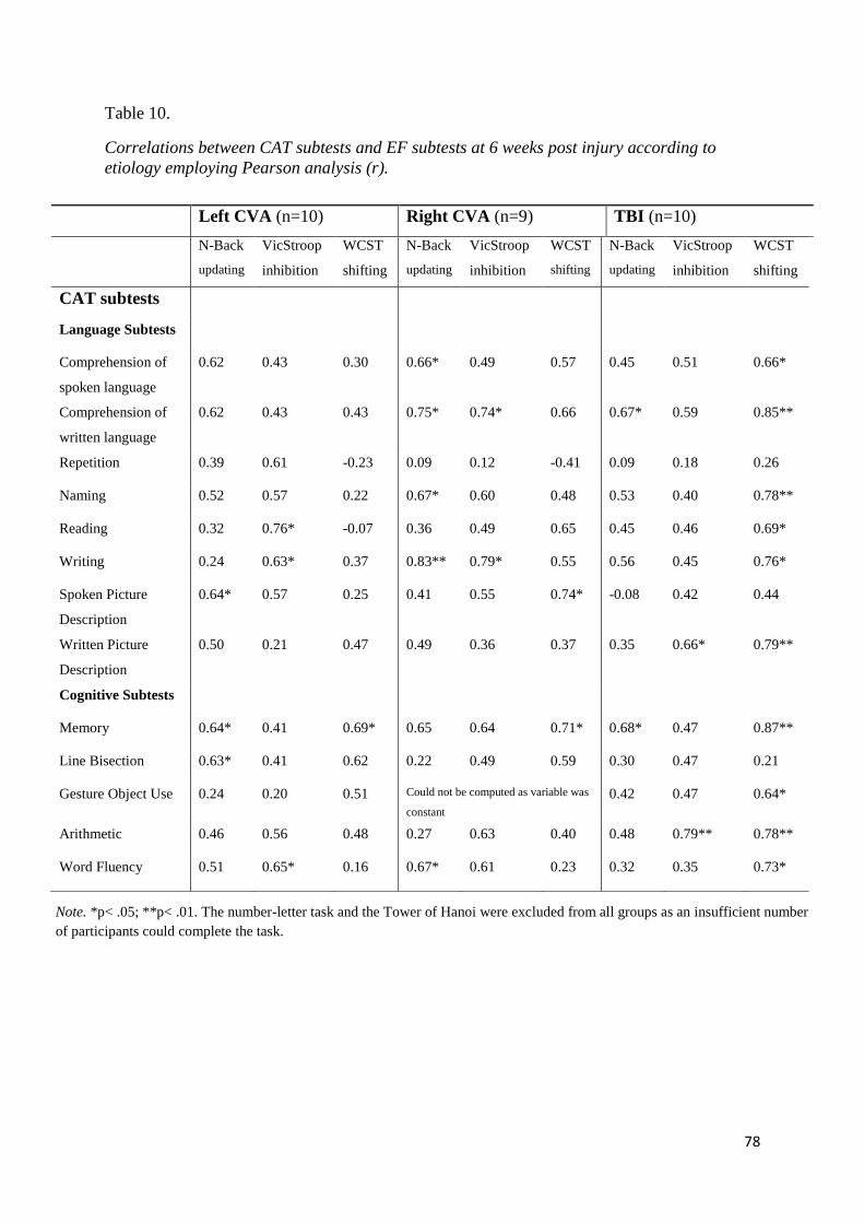
78
Table 10.
Correlations between CAT subtests and EF subtests at 6 weeks post injury according to
etiology employing Pearson analysis (r).
Left CVA (n=10) Right CVA (n=9) TBI (n=10)
N-Back
updating
VicStroop
inhibition
WCST
shifting
N-Back
updating
VicStroop
inhibition
WCST
shifting
N-Back
updating
VicStroop
inhibition
WCST
shifting
CAT subtests
Language Subtests
Comprehension of
spoken language
0.62 0.43 0.30 0.66* 0.49 0.57 0.45 0.51 0.66*
Comprehension of
written language
0.62 0.43 0.43 0.75* 0.74* 0.66 0.67* 0.59 0.85**
Repetition 0.39 0.61 -0.23 0.09 0.12 -0.41 0.09 0.18 0.26
Naming 0.52 0.57 0.22 0.67* 0.60 0.48 0.53 0.40 0.78**
Reading 0.32 0.76* -0.07 0.36 0.49 0.65 0.45 0.46 0.69*
Writing 0.24 0.63* 0.37 0.83** 0.79* 0.55 0.56 0.45 0.76*
Spoken Picture
Description
0.64* 0.57 0.25 0.41 0.55 0.74* -0.08 0.42 0.44
Written Picture
Description
0.50 0.21 0.47 0.49 0.36 0.37 0.35 0.66* 0.79**
Cognitive Subtests
Memory 0.64* 0.41 0.69* 0.65 0.64 0.71* 0.68* 0.47 0.87**
Line Bisection 0.63* 0.41 0.62 0.22 0.49 0.59 0.30 0.47 0.21
Gesture Object Use 0.24 0.20 0.51 Could not be computed as variable was
constant
0.42 0.47 0.64*
Arithmetic 0.46 0.56 0.48 0.27 0.63 0.40 0.48 0.79** 0.78**
Word Fluency 0.51 0.65* 0.16 0.67* 0.61 0.23 0.32 0.35 0.73*
Note. *p< .05; **p< .01. The number-letter task and the Tower of Hanoi were excluded from all groups as an insufficient number
of participants could complete the task.

79
Table 11.
Correlations between CAT language subtests and EF subtests at 12 weeks post injury
according to etiology employing Pearson analysis (r).
Left CVA (n=10) Right CVA (n=9) TBI (n=10)
N-Back
(updating)
VicStroop
(inhibition)
WCST
(shifting)
N-Back
(updating)
VicStroop
(inhibition)
WCST
((shifting)
N-Back
(updating)
VicStroop
(inhibition)
WCST
(shifting)
CAT subtests
Language Subtests
Comprehension of
spoken language
0.43 0.33 0.69* 0.65 0.04 0.25 0.15 0.12 0.39
Comprehension of
written language
0.64* 0.42 0.67* 0.51 -0.03 0.10 -0.05 0.01 0.34
Repetition 0.57 0.66* 0.30 0.13 0.59 0.31 0.09 0.11 0.37
Naming 0.49 0.49 0.53 0.73* 0.05 0.27 0.30 0.19 0.37
Reading 0.65* 0.35 0.44 0.08 0.42 -0.01 0.04 0.05 0.62
Writing 0.68* 0.44 0.59 0.83** -0.18 0.74* 0.08 0.17 0.30
Spoken Picture
Description
0.70* 0.65* 0.46 0.59 0.02 0.47 -0.08 0.07 0.52
Written Picture
Description
0.52 0.21 0.62 0.49 0.09 0.04 -0.06 0.03 0.12
Cognitive Subtests
Memory 0.73** 0.60* 0.62* 0.73* -0.18 0.12 0.24 0.11 0.24
Line Bisection 0.68* 0.11 0.68* 0.46 -0.05 -0.01 -0.16 0.20 0.54
Gesture Object use 0.28 0.22 0.53 Could not be computed as variable was
constant
-0.09 0.13 -0.53
Arithmetic 0.62* 0.34 0.56 0.11 -0.41 0.38 0.34 0.17 0.21
Word Fluency 0.43 0.22 0.43 0.84** -0.23 0.42 0.17 -0.02 0.27
Note. *p< .05; **p< .01. The number-letter task and the Tower of Hanoi were excluded from all groups as an insufficient number
of participants could complete the task.

80
Time Period Left CVA
(n=10)
Right CVA
(n=9)
TBI
(n=10)
N- Back 6 weeks
(Updating)
Memory
Line Bisection
Spoken Picture Description
Comprehension of spoken
language
Comprehension of written
language
Naming
Writing
Word Fluency
Memory
Comprehension of written
language
12 weeks Memory
Line Bisection
Arithmetic
Comprehension of written
language
Reading
Writing
Spoken Picture Description
Memory
Naming
Writing
Word Fluency
None
VicStroop 6 weeks
(Inhibition)
Reading
Writing
Word Fluency
Comprehension of written
language
Writing
Written Picture Description
Arithmetic
12 weeks Memory
Spoken Picture Description
Repetition
None None
WCST 6 weeks
(Shifting)
Memory Memory
Spoken Picture Description
Memory
Comprehension of spoken
language
Comprehension of written
language
Naming
Reading
Writing
Written Picture Description
Gesture object use
Arithmetic
Word Fluency
12 weeks Memory
Line Bisection
Comprehension of spoken
language
Comprehension of written
language
Writing None
Table 12.
The different patterns of CAT subtests which correlate with EF subtests according to time of assessment.
81
4.1 Correlations between the CAT and EF subtests according to clinical group
The results of the Pearson correlation tests reveal a unique cognitive and linguistic profile per
etiology when determining which executive function tasks correlate with specific language
tasks.
In the group of participants with a left hemisphere stroke, it was observed at 6 weeks post
stroke that three CAT subtests (memory, line bisection and spoken picture description)
correlated with the n-back task whilst at 12 weeks post stroke, seven CAT subtests (memory,
line bisection, arithmetic, comprehension of written language, reading, writing and spoken
picture description) correlated with the n-back task (updating). The CAT subtests of word
fluency, reading and writing correlated with the VicStroop task at 6 weeks post stroke whilst
at 12 weeks post stroke, repetition and spoken language correlated with the VicStroop task
(inhibition). The memory subtest correlated with the WCST (complex mental shifting task) at
6 weeks post stroke. Comprehension of spoken language and comprehension of written
language as well as the line bisection subtest correlated with WCST at 12 weeks post stroke.
None of the language subtests correlated with all three EF subtests at 6 or 12 weeks post
stroke. Limited subtests from the cognitive screener correlated with the EF subtests.
In the group of participants with a right hemisphere stroke, it was noted that there was a
correlation between the language subtests of comprehension of spoken language,
comprehension of written language, naming and writing with the n-back task at 6 weeks post
stroke. Naming, writing and memory correlated with the n-back task (updating) at 12 weeks
post stroke. A correlation was present between the WCST scores and the two CAT subtests of
memory and the spoken picture description at 6 weeks subsequent to the stroke. A correlation
between writing and WCST (shifting) was present at 12 weeks post stroke. At 6 weeks
subsequent to the right CVA, comprehension of written language and writing subtest
correlated with the VicStroop task, however at 12 weeks subsequent to the stroke, none of the
language subtests in this group of participants correlated with VicStroop (inhibition). Limited
subtests from the cognitive screener of the CAT correlated with EF subtests. The only
correlation was between the n-back task and the word fluency subtest at 6 and 12 weeks post
injury.
82
At 6 weeks post TBI, the memory subtest and comprehension of written language correlated
with the n-back task. The written picture description subtest correlated with the VicStroop
task. The WCST correlated with the following language subtests: memory, comprehension of
spoken language, comprehension of written language, naming, reading, writing and written
picture description. Limited subtests from the cognitive screener of the CAT correlated with
EF subtests. The only correlations that occurred at 6 weeks were between the VicStroop and
arithmetic and between the WCST and the gesture object use, arithmetic and word fluency
subtests. None of the language or cognitive subtests of the CAT correlated with any of the EF
subtests at 12 weeks subsequent to the TBI.
There were fewer correlations between language and EF subtests at 6 weeks post left CVA
than at 12 weeks post left CVA. In the participants with right CVA, there were more
correlations between language and EF subtests at 6 weeks post stroke with a number of
language subtests correlating with the n-back task. At 12 weeks post right CVA, there were
fewer correlations between language and EF subtests and the majority of correlations were
between the language subtests and the updating subtest. There were many correlations
between language subtests and EF subtests at 6 weeks subsequent to a TBI with the majority
of correlations between the WCST task and language subtests. There were no correlations
between the language subtests and the EF subtests at 12 weeks subsequent to a TBI. This
reveals different patterns of recovery which a clinician may need to consider when planning
treatment protocols. The limited correlations between the cognitive subtests of the CAT and
the EF subtests across etiology reveal that the cognitive screener of the CAT assesses
different cognitive skills to the EF battery. Completing the cognitive screener as well as the
EF battery will provide a clinician with comprehensive cognitive profile that could be useful
in planning effective treatment in the acute phase of recovery for bilinguals.
4.2 Correlations between the CAT and EF subtests in the control group
At the initial assessment there were correlations between n-back task and comprehension of
written language, repetition, naming, reading, writing and written picture description. There
were correlations between the VicStroop task and the language subtests of reading, spoken
picture description and written picture description. The WCST correlated with the following
language subtests: comprehension of written language, naming, reading and written picture
description. Correlations between the cognitive subtests of the CAT and the EF subtests were

83
limited. There was one correlation between the WCST and word fluency. At the 6 week
reassessment, the control group results revealed a correlation between the written picture
description subtest and the VicStroop, and between the comprehension of spoken language
subtest and WCST. Limited correlations reveal that in the control group each subtest is
assessing different parameters. There were again limited correlations between cognitive
subtests of the CAT and the EF subtests. There was a correlation between n-back and
memory subtest as well as the WCST and the word fluency subtest. This again reveals that
the cognitive screener has limitations and that further assessment using the EF battery will
provide a more comprehensive cognitive profile. Table 13 provides the r values to reflect the
correlation.
Table 13.
Correlations between CAT language subtests and EF subtests of control group at the initial
assessment and at the reassessment 6 weeks later employing Pearson analysis (r). (n=19)
N-back
(Updating)
Initial
Assessment
VicStroop
(Inhibition)
Initial
Assessment
WCST
(Shifting)
Initial
Assessment
N-back
(Updating)
Re
Assessment
VicStroop
(Inhibition)
Re
Assessment
WCST
(Shifting)
Re
Assessment
CAT subtests
Language subtests
Comprehension of
spoken language 0.20 0.07 0.39 0.41 0.24 0.65**
Comprehension of
written language 0.53* 0.44 0.68** 0.28 0.43 0.33
Repetition 0.52* 0.30 0.21 -0.24 0.15 0.11
Naming 0.53* 0.29 0.54* 0.19 0.22 0.39
Reading 0.53* 0.64** 0.62** 0.22 0.33 0.18
Writing 0.70** 0.37 0.44 0.20 0.23 0.11
Spoken Picture
Description 0.31 0.53* 0.30 0.21 -0.01 0.11
Written Picture
Description 0.56* 0.67** 0.71** -0.12 0.50* -0.01
Cognitive subtests
Memory 0.25 -0.16 0.17 0.46* -0.02 0.19
Line Bisection -0.07 0.37 0.34 0.19 0.25 -0.16
Gesture Object Use Could not be computed as variable was constant
Arithmetic 0.25 0.38 0.30 -0.24 0.40 -0.12
Word Fluency 0.37 0.23 0.47* 0.37 0.19 0.62*
Note. *p< .05; **p< .01
84
5. Summary of results
The results of the research reveals that the majority of the battery seem appropriate for use on
South African bilingual, second language English speakers. This was observed in the analysis
of the pattern of results of the control group and through the RM ANOVA. The control group
scored normal results on both the CAT and the non-verbal EF battery. This reveals that even
though the participants were second language English speakers who were culturally diverse,
valid results were obtained. The ToH seemed inappropriate for the South African population
and the cultural context. The number-letter task also did not appear appropriate as
perseveration in the participants skewed results. This task is more of a reflection of
perseveration symptoms associated with prefrontal lobe deficits in the acute phase as opposed
to shifting (Fuster, 1997).
The linguistic profiles obtained in the clinical groups by employing the CAT to assess
language skills in the acute phase reflected linguistic profiles of each etiology observed in the
literature. It was positive to identify that the CAT was able to provide a different pattern/
profile according to etiology in the acute phase. This highlights that the CAT is sensitive to
etiology in the acute phase.
The EF profile for the participants with a left hemisphere stroke reflected the profiles
observed in the literature. However, the EF profile for participants with a right hemisphere
stroke and TBI showed different profiles from those in the literature. The intact inhibition
skills at the 12 week assessment were a notable difference. Intact inhibition skills in the
bilingual population may reflect bilingual advantage even when there is a neuronal lesion.
When comparing the results at week 12 with the results at week 6, the language and EF
profiles of change were unique according to etiology. It must be noted that in the control
group there was statistically significant change in the subtests of comprehension of spoken
language, spoken picture description, and written picture description as well as the WCST.
This is not necessarily an error and may be related to a person’s learning potential (Duff et
al., 2007) and thus if changes occur in these areas in participants regardless of etiology, it
may reflect not only the recovery occurring but also their potential to learn.

85
It was also observed that there are unique interactions between CAT and EF subtests
according to etiology. It was important to note that these patterns of interaction changed over
a relatively short period (6 weeks). These interactions and the way that they change may be
important for therapy planning as well as recognising the amount of change which occurs in
the first 12 weeks post stroke and how therapy may need to be revised.

86
Chapter 5
Discussion
There is a burgeoning body of research on the impact of very early intervention in aphasia
and related disorders (Laska et al., 2008;Godecke et al., 2012). The process of recovery in the
acute phase is important to understand as it will impact the choice, timing, and nature of such
therapy. This is particularly the case for bilingual persons, whose recovery patterns are
different to monolinguals (Fabbro, 2001) and often create a challenge for the monolingual
clinician. This study was designed to investigate recovery profiles and determine possible
relationships between linguistic and non-linguistic factors in order to determine influential
components that may contribute to the recovery profiles observed. The assessment battery
developed for this research project will be discussed with regard to its use on South African
bilingual, second language English speakers. The importance of linguistic and EF profiling of
bilinguals in the acute phase will be deliberated. The importance of profiling will be
discussed in order to support understanding of the underlying recovery process which occurs
in the early stages after neuronal injury and their role in decision making for therapy in the
acute phase. Based on the profiles and changes which are observed, insights regarding the
bilingual population will be offered.
1. Assessment of South African bilinguals using the CAT and non-verbal EF battery
Previous research on monolingual patients has established the need to consider EF in
assessment and treatment of persons with ANCD as discussed in Chapter 2. It has been
suggested by Ansaldo et al (2008) and Weekes (2010) that assessment results describing the
preserved and disrupted processes and underlying neural networks may aid clinical decisions
and optimise intervention. This indicates the need for a clinician to complete a diagnostic
assessment that will guide and optimise intervention.
It has been recommended that in order to determine valuable prognostic information and to
develop an appropriate intervention plan it is essential to profile linguistic, cognitive and
communicative strengths and weaknesses (Cumming, Marshall and Lazar, 2013; Murray,
2012; Kohnert, 2004; Helm-Estabrooks, 2002). As discussed in Chapter 2 there have been
some limitations with regards to speech language pathology service provision in the South

87
African context due to the socio-political history of the country. This history has resulted in
pervasive consequences which affect current service delivery of speech-language
pathologists.
The results of this research, although exploratory due to a limited sample size, reflect that the
CAT and non-verbal EF battery (comprising the n-back task, VicStroop and WCST) may be
useful assessment measures when assessing bilingual, second language English speakers who
have had at least 12 years of education. The results of the control group reflected language
and EF skills that were within the normal limits according to the norms of the assessment
measures. The finding that testing bilingual participants in their second language could reveal
useful clinical information is reassuring, given the linguistic constraints of most South
African clinicians as well as a lack of suitable translated material for languages in this
specific multicultural and multilingual context. Roberts (1998) highlights that often
translations of published aphasia tests are usually standardised on native, monolingual
speakers of the translated test and not the bilingual speakers.
The use of a control group has helped to increase confidence in the results. Significant
differences were observed in the CAT and EF battery scores between the control group and
the participants with brain injury, highlighting that the assessment battery is useful in
distinguishing pathological from normal individuals. In addition to being able to differentiate
normal from pathological, the patterns observed according to etiology were different and
reflected patterns observed in the literature for these pathologies. This indicates that the CAT
and non-verbal EF battery could differentiate between patients with left CVA, right CVA and
TBI.
During standardisation of the CAT, the control group was assessed once. The control group
in this study was tested repeatedly using the CAT and the non-verbal EF battery. The
completion of a reassessment at six weeks after the initial assessment was deemed necessary
to determine what effect repeated exposures to the assessment materials might have. This
repeated testing further increased confidence in the results as it allowed the researcher to
observe changes in the control group and the possible reasons for the changes as discussed in
Chapter 4. Therefore when analysing repeated measures of this assessment battery, if a
clinician determines that there has been improvement, the clinician can consider not only the

88
spontaneous recovery occurring but also the patient’s learning ability as evidenced by
possible practice effects in certain subtests. It is also important for the clinician to be aware
that the WCST is also prone to practice effects in both normal and clinical groups and should
interpret the changes cautiously.
Roberts (1998) highlighted that translations of published tests are usually standardized on
native, monolingual speakers of the translated test and not necessarily on bilingual speakers.
Hence a strength of this research study and the finding that the assessment battery was able to
obtain valid results, is that the controls where second language English speakers. Thus it
provided evidence for how neurologically intact bilingual second language English speakers
would perform on the assessment battery and added validity to the use of this assessment
battery with this specific population.
The Tower of Hanoi (ToH) test proved to be an inappropriate assessment measure for the
South African context. The majority of participants in the acute phase post stroke or TBI
could not complete the task. In addition, the neurologically intact control group also had
difficulty completing the task. Only five out of the nineteen control group participants were
able to complete the task within the normal limits, thus indicating that for the South African
population, the ToH may not be culturally appropriate to include in the assessment battery.
This may be due to different educational and cultural background of the South African
population as compared to the population the assessment measured was normed on. The
number-letter task did not appear to provide precise information as often a participant would
perseverate on the task and due to the nature of measuring the speed of response the
participant would often achieve the normal speed of the task with poor accuracy. This task is
more a reflection of perseveration symptoms associated with prefrontal lobe deficits in the
acute phase as opposed to shifting (Fuster, 1997). Thus the number-letter task is not an
appropriate assessment tool for the acute phase subsequent to a brain injury.
Miyake et al. (2000) have suggested that multiple assessments of EFs are necessary to
produce an accurate characterisation of EF, and this research initially used multiple
assessment tasks to assess shifting, updating and inhibition. However, based on the limited
validity and reliability of results seen in this study from the ToH and the number-letter task,
the use of multiple tests in the acute phase may be redundant and ineffective. Therefore the

89
EF battery could possibly be streamlined for the assessment in the acute phase to include the
n-back task, Victoria Stroop and the Wisconsin card sorting test in order to provide the initial
EF findings for updating, mental shifting and inhibition. A further positive characteristic of
this battery is that it did not rely on timed tasks that identify processing speed deficits which
are often present in persons with any form of brain injury (Cumming, Marshall & Lazar,
2013).
The cognitive subtest of the CAT revealed limited correlations with the EF tasks for all
groups of participants. This result reflects that the cognitive skills assessed by the CAT
cognitive subtests are different to the skills being assessed by the EF subtests. The line
bisection subtest was employed to assess for visuospatial deficits, the arithmetic subtest was
used to assess for acalculia, the gesture object use subject was used to asses for
ideomotor/ideational apraxia and word fluency was used to assess generative naming. These
are specific cognitive skills that may be present subsequent to a head injury (Howard,
Swinburn, & Porter, 2010). The fact that the EF subtests do not correlate with these subtests,
reflects the importance of completing these subtests of the CAT in order to obtain further
information about additional aspects of a patient’s cognition that may affect assessment and
treatment. Neither the EF subtests nor the cognitive subtests of the CAT are redundant tests
and both provide specific sets of different cognitive information regarding the patient.
In summary, when identifying the linguistic and EF profiles in the acute phase, a clinician
may need to consider the assessments utilised as there may be some redundancy in employing
an extensive battery with multiple EF assessment measures. The majority of research
regarding profiling of linguistic and non-linguistic skills has been completed six months or
more subsequent to the brain injury. There is limited research on the assessment of cognitive
impairments which are present in the first few months post brain injury (Rasquin, Welter &
van Heugten, 2013; Zinn et al., 2007). Therefore although it is recommended that extensive,
individual neuropsychological evaluations should be completed to identify linguistic and
cognitive profiles, this recommendation has generally been made when referring to patients
in the chronic phase as discussed in Chapter 2.
Extensive profiling may not be feasible or ethical in the acute phase. At 6 weeks subsequent
to the injury, participants seemed to be more stressed during the assessment tasks as opposed

90
to the 12 week reassessment. Although this assessment battery was relatively short, it seemed
to be impacted by fatigue and it created an increased level of stress in some of the
participants. Thus completing a long extensive neuropsychological battery in the acute phase
may not be feasible or ethical due to stress levels as well the fatigue that a patient experiences
during this phase of recovery (Rossini & Del Forno, 2004). A clinician may be able to
employ a shortened battery such as the battery developed in this research in the acute phase in
order to profile patients’ linguistic and cognitive functions. The streamlined battery that was
found to be beneficial and sensitive to the multicultural and multilingual nature of South
Africa comprised the CAT as the language assessment and the n-back task (updating),
Victoria Stroop (inhibition) and WCST (shifting) as the EF assessment battery in the acute
phase.
The most significant finding is that this assessment battery was suitable for bilinguals of all
cultural and linguistic backgrounds. This finding fares well for the use of this battery in the
South African context which is multilingual and multicultural. It also reinforces the impact of
education on assessment batteries. The assessment battery was completed on patients with a
minimum of 12 years of education therefore further testing on individuals with less education
should be completed to identify the impact of education on these specific assessment
measures. It should be remembered that due to the sample size of the control group, further
research using this battery on bilingual, second language English-speaking South Africans is
required. In addition the clinical and control groups represented the population of bilingual
speakers in an urban South African setting and therefore further research would be required
to determine if these results could be replicated in a rural South African setting.
2. The importance of linguistic and EF profiling of bilinguals in the acute phase
The findings of this research provide some interesting insights into the need to complete
linguistic and cognitive profiling of bilingual persons who have had a left CVA, right CVA or
TBI in the acute phase of recovery. The acute profiles observed in this research study may
add value to the assumption by Lambon Ralph et al. (2010) that both the severity of language
and cognitive deficits are an important predictor of treatment outcomes in not only
monolinguals but also bilinguals. There is a paucity of research regarding the recovery
profiles of bilingual persons who have had a right CVA or TBI in the acute phase of recovery
and thus this research raises potentially important clinical implications of the need for acute

91
phase linguistic and cognitive profiling not only in bilingual persons with a left CVA but also
for those with a right CVA or TBI.
The acute recovery profile of a bilingual with a left CVA based on the correlations between
CAT and EF subtests was interesting. The correlations between language subtests and EF
subtests at 6 weeks post injury, changed significantly at 12 weeks post injury. In the same
way that the Martin et al (2012) study identified that verbal working memory could be used
as part of a therapy protocol, identifying the interaction patterns between EF and language
skills at both the 6 week stage and the 12 week stage may assist developing treatment
protocols for different phases post neuronal injury.
In participants with a right CVA, a different pattern in correlations between language subtests
and EF subtests was observed from 6 weeks to 12 weeks. This change in pattern was
discussed in Chapter 4. As recovery occurs in the acute phase and there is recovery in EF
skills, and possibly those skills no longer correlate with language functioning. It is known
that in monolingual persons with a right CVA, language deficits are more significant when
attention and/or working memory are taxed (Tompkins et al., 2002). Thus if there are
improvements in the EF skills, there may be improvements in language skills. It is important
for a clinician to understand which EFs are correlating with language subtests in the acute
phase as the deficits in these EF skills may relate to the language symptoms observed. Thus
integrating EF tasks into therapeutic tasks may be vital in employing the possible bilingual
advantage during the acute recovery process.
The pattern of correlations also changed in the acute phase for participants with a TBI. There
were many language subtests that correlated with shifting at 6 weeks post TBI possibly
highlighting the important role shifting has in bilinguals. It is known that in bilinguals they
are required to shift between languages and suppress the language that is not required for that
specific context (Garbin et al., 2010) and therefore shifting may be important to consider in
the acute phase. At 12 weeks post injury, no language subtests correlated with any of the EF
subtests, possibly highlighting that in the acute phase EF skills are essential for successful
language functioning, just as is known for the chronic phase. Again, incorporating these EF
skills into the therapeutic process may be necessary but further research is required to

92
determine how the incorporate the EF load into therapeutic tasks in the acute phase of
rehabilitation.
When a neuronal lesion occurs, some neurons essential to language and cognitive processing
are deleted, disconnected or functionally impaired. Individuals with neuronal lesions may
regain functionality by setting up new links and strengthening remaining links (Pulvermüller
& Berthier, 2008). In order to make use of learning patterns in therapy that will enable brain
repair, a clinician needs to know which areas of language and EF have deficits and how they
interact, hence the importance of the linguistic and non-linguistic profiling that a clinician can
achieve when employing this assessment battery. These deficits need to be known so that
tasks can be formulated in order to induce the relevant coincidence of neural firing in order to
train a patient successfully. Furthermore if a clinician is aware of not only the profile of
deficit but also the profile of correlation and interaction, the therapy they provide can support
functional reorganisation by strengthening remaining neural circuits through internal links
and by allowing neural circuits to incorporate additional neurons to compensate for those
which were lost due to the lesion (Pulvermüller & Berthier, 2008).
The change in correlations across time may also reflect the different compensatory networks
that are engaged at different time periods in the acute recovery process. Sebastian, Kiran and
Sandberg (2012), highlighted that there are additional neural substrates activated during
language processing of bilinguals and the neural areas they identified were areas which also
had a role in executive function and cognition. Different EF skills may be required to
successfully process language at different stages of the acute recovery process. This is
important to acknowledge as it may support therapeutic interventions and enable a SLP to
make use of the compensatory networks which may sub serve successful language
processing. Pettigrew and Hillis (2014) identified that the relationship between short term
memory and sentence comprehension may be correlational rather that causal because they
rely on a subset of the same neurological regions, so this research and the different
correlations identified may highlight the different language and EF skills that rely on similar
neural regions and how these change over time. The changes in correlation over time may be
due to the compensatory strategies which develop and the reorganisation of the cognitive
process underlying the language processing which occurs in the recovery process subsequent
to a neuronal injury.

93
Knowledge of the change in the language and EF patterns in the acute phase may be
particularly important when having to establish a treatment protocol in the South African
setting where there is limited access to speech-language therapy after the acute phase of
injury as discussed in Chapter 1 and 2. It is also essential to be aware of the interactions
between EF and language skills in the acute phase as a clinician needs to provide therapy in
line with neuroplasticity principles in order to provide therapy which will support the
spontaneous recovery and neuroplasticity which is occurring in the acute phase post stroke
(Kleim & Jones, 2008). Awareness of the interactions will enable a clinician to determine
appropriate tasks and stimulus materials in order to take into account the linguistic and non-
linguistic parameters which are relevant to the patient. As discussed extensively in Chapter 2,
animal research has provided evidence that administering the incorrect type of therapy in the
early stage after a neuronal injury can result in negative outcomes (Kleim & Jones, 2008).
Hence as clinicians know, it is essential to provide therapy that does not result in negative
outcomes.
In summary, the results revealed the linguistic and cognitive profiles in the acute phase were
differential and the different patterns of change in EF recovery profiles may be responsible
for the different language recovery profiles observed per etiology. This finding contributes to
research that EF may have a role in language processing and language recovery. Furthermore,
it provides preliminary evidence for the different EFs that may play a role according to
etiology. Knowledge of the specific EFs that interact with language recovery per etiology can
assist a clinician in providing effective therapy. This supports the research regarding the need
to include non-linguistic assessments in the assessment battery (Helm-Estabrooks, 2002;
Murray, 2012) and to consider EF in treatment planning (Nicholas, Sinotte & Helm-
Estabrooks, 2005; Purdy & Koch, 2006) of persons with a left hemisphere stroke as well as
right CVA and TBI. The role of EF in recovery would be an interesting phenomenon to
research further in order to determine if the different profiles of EF have a very definitive
impact on the different language recovery profiles.
3. Insights into bilingualism
The pattern of change in EF appeared to be different from that of monolinguals in the acute
phase. Research by Rasquin et al. (2013) determined that in the acute phase there were no

94
significant changes in EF in monolingual participants. In contrast, the results of this research
suggest there are changes (although different per etiology) in EF in the first 12 weeks post
injury. For bilingual participants with a left CVA, updating and shifting changed whilst for
participants with a right CVA, updating and inhibition changed. Inhibition and shifting
changed in participants with a TBI.
Recent research provides some evidence for these differences as it has been revealed that
there may be differences in cognitive control in bilingual and monolingual brains. The ability
to control receptive and expressive language in a specific context at a specific time is a
fundamental feature of the human bilingual brain (Abutalebi et al., 2008). It has been
identified that processes of inhibition, updating and mental shifting may be important
processes which are activated during language tasks in bilinguals (Bialystok, 2009).
If there is a bilingual advantage present in persons with ANCD then this may direct the
treatment protocol that clinicians may select for a bilingual. However, this is an exploratory
study and the sample sizes are limited thus, further research in the acute phase for bilinguals
who have had a TBI, left CVA or right CVA is required.
Controversies still remain regarding the treatment of bilingual patients. The use of linguistic
and cognitive profiling may provide the clinician with some insight into what therapy to
employ with a bilingual patient- whether it is monolingual versus bilingual therapy based on
the level of EF skills or whether one needs to target the cognitive or linguistic components
that underlie the language processing skills present in a bilingual person. Penn et al. (2010)
highlighted that EF may play a role in the chronic phase to support the decision of whether to
provide bilingual therapy to patients with well persevered EFs as the compensatory and
shifting strategies that were present prior to the stroke may support language recovery and
interactions. However, it was also cautioned that patients who have limited EF skills may not
benefit from bilingual therapy due to the cognitive resources required (Penn et al., 2010). The
results of the current study based on the changes in EF observed as well as the changes in
correlations between EF subtests and language subtests from week 6 to week 12, agree with
the above hypothesis that incorporating EF into early treatment paradigms may firstly assist
the clinician in determining whether treatment should be bilingual or monolingual and
secondly, may support retrieval of previous cognitive strategies inherently utilised by the

95
bilingual speaker. The possible bilingual advantage observed may be useful for the clinician
to take advantage of when developing a treatment programme. EF, in conjunction with other
identified factors (age, site of lesion, extent of lesion, language proficiency) may play a role
in the variety of language recovery patterns identified in bilingual patients.
Further understanding of the role of updating, shifting and inhibition in bilingual language
processing and recovery could support a clinician’s knowledge regarding how the EF deficits
may be contributing to the language behaviour and how the EF deficits may support the
recovery process. Inhibition appeared to provide insight into language recovery patterns of
right CVA and TBI as compared to left CVA. Inhibition also had important implications for
the selection of monolingual versus bilingual therapy. These implications included
considering monolingual therapy in the patient’s stronger language if inhibition is not intact
whilst intact inhibition may allow for bilingual therapy or therapy in the patient’s weaker
language. This will be discussed further in Chapter 6.

96
Chapter 6
Implications and Conclusions
1. Implications for Assessment and Therapy
1.1 Implications for Assessment
When identifying the linguistic and EF profiles in the acute phase, a clinician may need to
consider the assessments utilised as there may be some redundancy in employing an
extensive battery with multiple EF assessment measures which may not be appropriate in this
phase when assessing language and EF. The results of this study revealed that the CAT and
the non-verbal EF battery comprising of the n-back task, VicStroop and WCST may be an
economical and efficient battery to assess language and EF skills of South African bilingual,
English second language speakers. This streamlined battery was found to be beneficial and
sensitive to the multicultural and multilingual nature of South Africa. The streamlined battery
could also support patient fatigue and stress levels. Extensive testing may be unethical and
inappropriate at this stage of recovery.
This battery would assist the first language English-speaking speech-language pathologists in
the South African context to assess bilingual patients and have reliable information regarding
the patients’ language and EF skills. It is suggested that clinicians attempt to use this battery
with bilingual, English second language speakers (particularly if they do not have an
interpreter), when profiling linguistic and non-linguistic skills in the acute phase post left
CVA, right CVA or TBI.
It also provides evidence that a clinician can repeatedly asses a patient in the acute phase in
order to monitor progress so that the therapy plan can be adjusted according to the patient’s
needs. Practice effects that may occur in selected subtests can be interpreted to reflect a
patient’s learning ability.

97
1.2 Implications for therapy based on the interactions between linguistic and EF profiles
There is an intricate relationship between language and EF and the way in which the bilingual
brain is organised. Knowledge of the relationship may be useful in developing treatment
protocols for bilingual persons in order to assist with achieving functional gains in all
languages spoken. Thus having a better knowledge of the brain organisation of bilinguals
together with the linguistic and non-linguistic profiles may assist the clinician in deciding
whether to initially target the underlying cognitive skills or the linguistic elements that are
required for both languages as hypothesised by Kohnert and Derr (2004). Therefore it would
be beneficial if monolingual clinicians could work in consultation with bilingual clinicians in
order to develop and provide efficacious treatment protocols that incorporate the bilingual
element of the patient as suggested by Kohnert (2004).
There are interactions between language and EF skills from early in the recovery phase.
Therefore language and EF should be incorporated into therapy from the acute phase. In
monolingual patients, treatment targeting cognitive skills has been found to have a positive
impact on functional communication (Ramsberger, 2005) and it has been suggested that new
treatment approaches incorporating cognitive skills in the context of language tasks may be
necessary to improve language skills (Helm-Estabrooks, Connor & Albert, 2000; McNeil et
al., 2006; Murray & Ramage, 2000). Kohnert (2004) supports this notion in the context of
bilinguals, based on the results that intervention targeting basic information processing skills
positively impacted on the language deficits in a bilingual person with severe non-fluent
aphasia. However, this intervention occurred in the chronic phase of recovery and the
findings of the current study potentially validate the need for inclusion of EF assessment and
treatment in bilinguals in the acute phase. It also raises interesting possibilities for further
research regarding the role of EF in recovery and treatment paradigms in bilingual
populations.
1.3. Implications based on the patterns of change in linguistic and EF profiles
It is important for clinicians to understand the interactions between language and EF skills at
6 weeks and then at 12 weeks as the patterns of interaction change significantly in a short
period of time. It seems necessary for clinicians to take into account the different EF patterns
being observed in order to support the planning of language therapy programmes at different
stages of the acute phase.

98
Specifically in patients with a left CVA, a clinician may need to control carefully for
updating and shifting demands in language treatment tasks at 6 weeks post stroke and
integrate aims to improve these EF skills within the treatment protocol. Due to the changes at
12 weeks post left hemisphere stroke, a clinician may be able to allow for more updating,
inhibition and shifting demands in the treatment protocol. In addition, a general pattern of
improvement was observed within auditory language skills in the acute phase but not in
written language skills in the group of participants with left CVA. This pattern is important to
consider in selecting AAC systems for patients for example communication aids dependent of
written language. When selecting an AAC system, a clinician needs to be cognisant of the
pattern of language skills and consider the implications if written language is an area of
deficit as many AAC systems rely on written language (Linebarger & Schwartz, 2005). The
level of a written AAC system needs to be carefully selected and implemented and therapy
provided to improve the language skills to support functional communication (Linebarger &
Schwartz, 2005). A clinician may even need to consider that AAC should not be the
immediate choice for intervention at the acute phase. Clinically it has been observed that
there is a lack of compliance in use of AAC systems in the acute phase subsequent to brain
injury. This recommendation is a cautionary finding for clinicians when considering
treatment plans in the acute phase.
Changes in EF subtests of the participants with right CVA were different from those found in
participants with a left CVA. The changes observed indicate a change in updating and
inhibition with poor shifting skills. Possibly there may have been an interaction between the
changes in updating and inhibition with the language skills as well as the impact of poor
shifting skills. Therefore it may be deemed necessary for a clinician to identify updating and
shifting deficits in the acute phase of a person with a right CVA in order to control the impact
of updating and shifting on therapy tasks so that the clinician is able to provide therapy with
the appropriate updating and shifting demands. Therapy may also need to consider increasing
updating and shifting demands as the patient improves.
In the TBI participants, changes occurred in the majority of language and EF domains.
Shifting and inhibition were the EF skills which changed over the six weeks and thus a

99
clinician may need to be aware of the role of these two EF skills at different stages in the
acute phase and the impact on language skills.
1.4 Implications of preserved inhibition in bilinguals with ANCD
Inhibition skills were within the normal range for both participants with a right CVA and a
TBI. This intact inhibition provides evidence that there may be a residual bilingual advantage
in bilingual persons who have sustained a right CVA or TBI. It may be important to consider
this when providing therapy as the clinician may be able to make use of the intact inhibition
skills to support strategies and improvements in other domains. It is also important to
consider that inhibition deficits may result in the weaker language being at risk as discussed
previously and thus for patients with intact inhibition, the weaker language prior to the neural
injury may not be at risk and could be employed in therapy. Further research with regards to
this hypothesis is required.
Furthermore inhibition needs to be considered in both persons with right CVA and TBI as it
has a role to play in the recovery patterns of bilinguals and thus clinicians should make use of
the role inhibition has in recovery patterns. This would assist clinicians in the acute phase.
Green (2005) proposed that inhibitory processes are involved in successful language use by
bilinguals. Lorenzen and Murray (2008), suggest that differing degrees of inhibition could
lead to varying language recovery patterns. Permanent inhibition may result in selective
recovery, temporary inhibition in sequential recovery, alternating inhibition in antagonistic
recovery, greater inhibition in one language may cause differential recovery and loss of
inhibition may result in blending of languages.
With inhibition being relatively intact, simultaneous recovery should occur in both languages
according to the above model. Therefore a clinician should make use of inhibitory control in
the therapeutic process with bilinguals who have had a right CVA or TBI. Intact inhibition
highlights that bilingual therapy may be beneficial. However for persons with a left CVA
who present with inhibition deficits, which lead to pathological language symptoms, perhaps
clinicians need to consider that bilingual therapy which would make use of inhibitory control
mechanisms may not be beneficial. In patients with inhibition deficits, a clinician may need
to target the cognitive skill of inhibition prior to commencing bilingual language therapy. The

100
inhibition deficit may also facilitate the decision process for the therapeutic language, as it is
known that the less proficient language premorbidly may be at risk and thus a clinician may
select to utilise the more proficient language premorbidly for the language of treatment
(Paradis, 2001).
Due to the exploratory nature of this research, further research into the role of inhibition to
assist in the decision making process for monolingual versus bilingual therapy as well as the
language selected for treatment is required. The important role of inhibition in language
recovery has also been observed by the results and clinicians may need to consider inhibition
in order to assist language recovery.
2. Further Research
The assessment battery comprising the CAT and the non-verbal EF battery should be
validated on a larger sample size. Its use on rural South African population should also be
explored as well as with people who have less than 12 years of education. Further research
regarding the role of updating, shifting and inhibition in the recovery process would be
beneficial, to further support decision making processes for bilingual therapy. Further
research regarding the role of updating, shifting and inhibition in bilingual language
processing and recovery could support a clinician’s knowledge regarding the role cognitive
deficits may be contributing to the language behaviours and the manner in which EF may
support the recovery process. The role of inhibition as possibly a preserved bilingual
advantage in persons who have had a right CVA or TBI should be explored further as well as
its role in recovery and therapy.
Follow up assessments of participants from this research study in the chronic phase of
recovery could provide insights regarding prognostic value of the assessment battery and the
long term role of the different EF skills in recovery. These assessments could employ further
neurological assessment and reassessment with the test battery in order to determine the
chronic implications. Furthermore investigation of individualised therapeutic interventions
based on EF profiles could be employed in order to evaluate which therapy approaches may
be more appropriate for a patient based on their linguistic and EF profiles.

101
3. Conclusions
This research provided evidence that the CAT and the non-verbal EF battery comprising the
n-back tack, VicStroop and WCST are linguistically and culturally appropriate for the
assessment of bilingual, second language English speakers in the South African context
regardless of the participants cultural background. The battery was able to differentiate
pathological from neurologically intact. It was also able to differentiate pathology. It is
acknowledged that this study was exploratory and that further research with increased
population sizes is required to further validate the findings.
There are unique linguistic and EF profiles for bilinguals who have sustained a left CVA,
right CVA and TBI and the relationships between the language subtests and the EF subtests
are also unique and change across time periods. The profiles of language and EF skills of
bilinguals in the acute phase have not been reported on prior to this research project. There is
some evidence for preserved bilingual advantage subsequent to a neuronal insult particularly
with regards to inhibition in patients with a right CVA or TBI. Clinicians may engage this
preserved EF in order to facilitate therapy.
Language treatment needs to be provided in combination with an understanding of recovery
patterns, what is driving that pattern, and which cognitive deficits are contributing to the
language behaviour (Green, 2005). In addition clinicians need to be aware of the impact of
updating, shifting and inhibition in a bilingual person as well as that bilingual advantage may
play a role in recovery and could be a possible tool to support the therapeutic process.
This research suggests that individualised profiling is both feasible and relevant in the early
stages with bilingual patients; and that a diagnostic battery can be non-redundant and
cultually appropriate.

102
References
Abutalebi, J. & Green,D. (2007). Bilingual language production: The neurocognition of
language representation and control. Journal of Neurolinguistics, 20, 242–275.
Abutalebi, J., Annoni, J-M., Zimine, I., Pegna, A.J., Seghier, M.L., Lee-Jahnke, H., Lazeyras,
F., Cappa, S.F. & Khateb, A. (2008). Language control and lexical competition in
Bilinguals: An event-related fMRI study. Cerebral Cortex, 18 (7), 1496-1505.
Abutalebi, J., Rosa, P.A.D., Tettamanti, M., Green, D.W., & Cappa, S.F. (2009). Bilingual
aphasia and language control: a follow-up fMRI and intrinsic connectivity study.
Brain and Language, 109(2/3), 141-156.
Aerts, A., Batens, K., Santens, P., Van Mierlo, P., Huysman, E., Hartsuiker, R., Hemelsoet,
D., Duyck, W., Raedt, R., Van Roost, D., & De Letter, M. (2015). Aphasia therapy
early after stroke: behavioural and neuropsychological changes in the acute and post
acute phases. Aphasiology, DOI: 10.1080/02687038.2014.996520.
Alptekin, C. (2010). Redefining multicompetence for bilingualism and ELF. International
Journal of Applied Linguistics, 20(1), 95-110.
Ansaldo, A.I., Marcotte, K., Scherer, L. & Raboyeau, G. (2008). Language therapy and
bilingual aphasia: Clinical implications of psycholinguistic and neuroimaging
research. Journal of Neurolinguistics, 21, 539-557.
Ansaldo, A.I., Ghazi Saidi, L., & Ruiz, A. (2010). Model-driven intervention in bilingual
aphasia: evidence from a case of pathological language mixing. Aphasiology, 24(2),
309-324.
Archer, B. (2006). Afrikaans aphasia: Some generational differences. Undergraduate
research project, University of Witwatersrand.
Arciniegas, D. B., Frey, K. L., Newman, J., & Wortzel, H. S. (2010). Evaluation and
management of posttraumatic cognitive impairments. Psychiatric Annals, 40(11),
540–552.
Baddeley, A.D., & Hitch, G. J. (1974). Working memory. In G. A. Bower (Ed.), Recent
advances inlearning and motivation (pp. 47–90). New York, NY: Academic Press.
Bialystok, E. (2001). Bilingualism in Development: Language, Literacy and Cognition.
Cambridge: Cambridge University Press.

103
Bialystok, E. (2009). Bilingualism: The good, the bad and the indifferent, Bilingualism:
Language and Cognition, 12(1), 3-11.
Bialystok, E (2011). Reshaping the mind: The benefits of bilingualism. Canadian Journal of
Experimental Psychology, 65, 229-235.
Bialystok, E. & Barac, R. (2012). Emerging bilingualism: Dissociating advantages for
metalinguistic awareness and executive control. Cognition, 122, 67-73.
Bialystok, E. & Craik, F.I.M. (2010). Cognitive and linguistic processing in the bilingual
mind. Current Directions in Psychological Science, 19(1), 19-23.
Bialystok, E., Craik, F.I.M. & Freedman, M. (2007). Bilingualism as protection against the
onset of symptoms of dementia. Neuropsychologia, 45, 459-464.
Bialystok, E., Craik, F.I.M. & Luk, G. (2012). Bilingualism: Consequences for mind and
brain. Trends in Cognitive Sciences, 16(4), 240-250.
Bialystok, E. & Feng, X. (2009). Language Proficiency and executive control in proactive
interference: evidence from monolingual and bilingual children and adults. Brain and
Language, 109, 93-100.
Bialystok, E., Luk, G. & Craik, F. (2008). Cognitive Control and Lexical Access in Younger
and Older Bilinguals. Journal of Experimental Psychology: Learning, Memory &
Cognition, 34(4), 859-873.
Bialystok, E. & Viswanathan, M. (2009). Components of executive control with advantages
for bilingual children in two cultures. Cognition, 112, 494-500.
Botvinick, M.M, Cohen, J.D., & Carter, C.S. (2004). Conflict monitoring and anterior
cingulate cortex: an update. Trends in Cognitive Sciences, 8, 539-546.
Bradley, K.A.L., King, K.E. & Hernandez, A.E. (2013). Language experience differentiates
prefrontal and subcortical activation of the cognitive control network in novel word
learning. NeuroImage, 67, 101-110.
Bradshaw, D., Groenewald, P., Laubscher, R., Nannan, N., Nojilana, B., & Norman, R.
(2003). Initial burden of disease estimates for South Africa 2000. MRC Report. Cape
Town: South African Medical Research Council.
Brennan, A., Worrall, L. & McKenna, K. (2005). The relationship between specific features
of aphasia-friendly written material and comprehension of written material for people
with aphasia: An exploratory study. Aphasiology, 19(8), 693-711.

104
Brown, D.D.O. & Nell, V. (1991). Epidemiology of traumatic brain injury in Johannesburg-
II. Morbidity, mortality and aetiology. Social Science & Medicine, 33(3), 289-296.
Bruce, C., & Edmundson, A. (2010). Letting the CAT out of the bag: A review of the
Comprehensive Aphasia Test. Commentary on Howard, Swinburn, and Porter,
“Putting the CAT out: What the Comprehensive Aphasia Test has to offer”.
Aphasiology, 24(1), 79-93.
Buchsbaum, B.R., Greer, S., Chang, W.L., & Berman, K.F. (2005). Meta-analysis of
neuroimaging studies of the Wisconsin card-sorting task and component processes.
Human Brain Mapping, 25(1), 35-45.
Busch, R.M., McBride, A., Curtiss, G., & Vanderploeg, R.D. Busch, R.M., McBride, A.,
Curtiss, G., & Vanderploeg, R.D. (2005). The components of executive functioning in
traumatic brain injury. Journal of Clinical & Experimental Neuropsychology, 27(8),
1022-1032. DOI: 10.1080/13803390490919263.
Cabeza, R., Dolcos, F., Graham, R., & Nyberg, L. (2002). Similarities and differences in the
neural correlates of episodic memory and working memory. NeuroImage, 16(2), 317-
330.
Cannizzaro, M., & Coelho, C. (2012). Communication following executive dysfunction. In
R.K. Peach & L.P. Shapiro (Eds.), Cognition and acquired language disorders: an
information processing approach (pp. 227-240). Missouri: Elsevier Inc.
Carlson, S.M. & Meltzoff, A.N. (2008). Bilingual experience and executive functioning in
young children. Developmental Science, 11 (2), 282-298.
Cavada, C., & Goldman-Rakic, P. S. (1989). Posterior parietal cortex in rhesus monkey: II.
Evidence from segregated corticocortical networks linking sensory and limbic areas
with the frontal lobe. Journal of Comparative Neurology, 287, 422–445.
Cavé, J., & Grieve, K. W. (2009). Quality of education and neuropsychological test
performance. New Voices in Psychology, 5(l), 29-48.
Channon, S. & Watts, M. (2003). Pragmatic language interpretation after closed head injury:
relationship to executive functioning. Cognitive neuropsychiatry, 8(4), 243-260.
Chapey, R., & Hallowell, B. (2001). Introduction to language intervention strategies in adult
aphasia. In R. Chapey (Ed.), Language Intervention Strategies in Aphasia and Related
Neurogenic Communication Disorders (pp3-17). Philadelphia: Lippincott Williams &
Wilkins.
105
Chopra, M., Lawn, J.E., Sanders, D., Barron, P., Karim, S.S.A., Bradshaw, D., Jewkes, R.,
Karim, Q.A., Flisher, A.J., Mayosi, B.M., Tollman, S.M., Churchyard, G.J.,
Coovadia, H. (2009). Achieving the health Millennium Development Goals for South
Africa: challenges and priorities. Lancet, 374, 1023-1031.
Christensen, S. C., & Wright, H. H. (2010). Verbal and non-verbal working memory in
aphasia: What three n-back tasks reveal. Aphasiology, 24, 752–762.
Coelho, C. A. (2007). Management of discourse deficits following TBI: Progress, caveats,
and needs. Seminars in Speech and Language, 28, 122–135.
Coelho, C.A., Lê, K., Mozeiko, J., Hamilton, M., Tyler, E., Krueger, F., & Grafman, J.
(2013). Characterising discourse deficits following penetrating head injury: a
preliminary model. American Journal of Speech-Language Pathology, 22, S438-
S448.
Coelho C. A., Ylvisaker, M., & Turkstra, L. S. (2005). Nonstandardised assessment
approaches for individuals with traumatic brain injuries. Seminars in Speech and
Language, 26, 223–241.
Cockcroft, K. & Alloway, T. (2012). Phonological awareness and working memory:
Comparisons between South African and British children, Southern African
Linguistics and Applied Language Studies, 309 (10), 13-24.
Connor, M. & Bryer, A. (2006). Stroke in South Africa. In J. Fourie, K. Steyn, & N.J.
Temple (Eds.), Chronic Diseases of lifestyle: in South Africa: 1995-2005 (pp195-
203). Tygerberg: Medical Research Council.
Coovadia, H., Jewkes, R., Barron, P., Sanders, D., & McIntyre, D. (2009). The health and
health system of South Africa: historical roots of current public health challenges. The
Lancet, 374(9692), 817–834.
Costa, A., Hernandez, M., Costa-Faidella, J. & Sebastian-Galles, N. (2009). On the bilingual
advantage in conflict processing: Now you see it, now you don’t. Cognition, 113,
135-149.
Costa, A., Hernandez, M. & Sebastian-Galles, N. (2008). Bilingualism aids conflict
resolution: Evidence from the ANT task. Cognition, 106, 59-86.
Costa, A., & Santesteban, M. (2004). Lexical access in bilingual speech production: Evidence
from language switching in highly proficient bilingual speakers and L2 learners.
Journal of Memory and Language, 50, 491-511.

106
Côté, H., Payer, M., Giroux, F., & Joanette, Y. (2007). Towards a description of clinical
communication impairment profiles following right-hemisphere damage.
Aphasiology, 21, 739–749.
Cumming, T.B., Marshall, R.S. & Lazar, R.M. (2013). Stroke, cognition deficits and
rehabilitation: still an incomplete picture. International Journal of Stroke, 8, 38-45.
Davis, G.A. (2012). The cognition of language and communication. In R.K. Peach & L.P.
Shapiro (Eds.), Cognition and acquired language disorders: an information
processing approach (pp. 1-12). Missouri: Elsevier Inc.
Davis, C., & Harrington, G. (2012). Is bilingualism a benefit for recovery from aphasia?
Procedia - Social and Behavioral Sciences, 61, 189 – 190.
Duff, K., Beglinger, L.J., Schultz, S.K., Moser, D.J., McCaffrey, R.J., Haase, R.F.,
Westervelt, H.J.K., Langbehn, D.R., & Paulsen, J.S. (2007). Practice effects in the
prediction of long-term cognitive outcome in three patient samples: A novel
prognostic index. Archives of Clinical Neuropsychology, 22, 15-24.
Duffy, J.R. (2005). Motor Speech Disorders. Substrates, differential diagnosis and
management. Missouri: Elsevier.
Elliot, R. (2003). Executive functions and their disorders. British Medical Bulletin, 65, 49-59.
Enderby, P., Wood, V. A., Wade, D. T., & Langton Hewer, R. (1987). The Frenchay Aphasia
Screening Test: A short, simple test for aphasia appropriate for non-specialists.
International Rehabilitation Medicine, 8, 116–170.
Fabbro, F. (2001). The Bilingual Brain: Bilingual aphasia. Brain and Language, 79, 201-210.
Faroqi-Shah, Y., Frymark, T., Mullen, R., & Wang, B. (2010). Effect of treatment for
bilinguals with aphasia: A systematic review of the evidence. Journal of
Neurolinguistics, 23, 319–341.
Fillingham, J. K., Sage, K., & Lambon Ralph, M. A. (2005a). Further explorations and an
overview of errorless and errorful therapy for aphasic word-finding difficulties: The
number of naming attempts during therapy affects outcome. Aphasiology, 19, 597–
614.
Fillingham, J. K., Sage, K., & Lambon Ralph, M. A. (2005b). Treatment of anomia using
errorless versus errorful learning: Are frontal executive skills and feedback important?
International Journal of Language & Communication Disorders, 40, 505–523.

107
Fillingham, J. K., Sage, K., & Lambon Ralph, M. A. (2006). The treatment of anomia using
errorless learning. Neuropsychological Rehabilitation, 16, 129–154.
Folstein, M. F., Folstein, S. E., & McHugh, P. R. (1975). Mini-Mental State: A practical
method for grading the state of patients for the clinician. Journal of Psychiatric
Research, 12, 189–198.
Foster, A.M., Worrall, L.E., Rose, M.L., O’Halloran, R. (2013), Turning the tide: putting
acute management back on the agenda through evidence-based practice. Aphasiology,
27(3), 420-443.
Frankel, T. & Penn, C. (2007). Perseveration and conversation in TBI: Response to
pharmacological intervention. Aphasiology, 21(10-11), 1039-1078.
Frankel, T., Penn, C., Ormond Brown, D. (2007). Executive dysfunction as an explanatory
basis for conversation symptoms of aphasia: a pilot study. Aphasiology, 21, 814-828.
Fridriksson, J., Nettles, C., Davis, M., Morrow, L. & Montgomery, A. (2006). Functional
Communication and Executive Function in Aphasia. Clinical Linguistics and
Phonetics, 20(6), 401-410.
Friedman, N.P., & Miyake, A. (2004). The relations among inhibition and interference
control functions: a latent-variable analysis. Journal of Experimental Psychology:
General, 133(1), 101-135.
Fucetola, R., Connor, L.T., Perry, J., Peter, L., Tucker, F.M., & Corbetta, M. (2006). Aphasia
severity, semantics, and depression predict functional communication in acquired
aphasia. Aphasiology, 20(5), 449-461.
Fucetola, R., Connor, L.T., Strube, M.J., & Corbetta, M. (2009). Unravelling non verbal
cognitive performance in acquired aphasia. Aphasiology, 23(12), 1418-1426.
Fuster, J. M. (1997). The prefrontal cortex: Anatomy, physiology and neuropsychology of the
frontal lobe. New York: Raven Press.
Gadd, C. & Phipps, W.D. (2012). A preliminary standardisation of the Wisconsin Card
Sorting Test on Setswana-speaking university students. South African Journal of
Psychology, 42(3), 389-398.
Garbin, G., Sanjuan, A., Forn, C. Bustamante, J.C., Rodriguez-Pujada, A., Belloch, V.,
Hernandez, M., Costa, A. & Avila, C. (2010). Bridging language and attention: Brain
bias of impact of bilingualism on cognitive control. NeuroImage, 53, 1272-1278.

108
Gioia, G.A., & Isquith, P.K. (2004). Ecological assessment of executive function in traumatic
brain injury. Developmental Neuropsychology, 25(1-2), 135-158.
Glosser, G., & Goodglass, H. (1990). Disorders in executive control functions among aphasic
and other brain-damaged patients. Journal of Clinical and Experimental
Neuropsychology, 12(4), 485-501.
Godecke, E., Ciccone, N.A., Granger, A.S., Rai, T., West, D., Cream, A., Cartwright, J., &
Hankey, G.J. (2014). A comparison of aphasia therapy outcomes before and after a
very early rehabilitation programme following stroke. International Journal of
Language and Communication Disorders, 49(2), 149-161.
Godecke, E., Hird, K., Lalor, E.E., Rai, T., & Phillips, M.R. (2012). Very early aphasia
therapy: a pilot randomized control efficacy trial. International Journal of Stroke,
7(8), 635-644.
Godefroy, O., & Stuss, D. (2007). Dysexecutive syndromes. In O. Godefroy & J.
Bogousslavsky (Eds.), The behavioral and cognitive neurology of stroke (pp. 369–
406). New York: CambridgeUniversity Press.
Godfrey, H.P.D., & Shum, D. (2000). Executive functioning and the application of social
skills following a TBI. Aphasiology, 14(4), 433-444.
Goel, V., & Grafman, J. (1995). Are the frontal lobes indicated in “planning” functions?
Interpreting data from the Tower of Hanoi. Neuropsychologia, 33, 623-642.
Goldman-Rakic, P. S., & Leung, H-C. (2002). Functional architecture of the dorsolateral
prefrontal cortex in monkeys and humans. In D. T. Stuss, & R. T. Knight (Eds.),
Principles of Frontal Lobe Function (pp. 85–95). New York, NY: Oxford University
Press.
Goldstein, F. C., Levin, H. S., Goldman, W. P., Clark, A. N., & Altonen, T. K. (2001).
Cognitive and neurobehavioural functioning after mild versus moderate traumatic
brain injury in older adults. Journal of the International Neuropsychological Society,
7, 373–383.
Goral, M., Rosas, J., Conner, P.S., Maul, K.K., & Obler, L.K. (2012). Effects of language
proficiency and language of the environment on aphasia therapy in a multilingual.
Journal of Neurolinguistic, 25, 538-551.

109
Grant, D.A. & Berg, E.A. (1948). A behavioural analysis of the degree of reinforcement and
ease of shifting to new responses in a Weigl-type card sorting problem. Journal of
Experimental Psychology, 38, 404-411.
Green, D.W. (2005). The Neurocognition of Recovery Patterns in Bilingual Aphasia. In J.F.
Kroll & A.M.B De Groot (Eds.), Handbook of Bilingualism: Psycholinguistic
Approaches (pp. 516-530). New York: Oxford University Press Inc.
Green, D.W. & Abutalebi, J. (2008). Understanding the link between bilingual aphasia &
language control. Journal of Neurolinguistics, 21, 558-576.
Green, D., Grogan, A., Crinion, J., Ali, N., Sutton, C., & Price, C. (2010). Language control
and parallel recovery of language in individuals with aphasia. Aphasiology, 24(2),
188-209.
Green, D.W., Ruffle, L., Grogan, A., Ali, N., Ramsden, S., Schofield, T., Leff, A.P., Crinion,
J., & Price, C.J. (2011). Parallel recovery in a trilingual speaker: The use of the
Bilingual Aphasia Test as a diagnostic complement to the Comprehensive Aphasia
Test. Clinical Linguistics and Phonetics, 25(6-7), 499-512.
Grogan, A., Green, D., Ali, N., Crinion, J.T., & Price, C.J. (2009). Structural correlates of
semantic and phonemic fluency ability in first and second languages. Cerebral
Cortex, 19, 2690-2698.
Grosjean, F. (1996). Living with Two Language and Two Cultures. In I. Parasnis (Ed.),
Cultural and Language Diversity and the Deaf Experience (pp.20-37). Cambridge:
Cambridge University Press.
Grosjean, F. (1985). The bilingual as a competent but specific speaker-hearer. Journal of
Multilingual and Multicultural Development, 6,467-77.
Hanten, G., Dennis, M., Zhang, L., Barnes, M., & Roberson, G. (2004). Childhood head
injury and metacognitive processes in language and memory. Developmental
Neuropsychology, 25(1&2), 85–106.
Harris Wright, H., Downey, R.A., Gravier, M., Love, T., & Shapiro, L.P. (2007). Processing
distinct linguistic information types in working memory in aphasia. Aphasiology,
21(6-8), 802-813.
Hasher, L, Zacks, R.T., & May, C.P. (1999). Inhibitory control, circadian arousal and age. In
D. Gopher & A. Koriat (Eds.), Attention and performance, XVII. Cognitive regulation

110
of performance: Interaction of therapy and applications (pp. 653-675). Cambridge,
MA: MIT Press.
Helm-Estabrooks, N. (2002). Cognition and aphasia: A discussion and a study. Journal of
Communication Disorders, 35(2), 171–186.
Helm-Estabrooks, N., Connor, L. T., & Albert, M. L. (2000). Treating attention to improve
auditory comprehension. Brain and Language, 74, 469–472.
Hernandez, M., Costa, A. & Humphreys, G.W. (2012). Escaping Capture: Bilingualism
modulates distraction from working memory. Cognition, 122, 37-50.
Hinchliffe, F. J., Murdoch, B. E., & Theodoros, D. G. (2001). Linguistic deficits in adults
subsequent to traumatic brain injury. In B. E. Murdoch & D. G. Theodoros (Eds.),
Traumatic brain injury: Associated speech, language and swallowing disorders (pp.
199–222). San Diego, CA: Singular Thomson Learning.
Hinckley, J. J., & Carr, T. H. (2001). Differential contributions of cognitive abilities to
success in skill-based versus context-based aphasia treatment. Brain and Language,
79, 3–6.
Hinckley, J., & Nash, C. (2007). Cognitive assessment and aphasia severity. Brain and
Language, 103, 195–196.
Hoffmann, T., & McKenna, K. (2001). Prediction of outcome after stroke: implications for
general practice. Physical and Occupational therapy in geriatrics, 19, 53-75.
Hofgren, C., Bjorkdahl,. A., Esbjornsson, E., & Sunnerhagen, K.S. (2007). Recovery after
stroke: cognition, ADL function and return to work. Acta Neurologica Scandinavica,
115, 73-80.
Holland, A., & Fridriksson, J. (2001). Aphasia management during the early phases of
recovery following stroke. American Journal of Speech-Language Pathology, 10(1),
19-27.
Hommel, M., Miguel, S.T., Naegele, B., Gonnet, N., & Jaillard, A. (2009). Cognitive
determinants of social functioning after a first ever mild to moderate stroke at
vocational age. Journal of neurology, neurosurgery and psychiatry, 80, 876-880.
Howard, D., Swinburn, K. & Porter, G. (2010): Putting the CAT out: What the
Comprehensive Aphasia Test has to offer, Aphasiology, 24(1), 56-74.

111
Hula, W. D., & McNeil, M. R. (2008). Models of attention and dual-task performance as
explanatory constructs in aphasia. Seminars in Speech and Language, 29(03), 169–
187.
Hunt, A.W., Turner, G.R., Polatajko, H., Bottari, C., & Dawson, D.R. (2013). Executive
function, self-regulation and attribution in acquired brain injury: a scoping review.
Neuropsychological Rehabilitation: An International Journal, 23(6), 914-932.
Huntington-Pompon, R., Kendall, D. & Moore, A.B. (2011). Examining attention and
cognitive processing in participants with self reported mild anomia. Aphasiology,
25(6-7), 800-812.
Irwin, W. H., Wertz, R. T., & Avent, J. R. (2002). Relationships among language
impairment, functional communication, and pragmatical performance in aphasia.
Aphasiology, 16, 823–835.
Isaki, E., & Turkstra, L. (2000). Communication abilities and work re-entry following
traumatic brain injury. Brain Injury, 14(5), 441–453.
Jessner, U. (1999). Metalinguistic Awareness in Multilinguals: Cognitive aspects of third
language learning. Language Awareness, 8 (3&4), 201-209.
Jodzio, K. (2008). Neuropsychology of intentional action: Theories of executive functions.
Warsaw, Poland: Science Publisher Scholar.
Jodzio, K. & Beichowska (2010). Wisconsin Card Sorting Test as a Measure of Executive
Function Impairments in Stroke Patients. Applied Neuropsychology, 17, 267-277.
Jodzio, K., Biechowska, D., & Gasecki, D. (2008). Symptomatic and neuroanatomical
characteristic of dysexecutive syndrome after stroke. Psychological Studies, 46, 19–
34. [In Polish with English summary].
Jonides, J., & Smith, E. E. (1997). The architecture of working memory. In M. D. Rugg (Ed.),
Cognitive neuroscience (pp. 243–276). Cambridge, MA: MIT Press.
Jordaan, H. (2011). Delving into the academic language by foundation phase English
language learners. (Unpublished doctoral thesis, University of the Witwatersrand,
Johannesburg)
Kalbe, E., Reinhold, N., Brand, M., Markowitsch, J., &Kessler, J. (2005). A new test battery
to assess aphasic disturbances and associated cognitive dysfunctions: German

112
normative data on the Aphasia Check List. Journal of Clinical and Experimental
Neuropsychology, 27, 779–794.
Kalmek, L. (2001). Narrative strategies in bilingual aphasia. Unpublished Masters
dissertation, University of Witwatersrand.
Kathard, H. & Pillay, M. (2013). Promoting change through political consciousness: A South
African speech-language pathology response to the World Report on Disability.
International Journal of Speech-Language Pathology, 15(1), 84-89.
Kecskes, I. (2010). Dual and Multilanguage Systems. International Journal of
Multilingualism, 7 (2), 91-109.
Keil, K. & Kaszniak, A.W. (2002). Examining executive function in individuals with brain
injury: a review. Aphasiology, 16(3), 305-335.
Kinsella, G., Storey, E., & Crawford, J.R. (2006). Executive .unction and its assessment.
In A.H.V. Schapira (Ed.), Neurology and Clinical Neuroscience (pp 83-96).
Philadelphia: Mosby Elsevier.
Kiran, S., Sandberg, C., Gray, T., Ascenso, E., & Kester, E. (2013). Rehabilitation in
bilingual persons with aphasia: Evidence for within- and between-language
generalisation. American Journal of Speech-Language Pathology, 22, S298-S309.
Kleim, J.A., & Jones, T.A. (2008). Principles of experience-dependent neural plasticity:
Implications for rehabilitation after brain damage. Journal of Speech, Language and
Hearing research, 51, S225-S239.
Koechlin E., Ody C., & Kouneiher F. (2003). The architecture of cognitive control in the
human prefrontal cortex. Science, 302, 1181-1185.
Kohnert, K. (2009). Cross-language generalization following treatment in bilingual speakers
with aphasia: A review. Seminars in Speech and Language, 30, 174–186.
Kohnert, K. (2004). Cognitive and cognate-based treatments for bilingual aphasia: a case
study. Brain and Language, 91, 294-302.
Kohnert, K., & Derr, A. (2004). Language intervention with bilingual children. In B.
Goldstein (Ed.), Bilingual language development and disorders in Spanish-English
speakers (pp.315-343). Baltimore: Brookes.

113
Kousaie, S. & Phillips, N.A. (2012). Conflict monitoring and resolution: Are two languages
better than one? Evidence from reaction time and event-related brain potentials. Brain
Research, 1446, 71-90.
Laher, S. & Cockcroft, K. (2014). Psychological assessment in post-apartheid South Africa:
the way forward. South African Journal of Psychology, 44(3), 303-314.
Lambon Ralph, M.A., Snell, C., Fillingham, J.K., Conroy, P., & Sage, K. (2010). Predicting
the outcome of anomia therapy for people with aphasia post CVA: both language and
cognitive status are key predictors. Neuropsychological Rehabilitation: An
International Journal, 20(2), 289-305. DOI: 10.1080/09602010903237875.
Laska, A.C., Kahan, T., Hellblom, A., Murray, V., & Von Arbin, M. (2011). A randomized
controlled trial on very early speech and language therapy in patients with acute
stroke and aphasia. Cerebrovascular Disease Extra, 1, 66-74.
Lee, A., Kannan, V., & Hillis, A.E. (2006). The contribution of neuroimaging to the study of
language and aphasia. Neuropsychology Review, 16, 171-183.
Lezak, M.D., Howieson, D.B.,& Loring, D.W. (2004). Neurological assessment (4th
Ed).
Oxford: University Press.
Linebarger, M., & Schwartz, M. (2005). AAC for hypothesis testing and treatment of aphasic
language production: Lessons from a “processing prosthesis”. Aphasiology, 19 (10-
11). 930-942. DOI: 10.1080/02687030544000146.
Lorenzen, B. & Murray, L.L. (2008). Bilingual Aphasia: a theoretical and clinical review.
American Journal of Speech-Language Pathology, 17(3), 299-317.
Lucas, M. & Buchanan, C. (2012). The Tinker Toy Test as a measure of the dysexecutive
syndrome in those from differing socioeconomic backgrounds. South African Journal
of Psychology, 42(3), 381-388.
Luo, L., Luk, G., & Bialystok, E. (2010). Effect of Language Proficiency and Executive
Control on Verbal Fluency Performance in Bilinguals. Cognition, 114, 29-41.
Maas, M.B., Lev,M.H., Ay,H., Singhal,A.B., Greer,D.M., Smith,W.S. Harris, G.J., Halpern,
E.F., Koroshetz, W.J. & Furie, K.L. (2012). The prognosis for aphasia in stroke.
Journal of Stroke & Cerebrovascular Disease, 21, 350-357.
Maas, A.I.R., Stocchetti, N., & Bullock, R. (2008). Moderate and severe traumatic brain
injury in adults. Lancet Neurology, 7, 728-741.
114
Marini, A., Galetto, V., Zampieri, E., Vorano, L., Zettin, M.,& Carlomagno, S. (2011).
Narrative language in traumatic brain injury. Neuropsychologia, 49, 2904-2910.
Martin, N., Kohen, F., Kalinyak-Fliszar, M., Soveri, A., & Laine, M. (2012). Effects of
working memory load on processing of sounds and meanings of words in aphasia.
Aphasiology, 26(3-4), 462-493.
Mayer, J. F., & Murray, L. L. (2012). Measuring working memory deficits in aphasia.
Journal of Communication Disorders, 45, 325–339.
McDonald, B. C., Flashman, L. A., & Saykin, A. J. (2002). Executive dysfunction following
traumatic brain injury: Neural substrates and treatment strategies.
NeuroRehabilitation, 17, 333–344.
McIntyre, D.I., Doherty, J., & Gilson, L. (2003). A tale of two visions: the changing fortunes
of Social Health Insurance in South Africa. Health Policy Plan, 18(1), 47-58.
McNeil, M. R.,Matthews, C. T., Hula,W. D., Doyle, P. J., & Fossett, R. D. (2006). Effects of
visual-manual tracking under dual-task conditions on auditory language
comprehension and story retelling in persons with aphasia. Aphasiology, 20, 167–174.
Mecklinger, A., von Cramon, D.Y., Springer, H., & Matthes-von Cramon, G. (1999).
Executive control functions in task switching: evidence from brain injured patients.
Journal of Clinical & Experimental Neuropsychology, 21(5), 606-619.
Meirim, G., Jordaan, H., Kallenbach, A. & Rijhumal, M. (2010). Development of semantic
processes for academic language in foundation phase EAL learners. South African
Journal of Communication Disorders, 57, 43-50.
Meteyard, L., Bruce, C., Edmundson, A., & Oakhill, J. (2015). Profiling text comprehension
impairments in aphasia. Aphasiology, 29(1), 1-28.
Meyer, M.J., Megyesi, J., Meythaler, J., Murie-Fernandez, M., Aubut, J., Foley, N., Salter,
K., Bayley, M., Marshall, S. & Teasell, R. (2010). Acute management of brain injury
part 1: an evidence-based review of nonpharmacological interventions. Brain Injury,
24(5), 694-670.
Middleton F.A, & Strick P.L. (2000). Basal ganglia output and cognition: evidence from
anatomical, behavioral, and clinical studies. Brain and Cognition, 42, 183-200.
115
Miozzo, M., Costa, A., Hernandez, M. & Rapp, B. (2010). Lexical Processing in the
Bilingual Brain: Evidence from grammatical/morphological deficits, Aphasiology, 24
(2), 262-287.
Miyake, A., Friedman, N. P., Emerson, M. J., Witzki, A. H. & Howerter, A. (2000). The
unity and diversity of executive functions and their contributions to complex “frontal
lobe” tasks: A latent variable analysis. Cognitive Psychology, 41, 49–100.
Mlcoch, A. G. & Metter, E.J. (2001). Medical Aspects of Stroke Rehabilitation. In R. Chapey
(Ed.), Language Intervention Strategies in Aphasia and Related Neurogenic
Communication Disorders (pp37-54). Philadelphia: Lippincott Williams & Wilkins.
Monsell, S. (1996). Control of mental processes. In V. Bruce (Ed.), Unsolved mysteries of the
mind: Tutorial essays in cognition (pp. 93–148). Hove, UK: Erlbaum.
Morales, J., Calvo, A. & Bialystok, E. (2013). Working memory development in monolingual
and bilingual children. Journal of Experimental Child Psychology, 114, 187-202.
Moreno, S., Bialystok, E., Wodniecka, Z. & Alain, C. (2010). Conflict resolution in sentence
processing by bilinguals. Journal of Neurolinguistics, 23, 564-579.
Morris, N., & Jones, D. M. (1990). Memory updating in working memory: The role of the
central executive. British Journal of Psychology, 81, 111–121.
Moscovitch, M., & Winocur, G. (2002). The frontal cortex and working with memory. In D.
T. Stuss, & R. T. Knight (Eds.), Principles of frontal lobe function (pp. 188–209).
New York, NY: Oxford University Press.
Mosdell, J., Balchin, R., & Ameen, O. (2010). Adaptation of aphasia tests for neurocognitive
screening in South Africa. South African Journal of Psychology, 40(3), 250-261.
Mozeiko, J., Le, K., Coelho, C., Krueger, F., & Grafman, J. (2011). The relationship of story
grammar and executive function following TBI. Aphasiology, 25(6-7), 826-835.
Mueller, S.T. (2012). The Psychology Experiment Building Language, Version 0.13.
Retrieved 28 November, 2013, from http://pebl.soourceforge.net.
Mukhuba, T.T. (2005). Bilingualism, Language Attitudes, Language Policy and Language
Planning: A sociolinguistic Perspective. Journal of Language and Learning, 3(2),
268-278.
116
Murray, L. L. (2000). The effects of varying attentional demands on the word-retrieval skills
of adults with aphasia, right hemisphere brain-damage or no brain-damage. Brain and
Language,72, 40–72.
Murray, L.L. (2012). Attention and other cognitive deficits in aphasia: Presence and relation
to language and communication measures. American Journal of Speech-Language
Pathology, 21, 551-564.
Murray, L. L., Holland, A. L., & Beeson, P. M. (1997). Grammaticality judgments of mildly
aphasic individuals under dualtask conditions. Aphasiology, 11, 993–1016.
Murray, L. L., & Ramage, A. E. (2000). Assessing the executive function abilities of adults
with neurogenic communication disorders. Seminars in Speech and Language, 21,
153–168.
Murteira, A., & Santos, M.E. (2013). Language processing in right hemisphere stroke
patients: response speed and quality. Aphasiology, 27(2), 145-156.
Myers, P. (1999). Right hemisphere damage: Disorders of communication and cognition.
London: Singular Publishing Group.
Myers, P.S. (2001). Communication disorders associated with right hemisphere damage. In
R. Chapey (Ed.), Language Intervention Strategies in Aphasia and Related
Neurogenic Communication Disorders (809-828). Philadelphia: Lippincott Williams
& Wilkins.
Nevandwe, L. & Odeku, K.O. (2014). Access to quality health care services for all South
Africans through the national health insurance scheme: Lessons from Singapore.
Mediterranean Journal of Social Sciences, 5(23), 2722-2731.
Naidoo, D. (2013). Traumatic brain injury: the South African landscape. South African
Medical Journal, 103(9), 613-614.
Nicholas, M., Sinotte, M.P. & Helm-Estabrooks, N. (2005). Using a computer to
communicate: effects of executive function impairments in people with severe
aphasia. Aphasiology, 19(10/11), 1052-1065.
Nigg, J. T. (2000). On inhibition/disinhibition in developmental psychopathology: Views
from cognitive and personality psychology and a working inhibition taxonomy.
Psychological Bulletin, 126, 220–246.

117
Nilipour, R., & Paradis, M. (1995). Breakdown of functional categories in three Farsi–
English bilingual aphasic patients. In M. Paradis (Ed.), Aspects of bilingual aphasia
(pp. 123–138). Oxford, UK: Pergamon Press.
Norman, D.A., & Shallice, T. (1986). Attention to action: willed and automatic control of
behaviour. In R. J. Davidson, G.E. Schwartz, & D. Shapiro (Eds.), consciousness and
self-regulation: Advances in research and theory (pp.1-18). New York: Plenum.
Novick, J.M., Hussey, E., Teubner-Rhodes, S., Harbison, J.I., & Bunting, M.F. (2014).
Clearing the garden-path: improving sentence processing through cognitive control
training. Language, Cognition & Neuroscience, 29(2), 186-217.
Oberauer, K. (2005). Binding and inhibition in working memory: Individual and age
differences in short term recognition. Journal of Experimental Psychology: General,
134(3), 368–387.
Ogilvy, D., von Bentheim, I., Venter, A., Ulatowska, H., & Penn, C. (2000). Discourse,
dialect, and aphasia in the Western Cape. South African Journal of Communication
Disorders: Special issue on communication disorders in multilingual populations, 47,
111-118.
Ownsworth, T., & Shum, D. (2008). Relationship between executive functions and
productivity outcomes following stroke. Disability and Rehabilitation, 30, 531–540.
Paap, K.R., & Greenberg, Z.I. (2013). There is no coherent evidence for a bilingual
advantage in executive processing. Cognitive psychology, 66(2), 232-258.
Packwood, S., Hodgetts, H.M., & Tremblay, S. (2011). A multiperspective approach to the
conceptualization of executive functions. Journal of Clinical and Experimental
Neuropschology, 33(4), 456-470.
Paradis, M. (1989). Bilingual and polyglot aphasia. In F. Boller, and J. Grafman (Eds.),
Handbook of Neuropsychology (pp.117-140). Amsterdam: Elsevier.
Paradis, M., & Libben, G. (1987). The assessment of bilingual aphasia. Hillsdale, NJ:
Erlbaum.
Paradis, M. (1998). Aphasia in bilinguals: How atypical is it?. In P.Coppens, Y. Lebrun, & A.
Basso (Eds.), Aphasia in atypical populations (pp. 35–66). Mahwah, NJ: Lawrence
Erlbaum.
118
Paradis, M.(2001).Bilingual and polyglotaphasia. In R.S.Berndt (Ed.), Handbook of
neuropsychology, 2nd edition, vol.3, language and aphasia (pp. 69–91). Amsterdam:
Elsevier Science.
Paradis, M. (2004). A neurolinguistic theory of bilingualism. Amsterdam: John Benjamins
Publishing Company.
Paradis, M. (2008). Language communication disorders in multilinguals. In B. Stemmer & H.
Whitaker (Eds.), Handbook of the neuroscience of language (Section V:2).
Amsterdam: Elsevier.
Paradis, M. (2011). Principles underlying the Bilingual Aphasia Test and its uses. Clinical
Linguistics & Phonetics, 25(6-7), 427-443.
Parker Jones, O., Green, D.W., Grogan, A., Pliatsikas, C., Filippopolitis, K., Ali, N., Lee,
H.L., Ramsden, S., Gazarian, K., Prejawa, S., Seghier, M.L. and Price, C.J. (2012).
Where, When and Why Brain Activation Differs for Bilinguals and Monolinguals
during Picture Naming and Reading Aloud, Cerebral Cortex, 22, 892-902.
Penn, C. (2014). Asking new questions and seeking new answers: The reality of aphasia
practice in South Africa. Topics in Language Disorders, 34(2), 168-181.
Penn, C., Frankel, T., Watermeyer, J. & Muller, M. (2009). Informed Consent and aphasia:
Evidence of pitfalls in the process. Aphasiology 23(1), 3-32.
Penn, C., Frankel, T., Watermeyer, J. & Russell, N. (2010). Executive function and
conversational strategies in bilingual aphasia, Aphasiology, 24(2), 288-308.
Penn, C., Venter, A., & Ogilvy, D. (2001). Aphasia in Afrikaans: A preliminary analysis. In
M. Paradis (Ed.), Manifestations of aphasic symptoms in different languages. Oxford,
UK: Pergamon Press.
Pillay , M. (2013). Can the subaltern speak? Visibility of international migrants with
communication and swallowing disabilities in the World Report on Disability.
International Journal of Speech-Language Pathology, 15 , 80 – 84 .
Plowman, E., Hentz, B., & Ellis, C. (2012). Post-stroke aphasia prognosis: a review of
patient-related and stroke-related factors. Journal of Evaluation in Clinical Practice,
18(3), 689-694.
Pohl, P.S., McDowd, J.M., Filion, D., Richards, L.G., Stiers, W., & Kluding, P. (2007). Task
switching after stroke. Physical Therapy, 87, 66-73.

119
Poirier, J., & Shapiro, L.P. (2012). Linguistic and Psycholinguistic Foundations. In R.K.
Peach & L.P. Shapiro (Eds.), Cognition and acquired language disorders: an
information processing approach (pp. 121-146). Missouri: Elsevier Inc.
Pulvermüller, F. & Berthier, M.L. (2008). Aphasia therapy on a neuroscience basis,
Aphasiology, 22(6), 563-599.
Purdy, M. (2002). Executive function ability in persons with aphasia. Aphasiology, 16, 549–
557.
Purdy, M. & Koch, A. (2006). Prediction of strategy usage by adults with aphasia.
Aphasiology, 20(2/3/4), 337-348.
Quinette, P., Guillery, B., Desgranges, B., de la Sayette, V., Viader, F., & Eustache, F.
(2003). Working memory and executive functions in transient global amnesia. Brain,
126, 1917-1934.
Raidt, E.H. (1999). Recovering from the legacy of colonialism and apartheid: The position of
Afrikaans in a multilingual society. In K.K. Prah (Ed.), Knowledge in Black and
White (pp.159-168). Cape Town: Centre for Advanced Studies of African Society.
Ralph, M.A., Snell, C., Fillingham, J.K., Conroy, P., & Sage, K. (2010). Predicting the
outcome of anomia therapy for people with aphasia post CVA: both language and
cognitive status are key predictors. Neuropsychological Rehabilitation: An
International Journal, 20(2), 289-305.
Ramsberger, G. (1994). Functional perspective for assessment and rehabilitation of persons
with severe aphasia. Seminars in Speech and Language, 15, 1–16.
Ramsberger, G. (2005). Achieving conversational success in aphasia by focusing on non-
linguistic cognitive skills: a potentially promising new approach, Aphasiology,
19(10/11), 1066-1073.
Rasquin, S.M.C., Welter, J., & van Heugten, C.M. (2013). Course of cognitive functioning
during stroke rehabilitation. Neuropsychological Rehabilitation, 23(6), 811-823.
Raymer, A., Beeson, P., Holland, A., Kendall, D., Maher, L., Martin, N., Murray, L., Rose,
M., Thompson, C.K., Turkstra, L., Altmann, L., Boyle, M., Conway, T., Hula, W.,
Kearns, K., Rapp, B., Simmonds-Mackie, N., & Gonzalez Rothi, C.J. (2008).
Translational research in aphasia: from neuroscience to neurorehabilitation. Journal of
speech, language and hearing research, S1, S259-S275.

120
Reck, S.G. & Hund, A.M. (2011). Sustained attention and age predict inhibitory control
during early childhood. Journal of Experimental Child Psychology, 108, 504-512.
Republic of South Africa. (1996). Constitution of the Republic of South Africa. Pretoria,
South Africa: Government Gazette.
Rigby, H., Gubitz, G., Eskes, G., Reidy, Y., Christian, C., Grover, V., et al. (2009). Caring
for stroke survivors: baseline and 1-year determinants of caregiver burden.
International Journal of Stroke, 4, 152-158.
Roberts, P. M. (2001). Aphasia assessment and treatment for bilingual and culturally diverse
clients. In R. Chapey (Ed.), Language intervention strategies in aphasia and related
neurogenic communication disorders (pp. 208–232). New York: Lippincott, Williams
& Wilkins.
Roberts, P.M. (1998). Clinical research needs and issues in bilingual aphasia. Aphasiology,
12(2), 119-146.
Rogers, R.D. & Monsell, S. (1995). Costs of a predictable switch between simple cognitive
tasks. Journal of Experimental Psychology: General, 123, 374-393.
Rossini, P.M., & Dal Forno, G.D. (2004). Neuronal post-stroke plasticity in the adult.
Restorative neurology and Neuroscience, 22, 193-206.
Rousseaux, M., Daveluy, W., & Kalowski, O. (2010). Communication in conversation in
stroke patients. Journal of Neurology, 257, 1099–1107.
Sachdev, P.S., Brodaty, H., & Valenzuela, M.J. (2004). The neuropsychological profile of
vascular cognitive impairment in stroke and TIA patients. Neurology, 62, 912-919.
Schiavetti, N. & Metz, D.E. (2002) Evaluating Research in Communication Disorders.
London: Allyn and Bacon.
Schweizer, T.A., Ware, J., Fischer, C.E., Craik, F.I.M & Bialystok, E. (2012). Bilingualism
as a contributor to cognitive reserve: Evidence from brain atrophy in Alzheimer’s
disease. Cortex, 48, 991-996.
Seekings, J. (2013). Democracy, Poverty and Inclusive Growth in South Africa since 1994.
Retrieved on 9 February, 2015, From
http://www.cssr.uct.ac.za/sites/cssr.uct.ac.za/files/WP%20321.pdf.
Sebastian, R., Kiran, S., & Sandberg, C. (2012). Semantic processing in Spanish-English
bilinguals with aphasia. Journal of Neurolinguistics, 25, 240-262.

121
Seniów, J., Litwin, M. & Leśniak, M. (2009). The relationship between non-linguistic
cognitive deficits and language recovery in patients with aphasia. Journal of
Neurological Sciences, 283, 91-94.
Shuttleworth-Edwards, A. (2012). Guidelines for the use of the WAIS-IV with WAIS-III
cross-cultural normative indications. South African Journal of Psychology, 42(3),
399-410.
Shuttleworth-Edwards, A., Radloff, S.E., Whitefield-Alexander, V.J., Smith, I.P., &
Horsman, M. (2014). Practice effects reveal visuomotor vulnerability in school and
university rugby players. Archives of clinical neuropsychology, 29(1), 86-99.
Shuttleworth-Edwards, A. & Whitefield- Alexander, V.J. (2012). Cerebral damage in diving:
taking the cue from sports concussion medicine. South African Journal of Sports
Medicine, 24(1), 27-29.
Shuttleworth-Edwards, A. & Whitefield, V.J. (2007). Ethically we can no longer sit on the
fence: A neuropsychological perspective on the cerebrally hazardous contact sports.
South African Journal of Sports Medicine, 19(2), 32-38.
Simmons-Mackie, N. (2001). Social Approaches to Aphasia Intervention. In R. Chapey (Ed.),
Language Intervention Strategies in Aphasia and Related Neurogenic Communication
Disorders (pp246-268). Philadelphia: Lippincott Williams & Wilkins.
Skidmore, E.R., Whyte, E.M., Holm, M.B., Becker, J.T., Butters, M.A., Dew, M.A., Munin,
M.C., & Lenze, E.J. (2010). Cognitive and affective predictors of rehabilitation
participation after stroke. Archives of physical and medical rehabilitation, 91(2), 203-
207.
Skuy, M., Schutte, E., Fridjhon, P. & O’ Carroll, S. (2001). Suitability of published
neuropsychological test norms for urban African secondary school students in South
Africa. Personality and Individual Differences, 30, 1413-1425.
Springer, L., & Mantey, S. (2010). The Comprehensive Aphasia Test: A review.
Commentary on Howard, Swinburn, and Porter, “Putting the CAT out: What the
Comprehensive Aphasia Test has to offer”. Aphasiology, 24(1), 75-78.
Square, P.A., Martin, R.E., & Bose, A. (2001). Nature and Treatment of Neuromotor Speech
Disorders in Aphasia. In R. Chapey (Ed.), Language Intervention Strategies in
Aphasia and Related Neurogenic Communication Disorders (pp 847-884).
Philadelphia: Lippincott Williams & Wilkins.

122
Strauss, E., Sherman, E.M.S., & Spreen, O. (2006). A compendium of neuropsychological
tests: Administration, norms, and commentary. Third Edition. Oxford: Oxford
University Press.
Swinburn, K., Porter, G., & Howard, D. (2005). The Comprehensive Aphasia Test. Hove,
UK: Psychology Press.
Tatemichi, T.K., Paik, M., Bagiella, E., Desmond, D.W., Pirro, M., & Hanzawa, L.K. (1994).
Dementia after stroke is a predictor of long term survival. Stroke, 25, 1915-1919.
Tipping, B., de Villiers, L., Wainwright, H., Candy, S., & Bryer, A. (2007). Stroke in patients
with human immunodeficiency virus infection. Journal of Neurology,, Neurosurgery
and Psychiatry, 78, 1320-1324.
Tompkins, C.A., & Lehman, M.T. (1998). Interpreting intended meanings after right
hemisphere brain damage: An analysis of evidence, potential accounts, and clinical
implications. Topics in Stroke Rehabilitation, 5, 29–47.
Tompkins, C.A., Baumgaertner, A., Lehman, M.T., & Fassbinder, W. (2000). Mechanisms of
discourse comprehension impairment after right hemisphere brain damage:
Suppression in lexical ambiguity resolution. Journal of Speech, Language, and
Hearing Research, 43, 62–78.
Tompkins, C.A., Lehman Blake, M.T., Baumgaertner, A., & Fassbinder, W. (2001).
Mechanisms of discourse comprehension impairment after right hemisphere brain
damage: Suppression in inferential ambiguity resolution. Journal of Speech,
Language, & Hearing Research, 44, 400–415.
Tompkins, C.A., Lehman, M.B., Baumgaertner, A., & Fassbinder, W. (2002a).
Characterising comprehension difficulties after right brain damage: attentional
demands of suppression function. Aphasiology, 16(4-6), 559-572.
Tompkins, C., Fassbinder,W., Lehman-Blake, M., & Baumgaertner, A. (2002b). The nature
and implications of the right hemisphere language disorders: Issues in search of
answers. In A. Hillis (Ed.), The handbook of language disorders (pp. 429–448). New
York: Psychology Press.
Tompkins, C., Fassbinder, W., Blake, M.L., Baumgaertner, A., & Jayaram, N. (2004).
Inference generation during text comprehension by adults with right hemisphere brain
damage: Activation failure versus multiple activation. Journal of Speech and Hearing
Research, 47, 1380-1395.
123
Valenzuela, M.J., & Sachdev, P. (2006). Brain reserve and dementia: A systematic review.
Psychological Medicine, 36, 441-454.
Van Heerden, F., & Schutte, E. (2014, August). Executive functioning of rural South African
adults as measured by the Tower of London. Paper presented at the South African
Clinical Neuopsychology Conference, Rosebank, South Africa.
Van Heuven, W.J.B., Schriefers, H., Dijkstra, T. & Hagoort, P. (2008). Language conflict in
the bilingual brain. Cerebral Cortex, 18, 2706-2716.
Wallace, S.E., Dietz, A., Hux, K. & Weissling, K. (2012). Augmented input: The effect of
visuographic supports on the auditory comprehension of people with chronic aphasia.
Aphsiology, 26, 162-176.
Wang, L., Kakigi, R., Hoshiyama, M. (2001). Neural activities during the Wisconsin Card
Sorting Test- MEG observations. Cognitive Brain Research, 12, 19-31.
Wang, Y., Kuhl, P.K., Chen, C. & Dong, Q. (2009). Sustained and Transient language
control in the bilingual brain. NeuroImage, 47 (1), 414-422.
Wang, L., Liu, X., Guise, K.G., Knight, R.T., Ghajar, J., & Fan, J. (2009). Effective
connectivity of the fronto-parietal network during attentional control. Journal of
cognitive neuroscience, 22, 543-553.
Wartenburger, I., Heekeren, H.R., Abutalebi, J., Cappa, S.F., Villringer, A. & Perani, D.
(2003). Early setting of grammatical processing in the bilingual brain. Neuron, 37,
159-170.
Wasserman, S., de Villiers, L., & Bryer, A. (2009). Community-based care of stroke patients
in a rural African setting. South African Medical Journal , 99, 579–583.
Watt, N. & Penn, C. (2000). Predictors and indicators of return to work following traumatic
brain injury in South Africa: Findings from a preliminary experimental database.
South African Journal of Psychology, 30(2), 27-37.
Weekes, B.S. (2010): Issues in bilingual aphasia: An introduction, Aphasiology, 24(2),
123-125.
Whelan, B-M., & Murdoch, B. E. (2006). The impact of mild traumatic brain injury (mTBI)
on language function: More than meets the eye? Brain and Language, 99(1–2), 171–
172.

124
Whelan, B-M., Murdoch, B. E., & Bellamy, N. (2007). Delineating communication
impairments associated with mild traumatic brain injury: A case report. Journal of
Head Trauma Rehabilitation, 22(3), 192–197.
Wong, M.N., Murdoch, B. & Whelan, B.M. (2010). Language disorders subsequent to mild
traumatic brain injury: evidence from four cases. Aphasiology, 24(10), 1155-1169.
Wright, H.H., & Fergadiotis, G. (2012).Conceptualising and measuring working memory and
its relationship to aphasia. Aphasiology, 26(3-4), 258-278.
Wylie , K. , McAllister , L. , Davidson , B. , & Marshall , J . (2013) .Changing practice:
Implications of the world report on disability for responding to communication
disability in underserved populations. International Journal of Speech-Language
Pathology , 15 , 1 – 13 .
Ylvisaker, M. & Feeney, T.J. (1998). Collaborative Brain Injury Intervention- Positive
Everyday Routines. San Diego: Singular Publishing Group, Inc.
Zinn, S., Bosworth, H.B., Hoenig, H.M., & Swartzwelder, H.S. (2007). Executive Function
deficits in acute stroke, Archives of Physical Medicine and Rehabilitation, 88(2),
173-180.

125
Appendix A.
University of the Witwatersrand Ethics Clearance Certificate

126
Appendix B.
Permission letter from Life Health Care Group

127
Appendix C
Participant and family information sheet for the participant who has sustained a CVA
or TBI
This information sheet was read during the first session with the participant. If the participant
has sustained a head injury and has aphasia, a family member was present and the specially
adapted pictorial information sheet with graphics was utilised. The participant kept the
information sheet.
Introduction
Good day, my name is Nancy Barber. I am a Masters student at the Speech-Language
Department at Wits University. I am inviting you to decide if you want to help me with a
study. This letter you are reading is an information sheet. I will explain what the study is
about and what you will need to do. This will take 15-30 minutes. You can ask questions at
any time and I will give you time at the end to ask any questions. I will also give you one
week to think about the study and then I will ask you again and make sure you understand the
important points of the study before you have to tell me if you want to do the study. If you
want to help me with the study, I will give you a form to sign and if you cannot write I will
put your fingerprint in place of your signature. I will make a copy of the form you sign for
you to keep. If you do not want to take part in the research, you will still get therapy. The
therapy manager who checks the therapy will make sure of this, as she is checks that all
patients at the rehabilitation hospital receive the correct amount of therapy.
Reason for the study
A lot of people in South Africa speak two or more languages. The fact that a person speaks
two or more languages may affect how their brain works. Strokes and head injuries happen a
lot in South Africa. When a person has a stroke or a head injury, they have difficulty talking
and they can also have problems with thinking. I want to find out more about the impact that
speaking two or more languages has on the thinking skills of people who have had a head
injury or stroke. I am inviting you to be a volunteer in this study as you speak two or more
languages and have had a stroke / head injury.
What do I have to do?

128
This study will happen over about three months. First you and your family will fill out a form
saying what languages you speak. This will take about 15-30 minutes. Secondly you will do
some tests at 4-6 weeks after your injury/ stroke and then you will do the tests again at 12
weeks after your injury. Each time you take the tests, they will take place over two days.
Each day you will work with me for about one hour in a quiet room at the rehabilitation
hospital. I will do all the tests with you and some will be timed. If it is difficult to undertsand
the English explaining how to do the tests, a trained research assistant will help translate the
instructions into your home language.
You will do two types of testing- (1) language tests and (2) thinking skills tests:
1. Language tests:
To see how you understand words and sentences
To see how you are able to name pictures and make sentences. During the
section of the language test where you have to describe a picture, your talking
will be recorded so that I can write it down exactly as you say it.
To see how you are able to read and write.
2. Thinking skills tests:
Look at disks and move them to match a picture
Name the colours that some words are printed in
Sort some cards.
Remember some pictures.
Look at letters and numbers
I will use information from your medical file: your age, the type of injury you had, where
your brain has been damaged and when the injury happened.
What happens to your test results?
Once I have finished the study, I will study the results and I will write them up as a report. I
will submit this report to Wits University for a Masters in Speech-Language Therapy. I will
also try to publish the results in a professional journal. I will give you your results and the
results of the study if you want to know them. I will not give your employer the results unless
you say I can. If your treating speech therapist, psychologist and/or doctor would like the
results, I will give them the results if you say I can.

129
What is the cost of this study?
There will be no cost involved in the study. If you have been sent home from this hospital
before three months after your injury, your travel costs for the follow up appointment will be
paid for.
Confidentiality
Your name will be protected in the research. Your name will be assigned a number and your
name will not appear on any of the testing forms. The audio material will be destroyed after it
has been analysed. No information will be put in the report that will link you to the study.
You have the right to:
1. Stop the tests at any time during the study. Stopping will not affect the therapy you
receive or the study.
2. Have the results of your specific tests as well as the research project.
3. Give your test results to your treating speech-language therapist, psychologist and
doctor.
4. Contact me at any time to discuss any questions or concerns.
5. Contact my supervisor, Professor Penn, if you want to discuss any confidential
matters regarding the research
If you have any questions, you are welocme to contact me or my supervisor, Professor Penn.
You can also contact the chairperson of the University of the Witwatersrand Human Research
Ethics committee (HREC) (medical). Below are the details:
Nancy Barber Professor Penn (supervisor) Prof Cleaton Jones
0827400349 0721827801 (Chairperson HREC)
011 717 2301

130
Appendix D
Informed Consent Form for Patients with a CVA/TBI
I _____________________________, agree in writing to take part in this research.
In giving consent, I understand the following:
1. This study is for a Master’s Degree at Wits University.
2. I want to take part in the study (voluntarily) and I have not been forced
to take part.
3. My personal details will be not be used in the study. My name will not be used
during or after the study.
4. The researcher can use my medical details.
5. I will fill in a form with a family member, telling about the languages I can
understand and speak. This will take 15-30 minutes.
6. I can stop being part of the study at any time and this will not affect me, the therapy
I receive or the study.

131
7. The study will run over about 3 months.
The first tests will be at 4-6 weeks after my injury.
The second tests will be at 12 weeks after my injury.
8. Each set of testing will take place over two days for about one hour
per day.
9. I will be tested in a quiet room at the hospital
10. I will be asked some questions to check how I understand words and sentences.
11. I will be asked some questions to check how I say words and sentences.
12. I will read and write words and sentences.
13. I will also have to do some tests that see how I am able to think.
14. There is no cost.
15. If I am discharged before the three month follow up tests, my travel costs will be
paid.
16. I agree that when I talk and say what is happening in a picture, it can be recorded
using a voice recorder.
17. The results for all the people who take part in the research will be written up as a
report for a Master’s degree.
18. I can ask for my results and for the results of the study. The results can be given to
my speech therapist, psychologist and/or doctor if I want them to.

132
19. I can contact the researcher, Nancy Barber (0827400349) at any time during the
study.
20. I know the reason for the research and I want to take part. Nancy Barber is the main
researcher and I agree to voluntarily take part in this research project.
Name of Participant: _______________________________________
Date and Place: _______________________________________
Signature: _______________________________________
Name of Researcher: _______________________________________
Date and Place: _______________________________________
Signature: _______________________________________
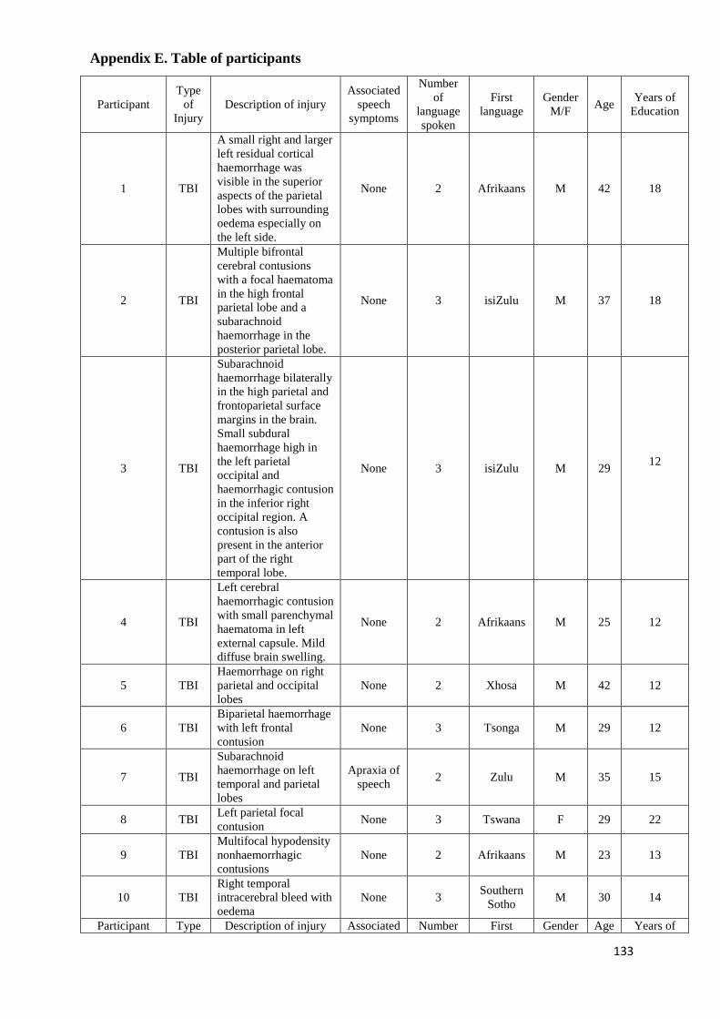
133
Participant
Type
of
Injury
Description of injury
Associated
speech
symptoms
Number
of
language
spoken
First
language
Gender
M/F Age
Years of
Education
1 TBI
A small right and larger
left residual cortical
haemorrhage was
visible in the superior
aspects of the parietal
lobes with surrounding
oedema especially on
the left side.
None 2 Afrikaans M 42 18
2 TBI
Multiple bifrontal
cerebral contusions
with a focal haematoma
in the high frontal
parietal lobe and a
subarachnoid
haemorrhage in the
posterior parietal lobe.
None 3 isiZulu M 37 18
3 TBI
Subarachnoid
haemorrhage bilaterally
in the high parietal and
frontoparietal surface
margins in the brain.
Small subdural
haemorrhage high in
the left parietal
occipital and
haemorrhagic contusion
in the inferior right
occipital region. A
contusion is also
present in the anterior
part of the right
temporal lobe.
None 3 isiZulu M 29 12
4 TBI
Left cerebral
haemorrhagic contusion
with small parenchymal
haematoma in left
external capsule. Mild
diffuse brain swelling.
None 2 Afrikaans M 25 12
5 TBI
Haemorrhage on right
parietal and occipital
lobes
None 2 Xhosa M 42 12
6 TBI
Biparietal haemorrhage
with left frontal
contusion
None 3 Tsonga M 29 12
7 TBI
Subarachnoid
haemorrhage on left
temporal and parietal
lobes
Apraxia of
speech 2 Zulu M 35 15
8 TBI Left parietal focal
contusion None 3 Tswana F 29 22
9 TBI
Multifocal hypodensity
nonhaemorrhagic
contusions
None 2 Afrikaans M 23 13
10 TBI
Right temporal
intracerebral bleed with
oedema
None 3 Southern
Sotho M 30 14
Participant Type Description of injury Associated Number First Gender Age Years of
Appendix E. Table of participants

134
of
Injury
speech
symptoms
of
language
spoken
language M/F Education
11 R
CVA
Right CVA based on
clinical signs. Unable
to observe on CT scan
None 5 Setswana F 58 20
12 R
CVA
Pre-contrast MRI no
signs of CVA but
presents with left
hemiplegia
None 3 Xhosa F 57 12
13 R
CVA
Non haemorrhagic
infarct in the right
MCA territory
None 3 Sepedi F 26 15
14 R
CVA R MCA infarct None 2 Afrikaans M 65 13
15 R
CVA
Large haematoma R
parietal region None 2 Afrikaans M 69 12
16 R
CVA
Subacute haemorrhage
in the right thalamus None 2 Afrikaans M 69 16
17 R
CVA
Right frontoparietal
infarct and pontine
medullary infarct
None 3 Sotho F 59 13
18 R
CVA
Right basal ganglia
infarct None 3 Tswana F 54 17
19 R
CVA R MCA infract None 2 Afrikaans M 60 19
20 L
CVA
Multiple cerebral
hyperintensities in the
left temporal, occipital
and periventricular
white matter
Dysarthria 3 Sepedi M 41
18
21 L
CVA
A left middle cerebral
artery infarct affecting
the left parietal region.
Apraxia of
speech 3 isiZulu F 27 14
22 L
CVA Left MCA infarct Dysarthria 3 Setswana M 51 12
23 L
CVA
L Acute subdural
haemorrhage on the
frontoparietal area
None 2 Zulu M 46 12
24 L
CVA Left MCA infarct
Apraxia of
speech 2 Xhosa M 45 15
25 L
CVA
Large left
haemorrhagic infarct
with oedema
None 2 Afrikaans F 65 12
26 L
CVA
L CVA of parieto-
occipital region Dysarthria 3 Zulu M 52 12
27 L
CVA
Deep white matter
ischaemia, white matter
demyelination and
progressive multifocal
leukoencephalopathy
and lunar infarcts in the
posterior limb of the
left internal capsule.
Dysarthria 2 isiZulu F 57 12
28 L
CVA
Left parieto-occipital
cerebral infarct
Apraxia of
speech 4 Tswana M 64 12
29 L
CVA L MCA Infarct
Apraxia of
speech 2 Afrikaans M 47 16
135
Appendix F
Participant information sheet for the control group.
This information sheet was read during the first session with the participant. The participant
kept the information sheet.
Introduction
Good day, my name is Nancy Barber. I am a Masters student at the Speech-Language
Department at Wits University. I am inviting you to consider participating voluntarily in my
research study. This document you are reading is an information sheet. I will explain the
purpose of the research and the methods of the research. You can ask questions throughout
the discussion and I will give you time at the end to also ask any questions. I will also give
you one week to think about the research and then I will contact you and reiterate the
important points of the research before you have to make a decision as to whether you want
to participate or not. If you agree to participate in the research, I will give you a form to sign.
I will make a copy of the form you sign for you to keep. If you do not want to participate in
the research, your job will not be affected in anyway. Your choice to be involved or not will
not be communicated to your employer.
Reason for the study
A lot of people in South Africa speak two or more languages. The fact that a person speaks
two or more languages may impact the way their brain works. Strokes and head injuries are
common in South Africa. When a person has a stroke or a head injury, not only do they have
difficulty talking and communicating, but they can also have problems with thinking. This
means that that people might have difficulty paying attention to things, remembering things,
organising and sequencing things as well as planning everyday activities. I am interested to
find out more about the impact that speaking two or more languages has on the thinking skills
of people who have had a head injury or stroke. The research needs a group of people who
have not had a head injury / stroke so that we can see if the tests we are using are appropriate
for South Africans. I am inviting you to be a volunteer in this research as you speak two or
more languages. If you agree to volunteer for this research study, you will be one of 20
people who will be asked to participate in the same manner.
What do I have to do?

136
This research will take place over about three months. First you will need to fill out a form
saying what languages you speak, when and how you learnt to speak them, what situations
you use them in and if you are able to read/write in them. This will take 15-30 minutes.
Secondly you will do some tests and then you will take the tests again at 12 weeks after the
first time we do the tests. The tests will take place over two days. Each day you will work
with me for about one hour in a quiet room at the rehabilitation hospital. I will do all the tests
with you and some will be timed. If it is difficult to undertsand the english explaining how to
do the tests, a trained research assistant will help translate the instructions into your home
language.
You will do two types of testing- (1) language tests and (2) thinking skills tests:
3. Language tests:
To see how you understand words and sentences
To see how you are able to name pictures and make sentences. During the
section of the language test where you have to describe a picture, your talking
will be recorded so that I can write it down exactly as you say it.
To see how you are able to read and write.
4. Thinking skills tests:
Look at disks and move them to match a picture
Name the colours that some words are printed in
Sort some cards.
Remember some pictures.
Look at letters and numbers
I need to also know your age and medical history to make sure you have not had any
injuries to your head. If you report to the researcher that you have had a head injury, this
will not be communicated to your employer and you will not participate in the research.
What happens to your test results?
Once I have completed the research, I will study the results and I will write them up as a
thesis. I will submit this thesis to Wits University for a Masters in Speech-Language Therapy.
I will also try to publish the results in a professional journal. I will give you your results and

137
the results of the study if you want to know them. I will not give your employer the results
unless you give consent.
What is the cost of this study?
There will be no cost involved in the study. Any travel costs to the rehabilitation hospital will
be compensated.
Confidentiality
Your identity will be protected in the research. Your name will be assigned a numeral code
and your name will not appear on any of the testing forms. The audio material will be
destroyed after it has been analysed. Your employer will not be informed of your
participation in the research. No information will be put in the thesis that will link you to the
research.
You have the right to:
6. Stop participating in the research at any time during the study. This withdrawal will
not affect you, your job or the research.
7. Have the results of your specific tests as well as the research project.
8. Contact me at any time to discuss any questions or concerns.
9. Contact my supervisor, Professor Penn, if you want to discuss any confidential
matters regarding the research
If you have any questions, you are welocme to contact me or my supervisor, Professor
Penn. You can also contact the chairperson of the University of the Witwatersrand
Human Research Ethics committee (HREC) (medical). Below are the details:
Nancy Barber Professor Penn (supervisor) Prof Cleaton Jones
0827400349 0721827801 (Chairperson HREC)
011 717 2301

138
Appendix G
Informed Consent Letter for the Control Group
I _____________________________, agree in writing to participate in this research.
In giving consent, I understand the following:
1. This research is for a Master’s Degree at Wits University.
2. My participation is voluntary and I have not been forced to participate.
3. My personal details will be confidential throughout the research. My name will not be
used during or after the study.
4. I will fill in a form telling details about the languages I can understand and speak.
5. I can stop participating in the research at any stage and this will not affect me, my job
or the study.
6. The research will run over about 2 months. The first set of testing will occur on a date
decided on by myself and the researcher. The second set of testing will be at 6 weeks
after my first assessment.
7. Each set of testing will take place over one day for one and a half hours.
8. I will be tested in a quiet room.
9. I will be asked some questions to check how I understand words and sentences.
10. I will be asked some questions to check how I say words and sentences.
11. I will read and write words and sentences.
12. I will also have to do some tasks that see how I am able to think. These tasks will
include saying numbers, saying colours, drawing lines, remembering pictures, moving
beads and connecting dots.
13. I agree that my verbal description of a picture can be recorded using a voice recorder.
14. My travel costs will be compensated.
15. My employer will not informed as to whether or not I am participating in the study.
16. The results for all the people who participate in the research will be written up as a
thesis for a degree. The results may also be published in a professional journal.
17. I can ask for my results and for the results of the study.
18. I can contact the researcher, Nancy Barber (0827400349) at any stage during the
research process.
139
19. I have understood the reason for the research and the tasks I have to be involved with
for the research. I recognise that Nancy Barber is the primary researcher and I agree to
voluntarily participate in this research project.
Name of Participant: _______________________________________
Date and Place: _______________________________________
Signature: _______________________________________
Name of Researcher: _______________________________________
Date and Place: _______________________________________
Signature: _______________________________________

140
Appendix H
Language Proficiency Questionnaire: Participant code: _______
Date of Birth: ____________________ Age: _______________ Male / Female
Number of years of schooling completed: ___________________
Please complete the following table starting with the first language as the language he/she
uses the most often.
To be completed by the researcher: (CVA ischaemic/ haemorrhagic) (TBI)
Date of injury: _________________
Admission to rehabilitation hospital: ___________________
Description of injury based on CT scans or MRI scans:
___________________________________________________________________________
___________________________________________________________________________
Language Age of
acquisition Environment
that the
language was
utilised
Manner of acquisition
(formal-via education
system or informal-via
family and friends)
Tick if the person was able to do the
following in the specified language Understand Speak Read Write
1.
2.
3.
4.
5.

141
Appendix I
Paragraphs for the Comprehension of Spoken Language subtest
The original word from the CAT is in parenthesis next to the culturally appropriate word
which is in italics.
Paragraph 1:
Sally and Richard had been on the train for over three hours. They were tired and fed up. The
train was already 45 minutes late, the tuckshop (buffet) had closed so there was no food and
the lady opposite was snoring.
No changes were made to the questions for paragraph 1.
Paragraph 2:
The explosion is Johannesburg (central London) caused havoc. Initially terrorists were
suspected but it turned out not to be a bomb. The cause was found to be a burst gas main that
ignited when someone had thrown down a lighted cigarette. People three kilometres (miles)
away heard the explosion and the damage is estimated at over a million rands (pounds).
Changes made to the questions included:
a. Was the explosion in Durban (Leicester)?
b. Was it caused by a bomb?
a. Was it in Johannesburg (London)?
b. Was the explosion caused by a gas main?

142
Appendix J
Detailed description of EF assessment battery
Number-letter task. This task was adapted from Rogers and Monsell (1995) as well as
Miyake et al (2000). A number-letter pair was presented in one of the four quadrants on the
iPad screen and the participant was required to respond by either pressing the yes or no
button. The number-letter pairs consisted of one of the following numbers: 2, 4, 6, 8, 3, 5, 7,
9 and one of the following letters: G, K, M, R, A, E, I, U. If the number-letter pair was
presented in the top two quadrants the participant was required to respond to the question “Is
the number even?”. If the number-letter pair was presented in the bottom two quadrants the
patient was required to respond to the question “Is the letter a vowel?”
The first session comprised 32 trials which were presented in the top two quadrants and then
32 trials which were presented in the bottom two quadrants. These trials required no task
shifting. The second session comprised letter-number pairs which were rotated clockwise
around all four blocks for 64 trials. Shifting was required by participants in these last 64
trials. Response times were recorded. The shift cost was the dependent measure for this task
and it was calculated using the difference in average response time for trials during the
second session where switching was required and the average response times of trials from
the first session where no switching was required.
Wisconsin Card Sorting Test (WCST). A computerised version of the WCST (Grant &
Berg, 1948) developed by Mueller (2012) was utilised on the laptop. The WCST was used as
the complex EF assessment that assessed shifting (Miyake et al., 2000). The WCST requires
sustained attention, set maintenance, concept formation, working memory, problem solving
and set switching (Jodzio & Biechowska, 2010). Despite the complexity of the WCST,
analysis by Miyake et al (2000) revealed that shifting skills contribute significantly to
performance on the WCST. The WCST has been found to be of high usefulness in patients
with aphasia however the impact of comprehension of instructions needs to be considered
when interpreting results (Keil & Kaszniak, 2002).
The WCST required the participants to match a series of target cards that were presented
individually to one of four reference cards that were positioned near the top of the screen.
Participants were instructed to sort the target card according to one of its attributes- colour
(red, yellow, blue, green), shape (triangle, cross, circle, star) or number (1, 2, 3, 4) and thus
determine the computer generated rule which governed categorisation. Participants were also

143
instructed that the computer generated rule would change after a certain number of
presentations and that the participant was then required to determine the new categorisation
rule. The computer would alter the rule once the patient had performed eight consecutive
sorts correctly. Visual feedback was provided with regards to whether their response was
correct or incorrect. There was no time limit on a participant’s response time.
“Perseverative error” was the domain utilised as the main dependent measure because this
indicates the number of times a participant failed to change sorting principles when a
category changed and the participant continued to sort according to previous sorting
principle.
The following tasks were employed to assess inhibition of prepotent responses:
Victoria Stroop test. A computerised version of the assessment as developed by Mueller
(2012) was utilised on the laptop. The Victoria Stroop test (VicStroop) can be analysed
independently of cognitive speed by using the error score and the interference ratio which
does not require time measures and therefore corrects for the slowed processing speed and
allows one to examine inhibition (Strauss, Sherman & Spreen, 2006). The VicStroop is also
brief and has reduced administration time. It may also be more preferable for identifying
response inhibition difficulties due to the fact that the participant does not get extended
practice on the task (Strauss, Sherman & Spreen, 2006).
On each screen of the VicStroop, a grey block highlighted the dot or word the participant
needed to respond to. Participants were required to push a number button (1, 2, 3, 4) in
response to a specific colour (red, green, blue, yellow). Initially this was practised. When the
test was commenced, the first screen, Part D, provided dots in the different colours (red,
green, blue, yellow) and participants were instructed to respond by naming the dots as
quickly as possible by pushing the corresponding button for the colour of the dot. The second
screen, Part W, provided random words in the different colours and participants were again
instructed to respond by naming the colour of the word as quickly as possible by pushing the
corresponding number button. The third screen, Part C, provided the names of the colours
typed in different colour ink. Participants were again instructed to respond to the ink colour
of the word and not to read the word.
The errors and time for each section were recorded. The dependent measure was time of Part
C/Part D to determine the ratio index of interference (Strauss, Sherman & Spreen, 2006).

144
Tower of Hanoi. The Tower of Hanoi (ToH) was selected as the complex EF assessment
to assess inhibition as Miyake et al (2000) determined that inhibition plays an important role
in performance on the ToH. Novel planning, problem solving and rule adherence are skills
also assessed by ToH (Glosser & Goodglass, 1990). Short term memory deficits and goal-
subgoal conflict resolution deficits may also be identified by this assessment measure (Goel
& Grafman, 1995). The ToH has been identified to have high usefulness with patients with
aphasia as there is limited language load (Keil & Kaszniak, 2002). In addition, the time
element does not need to be used when employing the ToH in patients with a CVA/TBI as
they may be slower due to general brain damage (Keil & Kaszniak, 2002).
Participants performed a computerised version of the Tower of Hanoi (Mueller, 2012) on the
laptop. In this task, participants were presented with two sets of three blocks with three disks
of consecutive sizes inside the blocks. The goal state of the disks was presented at the top of
the screen and the initial state of the disks was presented at the bottom of the screen. It was
explained to participants that they were required to manipulate the bottom set of disks in
order to arrange them to be identical to the top set of disks. The rules were explained using
simple language with reduced length of sentences to support comprehension. The rules
included: (1) only one disk can be moved at a time, (2) only a smaller disk can be placed on a
larger disk, (3) disks must be placed in a box and cannot be left in mid-air. There was no time
limit. The dependent variable was the total number of moves used to reach the goal state.
The following test was used to assess updating:
N-back test. During N-back testing the patient is required to monitor stimulus input and
update information in working memory in a flexible manner in order to produce an
appropriate response (Elliot, 2003). A computerised version of the N-back task adapted from
Quinette et al (2004) was employed on the iPad with N being 2. The participant was required
to touch the iPad screen when the picture was the same as two pictures prior. Participants had
two trials sets and then ten sets of pictures were employed. Other updating tasks could not be
employed as the speech and language demands placed on the participant could confound the
results. The dependent measure for this test was the accuracy rate.

145
M
6
M
12
CS
6
CS
12
CW
6
CW
12
Rep
6
Rep
12
N
6
N
12
R
6
R
12
W
6
W
12
SPD
6
SPD
12
WPD
6
WPD
12
LB
6
LB
12
GO
6
GO
12
A
6
A
12
WF
6
WF
12
C1 62 62 67 74 66 68 72 72 75 74 71 71 64 64 64 65 73 72 66 66 68 68 57 57 74 72
C2 62 62 53 59 51 52 62 66 62 63 60 63 58 57 56 60 61 65 53 53 68 68 57 57 64 64
C3 62 62 63 74 73 73 72 72 75 75 71 71 69 69 60 74 75 75 66 66 68 68 65 65 75 75
C4 54 54 65 63 68 63 66 66 68 69 71 71 58 59 60 62 70 71 66 66 68 68 57 57 71 71
C5 62 62 63 74 73 73 72 72 75 74 65 66 64 64 64 70 73 75 53 53 68 68 57 57 75 75
C6 50 50 47 46 54 53 62 62 55 58 62 62 57 58 59 60 66 68 66 66 68 68 57 57 60 69
C7 62 62 55 63 65 68 66 66 75 69 71 65 60 60 60 60 66 68 66 66 68 68 65 65 75 75
C8 62 62 62 67 68 73 62 62 72 69 67 71 65 65 58 61 71 74 53 53 68 68 65 65 72 70
C9 62 62 61 60 73 70 62 66 66 70 71 71 69 69 62 67 72 74 66 66 68 68 65 65 63 70
C10 62 62 65 60 63 70 66 72 71 68 71 71 59 60 61 66 72 75 66 66 68 68 65 65 70 67
C11 62 62 66 66 55 55 60 60 61 57 60 60 55 55 61 61 56 66 53 53 68 68 57 57 67 67
C12 62 62 63 63 65 65 66 62 64 65 66 71 58 64 61 62 72 73 66 66 68 68 65 65 68 67
C13 62 62 63 63 63 68 62 72 64 69 71 71 60 61 61 62 65 71 53 53 68 68 65 65 68 71
C14 54 54 59 63 67 68 72 72 74 72 60 57 59 61 60 67 70 75 53 53 68 68 65 65 74 71
C15 62 62 63 65 66 70 66 66 71 71 71 71 65 65 62 67 72 70 44 44 68 68 65 65 71 71
C16 54 54 52 57 56 61 72 72 69 68 71 71 69 69 63 62 72 73 53 53 68 68 65 65 71 67
C17 62 62 60 65 73 63 62 66 74 74 66 67 61 61 60 60 72 70 53 53 68 68 57 65 74 72
C18 62 62 74 74 68 68 66 66 74 74 71 71 65 65 62 61 69 70 66 66 68 68 65 65 75 75
C19 46 47 61 60 65 65 62 66 73 73 66 67 60 60 60 60 70 70 66 66 68 68 65 65 74 74
Appendix K.
Table detailing the CAT subtest scores for the control group at initial and follow up assessment
Key for subtests
6= Initial assessment Grey shaded blocks= within normal limits
12= Re assessment 6 weeks later
M= Memory subtest R= Reading LB= Line Bisection
CS= Comprehension of Spoken Language W= Writing
CW= Comprehension of Written Language SPD= Spoken Picture Description
Rep= Repetition GO= Gesture Object Use
N= Naming A= Arithmetic
WPD= Written Picture Description WF= Word Fluency

146
N-Back
6
N-Back
12
VicStroop
6
VicStroop
12
WCST
6
WCST
12
Num-let
6
Num-let
12
ToH
6
ToH
6
ToH
12
ToH
12
Scaled
score
Scaled
score
Standard
score
Standard
score mS mS
required num
of moves
Pt num of
moves
required num of
moves
Pt num of
moves
C1 0.94 0.91 16 16 87 98 387.81 537.65 35 40 38 44
C2 0.8 0.87 11 11 55 87 -61.71 233.28 34 45 34 78
C3 0.96 0.9 15 15 103 100 349.37 36.71 42 82 44 61
C4 0.95 0.8 16 16 92 96 -29.53 428.9 27 39 35 53
C5 1 0.92 16 16 103 103 456.25 375.46 38 39 38 45
C6 0.7 0.8 16 13 85 92 566.25 661.87 32 34 41 64
C7 0.91 0.91 16 16 93 94 370.16 132.56 46 121 35 54
C8 1 0.92 14 17 103 100 156.25 245.93 35 47 48 89
C9 0.98 0.93 17 17 86 79 378.12 545.15 42 132 44 66
C10 0.81 0.76 17 17 103 89 445.31 684.37 32 34 34 42
C11 0.69 0.77 11 17 57 95 49.21 177.65 43 91 37 42
C12 0.76 0.7 17 17 89 87 763.59 78.43 35 46 43 78
C13 0.8 0.64 17 17 80 98 485.93 209.21 27 39 34 107
C14 0.86 0.57 15 16 65 89 853.43 620 28 105 37 90
C15 0.98 0.91 15 13 96 98 282.96 -13.28 30 46 41 48
C16 0.9 0.53 18 18 76 89 364.84 664.22 36 66 34 79
C17 0.71 0.79 17 15 93 96 350.46 51.09 41 58 45 73
C18 0.84 0.88 17 17 105 105 236.74 220.51 46 52 38 44
C19 0.77 0.75 16 17 90 93 583.16 575.81 37 50 34 47
Appendix K continued.
Table detailing the EF subtest scores for the control group at initial and follow up assessment
Key for subtests
6= Initial assessment Grey shaded blocks= within normal limits
12= Re assessment 6 weeks later
N-Back (updating) WCST= Wisconsin Card Sorting Test (shifting)
VicStroop= Victoria Stroop (inhibition) ToH= Tower of Hanoi (inhibition)
Num-Let= Number-letter task (shifting) mS= milliseconds

147
Participant
M
6
M
12
CS
6
CS
12
CW
6
CW
12
Rep
6
Rep
12
N
6
N
12
R
6
R
12
W
6
W
12
SPD
6
SPD
12
WPD
6
WPD
12
LB
6
LB
12
GO
6
GO
12
A
6
A
12
WF
6
WF
12
20 43 47 50 52 49 53 66 62 60 64 53 59 57 58 49 51 55 56 53 39 60 68 53 57 63 62
21 54 54 43 52 47 52 41 54 51 56 44 54 53 54 47 50 54 54 66 66 68 68 57 57 51 54
22 45 47 36 48 39 52 54 66 49 58 66 71 52 58 39 51 42 56 53 44 47 68 57 57 54 67
23 33 36 33 39 32 34 50 57 42 45 42 44 44 46 47 48 42 42 26 44 69 60 40 57 37 37
24 28 31 33 33 31 25 44 46 35 35 38 38 34 34 39 39 42 42 25 25 37 38 44 44 37 37
25 30 54 32 48 32 50 44 58 43 52 38 65 34 61 39 55 42 58 39 66 31 55 34 65 37 54
26 41 54 41 38 40 38 59 58 51 51 45 47 34 41 47 52 42 42 44 44 40 42 44 40 48 47
27 36 38 43 51 45 49 54 53 51 55 56 66 50 54 50 52 55 59 44 44 51 51 49 53 57 64
28 31 28 29 33 34 25 34 38 44 43 38 40 41 41 39 39 42 42 25 44 43 60 44 44 45 47
29 26 27 25 25 25 25 34 34 35 35 38 38 34 40 39 39 42 42 44 53 31 35 34 49 37 37
Appendix L.
Table detailing the CAT subtest scores for the participants with a left CVA at the 6 week and 12 week assessment
Key for subtests
6= Assessment at week 6 Grey shaded blocks= within normal limits
12= Assessment at week 12
M= Memory subtest LB= Line Bisection
CS= Comprehension of Spoken Language GO= Gesture Object Use
CW= Comprehension of Written Language A= Arithmetic
Rep= Repetition WF= Word Fluency
N= Naming
R= Reading
W= Writing
SPD= Spoken Picture Description
WPD= Written Picture Description
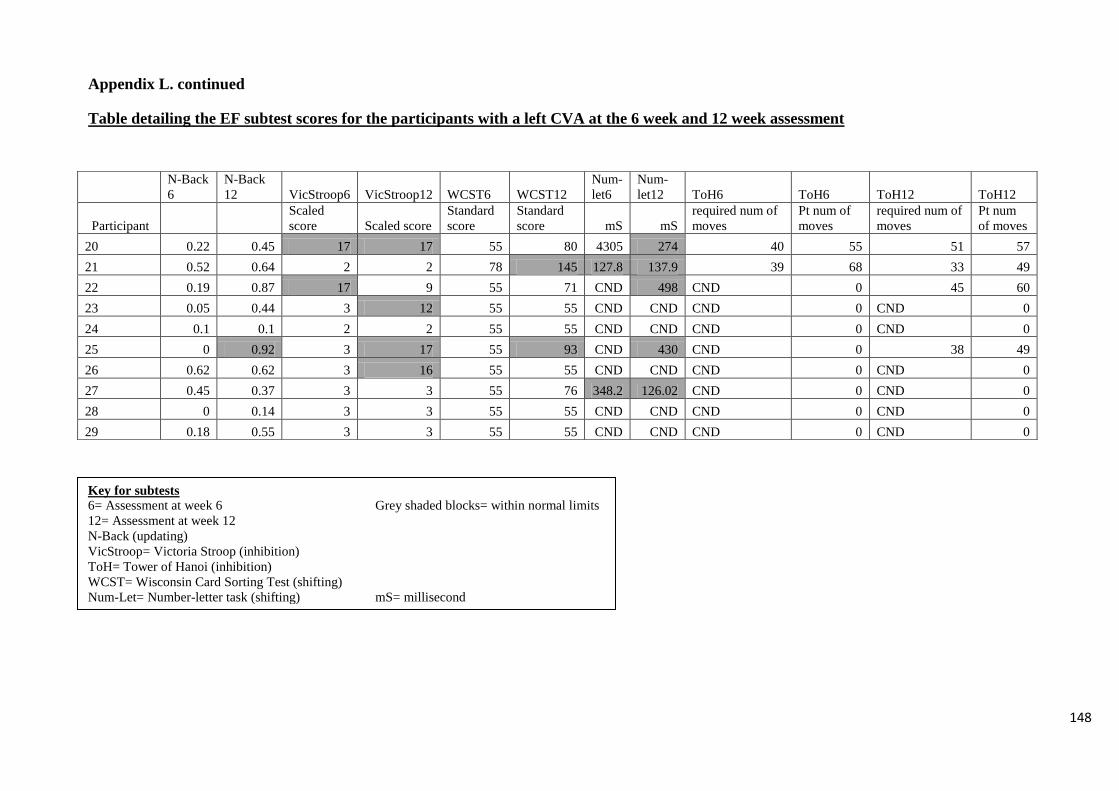
148
N-Back
6
N-Back
12 VicStroop6 VicStroop12 WCST6 WCST12
Num-
let6
Num-
let12 ToH6 ToH6 ToH12 ToH12
Participant
Scaled
score Scaled score
Standard
score
Standard
score mS mS
required num of
moves
Pt num of
moves
required num of
moves
Pt num
of moves
20 0.22 0.45 17 17 55 80 4305 274 40 55 51 57
21 0.52 0.64 2 2 78 145 127.8 137.9 39 68 33 49
22 0.19 0.87 17 9 55 71 CND 498 CND 0 45 60
23 0.05 0.44 3 12 55 55 CND CND CND 0 CND 0
24 0.1 0.1 2 2 55 55 CND CND CND 0 CND 0
25 0 0.92 3 17 55 93 CND 430 CND 0 38 49
26 0.62 0.62 3 16 55 55 CND CND CND 0 CND 0
27 0.45 0.37 3 3 55 76 348.2 126.02 CND 0 CND 0
28 0 0.14 3 3 55 55 CND CND CND 0 CND 0
29 0.18 0.55 3 3 55 55 CND CND CND 0 CND 0
Appendix L. continued
Table detailing the EF subtest scores for the participants with a left CVA at the 6 week and 12 week assessment
Key for subtests
6= Assessment at week 6 Grey shaded blocks= within normal limits
12= Assessment at week 12
N-Back (updating)
VicStroop= Victoria Stroop (inhibition)
ToH= Tower of Hanoi (inhibition)
WCST= Wisconsin Card Sorting Test (shifting)
Num-Let= Number-letter task (shifting) mS= millisecond

149
Participant
M
6
M
12
CS
6
CS
12
CW
6
CW
12
Rep
6
Rep
12
N
6
N
12
R
6
R
12
W
6
W
12
SPD
6
SPD
12
WPD
6
WPD
12
LB
6
LB
12
GO
6
GO
12
A
6
A
12
WF
6
WF
12
11 45 62 53 62 62 66 62 66 72 73 64 71 60 58 59 56 72 72 53 66 68 68 57 53 74 69
12 39 41 46 50 34 54 66 60 55 60 63 66 58 55 53 53 55 60 53 53 68 68 53 65 55 62
13 54 54 52 47 50 53 68 72 58 59 63 66 55 57 51 54 60 61 53 44 68 68 53 53 60 62
14 62 62 67 62 68 66 72 66 74 74 67 71 69 69 61 60 62 70 66 66 68 68 65 65 74 74
15 54 62 60 61 56 65 62 62 66 69 66 62 64 64 54 55 61 66 44 53 68 68 49 57 67 68
16 62 54 60 65 66 68 60 72 67 70 71 71 69 69 61 64 66 73 66 66 68 68 65 65 62 69
17 41 47 41 45 37 41 66 66 56 63 51 49 64 59 49 55 61 65 25 39 68 68 65 65 66 67
18 54 54 47 53 52 60 60 60 61 61 65 62 57 61 55 56 57 60 53 66 68 68 65 65 66 68
19 54 62 52 61 61 63 72 72 62 73 71 71 64 69 52 57 61 61 66 66 68 68 57 57 69 75
Key for subtests
6= Assessment at week 6 Grey shaded blocks= within normal limits
12= Assessment at week 12
M= Memory subtest LB= Line Bisection
CS= Comprehension of Spoken Language GO= Gesture Object Use
CW= Comprehension of Written Language A= Arithmetic
Rep= Repetition WF= Word Fluency
N= Naming
R= Reading
W= Writing
SPD= Spoken Picture Description
WPD= Written Picture Description
Appendix M.
Table detailing the CAT subtest scores for the participants with a right CVA at the 6 week and 12 week assessment
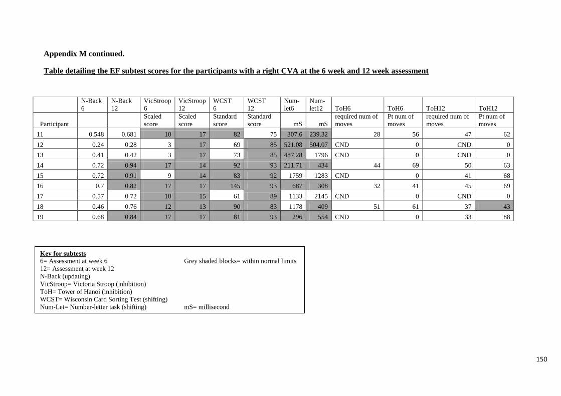
150
N-Back
6
N-Back
12
VicStroop
6
VicStroop
12
WCST
6
WCST
12
Num-
let6
Num-
let12 ToH6 ToH6 ToH12 ToH12
Participant
Scaled
score
Scaled
score
Standard
score
Standard
score mS mS
required num of
moves
Pt num of
moves
required num of
moves
Pt num of
moves
11 0.548 0.681 10 17 82 75 307.6 239.32 28 56 47 62
12 0.24 0.28 3 17 69 85 521.08 504.07 CND 0 CND 0
13 0.41 0.42 3 17 73 85 487.28 1796 CND 0 CND 0
14 0.72 0.94 17 14 92 93 211.71 434 44 69 50 63
15 0.72 0.91 9 14 83 92 1759 1283 CND 0 41 68
16 0.7 0.82 17 17 145 93 687 308 32 41 45 69
17 0.57 0.72 10 15 61 89 1133 2145 CND 0 CND 0
18 0.46 0.76 12 13 90 83 1178 409 51 61 37 43
19 0.68 0.84 17 17 81 93 296 554 CND 0 33 88
Appendix M continued.
Table detailing the EF subtest scores for the participants with a right CVA at the 6 week and 12 week assessment
Key for subtests
6= Assessment at week 6 Grey shaded blocks= within normal limits
12= Assessment at week 12
N-Back (updating)
VicStroop= Victoria Stroop (inhibition)
ToH= Tower of Hanoi (inhibition)
WCST= Wisconsin Card Sorting Test (shifting)
Num-Let= Number-letter task (shifting) mS= millisecond

151
Participant
M
6
M
12
CS
6
CS
12
CW
6
CW
12
Rep
6
Rep
12
N
6
N
12
R
6
R
12
W
6
W
12
SPD
6
SPD
12
WPD
6
WPD
12
LB
6
LB
12
GO
6
GO
12
A
6
A
12
WF
6
WF
12
1 39 62 46 62 50 65 59 60 50 65 55 67 48 73 49 61 55 73 48 66 40 68 49 65 48 67
2 41 47 50 52 48 53 62 66 54 62 66 71 57 58 52 56 54 55 66 66 55 55 49 53 51 62
3 34 45 40 53 40 52 72 72 48 68 47 61 55 57 48 52 55 66 26 39 60 68 40 65 52 69
4 50 62 52 56 63 65 66 66 57 66 62 64 61 61 49 56 61 63 53 66 68 68 57 65 54 68
5 34 38 45 38 43 44 54 61 53 56 52 60 51 59 52 49 60 59 66 66 55 68 49 40 56 58
6 45 50 35 44 44 49 55 60 49 52 56 58 53 52 48 50 57 57 33 53 68 68 49 53 50 56
7 31 39 25 31 34 39 44 48 42 44 38 38 42 42 38 38 42 42 39 39 45 60 34 44 37 45
8 54 54 56 67 70 73 66 72 59 65 66 67 69 58 55 62 68 72 44 66 68 68 57 65 60 66
9 38 47 46 53 48 60 54 62 57 63 59 63 52 59 53 53 54 64 53 53 45 60 44 53 57 69
10 62 62 60 74 61 66 62 66 69 71 71 71 64 64 49 56 67 72 66 66 68 68 57 65 72 71
Key for subtests
6= Assessment at week 6 Grey shaded blocks= within normal limits
12= Assessment at week 12
M= Memory subtest LB= Line Bisection
CS= Comprehension of Spoken Language GO= Gesture Object Use
CW= Comprehension of Written Language A= Arithmetic
Rep= Repetition WF= Word Fluency
N= Naming
R= Reading
W= Writing
SPD= Spoken Picture Description
WPD= Written Picture Description
Appendix N.
Table detailing the CAT subtest scores for the participants with a TBI at the 6 week and 12 week assessment

152
N-
Back6
N-Back
12
VicStroop
6
VicStroop
12 WCST6 WCST12
Num-
let6
Num-
let12 ToH6 ToH6 ToH12 ToH12
Participant
Scaled
score Scaled score
Standard
score
Standard
score mS mS
required num
of moves
Pt num of
moves
required num
of moves
Pt num of
moves
1 0.264 0.54 10 16 62 96 301.14 93.2 CND 0 28 31
2 0.531 0.735 8 16 55 143 444.4 228.8 CND 0 44 84
3 0.154 0.881 2 16 55 81 313.06 156.48 CND 0 40 64
4 0.94 0.8 16 16 90 94 550.03 490.32 43 66 44 69
5 0.22 0.25 16 16 66 81 CND 1812 CND 0 CND 0
6 0.24 0.19 10 11 78 78 2944.37 2480.62 CND 0 CND 0
7 0.74 0.66 3 16 55 77 CND CND CND 0 CND 0
8 0.9 0.1 13 15 98 103 517.81 401.66 37 61 38 66
9 0.36 0.16 3 10 80 88 570.4 -134.35 CND 0 42 93
10 0.94 0.84 10 16 96 98 -134.06 292.67 38 43 42 55
Appendix N continued
Table detailing the EF subtest scores for the participants with a TBI at the 6 week and 12 week assessment
Key for subtests
6= Assessment at week 6 Grey shaded blocks= within normal limits
12= Assessment at week 12
N-Back (updating)
VicStroop= Victoria Stroop (inhibition)
ToH= Tower of Hanoi (inhibition)
WCST= Wisconsin Card Sorting Test (shifting)
Num-Let= Number-letter task (shifting) mS= millisecond


