DZHK-SOP-K-02 Anamnesis/Clinical Diagnoses · Anamnesis/Clinical Diagnoses DZHK-SOP-K-02 Valid ......
49
Anamnesis/Clinical Diagnoses DZHK-SOP-K-02 Valid as of: 01.09.2014 Version: V1.0 Author: R. Wachter Page 1 of 49 *In this eCRF, items marked with two asterisks (**) must be completed (basic data set). Items marked with one asterisk (*) should be completed to the greatest extent possible. DZHK-SOP-K-02 Anamnesis/Clinical Diagnoses Expert Author Expert Review Endorsed by Section Head Approved by DZHK Name Rolf Wachter (Göttingen)* Sebastian Kufner (Munich) Matthias Nauck Thomas Eschenhagen Date 26/08/2014 26/08/2014 26/08/2014 26/08/2014 Signature *Adapted from the SOPs of the Competence Network Heart Failure Version: V1.0 Valid as of: 01.09.2014 Replaces version: - dated: - Modification notice: -
Transcript of DZHK-SOP-K-02 Anamnesis/Clinical Diagnoses · Anamnesis/Clinical Diagnoses DZHK-SOP-K-02 Valid ......
Anamnesis/Clinical Diagnoses
DZHK-SOP-K-02 Valid as of: 01.09.2014
Version: V1.0 Author: R. Wachter Page 1 of 49
*In this eCRF, items marked with two asterisks (**) must be completed (basic data set). Items marked with one asterisk (*) should be completed to the greatest extent possible.
DZHK-SOP-K-02
Anamnesis/Clinical Diagnoses
Expert Author Expert Review Endorsed by
Section Head
Approved by
DZHK
Name Rolf Wachter
(Göttingen)*
Sebastian Kufner (Munich)
Matthias Nauck Thomas
Eschenhagen
Date 26/08/2014 26/08/2014 26/08/2014 26/08/2014
Signature
*Adapted from the SOPs of the Competence Network Heart Failure
Version: V1.0 Valid as of: 01.09.2014
Replaces version: - dated: -
Modification notice: -
DZHK-SOP-K-02 Valid as of: 01.09.2014
Version: V1.0 Author: R. Wachter Page 2 of 49
*In this eCRF, items marked with two asterisks (**) must be completed (basic data set). Items marked with one asterisk (*) should be completed to the greatest extent possible.
CONTENTS 1 Introduction ..................................................................................................................................... 3
1.1 List of abbreviations ................................................................................................................ 3
1.2 Purpose .................................................................................................................................... 4
1.3 Target Group ........................................................................................................................... 4
Inclusion Criteria .............................................................................................................. 4
Exclusion Criteria ............................................................................................................. 4
1.4 Application and Tasks .............................................................................................................. 4
1.5 Terms, Definitions and Explanations for the eCRF Module .................................................... 4
1.6 Correlations to Other Examinations ...................................................................................... 14
1.7 Level of Quality ...................................................................................................................... 14
2 Examination Conditions................................................................................................................. 15
2.1 Requirements for Rooms/Equipment ................................................................................... 15
2.2 Equipment/Hardware ............................................................................................................ 15
2.3 Special Clinical Consumables ................................................................................................. 15
2.4 Documents Required ............................................................................................................. 15
2.5 Information Required ............................................................................................................ 15
2.6 Personnel ............................................................................................................................... 16
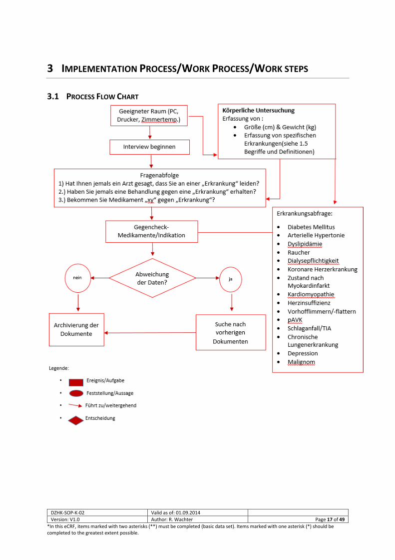
3 Implementation Process/Work Process/Work steps .................................................................... 17
3.1 Process Flow Chart ................................................................................................................ 17
3.2 Preparing for the Examination .............................................................................................. 18
Preparing the Work Space ............................................................................................. 18
Preparing the Equipment .............................................................................................. 18
Principles for Preparing the Proband for the Examination ........................................... 18
3.3 Carrying out the Examination ................................................................................................ 18
3.4 Post-Processing and Registering the Data ............................................................................. 19
3.5 Dealing with Deviations ......................................................................................................... 19
4 Literature and References ............................................................................................................. 20
5 Modifications ................................................................................................................................. 20
6 List of Contributors ........................................................................................................................ 20
DZHK-SOP-K-02 Valid as of: 01.09.2014
Version: V1.0 Author: R. Wachter Page 3 of 49
*In this eCRF, items marked with two asterisks (**) must be completed (basic data set). Items marked with one asterisk (*) should be completed to the greatest extent possible.
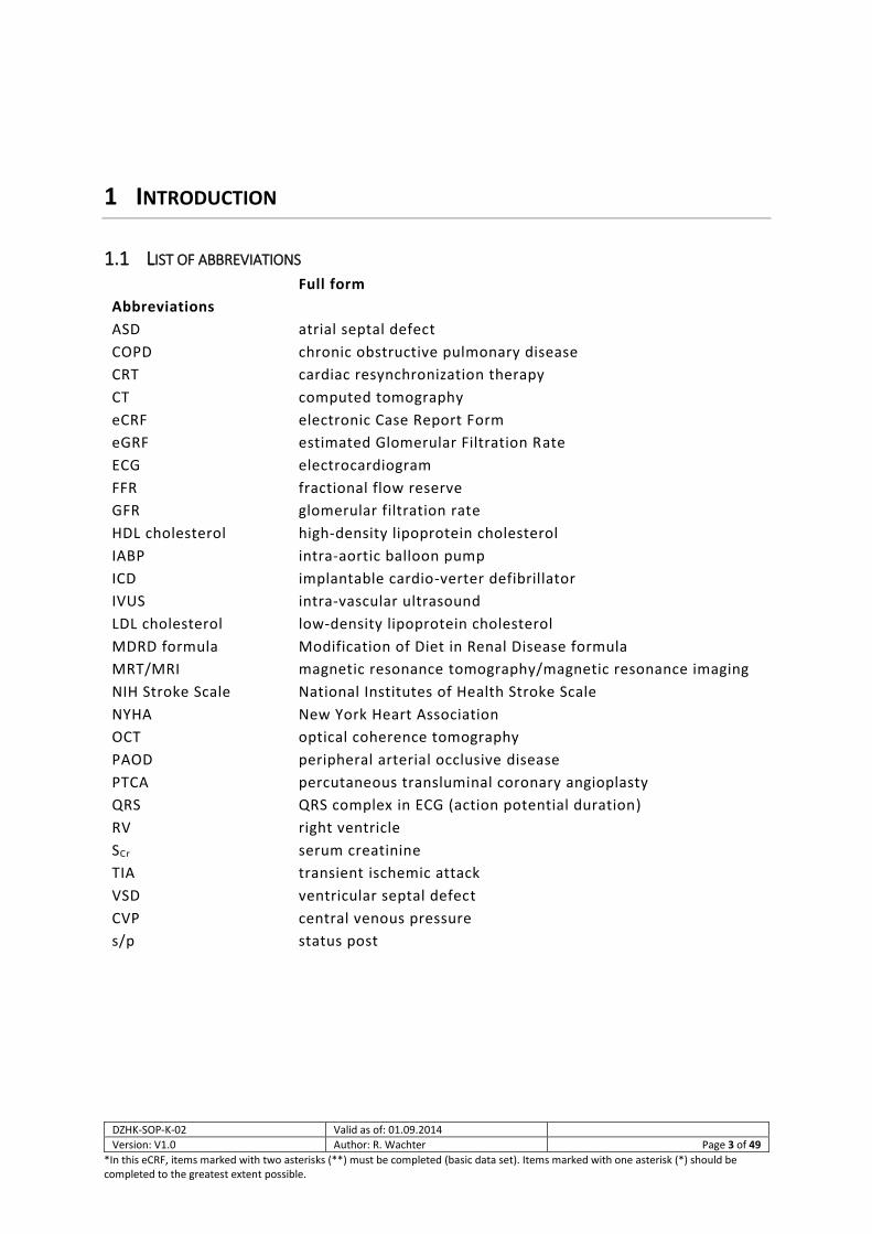
1 INTRODUCTION
1.1 LIST OF ABBREVIATIONS
Abbreviations
Full form
ASD atrial septal defect
COPD chronic obstructive pulmonary disease
CRT cardiac resynchronization therapy
CT computed tomography
eCRF electronic Case Report Form
eGRF estimated Glomerular Filtration Rate
ECG electrocardiogram
FFR fractional flow reserve
GFR glomerular filtration rate
HDL cholesterol high-density lipoprotein cholesterol
IABP intra-aortic balloon pump
ICD implantable cardio-verter defibrillator
IVUS intra-vascular ultrasound
LDL cholesterol low-density lipoprotein cholesterol
MDRD formula Modification of Diet in Renal Disease formula
MRT/MRI magnetic resonance tomography/magnetic resonance imaging
NIH Stroke Scale National Institutes of Health Stroke Scale
NYHA New York Heart Association
OCT optical coherence tomography
PAOD peripheral arterial occlusive disease
PTCA percutaneous transluminal coronary angioplasty
QRS QRS complex in ECG (action potential duration)
RV right ventricle
SCr serum creatinine
TIA transient ischemic attack
VSD ventricular septal defect
CVP central venous pressure
s/p status post
DZHK-SOP-K-02 Valid as of: 01.09.2014
Version: V1.0 Author: R. Wachter Page 4 of 49
*In this eCRF, items marked with two asterisks (**) must be completed (basic data set). Items marked with one asterisk (*) should be completed to the greatest extent possible.
1.2 PURPOSE Uniform definitions are proposed in the context of this SOP when a corresponding risk
factor/clinical diagnosis is considered to be present.
1.3 TARGET GROUP These SOPs are targeted at individuals responsible for entering data into the basic data
module “Anamnesis“. These may be e.g. doctors or study nurses.
Inclusion Criteria
Included are all patients who meet the respective inclusion/exclusion criteria of the
respective study.
Exclusion Criteria
None. If information cannot be collected in full, it should be collected to the greatest extent
possible.
1.4 APPLICATION AND TASKS The purpose of the anamnesis/clinical diagnoses is to accurately record known cardiovascular risk
factors. The anamnesis is a core element of medical diagnostics. The evidence collected during the
anamnesis enables a detailed assessment of a patient’s cardiovascular risk.
Collection of the anamnesis/clinical diagnoses is an integral part of all observational and clinical studies
of the DZHK.
1.5 TERMS, DEFINITIONS AND EXPLANATIONS FOR THE ECRF MODULE Date of examination
is defined as the date on which the examination takes place.
Sex and date of birth
are defined as the data which appear on the person’s identity card.
Height and weight
Height is measured in the standing position, without shoes and without head covering. Weight
is measured in normal street clothing, without a jacket and without shoes. Preferentially,
measured data should be collected; only when this is not possible (e.g. in the case of bed-
ridden patients) should one estimate the values or resort to information provided by the
proband.
DZHK-SOP-K-02 Valid as of: 01.09.2014
Version: V1.0 Author: R. Wachter Page 5 of 49
*In this eCRF, items marked with two asterisks (**) must be completed (basic data set). Items marked with one asterisk (*) should be completed to the greatest extent possible.
Ethnicity and skin colour
A person’s ethnic origin is defined by their ancestry in relation to a specific ethnic group. This
can be determined biologically and/or geographically on the basis of membership of a certain
settlement group. Accordingly, a person’s skin colour can also be broadly defined. The colour
spectrum can be differentiated from light to dark skin colour.
Family history of myocardial infarction or stroke
is defined as a medically diagnosed myocardial infarction or stroke in one or both biological
parents, biological siblings (including half-siblings) or biological children, provided the female
relative was under age 65, or the male relative under age 55 (when the myocardial
infarction/stroke occurred).
Diabetes mellitus
is defined as diabetes which has been diagnosed and/or treated by a doctor.
o The American Diabetes Association criteria are:
haemoglobin A1c ≥ 6.5 % or a fasting blood glucose level of ≥ 126 mg/dl or a
2-hour blood glucose level of ≥ 200 mg/dl in the oral glucose tolerance test.
Arterial hypertension
is defined as a current or previous diagnosis of arterial hypertension which was diagnosed
and/or is being treated by a doctor. Treatment can consist of e.g. dietary changes, physical
activity and/or medication. Systolic blood pressure values ≥ 140 mmHg and/or diastolic blood
pressure values ≥ 90mmHg measured by a doctor on at least two separate days after a 5-
minute resting phase qualify for a diagnosis of arterial hypertension.
Dyslipidaemia
is defined as a current or previous diagnosis of dyslipidaemia which was diagnosed and/or is
being treated by a doctor.
One or more of the following criteria:
total cholesterol ≥ 200 mg/dl,
LDL cholesterol ≥ 130 mg/dl,
HDL cholesterol < 40 mg/dl (men) and < 50 mg/dl (women).
Smoker
is defined as current or previous use of cigarettes, cigars, pipes or smokeless tobacco.
“Yes” for daily or occasional smoking (≥ 1x/month);
“Ex-smoker“ for abstinence of more than 6 months; ex-smoker since …;
“No“ for “never smoked“.
Pack years
is the product of the number of years of cigarette smoking multiplied by the average number
of packs smoked per day.
DZHK-SOP-K-02 Valid as of: 01.09.2014
Version: V1.0 Author: R. Wachter Page 6 of 49
*In this eCRF, items marked with two asterisks (**) must be completed (basic data set). Items marked with one asterisk (*) should be completed to the greatest extent possible.
Example: A patient who has smoked 2 packets of cigarettes per day for 20 years has 40 pack
years.
Drinks per week
is the number of alcoholic drinks consumed per week. One drink is defined as e.g. 0.25 l of
beer, 0.1 l of wine or 0.02 l of spirits. Example: A patient who drinks 0.5 l beer on average two
times every week has 4 drinks per week.
Medically diagnosed alcoholism
is defined as a current or previous diagnosis of alcoholism which was diagnosed and/or is
being treated by a doctor.
Renal failure
This includes all patients who exhibit reduced renal function. If known, the degree of renal
dysfunction should be quantified by the estimated Glomerular Filtration Rate (eGFR). Different
estimation methods exist; if available, the formula that follows the MDRD formula should be
used. This is:
eGFR: estimated Glomerular Filtration Rate
: serum creatinine in mg/dl
age: age in years
Current dialysis dependency
is defined as current regular, at least weekly, renal replacement therapy (including
haemodialysis and peritoneal dialysis) within the last 30 days.
Coronary heart disease
is defined as a current or previous diagnosis by a doctor with one or more of the following
criteria:
coronary artery stenosis of ≥ 50 % (diagnosed by cardiac catheterization or another
direct coronary artery imaging method),
prior coronary artery bypass operation,
prior percutaneous coronary intervention,
arteriosclerosis-induced myocardial infarction.
Status post myocardial infarction
DZHK-SOP-K-02 Valid as of: 01.09.2014
Version: V1.0 Author: R. Wachter Page 7 of 49
*In this eCRF, items marked with two asterisks (**) must be completed (basic data set). Items marked with one asterisk (*) should be completed to the greatest extent possible.
is a diagnosis of the disease by a doctor. Explanation: Acute myocardial infarction is defined as
demonstrated evidence of myocardial necrosis in a clinical setting which is consistent with
myocardial infarction.
One or more of the following criteria must apply:
Evidence of an increase or decrease of a cardiac biomarker (preferably troponin) with
at least one value above the 99 % percentile of the upper reference limit and,
additionally, at least one of the following factors:
symptoms of ischaemia, angina pectoris,
ECG changes indicative of new ischaemia, e.g. ST segment elevations or a new
left bundle branch block, development of pathological Q waves in the ECG,
imaging studies show a loss of viable myocardial tissue or new regional wall
motion abnormalities,
angiographic evidence of stenosis/blood vessel blockage.
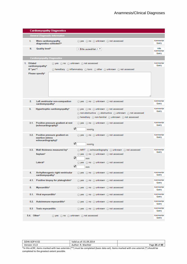
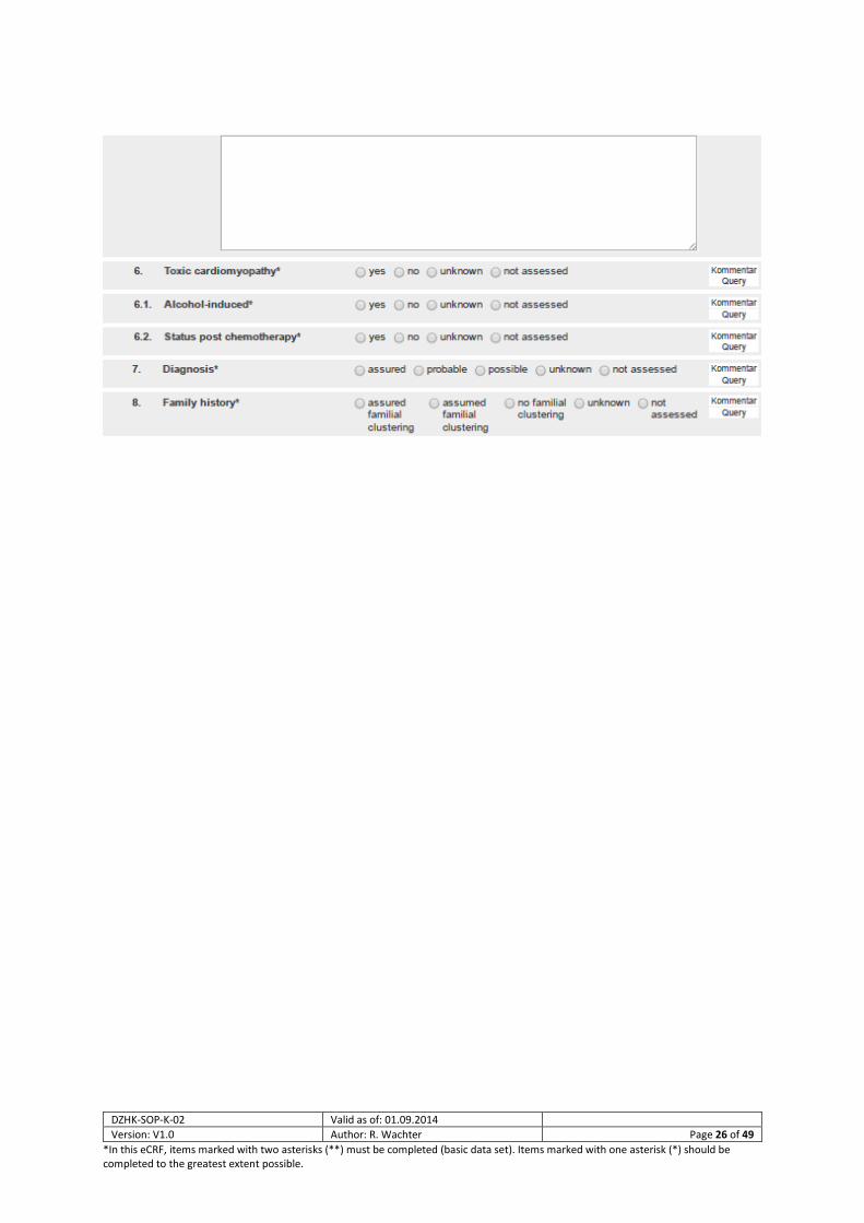
Cardiomyopathy
is defined as a diagnosis by a doctor of a primary heart muscle disease. If the response to this
question is “yes”, further data is collected in the “Cardiomyopathy Diagnostics” form.
Heart failure
is defined as a current or previous diagnosis and documentation by a doctor of heart failure,
based on the following symptoms: shortness of breath with mild exertion, recurrent shortness
of breath when sitting, fluid overload or pulmonary rales, distention of the neck veins,
pulmonary oedema on physical examination or pulmonary oedema on chest x-rays.
Documentation of reduced left ventricular function alone in the absence of clinical signs of
heart failure does not meet the criteria for heart failure.
Status post decompensation is defined as any previous admission to a hospital with symptoms of heart
failure (see above).
Initial diagnosis of heart failure is defined as the time point when heart failure was diagnosed for the
first time by a doctor. Hence it does not refer to the time point of first onset of symptoms, which is
often much earlier.
NYHA class: Classification of the patient’s symptoms based on the New York Heart Association classification of heart failure:
NYHA I: No symptoms
NYHA II: Symptoms with heavy physical exertion
NYHA III: Symptoms with light physical exertion
NYHA IV: Symptoms while at rest
Atrial fibrillation/flutter
DZHK-SOP-K-02 Valid as of: 01.09.2014
Version: V1.0 Author: R. Wachter Page 8 of 49
*In this eCRF, items marked with two asterisks (**) must be completed (basic data set). Items marked with one asterisk (*) should be completed to the greatest extent possible.
is defined as a current or previous diagnosis by a doctor of atrial fibrillation or atrial flutter. It
is defined as an episode of atrial fibrillation or atrial flutter lasting at least 30 seconds or atrial
fibrillation with evidence on the surface ECG or during pacemaker interrogation.
Current or previous medical diagnosis of heart valve disease
is defined as heart valve disease (incompetence or stenosis), which has been diagnosed and/or
treated by a doctor. A more precise differentiation and classification of the degree of severity
of the heart valve disease is conducted using the “Echocardiogram” form, if an echocardiogram
is to be documented in the context of the study.
Diagnosis by a doctor of endocarditis
If at any time, currently or in their previous medical history, a patient has been diagnosed by
a doctor with endocarditis (heart valve inflammation), it will be documented here.
Diagnosis by a doctor of a congenital heart defect
If a patient has a known congenital heart defect, it will be coded here. Congenital heart defects
include shunt defects (e.g. ASD, VSD), congenital valvular heart diseases (e.g. pulmonary
stenosis) and cardiomyopathies diagnosed in the first five years of life. Patent foramen ovale
does not belong to the class of congenital heart defects.
Interventional coronary revascularization
is defined as a percutaneously performed intervention on a coronary artery, e.g. PTCA, stent
implantation, rotablation et cetera. Purely diagnostic measures (intravascular ultrasound
(IVUS), optical coherence tomography (OCT)) as well as functional measurements (e.g.
fractional flow reserve (FFR) measurements) are not interventional coronary revascularization
procedures. Where applicable, the date of the last intervention should be entered.
Peripheral revascularization
is defined as a percutaneously performed intervention on a peripheral artery (not including
coronary arteries or bypass grafts) e.g. PTA, stent implantation, rotablation et cetera. Where
applicable, the date of the most recent intervention should be entered. Ablation procedures
(e.g. renal denervation) are not peripheral revascularization procedures. Where applicable,
the date of the most recent intervention should be entered.
DZHK-SOP-K-02 Valid as of: 01.09.2014
Version: V1.0 Author: R. Wachter Page 9 of 49
*In this eCRF, items marked with two asterisks (**) must be completed (basic data set). Items marked with one asterisk (*) should be completed to the greatest extent possible.
Coronary bypass operation
is defined as operative myocardial revascularization by means of a bypass graft (e.g. from the
internal thoracic artery or using arterial/venous grafts). Where applicable, the date of the most
recent operation should be entered.
Other vascular operation
is defined as an operation of any kind on non-coronary blood vessels. Where applicable, the
date of the most recent operation should be entered.
Heart valve operation
is defined as a minimally invasive percutaneous (catheter-based) or open surgical procedure on a
heart valve. This includes the surgical reconstruction/replacement of heart valves, valvuloplasty
procedures as well as interventional treatment of heart valve diseases (e.g. dilation, implantation
of protheses, heart valve repair). Where applicable, the date of the most recent operation should
be entered. The most recent event is to be coded according to type, whereby any transapical aortic
valve replacements are to be coded as “catheter-based“. In addition, details of the surgical
procedure should be given.
Implantable cardiac pacemaker or defibrillator
is defined as status post implantation of a cardiac pacemaker or cardio-verter defibrillator
(ICD). Where applicable, the date of the most recent operation (implantation/exchange) is to
be entered. In addition, the number of leads currently connected to the pacemaker power
supply should be coded. A device with only one lead should be coded as a 1-chamber
pacemaker, a device with an atrial and a ventricular lead should be coded as a 2-chamber
pacemaker. Devices for cardiac resynchronization therapy, with 2 ventricular leads, should be
coded as biventricular (CRT) pacemakers.
Other devices
are defined as other implantable devices for cardiac/vascular support. This includes devices
for cardiac contractility modulation, for neuromodulation (e.g. vagus nerve stimulator,
baroreceptor stimulator), intra-aortic balloon pumps and left ventricular cardiac assist devices.
Status post myocardial biopsy
is defined as status post bioptic removal of tissue from the heart muscle (e.g. during a right/left
catheter examination or operation). Where applicable, the sampling site as well as the date of
the most recent myocardial biopsy should be coded.
CURRENT SECONDARY DIAGNOSES
PAOD
DZHK-SOP-K-02 Valid as of: 01.09.2014
Version: V1.0 Author: R. Wachter Page 10 of 49
*In this eCRF, items marked with two asterisks (**) must be completed (basic data set). Items marked with one asterisk (*) should be completed to the greatest extent possible.
is defined as a current or previous diagnosis by a doctor of peripheral arterial occlusive disease
(in the blood vessels of the pelvis and legs, or from the upper extremity of the subclavian artery
to the distal extremity). Renal, coronary, cerebral and mesenteric blood vessels and aneurysms
are excluded. Possible symptoms are:
intermittent claudication,
pain at rest,
amputation due to severe arterial vascular insufficiency,
vascular reconstruction, bypass operation or percutaneous revascularization,
a positive non-invasive test (e.g. ankle-brachial index of ≤ 0.9, pathological TCPO2
measurement, evidence of 50 % or greater stenosis of a peripheral artery by
Doppler/duplex sonography, CT, MRT, or angiography).
Classification of the degree of severity is done according to the Fontaine classification:
Classification according to Fontaine
Stage Clinical Picture
I. Asymptomatic PAOD
II. Intermittent claudication
- with walking distances > 200 metres (Stage IIa)
- with walking distances < 200 metres (Stage IIb)
III. Pain at rest
IV. Necrosis, gangrene
- IVa: trophic disorder, dry necroses
- IVb: bacterial infection of the necrosis, wet gangrene
Acute ischaemic occlusion describes a recent (in the last 30 days) occurrence of demonstrated
acute ischaemic occlusion of a peripheral arterial vessel.
Stroke/TIA
is defined as a current or previous diagnosis by a doctor of:
Ischaemic stroke: Infarction of tissue of the central nervous system, either symptomatic or silent
(asymptomatic).
Transient ischaemic attack (TIA): A transient episode of neurological dysfunction caused by focal
brain, spinal cord or retinal ischaemia without acute infarction which resolves completely within
24 hours. This definition is not met by chronic (non-vascular) neurological diseases or other acute
neurological diseases such as metabolic or ischaemic encephalopathy resulting from general
hypoxia (e.g. in the case of respiratory insufficiency, following a cardiac/circulatory arrest).
Haemorrhagic stroke: Neurological dysfunction caused by intra-cranial bleeding.
Stroke where there is uncertainty as to whether the cause was haemorrhagic or ischaemic.
DZHK-SOP-K-02 Valid as of: 01.09.2014
Version: V1.0 Author: R. Wachter Page 11 of 49
*In this eCRF, items marked with two asterisks (**) must be completed (basic data set). Items marked with one asterisk (*) should be completed to the greatest extent possible.
Severity of the stroke: A stroke is described as “minor“ when the neurological symptoms can be
completely reversed within 30 days or the change in the NIH Stroke Scale (see Appendix 7.3 NIH Stroke
Scale) amounts to less than 3 points in comparison with the NIH Stroke Scale before the stroke. A
stroke is described as “major” when a neurological deficit can still be demonstrated 30 days after the
event or the NIH Stroke Scale is at least 3 points higher than prior to the stroke.
Consequences of the stroke: A stroke is described as “disabling” when more than 2 points are scored
on modified Rankin Scale 90 days after the stroke. If the modified Rankin Scale score is 2 points or less
90 days after the stroke, the stroke is described as “non-disabling”.
The modified Rankin Scale from 0 to 6 describes states from full health to death.
0 - No symptoms. 1 - No significant disability. Can perform day-to-day activities despite some symptoms. 2 - Slight disability. Is able to care for him or herself without assistance, but is restricted in
day-to-day activities. 3 - Moderate disability. Requires assistance in daily life, but is able to walk without
assistance. 4 - High level of disability. Requires assistance with personal hygiene; is not able to walk
without assistance. 5 - Severe disability. Confined to bed, incontinent, requires constant nursing care. 6 – Death caused by apoplexy.
Chronic lung disease
is defined as a diagnosis by a doctor of a chronic lung disease (e.g. COPD, chronic bronchitis,
pulmonary fibrosis) and/or their pharmacological treatment, for example, with inhalable or
oral pharmaceuticals (e.g. betamimetics, anti-inflammatory drugs, leukotriene receptor
antagonists, or steroids).
Primary pulmonary hypertension
is defined as a diagnosis and/or treatment by a doctor of primary pulmonary hypertension.
DZHK-SOP-K-02 Valid as of: 01.09.2014
Version: V1.0 Author: R. Wachter Page 12 of 49
*In this eCRF, items marked with two asterisks (**) must be completed (basic data set). Items marked with one asterisk (*) should be completed to the greatest extent possible.
Depression
is defined as a current or previous diagnosis by a doctor. The administration of antidepressants
alone does not qualify for a diagnosis of depression.
Cancer more than 5 years ago
is defined as a current or previous diagnosis of a malignant cancer. Basal cell carcinoma is not
counted as a malignancy.
Cancer within the last 5 years
is defined as malignant cancer diagnosed by a doctor less than 5 years ago. Basal cell carcinoma
is not counted as a malignancy.
Additional details; women only
Menopause
is defined as the time point of the last spontaneous menstrual period in the life of a woman
after which no further bleeding from the uterus induced by the ovaries occurs for at least 12
months. The year in which the menopause began is to be coded. The day on which the last
menstrual period began is required only for perimenopausal women.
PHYSICAL EXAMINATION
Blood pressure
The systolic blood pressure should be measured using a blood pressure monitor that is
serviced and calibrated on a regular basis. Where possible, tested devices (e.g. Omron 705 IT)
should be used for epidemiological trials. Blood pressure measurement begins after the
patient has been at rest for at least 5 minutes. Three readings are taken at intervals of 2
minutes; the average values of the second and third readings are entered into the CRF.
Heart rate
Measurement of the heart rate begins after the patient has been sitting down for at least 5
minutes. This should take place after measuring the blood pressure. This should be done
manually by counting the radial pulse for 30 seconds; that value multiplied by two should be
entered into the CRF (beats/minute).
DZHK-SOP-K-02 Valid as of: 01.09.2014
Version: V1.0 Author: R. Wachter Page 13 of 49
*In this eCRF, items marked with two asterisks (**) must be completed (basic data set). Items marked with one asterisk (*) should be completed to the greatest extent possible.
Clinical Symptoms and Examination Findings
Dyspnoea on exertion
A patient who complains of shortness of breath with physical exertion within the last 14 days
and/or at present. In cases of known heart failure, for patients in NYHA stages II-IV, dyspnoea
on exertion should be coded.
Dyspnoea at rest
A patient who complains of shortness of breath even when at rest (e.g. when talking) within
the last 14 days and/or at present. In cases of known heart failure, for patients in NYHA stage
IV, dyspnoea at rest should be coded.
Peripheral oedema
A patient who complains of bilateral accumulation of fluid in the extremities within the last 14
days and/or at present, whether clinically observed or perceived by the patient.
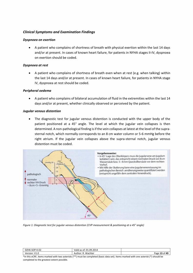
Jugular venous distention
The diagnostic test for jugular venous distention is conducted with the upper body of the
patient positioned at a 45° angle. The level at which the jugular vein collapses is then
determined. A non-pathological finding is if the vein collapses at latest at the level of the supra-
sternal notch, which normally corresponds to an 8 cm water column or 5-6 mmHg before the
right atrium. If the jugular vein collapses above the supra-sternal notch, jugular venous
distention must be coded.
Figure 1: Diagnostic test for jugular venous distention (CVP measurement & positioning at a 45° angle)
DZHK-SOP-K-02 Valid as of: 01.09.2014
Version: V1.0 Author: R. Wachter Page 14 of 49
*In this eCRF, items marked with two asterisks (**) must be completed (basic data set). Items marked with one asterisk (*) should be completed to the greatest extent possible.
Pulmonary rales
are defined as sounds heard over the lung during auscultation which are created by the
movement of fluids and/or secretions during inspiration and expiration. They belong to the
category of adventitious breath sounds overlying normal breath sounds and indicate a
pathological change in the lung.
1.6 CORRELATIONS TO OTHER EXAMINATIONS Here the correlations between the individual SOPs and other examination procedures are
described.
Mandatory preliminary examination (SOP
…):
Recommended preliminary examination
(SOP …):
Exclusion of preliminary examination (SOP
):
Adverse effects on other parts of the
examination:
Mandatory following examination (SOP …):
Recommended following examination (SOP
…):
Exclusion of following examination (SOP):
1.7 LEVEL OF QUALITY Quality of the data collection method
This SOP describes a data collection method that corresponds to quality level 2 of the DZHK. A higher
quality level could possibly be achieved if, for example, standardized interviews such as those used in
the German National Cohort were used. Because the studies planned so far in the DZHK do not require
a quality level higher than 2, initially only one SOP for that level has been drafted.
DZHK Quality Levels
Implementation
Level 1 The examination is performed in accordance with the guidelines of the medical
associations.
DZHK-SOP-K-02 Valid as of: 01.09.2014
Version: V1.0 Author: R. Wachter Page 15 of 49
*In this eCRF, items marked with two asterisks (**) must be completed (basic data set). Items marked with one asterisk (*) should be completed to the greatest extent possible.
Level 2 The examination is performed in accordance with the specifications of the DZHK
SOP. Minimum requirements to ensure the quality of the implementation and
the examiners are defined in the SOP.
Level 3 The examination is performed in accordance with the specifications of the DZHK
SOP and certification of the examiners: Definition of intra-observer and inter-
observer variability (standard of epidemiological studies).
2 EXAMINATION CONDITIONS
All circumstances are taken into account in order to ensure that the examination is
conducted under suitable conditions.
2.1 REQUIREMENTS FOR ROOMS/EQUIPMENT The examination room should have a room temperature of 22-26 °C. Generally, the room
should have a table at which the proband and the interviewer can sit in a comfortable
atmosphere in order to conduct the interview.
2.2 EQUIPMENT/HARDWARE PC with a monitor, keyboard, mouse, printer and printer paper. Depending on the respective
study, the forms for standardized documentation of the proband’s responses should be
available as source files, if needed.
2.3 SPECIAL CLINICAL CONSUMABLES None.
2.4 DOCUMENTS REQUIRED Routing slip
Scan barcode
2.5 INFORMATION REQUIRED Examiner number
Survey number (label)
Beginning of examination
Proband number
DZHK-SOP-K-02 Valid as of: 01.09.2014
Version: V1.0 Author: R. Wachter Page 16 of 49
*In this eCRF, items marked with two asterisks (**) must be completed (basic data set). Items marked with one asterisk (*) should be completed to the greatest extent possible.
2.6 PERSONNEL Persons using this SOP must have completed their training in the medical field (e.g. medical assistant,
nurse, qualified doctor). Students of medicine may use this SOP after they have successfully passed
their first medical examination (Physikum).
All users must have completed a prior course of instruction/certification for this SOP.
DZHK-SOP-K-02 Valid as of: 01.09.2014
Version: V1.0 Author: R. Wachter Page 17 of 49
*In this eCRF, items marked with two asterisks (**) must be completed (basic data set). Items marked with one asterisk (*) should be completed to the greatest extent possible.
3 IMPLEMENTATION PROCESS/WORK PROCESS/WORK STEPS
3.1 PROCESS FLOW CHART
DZHK-SOP-K-02 Valid as of: 01.09.2014
Version: V1.0 Author: R. Wachter Page 18 of 49
*In this eCRF, items marked with two asterisks (**) must be completed (basic data set). Items marked with one asterisk (*) should be completed to the greatest extent possible.
3.2 PREPARING FOR THE EXAMINATION
Preparing the Work Space
Seek out a suitable room with a table. Bring the temperature in the room to between 22 and
26 °C.
Preparing the Equipment
All equipment (PC/laptop/printer) should be switched on and operational. A form
(documentation of the source data) should be at hand.
Principles for Preparing the Proband for the Examination
Special preparation of the proband is not necessary.
3.3 CARRYING OUT THE EXAMINATION Physical examination – anthropometry
Height (in cm) and weight (in kg) are given either as self-reported values (Level 1) or as
measured values (Level 2). Whether the values given are based on anamnestic or measured
values shall be marked in the CRF.
A diagnosis is regarded as given if diagnosed by a medical doctor and/or therapy is being administered
which is considered to specifically target a certain disease. All documentation in medical documents
(e.g. doctor‘s reports) justifies accepting the diagnosis in question as given.
When carrying out the examination, for each clinical diagnosis, the following questions should be asked
in the interview:
1. Has a doctor ever told you that you suffer from a “disease”?
2. Have you ever received treatment for a “disease”?
3. Are you taking drug “xy“ for the “disease“?
As a “counter-check“, the interviewer should inquire about and document the indication for each
medication the patient is taking. A validation rule will be added to the database which will produce a
notification when inconsistencies arise (e.g. negative responses to questions 1-3, but the subject is
taking the corresponding medication).
When uncertainties arise (e.g. as to whether the relevant diagnoses have been made, but the subject
has consulted doctors for clarification), when and where those consultations took place should be
noted as precisely as possible in the remarks field.
DZHK-SOP-K-02 Valid as of: 01.09.2014
Version: V1.0 Author: R. Wachter Page 19 of 49
*In this eCRF, items marked with two asterisks (**) must be completed (basic data set). Items marked with one asterisk (*) should be completed to the greatest extent possible.
Inquiry about the following specific diseases , see section 1.5:
Diabetes mellitus
Arterial hypertension
Dyslipidaemia
Smoker
Positive family history of cardiovascular disease
Dialysis dependency
Coronary heart disease
Status post myocardial infarction
Cardiomyopathy
Heart failure
Atrial fibrillation/flutter
PAOD
Stroke/TIA
Chronic lung disease
Depression
Malignancy
3.4 POST-PROCESSING AND REGISTERING THE DATA A special debriefing session is not planned. The data should be entered without delay
(usually within 7 days).
3.5 DEALING WITH DEVIATIONS If a clear answer cannot be obtained for certain questions, this should be documented.
General particularities should always be noted in the commentary/notes field.

DZHK-SOP-K-02 Valid as of: 01.09.2014
Version: V1.0 Author: R. Wachter Page 20 of 49
*In this eCRF, items marked with two asterisks (**) must be completed (basic data set). Items marked with one asterisk (*) should be completed to the greatest extent possible.
4 LITERATURE AND REFERENCES
ACCF/AHA Guidelines Circulation 2011;124:103-123
5 MODIFICATIONS
Modifications compared with the previous version.
Section Description of the modification compared with the previous version
2.1
….
6 LIST OF CONTRIBUTORS
Name Function Contribution
PD Dr. Rolf Wachter Author Drafted the SOP
Dr. Sebastian Kufner Reviewer Expert review
Prof. Dr. Matthias Nauck Head of Scientific Infrastructure Expert review
PD Dr. Andreas Dösch Phenotyping & QM Group Expert review
Dr. Martin Hadamitzky Phenotyping & QM Group Expert review
Dr. Sabine Hübler Phenotyping & QM Group Expert review
Prof. Dr. Carsten Tschöpe Phenotyping & QM Group Expert review
Dr. Benjamin Meder Phenotyping & QM Group Expert review
Dr. Christina Dösch Phenotyping & QM Group Expert review
Prof. Dr. Christoph Knosalla Phenotyping & QM Group Expert review
Prof. Dr. Marcus Dörr Phenotyping & QM Group Expert review
Prof. Dr. Philipp Wild Phenotyping & QM Group Expert review
PD Dr. Renate Schnabel Phenotyping & QM Group Expert review
PD Dr. Stefanie Schulz Phenotyping & QM Group Expert review
Dr. Günther Schmidt Phenotyping & QM Group Expert review
Dr. Annika Jagodzinski Phenotyping & QM Group Expert review
Dr. Matthias Lutz Phenotyping & QM Group Expert review
Dr. Elham Kayvanpour Phenotyping & QM Group Expert review
Dr. Alexander Joost Phenotyping & QM Group Expert review
Matthias Quade Phenotyping & QM Group IT implementation
Mahsa Lee Phenotyping & QM Group IT implementation
Linda Gusky Phenotyping & QM Group IT implementation
Sophia Lamp Phenotyping & QM Group
Daniel Engler Layout, coordination
Dr. Stephanie Lesser Coordinator
DZHK-SOP-K-02 Valid as of: 01.09.2014
Version: V1.0 Author: R. Wachter Page 21 of 49
*In this eCRF, items marked with two asterisks (**) must be completed (basic data set). Items marked with one asterisk (*) should be completed to the greatest extent possible.
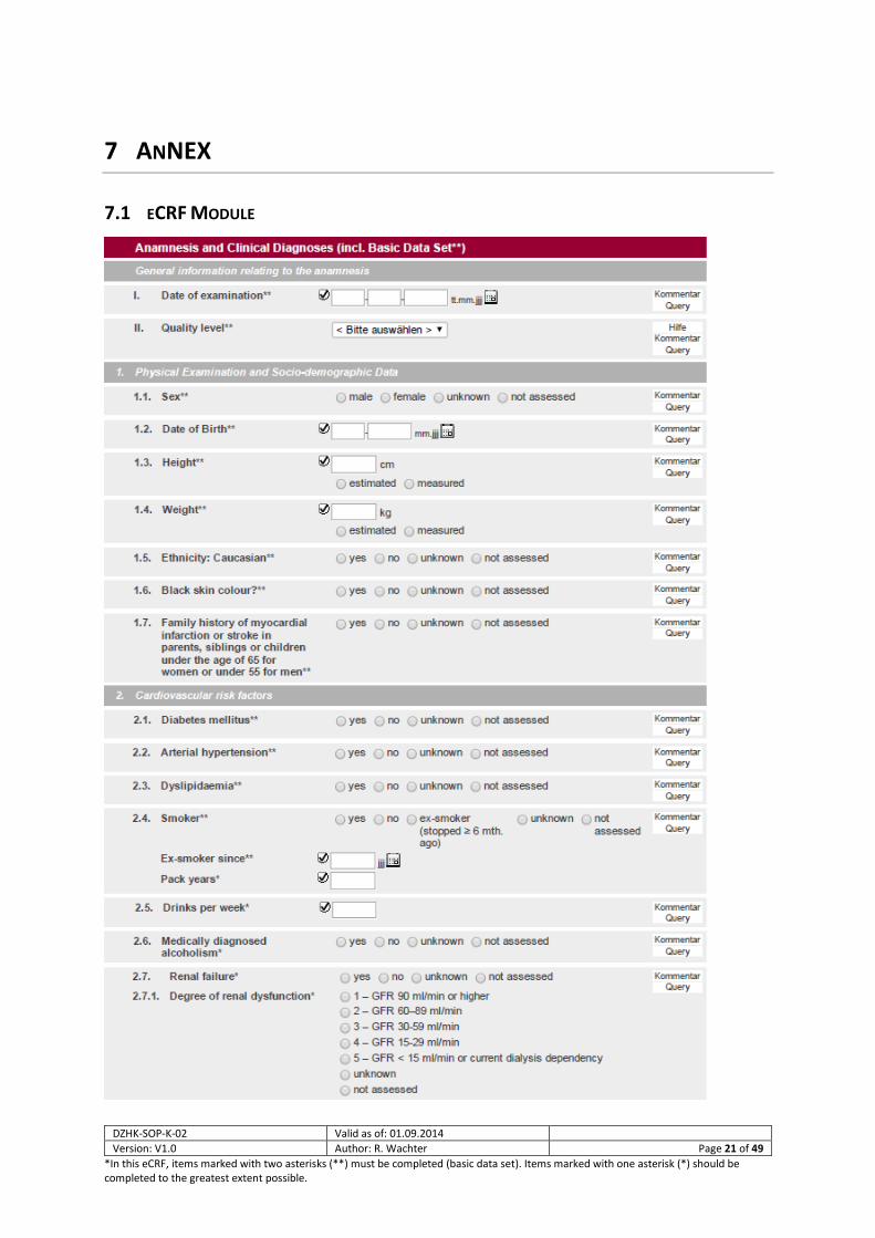
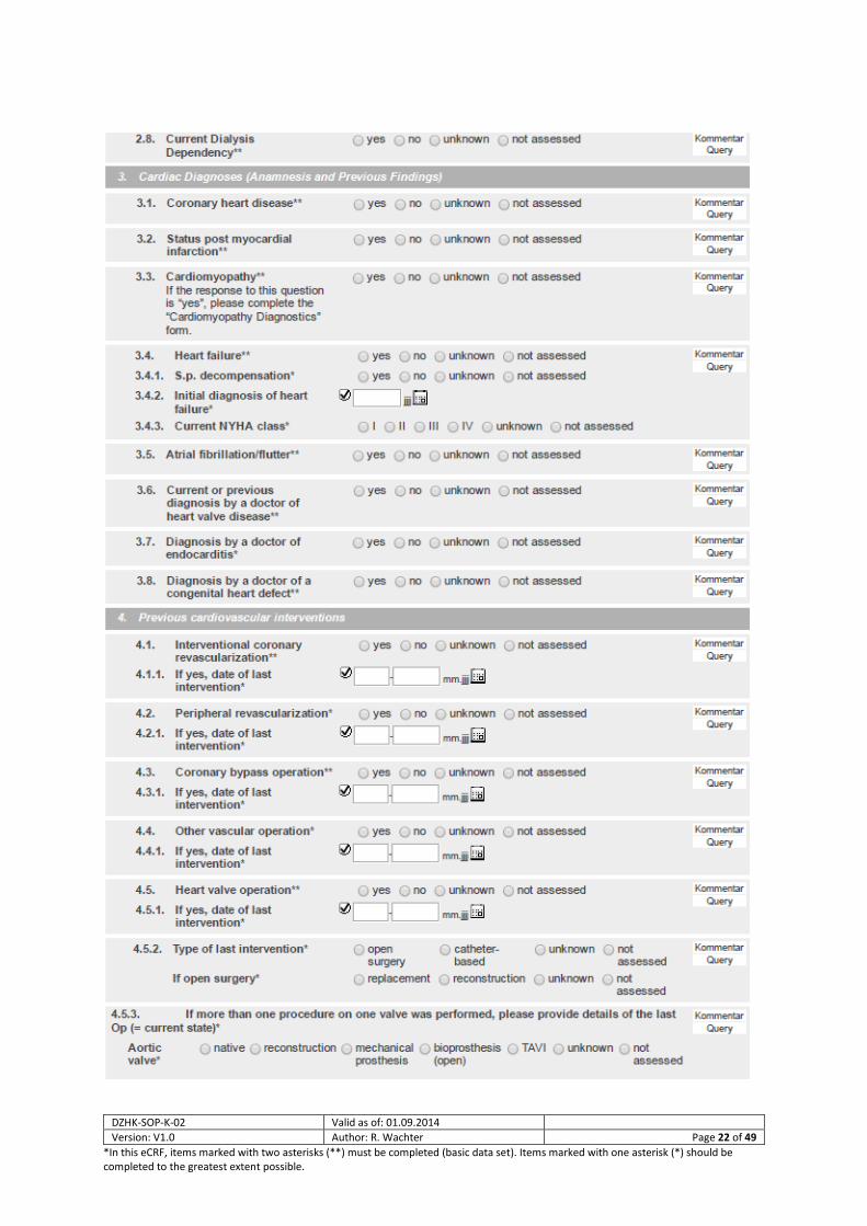
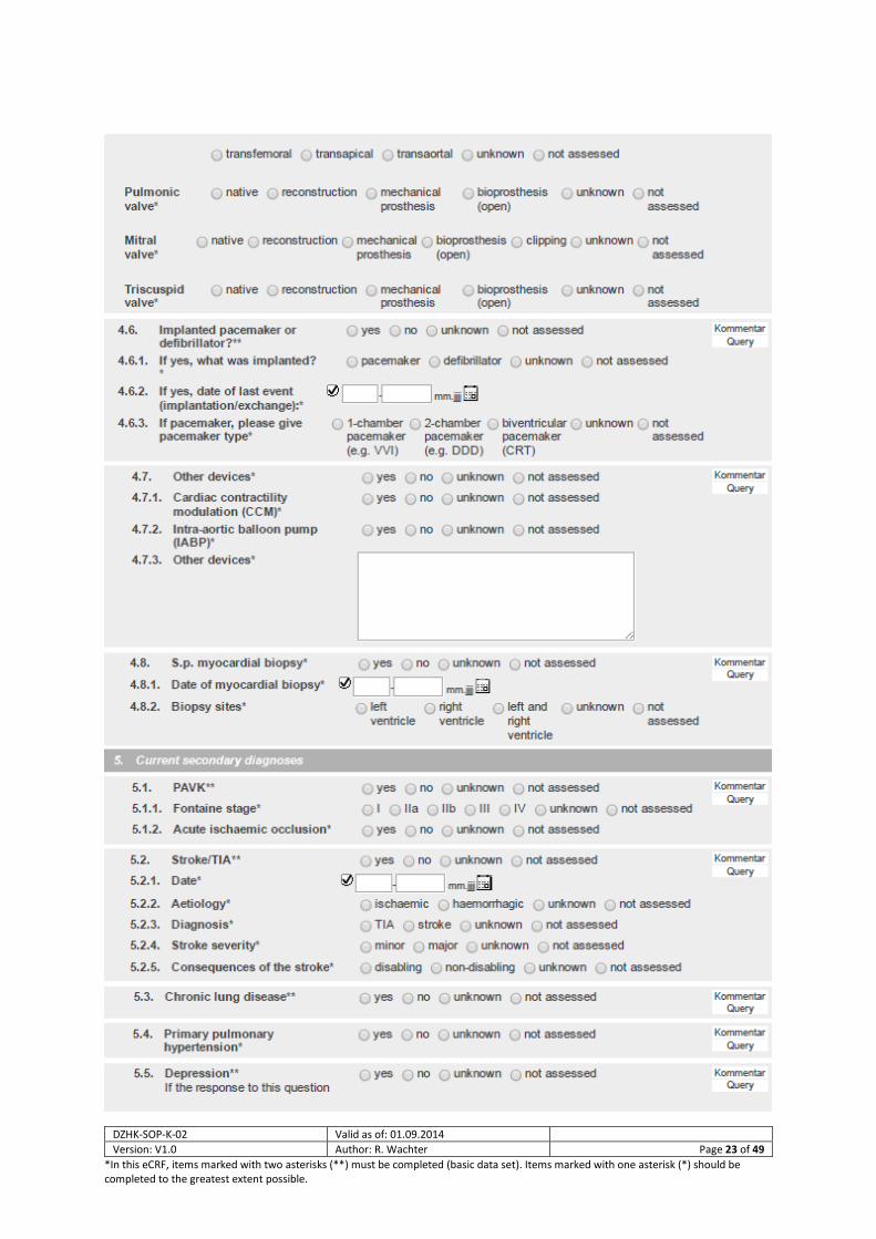
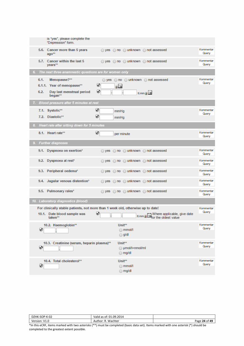
7 ANNEX
7.1 ECRF MODULE
DZHK-SOP-K-02 Valid as of: 01.09.2014
Version: V1.0 Author: R. Wachter Page 22 of 49
*In this eCRF, items marked with two asterisks (**) must be completed (basic data set). Items marked with one asterisk (*) should be completed to the greatest extent possible.
DZHK-SOP-K-02 Valid as of: 01.09.2014
Version: V1.0 Author: R. Wachter Page 23 of 49
*In this eCRF, items marked with two asterisks (**) must be completed (basic data set). Items marked with one asterisk (*) should be completed to the greatest extent possible.
DZHK-SOP-K-02 Valid as of: 01.09.2014
Version: V1.0 Author: R. Wachter Page 24 of 49
*In this eCRF, items marked with two asterisks (**) must be completed (basic data set). Items marked with one asterisk (*) should be completed to the greatest extent possible.
Anamnesis/Clinical Diagnoses
DZHK-SOP-K-02 Valid as of: 01.09.2014
Version: V1.0 Author: R. Wachter Page 25 of 49
*In this eCRF, items marked with two asterisks (**) must be completed (basic data set). Items marked with one asterisk (*) should be completed to the greatest extent possible.
DZHK-SOP-K-02 Valid as of: 01.09.2014
Version: V1.0 Author: R. Wachter Page 26 of 49
*In this eCRF, items marked with two asterisks (**) must be completed (basic data set). Items marked with one asterisk (*) should be completed to the greatest extent possible.
Anamnese/Klinische Diagnosen
DZHK-SOP-K-02 Gültig ab: 01.09.2014
Version: V1.0 Autor: R. Wachter Seite 27 von 49
*Die in diesem eCRF mit ** gekennzeichneten Items sind verpflichtend einzuhalten (= Basisdatensatz). Die mit * gekennzeichneten Items sind nach Möglichkeit einzuhalten.
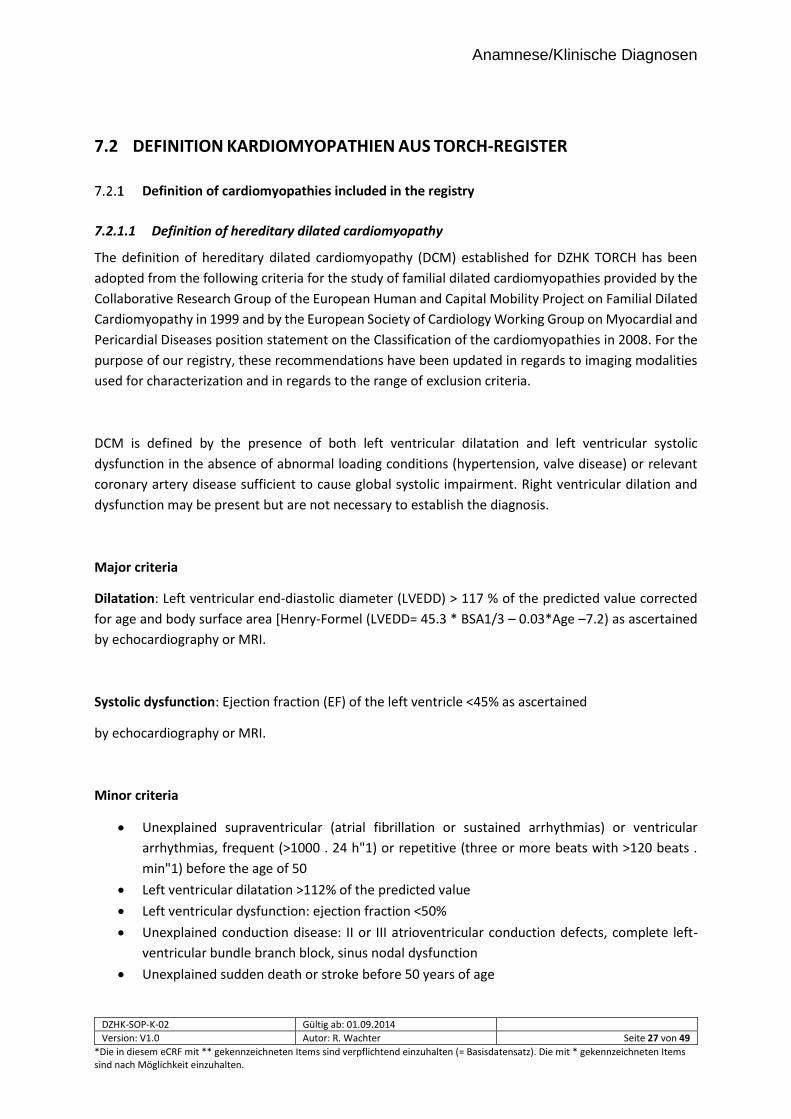
7.2 DEFINITION KARDIOMYOPATHIEN AUS TORCH-REGISTER
Definition of cardiomyopathies included in the registry
7.2.1.1 Definition of hereditary dilated cardiomyopathy
The definition of hereditary dilated cardiomyopathy (DCM) established for DZHK TORCH has been
adopted from the following criteria for the study of familial dilated cardiomyopathies provided by the
Collaborative Research Group of the European Human and Capital Mobility Project on Familial Dilated
Cardiomyopathy in 1999 and by the European Society of Cardiology Working Group on Myocardial and
Pericardial Diseases position statement on the Classification of the cardiomyopathies in 2008. For the
purpose of our registry, these recommendations have been updated in regards to imaging modalities
used for characterization and in regards to the range of exclusion criteria.
DCM is defined by the presence of both left ventricular dilatation and left ventricular systolic
dysfunction in the absence of abnormal loading conditions (hypertension, valve disease) or relevant
coronary artery disease sufficient to cause global systolic impairment. Right ventricular dilation and
dysfunction may be present but are not necessary to establish the diagnosis.
Major criteria
Dilatation: Left ventricular end-diastolic diameter (LVEDD) > 117 % of the predicted value corrected
for age and body surface area [Henry-Formel (LVEDD= 45.3 * BSA1/3 – 0.03*Age –7.2) as ascertained
by echocardiography or MRI.
Systolic dysfunction: Ejection fraction (EF) of the left ventricle <45% as ascertained
by echocardiography or MRI.
Minor criteria
Unexplained supraventricular (atrial fibrillation or sustained arrhythmias) or ventricular
arrhythmias, frequent (>1000 . 24 h"1) or repetitive (three or more beats with >120 beats .
min"1) before the age of 50
Left ventricular dilatation >112% of the predicted value
Left ventricular dysfunction: ejection fraction <50%
Unexplained conduction disease: II or III atrioventricular conduction defects, complete left-
ventricular bundle branch block, sinus nodal dysfunction
Unexplained sudden death or stroke before 50 years of age
DZHK-SOP-K-02 Gültig ab: 01.09.2014
Version: V1.0 Autor: R. Wachter Seite 28 von 49
*Die in diesem eCRF mit ** gekennzeichneten Items sind verpflichtend einzuhalten (= Basisdatensatz). Die mit * gekennzeichneten Items sind nach Möglichkeit einzuhalten.
Segmental wall motion abnormalities (<1 segment, or 1 if not previously present) in the
absence of intraventricular conduction defect or ischaemic heart disease.
Elevated NTproBNP: In patients presenting with non-acute dyspnea (> 14 days), a value
exceeding 125 ng/L (age < 75 years) or 450 ng/L (> 75 years) is considered abnormal. In patients
with acute dyspnea or signs of heart failure, a value below 300 ng/L excludes acute heart
failure (age-independent rule-out). An age-dependent value exceeding 900 ng/L (<50 years),
1,200 ng/L (50-69 years) or 1,800 ng/L (>70 years) is considered abnormal. Values between
rule-out and age-dependent rule-in cutoff are called greyzone values and merit attention. Cut-
off for BNP are different and are not dependent on age or gender. A value > 35 ng/L is
considered abnormal for non-acute presentation, and 100 ng/L for acute manifestation.
Data for cardiac troponins are less established: Detectable cTn concentrations are associated
with midterm and longterm adverse outcomes. For hscTn hazard for death and hospitalization
for heart failure has been reported to start below the 99th percentile value. A value > 99th
percentile, e.g. 14 ng/L for hscTnT is definitely elevated and presumably of prognostic
importance.
Cardiac limitation during spiroergometry:
o peakVO2 > 85% predicted value
o VO2 at anaerobic threshold (AT) < 40% predicted VO2
o Breathing Reserve (BR) ≥ 30% (at least ≥ 15 L/min)
o Heart Rate Reserve (HRR) > 15/min
o Aerobic Capacity (dVO2/dWR) ≤ 8 mL/min*W
o Relative Dead Space Ventilation (VD/VT) ≤ 35% at rest and exercise
Exclusion criteria
Pre-existing other cardiac diseases such as significant valvular, congenital, ischemic or
pericardial diseases
Severe arterial hypertension (RR> 160/100mmHg or hypertension despite therapy with at least
3 different drugs)
Primary pulmonary artery hypertension
Chronic advanced disorders requiring treatment or being the predominant clinical finding on
initial presentation (rheumatic, autoimmune, malignancy, insulin dependent DM, endocrine,
ESDR, liver failure, etc.)
History of treatment with cardiotoxic agents and radiation
Drug and alcohol abuse
Categorization which will be applied in the registry:
Definite DCM: An individual is defined as definitely affected in the presence of both major or left
ventricular dilatation (>117%) plus one minor criterion or three minor criteria – without the
presence of an exclusion criterion.
DZHK-SOP-K-02 Gültig ab: 01.09.2014
Version: V1.0 Autor: R. Wachter Seite 29 von 49
*Die in diesem eCRF mit ** gekennzeichneten Items sind verpflichtend einzuhalten (= Basisdatensatz). Die mit * gekennzeichneten Items sind nach Möglichkeit einzuhalten.
Probable DCM: An individual is defined as probably affected in the presence of left ventricular
dilatation (>112% of the predicted value) and left ventricular dysfunction (ejection fraction
<50%) – without the presence of an exclusion criterion.
Possible DCM: An individual is defined as possibly affected in the presence of left ventricular
dysfunction (ejection fraction <50%) – without the presence of an exclusion criterion.
See references no. 41-45
7.2.1.2 Clinical and biopsy-based definition of inflammatory dilated cardiomyopathy and acute
myocarditis
The definitions of inflammatory dilated cardiomyopathy (DCMi) and acute myocarditis established for
DZHK TORCH have been adopted from criteria described in the 1995 Report of the World Health
Organization/International Society and Federation of Cardiology Task Force on the Definition and
Classification of Cardiomyopathies, the World Heart Federation consensus conferences’ definition of
inflammatory cardiomyopathy (myocarditis) in 1999 (Marburg Classification) and in the European
Society of Cardiology Working Group on Myocardial and Pericardial Diseases position statement on
the Classification of the cardiomyopathies in 2008.
Myocardial inflammation (autoimmune, viral, or postviral) is mediated by the effector cells of the
immune system. In contrast to active myocarditis, which is by definition an acute inflammatory
disorder with inflammatory cell associated myocyte necrosis of the heart, with often preserved left
ventricular size, inflammatory DCM is defined by the presence of inflammatory cells in association with
left ventricular dilatation and reduced systolic function (dilatation and systolic function analog to
definition of hereditary or post-inflammatory/infectious DCM). Histology and/or
immunocytochemistry are required for the diagnosis. A proportion of individuals with inflammatory
DCM have persistence of viral genomes or proteins in the myocardium. (The term viral persistence in
DCM should only be applied in those cases, in which viral RNA or DNA but no inflammation is present.)
Viral persistence can be associated with or without inflammation.
World Health Organization Marburg Classification
First biopsy:
Acute/active myocarditis: a clear-cut infiltrate (diffuse, focal or confluent) of >14
leukocytes/mm2 (preferably activated T cells). The amount of the infiltrate should be
quantified by immunohistochemistry. Necrosis or degeneration is compulsory; fibrosis may
be absent or present and should be graded.
Chronic myocarditis (histologically described as borderline myocarditis): an infiltrate of >14
leukocytes/mm2 (diffuse, focal or confluent, preferably activated T cells). Quantification
DZHK-SOP-K-02 Gültig ab: 01.09.2014
Version: V1.0 Autor: R. Wachter Seite 30 von 49
*Die in diesem eCRF mit ** gekennzeichneten Items sind verpflichtend einzuhalten (= Basisdatensatz). Die mit * gekennzeichneten Items sind nach Möglichkeit einzuhalten.
should be made by immunohistochemistry. Necrosis or degeneration is usually not evident;
fibrosis may be absent or present and should be graded.
No myocarditis: No infiltrating cells or <14 leukocytes/mm2.
Subsequent biopsies: (histology and immunohistochemistry)
Ongoing (persistent) myocarditis. Criteria as in active or chronic myocarditis.
Resolving (healing) myocarditis. Criteria as in acute or chronic myocarditis, but the
immunologic process is sparser than in the first biopsy.
Resolved (healed) myocarditis. Corresponds to the Dallas classification and the
immunohistochemical evaluation.
The amount and distribution of fibrosis should be described similarly as no (grade 0), mild (grade 1),
moderate (grade 2), or severe (grade 3). Localisation or formation of fibrosis should be outlined as
endocardial, replacement or interstitial.
Expanded criteria for clinical and biopsy-based diagnosis of myocarditis
Suspicious for myocarditis = 2 positive categories
Compatible with myocarditis = 3 positive categories
High probability of being myocarditis = all 4 categories positive.
Definite proof of myocardial inflammation and/or viral infection demands biopsy analysis
(positive category 4)
NOTE: Any matching feature in category = positive for category; the categories I-III define the clinical
diagnosis of myocarditis/inflammatory CMP only. A definite proof demands biopsy analysis (positive
for category IV).
Category I: clinical symptoms
Clinical heart failure
Fever
Viral prodrome
Fatigue
Dyspnoea on exertion
Chest pain
Palpitations
Pre-syncope or syncope
DZHK-SOP-K-02 Gültig ab: 01.09.2014
Version: V1.0 Autor: R. Wachter Seite 31 von 49
*Die in diesem eCRF mit ** gekennzeichneten Items sind verpflichtend einzuhalten (= Basisdatensatz). Die mit * gekennzeichneten Items sind nach Möglichkeit einzuhalten.
Category II: clinical evidence of cardiac structural/functional perturbation in the absence of regional
coronary ischaemia
Echo evidence
Regional wall motion abnormalities
Cardiac dilation
Regional cardiac hypertrophy
Troponin release
Troponin result has high sensitivity (>0.1 nanogram/mL)
Positive indium-111 antimyosin scintigraphy and normal coronary angiography or absence of
reversible ischaemia by coronary distribution on perfusion scan
Category III: cardiac MRI
Increased myocardial T2 signal on inversion recovery sequence
Delayed contrast enhancement following gadolinium-diethylenetriamine pentaacetic acid
(DTPA) infusion.
Category IV: myocardial biopsy, pathologic or molecular analysis as definite proof of myocardial
inflammation and viral infection
Pathology findings compatible with Dallas criteria supplemented by immunohistochemistry
Presence of viral genome by PCR or in situ hybridisation.
See references no. 46-51
7.2.1.3 Definition of hypertrophic cardiomyopathy
The definition of hypertrophic cardiomyopathy (HCM) and in specific, hypertrophic obstructive
cardiomyopathy (HOCM) established for DZHK TORCH has been adopted from the American/European
Consensus Document on Hypertrophic Cardiomyopathy in 2003 referenced below. For the purpose of
our registry, these recommendations have been updated in regards to imaging modalities used for
characterization and in regards to the range of exclusion criteria.
Evidence of left ventricular hypertrophy and/or increased left ventricular mass.
Definition of hypertrophy:
Wall thickness (including asymmetric hypertrophy in individual segments) ≥15mm
septal/posterior wall thickness ratio >1.3 in normotensive patients, or
septal/posterior wall thickness ratio >1.5 in hypertensive patients.

DZHK-SOP-K-02 Gültig ab: 01.09.2014
Version: V1.0 Autor: R. Wachter Seite 32 von 49
*Die in diesem eCRF mit ** gekennzeichneten Items sind verpflichtend einzuhalten (= Basisdatensatz). Die mit * gekennzeichneten Items sind nach Möglichkeit einzuhalten.
Exclusion criteria:
Hemodynamic stressors sufficient to explain hypertrophy
systemic arterial hypertension
Valvular disease
athlete's heart
Systemic storage disorders
Amyloidosis
Glycogen storage disease
Anderson-Fabry disease
Categorization which will be applied in the registry:
Definite HCM: An individual is defined as definitely affected in the presence of left ventricular
hypertrophy as stated above and/or increased left ventricular mass between ≥122 g/m²
(women) and ≥149 g/m² (men) and impaired longitudinal function – without the presence of
an exclusion criterion.
Probable HCM: An individual is defined as probably affected in the presence of left ventricular
hypertrophy with a wall thickness (including asymmetric hypertrophy in individual segments)
between 11 – 14 mm (women) and 12 – 14 mm (men) and/or increased left ventricular mass
between 109-121 g/m² (women) and 132-148 g/m² (men) and impaired longitudinal function
– without the presence of an exclusion criterion.
Possible HCM: An individual is defined as probably affected in the presence of left ventricular
hypertrophy with a wall thickness (including asymmetric hypertrophy in individual segments)
between 10 – 11 mm (women) and 11 – 12 mm (men) and/or increased left ventricular mass
between 96-108 g/m² (women) and 116-131 g/m² (men) and impaired longitudinal function –
without the presence of an exclusion criterion.
Specific: Hypertrophic obstructive cardiomyopathy
Evidence of HCM according to criteria listed above
AND
Evidence of a significant left ventricular outflow tract obstruction (gradient ≥ 30 mmHg) at rest
during stable pre-/afterload
DZHK-SOP-K-02 Gültig ab: 01.09.2014
Version: V1.0 Autor: R. Wachter Seite 33 von 49
*Die in diesem eCRF mit ** gekennzeichneten Items sind verpflichtend einzuhalten (= Basisdatensatz). Die mit * gekennzeichneten Items sind nach Möglichkeit einzuhalten.
Evidence of a significant dynamic left ventricular outflow tract obstruction (gradient
≥50mmHg) (either during exercise, after glyceryl trinitrate (GTN) administration, or the
Valsalva maneuver) during stable pre-/afterload
Specific: Suspected familial HCM
In family members of a HCM index patient, the following criteria are applied to define suspected HCM
cases (1 major or 2 minor echocardiographic criteria, or 1 major echocardiographic criterion and 2
minor electrocardiographic criteria).
See references no. 52-69
7.2.1.4 Definition of left ventricular non-compaction cardiomyopathy
For the lack of common standardized diagnostic criteria for the left ventricular non-compaction
cardiomyopathy (LVNC) following definition was established for DZHK TORCH according to the
published studies.
To prevent over diagnosing of LVNC the results by echocardiography and cardiac MRI must be
concordant.
Diagnosis is considered definite when the following criteria are present:
1. Absence of congenital heart disease, infiltrative/hypertrophic cardiomyopathy or documented
coronary artery disease
2. Echocardiographic diagnostic features
European Echo criteria European ECG criteria
Major: - MWT ≥ 13mm anteroseptal or
posterior
- MWT ≥ 15mm posteroseptal, lateral
or severe SAM
Major: - Abnormal Q-waves ≥ 2 leads
- T-wave Inversion ≥ 2 leads
- LV hypertrophy signs
Minor: - MWT ≥ 12mm anteroseptal or
posterior
- MWT ≥ 14mm posteroseptal, lateral
or moderate SAM
Minor: - deep S in lead V2
- repolarization changes
- bundle brunch blockage
MWT = „myokardial wall thickness“; SAM = „systolic anterior motion“
DZHK-SOP-K-02 Gültig ab: 01.09.2014
Version: V1.0 Autor: R. Wachter Seite 34 von 49
*Die in diesem eCRF mit ** gekennzeichneten Items sind verpflichtend einzuhalten (= Basisdatensatz). Die mit * gekennzeichneten Items sind nach Möglichkeit einzuhalten.
According to Stöllberger et al.: More than three confirmed trabeculations within one image plane,
apical to the insertion of the papillary muscles. Trabeculations with the same echogenicity as the
myocardium and synchronous movement with ventricular contractions. Perfusion of the
intertrabecular spaces from the left ventricular cavity. Ratio of compacted to non-compacted
segment at least 1:2 (≤ 0.5). Acquisition of the images: apical four chamber view and three
chamber view; angulation of the transducer and acquisition of pictures in atypical views to obtain
the technically best picture quality for differentiation between false chords/aberrant bands and
trabeculations.
3. MRI diagnostic features
Petersen et al.: Ratio between the non-compacted and compacted layer > 2.3. Measurement: at
end-diastole.
See references no. 70-76
7.2.1.5 Definition of arrhythmogenic right ventricular cardiomyopathy
The definitions of arrhythmogenic right ventricular cardiomyopathy (ARVC), also called
arrhythmogenic right ventricular dysplasia (ARVD), established for DZHK TORCH have been adopted
from criteria described in the 2010 revised Task Force Criteria by Marcus et al. (Original International
Task Force criteria from the European Society of Cardiology and the International Society and
Federation of Cardiology published in 1994).
Presence of ARVC/ARVD is established following the combination of the below listed criteria as:
o definite:
two major criteria, or
one major plus two minor criteria, or
four minor criteria
with each criterion being from a different category
o borderline:
one major and one minor, or
three minor criteria
with each criterion being from a different category
o possible:
one major, or
two minor criteria
with the criteria being from a different category

DZHK-SOP-K-02 Gültig ab: 01.09.2014
Version: V1.0 Autor: R. Wachter Seite 35 von 49
*Die in diesem eCRF mit ** gekennzeichneten Items sind verpflichtend einzuhalten (= Basisdatensatz). Die mit * gekennzeichneten Items sind nach Möglichkeit einzuhalten.
I. Global or regional dysfunction and structural alterations
Major
By 2D echo:
• Regional RV akinesia, dyskinesia, or aneurysm and 1 of the following (end diastole):
- PLAX RVOT ≥32 mm (corrected for body size [PLAX/BSA] ≥19 mm/m2)
- PSAX RVOT ≥36 mm (corrected for body size [PSAX/BSA] ≥21 mm/m2)
- or fractional area change ≤33 percent
By MRI:
• Regional RV akinesia or dyskinesia or dyssynchronous RV contraction and 1 of the following:
- Ratio of RV end-diastolic volume to BSA ≥110 mL/m2 (male) or ≥100 mL/m2 (female)
- or RV ejection fraction ≤40 percent
By RV angiography:
• Regional RV akinesia, dyskinesia, or aneurysm
Minor
By 2D echo:
• Regional RV akinesia or dyskinesia and 1 of the following (end diastole):
- PLAX RVOT ≥29 to <32 mm (corrected for body size [PLAX/BSA] ≥16 to <19 mm/m2)
- PSAX RVOT ≥32 to <36 mm (corrected for body size [PSAX/BSA] ≥18 to <21 mm/m2)
- or fractional area change >33 percent to ≤40 percent
By MRI:
• Regional RV akinesia or dyskinesia or dyssynchronous RV contraction and 1 of the following:
- Ratio of RV end-diastolic volume to BSA ≥100 to <110 mL/m2 (male) or ≥90 to <100 mL/m2
(female)
- or RV ejection fraction >40 percent to ≤45 percent
II. Tissue characterization of wall
Major
• Residual myocytes <60 percent by morphometric analysis (or <50 percent if estimated), with
fibrous replacement of the RV free wall myocardium in ≥1 sample, with or without fatty replacement
of tissue on endomyocardial biopsy
DZHK-SOP-K-02 Gültig ab: 01.09.2014
Version: V1.0 Autor: R. Wachter Seite 36 von 49
*Die in diesem eCRF mit ** gekennzeichneten Items sind verpflichtend einzuhalten (= Basisdatensatz). Die mit * gekennzeichneten Items sind nach Möglichkeit einzuhalten.
Minor
• Residual myocytes 60 percent to 75 percent by morphometric analysis (or 50 percent to 65 percent
if estimated), with fibrous replacement of the RV free wall myocardium in ≥1 sample, with or without
fatty replacement of tissue on endomyocardial biopsy
III. Repolarization abnormalities
Major
• Inverted T waves in right precordial leads (V1, V2, and V3) or beyond in individuals >14 years of age
(in the absence of complete right bundle-branch block QRS ≥120 ms)
Minor
• Inverted T waves in leads V1 and V2 in individuals >14 years of age (in the absence of complete right
bundle-branch block) or in V4, V5, or V6
• Inverted T waves in leads V1, V2, V3, and V4 in individuals >14 years of age in the presence of
complete right bundle-branch block
IV. Depolarization/conduction abnormalities
Major
• Epsilon wave (reproducible low-amplitude signals between end of QRS complex to onset of the T
wave) in the right precordial leads (V1 to V3)
Minor
• Late potentials by SAECG in ≥1 of the following 3 parameters in the absence of a QRS duration of
≥110 ms on the standard ECG
- Filtered QRS duration (fQRS) ≥114 ms
- Duration of terminal QRS <40 µV (low-amplitude signal duration) ≥38 ms
- Root-mean-square voltage of terminal 40 ms ≤20 µV
• Terminal activation duration of QRS ≥55 ms measured from the nadir of the S wave to the end of
the QRS, including R', in V1, V2, or V3, in the absence of complete right bundle-branch block

DZHK-SOP-K-02 Gültig ab: 01.09.2014
Version: V1.0 Autor: R. Wachter Seite 37 von 49
*Die in diesem eCRF mit ** gekennzeichneten Items sind verpflichtend einzuhalten (= Basisdatensatz). Die mit * gekennzeichneten Items sind nach Möglichkeit einzuhalten.
V. Arrhythmias
Major
• Nonsustained or sustained ventricular tachycardia of left bundle-branch morphology with superior
axis (negative or indeterminate QRS in leads II, III, and aVF and positive in lead aVL)
Minor
• Nonsustained or sustained ventricular tachycardia of RV outflow configuration, left bundle-branch
block morphology with inferior axis (positive QRS in leads II, III, and aVF and negative in lead aVL) or
of unknown axis
• >500 ventricular extrasystoles per 24 hours (Holter)
VI. Family history
Major
• ARVC/D confirmed in a first-degree relative who meets current Task Force criteria
• ARVC/D confirmed pathologically at autopsy or surgery in a first-degree relative
• Identification of a pathogenic mutationΔ categorized as associated or probably associated with
ARVC/D in the patient under evaluation
Minor
• History of ARVC/D in a first-degree relative in whom it is not possible or practical to determine
whether the family member meets current Task Force criteria
• Premature sudden death (<35 years of age) due to suspected ARVC/D in a first-degree relative
• ARVC/D confirmed pathologically or by current Task Force Criteria in second-degree relative
See references nr. 77-78
DZHK-SOP-K-02 Gültig ab: 01.09.2014
Version: V1.0 Autor: R. Wachter Seite 38 von 49
*Die in diesem eCRF mit ** gekennzeichneten Items sind verpflichtend einzuhalten (= Basisdatensatz). Die mit * gekennzeichneten Items sind nach Möglichkeit einzuhalten.
7.2.1.6 Definitions for biopsy diagnosis of cardiomyopathies
Active Myocarditis:
Infiltrating lymphocytes (CD3) and/or monocytes/macrophages (CD68 in paraffin fixed tissues, CD11b
in unfixed/frozen tissues) + inflammatory cell associated myocyte necrosis. Focally or diffusely
enhanced expression of cell adhesion molecules.
Specific disease entities:
Giant cell myocarditis, eosinophilic myocarditis, granulomateous myocarditis (e.g. sarcoidosis)
Borderline-Myocarditis/inflammatory cardiomyopathy:
>14 infiltrating leukocytes with up to 4 monocytes/mm2 with the presence of CD 3 positive T-
lymphocytes ≥7 cells/mm2 or > 35 monocytes/macrophages (CD68 in paraffin fixed tissues, CD11b in
unfixed/frozen tissues) without inflammatory cell associated myocyte necrosis in addition to an
enhanced expression of cell adhesion molecules (HLA-1 or HLA-DR, CD54/ICAM-1, CD106/VCAM-1)
or
Focal infiltrates of inflammatory cells (lymphocytes, monocytes/macrophages, leukocytes) in
histologically (paraffin) or immunohistologically (frozen) stained tissues.
No Myocarditis/DCM:
Cell numbers of infiltrating lymphocytes or monocytes/macrophages are below those defining
Borderline-Myocarditis or inflammatory CMP; a mildly enhanced expression of cell adhesion molecules
(HLA-I/-DR and CD54/ICAM-1) may be present in postinflammatory tissues (resolved inflammatory cell
infiltrates).
No focal inflammatory cell infiltrates in histologically or immunohistochemically analyzed tissues
Histology: cardiomyocyte hypertrophy, interstitial fibrosis, and scars may be present and indicate
progressive disease
Viral myocarditis/cardiomyopathy:
Positive proof of viral genomes (PCR) with or without myocardial inflammation. Consideration of virus
subtypes, virus loads, and replicative intermediates (mRNA) indicating active/recent infection or virus
reactivation (myocardial tissue, blood).
DZHK-SOP-K-02 Gültig ab: 01.09.2014
Version: V1.0 Autor: R. Wachter Seite 39 von 49
*Die in diesem eCRF mit ** gekennzeichneten Items sind verpflichtend einzuhalten (= Basisdatensatz). Die mit * gekennzeichneten Items sind nach Möglichkeit einzuhalten.
HCM:
Often no specific histological or immunohistochemical features, since endomyocardial biopsy may be
regular. Myocyte hypertrophy, fibroses, scars, myocardial inflammation and viral genomes may be
present. Amyloidosis and storage diseases have to be excluded.
ARVD/C:
Due to the main localization of the disease process, there are often no specific histological or
immunohistochemical features and endomyocardial biopsy specimens may be regular. Myocyte
hypertrophy or atrophy, fibrosis, scars, myocardial inflammation and viral genomes may be present. A
reduced expression of gap junction proteins (immunohistochemistry) may indicate ARVD. In the
advanced stage of the disease, fibro-fatty degeneration of myocardial tissue proves ARVD.
Genetic/heriditary:
Genetic testing for specific gene defects/SNPs. In addition, histology, immunohistochemistry and
molecular biology as defined above.
See references nr. 46-51
References
1. FRIEDRICHS F, ZUGCK C, RAUCH GJ, IVANDIC B, WEICHENHAN D, MÜLLER-BARDORFF M, MEDER B, EL MOKHTARI
NE, REGITZ- ZAGROSEK V, HETZER R, SCHÄFER A, SCHREIBER S, CHEN J, NEU- HAUS I, JI R, SIEMERS NO, FREY N,
ROTTBAUER W, KATUS HA, STOLL M (2009). HBEGF, SRA1, AND IK: THREE COSEGREGATING GENES AS
DETERMINANTS OF CARDIOMYOPATHY. GENOME RES, 19:395-403.
2. HASSEL D, DAHME T, ERDMANN J, MEDER B, HUGE A, STOLL M, GRIMMLER M, JUST S, HESS A, EHLERMANN P,
WEICHENHAN D, LIPTAU H, HETZER R, REGITZ-ZAGROSEK V, FISCHER C, NÜRNBERG P, SCHUNKERT H, KATUS HA,
ROTTBAUER W (2009). NEXILIN MUTATIONS DESTABI LIZE CARDIAC Z-DISKS AND LEAD TO DILATED
CARDIOMYOPATHY. NATURE MEDICINE 15:1281- 1288.
3. FRANKENSTEIN L, REMPPIS A, FLUEGEL A, DOESCH A, KATUS HA, SENGES J, ZUGCK C.THE ASSOCIATION BETWEEN
LONG-TERM LONGITUDINAL TRENDS IN GUIDELINE ADHERENCE AND MORTALITY IN RELATION TO AGE AND SEX. EUR J
HEART FAIL 2010 JUN;12(6):574-580.
4. RÖTTGEN R, CHRISTIANI R, FREYHARDT P, GUTBERLET M, SCHULTHEISS HP, HAMM B, KÜHL U.MAGNETIC
RESONANCE IMAGING FINDINGS IN ACUTE MYOCARDITIS AND CORRELATION WITH IMMUNOHISTOLOGICAL
PARAMETERS. EUR RADIOL. 2011 JUN;21(6):1259-66. EPUB 2010 NOV 30.
DZHK-SOP-K-02 Gültig ab: 01.09.2014
Version: V1.0 Autor: R. Wachter Seite 40 von 49
*Die in diesem eCRF mit ** gekennzeichneten Items sind verpflichtend einzuhalten (= Basisdatensatz). Die mit * gekennzeichneten Items sind nach Möglichkeit einzuhalten.
5. KÜHL U, PAUSCHINGER M, SEEBERG B, LASSNER D, NOUTSIAS M, POLLER W, SCHULTHEISS HP.VIRAL PERSISTENCE
IN THE MYOCARDIUM IS ASSOCIATED WITH PROGRESSIVE CARDIAC DYSFUNCTION.CIRCULATION. 2005 SEP
27;112(13):1965-70. EPUB 2005 SEP 19
6. RIAD A, MEYER ZU SCHWABEDISSEN H, WEITMANN K, HERDA LR, DÖRR M, EMPEN K, KIEBACK A, HUMMEL
A, REINTHALER M, GRUBE M, KLINGEL K, NAUCK M, KANDOLF R,HOFFMANN W, KROEMER HK, FELIX SB.
VARIANTS OF TOLL-LIKE RECEPTOR 4 PREDICT CARDIAC RECOVERY IN PATIENTS WITH DILATED CARDIOMYOPATHY. J
BIOL CHEM. 2012 MAY 29. [EPUB AHEAD OF PRINT]
7. FREY N, BARRIENTOS T, SHELTON JM, FRANK D, RÜTTEN H, GEHRING D, KUHN C, LUTZ M, RO- THERMEL B,
BASSEL-DUBY R, RICHARDSON JA, KATUS HA, HILL JA, OLSON EN. MICE LACKING CALSARCIN-1 ARE SENSITIZED TO
CALCINEURIN SIGNALING AND SHOW ACCELERATED CARDIOMYOPA-THY IN RESPONSE TO PATHOLOGICAL
BIOMECHANICAL STRESS. NAT MED. 2004 DEC;10(12):1336- 43.
8. HOFFMANN W, VAN DEN BERG N, THYRIAN JR, FISS T. FREQUENCY AND DETERMINANTS OF POTENTIAL DRUG-DRUG
INTERACTIONS IN AN ELDERLY POPULATION RECEIVING REGULAR HOME VISITS BY GPS--RESULTS OF THE HOME
MEDICATION REVIEW IN THE AGNES-STUDIES. PHARMACOEPIDEMIOL DRUG SAF. 2011 DEC;20(12):1311-8. DOI:
10.1002/PDS.2224. EPUB 2011 SEP 15.
9. ANGELOW A, SCHMIDT M, HOFFMANN W. TOWARDS RISK FACTOR ASSESSMENT IN INFLAMMATORY DILATED
CARDIOMYOPATHY: THE SFB/TR 19 STUDY. EUR J CARDIOVASC PREV REHABIL. 2007 OCT;14(5):686-93.
10. HAUPT CM, ALTE D, DÖRR M, ROBINSON DM, FELIX SB, JOHN U, VÖLZKE H. THE RELATION OF EXPOSURE TO SHIFT
WORK WITH ATHEROSCLEROSIS AND MYOCARDIAL INFARCTION IN A GENERAL POPULATION. ATHEROSCLEROSIS. 2008
NOV;201(1):205-11. EPUB 2008 MAR 5.
11. FREY N, LUEDDE M, KATUS HA. MECHANISMS OF DISEASE: HYPERTROPHIC CARDIOMYOPATHY. NAT REV CARDIOL.
2011 OCT 25;9(2):91-100. DOI: 10.1038/NRCARDIO.2011.159.
12. SINNING D, KASNER M, WESTERMANN D, SCHULZE K, SCHULTHEISS HP, TSCHÖPE C. INCREASED LEFT VENTRICULAR
STIFFNESS IMPAIRS EXERCISE CAPACITY IN PATIENTS WITH HEART FAILURE SYMPTOMS DESPITE NORMAL LEFT
VENTRICULAR EJECTION FRACTION. CARIOL RES PRACT. 2011 MAR 2;2011:692862.
13. BOBBERT P, SCHEIBENBOGEN C, JENKE A, KANIA G, WILK S, KROHN S, STEHR J, KUEHL U, RAUCH U, ERIKSSON U,
SCHULTHEISS HP, POLLER W, SKURK C. ADIPONECTIN EXPRESSION IN PATIENTS WITH INFLAMMATORY
CARDIOMYOPATHY INDICATES FAVOURABLE OUTCOME AND INFLAMMATION CONTROL. EUR HEART J. 2011
MAY;32(9):1134-47. EPUB 2011 JAN 29
14. FRANZ WM, MULLER OJ, KATUS HA. CARDIOMYOPATHIES: FROM GENETICS TO THE PROSPECT OF TREATMENT.
LANCET. 2001;358:1627-37
15. F. AHMAD, J.G. SEIDMAN, C.E. SEIDMAN, THE GENETIC BASIS FOR CARDIAC REMODELING. ANNU REV GENOMICS
HUM GENET. 2005;6:185-216.
16. GRUNIG E, TASMAN JA, KUCHERER H, FRANZ W, KUBLER W, KATUS HA. FREQUENCY AND PHENOTYPES OF
FAMILIAL DILATED CARDIOMYOPATHY. J AM COLL CARDIOL. 1998;31:186-94.
17. LANG RM, BIERIG M, DEVEREUX RB, FLACHSKAMPF FA, FOSTER E, PELLIKKA PA, PICARD MH, ROMAN MJ,
SEWARD J, SHANEWISE JS, SOLOMON SD, SPENCER KT, SUTTON MS, STEWART WJ RECOMMENDATIONS FOR
CHAMBER QUANTIFICATION: A REPORT FROM THE AMERICAN SOCIETY OF ECHOCARDIOGRAPHY'S GUIDELINES AND
STANDARDS COMMITTEE AND THE CHAMBER QUANTIFICATION WRITING GROUP, DEVELOPED IN CONJUNCTION
WITH THE EUROPEAN ASSOCIATION OF ECHOCARDIOGRAPHY, A BRANCH OF THE EUROPEAN SOCIETY OF
CARDIOLOGY. J AM SOC ECHOCARDIOGR 2005;18:1440-1463.
DZHK-SOP-K-02 Gültig ab: 01.09.2014
Version: V1.0 Autor: R. Wachter Seite 41 von 49
*Die in diesem eCRF mit ** gekennzeichneten Items sind verpflichtend einzuhalten (= Basisdatensatz). Die mit * gekennzeichneten Items sind nach Möglichkeit einzuhalten.
18. LESTER SJ, TAJIK AJ, NISHIMURA RA, OH JK, KHANDHERIA BK, SEWARD JB. UNLOCKING THE MYSTERIES OF
DIASTOLIC FUNCTION: DECIPHERING THE ROSETTA STONE 10 YEARS LATER. J AM COLL CARDIOL 2008;51:679-689.
19. NAGUEH SF, APPLETON CP, GILLEBERT TC, MARINO PN, OH JK, SMISETH OA, WAGGONER AD, FLACHSKAMPF
FA, PELLIKKA PA, EVANGELISTA A. RECOMMENDATIONS FOR THE EVALUATION OF LEFT VENTRICULAR DIASTOLIC
FUNCTION BY ECHOCARDIOGRAPHY. J AM SOC ECHOCARDIOGR 2009;22:107-133.
20. PAI RG, BODENHEIMER MM, PAI SM, KOSS JH, ADAMICK RD. USEFULNESS OF SYSTOLIC EXCURSION OF THE MITRAL
ANULUS AS AN INDEX OF LEFT VENTRICULAR SYSTOLIC FUNCTION. AM J CARDIOL 1991;67:222-224.
21. PAULUS WJ, TSCHÖPE C, SANDERSON JE, RUSCONI C, FLACHSKAMPF FA, RADEMAKERS FE, MARINO P, SMISETH
OA, DE KEULENAER G, LEITE-MOREIRA AF, BORBÉLY A, EDES I, HANDOKO ML, HEYMANS S, PEZZALI N, PIESKE B,
DICKSTEIN K, FRASER AG, BRUTSAERT DL. HOW TO DIAGNOSE DIASTOLIC HEART FAILURE: A CONSENSUS STATEMENT
ON THE DIAGNOSIS OF HEART FAILURE WITH NORMAL LEFT VENTRICULAR EJECTION FRACTION BY THE HEART FAILURE
AND ECHOCARDIOGRAPHY ASSOCIATIONS OF THE EUROPEAN SOCIETY OF CARDIOLOGY. EUR HEART J
2007;28:2539-2550.
22. REDFIELD MM, JACOBSEN SJ, BURNETT JC JR, MAHONEY DW, BAILEY KR, RODEHEFFER RJ. BURDEN OF SYSTOLIC
AND DIASTOLIC VENTRICULAR DYSFUNCTION IN THE COMMUNITY: APPRECIATING THE SCOPE OF THE HEART FAILURE
EPIDEMIC. JAMA 2003;289:194-202.
23. RUDSKI LG, LAI WW, AFILALO J, HUA L, HANDSCHUMACHER MD, CHANDRASEKARAN K, SOLOMON SD, LOUIE EK,
SCHILLER NB. GUIDELINES FOR THE ECHOCARDIOGRAPHIC ASSESSMENT OF THE RIGHT HEART IN ADULTS: A REPORT
FROM THE AMERICAN SOCIETY OF ECHOCARDIOGRAPHY ENDORSED BY THE EUROPEAN ASSOCIATION OF
ECHOCARDIOGRAPHY, A REGISTERED BRANCH OF THE EUROPEAN SOCIETY OF CARDIOLOGY, AND THE CANADIAN
SOCIETY OF ECHOCARDIOGRAPHY. J AM SOC ECHOCARDIOGR 2010;23:685-713.
24. VINEREANU D, LIM PO, FRENNEAUX MP, FRASER AG. REDUCED MYOCARDIAL VELOCITIES OF LEFT VENTRICULAR
LONG-AXIS CONTRACTION IDENTIFY BOTH SYSTOLIC AND DIASTOLIC HEART FAILURE – A COMPARISON WITH BRAIN
NATRIURETIC PEPTIDE. EUR J HEART FAIL. 2005;7:512-519.
25. YOCK PG, POPP RL. NONINVASIVE ESTIMATION OF RIGHT VENTRICULAR SYSTOLIC PRESSURE BY DOPPLER
ULTRASOUND IN PATIENTS WITH TRICUSPID REGURGITATION. CIRCULATION. 1984;70:657-662.
26. ROSSI A, CICOIRA M, ZANOLLA L, SANDRINI R, GOLIA G, ZARDINI P, ENRIQUEZ-SARANO M. DETERMINANTS AND
PROGNOSTIC VALUE OF LEFT ATRIAL VOLUME IN PATIENTS WITH DILATED CARDIOMYOPATHY. J AM COLL CARDIOL.
2002 OCT 16;40(8):1425.
27. MACEIRA AM, COSÍN-SALES J, ROUGHTON M, PRASAD SK, PENNELL DJ. LEFT ATRIAL DIMENSIONS AND VOLUMES
BY STEADY STATE FREE PRECESSION CARDIOVASCULAR MAGNETIC RESONANCE. J CARDIOVASC MAGN RESON. 2010
NOV 11;12:65.
28. ASSOMULL RG, PRASAD SK, LYNE J, SMITH G, BURMAN ED, KHAN M, SHEPPARD MN, POOLE-WILSON PA,
PENNELL DJ CARDIOVASCULAR MAGNETIC RESONANCE, FIBROSIS, AND PROGNOSIS IN DILATED CARDIOMYOPATHY.J
AM COLL CARDIOL. 2006 NOV 21;48(10):1977-85. 31.
29. UGANDER M, OKI AJ, HSU LY, KELLMAN P, GREISER A, ALETRAS AH, SIBLEY CT, CHEN MY, BANDETTINI WP, ARAI
AE EXTRACELLULAR VOLUME IMAGING BY MAGNETIC RESONANCE IMAGING PROVIDES INSIGHTS INTO OVERT AND
SUB-CLINICAL MYOCARDIAL PATHOLOGY..EUR HEART J. 2012 JAN 24. [EPUB AHEAD OF PRINT]
30. ILES L, PFLUGER H, PHROMMINTIKUL A, CHERAYATH J, AKSIT P, GUPTA SN, KAYE DM, TAYLOR AJ. J AM COLL
CARDIOL. EVALUATION OF DIFFUSE MYOCARDIAL FIBROSIS IN HEART FAILURE WITH CARDIAC MAGNETIC RESONANCE
CONTRAST-ENHANCED T1 MAPPING. 2008 NOV 4;52(19):1574-80.

DZHK-SOP-K-02 Gültig ab: 01.09.2014
Version: V1.0 Autor: R. Wachter Seite 42 von 49
*Die in diesem eCRF mit ** gekennzeichneten Items sind verpflichtend einzuhalten (= Basisdatensatz). Die mit * gekennzeichneten Items sind nach Möglichkeit einzuhalten.
31. ASSOMULL RG, PRASAD SK, LYNE J, SMITH G, BURMAN ED, KHAN M, SHEPPARD MN, POOLE-WILSON PA,
PENNELL DJ. CARDIOVASCULAR MAGNETIC RESONANCE, FIBROSIS, AND PROGNOSIS IN DILATED CARDIOMYOPATHY.
J AM COLL CARDIOL. 2006 NOV 21;48(10):1977-85. 31.
32. TANDRI H, FRIEDRICH MG, CALKINS H, BLUEMKE DA MRI OF ARRHYTHMOGENIC RIGHT VENTRICULAR
CARDIOMYOPATHY/DYSPLASIA.. J CARDIOVASC MAGN RESON. 2004;6(2):557-63.
33. SEN-CHOWDHRY S, PRASAD SK, SYRRIS P, WAGE R, WARD D, MERRIFIELD R, SMITH GC, FIRMIN DN, PENNELL DJ,
MCKENNA WJ. CARDIOVASCULAR MAGNETIC RESONANCE IN ARRHYTHMOGENIC RIGHT VENTRICULAR
CARDIOMYOPATHY REVISITED: COMPARISON WITH TASK FORCE CRITERIA AND GENOTYPE..J AM COLL CARDIOL. 2006
NOV 21;48(10):2132-40.
34. TANDRI H, SARANATHAN M, RODRIGUEZ ER, MARTINEZ C, BOMMA C, NASIR K, ROSEN B, LIMA JA, CALKINS H,
BLUEMKE DA. NONINVASIVE DETECTION OF MYOCARDIAL FIBROSIS IN ARRHYTHMOGENIC RIGHT VENTRICULAR
CARDIOMYOPATHY USING DELAYED-ENHANCEMENT MAGNETIC RESONANCE IMAGING.. J AM COLL CARDIOL. 2005
JAN 4;45(1):98-103.
35. ABDEL-ATY H, COCKER M, STROHM O, FILIPCHUK N, FRIEDRICH MG. ABNORMALITIES IN T2-WEIGHTED
CARDIOVASCULAR MAGNETIC RESONANCE IMAGES OF HYPERTROPHIC CARDIOMYOPATHY: REGIONAL DISTRIBUTION
AND RELATION TO LATE GADOLINIUM ENHANCEMENT AND SEVERITY OF HYPERTROPHY. J MAGN RESON IMAGING.
2008 JUL;28(1):242-5.
36. BRUDER O, WAGNER A, JENSEN CJ, SCHNEIDER S, ONG P, KISPERT EM, NASSENSTEIN K, SCHLOSSER T, SABIN GV,
SECHTEM U, MAHRHOLDT H. MYOCARDIAL SCAR VISUALIZED BY CARDIOVASCULAR MAGNETIC RESONANCE IMAGING
PREDICTS MAJOR ADVERSE EVENTS IN PATIENTS WITH HYPERTROPHIC CARDIOMYOPATHY. J AM COLL CARDIOL. 2010
SEP 7;56(11):875-87.
37. O'HANLON R, GRASSO A, ROUGHTON M, MOON JC, CLARK S, WAGE R, WEBB J, KULKARNI M, DAWSON D,
SULAIBEEKH L, CHANDRASEKARAN B, BUCCIARELLI-DUCCI C, PASQUALE F, COWIE MR, MCKENNA WJ, SHEPPARD
MN, ELLIOTT PM, PENNELL DJ, PRASAD SK. PROGNOSTIC SIGNIFICANCE OF MYOCARDIAL FIBROSIS IN
HYPERTROPHIC CARDIOMYOPATHY. J AM COLL CARDIOL. 2010 SEP 7;56(11):867-74.
38. ABDEL-ATY H, BOYÉ P, ZAGROSEK A, WASSMUTH R, KUMAR A, MESSROGHLI D, BOCK P, DIETZ R, FRIEDRICH MG,
SCHULZ-MENGER J DIAGNOSTIC PERFORMANCE OF CARDIOVASCULAR MAGNETIC RESONANCE IN PATIENTS WITH
SUSPECTED ACUTE MYOCARDITIS: COMPARISON OF DIFFERENT APPROACHES. J AM COLL CARDIOL. 2005 JUN
7;45(11):1815-22.
39. FRIEDRICH MG, SECHTEM U, SCHULZ-MENGER J, HOLMVANG G, ALAKIJA P, COOPER LT, WHITE JA, ABDEL-ATY
H, GUTBERLET M, ET AL.; INTERNATIONAL CONSENSUS GROUP ON CARDIOVASCULAR MAGNETIC RESONANCE IN
MYOCARDITIS. CARDIOVASCULAR MAGNETIC RESONANCE IN MYOCARDITIS: A JACC WHITE PAPER J AM COLL
CARDIOL. 2009 APR 28;53(17):1475-87.
40. MAHRHOLDT H, WAGNER A, DELUIGI CC, KISPERT E, HAGER S, MEINHARDT G, VOGELSBERG H, FRITZ P, DIPPON
J, BOCK CT, KLINGEL K, KANDOLF R, SECHTEM U. PRESENTATION, PATTERNS OF MYOCARDIAL DAMAGE, AND
CLINICAL COURSE OF VIRAL MYOCARDITIS..CIRCULATION. 2006 OCT 10;114(15):1581-90.
41. MESTRONI L, MAISCH B, MCKENNA WJ, SCHWARTZ K, CHARRON P, ROCCO C, TESSON F, RICHTER A, WILKE A,
KOMAJDA M. GUIDELINES FOR THE STUDY OF FAMILIAL DILATED CARDIOMYOPATHIES. COLLABORATIVE RESEARCH
GROUP OF THE EUROPEAN HUMAN AND CAPITAL MOBILITY PROJECT ON FAMILIAL DILATED CARDIOMYOPATHY. EUR
HEART J 1999;20:93–102.
DZHK-SOP-K-02 Gültig ab: 01.09.2014
Version: V1.0 Autor: R. Wachter Seite 43 von 49
*Die in diesem eCRF mit ** gekennzeichneten Items sind verpflichtend einzuhalten (= Basisdatensatz). Die mit * gekennzeichneten Items sind nach Möglichkeit einzuhalten.
42. ELLIOTT P, ANDERSSON B, ARBUSTIN E, ET AL. CLASSIFICATION OF THE CARDIOMYOPATHIES: A POSITION STATEMENT
FROM THE EUROPEAN SOCIETY OF CARDIOLOGY WORKING GROUP ON MYOCARDIAL AND PERICARDIAL DISEASES.
EUR HEART J 2008;29:270–6.
43. PALANGE P, CARLONE S, FORTE S, GALASSETTI P, SERRA P. CARDIOPULMONARY EXERCISE TESTING IN THE
EVALUATION OF PATIENTS WITH VENTILATORY VS CIRCULATORY CAUSES OF REDUCED EXERCISE TOLERANCE. CHEST.
1994 APR;105(4):1122-6.
44. WORKING GROUP ON CARDIAC REHABILITATION & EXCERCISE PHYSIOLOGY AND WORKING GROUP ON HEART
FAILURE OF THE EUROPEAN SOCIETY OF CARDIOLOGY. RECOMMENDATIONS FOR EXERCISE TESTING IN CHRONIC
HEART FAILURE PATIENTS. EUR HEART J. 2001 JAN;22(1):37-45.
45. MEZZANI A, AGOSTONI P, COHEN-SOLAL A, CORRÀ U, JEGIER A, KOUIDI E, MAZIC S, MEURIN P, PIEPOLI M, SIMON
A, LAETHEM CV, VANHEES L. STANDARDS FOR THE USE OF CARDIOPULMONARY EXERCISE TESTING FOR THE
FUNCTIONAL EVALUATION OF CARDIAC PATIENTS: A REPORT FROM THE EXERCISE PHYSIOLOGY SECTION OF THE
EUROPEAN ASSOCIATION FOR CARDIOVASCULAR PREVENTION AND REHABILITATION. EUR J CARDIOVASC PREV
REHABIL. 2009 JUN;16(3):249-67.
46. RICHARDSON P, MCKENNA W, BRISTOW M, ET AL. REPORT OF THE 1995 WORLD HEALTH
ORGANIZATION/INTERNATIONAL SOCIETY AND FEDERATION OF CARDIOLOGY TASK FORCE ON THE DEFINITION AND
CLASSIFICATION OF CARDIOMYOPATHIES. CIRCULATION 1996;93:841–2.
47. MAISCH B, BÜLTMAN, FACTOR S, ET AL. WORLD HEART FEDERATION CONSENSUS CONFERENCES’S DEFINITION OF
INFLAMMATORY CARDIOMYOPATHY (MYOCARDITIS): REPORT FROM TWO EXPERT COMMITTEES ON HISTOLOGY AND
VIRAL CARDIOMYOPATHY. HEARTBEAT 1999;4:3–4.
48. ELLIOTT P, ANDERSSON B, ARBUSTIN E, ET AL. CLASSIFICATION OF THE CARDIOMYOPATHIES: A POSITION STATEMENT
FROM THE EUROPEAN SOCIETY OF CARDIOLOGY WORKING GROUP ON MYOCARDIAL AND PERICARDIAL DISEASES.
EUR HEART J 2008;29:270–6.
49. ARETZ HT, BILLINGHAM ME, EDWARDS WD, FACTOR SM, FALLON JT, FENOGLIO JJ JR, OLSEN EG, SCHOEN FJ.
MYOCARDITIS. A HISTOPATHOLOGIC DEFINITION AND CLASSIFICATION. AM J CARDIOVASC PATHOL. 1987
JAN;1(1):3-14.
50. MAISCH B, RICHTER A, SANDMÖLLER A, PORTIG I, PANKUWEIT S; BMBF-HEART FAILURE NETWORK.
INFLAMMATORY DILATED CARDIOMYOPATHY (DCMI). HERZ. 2005 SEP;30(6):535-44.
51. LIU PP, SCHULTHEISS HP. MYOCARDITIS. IN: LIBBY P, BONOW RO, MANN DL, ZIPES DP. BRAUNWALD'S HEART
DISEASE: A TEXTBOOK OF CARDIOVASCULAR MEDICINE. 8TH ED. PHILADELPHIA, PA: SAUNDERS ELSEVIER;
2007:CHAP 66.
52. DESAI MY, OMMEN SR, MCKENNA WJ, LEVER HM, ELLIOTT PM. IMAGING PHENOTYPE VERSUS GENOTYPE IN
HYPERTROPHIC CARDIOMYOPATHY. CIRC CARDIOVASC IMAGING 2011;4:156-168.
53. DOI YL, DEANFIELD JE, MCKENNA WJ, DARGIE HJ, OAKLEY CM, GOODWIN JF. ECHOCARDIOGRAPHIC
DIFFERENTIATION OF HYPERTENSIVE HEART DISEASE AND HYPERTROPHIC CARDIOMYOPATHY. BR HEART J
1980;44:395-400.
54. ELLIOTT P, ANDERSSON B, ARBUSTINI E, BILINSKA Z, CECCHI F, CHARRON P, DUBOURG O, KÜHL U, MAISCH B,
MCKENNA WJ, MONSERRAT L, PANKUWEIT S, RAPEZZI C, SEFEROVIC P, TAVAZZI L, KEREN A. CLASSIFICATION OF
THE CARDIOMYOPATHIES: A POSITION STATEMENT FROM THE EUROPEAN SOCIETY OF CARDIOLOGY WORKING
GROUP ON MYOCARDIAL AND PERICARDIAL DISEASES. EUR HEART J 2008;29:270-276.FUSTER V, VAN DER ZEE S,
DZHK-SOP-K-02 Gültig ab: 01.09.2014
Version: V1.0 Autor: R. Wachter Seite 44 von 49
*Die in diesem eCRF mit ** gekennzeichneten Items sind verpflichtend einzuhalten (= Basisdatensatz). Die mit * gekennzeichneten Items sind nach Möglichkeit einzuhalten.
MILLER MA. EVOLVING ANATOMIC, FUNCTIONAL, AND MOLECULAR IMAGING IN THE EARLY DETECTION AND
PROGNOSIS OF HYPERTROPHIC CARDIOMYOPATHY. J CARDIOVASC TRANSL RES 2009;2:398-406.
55. HENRY WL, GARDIN JM, WARE JH. ECHOCARDIOGRAPHIC MEASUREMENTS IN NORMAL SUBJECTS FROM INFANCY
TO OLD AGE. CIRCULATION 1980;62:1054-1061.
56. HO CY. HYPERTROPHIC CARDIOMYOPATHY FOR HEART FAILURE CLINICS: GENETICS OF CARDIOMYOPATHY AND HEART
FAILURE. HEART FAIL CLIN 2010; 6: 141–159.
57. HO CY. HYPERTROPHIC CARDIOMYOPATHY: PRECLINICAL AND EARLY PHENOTYPE. J OF CARDIOVASC TRANS RES
2009;2:462-470.
58. KLUES HG, SCHIFFERS A, MARON BJ. PHENOTYPIC SPECTRUM AND PATTERNS OF LEFT VENTRICULAR HYPERTROPHY
IN HYPERTROPHIC CARDIOMYOPATHY: MORPHOLOGIC OBSERVATIONS AND SIGNIFICANCE AS ASSESSED BY TWO-
DIMENSIONAL ECHOCARDIOGRAPHY IN 600 PATIENTS. J AM COLL CARDIOL 1995;26:1699-1708.
59. LANG RM, BIERIG M, DEVEREUX RB, FLACHSKAMPF FA, FOSTER E, PELLIKKA PA, PICARD MH, ROMAN MJ,
SEWARD J, SHANEWISE JS, SOLOMON SD, SPENCER KT, SUTTON MS, STEWART WJ. RECOMMENDATIONS FOR
CHAMBER QUANTIFICATION: A REPORT FROM THE AMERICAN SOCIETY OF ECHOCARDIOGRAPHY'S GUIDELINES AND
STANDARDS COMMITTEE AND THE CHAMBER QUANTIFICATION WRITING GROUP, DEVELOPED IN CONJUNCTION
WITH THE EUROPEAN ASSOCIATION OF ECHOCARDIOGRAPHY, A BRANCH OF THE EUROPEAN SOCIETY OF
CARDIOLOGY. J AM SOC ECHOCARDIOGR 2005;18:1440-1463.
60. MEDER B, KATUS HA, ROTTBAUER, W. GENETIK DER HYPERTROPHISCHEN KARDIOMYOPATHIE. JOURNAL FÜR
KARDIOLOGIE 2009;16 (7-8), 274-278.
61. MARON MS, OLIVOTTO I, BETOCCHI S, CASEY SA, LESSER JR, LOSI MA, CECCHI F, MARON BJ. EFFECT OF LEFT
VENTRICULAR OUTFLOW TRACT OBSTRUCTION ON CLINICAL OUTCOME IN HYPERTROPHIC CARDIOMYOPATHY. N ENGL
J MED 2003;348:295-303.
62. MARON BJ, MCKENNA WJ, DANIELSON GK, KAPPENBERGER LJ, KUHN HJ, SEIDMAN CE, SHAH PM, SPENCER WH,
3RD, SPIRITO P, TEN CATE FJ, WIGLE ED. AMERICAN COLLEGE OF CARDIOLOGY/EUROPEAN SOCIETY OF
CARDIOLOGY CLINICAL EXPERT CONSENSUS DOCUMENT ON HYPERTROPHIC CARDIOMYOPATHY. A REPORT OF THE
AMERICAN COLLEGE OF CARDIOLOGY FOUNDATION TASK FORCE ON CLINICAL EXPERT CONSENSUS DOCUMENTS
AND THE EUROPEAN SOCIETY OF CARDIOLOGY COMMITTEE FOR PRACTICE GUIDELINES. EUR HEART J.
2003;24:1965-91.
63. NISTRI S, OLIVOTTO I, GIROLAMI F, TORRICELLI F, CECCHI F, YACOUB MH. LOOKING FOR HYPERTROPHIC
CARDIOMYOPATHY IN THE COMMUNITY: WHY IS IT IMPORTANT? J CARDIOVASC TRANSL RES 2009;2:392-397.
64. OLIVOTTO I, GIROLAMI F, NISTRI S, ROSSI A, REGA L, GARBINI F, GRIFONI C, CECCHI F, YACOUB MH. THE MANY
FACES OF HYPERTROPHIC CARDIOMYOPATHY: FROM DEVELOPMENTAL BIOLOGY TO CLINICAL PRACTICE. J CARDIOVASC
TRANSL RES 2009;2:349-367.
65. RAJU H, ALBERG C, SAGOO GS, BURTON H, BEHR ER. INHERITED CARDIOMYOPATHIES. BMJ 2011;343:D6966.
66. WATKINS H, ASHRAFIAN H, REDWOOD C. INHERITED CARDIOMYOPATHIES. N ENGL J MED 2011;364:1643-1656.
67. WILLIAMS LK, FRENNEAUX MP, STEEDS RP. ECHOCARDIOGRAPHY IN HYPERTROPHIC CARDIOMYOPATHY DIAGNOSIS,
PROGNOSIS, AND ROLE IN MANAGEMENT. EUR J ECHOCARDIOGR 2009;10(8):III9-14.
68. YACOUB MH, EL-HAMAMSY I, SAID K, MAGDI G, ABUL ENEIN F, GEORGE R, ROSSI A, OLIVOTTO I, CECCHI F. THE
LEFT VENTRICULAR OUTFLOW IN HYPERTROPHIC CARDIOMYOPATHY: FROM STRUCTURE TO FUNCTION. J CARDIOVASC
TRANSL RES 2009;2:510-517.
DZHK-SOP-K-02 Gültig ab: 01.09.2014
Version: V1.0 Autor: R. Wachter Seite 45 von 49
*Die in diesem eCRF mit ** gekennzeichneten Items sind verpflichtend einzuhalten (= Basisdatensatz). Die mit * gekennzeichneten Items sind nach Möglichkeit einzuhalten.
69. CHIN TK, PERLOFF JK, WILLIAMS RG, JUE K, MOHRMANN R. ISOLATED NONCOMPACTION OF LEFT VENTRICULAR
MYOCARDIUM. A STUDY OF EIGHT CASES. CIRCULATION. 1990 AUG;82(2):507-13.
70. JENNI R, OECHSLIN E, SCHNEIDER J, ATTENHOFER JOST C, KAUFMANN PA. ECHOCARDIOGRAPHIC AND
PATHOANATOMICAL CHARACTERISTICS OF ISOLATED LEFT VENTRICULAR NON-COMPACTION: A STEP TOWARDS
CLASSIFICATION AS A DISTINCT CARDIOMYOPATHY. HEART. 2001 DEC;86(6):666-71.
71. STÖLLBERGER C, WINKLER-DWORAK M, BLAZEK G, FINSTERER J. LEFT VENTRICULAR
HYPERTRABECULATION/NONCOMPACTION WITH AND WITHOUT NEUROMUSCULAR DISORDERS. INT J CARDIOL. 2004
OCT;97(1):89-92.
72. PETERSEN SE, SELVANAYAGAM JB, WIESMANN F, ROBSON MD, FRANCIS JM, ANDERSON RH, WATKINS H,
NEUBAUER S. LEFT VENTRICULAR NON-COMPACTION: INSIGHTS FROM CARDIOVASCULAR MAGNETIC RESONANCE
IMAGING. J AM COLL CARDIOL. 2005 JUL 5;46(1):101-5.
73. JACQUIER A, THUNY F, JOP B, GIORGI R, COHEN F, GAUBERT JY, VIDAL V, BARTOLI JM, HABIB G, MOULIN G.
MEASUREMENT OF TRABECULATED LEFT VENTRICULAR MASS USING CARDIAC MAGNETIC RESONANCE IMAGING IN THE
DIAGNOSIS OF LEFT VENTRICULAR NON-COMPACTION. EUR HEART J. 2010 MAY;31(9):1098-104. EPUB 2010 JAN
19.
74. OECHSLIN E, JENNI R. LEFT VENTRICULAR NON-COMPACTION REVISITED: A DISTINCT PHENOTYPE WITH GENETIC
HETEROGENEITY? EUR HEART J. 2011 JUN;32(12):1446-56. EPUB 2011 JAN 31.
75. CAPTUR G, NIHOYANNOPOULOS P. LEFT VENTRICULAR NON-COMPACTION: GENETIC HETEROGENEITY, DIAGNOSIS
AND CLINICAL COURSE. INT J CARDIOL. 2010 APR 15;140(2):145-53. EPUB 2009 AUG6.
76. MARCUS FI, MCKENNA WJ, SHERRILL D, ET AL. DIAGNOSIS OF ARRHYTHMOGENIC RIGHT VENTRICULAR
CARDIOMYOPATHY/DYSPLASIA: PROPOSED MODIFICATION OF THE TASK FORCE CRITERIA. CIRCULATION 2010;
121:1533.
77. MCKENNA WJ, THIENE G, NAVA A, ET AL. DIAGNOSIS OF ARRHYTHMOGENIC RIGHT VENTRICULAR
DYSPLASIA/CARDIOMYOPATHY. TASK FORCE OF THE WORKING GROUP MYOCARDIAL AND PERICARDIAL DISEASE OF
THE EUROPEAN SOCIETY OF CARDIOLOGY AND OF THE SCIENTIFIC COUNCIL ON CARDIOMYOPATHIES OF THE
INTERNATIONAL SOCIETY AND FEDERATION OF CARDIOLOGY. BR HEART J 1994; 71:215.
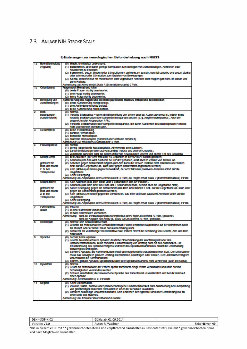
DZHK-SOP-K-02 Gültig ab: 01.09.2014
Version: V1.0 Autor: R. Wachter Seite 46 von 49
*Die in diesem eCRF mit ** gekennzeichneten Items sind verpflichtend einzuhalten (= Basisdatensatz). Die mit * gekennzeichneten Items sind nach Möglichkeit einzuhalten.
7.3 ANLAGE NIH STROKE SCALE
Anamnese/Klinische Diagnosen
DZHK-SOP-K-02 Gültig ab: 01.09.2014
Version: V1.0 Autor: R. Wachter Seite 47 von 49
*Die in diesem eCRF mit ** gekennzeichneten Items sind verpflichtend einzuhalten (= Basisdatensatz). Die mit * gekennzeichneten Items sind nach Möglichkeit einzuhalten.

DZHK-SOP-K-02 Gültig ab: 01.09.2014
Version: V1.0 Autor: R. Wachter Seite 48 von 49
*Die in diesem eCRF mit ** gekennzeichneten Items sind verpflichtend einzuhalten (= Basisdatensatz). Die mit * gekennzeichneten Items sind nach Möglichkeit einzuhalten.
DZHK-SOP-K-02 Gültig ab: 01.09.2014
Version: V1.0 Autor: R. Wachter Seite 49 von 49
*Die in diesem eCRF mit ** gekennzeichneten Items sind verpflichtend einzuhalten (= Basisdatensatz). Die mit * gekennzeichneten Items sind nach Möglichkeit einzuhalten.