DYSONTOGENETIC PITUITARY CYSTS - Europe PubMed Central
13
DYSONTOGENETIC PITUITARY CYSTS (PITUITARY CACHEXIA IN CHILDHOOD) BY H. S. BAAR, M.D. (From The Children's Hospital, Birmingham) . The confused classification of pituitary tumours was clarified at the beginning of this century by the work of Erdheim (1904, 1926). In a series of exemplary papers, he gave proofs that a variety of solid and cystic tumours of the pituitary gland and its stalk have a common origin in the displaced stratified squamous epithelium of the hypophyseal duct. This histogenesis is well expressed in the designation ' hypophyseal duct tumours ' (Erdheim) or craniopharyngeomata. Recently Ingraham and Scott (1946) rightly objected to the term cranio- pharyngeoma, pointing out that the hypophyseal 'duct develops from the ectodermal stomodeum and not from the entodermal primitive pharynx. They suggested tentatively the term ' craniostomodeoma.' The designation ' craniopharyngeoma ' however, being in common use, will be retained in this paper. While the experimental work on the pituitary has made spectacular progress, very little has been added since Erdheim's classical monograph (1926) to our knowledge of the morphogenesis of pituitary tumours. Unfortunately much confusion has been caused by the use of such names as ' tumours of Rathke's pouch,' ' cysts of Rathke's pouch;' ' adamantinomata of the pituitary,' etc. The use of the term ' tumour of Rathke's pouch' as synonymous with craniopharyngeoma is particularly deplorable, as will be obvious by reference to the developmental relationship of Rathke's pouch and its cavity to various parts of the pituitary gland. This shows that Rathke's pouch is at first a rather flat bag which, after frontal turning of the lateral and later cranial seams, resembles a basket. In the frontal wall of the basket a midsagittal crest is then formed, which in cross section appears as a solid process. By proliferation of the crest and of the lateral (frontally turned over) seams, a large number of solid glandular columns and narrow tubules are produced, which, with the invaded vascular con- nective tissue, form the main mass of the adeno- hypophysis. The upper seam with a few wider tubules probably gives rise to the pars tubularis and some of the cysts of the medullary zone (Hochstetter, 1924; Benda, 1932; Guizetti 1933). After obliteration of the craniopharyngeal duct, the original cavity of Rathke's pouch is represented in embryonic life by the 'primary Rathke's cyst,' which in young infants still persists in the form of Rathke's cleft (Koelliker's space =Frazer's pituitary lake, Frazer, 1921). By progressive segregation from the cleft, which was originally single, small colloid cysts are formed in the medullary or inter- mediate zone of the pituitary; these Kraus (1926) calls ' secondary Rathke's cysts.' However, the cysts of the medullary zone, which latter is often erroneously called 'pars intermedia,' only in part originate from the segregation and partial oblitera- tion of Rathke's cleft; some are the product of cellular degeneration, and others probably arise from hollow glandular buds of the cranial seam of the pituitary basket (Collin, 1923; Benda, 1932; Berblinger, 1932; Guizzetti, 1933; Selye, 1943). Usually the origin of the cysts is not recognizable from their morphology. Bailey (1932) says that cysts deriving from Rathke's cleft may be dis- tinguished from other cavities in this region by their being lined by ciliated epithelium; this state- ment is misleading, because an intact Rathke's cleft is often seen in young infants, where ciliated epithelium is found only occasionally and in limited areas. Cysts lined by ciliated epithelium are, except for the suprasellar ones, certainly derived from the original Rathke's cyst, but the majority of colloid cysts, produced by the segregation of Rathke's cleft, are lined by cubical and not by ciliated epithelium. The craniopharyngeal duct is pulled upwards anteriorly during the proliferation of the anterior wall of Rathke's pouch, and in the rare cases of persisting craniopharyngeal duct the cranial end of the intrasphenoid canal is situated in the anterior part of the sella, in front of the eminentia olivaris. 'The region of the insertion of the erstwhile hypophyseal duct is carried upwards, by the . . . rotation of the developing gland, to the anterior infundibular and upper pars anterior surfaces' (Duffy, 1920). How far upwards the insertion of 118
Transcript of DYSONTOGENETIC PITUITARY CYSTS - Europe PubMed Central

DYSONTOGENETIC PITUITARY CYSTS(PITUITARY CACHEXIA IN CHILDHOOD)
BY
H. S. BAAR, M.D.
(From The Children's Hospital, Birmingham)
. The confused classification of pituitary tumourswas clarified at the beginning of this century by thework of Erdheim (1904, 1926). In a series ofexemplary papers, he gave proofs that a varietyof solid and cystic tumours of the pituitary glandand its stalk have a common origin in the displacedstratified squamous epithelium of the hypophysealduct. This histogenesis is well expressed in thedesignation ' hypophyseal duct tumours ' (Erdheim)or craniopharyngeomata. Recently Ingraham andScott (1946) rightly objected to the term cranio-pharyngeoma, pointing out that the hypophyseal'duct develops from the ectodermal stomodeum andnot from the entodermal primitive pharynx. Theysuggested tentatively the term ' craniostomodeoma.'The designation ' craniopharyngeoma ' however,being in common use, will be retained in this paper.While the experimental work on the pituitary hasmade spectacular progress, very little has been addedsince Erdheim's classical monograph (1926) to ourknowledge of the morphogenesis of pituitarytumours. Unfortunately much confusion has beencaused by the use of such names as ' tumours ofRathke's pouch,' ' cysts of Rathke's pouch;'' adamantinomata of the pituitary,' etc. The useof the term ' tumour of Rathke's pouch' assynonymous with craniopharyngeoma is particularlydeplorable, as will be obvious by reference to thedevelopmental relationship of Rathke's pouch andits cavity to various parts of the pituitary gland.This shows that Rathke's pouch is at first a ratherflat bag which, after frontal turning of the lateraland later cranial seams, resembles a basket. In thefrontal wall of the basket a midsagittal crest is thenformed, which in cross section appears as a solidprocess. By proliferation of the crest and of thelateral (frontally turned over) seams, a large numberof solid glandular columns and narrow tubules areproduced, which, with the invaded vascular con-nective tissue, form the main mass of the adeno-hypophysis. The upper seam with a few widertubules probably gives rise to the pars tubularis andsome of the cysts of the medullary zone
(Hochstetter, 1924; Benda, 1932; Guizetti 1933).After obliteration of the craniopharyngeal duct, theoriginal cavity of Rathke's pouch is represented inembryonic life by the 'primary Rathke's cyst,'which in young infants still persists in the form ofRathke's cleft (Koelliker's space =Frazer's pituitarylake, Frazer, 1921). By progressive segregationfrom the cleft, which was originally single, smallcolloid cysts are formed in the medullary or inter-mediate zone of the pituitary; these Kraus (1926)calls ' secondary Rathke's cysts.' However, thecysts of the medullary zone, which latter is oftenerroneously called 'pars intermedia,' only in partoriginate from the segregation and partial oblitera-tion of Rathke's cleft; some are the product ofcellular degeneration, and others probably arisefrom hollow glandular buds of the cranial seam ofthe pituitary basket (Collin, 1923; Benda, 1932;Berblinger, 1932; Guizzetti, 1933; Selye, 1943).Usually the origin of the cysts is not recognizablefrom their morphology. Bailey (1932) says thatcysts deriving from Rathke's cleft may be dis-tinguished from other cavities in this region bytheir being lined by ciliated epithelium; this state-ment is misleading, because an intact Rathke's cleftis often seen in young infants, where ciliatedepithelium is found only occasionally and in limitedareas. Cysts lined by ciliated epithelium are, exceptfor the suprasellar ones, certainly derived from theoriginal Rathke's cyst, but the majority of colloidcysts, produced by the segregation of Rathke's cleft,are lined by cubical and not by ciliated epithelium.The craniopharyngeal duct is pulled upwards
anteriorly during the proliferation of the anteriorwall of Rathke's pouch, and in the rare cases ofpersisting craniopharyngeal duct the cranial end ofthe intrasphenoid canal is situated in the anteriorpart of the sella, in front of the eminentia olivaris.'The region of the insertion of the erstwhilehypophyseal duct is carried upwards, by the . . .
rotation of the developing gland, to the anteriorinfundibular and upper pars anterior surfaces'(Duffy, 1920). How far upwards the insertion of
118

PITUITARY CACHEXIAthe craniopharyngeal duct, which is originallysituated at the caudal end of the pouch, can becarried by the developing glandular mass is wellseen in a figure of Hochstetter's, where a persistingrest of the hypophyseal duct is seen inserted high(i.e. cranially) on the ventral aspect of the adeno-hypophysis. This explains the main localization ofdisplaced squamous epithelium on the anterioraspect of the pituitary, and especially of its stalkand the corresponding situations of craniopharyn-geomata. Schematic drawings and reproductionsof wax models ijiustrating the developmentalanatomy of the pituitary may be found in papersof Mihalkovics (1875), Hochstetter (1924), Erdheim(1926) and Benda (1932). The whole anterior lobeof the pituitary being derived from the wall ofRathke's pouch, the designation- 'tumours ofRathke's pouch' could, in fact, be applied withmore justification to adenomata of the pituitary thanto craniopharyngeomata, which are derived fromdisplaced squamous epithelium of the cranio-pharyngeal duct, which in embryos of 12 to16 mm. crown-rump length is already welldifferentiated from Rathke's pouch proper. If, onthe other hand, the term ' Rathke's pouch ' is usedas synonymous with ' primary Rathke's cyst,' whichis done by Worster-Drought et al. (1927) thedesignation ' cysts and tumours of Rathke's pouch 'can be applied only to those arising from thepituitary cleft and its lining epithelium, and not tothe vast majority of craniopharyngeomata. Theterm ' tumour of Rathke's pouch ' thus leads toconfusion of craniopharyngeomata with cysts(either colloid or ciliated epithelial) which developfrom the primary or secondary Rathke's cysts andwhich have histogenetically nothing in commonwith the former. Frazier and Alpers (1934), whodo not agree with the present classification ofpituitary tumours, proposed a classification into(1) adenomata, (2) tumours of the pituitary stalk,and (3) tumours of Rathke's cleft. Except for theadenomata, this classification is purely topo-graphical, makes no distinction between neoplasticand non-neoplastic cysts, and cannot be consideredas satisfactory when a morphogenetic classificationis possible.
It is hoped that the publication of the twofollowing cases may contribute to a more satis-factory classification of pituitary cysts. Moreoverthe cases present several other points of interest.In the first place they are examples of the very rare
pituitary cachexia in young children. The few casesof Simmonds' disease in childhood (Thomas, 1933;Goebel, 1932; Geldrich, 1939; and others) were
almost all only clinically observed. Only one case
(Simmonds, 1916) is mentioned in Graubner's
(1925) review and Escamillo and Lisser (1942),reviewing all cases of pituitary cachexia, foundamong 111 cases verified by autopsy, only one caseunder ten years. Secondly, it is exceptional foreven large colloid cysts, such as those described inthis paper, to cause cinical symptoms. Goldzieher(1913) described a large colloid cyst causing diabetesinsipidus, and Kiyono (1926) a case of pituitarycachexia due to similar cyst.* Merz(1930)describeda case of pituitary cachexia due to a pea-sizedcolloid cyst between the anterior and posterior lobes,but the presence of sclerotic changes at the insertionof the pituitary stalk makes the interpretation of hiscase somewhat uncertain. Finally, in one patientdescribed in the present paper, the pituitary cyst wasassociated with syringomyelia, and in the other withfibrocystic dystrophy of the pancreas. While thefirst association can be regarded as a simultaneousoccurrence of two developmental disturbances, thepossibility of a causal connexion between pituitaryand pancreatic disease cannot be ruled out and willbe discussed below.
Case 1After seven months' gestation, a boy was born
on March 15, 1941; he was admitted to theChildren's Hospital, Birmingham, on July 20, 1945.The child thrived normally until the age of eightmonths, when he developed pneumonia, for whichhe was in hospital for several months. Before thishe could stand and walk with assistance. A fewweeks after his discharge he showed so little improve-ment that he was readmitted. He remained inhospital until the age of two and a half years. Healways had a cough, variable in severity, and a poorappetite. He had no difficulty in swallowing, butthe act often produced coughing, which in its turncaused vomiting; he never vomited except aftercough. He drank well. He could not sit up byhimself, but was able to feed himself when he feltstrong enough. He liked to play with an engine,and he would look at a picture book for hoursand was not destructive. At the time of his dis-charge from hospital he was not able to talk much,but uttered words such as ' gee gee.' His sleep wasrestless. He had no fits. Bowel and urinaryfunctions were good, but if he had been vomitingduring the day he had nocturnal enuresis.
Findings on admission. He was an emaciatedchild who did not smile. He knew the names of afew common objects, but the speech was confinedto single words. He seemed to want to rousehimself, but could not succeed. He kept his headon one side, being unable to hold it up, and he satonly with support. There was some nystagmus inall directions, and ? fibrillation of the tongue.There was frequent, unexplosive cough, the chestwas full of bronchial sounds, the abdomen normal,
* The paper of Jedlidka (Sborn. ilk., 1924, 25, 149) describing anapparently similar case was unfortunately not available.
119

ARCHIVES OF DISEASE IN CHILDHOOD
all reflexes brisk, and the voice monotonous. OnJuly 21, 1945, the tongue protruded with difficultyand there was fibrillation, though there was nonein the hands, nor any wasting of small musclegroups. On July 22 he regurgitiated through thenose, and the next day he was weaker and thetemperature, pulse, and respiration rates were raisedand the colour poor. He died on July 24, 1945,and necropsy was performed nine hours after death.
Post-mortem findings. The body was of anextremely emaciated, pale boy. It measured 87 5 cm.long (normal average, 102 cm.). The sitting heightwas 50 cm. (normal, 56 cm.), and the length ofthe legfrom the anterior superior iliac spine to the internalmalleolus was 42 5 cm. (normal, 47 5 cm.). (Thenormal figures for body length are taken fromKornfeld, 1929; and other measurements fromBrock, 1932; and the weights of normal organs inchildhood from Copolletta and Wolbach, 1933.)The thyroid and parathyroid glands appeared
normal to the naked eye. The thymus was grosslyatrophic. There were extensive and firm adhesionsbetween the pulmonary pleura over the right lungand the costal pleura, and some fibrinous exudateover the lower lobe of the left lung. Moderatelydilated bronchi, filled with pus, were seen on thecut surface of both lungs. In the lower lobe of theleft lung there were numerous areas of consolida-tion, brownish-red in colour; there were areas ofcollapse in the right lung. The tracheo-bronchiallymph nodes were markedly enlarged, soft andhyperaemic. The largest was at the bifurcationand was almost as large as a walnut. The peri-cardium was normal. The liver was moderatelycongested. The testicles, each about the size of apea, were in theinguinal canals.The sella turcica was normal in sagittal and
frontal diameters. The pituitary appeared (oninspection after an incision into the diaphragmsellae) to be replaced by a lArge pea-sized, thin-walled cyst which apparently filled the whole sella.It was not dissected immediately, but the wholebody of the sphenoid bone was removed, fixed informol saline, decalcified, and dissected in the mid-saggital plane, one half being embedded in paraffinand the other saved. The spinal cord was savedbut not dissected immediately.The costo-chondral junctions were normal on
naked-eye examination. The bone mar ow of theright femur was dark red throughout.
Histological findingsPITUITARY. There was considerable shrinkage of
the pituitary affecting mainly the cyst, 'which had anantero-posterior diameter of 3 mm. The anteriorlobe was reduced to a narrow band which onsagittal section had the shape of a sickle. Thepointed end of the sickle was at the floor of the sellaturcica, and the diameter increased gradually andreached its maximum of I 2 mm. at the antero-supenor aspect of the cyst. The posterior lobe had
a maximal diameter of I 5 mm. The cyst wassituated between the two lobes of the pituitary andextended downwards to the periostium of the sella.(Fig. 1, and Plate Ia.) It was filled with a homo-genous material, which appeared pale pink in
FIG. 1.-Schematic reconstruction of the immediatepost-mortem findings on the pituitary in case 1.A.L.=anterior lobe; C.C.=colloid cyst; P.L.=pos-terior lobe; D.S.=dorsum sellae; S.T.=stalk.
haematoxyline-eosin-stained sections and pale bluishin trichrome stain, and which showed fuchsinophil,fuchsinophobe, and tannin-fast areas by Kraus'(1914) colloid stain. The lining of the cyst con-sisted of a single, in a few places double, layer offlat or cubical cells which did not show any granula-tion in their cytoplasm (Plate Ib). Their nucleiwere dark, oval or spherical, their cytoplasm paleand amphophil. A few small colloid cysts werealso seen along the anterior aspect of the posteriorlobe. Here, and occasionally on the posterioraspect, several glands were seen which had theappearance of salivary glands. Such glands are notuncommon in pituitaries. Erdheim (1903) con-sidered them as identical with salivary glands. Thestructure of the posterior lobe was normal. In theanterior lobe the cells were arranged in parallel,longitudinal strands, instead of the normal tortuouscolumns, as if compressed between the bone andthe cyst. The sinusoids between the cellular strands,however, were wide and engorged, thus suggestingthat the cellular disarrangement was due to achronic pressure directing the growth of the glandand not to an immediate mechanical effect. Thenumerical relationship between the acidophil andbasophil cells was approximately normal. Thechromophobe cells were rather scanty. There wasno follicle formation in the anterior lobe, andnowhere within this part was there any evidence ofcolloid degeneration.THYROID. The vesicles of the thyroid were
normal in size; theywere lined bycuboid or columnarepithelium. Colloid, which was present in a fewvesicles only, showed a pale eosin stain and had insome places a vacuolated appearance. Desquamatedepithelial cells were seen in many vesicles.
PARATHYROID. Only one parathyroid gland wasexamined. It measured 1I7 by 0-7 mm. ThereN
120

PITUITARY CACHEXIAwere fairly large areas of fat tissue between the cells,which had all the character of water-clear ' chiefcells ' (Plate Ic). Normally no fat cells are seen inthe interstitial tissue of the parathyroids before thefifth year of life (Erdheim, 1903).
SUPRARENALS. When the suprarenal glands wereexamined, only the cells of the zona glomerulosashowed an approximately normal foamy appearance.The cells of the zona fasiculata and reticularis wereswollen, their cytoplasm was slightly granular, andpale pinkish in haematoxylineosin stained sections.The seminiferous tubules of the testis were
narrow, lined by one to three layers of cubical orcolumnar cells with dark nuclei, and separated bybroad strands of interstitial tissue. The latter wascellular with numerous fibroblasts in some areas,acellular with broad collagenous fibres in others.Interstitial cells of Leydig were seen only occasion-ally (Plate Id).THE MEDULLA. This showed an increased amount
of glia at the floor of the fourth ventricle, andspecially in the region of the hypoglossal nuclei.There was marked rarefaction, chromatolysis ofnerve cells, and 'fading away ' of nerve cells withformation of ghost cells.THE LUMBAR CoRD, The lumbar cord showed
the central canal replaced by a cavity 5i by 3 mm.in diameter in its widest part. The cavity containedsome disintegrated nerve tissue. Small parts of itscircumference were lined by a single layer of cuboidor columnar cells in epithelial arrangement, thecells with very few exceptions not being ciliated.There was a ring of fibrillary glia, about 0 3 mm. indiameter, surrounding the cavity (Plate le and f).The glia fibres were in loose arrangement, but hereand there formed patches of dense network. Theastrocytes were poorly impregnated by the Cajalmethod; their processes were irregular and occa-sionally fragmented. Except for a slight increaseof the glia aroumd the central canal, the dorsal andcervical cord showed no pathological changes.
RIBs. The proliferation zone proper of thecartilage was very poorly developed, in many placesscarcely recognizable as such. The columnar zonewas normal in width, but in numerous places wholecolumns had undergone severe degenerative changesand the cells had fused with the ground substanceof the cartilage. The provisional zone of calcifica-tion was interrupted in a few places, and in theseit consisted of small isolated patches of calcifiedground substance. In several places the osteoidseams of the bone trabeculae were definitely broaderthan normal. (Completely decalcified ribs, andribs partially decalcified by Mueller's solution,were examined.)BONE MARROW. In the rib there was a very
cellular bone marrow with numerous normoblastsand myelocytes. In the distal end of the femurthere was fat marrow.Summary of Findings. A four-year-old boy who
suffered from his eighth month of life from indefinite
symptoms of retarded development, muscular weak-ness, loss of appetite, and wasting, died withsymptoms suggestive of bulbar palsy. Necropsyrevealed a large colloid cyst of the pituitary causinga severe atrophy of the anterior lobe. This wasassociated with syringomyelia of the lumbar cordand a mild gliosis and rarefaction in the region ofthe hypoglossal nuclei. At the time of death he was14- 5 cm. shorter than the average for his age. Hisbody length was 86 per cent. and the length of thelower limbs 89 per cent. of the normal figure.He may, therefore, be considered as of normalproportion. There was a lipomatosis of a para-thyroid, an incomplete descent of testicles withatrophy and interstitial fibrosis of the latter. Inaddition there was a chronic ulcero-necroticbronchitis, peribronchitis, bronchiectasis, andadhesive pleurisy.
Case 2A girl of eight years of age was well until April,
1945, when she started to have eight or nine veryoffensive, loose, yellow stools daily. She lost herappetite. She had an attack of diarrhoea twelvemonths before admission, but recovered in threeor four days. On admission on July 17, 1945, shewas very emaciated, and talked with a slow,monotonous voice; her feet were pale and cold,and there was excessive muscular wasting inher legs. There was slight oedema, and a purpuricrash. On the knees were depressed scales aboutthe size of the head of a pin, surrounded by narrowrings of erythema. There was latent tetany, butno other relevant findings. A five days' collectionof stools showed a daily output of 18 4 g. of fat.The total fat was 43 -2 per cent. of the dried faeces,and the neutral fat 15 8 per cent. The blood ureawas 29 mg. per 100 c.cm., the albumin 1-77 g., andthe globulin 1 15 g. There was 248 mg. sodiumper 100 c.cm. serum, 477 mg. chlorine (calculatedas sodium chloride), 10 mg. pyruvic acid, 6-0 mg.calcium, 4 - 4 mg. phosphorus, and 8 * 5 mg. inorganicphosphorus liberated in three hours, at 370 C. Thewater-elimination test for Addison's disease wasinconclusive. The test of Robinson et al. (1941)was performed on Aug. 12 and 13, and the resultingquotient was 5 * 7 (normal more than 25). Anotherestimation of faecal fat in the period from Aug. 9to 14 showed a daily output of 24-8 g., with thetotal fat forming 52 3 per cent. and the neutralfat 18 0 per cent. of the dried faeces.The blood findings are shown in the table and in
the Price Jones curve (fig. 2). The mean corpuscularvolume was 81 -6 to 85-2 p3. The blpeding timewas one and a half minutes and the clotting timethree minutes (Aug. 17, 1945). An oral and anintravenous glucose tolerance curve showed no grossabnormality. Two units of insulin injected intra-venously caused a depression of the blood-sugarlevel from the fasting level of 104 mg. per 100 c.cm.to 42 mg. per 100 c.cm. after twenty-three minutes.and after sixty-two minutes it was 62 mg. per
121

ARCHIVES OF DISEASE IN CHILDHOOD100 c.cm. The Mantoux test was negative with approximately in the middle. The right lobe of the0 1 and with 1 mg. tuberculin. The child was given thyroid measured 30 by 12 by 9 mm., the leftplasma intravenously, calcium chloride by mouth, 28 by 9 by 6 mm., the cut surface being moist,and, from Aug. 18, 5 mg. percortin daily. There granular, and lustrous yellowish pink.was a transient improvement after the percortin The thymus was grossly atrophic. The hearttreatment, but after this a rapid deterioration, and measured 8* 2 by 7* 5 by 3 * 6 cm., and weighed 67 g.the child died on Sept. 20, 1945. Her weight was (empty) (normal, 80 to 130 g.). The wall of the
left ventricle was 9 mm. and that of the right 2 mm.thick. The heart muscle was pale, flabby, and
so MD-934 MACROCYTOSBS -65 /. friable. The valves and -the septa were normal.-I42,.A MICROCtTOSS -0 2Y/ The ductus arteriosus was obliterated, and a shallow
40 v-sZ pit on the aortic side was noticeable.
30 4 / \ There was one pinhead-sized subserous haemor-rhage on the anterior aspect of the stomach near the
20 / \ greater curvature. Very little fat was present in the20 \great omentum and in the appendices epiploicae./0 The anus was rather wide; the blood vessels of the
mesentery somewhat distended and engorged. The0 5 6 7 a 9 If 12 13,r stomach was distended and contained about a
FIG. 2.-Price Jones curve of case 2. handful of a greyish material of the consistency ofporridge. Its mucous membrane was normal.The contents of the duodenum were bile-stained,
3 st. 1 1 lb. one week after the onset of the disease, and bile was easily expressed from the gall bladderand 1 st. 10 lb. 14 oz. two days before death. by manual pressure. The small intestine was
Post-Mortem findings. The body, which was of collapsed, and the large intestine, especially thean extremely emaciated girl, measured 116 cm. long pelvic colon, considerably distended. The contents(average, 123 cm.). The distance from the acromion of the small intestine were green, liquid, and some-
to the tip of the middle fingers was 50 cm. (normal, what slimy. The large intestine contained watery-
53 * 5 cm.); and from the spina iliaca anterior to the green faeces. The mucous membrane of themalleous externus, 56 25 cm. (normal, 62 5 cm.). intestine was normal, and the lymphatic apparatusThe skin was pale, with a greyish-yellow tinge. rather atrophic. The mesenteric lymph nodes were
Ecchymoses, pin-head to millet-sized, occasionally lentil to pea-sized, greyish-white, and normal inlentil-sized, and not sharply demarcated, were consistency.situated mainly on the anterior aspect of the chest; The liver weighed 1 lb. and showed fatty changesbut there were a few on the anterior abdominal wall and some congestion. The bile ducts were patent.and on the limbs, a single one 11 by 5 mm. in The pancreas weighed 34 g. and was rather firm,diameter on the interior aspect of the lower third of its cut surface being normal in appearance. Thethe left thigh, and another measuring 10 by 5 mm. spleen measured 6 5 by 49 5 by 1*5 cm. and weighedon the radial side of the back of left hand, between 27 g. (normal, 69 g.). The capsule was smooth,the metacarpes of the thumb and the second finger. the colour pale salmon red, and the consistencyThere were no pubic and axillary hairs. The normal. On the cut surface the Malpighian bodiessubcutaneous fat tissue was almost absent, being were fairly large, and the pulp bright red and notreduced to small patches, saffron yellow in colour. diffluent. The right adrenal weighed 3 * 3 g., andThe bone marrow of the sternum was a dark the left 3 5.; their cortex was greyish and poor inraspberry red. The tongue showed atrophy of the lipoids. The left kidiey measured 7 by 4- 5 by 3 cm.
filiform papillae and several blackish-grey patches and weighed 59 g. (normal, 75 g.); the right
TABLE
NeutrophilHb. g. per Erythro- Colour Reticu- Leuco- Plate- Eosino- Mono- Lympho-
Date c.cm. of cytes Index cytes cytes lets band sgm. phils cytes cytesblood (million) %% iX % % % %
July 23 9 3 ? 3 6 ?0 88 1 8 6,250 290,000 2-0 44-5 00 6 0 47 5
Aug. 17 9 6 2 78 1 22 3 4 3,900 222,000 2-0 42-0 3 0 3 0 50.0
Aug. 23 9 8 2 98 1 18 6|6 6,000 307,000 1 0 57 0 1 0 5 0 36-0
Sept. 6 10 8 2-58 1 52 0 8 11,250 337,000 3 0 8400 00 10 12 0
122

PITUITARYmeasured 8 by 4 5 by 3 5 cm., and weighed 61 g.(normal, 74 g.). Their capsules were easily strippedoff, and the surface was smooth and pale yellowish-red. On the cut surface the medulla was dark red;the normal markings of the cortex were indistinct.The renal pelves appeared normal. The right ovaryweighed 0-71 g., the left 0 66 g., and they appearednormal.The vault of the skull showed on both sides of
the sagittal suture, close to the coronary suture,triangle-shaped, thin, white, transparent, non-flexible areas. In other areas dark red bone marrowwas visible in transmitted light. The thickness ofthe vault was between 1 and 2 mm. The appearanceof the internal aspect was normal. The distancebetween the anterior cinoid processes was 24 mm.,that between the posterior 16 mm. The pituitaryappeared normal from above, but when it wasremoved from the sella a thin-walled cyst, 8 by8 by 3 mm. with clear, yellowish, apparentlygelatinous contents was found replacing a consider-able part of its anterior lobe. The size of the wholepituitary was 11 by 8 by 3 mm., its weight being0 341 g. The depth of the sella turcica was 7 mm.and its midsagittal diameter 9 mm.The pineal gland measured 14 by 8 by 2 mm. and
appeared normal. The labia majora were scarcelydiscernible, the clithoris rather small, and thevagina and uterus normal. On the sixth left rib theproliferation zone of the cartilage was 2 mm. indepth, and the provisional calcification zone wasstraight, sharply defined, and about 0 25 mm. inwidth.The femur was easily sawn through. Its cortex
and the trabecu6ae of the cancellous bone were thin.The epiphyses were ossified, leaving a narrowepiphyseal cartilage. The bone marrow of theproximal three quarters of the femur was raspberry-red and that of the distal quarter was pale, greyish-white, and gelatinous. The marrow of the proximalepiphysis was red, and that of the distal epiphysispale greyish-white.
Histological findingsPITUITARY. The Rawthke's cleft was very con-
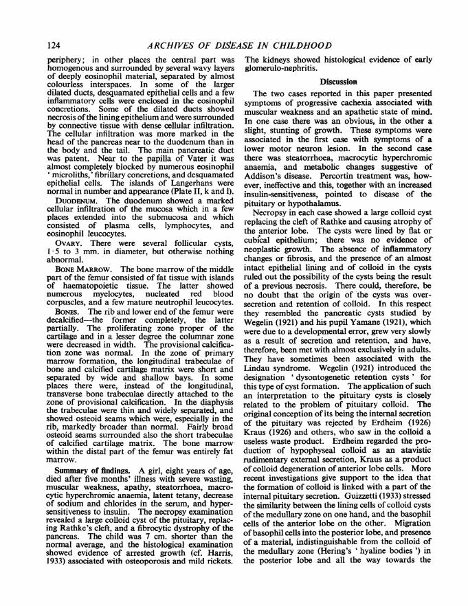
siderably dilated, and, on midsagittal section,triangle-shaped. It contained a large amount offuchsinophobe colloid with a small admixture offuchsinophil and tanninfast colloid. The wall ofthe cyst was ruptured and the cyst collapsed, and aconsiderable amount of colloid was seen outsidethe cyst. The lining epithelium of cyst was cubical,and the cells showed no granulation. There were afew small colloid cysts between the posterior wallof the main cyst and the posterior lobe of thepituitary (Plate I, g, h, Plate II, j, and fig. 3). Theanterior lobehad amaximalantero-posterior diameterof2 - 5 mm. Basophil cells were plentiful in all areas.In some areas the eosinophil cells were almostabsent, while in other areas they were increased innumber at the expense of chromophobe cells(compensatory hypertrophy: Berblinger, 1927).
CACHEXIA 123THYMus. There was atrophy of the thymus, with
marked lipomatous metamorphosis.THYROID. The vesicles were large and filled with
a colloid which in some places showed a few largevacuoles, in other places numerous small vacuoles.
FiG. 3.-Schematic reconstruction of the immediatepost-mortem findings on the pituitary in case 2. A.L.= anterior lobe; P.L.== posterior lobe; ST. =stalk;C.C.=colloid cyst; D.S. =dorsum sellae.
KIDNEY. The glomeruli showed a slight intra-capillary hyalinization. The hyaline was presentonly in a few loops of the tuft, the majority of loopsbeing widely patent. The first convoluted tubuliwere markedly dilated and filled either with a pale,foamy material or with hyaline casts. The liningepithelium was cuboid or flat. Many of theepithelial cells showed large vacuoles (no fat stainmade). A foamy material similar to that in thefirst convoluted tubuli was also seen in the Henleloops and in the collecting tubuli. The renal pelvisshowed an epithelial proliferation, and in a fewplaces a modified epithelium with the characters ofa stratified squamous epithelium without comifica-tion.
SPLEEN. The malpighian bodies were normal,the venous sinuses narrow, and Billroth's cords verycellular with numerous polymorphonuclear leuco-cytes.
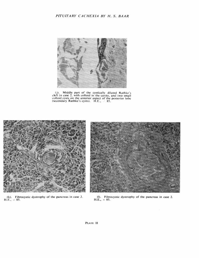
PANCREAS. The interlobular connective tissuewas considerably increased in amount. It consistedoffibroblasts, thick collagenous fibres, and numeroussmall round cells with an occasional polymorphonu-clear leucocyte. This connective tissue invaded theindividual lobules, which showed an advancedatrophy of the secretory tissue. They consisted ofducts, infundibuli, and small, usually crescent-shaped, groups of secretory cells attached to thetips of the infundibuli. The acinus cells weremodified and more narrow than normal, andzymogen granules were absent. When they wereisolated, it was often difficult to decide whether wehad to deal with an acinus or a cross section of aduct. Between these parenchyma cells there was alarge amount of loose connective tissue with patchesof round-cell infiltration. Many of the inter- andintralobular ducts were markedly dilated and filledwith an eosinophil homogenous or stratified material.In some places the latter showed a pale granularcentre and a homogenous, intensely eosinophil
ARCHIVES OF DISEASE IN CHILDHOOD
periphery; in other places the central part washomogenous and surrounded by several wavy layersof deeply eosinophil material, separated by almostcolourless interspaces. In some of the largerdilated ducts, desquamated epithelial cells and a fewinflammatory cells were enclosed in the eosinophilconcretions. Some of the dilated ducts showednecrosis ofthe lining epithelium and were surroundedby connective tissue with dense cellular infiltration.The cellular infiltration was more marked in thehead of the pancreas near to the duodenum than inthe body and the tail. The main pancreatic ductwas patent. Near to the papilla of Vater it wasalmost completely blocked by numerous eosinophil' microliths,' fibrillary concretions, and desquamatedepithelial cells. The islands of Langerhans werenormal in number and appearance (Plate II, k and 1).DUODENUM. The duodenum showed a marked
cellular infiltration of the mucosa which in a fewplaces extended into the submucosa and whichconsisted of plasma cells, lymphocytes, andeosinophil leucocytes.OVARY. There were several follicular cysts,
1*5 to 3 mm. in diameter, but otherwise nothingabnormal.BONE MARROW. The bone marrow of the middle
part of the femur consisted of fat tissue with islandsof haematopoietic tissue. The latter showednumerous myelocytes, nucleated red bloodcorpuscles, and a few mature neutrophil leucocytes.
BoNEs. The rib and lower end of the femur weredecalcified-the former completely, the latterpartially. The proliferating zone proper of thecartilage and in a lesser degree the columnar zonewere decreased in width. The provisional calcifica-tion zone was normal. In the zone of primarymarrow formation, the longitudinal trabeculae ofbone and calcified cartilage matrix were short andseparated by wide and shallow bays. In someplaces there were, instead of the longitudinal,transverse bone trabeculae directly attached to thezone of provisional calcification. In the diaphysisthe trabeculae were thin and widely separated, andshowed osteoid seams which were, especially in therib, markedly broader than normal. Fairly broadosteoid seams surrounded also the short trabeculaeof calcified cartilage matrix. The bone marrowwithin the distal part of the femur was entirely fatmarrow.Summary of findings, A girl, eight years of age,
died after five months' illness with severe wasting,muscular weakness, apathy, steatorrhoea, macro-cytic hyperchromic anaemia, latent tetany, decreaseof sodium and chlorides in the serum, and hyper-sensitiveness to insulin. The necropsy examinationrevealed a large colloid cyst of the pituitary, replac-ing Rathke's cleft, and a fibrocytic dystrophy of thepancreas. The child was 7 cm. shorter than thenormal average, and the histological examinationshowed evidence of arrested growth (cf. Harris,1933) associated with osteoporosis and mild rickets.
The kidneys showed histological evidence of earlyglomerulo-nephritis.
DiscussionThe two cases reported in this paper presented
symptoms of progressive cachexia associated withmuscular weakness and an apathetic state of mind.In one case there was an obvious, in the other aslight, stunting of growth. These symptoms wereassociated in the first case with symptoms of alower motor neuron lesion. In the second casethere was steatorrhoea, macrocytic hyperchromicanaemia, and metabolic changes suggestive ofAddison's disease. Percortin treatment was, how-ever, ineffective and this, together with an increasedinsulin-sensitiveness, pointed to disease of thepituitary or hypothalamus.
Necropsy in each case showed a large colloid cystreplacing the cleft of Rathke and causing atrophy ofthe anterior lobe. The cysts were lined by flat orcubical epithelium; there was no evidence ofneoplastic growth. The absence of inflammatorychanges or fibrosis, and the presence of an alnostintact epithelial lining and of colloid in the cystsruled out the possibility of the cysts being the resultof a previous necrosis. There could, therefore, beno doubt that the origin of 'the cysts was over-secretion and retention of colloid. In this respectthey resembled the pancreatic cysts studied byWegelin (1921) and his pupil Yamane (1921), whichwere due to a developmental error, grew very slowlyas a result of secretion and retention, and have,therefore, been met with almost exclusively in adults.They have sometimes been associated with theLindau syndrome. Wegelin (1921) introduced thedesignation ' dysontogenetic retention cysts ' forthis type of cyst formation. The application ofsuchan interpretation to the pituitary cysts is closelyrelated to the problem of pituitary colloid. Theoriginal conception of its being the internal secretionof the pituitary was rejected by Erdheim (1926)Kraus (1926) and others, who saw in the colloid auseless waste product. Erdheim regarded the pro-duction of hypophyseal colloid as an atavisticrudimentary external secretion, Kraus as a productof colloid degeneration of anterior lobe cells. Morerecent investigations give support to the idea thatthe formation of colloid is linked with a part of theinternal pituitary secretion. Guizzetti (1933) stressedthe similarity between the lining cells of colloid cystsof the medullary zone on one hand, and the basophilcells of the anterior lobe on the other. Migrationof basophil cells into the posterior lobe, and presenceof a tnaterial, indistinguishable from the colloid ofthe medullary zone (Hering's 'hyaline bodies') inthe posterior lobe and all the way towards the
124

PITUITARY CACHEXIAhypothalamus, has been demonstrated by severalauthors (Kraus, 1926; Cushing, 1933; Benda, 1942;Selye, 1943). The inundation of hypothalamiccentres with colloid is very marked after removal ofthe upper cervical sympathetic ganglion (Popjak,1940). Selye (1943) has shown that intravenousinjections of hypertonic NaCI-solutions into ratscaused swelling of basophil cells in the anterior lobeof the pituitary gland, eventually with degenerativechanges and at the same time considerable distensionof Rathke's cleft with colloid. On the other handthe presence of symptoms of pituitary cachexia, ofarrested growth, of suprarenal, testicular, andparathyroid changes in our two cases and in thecase of Kiyono (1926), where a large amount ofcolloid was. present in the cystic dilatation ofRathke's cleft, are strong evidence that the mainhormones of the anterior pituitary lobe are notpresent in the colloid secretion. It is also worthyof note that in the two cases presented in this paperthere was no evidence of colloid degeneration in theanterior lobe, and the whole amount of colloid wasapparently the product of the epithelial lining of thecysts. It appears, therefore, conclusive (1) that thecolloid is formed by both apocrine secretion of thelining cells of Rathke's cleft and by holocrine(possibly also apocrine) secretion of basophil cellsof the anterior lobe; and (2) that the colloidformation is linked with only a part of pituitaryhormnone production, probably that associated withthe activity of posterior lobe and the hypothalamiccentres. The problem of 'neurocrine ' formationof colloid by pituicytes and the hypothalamus(Scharrer's (1941) diencephalic gland) is for ourproblem irrelevant.
In the present two cases the cleft of Rathke didnot undergo the normal segregation into a series ofsmall cysts; but instead, as a result of a continuous,increased colloid secretion by its lining epithelium,it became transformed into a large retention cyst,thus causing atrophy and hypofunction of theanterior lobe and an accumulation of a secretionwhich may have an influence on the hypothalamiccentres.As mentioned in the introduction, even com-
paratively large colloid cysts do not usually causesymptoms of hypo- or apituitarism. Why they doso in rare cases is still a debatable problem. It hasbeen repeatedly assumed (Kiyono, 1926 ; Merz,1930) that a separation of the anterior lobe fromthe posterior has similar effects to disease of theanterior lobe. In the first of the two cases describedin this paper there was complete separation, but inthe second a strip of anterior lobe tissue was seenclosely attached to the superior pole of the neuro-hypophysis. Possibly the presence of pharyngeal
hypophysis accounts for the fact that some largecolloid cysts cause no clinical symptoms.
In the first case there was a marked atrophy ofthe testicles, morphological changes in the supra-renals, lipomatosis of at least one parathyroid, andsyringomyelia of lumbar cord. The parathyroidchanges are noteworthy because of the still contestedformation of a parathyrotropic hormone by theanterior pituitary lobe (see Cameron, 1945; Perlman,1944). The changes in the testicles, although verymarked, occur also in other cases of undescendedtesticles (Kyrle, 1910). It is, however, possible,that these are also related to pituitary dysfunction,and according to Gruenwald (1946) they are lessfrequent than previously assumed and changes lessmarked than those found in the present case areconsidered by this author as evidence of under-development. Syringomyelia is apparently anindependent, associated condition. As syringo-myelia is now considered by most authors to be adevelopmental error with persistence of primitiveglia (Tamaki and Lubin, 1938), this associationsupports the interpretation of the pituitary cyst inthe present case as 'dysontogenetic ' in origin.The slight gliosis and rarefaction at the floor of thefourth ventricle is apparently of the same natureas the cavity formation in the lumbar cord. Thereis no reason to associate the changes in the spinalcord of this case with the condition described as' pseudotabes pituitaria ' (Otto, 1936; Snapper et al.,1937).In the second case the colloid cyst of the pituitary
was associated with atrophy and fibrocysticdegeneration of the pancreas. The relationship ofthese two conditions can, in the present state of ourknowledge, only be a matter of speculation. Thepossibility of a causal link between these diseasescannot, however, be disregarded. In a paper readbefore the Association of Clinical Pathologists it waspointed out (Baar, 1944) that what is now generallycalled 'fibrocystic disease of the pancreas' comprisestwo pathogenetically different conditions. In veryrare cases there is histological evidence that the cystsare due to segregation of ducts, pathological pro-liferation of their epithelium, and retention of athin, mucoid, secreted material. This has beenconsidered as the infantile form of the conditiondescribed by Wegelin (1921) and Yamane (1921) as' dysontogenetic pancreatic cystosis.' Wissler andZollinger (1945) recently came to the same conclu-sion. For the common form of the infantilefibrocystic disease of the pancreas, histologicalevidence has been presented to support the opinionof Blackfan and Wolbach (1933) and Blackfan andMay (1938) that a pathological change in thesecretion leading to its inspissation is the primary
125

ARCHIVES OF DISEASE IN CHILDHOODchange, resulting in stagnation of the secretion,cystic dilatation of ducts, infundibuli and acini,their secondary segregation, occasionally necrosisof the epithelium and rupture of ducts, atrophy ofsecretory tissue with inter- and intracinous fibrosis,and secondary inflammatory cellular infiltration.The present case belongs to this second type, forwhich the designation ' fibrocystic dystrophy of thepancreas' was proposed. An attempt was madeto explain these findings in a way which would dojustice to the identical condition in the early neonatalperiod, which is invariably associated with either ameconium ileus or a congenital obliteration withinthe small intestine. A hypothesis was put forwardthat the primary cause is a disturbance in the balancebetween the autonomic and secretin stimuli infavour of the former. In the present case it ispossible that the increased formation of pituitarycolloid caused an increased stimulation of theposterior lobe and of the parasympathetic hypo-thalamic centres. It may be interestipg in thisconnexion to observe that proliferative changes inthe neurohypophysis have been actually describedin Simmonds' disease (Jacob, 1923; Meng, 1928).The writer is fully aware of the hypothetical natureof the views presented but hopes that they maystimulate a more thorough investigation of faecalfats and of changes in the secretory pancreatictissue in cases of pituitary disease.The presence of a disturbance in the electrolytes
resembling that in Addison's disease is of particularinterest. Although hypophysectomy causes anatrophy of the suprarenal cortex, no change occursin the blood sodium, chloride, or potassium levels(Swan, 1940). Cameron (1945) calls cases ofSimmonds' disease with marked symptoms of hypo-corticoadrenalism pituitary Addison's disease, butthe writer is aware of only one case (Moss, 1942) inwhich changes in blood electrolytes similar to thosein the present case were found.The macrocytic hyperchromic anaemia in this
case is probably due to the steatorrhoea and notdirectly to the pituitary disease. Snapper et al.described cases of hyperchromic anaemia andhistamine-refractory achlorhydria in chronicpituitary insufficiency. They stress, however, thefact that the anaemia was a late symptom in thecourse of the disease and developed only afterachlorhydria was present for a considerable period.The hypocalcaemia and latent tetany have alsotheir reasonable explanation in the pancreaticsteatorrhoea and formation of excess calcium soapsin the intestine. The blood was not examined inthe first case, but the presence of haemosiderosisand extramedullary haematopoiesis are evidence ofa haemolytic anaemia.
With regard to the histogenesis of the twopituitary cysts, there can be but little doubt that theyare due to a developmental error. There is not theslightest evidence of neoplastic growth. Both cystshave developed in a position where in embryoniclife there is a flat cavity, the ' primary Rathke'scyst.'. Instead of becoming rudimentary, changinginto the Rathke's cleft and finally segregating intoa few small 'secondary Rathke's cysts,' in thesecases the lining epithelium continued to grow assuch, and to produce a colloid secretion whichaccumulated and caused the formation of a cystgradually increasing in size. In analogy with thenomenclature of Wegelin mentioned above, thedesignation ' dysontogenetic pituitary cysts ' for thistype and the following classification of pituitarycysts is, therefore, suggested.
1. Neoplastic (cystic craniopharyngeoma, themost common form among the pathological cystsof the pituitary, and very occasionally adenoma withcystic degeneration).
2. Dysontogenetic (pathological colloid cysts andciliated epithelial cysts, cases of Goldzieher, Kiyono,Frazier and Alpers and the present two cases, and? Merz's first case).
3. Dystrophic (due to embolism or thrombosisfollowed by necrosis and cystic degeneration:Falta, 1913; Simmonds, 1919; ? Merz, 1930).
4. A combination of 2 and 3 (the only case ofthis type has been described by Priesel in 1920. Inthis case a developmental dystopy of the pituitarycaused a defective nutrition with secondary cystformation and symptoms of pituitary dwarfism).
5. Combination of 2 and 1 (this is represented bythe remarkable case of Worster-Drought et al.,1927). In a girl aged nineteen with pituitarydwarfism, three ciliated epithelial cysts were found,and a neoplastic growth arising from the primitiveepithelium of Rathke's cyst or from the ependymaof the third ventricle (? Duffy's fourth case).
SummaryTwo cases of pituitary cachexia in childhood are
described. Both were due to large colloid cysts ofRathke's cleft with pressure atrophy of the anteriorlobe. The designation ' dysontogenetic pituitarycysts ' for this type, and a classification of thevarious pituitary cysts are proposed. The reasonsare pointed out why the designation 'tumours ofRathke's pouch' for carniapharyngeomata is con-sidered to be a misnomer. The pituitary cachexiawas associated with syringomyelia in one case andwith fibrocystic dystrophy of the pancreas in theother. The possibility of a causal link between thelatter and the pituitary disease is discussed.
126

PITUITARY CACHEXIA 127It is my pleasant duty to thank Sir Leonard G.
Parsons for his helpful criticism and advice;Professor K. D. Wilkinson and Dr. A. V. Neale forpermission to publish their cases, Mr. A. R.Dethendge for cuitting and staining the sections, andMr. J. Gregory Williamson for the photographs.
REFERENCESBaar, H. S. (1944). Meeting of the Association of
Clinical Pathology. London. January.Bailey, P. (1928). Cowdry's spec. Cytol., 2, 484. New
York.(1932). In 'Penfield's Cytology and cellularPathology of the nervous system,' 3, 1133. NewYork.
Benda,,C. (1932). Hirsch's Handb. inneren Sekr., 1,part 6. Leipzig.
Benda' C. E. (1942). J. clin. Endocrinol., 2, 737.Berblinger, W. (1932). Hirsch's Handb. inneren Sekr., 1,
part 6. Leipzig.(1927). Verhandl. Dtsch. path. Ges., 22, 191.
Blackfan, K. D. and Wolbach, S. B. (1933). J. Pediat.,3, 679.
- and May, C. D. (1938). Ibid., 13, 627.Brock, J. (1932). Biol. Daten far den Kinderarzt., 1,
Springer. Berlin.Cameron, A. T. (1945). Recent advances in Endocrin-
ology. London. Churchill.Collin, R. (1923). C.R. Soc. Biol., 88, 92.Coppoletta, J. M. and Wolbach, S. B. (1933). Amer.
J. Path., 9, 55.Cushing, H. (1933). Ibid., 9, 539.Duffy, W. C. (1920). Ann. Surg., 72, 537, and 725.Erdheim, J. (1903). Beitr. path. Anat. allg. Path., 33, 158,
(1904). Sitzungsber. Kais. Akad. Wiss. Wien., 113,537.
(1926). Ergebn. allg. Path. path. Anat., 21, 482.Escamilla, R. F. and Lisser, H. (1942). J. clin. Enocrinol.,
2, 65.Falta, W. (1913). Erkrankungen der Blutdruesen.
Berlin.Fraser, J. (1921). Edinb. med. J., 27, 136.Frazier, Ch. H. and Alpers, B. J. (1934). Arch. Neurol.
Psychiat., Chicago, 32, 973.Geldrich, J. (1939). Mschr. Kinderheilk., 80, 103.Goebel, F. (1932). Z. Kinderheilk., 53, 575.Goldzieher, M. (1913). Verhandl. Dtsch. path. Ges.,
Sixteenth meeting, 281.
Graubner, W. (1925). Z. klin. Med., 101, 249.Gruenwald, P. (1946). Arch. Pathol., 42, 55.Guizzetti, P. (1933). Pathologica., 25, 1.Haris, H. A. (1933). Bone growth in health and disease.
Oxford Medical Publications. London.Hertwig, W. A. 0. (1906). Lehrb. Entwickelungsgesch.
Menschen Wirbeltiere. Jena.Hochstetter, F. (1924). Beilr. Entwickelungsgesch.
mensch. Gehirnes, 2. Vienna. Leipzig. Deu-ticke.
Ingraham, F. D. and Scott, H. W. (1946). J. Pediat.,29, 95.
Jacob, A. (1923). Virchows Arch., 246, 151.Kiyono, H. (1926). Ibid., 259, 388.Komfeld, W. (1929). Z. Kinderheilk., 48, 188.Kyrle, J. (1910). Wien. klin. Wschr., 23, 1583.Kraus, E. J. (1914). Virchows Arch., 218, 107.
(1926). Henke-Lubarsch's Handb. path. Anat.Histol., 8. Berlin.
Meng, H. (1928). 'Frankf. Z. Pathol., 36, 650.Merz, W. (1930). Ibid., 40, 452.Mihilkovics, V.v. (1875). Arch. mikrisk. Anat., 11, 439.Moss, R. E. (1942). J. clin. Endocrinol., 2, 395.Otto, H. (1936). Dtsch. Arch. klin. Med., 178, 453.Perlman, R. M. (1944). Arch. Pathol., 38, 20.Pietsch, K. (1930). Z. mikrosk. anat. Forsch., 22, 227.Popjak, G. (1940). J. Path. Bact., 51, 83.Priesel, A. (1920). Beitr. path. Anat. allg. Path., 57, 220.Robinson, F. J., Power, M. H. and Kepler, E. J. (1941).
Proc. Mayo Clin., 16, 577.Scharrer, E. (1941). J. comp. Neurol., 74, 81 and 93.Selye, H. (1943). Anat. Rec., 86, 109.Simmonds, M. (1916). Dtsch. med. Wschr., 42, 190.- (1919). Ibid., 45, 487.
Snapper, I., Groen, J., Hunter, D. and Witts, L. J. (1937).Quart. J. Med., N.S., 6, 195.
Swan, H. G. (1940). Physiol. Rev., 20, 493.Tamaki, K. and Lubin, A. J. (1938). Arch. Neurol.
Psychiat., Chicago, 40, 748.Thomas, E. (1933). Hirsch's Handb. inneren. Sekretion.
2, part 2. Leipzig.Wegelin, C. (1921). Verhandl. Dtsch. pathol. Ges., 18,
169.Wissler, H. and Zollinger, H. U. (1945). Die familiare
kongenitale. cystische Pankreasfibrose. Basel.Worster-Drought, C., Carnegie Dickson, W. E. and
Crowhurst Archer, B. W. (1927). Brain, 50, 704.Yamane, M. (1921). Beitr. Kentnis primaren Leberrebes,
Berne.

PITUITARY CACHEXIA BY H. S. BAAR
(a). Midsagittal section of the sella turcica with the (b). Posterior wall of the cyst in case 1. H.E., x 670.pituitary cyst in case 1. The cyst shows considerableshrinkage. Originally the cyst with the posterior andthe atrophic anterior lobes filled the whole sella.A.L.= anterior lobe; C=colloid cyst; P.L.== posteriorlobe. H.E., x 4.
(c). Part of the thyroid and a parathyroid in case 1, (d). Testicle in case 1. H.E., x 65.showing lipomatosis of the latter. H.E., x 65.
PLATE I

PITUITARY CACHEXIA BY H. S. BAAR
(e). Cross section of the lumbar cord in case I,showing the central cavity. H.E., x 4.
(f). Part of the wall of the cavity in the lumbar cordofcase 1. This part is lined by ependyma and surroundedby an area of dense gliosis. Mallory's phospho-tungstic acid-haematoxylin. x 670.
(h). Upper part of Rathke's cleft in case 2. H.E., x 85.
(g). Midsagittal section of the pituitary in case 2.The cyst is ruptured and collapsed, most of the colloidis outside the cavity of the cyst. A.L.=anterior lobe;P.L.=posterior lobe; Ar=artifact; c=colloid. H.E.,x 4.
PLATE I
PITUITARY CACHEXIA BY H. S. BAAR
(j). Middle part of the cystically dilated Rathke'scleft in case 2, with colloid in the cavity, and two smallcolloid cysts on the anterior aspect of the posterior lobe(secondary Rathke's cysts). H.E., x 85.
0 -
(k). Fibrocystic dystrophy of the pancreas in case 2.H.E., x 85.
(1). Fibrocystic dystrophy of the pancreas in case 2.H.E., x 85.
PLATE II