DRY EYE DISEASE (DED) · ght and individualized therapy. We have evaluated more than a thousand pa...
4
A BASIC PROTOCOL FOR FUNCTIONAL ASSESSMENT OF DED. TO EVALUATE NEW ASSESSMENT TECHNIQUES. TO INVESTIGATE THE EFFICACY OF TREATMENTS. DRY EYE DISEASE (DED)
-
Upload
truongduong -
Category
Documents
-
view
214 -
download
0
Transcript of DRY EYE DISEASE (DED) · ght and individualized therapy. We have evaluated more than a thousand pa...

A BASIC PROTOCOL FOR FUNCTIONAL ASSESSMENT OF DED.
TO EVALUATE NEW ASSESSMENT TECHNIQUES.
TO INVESTIGATE THE EFFICACY OF TREATMENTS.
DRY EYE DISEASE (DED)
Dry Eye Disease is a multifactorial pathological condition that involves lacrimal dysfunction and ocular surface alterations and causes im-portant subjective symptoms, visual distur-bances, tear film fluctuations and ocular surfa-ce damage and inflammation.
The level of severity and discomfort for the pa-tient and the causes may be different.
DED may be caused by poor tear production or by excessive evaporation with or without Mei-bomian Gland dysfunction (MGD).
This condition is getting more and more com-mon among pc users and residents of big pol-luted cities. Symptoms are in a wide range but it is important to detect also mild diseases especially in case of patients scheduled for ca-taract or refractive surgery. Results and symp-toms may be conditioned and become much worse after surgery.
DED requires an appropriate diagnosis and a specific, individualized multidisciplinary treat-ment to achieve good results for the patient and for doctor satisfaction.
We founded a dry eye center in Milan one year and half ago in order to answer our patients’ requests and solve their problems and at the same time to achieve good results in case of cataract and refractive surgery.
Since then I personally evaluated more than a thousand of patients, using different diagno-stic tools to make a proper diagnosis and to give them an individual specific and effective therapy.
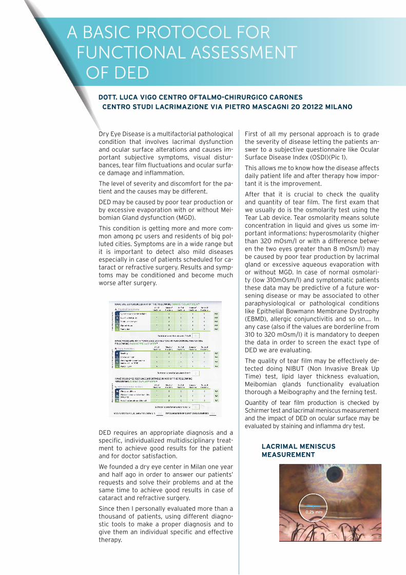
First of all my personal approach is to grade the severity of disease letting the patients an-swer to a subjective questionnaire like Ocular Surface Disease Index (OSDI)(Pic 1).
This allows me to know how the disease affects daily patient life and after therapy how impor-tant it is the improvement.
After that it is crucial to check the quality and quantity of tear film. The first exam that we usually do is the osmolarity test using the Tear Lab device. Tear osmolarity means solute concentration in liquid and gives us some im-portant informations: hyperosmolarity (higher than 320 mOsm/l or with a difference betwe-en the two eyes greater than 8 mOsm/l) may be caused by poor tear production by lacrimal gland or excessive aqueous evaporation with or without MGD. In case of normal osmolari-ty (low 310mOsm/l) and symptomatic patients these data may be predictive of a future wor-sening disease or may be associated to other paraphysiological or pathological conditions like Epithelial Bowmann Membrane Dystrophy (EBMD), allergic conjunctivitis and so on.… In any case (also if the values are borderline from 310 to 320 mOsm/l) it is mandatory to deepen the data in order to screen the exact type of DED we are evaluating.
The quality of tear film may be effectively de-tected doing NIBUT (Non Invasive Break Up Time) test, lipid layer thickness evaluation, Meibomian glands functionality evaluation thorough a Meibography and the ferning test.
Quantity of tear film production is checked by Schirmer test and lacrimal meniscus measurement and the impact of DED on ocular surface may be evaluated by staining and inflamma dry test.
A BASIC PROTOCOL FOR FUNCTIONAL ASSESSMENT OF DED
DOTT. LUCA VIGO CENTRO OFTALMO-CHIRURGICO CARONES CENTRO STUDI LACRIMAZIONE VIA PIETRO MASCAGNI 20 20122 MILANO
LACRIMAL MENISCUS MEASUREMENT

Due to the large number of patients referring to our center I think that it is very important and useful to have screening and diagnostic devices easy and comfortable to handle and that provide precise and reliable results very quickly.
In our Dry Eye Center we have been using for over one year I.C.P. devices to make the proper diagnosis and provide the patient with the ri-ght and individualized therapy.
We have evaluated more than a thousand pa-tients this year using ICP MGD analyzer (Pic 3 with table of features), an infrared camera, to evaluate Meibomian Gland functionality, ICP tearscope (Pic 4 with table of features) to analyze lipid layer thickness and Non Inva-sive Break Up Time (NIBUT) and ferning test (5 with table of features) to check the mucus and indirectly the tear film stability.
The infrared camera is very easy to use and provide auto-analyzed images that allow us to have standard and repeatable results as well tear scope is comfortable to handle and the acquired images and values are realistic and very precise. In our practice 68% of Dry Eye patients have a tear film with a lipid layer 30nm or less thick and 85% of patients with lipid layer thickness within 80 nm. That means that the majority of Dry Eye patients have a Dry Eye Disease due to excessive evaporation and a Meibomian Gland dysfunction.
These patients treated with artificial tears with fat acids, warm compresses, omega 3 dietary supplements and Intense Regulated Pulsed Li-ght (IRPL by E-Swin Technology) show, during follow-up visits, an important improvement of lipid layer thickness (thicker than 80nm), NIBUT and a significant decrease of osmolarity;
a multifactorial therapy shows good results and provide a significant improvement in pa-tient comfort.
The connection of these diagnostic instrumen-ts with an Apple or Windows platform (through an I-Pad or a PC) (creates graphs, that can be shown and explained to our patients, and ge-nerates a PDF file that can be printed or sent by e-mail (Pic 8).
The combination of all these data (OSDI, NI-BUT, MGD functionality, lipid layer thickness, osmolarity, ferning values) with Schirmer test data and staining values give us the possibility to decide the right therapy or if other exams (like blood examinations for immunological diseases) are needed to find out the precise etiology of Dry Eye Disease.
Explaining the patient the grade of severity, showing him the Pdf file with colored graphs, makes us easier to give a therapy and to have the right patient compliance and satisfaction during the first consultation and follow-up visits.
Appropriate therapy is our goal that we can achieve only with good, precise, reliable dia-gnostic instruments. The choice of using a combination of different approaches (topical, systemic, pharmacological or mechanical) de-pends on DED severity and on the whole data that we got with our diagnostic tools.
Dry Eye Disease is nowadays a common condi-tion, with multifactorial etiology and that so-metimes requires a multidisciplinary approa-ch, that is the reason why to find the effective therapy it is mandatory to perform the right diagnosis evaluating local and systemic condi-tions.
EVALUATE MEIBOMIAN GLAND FUNCTIONALITY
QUALITATIVE TESTS, WHICH ASSESS THE FUNCTIONALITY AND STABILITY OF THE TEAR FILM
FERNING TEST

ABSTRACTAIM
Dry Eye Disease is a multifactorial pathological condition that involves lacrimal dysfunction and ocular surface alterations and causes important subjective symptoms, visual disturbances, tear film fluctuations and ocular surface damage and inflammation.
METHOD
Evaluated more than a thousand patients, between 2016 and 2017 using different diagnostic tools like Tearscope, MGD, OSA (by Sbm Sistemi) to make a proper diagnosis and to give them an individual specific and effective therapy.
RESULTS
During the study period 1052 exams were carried out. The standard protocol was employed in 100% of the cases. The median number of investigated dry eye disease was 68%: 11% of the Dry Eye affected was symptomless, between 68-80% was borderline and used Eye Drops, 79% had some lacrimal dysfunction Only 21% of the patient had a correct lacrimation.
CONCLUSION
The use of the standard protocol procedure leads to detect more patients suffering from dry eye disease and may also improve the long-term outcome for patients.
Strada Torino, 43 10043 Orbassano (Torino) ItalyTel. +39 011 19923378

![Dr.Aghaie-management of dry eye [Read-Only]erc.mums.ac.ir/images/erc/eyecenter/Dr.aghaei.lacri.pdf · Dry Eye : treatment Dry eye /ocular surface disorder is : progressive , life-long](https://static.fdocuments.us/doc/165x107/5c5f186309d3f2515c8d2852/draghaie-management-of-dry-eye-read-onlyercmumsacirimageserceyecenterdr.jpg)