Drug-Induced Kidney Disease: An Update - nysn …nysn-online.org/ARCHIVES/2007-2008/Drug...
58
Drug-Induced Kidney Disease: An Update Mark A. Perazella, MD, FACP Associate Professor of Medicine Director, Renal Fellowship Program Yale University School of Medicine
Transcript of Drug-Induced Kidney Disease: An Update - nysn …nysn-online.org/ARCHIVES/2007-2008/Drug...
Drug-Induced Kidney Disease:An Update
Mark A. Perazella, MD, FACPAssociate Professor of Medicine
Director, Renal Fellowship ProgramYale University School of Medicine
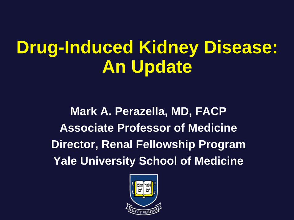
• Acute & chronic kidney disease commonly complicate drug therapy
• Potentially nephrotoxic medications are constantly being released for use in clinical practice
• These drugs are often not tested in the patient populations who subsequently receive them
Drug-induced Kidney DiseaseBackground
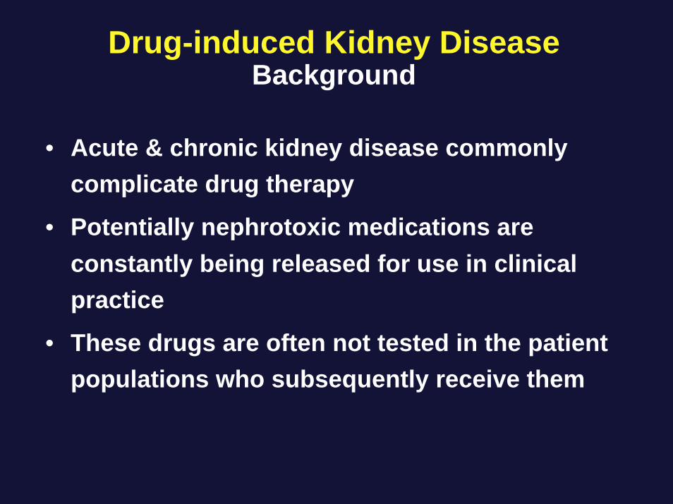
• Culprit Drugs & Nephrotoxins– Therapeutic Medications
• Prescribed• Over-the-counter
– Diagnostic Agents• Contrast material
– Alternative Medications• Herbal Remedies/Supplements• Contaminants
– Environmental Pollutants• Heavy Metals• Organic Solvents
Drug-induced Kidney DiseaseBackground
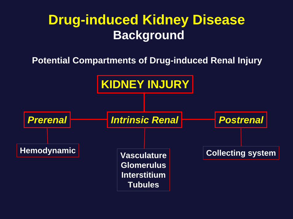
Prerenal Intrinsic Renal Postrenal
KIDNEY INJURY
Hemodynamic Collecting systemVasculatureGlomerulusInterstitium
Tubules
Potential Compartments of Drug-induced Renal Injury
Drug-induced Kidney DiseaseBackground
• Categories of Drug-induced Kidney Disease– Acute Kidney Injury (AKI)
– Proteinuria/Nephrotic Syndrome
– Tubulopathies
– Chronic Kidney Disease
Drug-induced Kidney DiseaseBackground
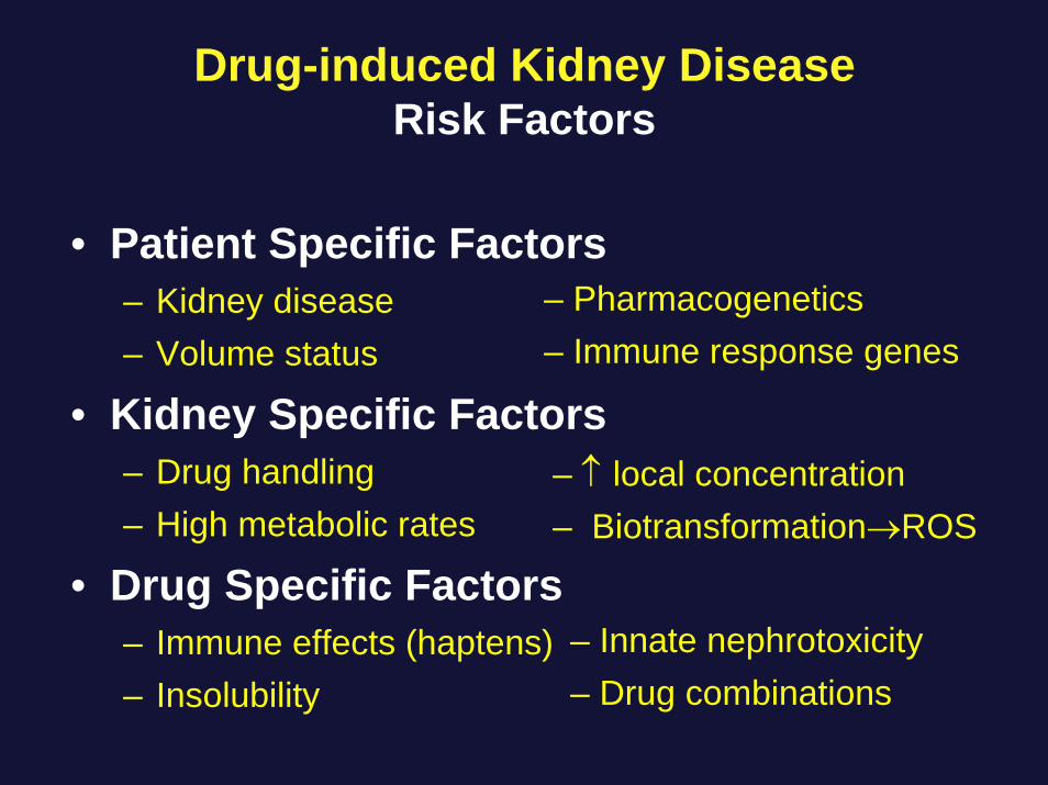
Drug-induced Kidney DiseaseRisk Factors
• Patient Specific Factors– Kidney disease– Volume status
• Kidney Specific Factors– Drug handling– High metabolic rates
• Drug Specific Factors– Immune effects (haptens)– Insolubility
– Pharmacogenetics– Immune response genes
– ↑ local concentration– Biotransformation→ROS
– Innate nephrotoxicity– Drug combinations
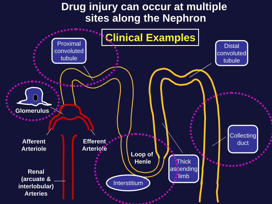
AfferentArteriole
Loop of Henle
EfferentArteriole
Proximalconvoluted
tubule
Distalconvoluted
tubule
Collectingduct
Thickascending
limbRenal
(arcuate &interlobular)
Arteries
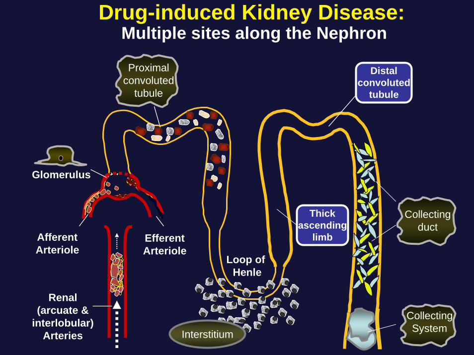
Drug injury can occur at multiple sites along the Nephron
Clinical Examples
Glomerulus
Interstitium
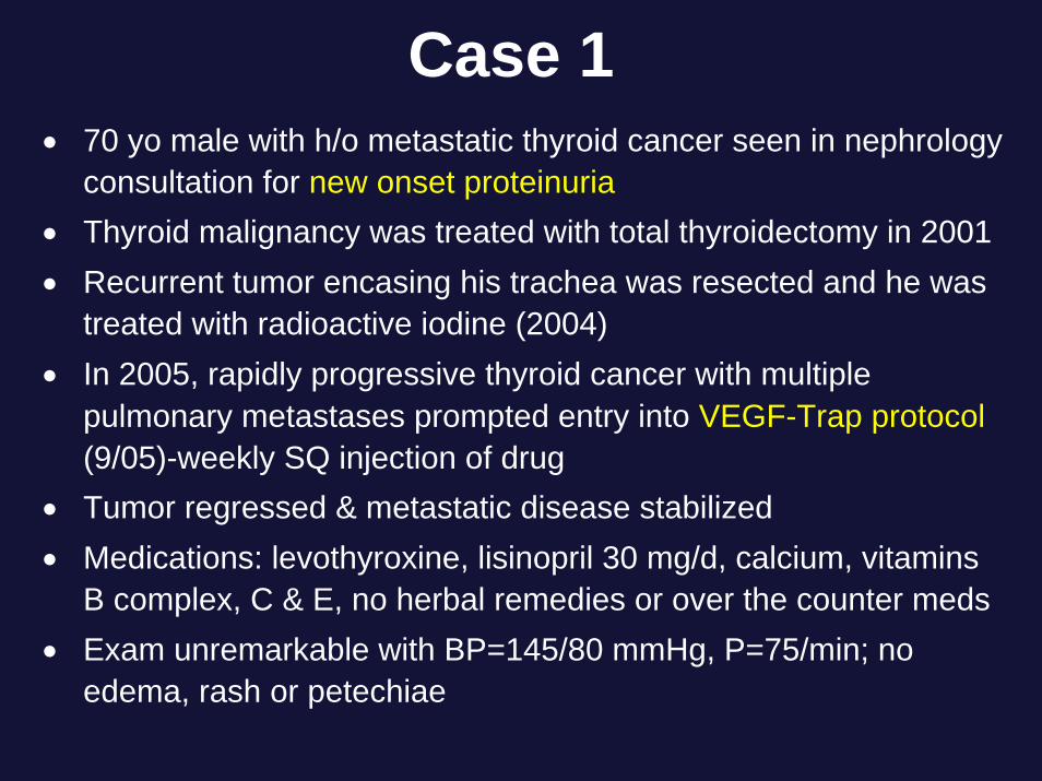
Case 1• 70 yo male with h/o metastatic thyroid cancer seen in nephrology
consultation for new onset proteinuria• Thyroid malignancy was treated with total thyroidectomy in 2001• Recurrent tumor encasing his trachea was resected and he was
treated with radioactive iodine (2004)• In 2005, rapidly progressive thyroid cancer with multiple
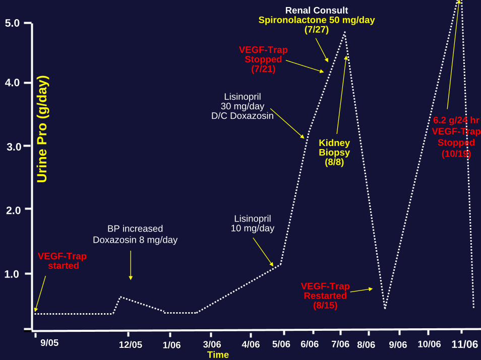
pulmonary metastases prompted entry into VEGF-Trap protocol(9/05)-weekly SQ injection of drug
• Tumor regressed & metastatic disease stabilized• Medications: levothyroxine, lisinopril 30 mg/d, calcium, vitamins
B complex, C & E, no herbal remedies or over the counter meds• Exam unremarkable with BP=145/80 mmHg, P=75/min; no
edema, rash or petechiae

Time
Urin
e Pr
o (g
/day
)
1.0
2.0
3.0
4.0
5.0
9/05 12/0512/05 1/061/06 3/063/06 4/064/06
HypertensionDoxazosin 8 mg/day
VEGF-Trapstarted
5/065/06
Lisinopril10 mg/day
(5/7)
6/066/06 7/067/06 8/068/06 9/069/06 10/0610/06
Lisinopril30 mg/day
D/C Doxazosin(6/5)
VEGF-TrapStopped
(7/21)
Renal ConsultSpironolactone 50 mg/day
(7/27)
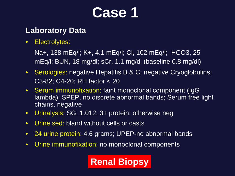
Case 1Laboratory Data• Electrolytes:
Na+, 138 mEq/l; K+, 4.1 mEq/l; Cl, 102 mEq/l; HCO3, 25 mEq/l; BUN, 18 mg/dl; sCr, 1.1 mg/dl (baseline 0.8 mg/dl)
• Serologies: negative Hepatitis B & C; negative Cryoglobulins; C3-82; C4-20; RH factor < 20
• Serum immunofixation: faint monoclonal component (IgGlambda); SPEP, no discrete abnormal bands; Serum free light chains, negative
• Urinalysis: SG, 1.012; 3+ protein; otherwise neg• Urine sed: bland without cells or casts• 24 urine protein: 4.6 grams; UPEP-no abnormal bands• Urine immunofixation: no monoclonal components
Renal Biopsy
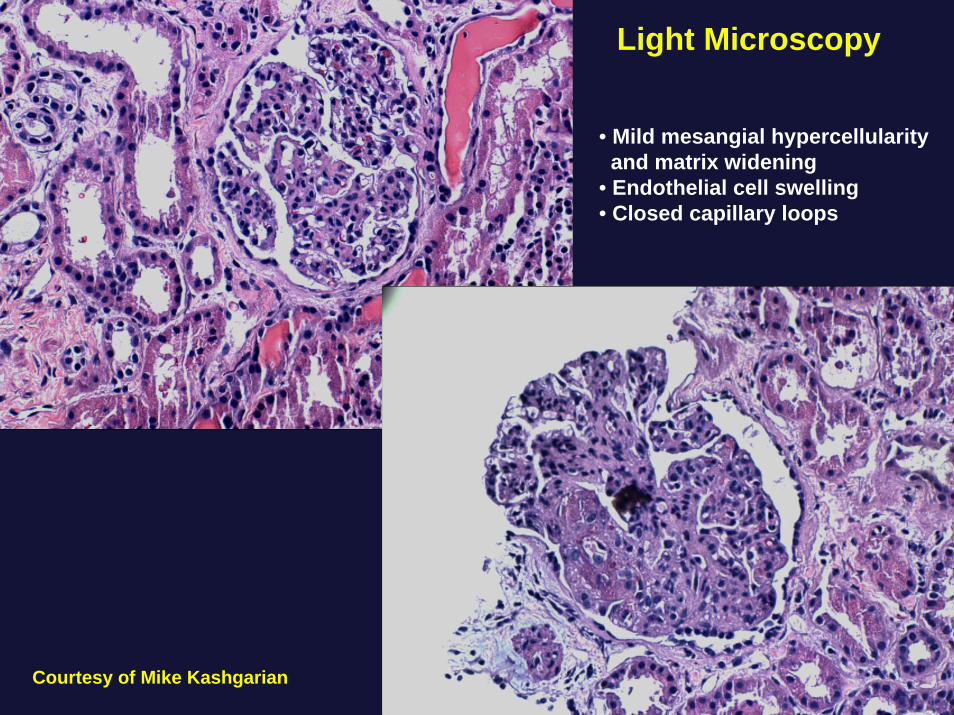
Courtesy of Mike Kashgarian
• Mild mesangial hypercellularityand matrix widening
• Endothelial cell swelling• Closed capillary loops
Light Microscopy
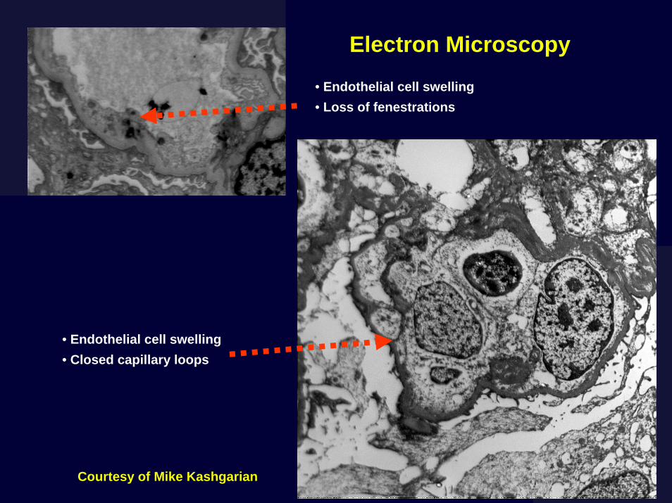
Endothelial cell swelling
Courtesy of Mike Kashgarian
Electron Microscopy
• Endothelial cell swelling• Loss of fenestrations
• Endothelial cell swelling• Closed capillary loops

Final Diagnosis• “Glomerular Endotheliosis” secondary to
Anti-VEGF therapy (VEGF-Trap)– Capillary loop occlusion
– Severe endothelial cell swelling
– Loss of endothelial cell fenestrations
– Fibrin deposition (not shown)
– Focal effacement foot processes
Time
Urin
e Pr
o (g
/day
)
1.0
2.0
3.0
4.0
5.0
BP increasedDoxazosin 8 mg/day
VEGF-Trapstarted
5/06
Lisinopril10 mg/day
6/06 7/06 8/06 9/06 10/06
Lisinopril30 mg/day
D/C Doxazosin
VEGF-TrapStopped
(7/21)
KidneyBiopsy
(8/8)
Renal ConsultSpironolactone 50 mg/day
(7/27)
VEGF-TrapRestarted
(8/15)
11/06
6.2 g/24 hrVEGF-Trap
Stopped(10/19)
9/05 12/0512/05 1/061/06 3/063/06 4/064/06
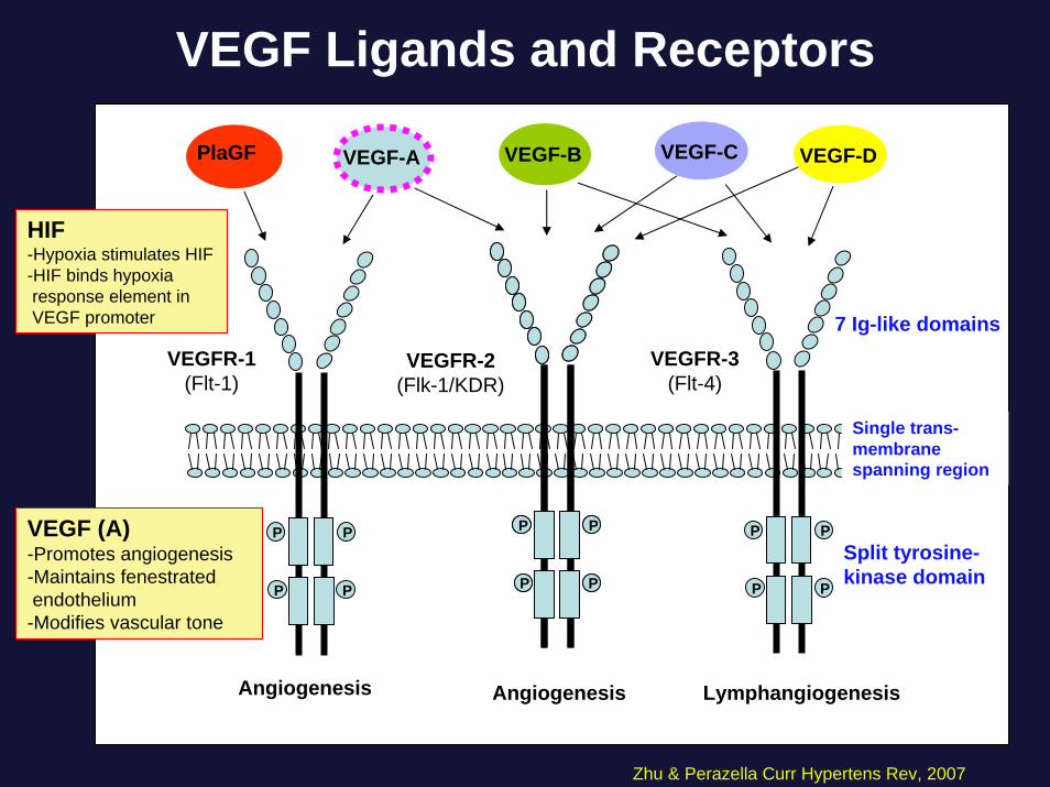
VEGF Ligands and Receptors
P
PP
PP
PP
P
VEGF-A
P
PP
P
VEGF-C
P
PP
P
PlaGF
Angiogenesis LymphangiogenesisAngiogenesis
VEGF-B
VEGFR-1(Flt-1)
VEGFR-2(Flk-1/KDR)
VEGFR-3(Flt-4)
VEGF-D
7 Ig-like domains
Split tyrosine-kinase domain
Single trans-membrane spanning region
Zhu & Perazella Curr Hypertens Rev, 2007
VEGF (A)-Promotes angiogenesis-Maintains fenestratedendothelium-Modifies vascular tone
HIF-Hypoxia stimulates HIF-HIF binds hypoxiaresponse element inVEGF promoter
P
PP
P
VEGF
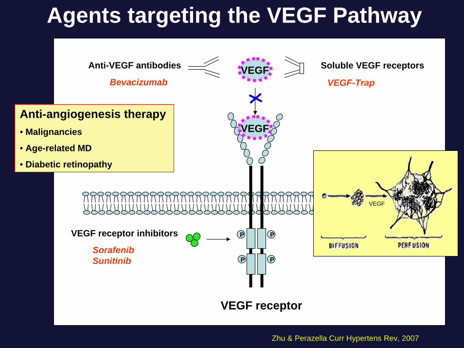
VEGFAnti-VEGF antibodies
Bevacizumab
Soluble VEGF receptors
VEGF-Trap
VEGF receptor inhibitors
SorafenibSunitinib
VEGF receptor
Agents targeting the VEGF Pathway
Zhu & Perazella Curr Hypertens Rev, 2007
Anti-angiogenesis therapy• Malignancies
• Age-related MD
• Diabetic retinopathy
VEGF
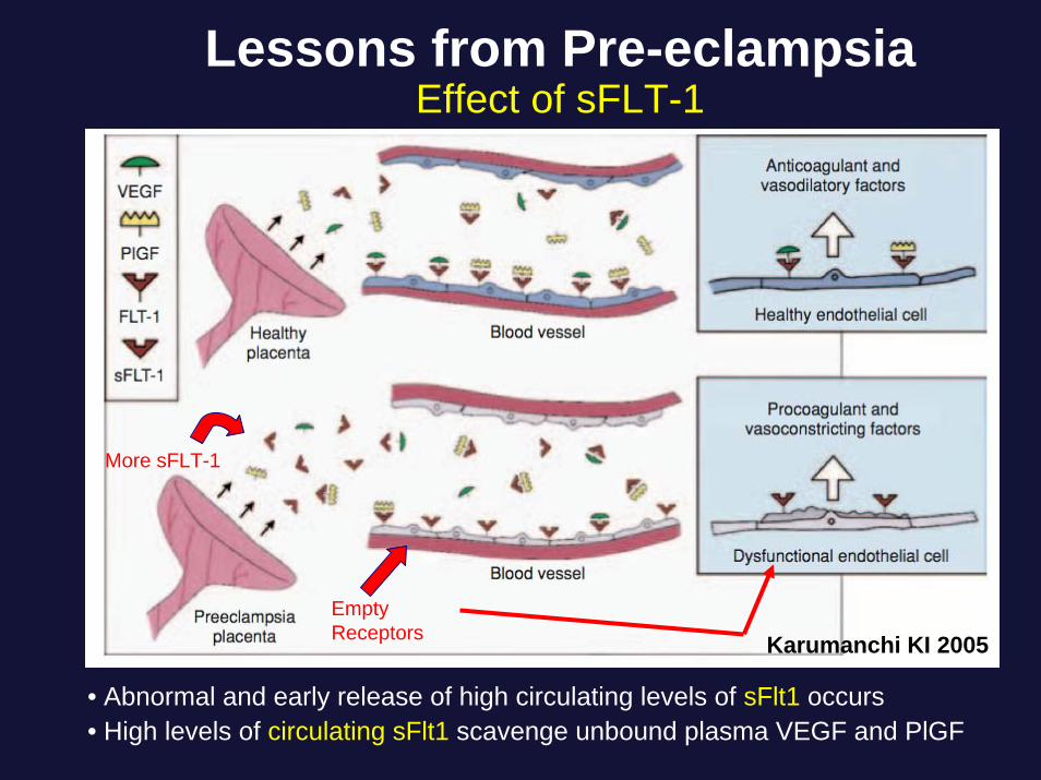
Lessons from Pre-eclampsiaEffect of sFLT-1
Karumanchi KI 2005Empty Receptors
More sFLT-1
• Abnormal and early release of high circulating levels of sFlt1 occurs• High levels of circulating sFlt1 scavenge unbound plasma VEGF and PlGF
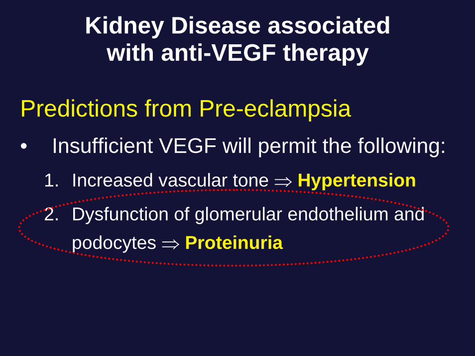
Kidney Disease associated with anti-VEGF therapy
Predictions from Pre-eclampsia• Insufficient VEGF will permit the following:
1. Increased vascular tone ⇒ Hypertension
2. Dysfunction of glomerular endothelium and podocytes ⇒ Proteinuria
VEGF in the Kidney
• VEGF is produced by visceral epithelial cells (podocytes)
• VEGF receptors are present on glomerular endothelium
• VEGF maintains normal functioning of glomerular endothelial cells and podocytes
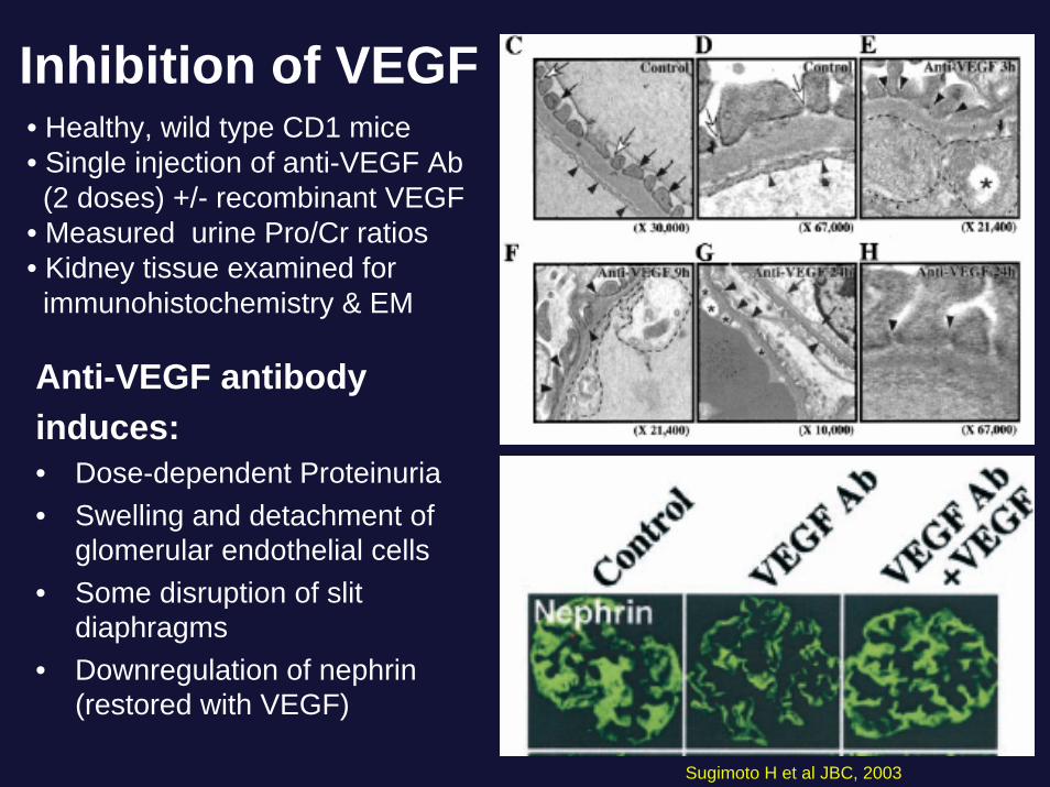
Inhibition of VEGF
Anti-VEGF antibodyinduces:• Dose-dependent Proteinuria• Swelling and detachment of
glomerular endothelial cells• Some disruption of slit
diaphragms• Downregulation of nephrin
(restored with VEGF)
• Healthy, wild type CD1 mice• Single injection of anti-VEGF Ab(2 doses) +/- recombinant VEGF
• Measured urine Pro/Cr ratios• Kidney tissue examined for immunohistochemistry & EM
Sugimoto H et al JBC, 2003
Podocyte
FootProcessGBM
EndothelialCell (EC)
EC
VEGF
Blood
Urine
VEGF
ECFlt-1 Flk-1
Podocyte
GBM
EndothelialCell (EC)
Blood
Urine
EC
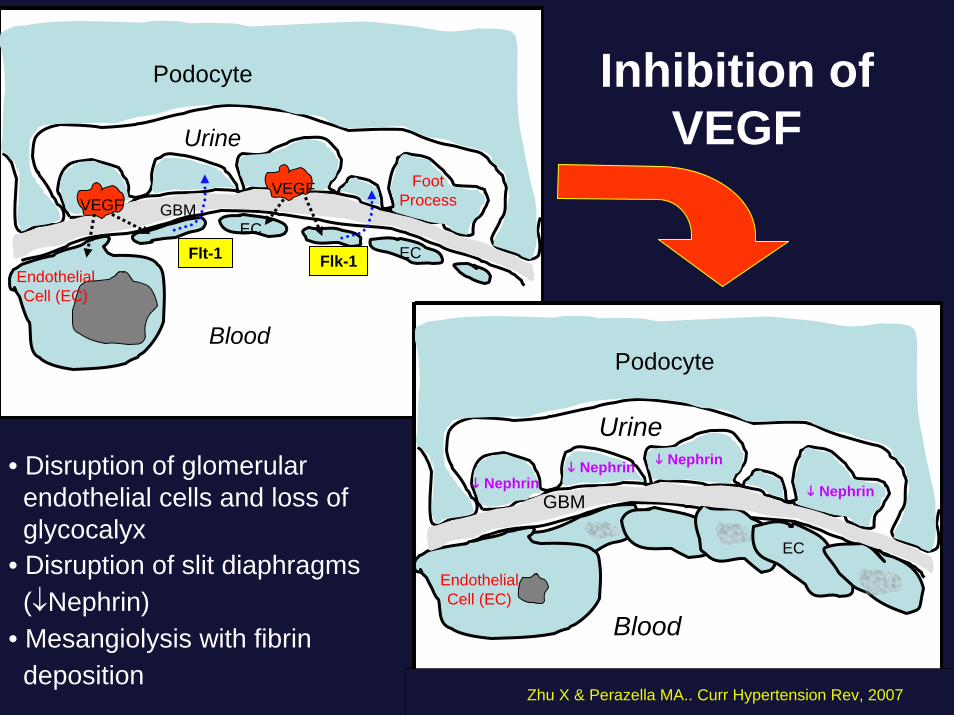
Inhibition of VEGF
NephrinNephrinNephrin Nephrin
Zhu X & Perazella MA.. Curr Hypertension Rev, 2007
• Disruption of glomerular endothelial cells and loss of glycocalyx
• Disruption of slit diaphragms (↓Nephrin)
• Mesangiolysis with fibrindeposition

BevacizumabTrials
Bevacizumab(mg/kg/dose)
Concurrent chemotherapy
ProteinuriaControl Low High
Kabbinavar et al., 2003 J Clin Oncol(Colorectal cancer)N=99
5 10 FU/LV 11% 23% 28%
Yang et al., 2003 NEJM (Renal cancer) N=116
5 10 none 38% 41%(5.4% grade III)
21% 42%(3% grade III)
Miller et al., 2005J Clin Onc(Breast cancer) N=444
15/3wk capecitabine 7.4 22.3%(0.9% grade III)
46%(1.8% grade III)
26.5%(0.8% grade III)
64%(7.7% grade III)
15
Kabbinavar et al., 2005 J Clin Onc(Colon cancer) N=204
5 FU/LV 23%(0% grade III)
Johnson et al., 2004 J Clin Onc(NSCLC) N=98
7.5 carboplatinpaclitxel
2%
Hurwitz et al., 2004NEJM(Colorectal cancer) N=790
5 irinotecanFU/LV
21.7%(0.8% grade III)
Zhu X. et al AJKD 2007
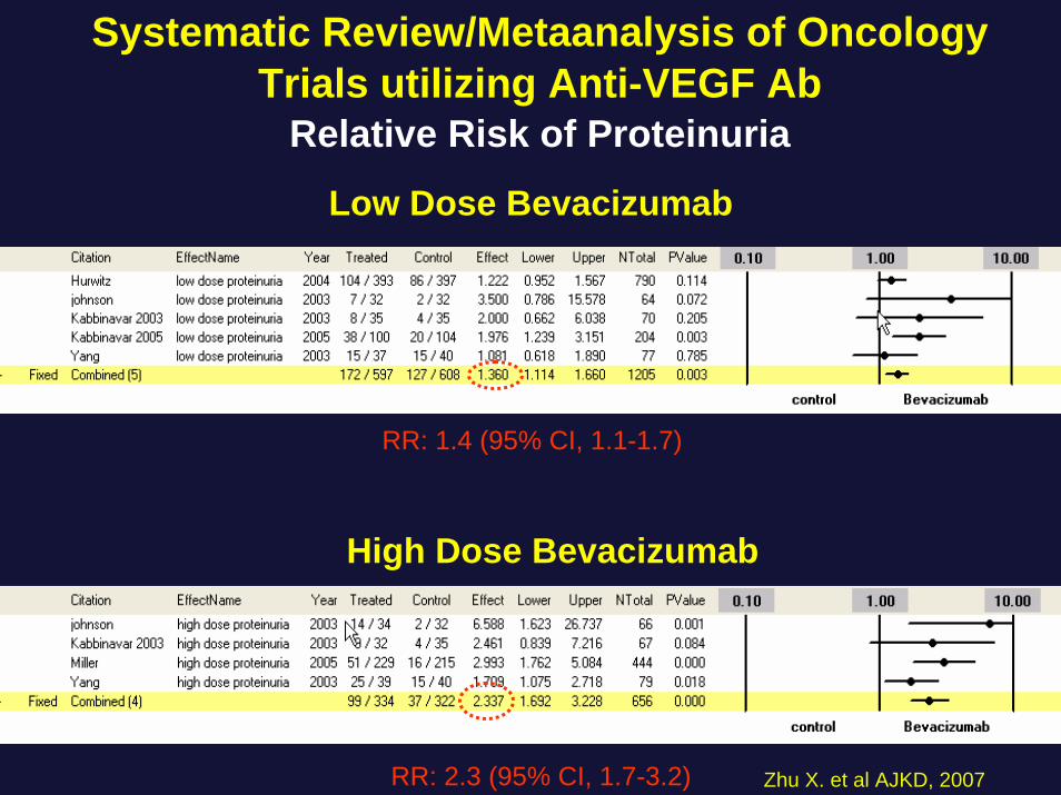
Systematic Review/Metaanalysis of Oncology Trials utilizing Anti-VEGF Ab
Relative Risk of Proteinuria
Low Dose Bevacizumab
High Dose Bevacizumab
RR: 1.4 (95% CI, 1.1-1.7)
RR: 2.3 (95% CI, 1.7-3.2) Zhu X. et al AJKD, 2007
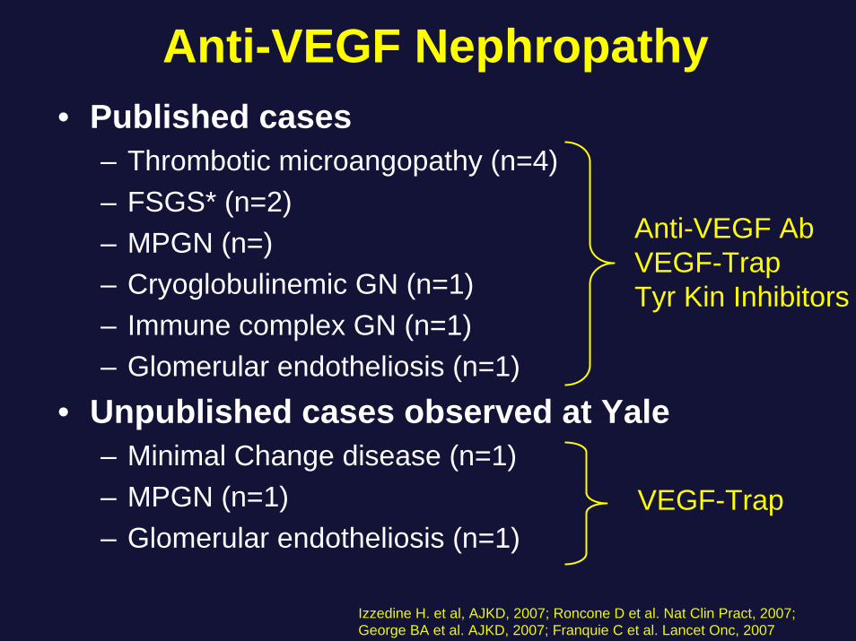
Anti-VEGF Nephropathy• Published cases
– Thrombotic microangopathy (n=4)– FSGS* (n=2)– MPGN (n=)– Cryoglobulinemic GN (n=1)– Immune complex GN (n=1)– Glomerular endotheliosis (n=1)
• Unpublished cases observed at Yale– Minimal Change disease (n=1)– MPGN (n=1)– Glomerular endotheliosis (n=1)
Izzedine H. et al, AJKD, 2007; Roncone D et al. Nat Clin Pract, 2007; George BA et al. AJKD, 2007; Franquie C et al. Lancet Onc, 2007
VEGF-Trap
Anti-VEGF AbVEGF-TrapTyr Kin Inhibitors
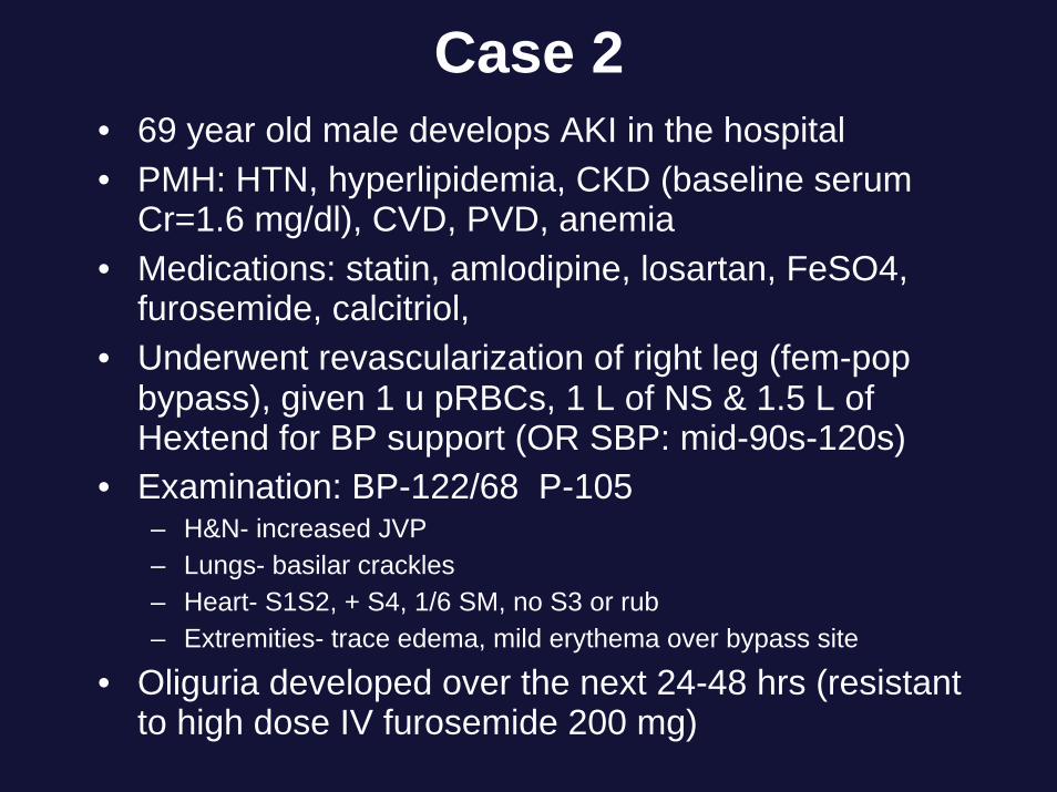
Case 2• 69 year old male develops AKI in the hospital• PMH: HTN, hyperlipidemia, CKD (baseline serum
Cr=1.6 mg/dl), CVD, PVD, anemia • Medications: statin, amlodipine, losartan, FeSO4,
furosemide, calcitriol, • Underwent revascularization of right leg (fem-pop
bypass), given 1 u pRBCs, 1 L of NS & 1.5 L of Hextend for BP support (OR SBP: mid-90s-120s)
• Examination: BP-122/68 P-105 – H&N- increased JVP– Lungs- basilar crackles– Heart- S1S2, + S4, 1/6 SM, no S3 or rub – Extremities- trace edema, mild erythema over bypass site
• Oliguria developed over the next 24-48 hrs (resistant to high dose IV furosemide 200 mg)
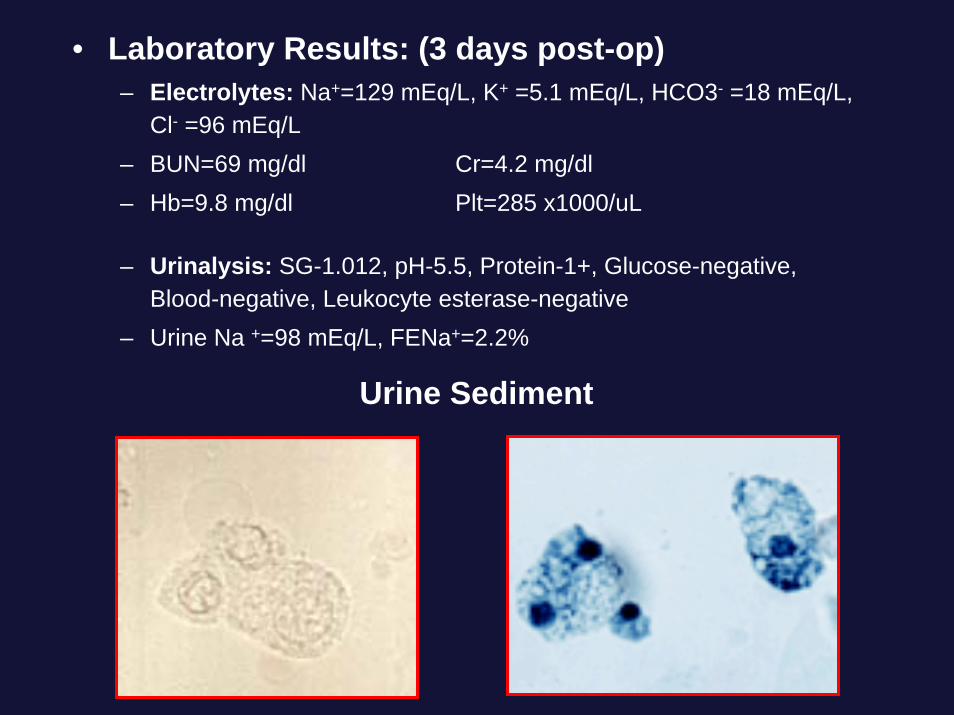
• Laboratory Results: (3 days post-op)– Electrolytes: Na+=129 mEq/L, K+ =5.1 mEq/L, HCO3- =18 mEq/L,
Cl- =96 mEq/L– BUN=69 mg/dl Cr=4.2 mg/dl– Hb=9.8 mg/dl Plt=285 x1000/uL
– Urinalysis: SG-1.012, pH-5.5, Protein-1+, Glucose-negative, Blood-negative, Leukocyte esterase-negative
– Urine Na +=98 mEq/L, FENa+=2.2%
Urine Sediment
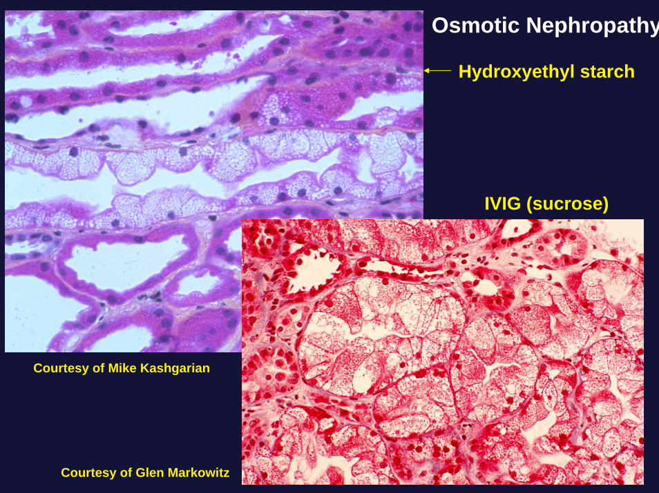
Osmotic Nephropathy
Hydroxyethyl starch
IVIG (sucrose)
Courtesy of Glen Markowitz
Courtesy of Mike Kashgarian
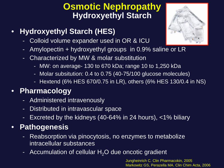
Osmotic NephropathyHydroxyethyl Starch
• Hydroxyethyl Starch (HES)- Colloid volume expander used in OR & ICU- Amylopectin + hydroxyethyl groups in 0.9% saline or LR- Characterized by MW & molar substitution
- MW: on average- 130 to 670 kDa; range 10 to 1,250 kDa- Molar subsitution: 0.4 to 0.75 (40-75/100 glucose molecules)- Hextend (6% HES 670/0.75 in LR), others (6% HES 130/0.4 in NS)
• Pharmacology- Administered intravenously- Distributed in intravascular space- Excreted by the kidneys (40-64% in 24 hours), <1% biliary
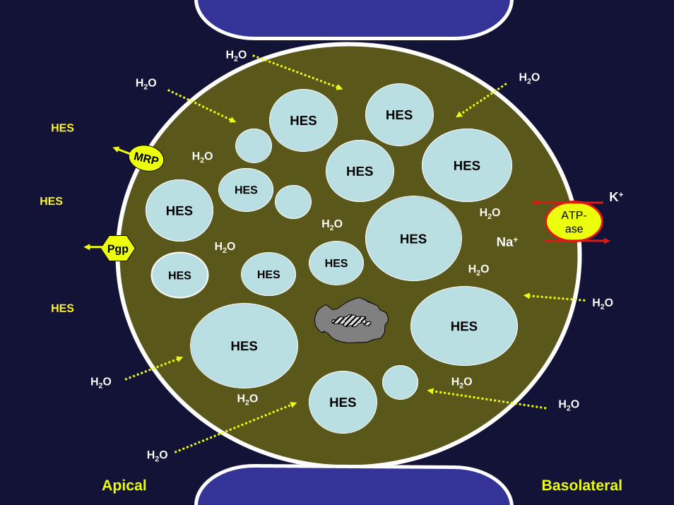
• Pathogenesis- Reabsorption via pinocytosis, no enzymes to metabolize
intracellular substances- Accumulation of cellular H2O due oncotic gradient
Jungheinrich C. Clin Pharmacokin, 2005Markowitz GS, Perazella MA. Clin Chim Acta, 2006
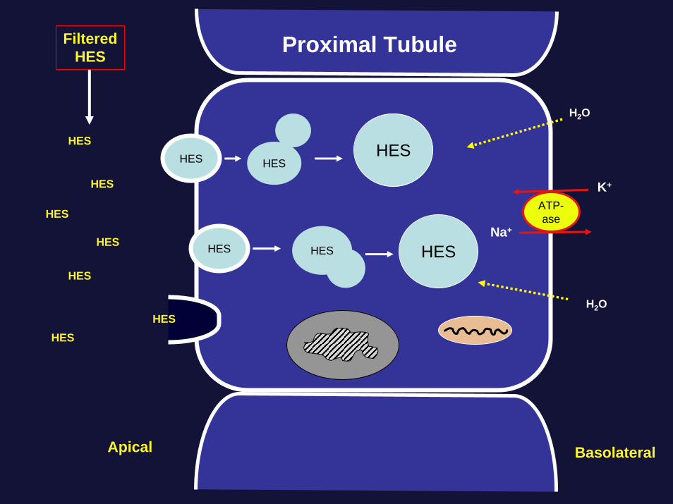
ATP-ase
HESHES
Na+
K+
HESHES
FilteredHES
Apical Basolateral
HES HESHES
H2O
H2O
Proximal Tubule
HES
HES
HES
HES
HES
HES
K+
Apical Basolateral
H2O
H2O
H2O
H2O
H2O
H2O
H2O
ATP-ase
HES
HES
HES
HES
H2O
H2O
H2OH2O
HES
HESHES
HES
HES
HESHES
HES
H2O
H2O
HES H2O
Na+Pgp
MRP
HES
HES
HES
Osmotic NephropathyHydroxyethyl Starch
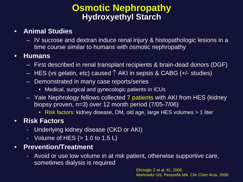
• Animal Studies– IV sucrose and dextran induce renal injury & histopathologic lesions in a
time course similar to humans with osmotic nephropathy• Humans
– First described in renal transplant recipients & brain-dead donors (DGF)– HES (vs gelatin, etc) caused ↑ AKI in sepsis & CABG (+/- studies)– Demonstrated in many case reports/series
• Medical, surgical and gynecologic patients in ICUs– Yale Nephrology fellows collected 7 patients with AKI from HES (kidney
biopsy proven, n=3) over 12 month period (7/05-7/06)• Risk factors: kidney disease, DM, old age, large HES volumes > 1 liter
• Risk Factors- Underlying kidney disease (CKD or AKI)- Volume of HES (> 1.0 to 1.5 L)
• Prevention/Treatment- Avoid or use low volume in at risk patient, otherwise supportive care,
sometimes dialysis is requiredEbcioglu Z et al. KI, 2006;Markowitz GS, Perazella MA. Clin Chim Acta, 2006

K+
Basolateral
H2O H2O
H2O
H2O
H2O
H2O
Apical
ATP-ase
D
D
D
D
H2O
H2O
H2OH2O
D
D D
D
DDD
D
H2O
H2O
DH2O
MRP
Drugs• Sucrose (IVIG)• Hydroxyethyl starch• Dextran• Mannitol• High or low osmolarradiocontrast
• Gadolinium contrast?
Osmotic Nephropathy
Pgp
Case 3• 57 year old obese female presents with malaise,
weakness, frequent “oily” stools for several weeks and AKI (increased serum Cr from baseline)
• PMH: HTN, T2-DM, arthritis, CKD (baseline serum Cr=2.1 mg/dl), GERD, asthma, hypothyroid
• Medications: diltiazem 240 mg qd, singular 10 mg qd, furosemide 40 mg qd, pantoprazole 20 mg qd, colchicine 0.6 mg prn, orlistat 120 mg tid (up from bid), no herbal products, NSAIDs or ascorbic acid
• Examination: BP-130/80 P-86 Afebrile– H&N- mild conjunctival pallor, normal JVP– Lungs-clear – Abdomen- obese – Extremities- Trace ankle edema, no rash
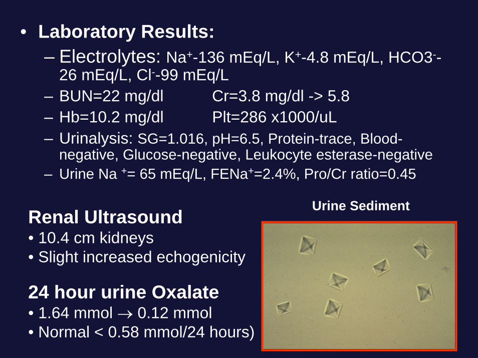
• Laboratory Results:– Electrolytes: Na+-136 mEq/L, K+-4.8 mEq/L, HCO3--
26 mEq/L, Cl--99 mEq/L– BUN=22 mg/dl Cr=3.8 mg/dl -> 5.8– Hb=10.2 mg/dl Plt=286 x1000/uL – Urinalysis: SG=1.016, pH=6.5, Protein-trace, Blood-
negative, Glucose-negative, Leukocyte esterase-negative– Urine Na += 65 mEq/L, FENa+=2.4%, Pro/Cr ratio=0.45
Urine SedimentRenal Ultrasound• 10.4 cm kidneys• Slight increased echogenicity
24 hour urine Oxalate• 1.64 mmol → 0.12 mmol• Normal < 0.58 mmol/24 hours)

Crystal NephropathyOrlistat- Calcium Oxalate
Singh et al. AJKD, 2006 Courtney et al. NDT, 2007
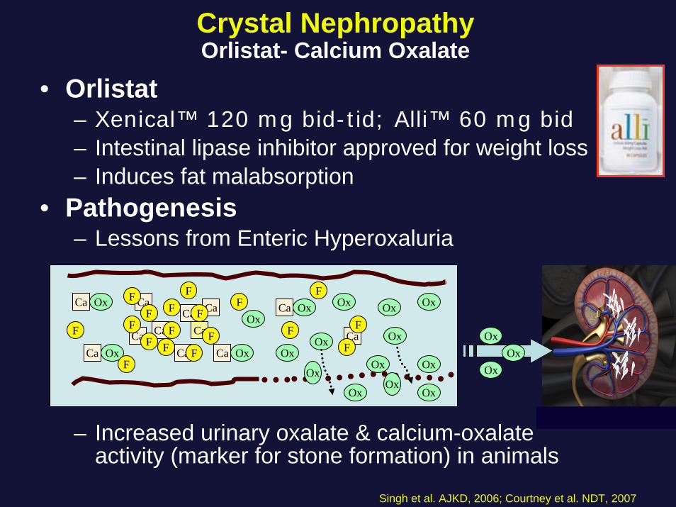
• Orlistat– Xenical™ 120 mg bid-tid; Alli™ 60 mg bid – Intestinal lipase inhibitor approved for weight loss– Induces fat malabsorption
• Pathogenesis– Lessons from Enteric Hyperoxaluria
– Increased urinary oxalate & calcium-oxalate activity (marker for stone formation) in animals
Crystal NephropathyOrlistat- Calcium Oxalate
Singh et al. AJKD, 2006; Courtney et al. NDT, 2007
F
F
CaCa
Ca
Ca FF
Ox
Ox
Ox
Ox
Ca
Ox
OxCa
CaOx
Ox
OxOxF
F F F
FF
Ox
Ox
Ox
CaFCaF
F
OxOx
Ox
CaCaF
F
Ox
FCa
F F
Ox
OxOx
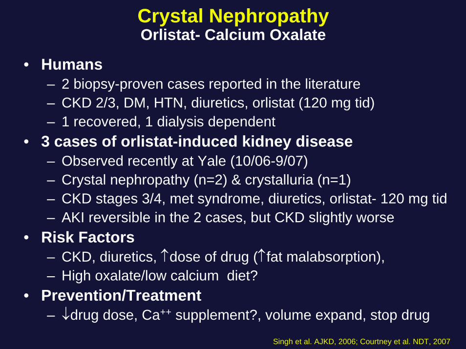
• Humans– 2 biopsy-proven cases reported in the literature– CKD 2/3, DM, HTN, diuretics, orlistat (120 mg tid)– 1 recovered, 1 dialysis dependent
• 3 cases of orlistat-induced kidney disease– Observed recently at Yale (10/06-9/07)– Crystal nephropathy (n=2) & crystalluria (n=1) – CKD stages 3/4, met syndrome, diuretics, orlistat- 120 mg tid– AKI reversible in the 2 cases, but CKD slightly worse
• Risk Factors– CKD, diuretics, ↑dose of drug (↑fat malabsorption),– High oxalate/low calcium diet?
• Prevention/Treatment– ↓drug dose, Ca++ supplement?, volume expand, stop drug
Crystal NephropathyOrlistat- Calcium Oxalate
Singh et al. AJKD, 2006; Courtney et al. NDT, 2007
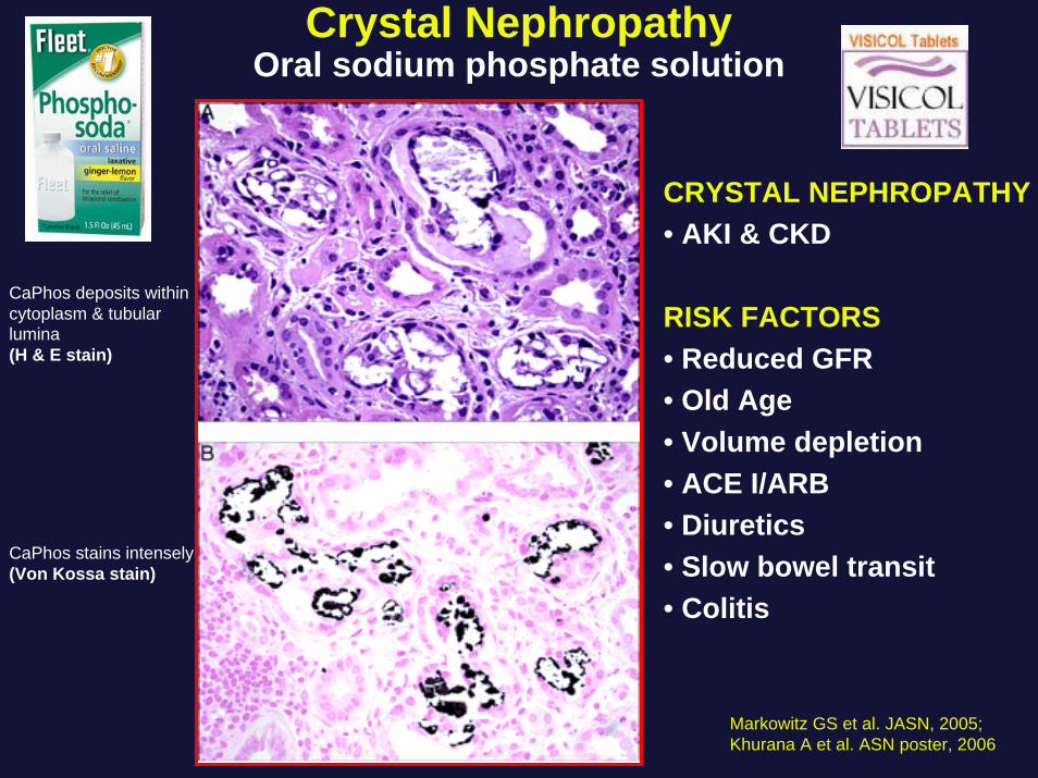
CaPhos deposits within cytoplasm & tubular lumina(H & E stain)
CaPhos stains intensely(Von Kossa stain)
CRYSTAL NEPHROPATHY• AKI & CKD
RISK FACTORS• Reduced GFR• Old Age• Volume depletion• ACE I/ARB• Diuretics• Slow bowel transit• Colitis
Markowitz GS et al. JASN, 2005;Khurana A et al. ASN poster, 2006
Crystal NephropathyOral sodium phosphate solution
Crystal NephropathyCiprofloxacin
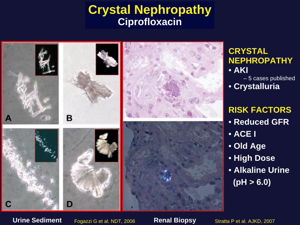
Urine Sediment Renal Biopsy
CRYSTAL NEPHROPATHY• AKI
– 5 cases published• Crystalluria
RISK FACTORS• Reduced GFR• ACE I• Old Age• High Dose• Alkaline Urine(pH > 6.0)
Stratta P et al. AJKD, 2007Fogazzi G et al. NDT, 2006
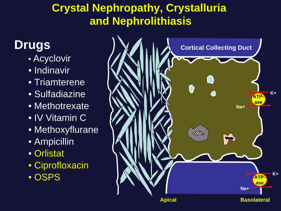
Crystal Nephropathy, Crystalluriaand Nephrolithiasis
ATP-ase
Na+
ATP-ase
Na+
K+
Apical Basolateral
Cortical Collecting Duct
K+
Drugs• Acyclovir • Indinavir• Triamterene• Sulfadiazine• Methotrexate• IV Vitamin C• Methoxyflurane• Ampicillin• Orlistat• Ciprofloxacin• OSPS

Case 4• 69 year old female presents with fatigue and renal failure• PMH: HTN, T2 DM, GERD, osteoarthritis, osteoporosis,
COPD. Developed severe GERD 6 months prior-initially took tums & OTC cimetidine X 2 months without relief
• Medications: enalapril, amlodipine, glyburide, combiventinhaler, lansoprazole (3 months), calcium tablets, no OTC or herbal products at this time
• No allergies• Examination: BP-135/82 P-89 T-98.7
– H&N- pink conjunctiva, normal JVP– Lungs- clear– Heart- S1S2, no S3, rub or 1/6 systolic ejection murmur– Abdomen- benign with normal BS, no flank tenderness– Extremities- no edema or rash

• Laboratory Results:– Electrolytes: Na+=132, K+=5.1, HCO3=18, Cl-=104– BUN=49 mg/dl Cr=2.9 mg/dl– Hb=11.2 mg/dl Plt=335 x1000/uL– WBCs=12 per mm3 Eosinophils=2%– Urinalysis: SG-1.015, pH-5.5, Protein-1+, Blood-trace positive,
Leukocyte esterase-positive– Urine Na +=55 mEq/L, FENa+=3.2%
Urine Sediment
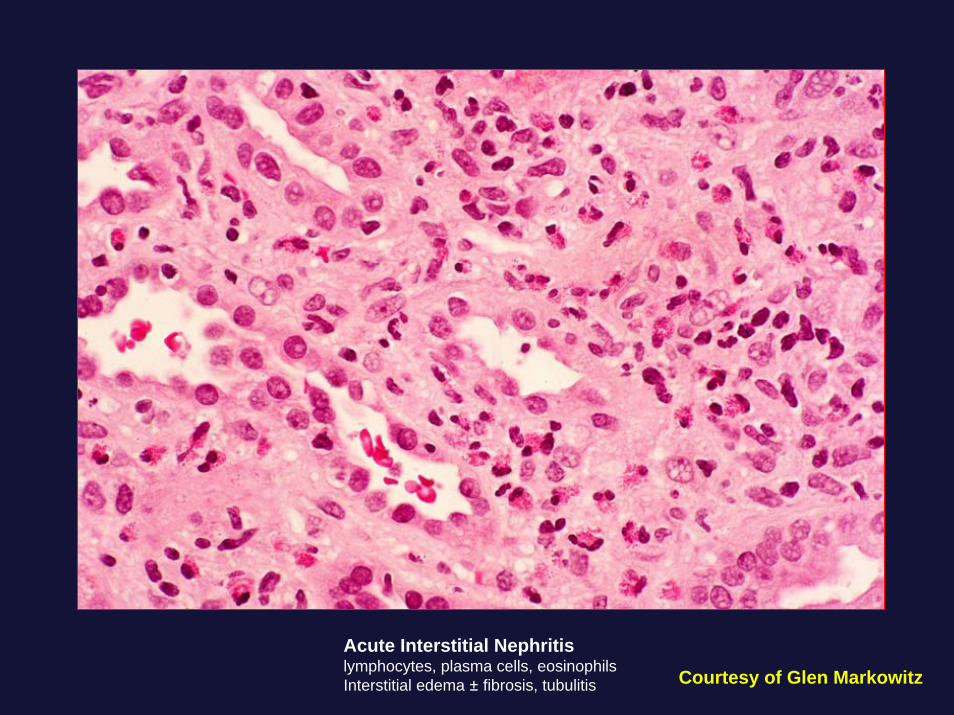
Acute Interstitial Nephritislymphocytes, plasma cells, eosinophilsInterstitial edema ± fibrosis, tubulitis Courtesy of Glen Markowitz
Drug-induced Acute Interstitial NephritisProton Pump Inhibitors (PPIs)
• AIN associated with omeprazole– First case of biopsy-proven AIN (confirmed with drug
rechallenge) was described with omeprazole in 19921
– Subsequent reports of AIN associated with omeprazole(n=29) appeared in the literature over the next 12 years
– Of these 29 cases, 23 were biopsy-proven AIN• AIN associated with other PPIs
– In 2004, first case reports of AIN from other PPIs– Lansoprazole (n=2), pantoprazole (n=2), rabeprazole
(n=1), esomeprazole (n=1)– All biopsy-proven cases
Drug-induced Acute Interstitial NephritisProton Pump Inhibitors (PPIs)
• Retrospective case review (1993-2003) in Australia– 18/28 (64%) cases of biopsy proven AIN were
associated with PPI use– Median age
• 74 years (65-79)– Mean duration of PPI exposure prior to diagnosis:
• 11 weeks (3-24)– Common presenting symptoms:
• Nonspecific in most, fatigue and nausea (39%), weight loss (22%)
– Biopsy findings:• Classic findings with interstitial eosinophils (83%)
Geevasinga et al. Clin Gastro Hep, 2006
Drug-induced Acute Interstitial NephritisProton Pump Inhibitors (PPIs)
• Retrospective case review (2002-2005) in Auckland, New Zealand– 15/87 (7.7%) cases of AIN were associated with PPIs– Median age
• 78 years (55-86)
– Duration of PPI exposure prior to diagnosis:• 10 days- 18 months
– Common presenting symptoms:• Nonspecific in 11, insidious onset of AKI, 4 with “fever/chills”
– Urine sediment with pyuria (sterile), some hematuria– Renal biopsy proven in 12/15
Simpson et al. Nephrology, 2006
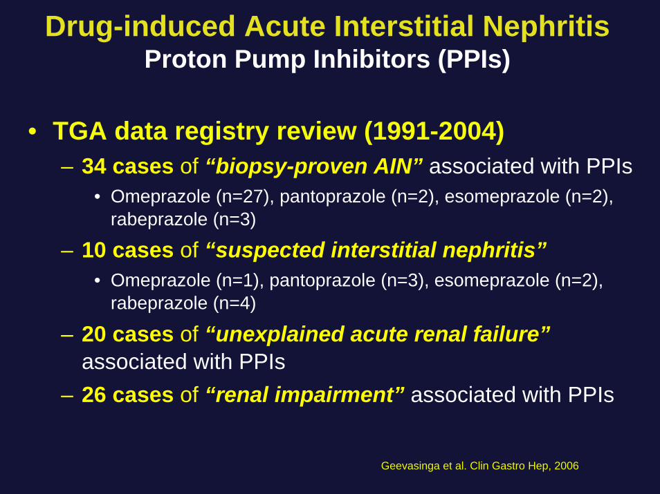
Drug-induced Acute Interstitial NephritisProton Pump Inhibitors (PPIs)
• TGA data registry review (1991-2004)– 34 cases of “biopsy-proven AIN” associated with PPIs
• Omeprazole (n=27), pantoprazole (n=2), esomeprazole (n=2), rabeprazole (n=3)
– 10 cases of “suspected interstitial nephritis”• Omeprazole (n=1), pantoprazole (n=3), esomeprazole (n=2),
rabeprazole (n=4)
– 20 cases of “unexplained acute renal failure”associated with PPIs
– 26 cases of “renal impairment” associated with PPIs
Geevasinga et al. Clin Gastro Hep, 2006
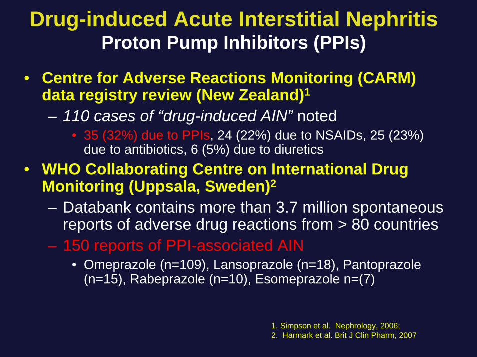
Drug-induced Acute Interstitial NephritisProton Pump Inhibitors (PPIs)
• Centre for Adverse Reactions Monitoring (CARM) data registry review (New Zealand)1
– 110 cases of “drug-induced AIN” noted• 35 (32%) due to PPIs, 24 (22%) due to NSAIDs, 25 (23%)
due to antibiotics, 6 (5%) due to diuretics• WHO Collaborating Centre on International Drug
Monitoring (Uppsala, Sweden)2
– Databank contains more than 3.7 million spontaneous reports of adverse drug reactions from > 80 countries
– 150 reports of PPI-associated AIN• Omeprazole (n=109), Lansoprazole (n=18), Pantoprazole
(n=15), Rabeprazole (n=10), Esomeprazole n=(7)
1. Simpson et al. Nephrology, 2006;2. Harmark et al. Brit J Clin Pharm, 2007
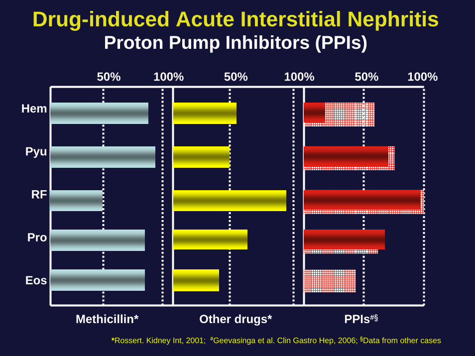
Drug-induced Acute Interstitial NephritisProton Pump Inhibitors (PPIs)
Hem
Pyu
RF
Pro
Eos
50% 50%100% 100% 100%
Methicillin* Other drugs* PPIs#§
*Rossert. Kidney Int, 2001; #Geevasinga et al. Clin Gastro Hep, 2006; §Data from other cases
50%
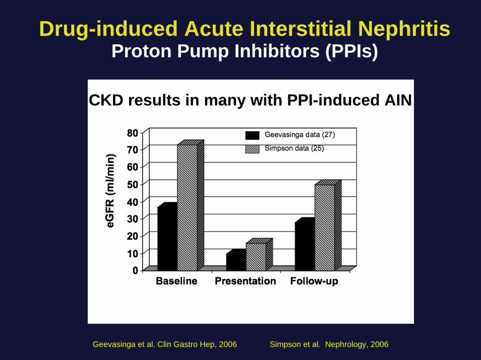
Drug-induced Acute Interstitial NephritisProton Pump Inhibitors (PPIs)
Geevasinga et al. Clin Gastro Hep, 2006 Simpson et al. Nephrology, 2006
CKD results in many with PPI-induced AIN
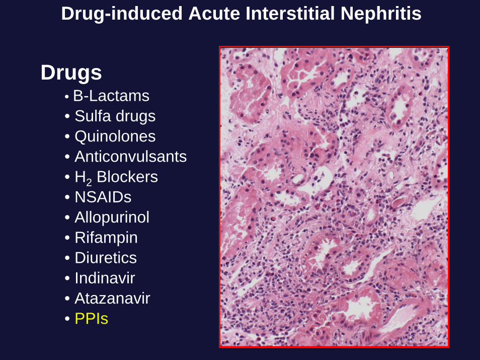
Drug-induced Acute Interstitial Nephritis
Drugs• B-Lactams• Sulfa drugs• Quinolones• Anticonvulsants• H2 Blockers• NSAIDs• Allopurinol• Rifampin• Diuretics• Indinavir• Atazanavir• PPIs
AfferentArteriole
Loop of Henle
EfferentArteriole
Distalconvoluted
tubule
Proximalconvoluted
tubule
Glomerulus
Collectingduct
Renal(arcuate &
interlobular)Arteries
Thickascending
limb
CollectingSystem
Drug-induced Kidney Disease:Multiple sites along the Nephron
Interstitium

Thank You
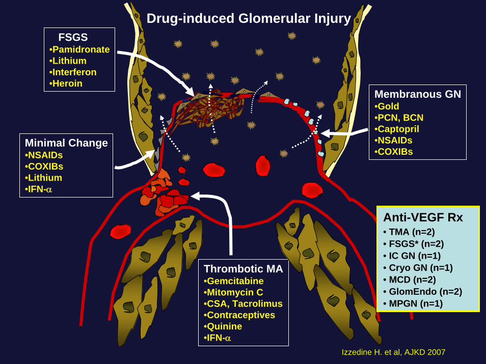
FSGS•Pamidronate•Lithium•Interferon•Heroin
Membranous GN•Gold•PCN, BCN•Captopril•NSAIDs•COXIBs
Thrombotic MA•Gemcitabine•Mitomycin C•CSA, Tacrolimus•Contraceptives•Quinine•IFN-α
Minimal Change•NSAIDs•COXIBs•Lithium•IFN-α
Drug-induced Glomerular Injury
Anti-VEGF Rx• TMA (n=2)• FSGS* (n=2)• IC GN (n=1)• Cryo GN (n=1)• MCD (n=2)• GlomEndo (n=2)• MPGN (n=1)
Izzedine H. et al, AJKD 2007
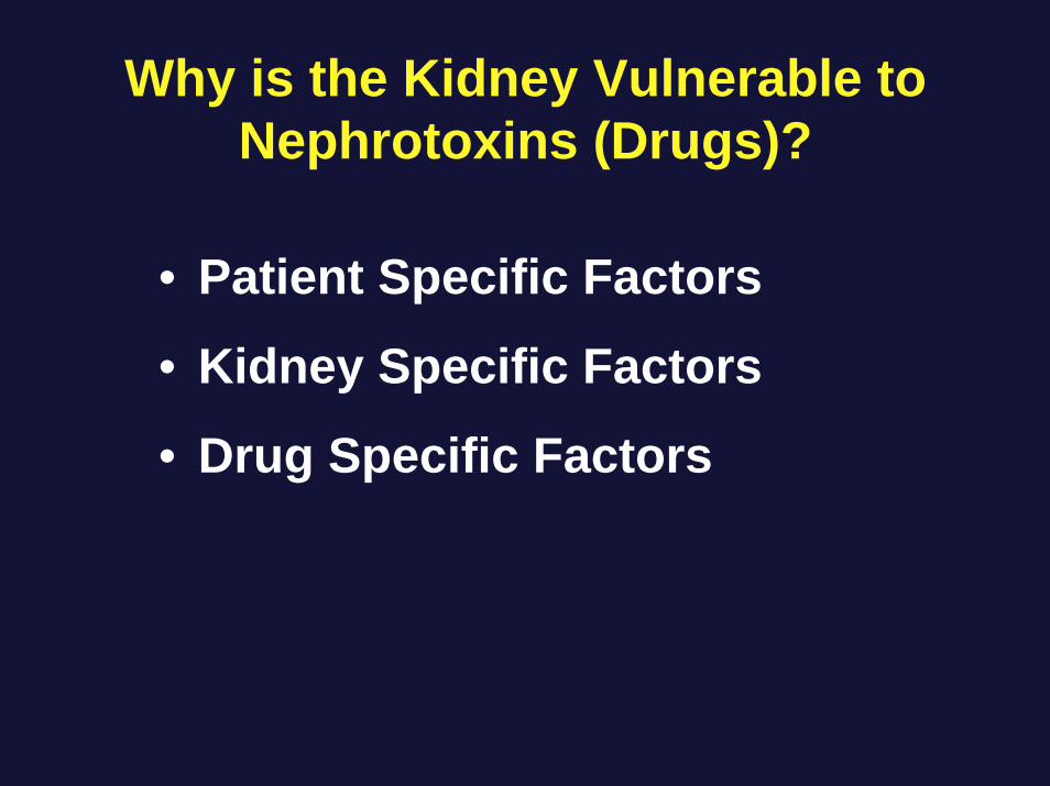
Why is the Kidney Vulnerable to Nephrotoxins (Drugs)?
• Patient Specific Factors
• Kidney Specific Factors
• Drug Specific Factors
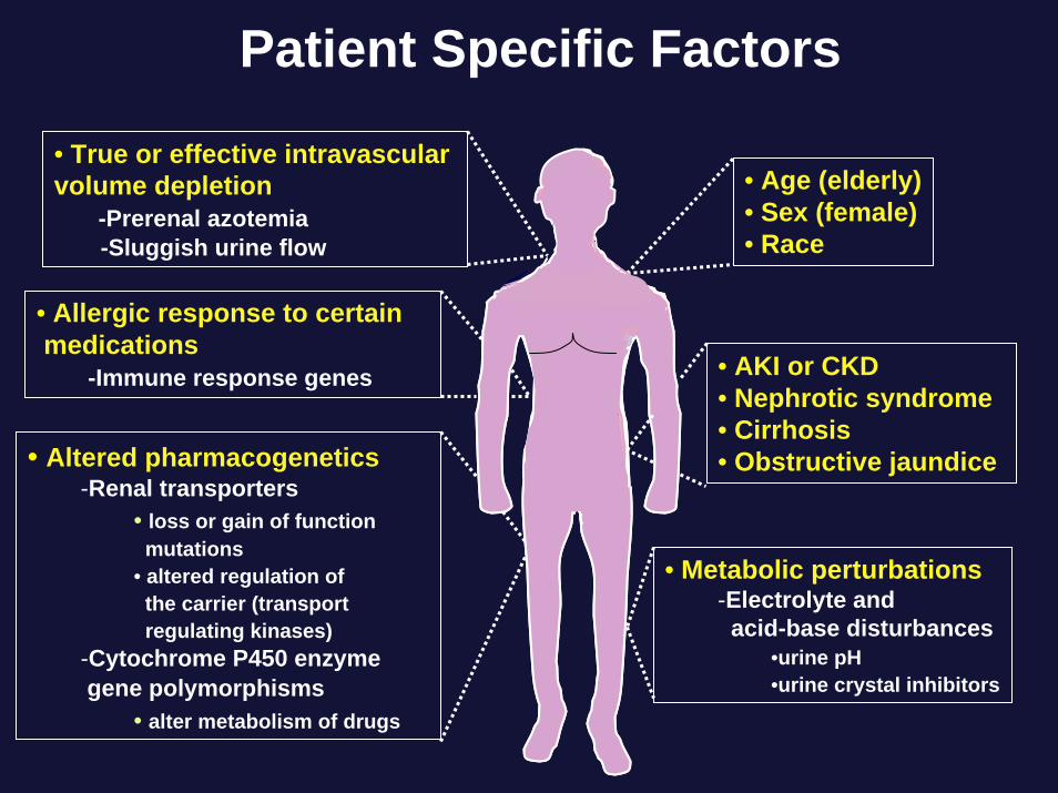
Patient Specific Factors
• Age (elderly)• Sex (female)• Race
• True or effective intravascular volume depletion
-Prerenal azotemia-Sluggish urine flow
• AKI or CKD• Nephrotic syndrome• Cirrhosis• Obstructive jaundice• Altered pharmacogenetics
-Renal transporters• loss or gain of function
mutations• altered regulation ofthe carrier (transportregulating kinases)
-Cytochrome P450 enzyme gene polymorphisms
• alter metabolism of drugs
• Metabolic perturbations-Electrolyte and
acid-base disturbances•urine pH•urine crystal inhibitors
• Allergic response to certainmedications
-Immune response genes
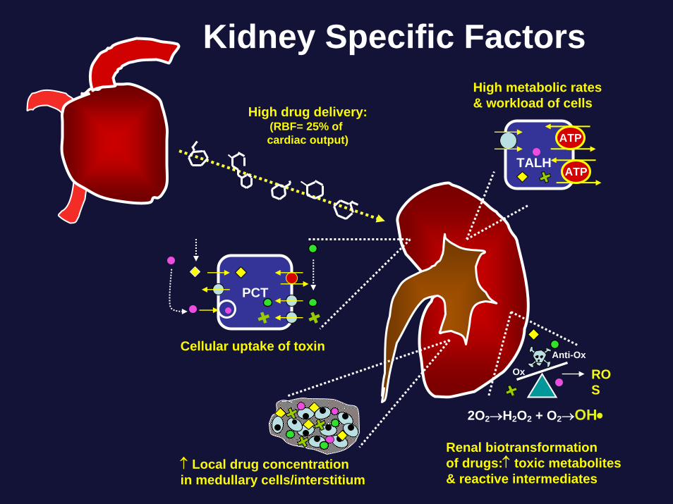
PCT
Cellular uptake of toxin
↑ Local drug concentrationin medullary cells/interstitium
High drug delivery:(RBF= 25% of cardiac output)
High metabolic rates& workload of cells
2O2→H2O2 + O2→OH•
Kidney Specific Factors
TALH
ATP
ATP
Renal biotransformationof drugs:↑ toxic metabolites & reactive intermediates
Anti-Ox
ROS
Ox
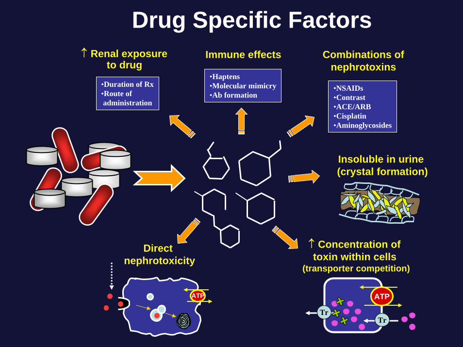
Drug Specific Factors
Insoluble in urine (crystal formation)
↑ Concentration of toxin within cells
(transporter competition)
↑ Renal exposureto drug
Combinations of nephrotoxins
Direct nephrotoxicity
ATP
TrTr
ATP
•NSAIDs•Contrast•ACE/ARB•Cisplatin•Aminoglycosides
•Duration of Rx•Route ofadministration
•Haptens•Molecular mimicry•Ab formation
Immune effects

![(9/21) Thambi Lecture: Drug-Induced Renal Disease ... · (9/21) Thambi Lecture: Drug-Induced Renal Disease Identifying Drug-Induced AKI [Drug-Induced] to be shorted to DI - Sx: Decreased](https://static.fdocuments.us/doc/165x107/5ee17160ad6a402d666c535c/921-thambi-lecture-drug-induced-renal-disease-921-thambi-lecture-drug-induced.jpg)