Dose comparison between Ziehm Vision RFD 3D, Medtronic O ... · Ziehm Vision RFD 3D CMOSline Ziehm...
12
White Paper No. 39 / 2018 Today, intraoperative 3D imaging is re- garded as the gold standard in complex, minimally invasive surgeries. Especially in sensitive areas such as the cervical spine, or even in complex orthopedic and trauma surgeries, reliable intra- operative imaging is essential for the safe placement of implants and the im- mediate control of results. As a flexible, space-saving and cost-effective alter- native to fixed installed 3D scanners, mobile 3D imaging systems are gaining in relevance in this clinical area of inter- est, which is why various medical device companies offer alternative mobile 3D imaging systems to fulfill the market’s requirements. At the same time, dose exposure is being increasingly discussed in our industry and in daily communication with cus- tomers and healthcare professionals. Organizations such as the International Commission on Radiology Protection (ICRP), the European ALARA Network, the American Association of Physicists in Medicine (AAPM) and Image Gently are instrumental in recognizing the importance of dose management and appropriate treatment. This paper is a compilation of dosimetry measurements on major current mobile 3D imaging technologies. Current situation For more than four decades, OR planning and control has been based on CT scans to display anatomical structures not only in 2D, but also in 3D. This is why they were recognized as the imaging standard for years. With the trend toward minimally invasive sur- geries, the demand for intraoperative 3D imag- ing has gained relevance during the last decade to acquire real-time imaging information within the operating theater and to be able to refer to the current anatomical situation. It has also become more important to control the place- ments of implants or revisions of reductions intraoperatively and to reduce the need for post- operative revision surgeries. Dose comparison between Ziehm Vision RFD 3D, Medtronic O-arm ® O2 and Samsung NeuroLogica BodyTom Discover significantly lower dose levels for the Ziehm Vision RFD 3D. 01
Transcript of Dose comparison between Ziehm Vision RFD 3D, Medtronic O ... · Ziehm Vision RFD 3D CMOSline Ziehm...

White Paper No. 39 / 2018
Today, intraoperative 3D imaging is re-garded as the gold standard in complex, minimally invasive surgeries. Especially in sensitive areas such as the cervical spine, or even in complex orthopedic and trauma surgeries, reliable intra-operative imaging is essential for the safe placement of implants and the im-mediate control of results. As a flexible, space-saving and cost-effective alter-native to fixed installed 3D scanners, mobile 3D imaging systems are gaining in relevance in this clinical area of inter-est, which is why various medical device companies offer alternative mobile 3D imaging systems to fulfill the market’s requirements.
At the same time, dose exposure is being increasingly discussed in our industry and in daily communication with cus-tomers and healthcare professionals. Organizations such as the International Commission on Radiology Protection (ICRP), the European ALARA Network, the American Association of Physicists
in Medicine (AAPM) and Image Gently are instrumental in recognizing the importance of dose management and appropriate treatment.
This paper is a compilation of dosimetry measurements on major current mobile 3D imaging technologies.
Current situation
For more than four decades, OR planning and control has been based on CT scans to display anatomical structures not only in 2D, but also in 3D. This is why they were recognized as the imaging standard for years.
With the trend toward minimally invasive sur-geries, the demand for intraoperative 3D imag-ing has gained relevance during the last decade to acquire real-time imaging information within the operating theater and to be able to refer to the current anatomical situation. It has also become more important to control the place-ments of implants or revisions of reductions intraoperatively and to reduce the need for post-operative revision surgeries.
Dose comparison between Ziehm Vision RFD 3D, Medtronic O-arm® O2 and Samsung NeuroLogica BodyTomDiscover significantly lower dose levels for the Ziehm Vision RFD 3D.
01
White Paper No. 39 / 2018
Data acquisition method
The following comparison of the Ziehm Vision RFD 3D, the enhanced Ziehm Vision RFD 3D CMOSline1, Samsung NeuroLogica BodyTom and Medtronic O-arm® O2 provides dose values for equivalent applications of leading intraopera-tive mobile 3D imaging technologies. There were also plans to measure the Siemens Healthineers ARCARDIS Orbic 3D, but it fell out of the evalua-tion process, as its X-ray beam and the resulting 3D volume (12 cm x 12 cm x 12 cm) was too small compared with the other imaging modalities, in particular when considering that the CTDI phan-tom has a diameter of 32 cm. Therefore no fair arrangements could be set for all systems.
Comparable CTDI measurements were per-formed together by Stryker and Ziehm Imaging for the Ziehm Vision RFD 3D and BodyTom. The Ziehm Vision RFD 3D CMOSline is an advanced version of the established Ziehm Vision RFD 3D. Measurements on this system were performed in-house at Ziehm Imaging under identical con-ditions. The CTDI values from the Medtronic O-arm® O2 refer to a reference document pro-vided by Medtronic (BI-160-00227 Rev 1, O-arm® O2 Imaging System Version 4.0 Dosimetry Report March 2015). All further measurements are either on file at Ziehm Imaging or publicly available.
Measurement procedure setup
For each measurement with the Ziehm Vision RFD 3D and Samsung NeuroLogica BodyTom, the same carbon fiber OR table from Stille (STILLE imagiQ2) was used. The carbon fiber OR table ensured that no metal parts were attached or part of the radiation field to avoid modified dose levels due to metal in the field of view. The measurements were done under realistic and maximally comparable conditions with the same experimental setup and comparable positioning of the phantom (PMMA) at the center of the 3D scan. As the measuring points (positioning of the measuring probe) were located in the center and
at 3, 6, 9 and 12 o’clock positions on the PMMA phantom just below the surface, the influences of the specific self-absorption and scattering of the mounting plate were taken into account.
Measurement equipment
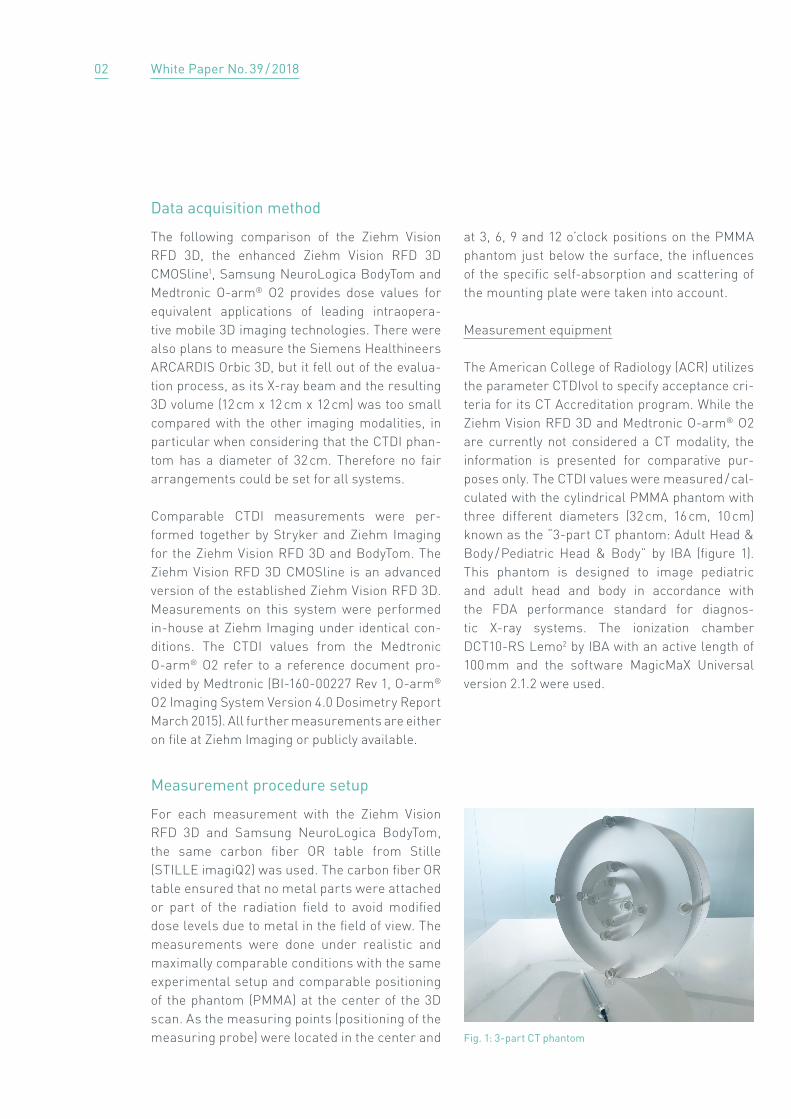
The American College of Radiology (ACR) utilizes the parameter CTDIvol to specify acceptance cri-teria for its CT Accreditation program. While the Ziehm Vision RFD 3D and Medtronic O-arm® O2 are currently not considered a CT modality, the information is presented for comparative pur-poses only. The CTDI values were measured / cal-culated with the cylindrical PMMA phantom with three different diameters (32 cm, 16 cm, 10 cm) known as the “3-part CT phantom: Adult Head & Body / Pediatric Head & Body” by IBA (figure 1). This phantom is designed to image pediatric and adult head and body in accordance with the FDA performance standard for diagnos-tic X-ray systems. The ionization chamber DCT10-RS Lemo2 by IBA with an active length of 100 mm and the software MagicMaX Universal version 2.1.2 were used.
Fig. 1: 3-part CT phantom
02

White Paper No. 39 / 2018
a. Ziehm Vision RFD 3D (CMOSline)
For the measurements, a Ziehm Vision RFD 3D with software version 6.073 was used. For in-house measurements with the Ziehm Vision RFD 3D CMOSline, software version 7.033 was used.
The Ziehm Vision RFD 3D has three different modes for the patient size: Pediatric / Low Dose, Adult, and Obese patients.
The dose length product was measured with a probe at the 3, 6, 9 and 12 o'clock positions and in the center of the phantom. After doing the five measurements, the CTDI values were calculated by the MagicMaX software. This is a standard procedure that is well-known and regularly per-formed in CTs to confirm the system’s stability and conduct other tests.
Scans were made for the following applications: head, cervical spine, thoracic spine / chest, shoul-der, lumbar spine / pelvis and extremities under Pediatric / Low Dose, Adult and Obese patient modifiers. 3D imaging was performed “live” for all anatomical regions. Due to the different size of the anatomical regions, the phantom diameters are listed in table 1.
Application Diameter of CTDI phantom
Head 16 cm
Cervical spine 16 cm
Thoracic spine / chest
32 cm
Lumbar spine / pelvis
32 cm
Shoulder 32 cm
Extremities 16 cm
Table 1: Application referenced to diameter CTDI phantom
Application Ziehm Vision RFD 3D program
Head Pediatric /Low Dose: headAdult: headObese patient: head
Cervical spine Pediatric / Low Dose: cervical spineAdult: cervical spineObese patient: cervical spine
Thoracic spine / chest
Pediatric / Low Dose: spineAdult: spineObese patient: spine
Lumbar spine / pelvis
Pediatric / Low Dose: spineAdult: spineObese patient: spine
Shoulder Pediatric / Low Dose: shoulderAdult: shoulderObese patient: shoulder
Extremities Pediatric / Low Dose: handAdult: handObese patient: hand
Table 2: Application referenced to the Ziehm Vision RFD 3D program
Pediatric / Low Dose is used for children and smaller adults up to 60 kg, Adult mode is used for adults from 61 kg up to 130 kg, and the Obese pa-tient modifier is used for adults weighing more than 130 kg. The following programs were mea-sured:
b. Samsung NeuroLogica BodyTom
The measurement took place in July 2017 at Bap-tist Health Medical Center in Little Rock, Arkan-sas, USA. The CTDI values were measured with Samsung NeuroLogica BodyTom3 and software version 1.08. The measurements were performed with the helical settings according to the manu-facturer’s recommendations for the specific application.
03

White Paper No. 39 / 2018
c. Medtronic O-arm O2
As described in the reference document “O-arm® O2 Imaging System, Version 4.0, Dosimetry Re-port March 2015”, the dose measurements were performed for the following anatomical sections with a field of view of 20 cm4: abdomen, chest, head and extremities. Besides the anatomical se-
lection, the settings are also defined by the 3D mode (standard 3D, HD3D, Low Dose 3D, enhanced 3D) and the patient size (small, medium, large, and extra large). Table 4 shows the available 3D image information and available patient sizes.
The following programs were used for the ex-amined applications:
Table 5: Application referenced to O-arm® O2 program
Application Medtronic O-arm® O2 anatomical section
Head Head
Cervical spine Chest
Thoracic spine / chest
Chest
Lumbar spine / pelvis
Abdomen
Shoulder Chest
Extremities Extremities
Table 4: Medtronic O-arm® O2 3D mode according to patient size
3D mode Patient size
Low Dose Small / medium / large / extra large
Standard Small / medium / large / extra large
HD3D Small / medium / large / extra large
Enhanced 3D(for anatomical section head only)
Small / medium / large / extra large
Table 3: Application referenced to the BodyTom protocol
Application Samsung NeuroLogica BodyTom protocol
Head Adult: helical headPediatric: helical head (14 yrs. +)
Cervical spine
Adult: helical cervical spinePediatric: helical cervical spine (14 yrs. +)
Thoracic spine / chest
Adult: helical chestPediatric: helical chest (60 + kg)Pediatric: helical chest (40 - 60 kg)
Lumbar spine / pelvis
Adult: helical lumbar spine Pediatric: TL spine (60 + kg)Pediatric: TL spine (30 - 60 kg)
Shoulder Adult: helical shoulderPediatric: helical shoulder
Extremities Adult: helical lower extremitiesPediatric: helical lower extremities
The Samsung NeuroLogica BodyTom is designed with different modes for the patient size and ap-plication. The following modes were selected for the comparison, according to the respective manufacturer’s recommendations: Adult, Pediat-ric (60 kg +), Pediatric (30 - 60 kg) and Pediatric (14 years +). As there was no Obese patient mode or HQ mode available, only Pediatric and Adult patient modes were measured. Table 3 shows the protocols used for the examined body regions.
®
04

White Paper No. 39 / 2018
Dosimetry Results
The following tables show the CTDI in mGy for the different application, named in each headline.
78.97
62.63
79.12
76.36
9.87 15.71
20.37 15.71
20.37
5.96
38.55
29.94
23.76
5.96
23.76
0
10
20
60
30
70
40
80
50
1.32 1.88
6.83
1.06 1.423.67
11.7
39.6
12.42
LD Standard HD LD Standard HD LD Standard LD Standard HD3D
Ziehm Vision RFD 3D CMOSline
Ziehm Vision RFD 3D
Samsung NeuroLogica BodyTom5
Medtronic O-arm® O2 (head)5
Small / medium / large / extra large
Enhanced 3D
a. Dosimetry data for head protocols
[mGy]
05

White Paper No. 39 / 2018
c. Dosimetry data for chest / thoracic spine protocols
b. Dosimetry data for cervical spine
16.73
8.38 10.65
5.28
26.63
13.36
6.63
16.55
21.31
Ziehm Vision RFD 3D CMOSline
Ziehm Vision RFD 3D
Samsung NeuroLogica BodyTom5
Medtronic O-arm® O2 (chest)5 Small / medium / large / extra large
0
10
20
30
40
13.08
17.22
35.37
4.87.8
23.725.8 26.59
42.08
13.36
5.5 5.1
50[mGy]
Ziehm Vision RFD 3D CMOSline
Ziehm Vision RFD 3D
Samsung NeuroLogica BodyTom5
Medtronic O-arm® O2 (chest)5 Small / medium / large / extra large
0
10
20
30
40
1.32 1.88
6.68
1.06 1.423.67
6.7
37.0
50[mGy]
16.73
8.38 10.65
5.28
26.63
13.36
6.63
16.55
21.31
26.59
42.08
13.36
LD Standard HD LD Standard HD LD Standard LD Standard HD
LD Standard HD LD Standard HD LD Pediatric
60 kg +
Standard LDLD Pediatric 30 - 60 kg
Standard HD
06

White Paper No. 39 / 2018
e. Dosimetry data for lumbar spine / pelvis protocols
d. Dosimetry data for shoulder protocols
Ziehm Vision RFD 3D CMOSline
Ziehm Vision RFD 3D
Samsung NeuroLogica BodyTom5
Medtronic O-arm® O2 (chest)5 Small / medium / large / extra large
18.59
24.13
36.57
6.97
11.18
29.87
16.73
8.38 10.65
5.28
26.63
13.36
6.63
16.55
21.31
0
10
20
30
40
50
1.76
12.0
26.59
42.08
13.36
16.73
6.63
26.6326.59
13.3610.73
5.28
42.08
16.55
Ziehm Vision RFD 3D CMOSline
Ziehm Vision RFD 3D
Samsung NeuroLogica BodyTom5
Medtronic O-arm® O2 (abdomen)5 Small / medium / large / extra large
0
10
20
30
40
50
60
14.75
19.59
36.72
4.757.8
23.74
6.0 7.4
24.6
33.8
53.19
16.73
LD Standard HD LD Standard HD LD Pediatric
60 kg +
Standard LDLD Pediatric 40 - 60 kg
Standard HD
[mGy]
[mGy]
LD Standard HD LD Standard HD LD Standard LD Standard HD
07

White Paper No. 39 / 2018
Dosimetry comparison
For the first time, we are able to compare stan-dardized measurements with the PMMA phan-tom and standardized measurement setups for the Ziehm Vision RFD 3D, Samsung NeuroLogica BodyTom and Medtronic O-arm® O2. This makes it possible to eliminate patient variability, which usually influences dose levels the most. The systems from Ziehm Imaging in particular offer an intelligent, real-time dose regulation that is influenced by different patient anatomy and con-stitution. The setup with a standardized PMMA phantom makes a real comparison possible.
1. The dose measurements comparing the three different systems clearly depict the profound advantage the Ziehm Vision RFD 3D has over the other systems. Ziehm Imaging is strongly driving the awareness of ALARA principles in the indus-try and among customers. That is why the Low
Dose mode is set as the default mode for all Ziehm Imaging systems. In addition, the CMOSline achieves higher spatial resolution due to smaller pixel sizes combined with lower noise levels. This makes interpolation unnecessary, especially in the magnification modes. There-fore, the Ziehm Vision RFD 3D offers not only minimal dose levels but also exceptional image quality.
2. Especially in the anatomical programs for head, cervical spine and extremities, the Ziehm Vision RFD 3D in its enhanced CMOSline version shows by far the best results with better dose values in all different patient modifiers compared with the other systems.
f. Dosimetry data for extremities
12.29.615.845.84
7.47
18.32
9.65
14.22 14.22
Ziehm Vision RFD 3D CMOSline
Ziehm Vision RFD 3D
Samsung NeuroLogica BodyTom5
Medtronic O-arm® O2 (extremities)5 Small / medium / large / extra large
0
10
20
30
40
1.53.65
8.72
1.25 1.974.7 3.4
15.6 15.32
23.19
12.16
LD Standard HD LD Standard HD LD Standard LD Standard HD
50[mGy]
08

White Paper No. 39 / 2018
3. Comparing the Ziehm Vision RFD 3D CMOSline with Samsung's NeuroLogica BodyTom, the Ziehm Imaging system shows better results in all compared cases, except one (shoulder LD).6
4. The Ziehm Vision RFD 3D shows advantages in CTDI values in all cases compared with Med-tronic's O-arm® O2 depending on the specialized patient program of the O-arm® O2. By compar-ing only the lowest-possible CTDI values in each program of Medtronic with the CTDI values of the Ziehm Vision RFD 3D, the C-arm shows bet-ter results in 67 of 76 available cases.7
5. In those programs where the Ziehm Vision RFD 3D shows higher CTDI values, the values are comparable to those values of the other sys-tems. Especially in the critical trunk area, the Ziehm Vision RFD 3D is able to invest higher dose values for obese patients to provide an optimized penetration and therefore improved image quality. Further, to fulfill the need of suf-ficient image quality at the lowest-possible dose in obese patients, Ziehm Imaging invests addi-tional dose levels for those challenging patient anatomies. But here as well, the intelligent dose regulation algorithms ensure optimized dose levels for each individual anatomy, depending on patient size, weight, constitution and scanned area as well as scan projections (AP or lateral projection). This is made possible by the real-time, patient-unique regulation strategy that allows different mA levels within one modifier (Pediatric / Low Dose, Adult, Obese patient).
This is why the Ziehm Vision RFD 3D manages to get along with only three different modifiers, as mA levels will be adapted to the optimum auto-matically and in a manner unique to the patient. As this is done automatically by the system and does not have to be set by the user, it provides a profound advantage compared with other intra-operative 3D imaging solutions in which staff variability complicates the correct use of the system and therefore the correct dose.
Image quality results
In addition to the dosimetry comparisons based on physical conditions, we also managed to achieve an exemplary image quality comparison with an anthropomorphic X-ray head phantom for high-contrast imaging on all three systems. For those high-contrast procedures, the differentia-tion between metal, bone and soft tissue is pivotal. The following images show the image quality of the three different systems. Image captures show the dose results of the measure-ments for the 16 cm CTDI phantom.
09

White Paper No. 39 / 2018
Low Dose, CTDIvol16 1.06 mGy
Ziehm Vision RFD 3D CMOSline (head, resolution 512³ voxel)
Medtronic O-arm® O2 (head, resolution 512 x 512 x 192 voxel)
Samsung NeuroLogica BodyTom (head, resolution: LD Pediatric 512 x 512 x 208 voxel, Standard 512 x 512 x 400 voxel)
Low Dose, small, CTDIvol16 5.96 mGy
Low Dose, (14 yrs.+), CTDIvol16 11.7 mGy
Standard, CTDIvol16 1.42 mGy
Standard, medium, CTDIvol16 15.71 mGy
Standard, CTDIvol16 39.6 mGy
HD, CTDIvol16 3.67 mGy
HD 3D, medium, CTDIvol16 23.79 mGy
10

White Paper No. 39 / 2018
Image quality comparison
The images show clearly that all three systems offer a comparable image quality and allow a clear differentiation of finest bone structures and soft tissue in the head. Furthermore, boundaries between cortical bone structures and the can-cellous bone can also be differentiated as well as hollow spaces in the sinus cavity.
The high-contrast characteristic is provided by all three different systems but variations be-tween image impression and homogeneity arise from different dose levels. Comparing the dose levels of those three exemplary phantom images (CTDIvol16 value for standard 3D mode8: Ziehm Vision RFD 3D: 1.42 mGy; Medtronic O-arm O2: 15.71 mGy; Samsung NeuroLogica BodyTom: 39.6 mGy), the data shows that comparable im-age quality does not result in comparable dose levels. As a result, the conclusion can be drawn that the Ziehm Vision RFD 3D works strictly ac-cording to ALARA principles offering sufficient image quality at the lowest-possible dose.
Conclusion
The Ziehm Vision RFD 3D offers unprecedented performance across the most varied and chal-lenging application spectrum and can be seen as one of the most important and dose-saving al-ternatives in the field of mobile intraoperative 3D imaging systems. In addition, the Ziehm Vision RFD 3D CMOSline comes with an enhanced ver-sion of our comprehensive SmartDose concept. A newly developed dose-saving technology called Beam Filtration9 supports the latest improve-ments in our enhanced CMOS imaging chain, thus enabling an exceptional reduction in the skin entrance dose. As a result, the Ziehm Vision RFD 3D CMOSline delivers excellent image qual-ity with a lower dose, which is also confirmed by our customers:
"Compared with systems with comparable im-age quality such as Medtronic's O-arm, the Ziehm Vision RFD 3D has superior OR usability and advanced radiation dose control, which ben-efits patients, surgeons and staff alike. The ex-cellent image quality results in increased patient safety. In addition, the ideal usability gives improved surgeon efficiency. Ziehm Imaging's dramatically superior price-performance ratio allows radiology purchasing decision-makers to get this technology in the hands of doctors and realize immediate return on investment,” said S. Raymond Golish, MD PhD MBA, Chief Quality Officer at Jupiter Medical Center in Palm Beach, Florida, USA.
11

White Paper No. 39 / 2018
1 CMOSline represents a system configuration that is based on a Ziehm Imaging CMOS flat-panel detector.
2 Information on calibration is available on request.3 Serial number is traceable and available on request. 4 For 20 cm field of view, the cylinder has a diameter of 212 ± 1 mm and a length of 160 ± 1 mm
5 The programs and settings used were in accordance with the respective manufacturer’s recommendations regarding the applied applications.
6 Case means patient modifier in a protocol; 4 protocols including 2 modifier comparing LD and Standard and 2 pro-tocols including 3 modifier comparing LD of Ziehm Vision RFD 3D with LD 30-60 kg and LD 60 kg+ of Samsung's NeuroLogica BodyTom .
7 Case means patient modifier in a protocol with different patient size; 5 protocols including 3 modifier à 4 patient sizes (cervical spine protocol, chest /thoracic spine pro-tocol, shoulder protocol, spine / pelvis protocol, extremi-ties protocol with modifier LowDose, Standard, HD in the patient sizes small, medium, large and extra large) and 1 protocol including 4 modifier à 4 patient sizes (head proto-col with modifier LowDose, Standard, HD3D and Enhanced 3D in the patient sizes small, medium, large and extra large) result in 76 cases.
8 It can be assumed that the absorption with a 16 cm PMMA CTDI phantom adequately represents the head phantom.
9 The technology Beam Filtration reduces dose exposure for all CMOSline systems in comparison with conventional fil-tration techniques (status before September 2017). Data on file. Results may vary.
Authors:Lena LochnerGlobal Product Manager 3D & NavigationZiehm Imaging GmbH
Anne-Kathrin MeierClinical Marketing ManagerZiehm Imaging GmbH
Günter StelzerDirector Special ProjectsZiehm Imaging GmbH
Kevin PackardStrategic Clinical Marketing ManagerStryker
12