State-of-the-art 3D imaging and navigation with the Ziehm ... · niated discs with...
6
White Paper No. 36/ 2016 State-of-the-art 3D imaging and navigation with the Ziehm Vision RFD 3D mobile C-arm and Brainlab Spine & Trauma 3D navigation. A workshop on human specimens. Lower reimbursement trends and the awareness of increased accuracy and reduced X-ray exposure in surgical pro- cedures are rising challenges in health- care. 1,2 Improving patient care as well as optimizing clinical workflows drive these challenges even further. 3 To overcome the challenges of demand- ing procedures in areas like cervical and upper thoracic spine as well as pelvic or minimally invasive surgeries, Ziehm Imaging, in cooperation with Brainlab, developed an interface to com- bine surgical navigation systems and intraoperative 3D imaging with the Ziehm Vision RFD 3D mobile C-arm. This paper will first describe the latest de- velopments in image-guided surgery for the Ziehm Vision RFD 3D in combination with Brainlab navigation. Second, it will give an overview of initial customer feed- back on results in human specimen cases. This paper will give an overview of the broad application area for the combination of Brainlab Spine & Trauma navigation and the Ziehm Vision RFD 3D, and shows how innovative technologies can help to over- come current challenges in healthcare. The rising trend of minimally invasive procedures and image-guided surgery A number of reasons – including an ever-increas- ing aging population – lead to a rising demand for efficient high-quality care. 4 The aim is to benefit from reduced costs as a result of shorter hospital stays through less-invasive approaches, and to improve patient outcomes. As a result, the demand for minimally invasive procedures is growing and posing increasing requirements on the development and equipment of imaging and navigation technologies. 5 Intraoperative 3D imaging is recognized as the latest imaging technology for mobile devices to achieve complete 3D CT-like image information in just one scan procedure, cost-effectiveness and lower exposure rates in orthopedic, trauma and spine surgeries in comparison to fixed systems. 6 In combination with dedicated navigation systems like Brainlab Spine & Trauma 3D, it enables the surgeon to perform demanding minimally inva- sive procedures with higher confidence and, more importantly with less dose exposure to patients and the surgical team. 7 In more than 21 % of complex anatomical osteo- synthesis procedures, an intraoperative improve- ment of the implant position or a revision of reduction has to be performed. 6 01
Transcript of State-of-the-art 3D imaging and navigation with the Ziehm ... · niated discs with...
White Paper No. 36/ 2016
State-of-the-art 3D imaging and navigation with the Ziehm Vision RFD 3D mobile C-arm and Brainlab Spine & Trauma 3D navigation.A workshop on human specimens.
Lower reimbursement trends and the awareness of increased accuracy and reduced X-ray exposure in surgical pro-cedures are rising challenges in health-care.1,2 Improving patient care as well as optimizing clinical workflows drive these challenges even further.3
To overcome the challenges of demand-ing procedures in areas like cervical and upper thoracic spine as well as pelvic or minimally invasive surgeries, Ziehm Imaging, in cooperation with Brainlab, developed an interface to com-bine surgical navigation systems and intraoperative 3D imaging with the Ziehm Vision RFD 3D mobile C-arm.
This paper will first describe the latest de-velopments in image-guided surgery for the Ziehm Vision RFD 3D in combination with Brainlab navigation. Second, it will give an overview of initial customer feed-back on results in human specimen cases.
This paper will give an overview of the broad application area for the combination of Brainlab Spine & Trauma navigation and the Ziehm Vision RFD 3D, and shows how innovative technologies can help to over-come current challenges in healthcare.
The rising trend of minimally invasive procedures and image-guided surgery
A number of reasons – including an ever-increas-ing aging population – lead to a rising demand for efficient high-quality care.4 The aim is to benefit from reduced costs as a result of shorter hospital stays through less-invasive approaches, and to improve patient outcomes.
As a result, the demand for minimally invasive procedures is growing and posing increasing requirements on the development and equipment of imaging and navigation technologies.5
Intraoperative 3D imaging is recognized as the latest imaging technology for mobile devices to achieve complete 3D CT-like image information in just one scan procedure, cost-effectiveness and lower exposure rates in orthopedic, trauma and spine surgeries in comparison to fixed systems.6
In combination with dedicated navigation systems like Brainlab Spine & Trauma 3D, it enables the surgeon to perform demanding minimally inva-sive procedures with higher confidence and, more importantly with less dose exposure to patients and the surgical team.7
In more than 21 % of complex anatomical osteo-synthesis procedures, an intraoperative improve-ment of the implant position or a revision of reduction has to be performed.6
01
White Paper No. 36/ 2016
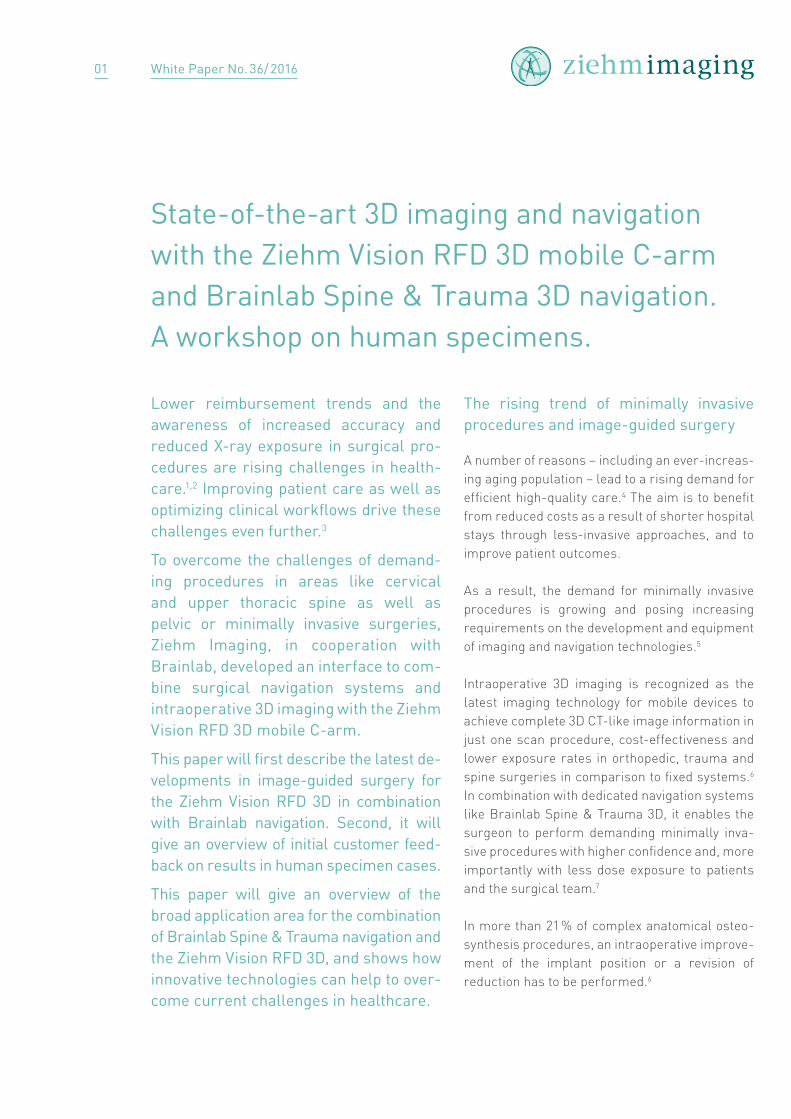
Step 1 – Setup of navigation systemThe navigation camera and the monitor cart can be positioned flexibly beside or in front of the patient’s table as they are separated from each other. A reference clamp with a reflective marker array is attached to the patient’s bony anatomy. A second array is firmly mounted to the detector housing of the Ziehm Vision RFD 3D. There are two dedicated registration arrays for each side of the flat-panel detector (FD), which can be mounted onto the han-dle at the FD housing. As the registration arrays are autoclavable and designed for sterile use they can be attached over the FD’s housing drape. The reference clamp, which is attached to the patient’s bone structure to be navigated upon, has to be vis-ible to the infrared camera.
Step 2 – Image acquisition with the Ziehm Vision RFD 3DWhen the set-up is done the Ziehm Vision RFD 3D generates a high-resolution 3D data-set in less than 5 minutes. This time includes draping, hyperoxygenation and breathing stop of the patient, the team leaving the OR, image acquisition, and reconstruction.
Image-guided surgery has been proven to poten-tially lower revision rates and increase accuracy. This applies especially to complex procedures.8,9
Navigation enables clinicians to improve patient outcomes, reduce X-ray exposure and improve efficiency in the OR by optimizing clinical work- flows.
Typical workflow
A common procedure for navigation is register-ing preoperative CT scans for navigation where a manual registration of the bone structure is required, as the patient’s position in surgery may differ from the scanning position.
In such cases, a postoperative scan is com-mon to verify whether the intervention was suc-cessful. Intraoperative 3D imaging with the new Ziehm Vision RFD 3D C-arm, eliminates the need for manual registration and delivers intraopera-tive real-time information, optimizing the clinical workflow and bringing new levels of image quality and efficiency to the OR.
Fig. 1: Example setup of the Ziehm Vision RFD 3D and Brainlab navigation system Kick®.
02
White Paper No. 36/ 2016
The aim was to confirm workflow and effi-ciency strategies, which were already tested under lab conditions, in a hospital environment. Furthermore, the workshop demonstrated a broader future application area and offered hands-on feedback from potential users on the integration of the Ziehm NaviPort interface, Brainlab Spine & Trauma 3D navigation and Fluoro 3D registration software.
Workflow and results
After setting up the systems (Ziehm Imaging and Brainlab), the C-arm has been draped, and a ref-erence array has been attached to the flat-panel detector of the C-arm and to the spinous pro-cess of the patient at level C2. After a successfull collision check the 3D scan was performed to register the dataset for navigation.
First, a spinal fusion of the first and second cer-vical vertebra was performed with the C-arm in lateral position to perform Margerl’s procedure for transarticular screw fixation. After image registration and automatic transfer of the data-set to the Brainlab navigation system Kick®, the accuracy of the registration has been verified thor- oughly and showed high accuracy. Prior to the navigated screw insertion, the two screws
Once the scan is finalized, the acquired 3D data-set is automatically transferred via the Ziehm NaviPort interface to the Brainlab navigation system to be automatically registered. That means that the position of the patient is set in relation to the acquired dataset. The automatic registration and transfer of the 3D dataset to the navigation system is performed in less than 1 minute.10
Step 3 – Navigation procedure with Brainlab Spine & Trauma 3D navigationAs soon as the dataset has been transferred to the navigation system, navigation can be started. Optical tracking of the C-arm is no longer re- quired. Surgical instruments are continuously tracked with the infrared camera and their posi-tion visualized on the patient data. This allows for more accurate procedures compared to con-ventional surgical techniques. The surgical pro-cedure on the 3D dataset is performed using various navigational instruments like a navigated drill guide from Brainlab.
Step 4 – Final control scan with the Ziehm Vision RFD 3DFor the final quality check of the results or for doc-umentation purposes, the Ziehm Vision RFD 3D can generate a control scan in the OR. Efficiency can be increased as postoperative CT scans may no longer be required.
First results in specimen
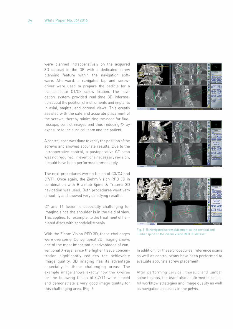
MethodsA 92-year-old female specimen was the subject of a one-day workshop held by both manufac-turers at the Institute of Anatomy, at Ludwig-Maximilians-University, Munich, Germany. Diffe- rent 3D scans followed by navigated screw place-ments in cervical, thoracic and lumbar spine as well as at the pelvis were performed using the Ziehm Vision RFD 3D and Brainlab Spine & Trauma 3D navigation software (Fig. 2 - 5).
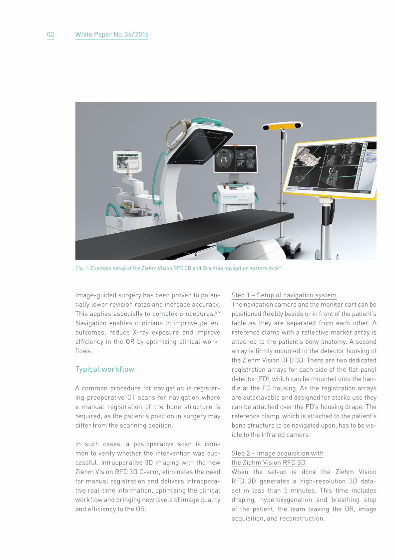
Fig. 2: Navigated screw placement with Brainlab Spine & Trauma 3D software at the cervical spine on the Ziehm Vision RFD 3D dataset.
03
White Paper No. 36/ 2016
In addition, for these procedures, reference scans as well as control scans have been performed to evaluate accurate screw placement.
After performing cervical, thoracic and lumbar spine fusions, the team also confirmed success-ful workflow strategies and image quality as well as navigation accuracy in the pelvis.
were planned intraoperatively on the acquired 3D dataset in the OR with a dedicated screw planning feature within the navigation soft-ware. Afterward, a navigated tap and screw-driver were used to prepare the pedicle for a transarticular C1 / C2 screw fixation. The navi-gation system provided real-time 3D informa- tion about the position of instruments and implants in axial, sagittal and coronal views. This greatly assisted with the safe and accurate placement of the screws, thereby minimizing the need for fluo-roscopic control images and thus reducing X-ray exposure to the surgical team and the patient.
A control scan was done to verify the position of the screws and showed accurate results. Due to the intraoperative control, a postoperative CT scan was not required. In event of a necessary revision, it could have been performed immediately.
The next procedures were a fusion of C3 / C4 and C7 / T1. Once again, the Ziehm Vision RFD 3D in combination with Brainlab Spine & Trauma 3D navigation was used. Both procedures went very smoothly and showed very satisfying results.
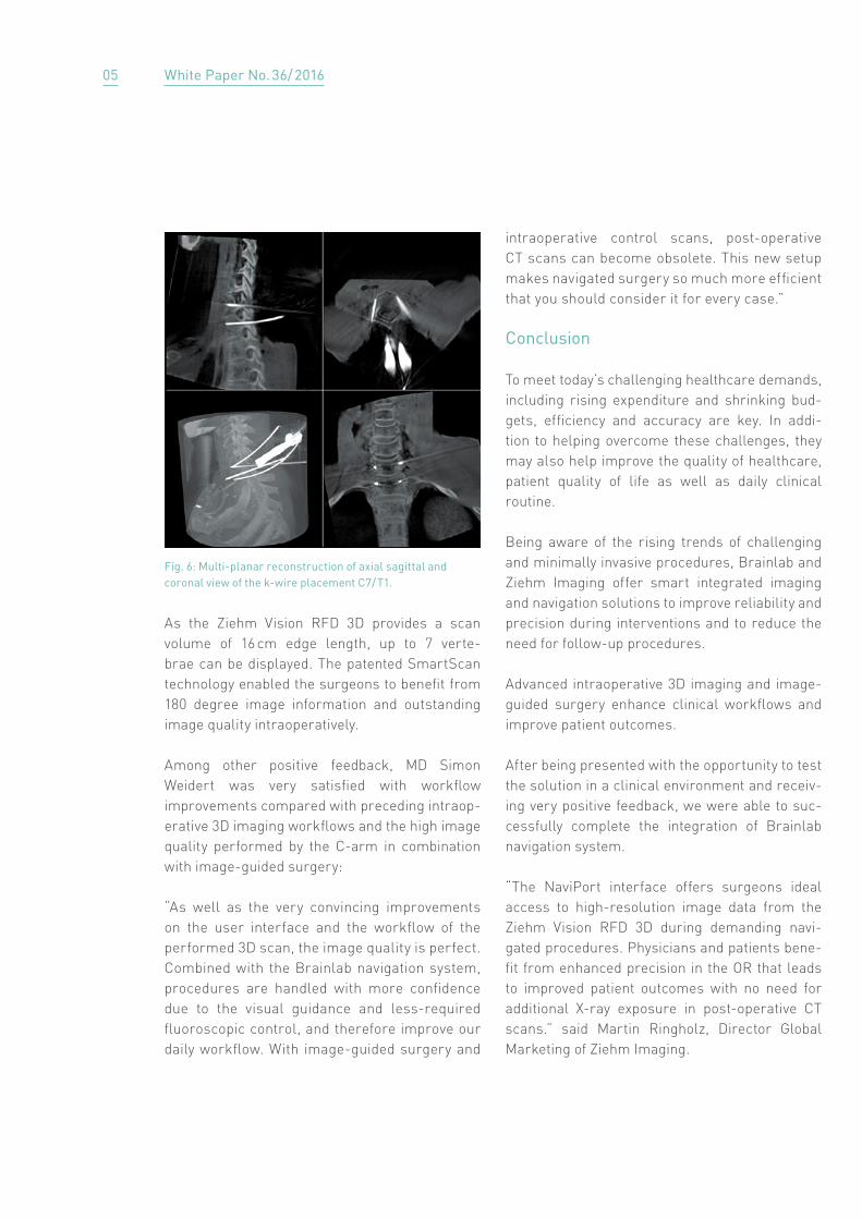
C7 and T1 fusion is especially challenging for imaging since the shoulder is in the field of view. This applies, for example, to the treatment of her-niated discs with spondylolisthesis.
With the Ziehm Vision RFD 3D, these challenges were overcome. Conventional 2D imaging shows one of the most important disadvantages of con-ventional X-rays, since the higher tissue concen- tration significantly reduces the achievable image quality. 3D imaging has its advantage especially in those challenging areas. The example image shows exactly how the k-wires for the following fusion of C7 / T1 were placed and demonstrate a very good image quality for this challenging area. (Fig. 6)
Fig. 3 - 5: Navigated screw placement at the cervical and lumbar spine on the Ziehm Vision RFD 3D dataset.
04
White Paper No. 36/ 2016
intraoperative control scans, post-operative CT scans can become obsolete. This new setup makes navigated surgery so much more efficient that you should consider it for every case.”
Conclusion
To meet today’s challenging healthcare demands, including rising expenditure and shrinking bud-gets, efficiency and accuracy are key. In addi-tion to helping overcome these challenges, they may also help improve the quality of healthcare, patient quality of life as well as daily clinical routine.
Being aware of the rising trends of challenging and minimally invasive procedures, Brainlab and Ziehm Imaging offer smart integrated imaging and navigation solutions to improve reliability and precision during interventions and to reduce the need for follow-up procedures.
Advanced intraoperative 3D imaging and image-guided surgery enhance clinical workflows and improve patient outcomes.
After being presented with the opportunity to test the solution in a clinical environment and receiv-ing very positive feedback, we were able to suc-cessfully complete the integration of Brainlab navigation system.
“The NaviPort interface offers surgeons ideal access to high-resolution image data from the Ziehm Vision RFD 3D during demanding navi-gated procedures. Physicians and patients bene-fit from enhanced precision in the OR that leads to improved patient outcomes with no need for additional X-ray exposure in post-operative CT scans.” said Martin Ringholz, Director Global Marketing of Ziehm Imaging.
As the Ziehm Vision RFD 3D provides a scan volume of 16 cm edge length, up to 7 verte-brae can be displayed. The patented SmartScan technology enabled the surgeons to benefit from 180 degree image information and outstanding image quality intraoperatively.
Among other positive feedback, MD Simon Weidert was very satisfied with workflow improvements compared with preceding intraop-erative 3D imaging workflows and the high image quality performed by the C-arm in combination with image-guided surgery:
“As well as the very convincing improvements on the user interface and the workflow of the performed 3D scan, the image quality is perfect. Combined with the Brainlab navigation system, procedures are handled with more confidence due to the visual guidance and less-required fluoroscopic control, and therefore improve our daily workflow. With image-guided surgery and
Fig. 6: Multi-planar reconstruction of axial sagittal and coronal view of the k-wire placement C7 / T1.
05
White Paper No. 36/ 2016
Authors:MD Simon WeidertDepartment for General, Trauma and Reconstructive SurgeryLudwig-Maximilians-University Hospital Munich, Germany
Eva-Maria IlgTeamlead 3D and NavigationZiehm Imaging GmbH
Anne-Kathrin ReifClinical Marketing ManagerZiehm Imaging GmbH
References
1 Richter et. al., Cervical pedicle screws: conventional
versus computer-assisted placement of cannulated
screws. Spine (PhilaPa 1976). 2005 Oct 15;30(20):2280-72 Gebhard et al., Does computer assisted spine
surgery reduce intraoperative radiation doses?
Spine (PhilaPa1976). 2006 Aug 1;31(17)3 PricewaterhouseCoopers LLP, 2013
Five megatrends and possible implications4 http://www.beckersspine.com/spine/item/
19610-the-future-of-spinal-fusion-where-
bmp-stands-today-tomorrow.html5 https://spine-operation.guide/operationen/
minimal-invasive-wirbelsaulenchirurgie/6 Recum von, J. et al., Unfallchirurg 2012, 115:196-201, Die
intraoperative 3D-C-Bogen-Anwendung. State of the art7 Kraus M.D. et al. Can computer-assisted surgery
reduce the effective dose for spinal fusion and sacro-
iliac screw insertion? CORR 2010, 468, 2419-24298 Watkins R.G. et al. Cost-Effectiveness of Image-Guided
Spine Surgery. Open Orthop J. 2010 Aug 6;4:228-33.9 Mason A, Paulsen R, Babuska JM, Rajpal S, Burneikiene
S, Nelson EL, Villavicencio AT. The accuracy of pedicle
screw placement using intraoperative image guidance
systems. J Neurosurg Spine. 2014 Feb;20(2):196-203.10 Data on file. This time includes image acquisition &
reconstruction of the 3D dataset.
06