Dopamine modification of interfaces between polymers · PDF fileDopamine Modification of...
137
Dopamine modification of interfaces between polymers and metals Ribena, D. DOI: 10.6100/IR735527 Published: 01/01/2012 Document Version Publisher’s PDF, also known as Version of Record (includes final page, issue and volume numbers) Please check the document version of this publication: • A submitted manuscript is the author's version of the article upon submission and before peer-review. There can be important differences between the submitted version and the official published version of record. People interested in the research are advised to contact the author for the final version of the publication, or visit the DOI to the publisher's website. • The final author version and the galley proof are versions of the publication after peer review. • The final published version features the final layout of the paper including the volume, issue and page numbers. Link to publication Citation for published version (APA): Ribena, D. (2012). Dopamine modification of interfaces between polymers and metals Eindhoven: Technische Universiteit Eindhoven DOI: 10.6100/IR735527 General rights Copyright and moral rights for the publications made accessible in the public portal are retained by the authors and/or other copyright owners and it is a condition of accessing publications that users recognise and abide by the legal requirements associated with these rights. • Users may download and print one copy of any publication from the public portal for the purpose of private study or research. • You may not further distribute the material or use it for any profit-making activity or commercial gain • You may freely distribute the URL identifying the publication in the public portal ? Take down policy If you believe that this document breaches copyright please contact us providing details, and we will remove access to the work immediately and investigate your claim. Download date: 20. Apr. 2018
Transcript of Dopamine modification of interfaces between polymers · PDF fileDopamine Modification of...
Dopamine modification of interfaces between polymersand metalsRibena, D.
DOI:10.6100/IR735527
Published: 01/01/2012
Document VersionPublisher’s PDF, also known as Version of Record (includes final page, issue and volume numbers)
Please check the document version of this publication:
• A submitted manuscript is the author's version of the article upon submission and before peer-review. There can be important differencesbetween the submitted version and the official published version of record. People interested in the research are advised to contact theauthor for the final version of the publication, or visit the DOI to the publisher's website.• The final author version and the galley proof are versions of the publication after peer review.• The final published version features the final layout of the paper including the volume, issue and page numbers.
Link to publication
Citation for published version (APA):Ribena, D. (2012). Dopamine modification of interfaces between polymers and metals Eindhoven: TechnischeUniversiteit Eindhoven DOI: 10.6100/IR735527
General rightsCopyright and moral rights for the publications made accessible in the public portal are retained by the authors and/or other copyright ownersand it is a condition of accessing publications that users recognise and abide by the legal requirements associated with these rights.
• Users may download and print one copy of any publication from the public portal for the purpose of private study or research. • You may not further distribute the material or use it for any profit-making activity or commercial gain • You may freely distribute the URL identifying the publication in the public portal ?
Take down policyIf you believe that this document breaches copyright please contact us providing details, and we will remove access to the work immediatelyand investigate your claim.
Download date: 20. Apr. 2018
Dopamine Modification of Interfaces between Polymers and Metals

Dopamine Modification of Interfaces between Polymers and Metals
PROEFSCHRIFT
ter verkrijging van de graad van doctor aan de Technische Universiteit Eindhoven, op gezag van de rector magnificus, prof.dr.ir. C.J. van Duijn, voor een
commissie aangewezen door het College voor Promoties in het openbaar te verdedigen
op donderdag 4 oktober 2012 om 16.00 uur
door
Dina Rībena
geboren te Talsi, Letland
Dit proefschrift is goedgekeurd door de promotor: Prof.dr. G. de With Copromotor: Dr. N.A.J.M. Sommerdijk Rībena, Dina Dopamine Modification of Interfaces between Polymers and Metals Eindhoven University of Technology, 2012 A catalogue record is available from the Eindhoven University of Technology Library. ISBN: 978-90-386-3233-9 The work described in this thesis has been carried out at the Laboratory of Materials and Interface Chemistry (SMG) within the Department of Chemical Technology, Eindhoven University of Technology, the Netherlands. This research forms part of the research programme of the Dutch Polymer Institute (DPI), project 672. Cover design: Dina Rībena Printed at the PrintService, Eindhoven University of Technology

"΄Επιστήμη ποιητική εύδαιμονίας" -Πλάτων
"Scientific knowledge is the creator of happiness" - Plato

TABLE OF CONTENTS
Chapter 1 Introduction ......................................................................................................................... 1
1.1 Marine mussel adhesion and DOPA...............................................................3 1.2 The reactivity of protein bound-DOPA..........................................................6 1.3 Synthetic mimics of mussel foot proteins ......................................................7 1.4 Recent developments in mussel-inspired adhesives.....................................14 1.5 The aim and outline of the thesis..................................................................16 1.6 References ....................................................................................................17
Chapter 2 Dopamine and polydopamine coatings on gold and sapphire ....................................... 21
2.1 Introduction ..................................................................................................22 2.2 Experimental.................................................................................................22 2.3 Results and Discussion .................................................................................25
2.3.1 Dopamine behavior in aqueous media .................................................25 2.3.2 Dopamine and polydopamine films......................................................26 2.3.3 Measuring practical adhesion ...............................................................39
2.4 Conclusion....................................................................................................42 2.5 References ....................................................................................................42
Chapter 3 N-stearoyldopamine monolayer on gold and sapphire .................................................. 46
3.1 Introduction ..................................................................................................47 3.2 Experimental............................................................................................47
3.3 Results and Discussion .................................................................................51 3.3.1 Monolayer formation using the Langmuir-Blodgett technique............51 3.3.2 Monolayer characterization with surface sensitive techniques ............54 3.3.3 Monolayer characterization by means of molecular simulations .........66
3.4 Conclusions ..................................................................................................70 3.5 References ....................................................................................................71
Chapter 4 4-stearylcatechol monolayer on gold and sapphire ........................................................ 74
4.1 Introduction ..................................................................................................75 4.2 Experimental.................................................................................................75 4.3 Results and Discussion .................................................................................79
4.3.1 Monolayer formation using the Langmuir-Blodgett technique............79 4.3.2 Monolayer characterization with surface sensitive techniques ............80 4.3.3 Monolayer characterization by means of molecular simulations .........90
4.4 Conclusions ..................................................................................................93
4.5 References ....................................................................................................94 Chapter 5 Adhesion on AA-2024-T.................................................................................................... 97
5.1 Introduction ..................................................................................................98 5.2 Experimental.................................................................................................99 5.3 Results and Discussion ...............................................................................101
5.3.1 Monolayer formation with Langmuir-Blodgett technique .................101 5.3.2 Top-coating formation and analysis ...................................................103 5.3.3 Measuring practical adhesion .............................................................107
5.4 Conclusions ................................................................................................113 5.5 References ..................................................................................................114 Conclusions and outlook ................................................................................117 Summary .........................................................................................................119 Samenvatting...................................................................................................121 Kopsavilkums..................................................................................................123 Acknowledgements .........................................................................................125 List of publications .........................................................................................127 Curriculum Vitae............................................................................................128
1
CHAPTER 1
Introduction
Adhesion is an interface physico-chemical phenomenon of attraction between dissimilar molecular species. Adhesion plays an enormous role in our everyday life and increasingly we are becoming dependent on adhesives. Adhesives provide the ability to join dissimilar materials, allow for better distribution of stresses in bonded joints, increased flexibility of design and improved cost effectiveness.
The physical properties of the adhesive joint depend strongly on the character of the substrate surface and how the adhesive interacts with the surface. It is well known in the field of adhesive bonding that water or moisture is a main contributor to adhesive failure. There are several main pathways in which water leads to the deterioration of performance in synthetic adhesive polymers such as moisture induced plasticization, swelling, erosion and hydrolysis of the adhesive material but also interfacial wicking and crazing. For example, protein-based adhesives were used in the early days with the disadvantage that these structures were sensitive to moisture. This problem was solved with the introduction of synthetic polymers (i.e., urea-formaldehyde and phenol-formaldehyde-based) adhesive systems. With the development of synthetic polymer materials, higher loaded joints in more demanding applications became possible.1
Increasingly Nature is a source of inspiration by providing many outstanding examples of adhesive strategies. Many marine organisms have developed adhesive strategies to deal with the dynamic ocean environment, particularly at the tidal interface. Mussel attachment with good adhesion properties in wet and dry environments was the subject of one of the earliest recorded observations of bioadhesion.2,3 They attach to inanimate and living surfaces in both fresh-water and marine environments by secreting adhesive proteins that harden in situ. There are no synthetic glues that can be similarly applied in aqueous environment and are impervious to water and turbulent forces.
In the past several decades’ scientists have performed detailed studies of the complex adhesion mechanisms of mussels. Individual proteins isolated from mussel byssus have been shown to bond with numerous substrates including glass, Teflon, wood, concrete,

2
plastics, metals, biological cell lines, bone, teeth and others. The precise mechanism for assembly of the proteins is not understood.4 Nevertheless the obtained knowledge broadens biomimetic strategies for synthesizing new practical adhesives. Although new biomimetic adhesives have the potential to influence many areas of material science, one of the more compelling outlets e.g. coating industry (as an adhesive primer) which will be the focus of this PhD thesis.
3
1.1 Marine mussel adhesion and DOPA
Mussels have been studied as a potential source for a water resistant bioadhesive.
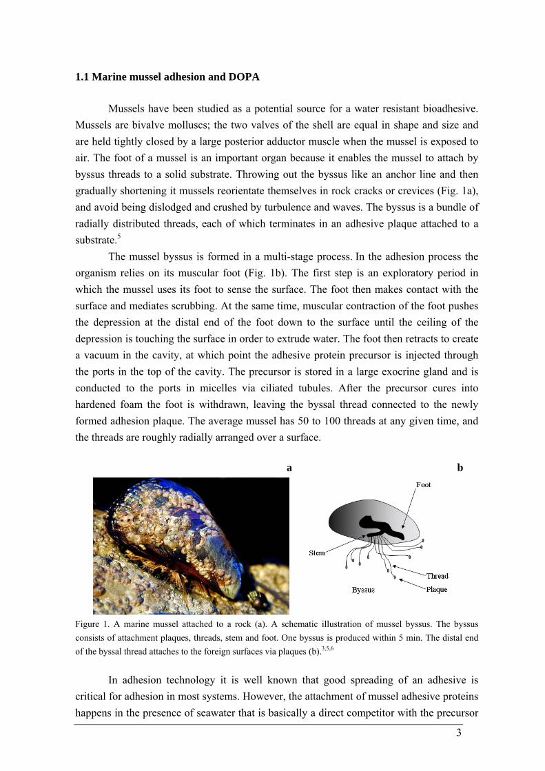
Mussels are bivalve molluscs; the two valves of the shell are equal in shape and size and are held tightly closed by a large posterior adductor muscle when the mussel is exposed to air. The foot of a mussel is an important organ because it enables the mussel to attach by byssus threads to a solid substrate. Throwing out the byssus like an anchor line and then gradually shortening it mussels reorientate themselves in rock cracks or crevices (Fig. 1a), and avoid being dislodged and crushed by turbulence and waves. The byssus is a bundle of radially distributed threads, each of which terminates in an adhesive plaque attached to a substrate.5
The mussel byssus is formed in a multi-stage process. In the adhesion process the
organism relies on its muscular foot (Fig. 1b). The first step is an exploratory period in which the mussel uses its foot to sense the surface. The foot then makes contact with the surface and mediates scrubbing. At the same time, muscular contraction of the foot pushes the depression at the distal end of the foot down to the surface until the ceiling of the depression is touching the surface in order to extrude water. The foot then retracts to create a vacuum in the cavity, at which point the adhesive protein precursor is injected through the ports in the top of the cavity. The precursor is stored in a large exocrine gland and is conducted to the ports in micelles via ciliated tubules. After the precursor cures into hardened foam the foot is withdrawn, leaving the byssal thread connected to the newly formed adhesion plaque. The average mussel has 50 to 100 threads at any given time, and the threads are roughly radially arranged over a surface.
a
b
Figure 1. A marine mussel attached to a rock (a). A schematic illustration of mussel byssus. The byssus consists of attachment plaques, threads, stem and foot. One byssus is produced within 5 min. The distal end of the byssal thread attaches to the foreign surfaces via plaques (b).3,5,6
In adhesion technology it is well known that good spreading of an adhesive is critical for adhesion in most systems. However, the attachment of mussel adhesive proteins happens in the presence of seawater that is basically a direct competitor with the precursor
4
for the substrate. Given that they attach to hard surfaces underwater, mussels appear to be able to remove weak boundary layers including water from surfaces and, once attached, their adhesion is not subverted by the dielectric constant of the medium all around them.3
The permanent and opportunistic byssal attachment of mussels is derived from an assortment of proteins. The chemistry of the proteins had been studied in detail in the last decades; especially large number of researchers had drawn attention to the marine blue mussel Mytilus edulis.3,4,7 Nevertheless, it has been reported that other organisms that are strictly aquatic such as Mytilus galloprovincialis, Mytilus coruscus, Mytilus californiaus, Geukensia demissa and some others also have DOPA-containing proteins.3,8,9
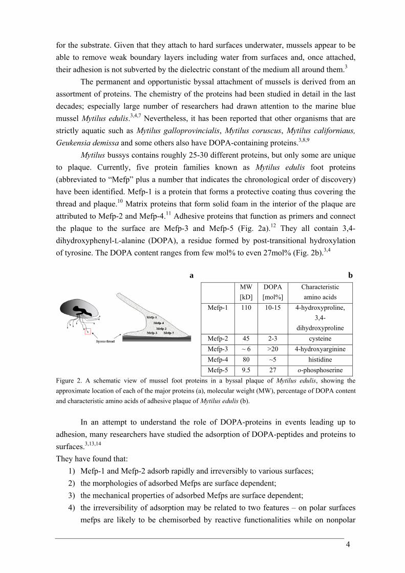
Mytilus bussys contains roughly 25-30 different proteins, but only some are unique to plaque. Currently, five protein families known as Mytilus edulis foot proteins (abbreviated to “Mefp” plus a number that indicates the chronological order of discovery) have been identified. Mefp-1 is a protein that forms a protective coating thus covering the thread and plaque.10 Matrix proteins that form solid foam in the interior of the plaque are attributed to Mefp-2 and Mefp-4.11 Adhesive proteins that function as primers and connect the plaque to the surface are Mefp-3 and Mefp-5 (Fig. 2a).12 They all contain 3,4-dihydroxyphenyl-L-alanine (DOPA), a residue formed by post-transitional hydroxylation of tyrosine. The DOPA content ranges from few mol% to even 27mol% (Fig. 2b).3,4
a
b MW
[kD] DOPA [mol%]
Characteristic amino acids
Mefp-1 110 10-15 4-hydroxyproline, 3,4-
dihydroxyproline Mefp-2 45 2-3 cysteine Mefp-3 ~ 6 >20 4-hydroxyarginine Mefp-4 80 ~5 histidine Mefp-5 9.5 27 o-phosphoserine
Figure 2. A schematic view of mussel foot proteins in a byssal plaque of Mytilus edulis, showing the approximate location of each of the major proteins (a), molecular weight (MW), percentage of DOPA content and characteristic amino acids of adhesive plaque of Mytilus edulis (b).
In an attempt to understand the role of DOPA-proteins in events leading up to adhesion, many researchers have studied the adsorption of DOPA-peptides and proteins to surfaces.3,13,14 They have found that:
1) Mefp-1 and Mefp-2 adsorb rapidly and irreversibly to various surfaces; 2) the morphologies of adsorbed Mefps are surface dependent; 3) the mechanical properties of adsorbed Mefps are surface dependent; 4) the irreversibility of adsorption may be related to two features – on polar surfaces
mefps are likely to be chemisorbed by reactive functionalities while on nonpolar
5
surfaces irreversibility of adsorption is enhanced by protein aggregation since aggregation is driven by increasing pH or peroxide oxidation. It is believed to involve charge transfer between DOPA and DOPA-quinone or aryl coupling (i.e., diDOPA);
5) given their strong net positive charge Mefps may be predisposed to co-adsorption with poly-anions.15-17
The most extensive research about the adhesive mechanical properties of mussels has been done with Mytilus edulis. M. edulis adhesive tensile strength measurements in aqueous environment on several substrates have been reported by J. R. Burkett et al.18 They described mussel plaque detachment force (pulled at 90o angle) per individual plaque. The adhesive performance of each plaque was reported by dividing the failure force (in Newton) by the area (in m2) to yield values in Pascal (Pa = N/m2) for each plaque, therefore the published results provide standard adhesion data that can be compared directly with other biological or synthetic adhesives. Mussel average adhesion on aluminum was 288 ± 110 kPa; it was the highest when compared with other tested materials such as glass (171 ± 65 kPa), polyvinyl chloride, PVC (144 ± 55 kPa), acrylic (133 ± 34 kPa). Such variations in tensile strengths values were explained from the differences arranging from the substrate surface energies, surface moduli (i.e., stiffness) as well as surface chemistry changes. Accordingly, lower energy surfaces (acrylic, PVC) minimized the animal’s ability to adhere while higher energy surfaces (glass, aluminum) maximized adhesion. More compliant surfaces decrease adhesion. Additionally, the particularly high adhesion on aluminum may be influenced by chemistry of both the surface and the adhesive itself. Metal oxides tend to have a slightly anionic character (i.e., negative charge) while the proteins known to constitute mussel adhesive harbor has somewhat cationic (i.e., positive charge). Such anion-cation interactions could contribute to enhanced adhesive bonding between the plaques and a surface. Other chemical contributions to strong adhesion on aluminum may include hydrogen bonding between proteins and the metal oxide surface as well as chelating of surface aluminum ions by DOPA amino acids of the mefp. A glass surface could also exhibit analogous hydrogen bonding and anion-cation interactions. The plastics (PVC, acrylic) are less likely to have such bonding modes available, thereby contributing to the decreased adhesive forces measured.18
For comparison, J. R. Burkett et al. measured synthetic adhesive tensile strengths. White polyvinyl acetate (PVA) glue on aluminum exhibited adhesion at 890 ± 260 kPa, which is roughly three times as great as mussel plaques on aluminum. The cyanoacrylate-based Krazy Glue yielded a lower limit of adhesion at ~ 8 MPa. These data show that this commercial adhesive is significantly stronger than the mussel glue. Although weaker than these synthetic materials, mussel adhesive does exhibit unique properties such as the ability to set well in a wet environment.18
6
The high content of DOPA in mussel adhesive proteins has brought an interest in the scientific society to examine the main chemical interactions of which DOPA appears to be capable.
1.2 The reactivity of protein bound-DOPA
Previous studies of mefp-s have proposed that the adhesion depends on DOPA,3,8
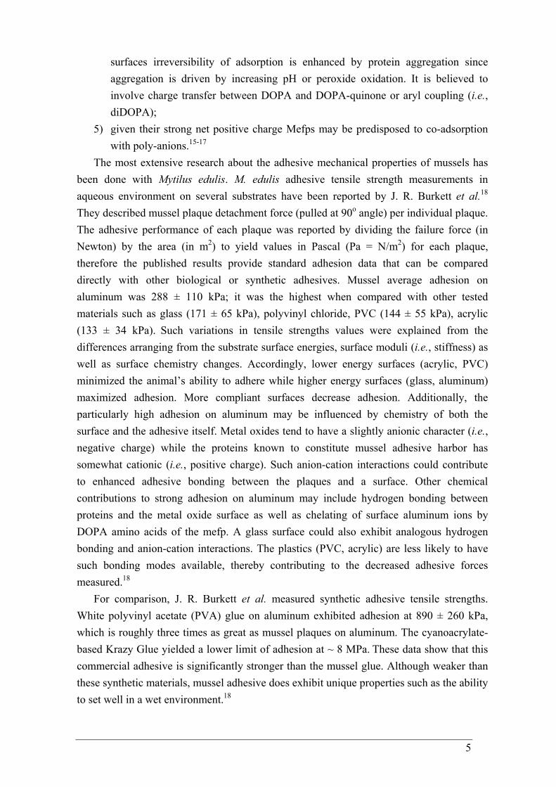
and synthetic analogs have generally confirmed that the higher the DOPA content, the stronger the adhesion.19-21 In light of the high content of DOPA in mussel adhesive proteins, it is useful to examine the reactivity of protein-bound DOPA residues: hydrogen bonding, metal-ligand complexation, Michael-type addition, Schiff base reaction and quinhydrone charge-transfer complexation. The reactivity of quinones possible under physiological conditions is briefly outlined in Fig. 3.9
OH OH
NH
O
O O
NH
O
O O
NH
O
Fe (III)
OH OH
NH
O
NHR
N O
NH
O
R
O
O O
NH
O
HH
N
H
R'R
O OH
NH
O
H
OH OH
NH
OOHOH
NH
O
.
A B C
DE
Figure 3. Hypothetical scheme of protein-bound DOPA residue reactivity showing the possible reaction pathways: metal chelates (A), Michael-type reaction (B), Schiff base (C), H-bonding (E) and dehydrodopa formation and phenol coupling (D).
7
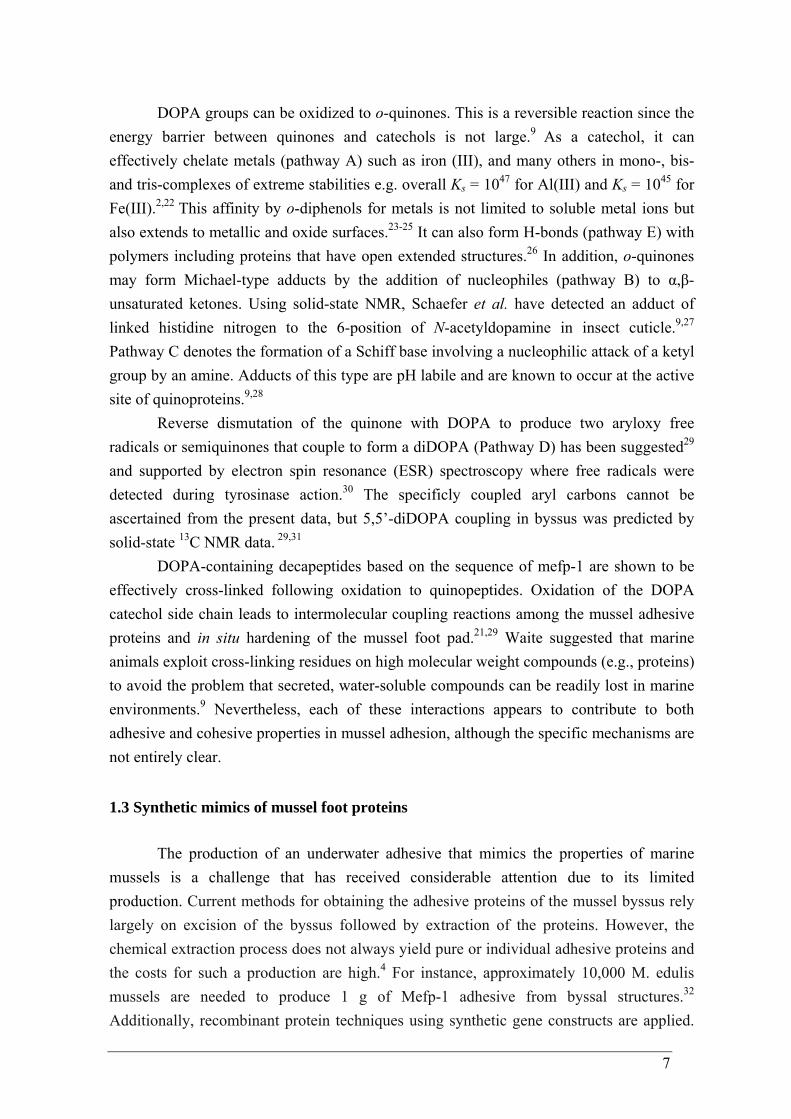
DOPA groups can be oxidized to o-quinones. This is a reversible reaction since the energy barrier between quinones and catechols is not large.9 As a catechol, it can effectively chelate metals (pathway A) such as iron (III), and many others in mono-, bis- and tris-complexes of extreme stabilities e.g. overall Ks = 1047 for Al(III) and Ks = 1045 for Fe(III).2,22 This affinity by o-diphenols for metals is not limited to soluble metal ions but also extends to metallic and oxide surfaces.23-25 It can also form H-bonds (pathway E) with polymers including proteins that have open extended structures.26 In addition, o-quinones may form Michael-type adducts by the addition of nucleophiles (pathway B) to α,β-unsaturated ketones. Using solid-state NMR, Schaefer et al. have detected an adduct of linked histidine nitrogen to the 6-position of N-acetyldopamine in insect cuticle.9,27 Pathway C denotes the formation of a Schiff base involving a nucleophilic attack of a ketyl group by an amine. Adducts of this type are pH labile and are known to occur at the active site of quinoproteins.9,28
Reverse dismutation of the quinone with DOPA to produce two aryloxy free radicals or semiquinones that couple to form a diDOPA (Pathway D) has been suggested29
and supported by electron spin resonance (ESR) spectroscopy where free radicals were detected during tyrosinase action.30 The specificly coupled aryl carbons cannot be ascertained from the present data, but 5,5’-diDOPA coupling in byssus was predicted by solid-state 13C NMR data. 29,31
DOPA-containing decapeptides based on the sequence of mefp-1 are shown to be effectively cross-linked following oxidation to quinopeptides. Oxidation of the DOPA catechol side chain leads to intermolecular coupling reactions among the mussel adhesive proteins and in situ hardening of the mussel foot pad.21,29 Waite suggested that marine animals exploit cross-linking residues on high molecular weight compounds (e.g., proteins) to avoid the problem that secreted, water-soluble compounds can be readily lost in marine environments.9 Nevertheless, each of these interactions appears to contribute to both adhesive and cohesive properties in mussel adhesion, although the specific mechanisms are not entirely clear.
1.3 Synthetic mimics of mussel foot proteins
The production of an underwater adhesive that mimics the properties of marine
mussels is a challenge that has received considerable attention due to its limited production. Current methods for obtaining the adhesive proteins of the mussel byssus rely largely on excision of the byssus followed by extraction of the proteins. However, the chemical extraction process does not always yield pure or individual adhesive proteins and the costs for such a production are high.4 For instance, approximately 10,000 M. edulis mussels are needed to produce 1 g of Mefp-1 adhesive from byssal structures.32 Additionally, recombinant protein techniques using synthetic gene constructs are applied.
8
But also this approach yields inconsistencies in the quality of the protein. The laboratory-prepared products have not demonstrated comparable strength to the natural protein.4 Meanwhile, the proposition of marine mussel adhesive proteins in interfacial contact formation plays an essential role, particularly as it relates to surface modification. Therefore the chemical synthesis of adhesive proteins and their analogues are of current interest worldwide.
Consequently, it has long been a goal of many scientists to develop useful synthetic polymer adhesives that exhibit the wet adhesive capabilities of mefps. There are many successful attempts reported in the literature. Their adhesion properties on a wide variety of substrates, including metals (Ag, Au)33,34 metal oxide surfaces (Ti, stainless steel),35,36 semiconductors (Si),37 and polymers such as poly(dimethylsiloxane),38 poly(carbonate) and poly(tetrafluoro ethylene)37 have been reported.
A popular way of engineering mussel mimetic synthetic polymers is to functionalize linear or branched polymers with DOPA, DOPA peptides or other catechol functional groups by employing standard chemical ligation chemistries. This approach is used mostly to avoid partial inhibition of free-radical polymerization by catechols in the presence of atmospheric oxygen.5
There are a number of publications regarding poly(ethylene glycol) (PEG)-based polymers. Herein the nonreactive PEG polymers serve as inert and biocompatible macromolecular supports and DOPA is often incorporated as a terminal group on each polymer chain thus bringing cohesive and adhesive characteristics. Solutions of branched and linear PEGs with DOPA end groups readily can form hydrogels when treated with either chemical (peroxidate) or enzymatic (horseradish peroxidase and mushroom tyrosinase) oxidants, such hydrogels are generally targeted as medical adhesives and sealants.5
Oxidation-free strategies have also been developed for mefps mimetic polymer hydrogel formation consequently broadening their applications. DOPA has been coupled to the ends of the poly(ethylene oxide)-poly(propylene oxide)-poly(ethylene oxide) (PEO-PPO-PEO) tribloc copolymer by activation of hydroxyl end groups of PEO-PPO-PEO with N,N’-disuccinimidyl carbonate and then coupling with DOPA or its methyl ester.39 Aqueous solutions of these copolymers usually exhibit temperature-included aggregation phenomena due to the hydrophobic nature of PPO block.40 A sol-gel transition occurs upon heating. This physical characteristic – thermal gelation, was one route used to produce a polymer matrix avoiding oxidative DOPA crosslinking. Meanwhile the un-oxidized DOPA end groups adhered to mucin coated surfaces thus showing a substantial increase in bioadhesion comparing to unmodified copolymer.
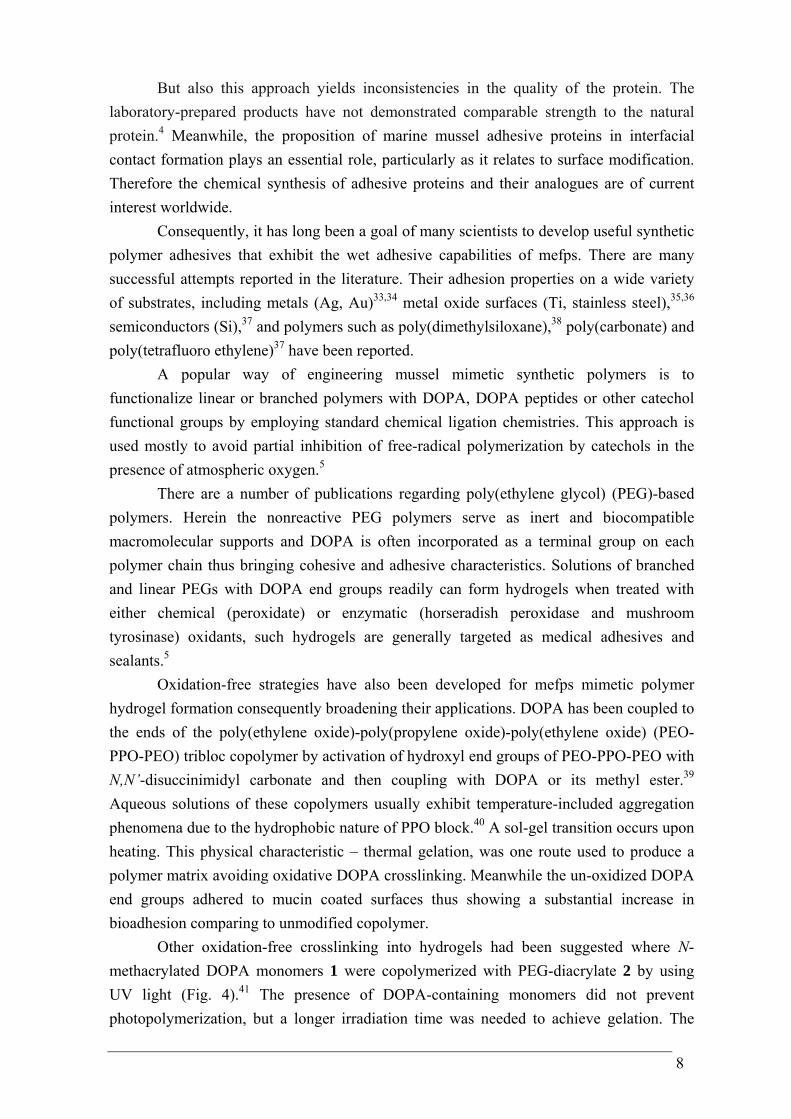
Other oxidation-free crosslinking into hydrogels had been suggested where N-methacrylated DOPA monomers 1 were copolymerized with PEG-diacrylate 2 by using UV light (Fig. 4).41 The presence of DOPA-containing monomers did not prevent photopolymerization, but a longer irradiation time was needed to achieve gelation. The
9
DOPA modified PEG hydrogels 3 remained un-oxidized for the purpose of adhesion to surfaces.
OHOH
NH O
OOH
O OO O
n
OH
OH
NH
O
OH
O
OH
OH
NH
O
OH
O
*
O OO
*
O
**
n
x
x
y
y
1
+
Photoinitiatorhv
2
3
Figure 4. Schematic representation of copolymerization of N-methacrylated DOPA monomer 1 with PEG-diacrylate 2.
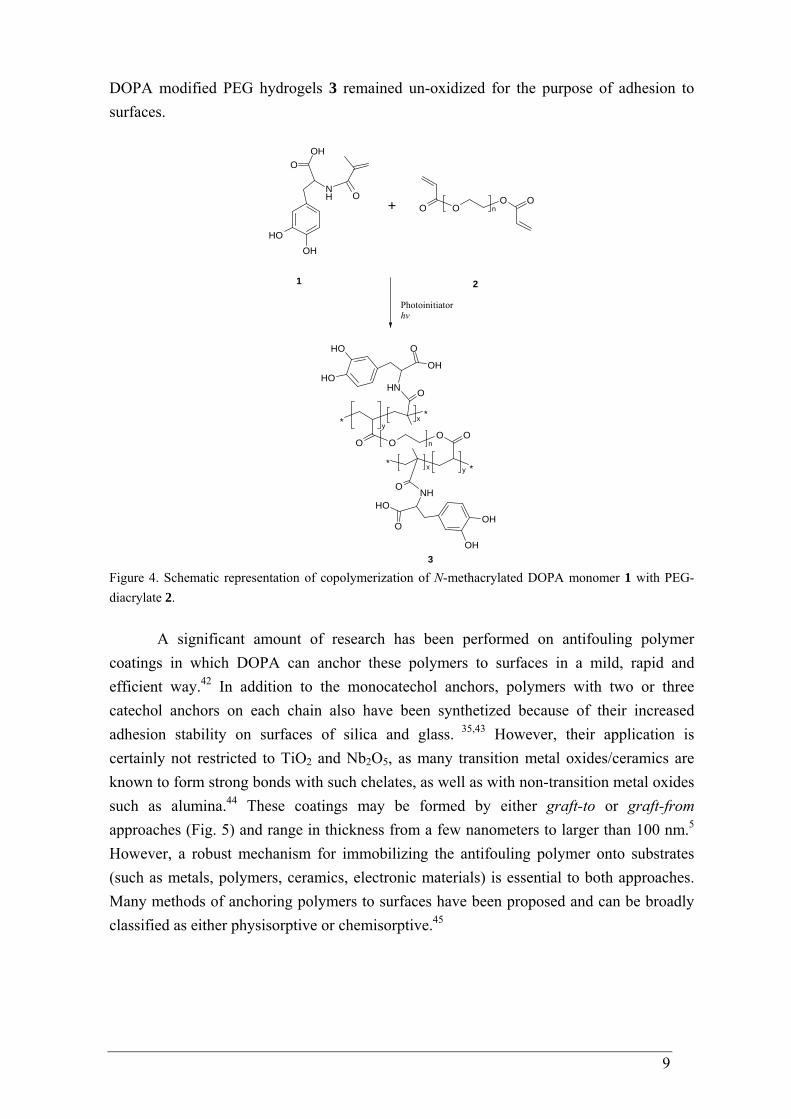
A significant amount of research has been performed on antifouling polymer coatings in which DOPA can anchor these polymers to surfaces in a mild, rapid and efficient way.42 In addition to the monocatechol anchors, polymers with two or three catechol anchors on each chain also have been synthetized because of their increased adhesion stability on surfaces of silica and glass. 35,43 However, their application is certainly not restricted to TiO2 and Nb2O5, as many transition metal oxides/ceramics are known to form strong bonds with such chelates, as well as with non-transition metal oxides such as alumina.44 These coatings may be formed by either graft-to or graft-from approaches (Fig. 5) and range in thickness from a few nanometers to larger than 100 nm.5 However, a robust mechanism for immobilizing the antifouling polymer onto substrates (such as metals, polymers, ceramics, electronic materials) is essential to both approaches. Many methods of anchoring polymers to surfaces have been proposed and can be broadly classified as either physisorptive or chemisorptive.45
10
OHOH
NH
O
OHOH
NH
OBrPolymer
Graft-to Graft-from
Figure 5. Schematic representation of graft-to and graf-from strategies for surface modification.
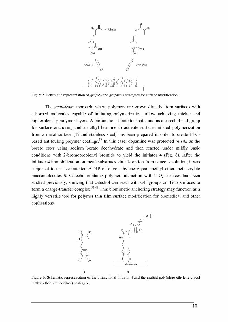
The graft-from approach, where polymers are grown directly from surfaces with adsorbed molecules capable of initiating polymerization, allow achieving thicker and higher-density polymer layers. A biofunctional initiator that contains a catechol end group for surface anchoring and an alkyl bromine to activate surface-initiated polymerization from a metal surface (Ti and stainless steel) has been prepared in order to create PEG-based antifouling polymer coatings.36 In this case, dopamine was protected in situ as the borate ester using sodium borate decahydrate and then reacted under mildly basic conditions with 2-bromopropionyl bromide to yield the initiator 4 (Fig. 6). After the initiator 4 immobilization on metal substrates via adsorption from aqueous solution, it was subjected to surface-initiated ATRP of oligo ethylene glycol methyl ether methacrylate macromolecules 5. Catechol-containg polymer interaction with TiO2 surfaces had been studied previously, showing that catechol can react with OH groups on TiO2 surfaces to form a charge-transfer complex.35,46 This biomimetic anchoring strategy may function as a highly versatile tool for polymer thin film surface modification for biomedical and other applications.
OHOH
NH
O Br
OO
NH
O Br
OO
O *n
n
4
Me substrate
5 Figure 6. Schematic representation of the bifunctional initiator 4 and the grafted poly(oligo ethylene glycol methyl ether methacrylate) coating 5.
11
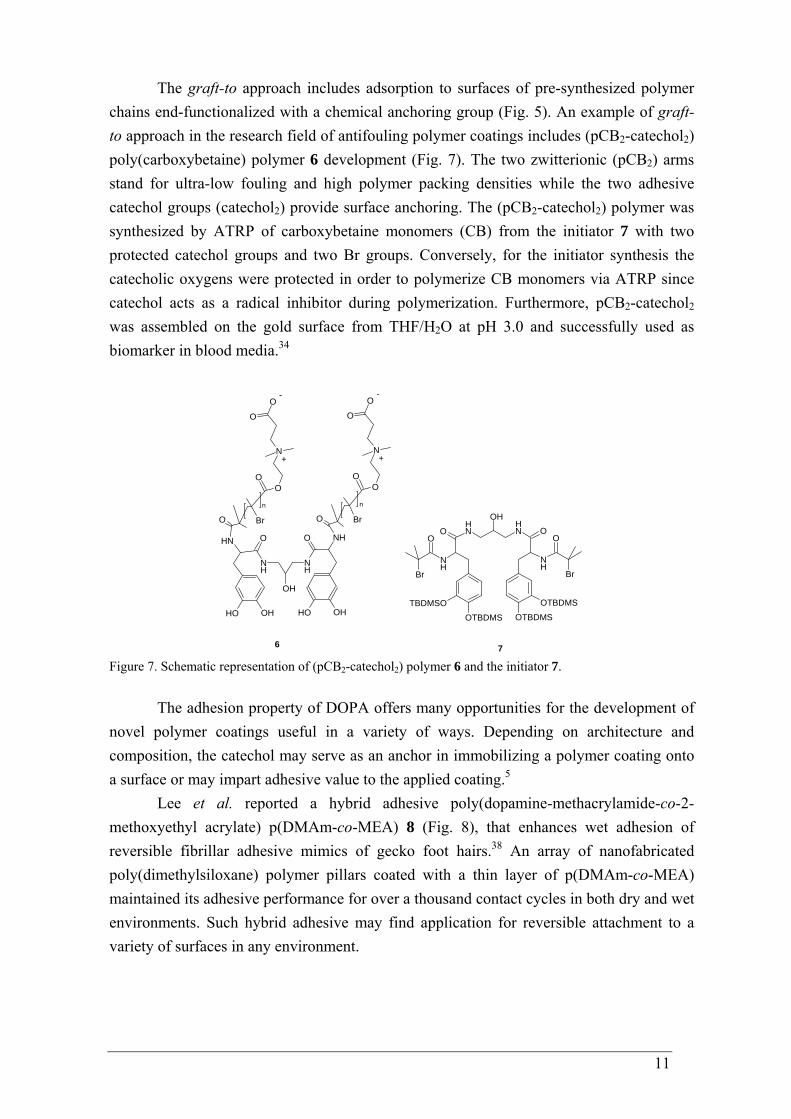
The graft-to approach includes adsorption to surfaces of pre-synthesized polymer chains end-functionalized with a chemical anchoring group (Fig. 5). An example of graft-to approach in the research field of antifouling polymer coatings includes (pCB2-catechol2) poly(carboxybetaine) polymer 6 development (Fig. 7). The two zwitterionic (pCB2) arms stand for ultra-low fouling and high polymer packing densities while the two adhesive catechol groups (catechol2) provide surface anchoring. The (pCB2-catechol2) polymer was synthesized by ATRP of carboxybetaine monomers (CB) from the initiator 7 with two protected catechol groups and two Br groups. Conversely, for the initiator synthesis the catecholic oxygens were protected in order to polymerize CB monomers via ATRP since catechol acts as a radical inhibitor during polymerization. Furthermore, pCB2-catechol2 was assembled on the gold surface from THF/H2O at pH 3.0 and successfully used as biomarker in blood media.34
OTBDMSTBDMSO
NH
Br
ONH
O
OHNH
O
NH
O
Br
OTBDMS
OTBDMS
O Br
OO
N
O
O
n
OHOH
NH
NH
O
OH OH
NHO
NH
OH
O Br
OO
N
O
O
n
6
+
-
+
-
7 Figure 7. Schematic representation of (pCB2-catechol2) polymer 6 and the initiator 7.
The adhesion property of DOPA offers many opportunities for the development of novel polymer coatings useful in a variety of ways. Depending on architecture and composition, the catechol may serve as an anchor in immobilizing a polymer coating onto a surface or may impart adhesive value to the applied coating.5
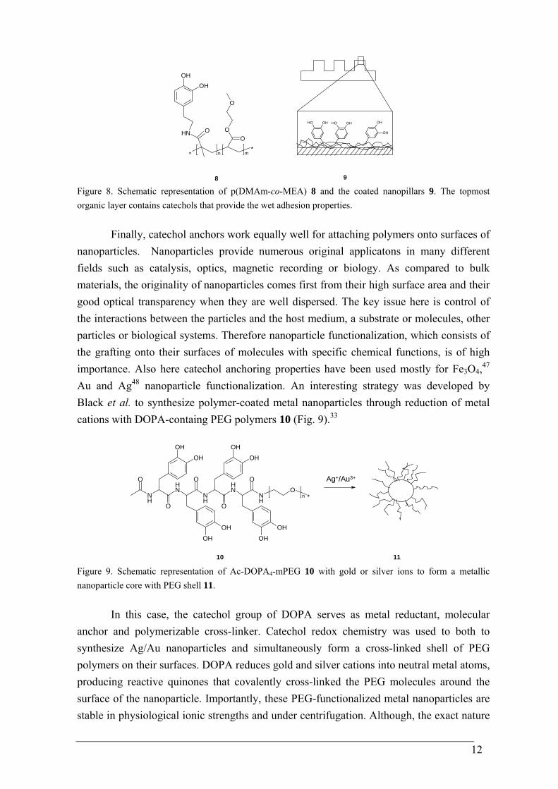
Lee et al. reported a hybrid adhesive poly(dopamine-methacrylamide-co-2-methoxyethyl acrylate) p(DMAm-co-MEA) 8 (Fig. 8), that enhances wet adhesion of reversible fibrillar adhesive mimics of gecko foot hairs.38 An array of nanofabricated poly(dimethylsiloxane) polymer pillars coated with a thin layer of p(DMAm-co-MEA) maintained its adhesive performance for over a thousand contact cycles in both dry and wet environments. Such hybrid adhesive may find application for reversible attachment to a variety of surfaces in any environment.
12
**
ONH
OHOH
OO
O
n
m
OH OH OH OH OH
OH
8 9 Figure 8. Schematic representation of p(DMAm-co-MEA) 8 and the coated nanopillars 9. The topmost organic layer contains catechols that provide the wet adhesion properties.
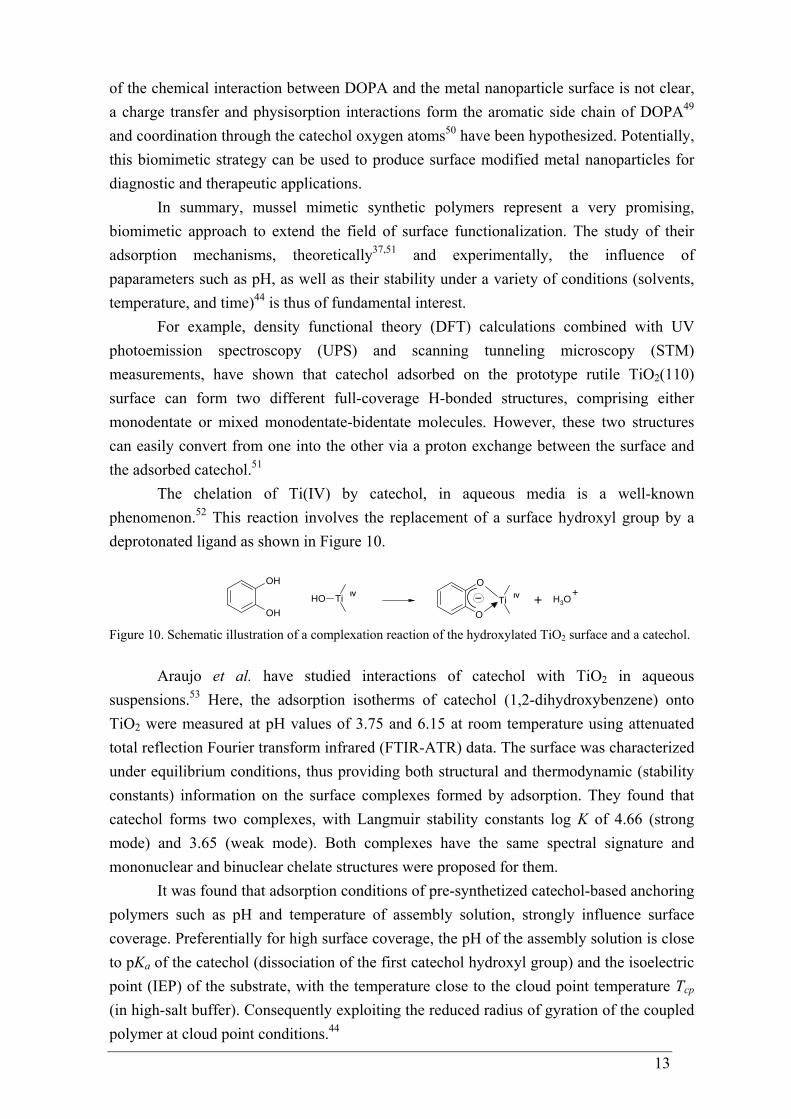
Finally, catechol anchors work equally well for attaching polymers onto surfaces of nanoparticles. Nanoparticles provide numerous original applicatons in many different fields such as catalysis, optics, magnetic recording or biology. As compared to bulk materials, the originality of nanoparticles comes first from their high surface area and their good optical transparency when they are well dispersed. The key issue here is control of the interactions between the particles and the host medium, a substrate or molecules, other particles or biological systems. Therefore nanoparticle functionalization, which consists of the grafting onto their surfaces of molecules with specific chemical functions, is of high importance. Also here catechol anchoring properties have been used mostly for Fe3O4,47 Au and Ag48 nanoparticle functionalization. An interesting strategy was developed by Black et al. to synthesize polymer-coated metal nanoparticles through reduction of metal cations with DOPA-containg PEG polymers 10 (Fig. 9).33
NH
O
O
NH
NH
ONH
ONH
OO
*
OHOH
OHOH OH
OH
OHOH
n
Ag+/Au3+
10 11 Figure 9. Schematic representation of Ac-DOPA4-mPEG 10 with gold or silver ions to form a metallic nanoparticle core with PEG shell 11.
In this case, the catechol group of DOPA serves as metal reductant, molecular anchor and polymerizable cross-linker. Catechol redox chemistry was used to both to synthesize Ag/Au nanoparticles and simultaneously form a cross-linked shell of PEG polymers on their surfaces. DOPA reduces gold and silver cations into neutral metal atoms, producing reactive quinones that covalently cross-linked the PEG molecules around the surface of the nanoparticle. Importantly, these PEG-functionalized metal nanoparticles are stable in physiological ionic strengths and under centrifugation. Although, the exact nature
13
of the chemical interaction between DOPA and the metal nanoparticle surface is not clear, a charge transfer and physisorption interactions form the aromatic side chain of DOPA49 and coordination through the catechol oxygen atoms50 have been hypothesized. Potentially, this biomimetic strategy can be used to produce surface modified metal nanoparticles for diagnostic and therapeutic applications.
In summary, mussel mimetic synthetic polymers represent a very promising, biomimetic approach to extend the field of surface functionalization. The study of their adsorption mechanisms, theoretically37,51 and experimentally, the influence of paparameters such as pH, as well as their stability under a variety of conditions (solvents, temperature, and time)44 is thus of fundamental interest.
For example, density functional theory (DFT) calculations combined with UV photoemission spectroscopy (UPS) and scanning tunneling microscopy (STM) measurements, have shown that catechol adsorbed on the prototype rutile TiO2(110) surface can form two different full-coverage H-bonded structures, comprising either monodentate or mixed monodentate-bidentate molecules. However, these two structures can easily convert from one into the other via a proton exchange between the surface and the adsorbed catechol.51
The chelation of Ti(IV) by catechol, in aqueous media is a well-known phenomenon.52 This reaction involves the replacement of a surface hydroxyl group by a deprotonated ligand as shown in Figure 10.
OH
OHTiOH
O
OTi
IV IV + H3O+
Figure 10. Schematic illustration of a complexation reaction of the hydroxylated TiO2 surface and a catechol.
Araujo et al. have studied interactions of catechol with TiO2 in aqueous suspensions.53 Here, the adsorption isotherms of catechol (1,2-dihydroxybenzene) onto TiO2 were measured at pH values of 3.75 and 6.15 at room temperature using attenuated total reflection Fourier transform infrared (FTIR-ATR) data. The surface was characterized under equilibrium conditions, thus providing both structural and thermodynamic (stability constants) information on the surface complexes formed by adsorption. They found that catechol forms two complexes, with Langmuir stability constants log K of 4.66 (strong mode) and 3.65 (weak mode). Both complexes have the same spectral signature and mononuclear and binuclear chelate structures were proposed for them.
It was found that adsorption conditions of pre-synthetized catechol-based anchoring polymers such as pH and temperature of assembly solution, strongly influence surface coverage. Preferentially for high surface coverage, the pH of the assembly solution is close to pKa of the catechol (dissociation of the first catechol hydroxyl group) and the isoelectric point (IEP) of the substrate, with the temperature close to the cloud point temperature Tcp (in high-salt buffer). Consequently exploiting the reduced radius of gyration of the coupled polymer at cloud point conditions.44
14
1.4 Recent developments in mussel-inspired adhesives
There has been a dramatic increase in the number of papers reporting exciting
progress in new adhesive materials inspired by mussel adhesive proteins in the last years. Perhaps the most significant result to date has been the demonstration that a simple DOPA protein mimic – dopamine, commonly known as neurotransmitter can be used to coat a wide range of inorganic and organic materials. Dopamine in dilute aqueous solution can spontaneously polymerize under alkaline conditions (pH 8.5) and form a hydrophilic film rapidly on various substrates, including noble metals, oxides, polymers, semiconductors, and ceramics by simple dip coating. The film thickness depends from the immersion time and can reach a value of up to 50 nm after 24 h at room temperature for 2 mg dopamine hydrochloride aqueous solution, buffered at pH 8.5.54
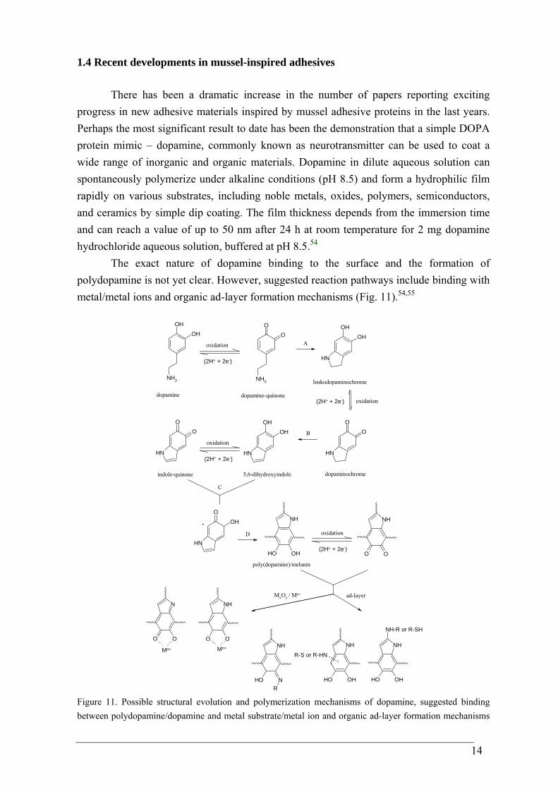
The exact nature of dopamine binding to the surface and the formation of polydopamine is not yet clear. However, suggested reaction pathways include binding with metal/metal ions and organic ad-layer formation mechanisms (Fig. 11).54,55
OHOH
NH2
OO
NH2
OHOH
NH
OO
NH
OHOH
NH
OO
NH
OOH
NH
OHOH
NH
OO
NH
OO
NH
OO
N
NOH
NH
ROHOH
NH
NH-R or R-SH
OHOH
NH
R-S or R-HN
dopamine
oxidation
(2H+ + 2e-)
dopamine-quinone
A
leukodopaminochrome
(2H+ + 2e-) oxidation
dopaminochrome
B
5,6-dihydroxyindoleindole-quinone
oxidation
(2H+ + 2e-)
C
poly(dopamine)/melanin
oxidation
(2H+ + 2e-)
.D
MxOy / Mn+
Mn+ Mn+
ad-layer
Figure 11. Possible structural evolution and polymerization mechanisms of dopamine, suggested binding between polydopamine/dopamine and metal substrate/metal ion and organic ad-layer formation mechanisms
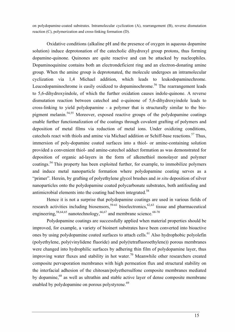
15
on polydopamine-coated substrates. Intramolecular cyclization (A), rearrangement (B), reverse dismutation reaction (C), polymerization and cross-linking formation (D).
Oxidative conditions (alkaline pH and the presence of oxygen in aqueous dopamine solution) induce deprotonation of the catecholic dihydroxyl group protons, thus forming dopamine-quinone. Quinones are quite reactive and can be attacked by nucleophiles. Dopaminoquinine contains both an electrondeficient ring and an electron-donating amine group. When the amine group is deprotonated, the molecule undergoes an intramolecular cyclization via 1,4 Michael addition, which leads to leukodopaminechrome. Leucodopaminochrome is easily oxidized to dopaminochrome.56 The rearrangement leads to 5,6-dihydroxyindole, of which the further oxidation causes indole-quinone. A reverse dismutation reaction between catechol and o-quinone of 5,6-dihydroxyindole leads to cross-linking to yield polydopamine - a polymer that is structurally similar to the bio-pigment melanin.54,55 Moreover, exposed reactive groups of the polydopamine coatings enable further functionalization of the coatings through covalent grafting of polymers and deposition of metal films via reduction of metal ions. Under oxidizing conditions, catechols react with thiols and amine via Michael addition or Schiff-base reactions.57 Thus, immersion of poly-dopamine coated surfaces into a thiol- or amine-containing solution provided a convenient thiol- and amine-catechol adduct formation as was demonstrated for deposition of organic ad-layers in the form of alkenethiol monolayer and polymer coatings.54 This property has been exploited further, for example, to immobilize polymers and induce metal nanoparticle formation where polydopamine coating serves as a “primer”. Herein, by grafting of polyethylene glycol brushes and in situ deposition of silver nanoparticles onto the polydopamine coated polycarbonate substrates, both antifouling and antimicrobial elements into the coating had been integrated.58
Hence it is not a surprise that polydopamine coatings are used in various fields of research activities including biosensors,59-61 bioelectronics,62,63 tissue and pharmaceutical engineering,58,64,65 nanotechnology,66,67 and membrane science.68-70
Polydopamine coatings are successfully applied when material properties should be improved, for example, a variety of bioinert substrates have been converted into bioactive ones by using polydopamine coated surfaces to attach cells.61 Also hydrophobic polyolefin (polyethylene, poly(vinylidene fluoride) and poly(tetrafluoroethylene)) porous membranes were changed into hydrophilic surfaces by adhering thin film of polydopamine layer, thus improving water fluxes and stability in hot water.70 Meanwhile other researchers created composite pervaporation membranes with high permeation flux and structural stability on the interfacial adhesion of the chitosan/polyethersulfone composite membranes mediated by dopamine,68 as well as ultrathin and stable active layer of dense composite membrane enabled by polydopamine on porous polystyrene.69
16
1.5 The aim and outline of the thesis
As apparent from the given overview, mussel adhesive proteins are being
increasingly regarded by chemists as masterpieces of wet adhesion functionality. Although the precise mechanism for assembly of the proteins is not understood, the obtained knowledge broadens biomimetic strategies for synthesizing new practical adhesives. In this project, dopamine-based compounds are of main interest due to their potential of being universal adhesion promoters, harmless to environment and applicants. Therefore the adherence of coatings using dopamine-based promoters on metals will be investigated in detail.
Chapter 2 presents the properties of dopamine and polydopamine coatings on metallic substrates. Dip coating from aqueous dopamine hydrochloride solutions at pH 4 and pH 8 was performed on gold (noble metal) and aluminium oxide (metal with native oxide layer). The formed coatings were analyzed with surface sensitive techniques concerning the coating composition and the oxidation state (by infrared reflection-absorption spectroscopy (IRRAS), surface enhanced Raman spectroscopy (SERS) and X-Ray photoelectron spectroscopy (XPS)), topography (by Atomic Force microscopy (AFM)), wetting (by contact angle (CA) measurements) and mechanical (by pull-off test) properties. The outcomes of these studies emphasized the need to generate a well defined system. Chapter 3 deals with the model molecule N-stearoyldopamine. Attention is devoted in studying the adsorption behavior of N-stearoyldopamine onto gold and aluminum oxide in terms of monolayer characteristics. The organization of Langmuir-Blodgett monolayers on these metal substrates as suggested by previous experimental evidence with marine mussel foot proteins was observed directly and studied in detail with several surface sensitive techniques. Our study shows that within the monolayer the catechols functions as a surface anchor on gold and the alkyl-chains appear to be tilted within a monolayer, while the amide functionality stabilizes the formed film. The statements are supported with the outcomes of molecular simulations of the monolayer on gold.
Chapter 4 investigates in catechol interaction with a metal surface by using a model molecule where the amide functionality is excluded. Monolayers of 4-stearylcatechol have been transferred from the air - water interface onto gold and aluminum oxide substrates using the Langmuir-Blodgett technique. The organization of the monolayers on a molecular scale was examined by several surface sensitive techniques and compared with the data for N-stearoyldopamine monolayers. Our study shows that, similar to N-stearoyldopamine monolayers, the catechols function as a surface anchor on gold and the alkyl-chains appear to be tilted within a monolayer. Although the alkyl- chains are tilted within the monolayer for both molecules, the irregularity of the 4-stearylcatechol film on gold leads to domain formation that is caused
17
by the absence of the amide functionality. On the contrary, from the molecular simulations it appeared that for both types of molecules parallel orientations of the catechols with the gold are also present. However, hydrogen bonds formed between the amide functionality and the catechol hydroxyl groups have a profound influence on the structure and regularity on the adsorbed layer.
In Chapter 5 attempts are made to quantify the adhesion strength of the anchoring catechols on metal oxide. A covalently bound top-coating was used to determine the adhesion of an N-linoleoyldopamine primer on an aluminum alloy. Several application conditions were tested for the top-coating and the primer. The resulting tensile strength values of the top-coating having an N-linoleoyldopamine monolayer primer showed an obvious improvement in adhesion whereas a negative impact on adhesion occurred when the primer was not applied in a uniform and controlled manner by the Langmuir-Blodgett technique.
1.6 References
1 Schwartz, M. Innovations in Materials Manufacturing, Fabrication, and
Enviromental Safety 2011, Taylor and Francis Group, USA. 2 Waite, J. H. Int. J. Biol. Macromol. 1990, 12, 139. 3 Waite, J.H. Integr. comp. biol. 2002, 42,1172. 4 Silverman, H. G.; Roberto, F. F. Marine Techn. 2007, 9, 661. 5 Lee, B. P.; Messersmith, P.B.; Israelachvili, J.N.; Waite, J.H. Annu. Rev. Mater.
Res. 2011, 41, 99. 6 Kamino, K. Mar. Biotechnol. 2008, 10, 111. 7 Moskovits, M. J. Chem. Phys. 1982, 77, 4408. 8 Deming, T. J. Curr Opin Chem Biol. 1999, 3, 100. 9 Waite, J. H. Comp. Biochem. Physiol. 1990, 97B, 19. 10 Waite, J. H.; Tanzer, M. L. Science 1981, 212,1038. 11 Rzepecki, L. M.; Hansen, K. M.; Waite, J. H. Biological Bulletin 1992, 183,123. 12 Papov, V. V.; Diamond, T. V.; Biemann, K.; Waite, J. H. The J. of Biol. Chem.
1995, 270, 20183. 13 Lin, Q.; Gourdon, D.; Sun, C.; Holten-Andersen, N.; Anderson, T. H.; Waite, J. H.;
Israelachvili, J. N. PNAS 2007, 104, 3782. 14 Cha, H. J.; Hwang, D. S.; Lim S. Biotechnol. J. 2008, 3, 631. 15 Höök, F.; Kasemo, B.; Nylander, T.; Fant, C.; Sott, K.; Elwing H. Anal. Chem.
2001, 73, 5796. 16 Suci, P.A.; Geesey, G. G. Colloids and Surfaces B: Biointerfaces 2001, 22, 159. 17 Suci, P.A.; Geesey, G. G. J. of Colloid and Interface Science 2000, 230, 340. 18 Burkett, J. R.; Wojtas, J. L.; Cloud, J. L.; Wilker, J. J. The Journal of Adhesion
2009, 85, 601.
18
19 Yu, M.; Deming, T. J. Macromolecules 1998, 31, 4739. 20 Lee, B. P.; Chao, C.-Y.; Nunalee, F. N.; Motan, E.; Shull, K. R.; Messersmith, P. B.
Macromolecules 2006, 39, 1740. 21 Yu, M.; Hwang, J.; Deming, T. J. J. Am. Chem. Soc. 1999, 121, 5825. 22 Pierpont, C. G.; Buchanan, R. M. Coordination Chemistry Reviews 1981, 38, 45. 23 Soriaga, M. P.; Hubbard, A. T. J. Am. Chem. Soc. 1982, 104, 2735. 24 Kummert, R.; Stumm, W. J. of Colloid and Interface Science 1980, 75, 373. 25 McBride, M. B.; Wesselink, L. G. Environ. Sci. Technol. 1988, 22, 703. 26 A. E. Hagerman, L. G. Butler The J. of Biological Chem. 1981, 256, 4494. 27 Schaefer, J.; Kramer, K. J.; Garbow, J. R.; Jacob, G. S.; Stejskal, E. O.; Hopkins, T.
L.; Speirs, R. D. Science 1987, 235, 1200. 28 Duine, J.; Jongejan J. A. Annual Rev. Biochem. 1989, 58, 403. 29 Burzio, L. A.; Waite, J. H. Biochemistry 2000, 39, 11147. 30 Koga, S.; Nakano, M.; Tero-Kubota, S. Archives of Biochem. and Biophys. 1992,
292, 570. 31 McDowell, L. M.; Burzio, L. A.; Waite, J. H.; Schaefer, J. The J. of Biol. Chem.
1999, 274, 20293. 32 Hwang, D. S.; Gim, Y.; Yoo, H. J.; Cha, H. J. Biomaterials 2007, 28, 3560. 33 Black, K. C. L.; Liu, Z.; Messersmith, P. B. Chem. Mater. 2011, 23, 1130. 34 Gao, C.; Li, G.; Xue, H.; Yang, W.; Zhang, F.; Jiang, S. Biomaterials 2010, 31,
1486. 35 Dalsin, J. L.; Lin, L.; Tosatti, S.; Vörös, J.; Textor, M.; Messersmith, P. B.
Langmuir 2005, 21, 640. 36 Fan, X.; Lin, L.; Dalsin, J. L.; Messersmith, P. B. J. Am. Chem. Soc. 2005, 127,
15843. 37 Lee, H.; Lee, K. D.; Pyo, K. B.; Park, S. Y.; Lee, H. Langmuir 2010, 26, 3790. 38 Lee, H.; Lee, B. P.; Messersmith, P. B. Nature 2007, 448, 338. 39 Huang, K.; Lee, B. P.; Ingram, D. R.; Messersmith, P. B. Biomacromolecules 2002,
3, 397. 40 Hvidt, S.; Jørgensen, E. B. J. Phys. Chem. 1994, 98, 12320. 41 Lee, B. P.; Huang, K.; Nunalee, F. N.; Shull, K. R.; Messersmith, P. B. J. Biomater.
Sci. Polymer Edn. 2004, 15, 449. 42 Lee, B. P.; Dalsin, J. L.; Messersmith, P. B. Biomacromolecules 2002, 3, 1038. 43 Wach, J.-Y.; Malisova, B.; Bonazzi, S.; Tosatti, S.; Textor, M.; Zürcher, S.;
Gademann, K. Chem. Eur. J. 2008, 14, 10579. 44 Malisova, B.; Tosatti, S.; Textor, M.; Gademann, K.; Zürcher, S. Langmuir 2010,
26, 4018. 45 Dalsin, J. L.; Messersmith, P. B. Materials Today 2005, 8, 38. 46 Rodríguez, R.; Blesa, M. A.; Regazzoni, A. E. J. of Colloid and Interface Science
1996, 177, 122.
19
47 Amstad, E.; Gillich, T.; Bilecka, I.; Textor, M.; Reimhult, E. Nano Letters 2009, 9, 4042.
48 Pande, S.; Jana, S.; Sinha, A.K.; Sarkar, S.; Basu, M.; Pradhan, M.; Pal, A.; Chowdhury, J.; Pal, T. J. Phys. Chem. C 2009, 113, 6989.
49 Weinhold, M.; Soubatch, S.; Temirov, R.; Rohlfing, M.; Jastorff, B.; Tautz, F. S.; Doose, C. J. Phys. Chem. B 2006, 110, 23756.
50 Ooka, A. A.; Garrell, R. L. Biopolymers 2000, 57, 92. 51 Li, S.-C.; Wang, J.-G.; Jacobson, P.; Gong, X.-Q.; Selloni, A.; Diebold, U. J. Am.
Chem. Soc. 2009, 131, 980. 52 Moser, J.; Punchihewa, S.; Infelta, P. P.; Gratzel, M. Langmuir 1991, 7, 3012. 53 Araujo, P. Z.; Morando, P. J.; Blesa, M. A. Langmuir 2005, 21, 3470. 54 Lee, H.; Dellatore, S. M.; Miller, W. M.; Messersmith, P. B. Science 2007, 318,
426. 55 Yu, F.; Chen, S.; Chen, Y.; Li, H.; Yang, L.; Chen, Y.; Yin, Y. Journal of
Molecular Structure 2010, 982,152. 56 Łuczak, T. Electrochimica Acta 2008, 53, 5725. 57 Burzio, L. A.; Waite, J. H. Biochemistry 2000, 39, 11147. 58 Sileika, T. S.; Kim, H.-D.; Maniak, P.; Messersmith, P. B. ACS Appl. Mater.
Interfaces 2011, 3, 4602. 59 Ouyang, R.; Lei, J.; Ju, H. Nanotechnology 2010, 21, 185502. 60 Zhou, W.-H.; Lu, C.-H.; Guo, X.-C.; Chen, F.-R.; Yang, H.-H.; Wang, X.-R. J.
Mater. Chem. 2010, 20, 880. 61 Ku, S. H.; Ryu, J.; Hong, S. K.; Lee, H.; Park, C. B. Biomaterials 2010, 31, 2535. 62 Ye, W.; Wang, D.; Zhang, H.; Zhou, F.; Liu, W. Electrochimica Acta 2010, 55,
2004. 63 Yu, B.; Liu, J.; Liu, S.; Zhou, F. Chem. Commun. 2010, 46, 5900. 64 Haller, C. M.; Buerzle, W.; Brubaker, C. E.; Messersmith, P. B.; Mazza, E.;
Ochsenbein-Koelble, N.; Zimmermann, R.; Ehrbar, M. Prenat. Diagn. 2011, 31, 654.
65 Su, J.; Chen, F.; Cryns, V. L.; Messersmith, P. B. J. Am. Chem. Soc. 2011, 133, 11850.
66 Ren, Y.; Rivera, J. G.; He, L.; Kulkarni, H.; Lee, D.-K.; Messersmith P. B. BMC Biotechnology 2011, 11, 63.
67 Fei, B.; Qian, B.; Yang, Z.; Wang, R.; Liu, W. C.; Mak, C. L.; Xin, J. H. Carbon 2008, 46, 1792.
68 Chen, J.; Chen, X.; Yin, X.; Ma, J.; Jiang, Z. Journal of Membrane Science 2009, 344, 136.
69 Li, B.; Liu, W.; Jiang, Z.; Dong, X.; Wang, B.; Zhong, Y. Langmuir 2009, 25, 7368.

20
70 Xi, Z.-Y.; Xu, Y.-Y.; Zhu, L.-P.; Wang, Y.; Zhu, B.-K. Journal of Membrane Science 2009, 327, 244.
21
CHAPTER 2
Dopamine and polydopamine coatings on gold and sapphire This chapter demonstrates that a simple DOPA protein mimic – dopamine, can be used to coat metal substrates and the properties of dopamine and polydopamine coatings on metals are studied in detail. Dip coating from aqueous dopamine hydrochloride solutions at pH 4.0 and pH 8.5 were performed on gold (noble metal without native oxide layer) and aluminium oxide (simple metal with native oxide layer). The formed coatings were analyzed with surface sensitive techniques dealing with coating composition and the oxidation state, as well as with mechanical measurements characterising the adhesion of coatings. The outcomes of these studies emphasized the need to design a better defined system for studies of adherence behavior of dopamine-based materials on metals.
22
2.1 Introduction
The efficiency of the adhesion of thin film coatings strongly depends on various
parameters, such as substrate and adsorbent type, solvent choice, adsorbent concentration, adsorption time, temperature, and pH, all of which need explicit consideration when designing adsorbents. In that respect, catecholamines are particularly attractive due to their remarkable adhesive properties, forming strong chemical interactions with organic and inorganic surfaces via simple dip coating at room temperature from aqueous solutions. The resulting idea that dopamine may establish moisture-resistant, strong interactions with most of the chemical groups present on metal, metal oxide, or polymer surfaces, allowed the introduction of a new surface modification approach, where a dopamine aqueous solution buffered at alkaline pH is put in contact with the material of interest, yielding a film of a few nanometers in thickness after a reaction time of some minutes to hours.1,2 This approach has become a versatile and attractive method for surface modification of solid materials and some of the outcomes are already discussed in Chapter 1.
Although there is no question about the practical appeal of this versatile adhesive platform, there are still many fundamental questions about the formation mechanism, structure and properties of the dopamine and polydopamine films. Therefore, in this Chapter dopamine and polydopamine films will be studied in detail.
2.2 Experimental
Methods and Materials. Dopamine hydrochloride (98%), and 2-Amino-2-(hydroxymethyl)-1,3-propanediol (>99.9%) were purchased from Sigma-Aldrich. Milli-Q water (resistivity = 18.2 MΩ⋅cm) was used in all experiments. Substrates and Substrate Preparation. Mica substrates 80 x 30 mm2 (G250-1, Agar Scientific Limited, United Kingdom) were cleaved by removing a mica sheet before use. C-plane sapphire polished (< 1 nm surface roughness) wafers with (0001) surfaces and dimensions of 10 x 10 mm x 2 mm (Valley Design Corp., USA) was pretreated before film formation, i.e. sonicated (ultrasonic bath, Branson 1510) in acetone, chloroform, methanol and milli-Q water for 10 min. One side polished silicon wafers 10 x 10 mm2 (resistivity of 1–5 ohm⋅cm, 500–550 μm thick, emerging plane’s indices 1 0 0) (Silicon Quest International, Inc., USA)) were coated with an approximately 20 nm thick layer of TiO2 (99.99% pure) (settings: 120 W RF power for 600 s at 7.10-3 mbar) and an approximately 20 nm thick layer of Au (99.99% pure) (settings: 60 W RF power for 240 s at 7.10-3 mbar) by physical vapor deposition using a Moorfield minilab magnetron sputtering device. These gold substrates were used immediately after sputtering. The modified substrates were dried at room temperature under a N2 atmosphere before the AFM, XPS, SERS, CA and IR measurements. Aluminum alloy 2024-T3 (ThyssenKrupp Aerospace) were
23
pretreated before film formation using sonication (ultrasonic bath, Branson 1510) in acetone, chloroform, methanol and milli-Q water for 10 min. Dopamine and polydopamine film formation. The substrates were directly dipped into a freshly prepared aqueous dopamine.HCl (2 mg/ml) solution at pH 8.5 by using Trizma Base (2-Amino-2-(hydroxymethyl)-1,3-propanediol) 10 mM buffer as described in the paper by Lee et al.1 The substrates were kept into solution for between 20 min to 20 h at room temperature (about 23 oC) and ambient oxygen, the solution was visually observed to change from colorless to brown due to oxidation of dopamine-catechol to dopamine-quinone. Then samples were rinsed (3 times) with milli-Q water and dried under N2. Sample preparation for adhesion measurements. Substrate pretreatment and modification. AA 2024-T3 sheets were cut into 2.6 cm × 2.0 cm rectangles, treated with sand paper (Scotch-Brite, 3M) and sonicated (ultrasonic bath, Branson 1510) in acetone, chloroform, methanol and milli-Q water for 10 min. The substrates were dipped into milli-Q water (for reference sample), dopamine.HCl (2 mg/ml) and dopamine.HCl (2 mg/ml) at pH 8.5 (buffered with Trizma Base10 mM) aqueous solutions for 20 h at room temperature and ambient oxygen. Then the substrates were rinsed with milli-Q water three times and dried in vacuum oven at 37 oC for 24 h. Add-layer formation. Epoxy coatings. Bisphenol A based epoxy (Epikote 828, Resolution Nederland BV) was mixed with Jeffamine D230 (Huntsman BV, Belgium) having an epoxy-to-amine (Ep/Jef) ratio of 1.05/1.00. After the mixture was magnetically stirred for 5 minutes, it was degassed in a sonicator for 10 minutes and spin coated with a spin rotation rate of 1000 min-1 and a rotation time of 30 seconds. Coated substrates were then cured at 100°C for 4 hours or at room temperature (RT) for 3 days. The final coating thicknesses measured were in the range of 50 ± 5 µm. Polyurethane coatings. The polyurethane coating, provided by Akzo Nobel, consisted of a hydroxyl functional poly-ester/acrylate, a tri-functional isocyanate and a number of additives. The coating was sprayed and cured at RT for 2 weeks. The final coating thicknesses measured were in the range of 44 ± 2 μm. Adhesion measurements. Pull-off studs (stainless steel, d = 8.13 mm) were glued to the coated AA 2024-T3 substrates with 3M DP scotch-weld 460 glue (cured for 3 days at room temperature). After solidification of the glue, a precut around the stud was made. A pull-off tester Model EZ-20 (Lloyd Instruments) was used. Tests were performed in an air atmosphere at room temperature using a tensile device cross-head velocity of 1 mm/min. Characterization. UV-Visible Spectroscopy (UV-Vis). Spectra were recorded at wavelengths from 190 to 900 nm, at a scan rate of 800 nm/min. All samples were recorded at room temperature under aerobic conditions using UV-2401 PC UV-Vis spectrometer by taking milli-Q water as a reference. X-ray Diffraction (XRD) and X-ray Reflectometry (XRR) measurements. X-ray measurements are performed on a Rigaku Geigerflex powder diffractometer as well on a Bruker D8 Discover diffractometer with GADDS, a two-dimensional (2D) detector system. The Rigaku powder diffractometer is a focusing beam Bragg-Bretano setup with a graphite
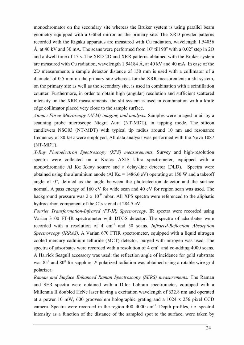
24
monochromator on the secondary site whereas the Bruker system is using parallel beam geometry equipped with a Göbel mirror on the primary site. The XRD powder patterns recorded with the Rigaku apparatus are measured with Cu radiation, wavelength 1.54056 Å, at 40 kV and 30 mA. The scans were performed from 10o till 90o with a 0.02o step in 2Θ and a dwell time of 15 s. The XRD-2D and XRR patterns obtained with the Bruker system are measured with Cu radiation, wavelength 1.54184 Å, at 40 kV and 40 mA. In case of the 2D measurements a sample detector distance of 150 mm is used with a collimator of a diameter of 0.5 mm on the primary site whereas for the XRR measurements a slit system, on the primary site as well as the secondary site, is used in combination with a scintillation counter. Furthermore, in order to obtain high (angular) resolution and sufficient scattered intensity on the XRR measurements, the slit system is used in combination with a knife edge collimator placed very close to the sample surface. Atomic Force Microscopy (AFM) imaging and analysis. Samples were imaged in air by a scanning probe microscope Ntegra Aura (NT-MDT), in tapping mode. The silicon cantilevers NSG03 (NT-MDT) with typical tip radius around 10 nm and resonance frequency of 80 kHz were employed. All data analysis was performed with the Nova 1087 (NT-MDT). X-Ray Photoelectron Spectroscopy (XPS) measurements. Survey and high-resolution spectra were collected on a Kratos AXIS Ultra spectrometer, equipped with a monochromatic Al Kα X-ray source and a delay-line detector (DLD). Spectra were obtained using the aluminium anode (Al Kα = 1486.6 eV) operating at 150 W and a takeoff angle of 0°, defined as the angle between the photoelectron detector and the surface normal. A pass energy of 160 eV for wide scan and 40 eV for region scan was used. The background pressure was 2 x 10⁻⁹ mbar. All XPS spectra were referenced to the aliphatic hydrocarbon component of the C1s signal at 284.5 eV. Fourier Transformation-Infrared (FT-IR) Spectroscopy. IR spectra were recorded using Varian 3100 FT-IR spectrometer with DTGS detector. The spectra of adsorbates were recorded with a resolution of 4 cm-1 and 50 scans. Infrared-Reflection Absorption Spectroscopy (IRRAS). A Varian 670 FTIR spectrometer, equipped with a liquid nitrogen cooled mercury cadmium telluride (MCT) detector, purged with nitrogen was used. The spectra of adsorbates were recorded with a resolution of 4 cm-1 and co-adding 4000 scans. A Harrick Seagull accessory was used; the reflection angle of incidence for gold substrate was 85o and 80o for sapphire. P-polarized radiation was obtained using a rotable wire grid polarizer. Raman and Surface Enhanced Raman Spectroscopy (SERS) measurements. The Raman and SER spectra were obtained with a Dilor Labram spectrometer, equipped with a Millennia II doubled HeNe laser having a excitation wavelength of 632.8 nm and operated at a power 10 mW, 600 grooves/mm holographic grating and a 1024 x 256 pixel CCD camera. Spectra were recorded in the region 400–4000 cm-1. Depth profiles, i.e. spectral intensity as a function of the distance of the sampled spot to the surface, were taken by
25
manually adjusting the distance between sample and objective. The recording time for a single spectrum is around 60 seconds. A 100x magnification, (Olympus, numerical aperture 0.8) ultra-long working distance objective was used. The Labram was equipped with a built-in camera, enabling the visualization of the reflected laser light from the inner side of the glass plate. The focal depth zero was determined by minimizing the spot size of this reflected light. Contact Angle (CA) Goniometry. Static contact angle measurements were performed on a video-based semi-automatic contact angle meter (DataPhysics OCA 30 Instrument Gm, Germany) at 22 oC and 42 % relative humidity, using milli-Q water as a probe liquid. Six drops were measured on each sample.
2.3 Results and Discussion
2.3.1 Dopamine behavior in aqueous media
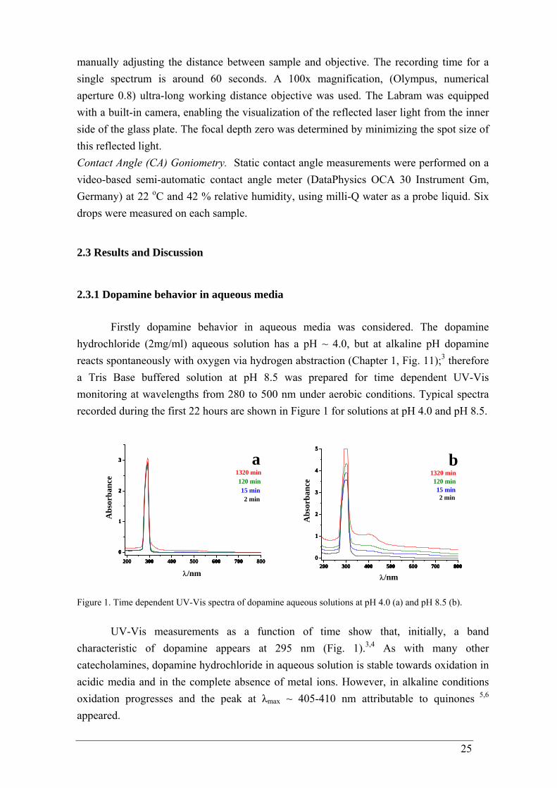
Firstly dopamine behavior in aqueous media was considered. The dopamine hydrochloride (2mg/ml) aqueous solution has a pH ~ 4.0, but at alkaline pH dopamine reacts spontaneously with oxygen via hydrogen abstraction (Chapter 1, Fig. 11);3 therefore a Tris Base buffered solution at pH 8.5 was prepared for time dependent UV-Vis monitoring at wavelengths from 280 to 500 nm under aerobic conditions. Typical spectra recorded during the first 22 hours are shown in Figure 1 for solutions at pH 4.0 and pH 8.5.
200 300 400 500 600 700 800
0
1
2
3
200 300 400 500 600 700 800
0
1
2
3
200 300 400 500 600 700 800
0
1
2
3
200 300 400 500 600 700 800
0
1
2
3 a1320 min120 min15 min2 min
Abs
orba
nce
λ/nm
200 300 400 500 600 700 8000
1
2
3
4
5
200 300 400 500 600 700 8000
1
2
3
4
5
200 300 400 500 600 700 8000
1
2
3
4
5
200 300 400 500 600 700 8000
1
2
3
4
5
b1320 min120 min15 min2 min
Abs
orba
nce
λ/nm
Figure 1. Time dependent UV-Vis spectra of dopamine aqueous solutions at pH 4.0 (a) and pH 8.5 (b).
UV-Vis measurements as a function of time show that, initially, a band characteristic of dopamine appears at 295 nm (Fig. 1).3,4 As with many other catecholamines, dopamine hydrochloride in aqueous solution is stable towards oxidation in acidic media and in the complete absence of metal ions. However, in alkaline conditions oxidation progresses and the peak at λmax ~ 405-410 nm attributable to quinones 5,6 appeared.
26
Visually the color of the alkaline dopamine hydrochloride solution changes from colorless via pink due to the appearance of dopaminochrome, to black within 15 minutes and after some hours sedimentation of a black deposit appears at the bottom of the vessel. Even with further sedimentation in the vessel, the solution always remains black. This behavior has been described in the literature3 and assigned to the formation of the biomolecule melanin by spontaneous oxidation of dopamine.
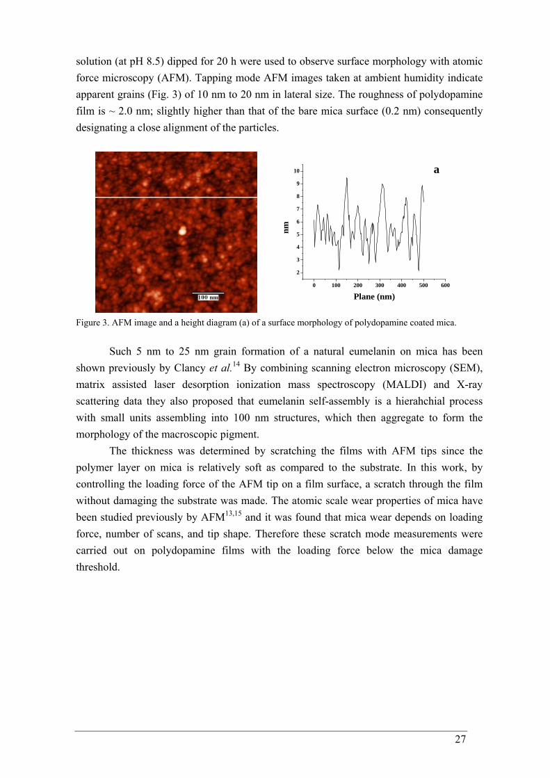
In vivo, the black melanin - eumelanin is produced from the amino acid tyrosine that is first enzymatically converted to 3,4-dihydroxy-L-phenylalanine (DOPA) and then in several steps to 5,6-dihydroxyindole carboxylic acid (DHICA) (Fig. 2), which polymerizes into eumelanin in a process that is not well understood yet.7,8 However, synthetic melanins are commonly produced by spontaneous oxidation of dopamine,3 DOPA9 or 5,6-dihydroxyindole (DHI).10,11
OH
OH
NH2 OH
OHNH2
OH
O
OH
OH NH
OH
OH NH
OH
O
dopamine DOPA DHIDHICA Figure 2. The main monomeric building blocks of eumelanin: dopamine, 3,4-dihydroxy-L-phenylalanine (DOPA), 5,6-dihydroxyindole carboxylic acid (DHICA) and 5,6-dihydroxyindole (DHI).
It is generally accepted that eumelamins are macromolecules of DHI and DHICA in proportions depending on the origin and preparation method of the eumelanin. Furthermore, it has been shown that eumelanin contains reduced (catechol) as well as oxidized (quinone) substructures.11
The reaction, initiated by the oxidation of dopamine (Chapter 1, Fig.11), occurs not only in the solution but also on the surface of the investigated material. Lee and others showed that an organic deposit grows on the surface of various materials when they are dipped into dopamine aqueous solutions in the presence of a buffer at pH 8.5.1 They supposed that the deposit, which reaches a plateau thickness of ~ 50 nm after 24 h of immersion, is composed of melanin. For convenience, the deposits obtained from dopamine buffered solutions in this work will be called “polydopamine”.
2.3.2 Dopamine and polydopamine films
The first surface modification experiments were performed on Muscovite mica. It is a hard, layered, crystalline mineral where the layered structure of aluminum silicate sheets are weakly bonded together by layers of potassium ions.12 When cleaved by physical removal of a layer, fracture along the weak atomic planes easily produce atomically flat surfaces of atoms having a regular lattice structure. AFM topographic images showed roughness of 0.2 nm.13 The cleaved sheets are hydrophilic and negatively charged in water. Polydopamine films built on a freshly cleaved mica substrate using an aqueous dopamine
27
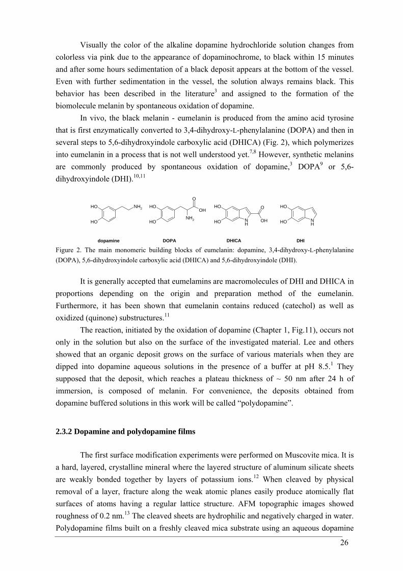
solution (at pH 8.5) dipped for 20 h were used to observe surface morphology with atomic force microscopy (AFM). Tapping mode AFM images taken at ambient humidity indicate apparent grains (Fig. 3) of 10 nm to 20 nm in lateral size. The roughness of polydopamine film is ~ 2.0 nm; slightly higher than that of the bare mica surface (0.2 nm) consequently designating a close alignment of the particles.
0 100 200 300 400 500 600
2
3
4
5
6
7
8
9
10
nmPlane (nm)
a
Figure 3. AFM image and a height diagram (a) of a surface morphology of polydopamine coated mica.
Such 5 nm to 25 nm grain formation of a natural eumelanin on mica has been shown previously by Clancy et al.14 By combining scanning electron microscopy (SEM), matrix assisted laser desorption ionization mass spectroscopy (MALDI) and X-ray scattering data they also proposed that eumelanin self-assembly is a hierahchial process with small units assembling into 100 nm structures, which then aggregate to form the morphology of the macroscopic pigment.
The thickness was determined by scratching the films with AFM tips since the polymer layer on mica is relatively soft as compared to the substrate. In this work, by controlling the loading force of the AFM tip on a film surface, a scratch through the film without damaging the substrate was made. The atomic scale wear properties of mica have been studied previously by AFM13,15 and it was found that mica wear depends on loading force, number of scans, and tip shape. Therefore these scratch mode measurements were carried out on polydopamine films with the loading force below the mica damage threshold.
28
0 200 400 600 800 1000 1200
0
10
20
30
40
50
60
70
80 a
nm
Plane (nm)
Figure 4. AFM scratch experiment and a height diagram (a) of polydopamine coated mica.
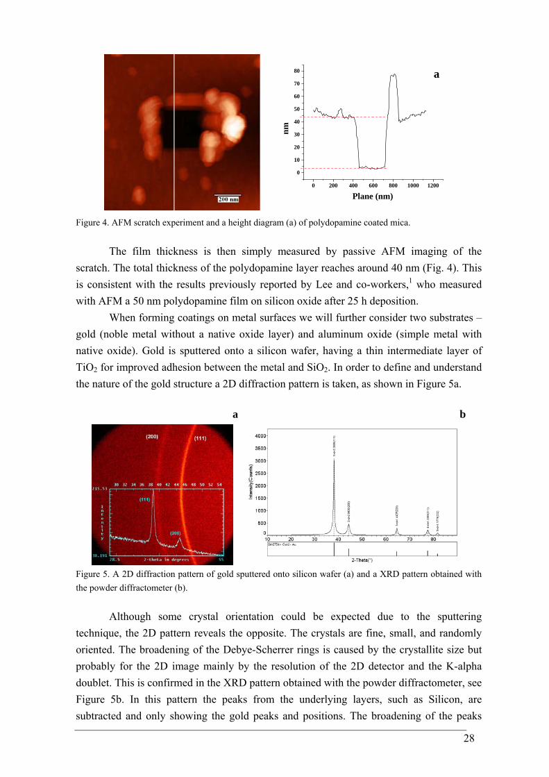
The film thickness is then simply measured by passive AFM imaging of the scratch. The total thickness of the polydopamine layer reaches around 40 nm (Fig. 4). This is consistent with the results previously reported by Lee and co-workers,1 who measured with AFM a 50 nm polydopamine film on silicon oxide after 25 h deposition.
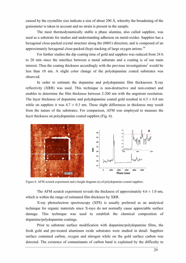
When forming coatings on metal surfaces we will further consider two substrates – gold (noble metal without a native oxide layer) and aluminum oxide (simple metal with native oxide). Gold is sputtered onto a silicon wafer, having a thin intermediate layer of TiO2 for improved adhesion between the metal and SiO2. In order to define and understand the nature of the gold structure a 2D diffraction pattern is taken, as shown in Figure 5a.
a
b
Figure 5. A 2D diffraction pattern of gold sputtered onto silicon wafer (a) and a XRD pattern obtained with the powder diffractometer (b).
Although some crystal orientation could be expected due to the sputtering technique, the 2D pattern reveals the opposite. The crystals are fine, small, and randomly oriented. The broadening of the Debye-Scherrer rings is caused by the crystallite size but probably for the 2D image mainly by the resolution of the 2D detector and the K-alpha doublet. This is confirmed in the XRD pattern obtained with the powder diffractometer, see Figure 5b. In this pattern the peaks from the underlying layers, such as Silicon, are subtracted and only showing the gold peaks and positions. The broadening of the peaks
29
caused by the crystallite size indicate a size of about 200 Å, whereby the broadening of the goniometer is taken in account and no strain is present in the sample.
The most thermodynamically stable α phase alumina, also called sapphire, was used as a substrate for studies and understanding adhesion on metal-oxides. Sapphire has a hexagonal close-packed crystal structure along the (0001) direction; and is composed of an approximately hexagonal close-packed (hcp) stacking of large oxygen anions.16
For further studies the dip coating time of gold and sapphire was reduced from 24 h to 20 min since the interface between a metal substrate and a coating is of our main interest. Thus the coating thickness accordingly with the previous investigations1 would be less than 10 nm. A slight color change of the polydopamine coated substrates was observed.
In order to estimate the dopamine and polydopamine film thicknesses X-ray reflectivity (XRR) was used. This technique is non-destructive and non-contact and enables to determine the film thickness between 2-200 nm with the angstrom resolution. The layer thickness of dopamine and polydopamine coated gold resulted in 6.5 ± 0.8 nm while on sapphire it was 4.7 ± 0.3 nm. These slight differences in thickness may result from the nature of the substrates. For comparison, AFM was employed to measure the layer thickness on polydopamine coated sapphire (Fig. 6).
0 1000 2000 3000 4000 5000
15
20
25
30
35
40
45
50a
3000 4000 500018
19
20
21
22
23
24
nm
Plane (nm)
Figure 6. AFM scratch experiment and a height diagram (a) of polydopamine coated sapphire.
The AFM scratch experiment reveals the thickness of approximately 4.6 ± 1.0 nm, which is within the range of estimated film thickness by XRR.
X-ray photoelectron spectroscopy (XPS) is usually preferred as an analytical technique for organic materials since X-rays do not normally cause appreciable surface damage. This technique was used to establish the chemical composition of dopamine/polydopamine coatings.
Prior to substrate surface modification with dopamine/polydopamine films, the fresh gold and pre-treated aluminum oxide substrates were studied in detail. Sapphire surface contained carbon, oxygen and nitrogen while on the gold surface carbon was detected. The existence of contaminants of carbon band is explained by the difficulty to

30
prepare a clean gold surface and maintain cleanliness near atmospheric pressure for XPS measurements.17
Analysis of the coated substrates also revealed signals specific to the substrates, indicating a coating formation of less than 10 nm in thickness. In order to extract the exact changes in elemental composition of films formed, the nitrogen-to-carbon signal ratios (N/C) were calculated. Surface atom percentages and N/C signal ratios are listed in table 1. Table 1. Relative atomic ratios for coated aluminum oxide (sapphire) and gold samples.
Sample Atomic ratio (mol%) N/C
Dopamine sapphire 0.4/10.3 = 1/26 = 0.039 Polydopamine sapphire 2.0/23.3 = 1/12 = 0.086
Dopamine gold 0.2/4.9 = 1/24 = 0.041 Polydopamine gold 0.7/8.9 = 1/13 = 0.079
From the table 1, it can be found that the N/C ratio of polydopamine coatings are comparable with each other when formed on gold and sapphire; the same situation is detected for dopamine coated samples. However, the theoretical N/C values of dopamine hydrochloride (N/C = 1/8 = 0.125) have not been approached in any case since the measured ratios are lower than the theoretical ones. The much higher carbon amount suggests an unavoidable presence of carbon-containing adventitious contamination when making samples under ambient conditions.
High resolution spectra were collected to obtain binding energy values for the respective functional groups and percentages from the relative area determinations. It is worth to note, that ability of XPS to detect and quantify a number of functional groups is limited in cases when relative chemical shifts of groups are below the energy resolution attainable, even with the new generation XP spectrometers, due to the existence of significant intrinsic peak widths.18,19 Therefore the data has only limited value for the determination of the contrenetation of a particular functional group. For instance, binding energy shifts of oxygen- or nitrogen-containing surface functional groups are smaller due to their similar electronegativity. The chemical shifts from a large variety of such species are present in a narrow binding energy window, and many of the peaks indicating these species overlap.20 Moreover, small binding energy shifts induced by double bonds are also present in this region, thus further complicates the analysis of the narrow scan of C 1s envelope. Similarly, the chemical shifts for nitrogen containing functionalities on the N 1s core levels are very small.21 Table 2 displays binding energy intervals obtained from the dopamine/polydopamine film measurements and assignments of corresponding functional groups, which are validated comparing with literature data. The binding energy scale was set by correcting the CHx peak of the carbon 1s envelope to 284.5 eV and all reported functional groups’ binding energies are relative to this position.
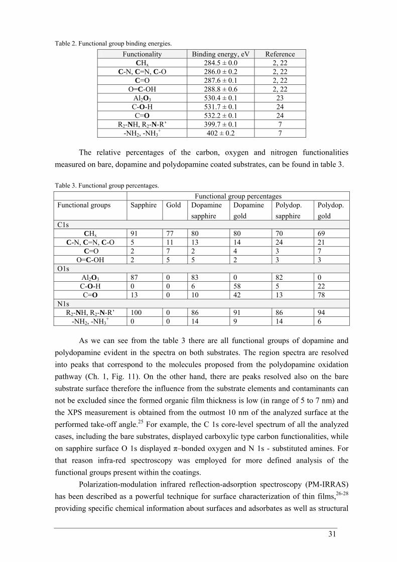
31
Table 2. Functional group binding energies. Functionality Binding energy, eV Reference
CHx 284.5 ± 0.0 2, 22 C-N, C=N, C-O 286.0 ± 0.2 2, 22
C=O 287.6 ± 0.1 2, 22 O=C-OH 288.8 ± 0.6 2, 22
Al2O3 530.4 ± 0.1 23 C-O-H 531.7 ± 0.1 24 C=O 532.2 ± 0.1 24
R2-NH, R2-N-R’ 399.7 ± 0.1 7 -NH2, -NH3
+ 402 ± 0.2 7
The relative percentages of the carbon, oxygen and nitrogen functionalities measured on bare, dopamine and polydopamine coated substrates, can be found in table 3. Table 3. Functional group percentages. Functional group percentages Functional groups Sapphire Gold Dopamine
sapphire Dopamine gold
Polydop. sapphire
Polydop. gold
C1s CHx 91 77 80 80 70 69
C-N, C=N, C-O 5 11 13 14 24 21 C=O 2 7 2 4 3 7
O=C-OH 2 5 5 2 3 3 O1s
Al2O3 87 0 83 0 82 0 C-O-H 0 0 6 58 5 22 C=O 13 0 10 42 13 78
N1s R2-NH, R2-N-R’ 100 0 86 91 86 94
-NH2, -NH3+ 0 0 14 9 14 6
As we can see from the table 3 there are all functional groups of dopamine and polydopamine evident in the spectra on both substrates. The region spectra are resolved into peaks that correspond to the molecules proposed from the polydopamine oxidation pathway (Ch. 1, Fig. 11). On the other hand, there are peaks resolved also on the bare substrate surface therefore the influence from the substrate elements and contaminants can not be excluded since the formed organic film thickness is low (in range of 5 to 7 nm) and the XPS measurement is obtained from the outmost 10 nm of the analyzed surface at the performed take-off angle.25 For example, the C 1s core-level spectrum of all the analyzed cases, including the bare substrates, displayed carboxylic type carbon functionalities, while on sapphire surface O 1s displayed π–bonded oxygen and N 1s - substituted amines. For that reason infra-red spectroscopy was employed for more defined analysis of the functional groups present within the coatings.
Polarization-modulation infrared reflection-adsorption spectroscopy (PM-IRRAS) has been described as a powerful technique for surface characterization of thin films,26-28 providing specific chemical information about surfaces and adsorbates as well as structural
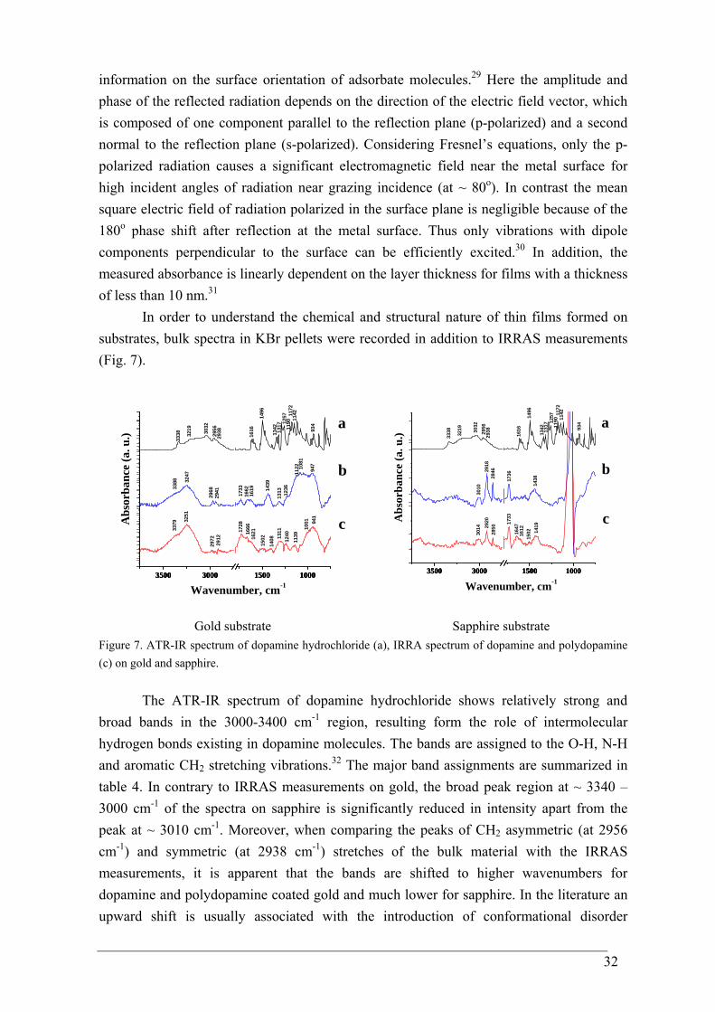
32
information on the surface orientation of adsorbate molecules.29 Here the amplitude and phase of the reflected radiation depends on the direction of the electric field vector, which is composed of one component parallel to the reflection plane (p-polarized) and a second normal to the reflection plane (s-polarized). Considering Fresnel’s equations, only the p-polarized radiation causes a significant electromagnetic field near the metal surface for high incident angles of radiation near grazing incidence (at ~ 80o). In contrast the mean square electric field of radiation polarized in the surface plane is negligible because of the 180o phase shift after reflection at the metal surface. Thus only vibrations with dipole components perpendicular to the surface can be efficiently excited.30 In addition, the measured absorbance is linearly dependent on the layer thickness for films with a thickness of less than 10 nm.31
In order to understand the chemical and structural nature of thin films formed on substrates, bulk spectra in KBr pellets were recorded in addition to IRRAS measurements (Fig. 7).
3500 3000 1500 10003500 3000 1500 10003500 3000 1500 1000
1172
1190
1342 93
4
1142
1257
1317
2938
1496
1616
29563032
3219
3338
947
1236
1662
1733
2941
2968
3247
3388
1502
1408 13
1112
4011
39
1001 94
1
16661728
2912
2972
3251
3379
1081
1122
131314
39
1619
1621
c
b
a
Abs
orba
nce
(a. u
.)
Wavenumber, cm-1
3500 3000 1500 10003500 3000 1500 10003500 3000 1500 1000
1190 a
1172
1342 93
4
1142
1257
1317
1496
16162956
3032
3219
3338
143817
36284629
1830
10
1419
1647
1733
285029
2030
14
2938
1612
1502
c
b
Abs
orba
nce
(a. u
.)
Wavenumber, cm-1
Gold substrate Sapphire substrate Figure 7. ATR-IR spectrum of dopamine hydrochloride (a), IRRA spectrum of dopamine and polydopamine (c) on gold and sapphire.
The ATR-IR spectrum of dopamine hydrochloride shows relatively strong and broad bands in the 3000-3400 cm-1 region, resulting form the role of intermolecular hydrogen bonds existing in dopamine molecules. The bands are assigned to the O-H, N-H and aromatic CH2 stretching vibrations.32 The major band assignments are summarized in table 4. In contrary to IRRAS measurements on gold, the broad peak region at ~ 3340 – 3000 cm-1 of the spectra on sapphire is significantly reduced in intensity apart from the peak at ~ 3010 cm-1. Moreover, when comparing the peaks of CH2 asymmetric (at 2956 cm-1) and symmetric (at 2938 cm-1) stretches of the bulk material with the IRRAS measurements, it is apparent that the bands are shifted to higher wavenumbers for dopamine and polydopamine coated gold and much lower for sapphire. In the literature an upward shift is usually associated with the introduction of conformational disorder
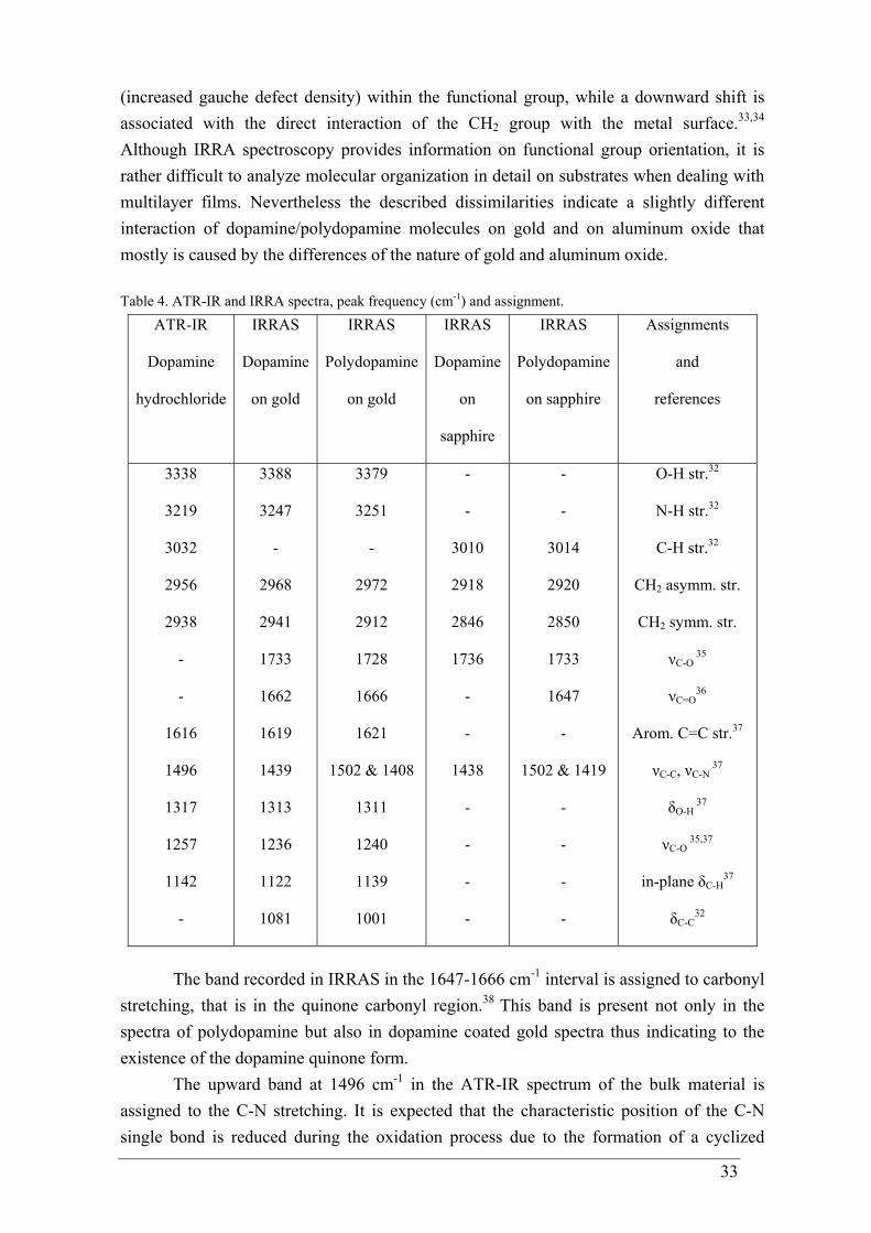
33
(increased gauche defect density) within the functional group, while a downward shift is associated with the direct interaction of the CH2 group with the metal surface.33,34 Although IRRA spectroscopy provides information on functional group orientation, it is rather difficult to analyze molecular organization in detail on substrates when dealing with multilayer films. Nevertheless the described dissimilarities indicate a slightly different interaction of dopamine/polydopamine molecules on gold and on aluminum oxide that mostly is caused by the differences of the nature of gold and aluminum oxide. Table 4. ATR-IR and IRRA spectra, peak frequency (cm-1) and assignment.
ATR-IR
Dopamine
hydrochloride
IRRAS
Dopamine
on gold
IRRAS
Polydopamine
on gold
IRRAS
Dopamine
on
sapphire
IRRAS
Polydopamine
on sapphire
Assignments
and
references
3338
3219
3032
2956
2938
-
-
1616
1496
1317
1257
1142
-
3388
3247
-
2968
2941
1733
1662
1619
1439
1313
1236
1122
1081
3379
3251
-
2972
2912
1728
1666
1621
1502 & 1408
1311
1240
1139
1001
-
-
3010
2918
2846
1736
-
-
1438
-
-
-
-
-
-
3014
2920
2850
1733
1647
-
1502 & 1419
-
-
-
-
O-H str.32
N-H str.32
C-H str.32
CH2 asymm. str.
CH2 symm. str.
νC-O 35
νC=O36
Arom. C=C str.37
νC-C, νC-N 37
δO-H 37
νC-O 35,37
in-plane δC-H37
δC-C32
The band recorded in IRRAS in the 1647-1666 cm-1 interval is assigned to carbonyl stretching, that is in the quinone carbonyl region.38 This band is present not only in the spectra of polydopamine but also in dopamine coated gold spectra thus indicating to the existence of the dopamine quinone form.
The upward band at 1496 cm-1 in the ATR-IR spectrum of the bulk material is assigned to the C-N stretching. It is expected that the characteristic position of the C-N single bond is reduced during the oxidation process due to the formation of a cyclized
34
products (Fig. 11, Ch. 1). Therefore the two bands at 1502 cm-1 and 1419-1408 cm-1 appear for the polydopamine coatings on gold and aluminum oxide substrates designating that dopamine intramolecular cyclization reactions have taken place and indole derivatives (dopaminechromes) are formed.39
The absence of a band at 1317 cm-1 in the IRRA spectra on sapphire, when compared with IRRAS data recorded on gold, may be an indication of a chelate complex formation on the aluminum oxide layer.40 However, the νC-O 1736-1728 cm-1 region stretch in IRRAS of dopamine and polydopamine coatings for both substrates corresponding with the νC-O stretch at 1257-1236 cm-1 region for coated gold, indicates the presence of catechols coordinated with gold surface; unfortunately the 1257-1236 cm-1 region is difficult for sapphire spectra. It is well documented that IRRAS experiments of thin layers on gold often result in high quality spectra, even for submolecular thickness of the material under investigation.41-43 However, samples of interest on a dielectric substrate, such as sapphire, are more complex for measuring thin films due to the small absorption coefficient.31,26 In contrast with metal surfaces both parallel and perpendicular vibrational components of the adsorbate can be detected. The p-polarized light, used in IRRAS measurements, is composed of a perpendicular and parallel component; it can be negative and positive depending on the orientation of the transition dipole moment and the angle of incidence. Both positive and negative peaks can occur in the spectrum of p-polarized light, depending on the orientations of the different transition dipole moments31,44 as illustrated by the negative band in ~ 1140 cm-1 region measured from the IRRAS spectrum of dopamine coated sapphire.
In combination with IR, Raman spectroscopy was used to observe molecular vibrations of dopamine/polydopamine coatings. IR and Raman spectra generally differ, owing to symmetry reasons and selection rules, and often yield complementary information. Vibrations of weakly polar and even symmetrically bonded atoms usually result in intense Raman bands because Raman activity is related with changes in polarizability. Raman scattering on thin organic films is an inherently weak process and as a result an enhancement mechanism is usually needed to directly observe the data. The importance of surface enhanced Raman spectroscopy (SERS) is that it is both surfaces selective and highly sensitive. Signal enhancement can usually be achieved by depositing films onto roughened coinage (Au, Ag, Cu) or alkali (Li, Na, K) metal surfaces. Although the phenomenon is not completely understood it is generally accepted that a major portion of the signal increase can be attributed to the field enhancement of the incident and scattered radiation only at the surface. Thus, the surface signal overwhelms the bulk signal, making bulk subtraction unnecessary.45 Two types of surfaces that are suitable for SERS may be distinguished: roughened gold or silver surfaces that are macroscopically flat and colloidal suspensions of gold or silver nanoparticles.46 In this study roughened gold surfaces that are macroscopically flat were used.
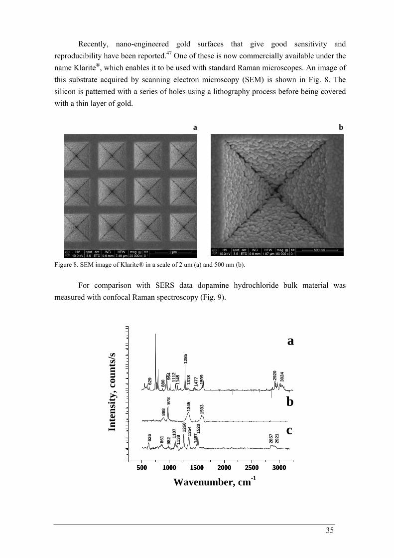
35
Recently, nano-engineered gold surfaces that give good sensitivity and reproducibility have been reported.47 One of these is now commercially available under the name Klarite®, which enables it to be used with standard Raman microscopes. An image of this substrate acquired by scanning electron microscopy (SEM) is shown in Fig. 8. The silicon is patterned with a series of holes using a lithography process before being covered with a thin layer of gold.
a b
Figure 8. SEM image of Klarite® in a scale of 2 um (a) and 500 nm (b).
For comparison with SERS data dopamine hydrochloride bulk material was measured with confocal Raman spectroscopy (Fig. 9).
500 1000 1500 2000 2500 3000500 1000 1500 2000 2500 3000500 1000 1500 2000 2500 3000
159313
45978
898
861626
Inte
nsity
, cou
nts/
s
2921
2857
1520
135412
6011
381107
982
1487
1112
880 30
2429
20
629 96
4
1145
1285
1318
1477 1599
c
b
a
Wavenumber, cm-1
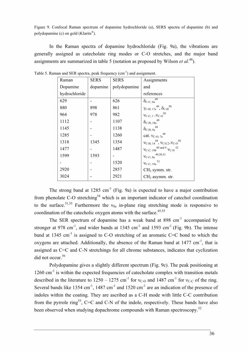
36
Figure 9. Confocal Raman spectrum of dopamine hydrochloride (a), SERS spectra of dopamine (b) and polydopamine (c) on gold (Klarite®).
In the Raman spectra of dopamine hydrochloride (Fig. 9a), the vibrations are generally assigned as catecholate ring modes or C-O stretches, and the major band assignments are summarized in table 5 (notation as proposed by Wilson et al.48).
Table 5. Raman and SER spectra, peak frequency (cm-1) and assignment. Raman Dopamine hydrochloride
SERS dopamine
SERS polydopamine
Assignments and references
629 880 964 1112 1145 1285 1318 1477 1599 - 2920 3024
- 898 978 - - - 1345 - 1593 - - -
626 861 982 1107 1138 1260 1354 1487 - 1520 2857 2921
δC-C; 6a49
γC-H; 17a49, δC-H
50 νC-C; 1 +νC-O
50
δC-H; 18a49
δC-H; 9a49
cat. νC-O; 7a49
νC-H; 1449, νC1C2+νC-O
50 νC-C; 19b
49 and 9 νC-N 52
νC-C; 8a 49,50,51
νC-C; 19a 51
CH2 symm. str. CH2 asymm. str.
The strong band at 1285 cm-1 (Fig. 9a) is expected to have a major contribution from phenolate C-O stretching54 which is an important indicator of catechol coordination to the surface.51,55 Furthermore the ν8a in-plane ring stretching mode is responsive to coordination of the catecholic oxygen atoms with the surface.45,55
The SER spectrum of dopamine has a weak band at 898 cm-1 accompanied by stronger at 978 cm-1, and wider bands at 1345 cm-1 and 1593 cm-1 (Fig. 9b). The intense band at 1345 cm-1 is assigned to C-O stretching of an aromatic C=C bond to which the oxygens are attached. Additionally, the absence of the Raman band at 1477 cm-1, that is assigned as C=C and C-N stretchings for all chrome substances, indicates that cyclization did not occur.56
Polydopamine gives a slightly different spectrum (Fig. 9c). The peak positioning at 1260 cm-1 is within the expected frequencies of catecholate complex with transition metals described in the literature to 1250 – 1275 cm-1 for νC-O and 1487 cm-1 for νC-C of the ring. Several bands like 1354 cm-1, 1487 cm-1 and 1520 cm-1 are an indication of the presence of indoles within the coating. They are ascribed as a C-H mode with little C-C contribution from the pyrrole ring53, C=C and C-N of the indole, respectively. These bands have also been observed when studying dopachrome compounds with Raman spectroscopy.52
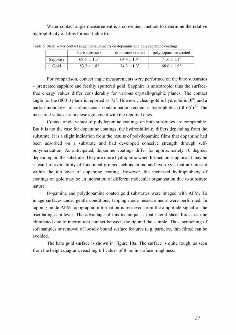
37
Water contact angle measurement is a convenient method to determine the relative hydrophilicity of films formed (table 6). Table 6. Static water contact angle measurements on dopamine and polydopamine coatings.
bare substrate dopamine coated polydopamine coated Sapphire 69.5 ± 1.3° 64.4 ± 1.4° 71.6 ± 1.1°
Gold 53.7 ± 1.8° 74.3 ± 1.3° 68.6 ± 1.9°
For comparison, contact angle measurements were performed on the bare substrates – pretreated sapphire and freshly sputtered gold. Sapphire is anisotropic; thus the surface-free energy values differ considerably for various crystallographic planes. The contact angle for the (0001) plane is reported as 72o. However, clean gold is hydrophilic (0o) and a partial monolayer of carbonaceous contamination renders it hydrophobic (till 66o).57 The measured values are in close agreement with the reported ones.
Contact angle values of polydopamine coatings on both substrates are comparable. But it is not the case for dopamine coatings; the hydrophilicilty differs depending from the substrate. It is a slight indication from the results of polydopamine films that dopamine had been adsorbed on a substrate and had developed cohesive strength through self-polymerization. As anticipated, dopamine coatings differ for approximately 10 degrees depending on the substrate. They are more hydrophilic when formed on sapphire. It may be a result of availability of functional groups such as amine and hydroxyls that are present within the top layer of dopamine coating. However, the increased hydrophobiciy of coatings on gold may be an indication of different molecular organization due to substrate nature.
Dopamine and polydopamine coated gold substrates were imaged with AFM. To image surfaces under gentle conditions, tapping mode measurements were performed. In tapping mode AFM topographic information is retrieved from the amplitude signal of the oscillating cantilever. The advantage of this technique is that lateral shear forces can be eliminated due to intermittent contact between the tip and the sample. Thus, scratching of soft samples or removal of loosely bound surface features (e.g. particles, thin films) can be avoided.
The bare gold surface is shown in Figure 10a. The surface is quite rough, as seen from the height diagram, reaching till values of 8 nm in surface roughness.
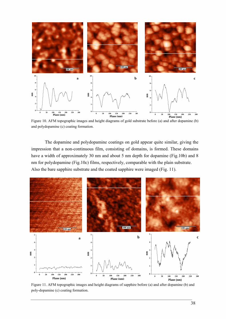
38
0 50 100 150 200 250 3000
2
4
6
8
10
a
nm
Plane (nm)
0 50 100 150 200 250 3000
2
4
6
8
10
b
nm
Plane (nm)
0 50 100 150 200 250 3000
2
4
6
8
10c
nm
Plane (nm) Figure 10. AFM topographic images and height diagrams of gold substrate before (a) and after dopamine (b) and polydopamine (c) coating formation.
The dopamine and polydopamine coatings on gold appear quite similar, giving the impression that a non-continuous film, consisting of domains, is formed. These domains have a width of approximately 30 nm and about 5 nm depth for dopamine (Fig.10b) and 8 nm for polydopamine (Fig.10c) films, respectively, comparable with the plain substrate. Also the bare sapphire substrate and the coated sapphire were imaged (Fig. 11).
0 50 100 150 200 250 3000
1
2
3
4
5
nm
Plane (nm)
a
0 50 100 150 200 250 3000
1
2
3
4
5b
nm
Plane (nm)
0 50 100 150 200 250 3000
1
2
3
4
5
c
nm
Plane (nm) Figure 11. AFM topographic images and height diagrams of sapphire before (a) and after dopamine (b) and poly-dopamine (c) coating formation.
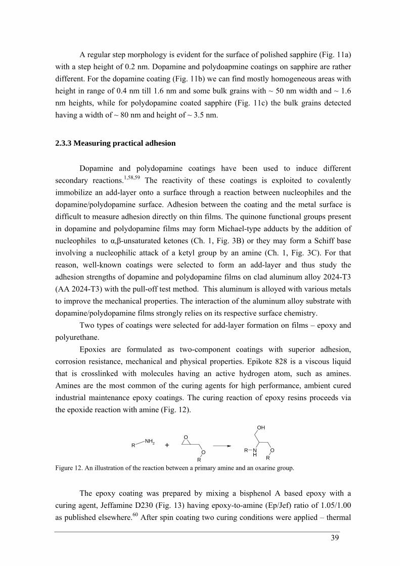
39
A regular step morphology is evident for the surface of polished sapphire (Fig. 11a) with a step height of 0.2 nm. Dopamine and polydoapmine coatings on sapphire are rather different. For the dopamine coating (Fig. 11b) we can find mostly homogeneous areas with height in range of 0.4 nm till 1.6 nm and some bulk grains with ~ 50 nm width and ~ 1.6 nm heights, while for polydopamine coated sapphire (Fig. 11c) the bulk grains detected having a width of ~ 80 nm and height of ~ 3.5 nm.
2.3.3 Measuring practical adhesion
Dopamine and polydopamine coatings have been used to induce different secondary reactions.1,58,59 The reactivity of these coatings is exploited to covalently immobilize an add-layer onto a surface through a reaction between nucleophiles and the dopamine/polydopamine surface. Adhesion between the coating and the metal surface is difficult to measure adhesion directly on thin films. The quinone functional groups present in dopamine and polydopamine films may form Michael-type adducts by the addition of nucleophiles to α,β-unsaturated ketones (Ch. 1, Fig. 3B) or they may form a Schiff base involving a nucleophilic attack of a ketyl group by an amine (Ch. 1, Fig. 3C). For that reason, well-known coatings were selected to form an add-layer and thus study the adhesion strengths of dopamine and polydopamine films on clad aluminum alloy 2024-T3 (AA 2024-T3) with the pull-off test method. This aluminum is alloyed with various metals to improve the mechanical properties. The interaction of the aluminum alloy substrate with dopamine/polydopamine films strongly relies on its respective surface chemistry.
Two types of coatings were selected for add-layer formation on films – epoxy and polyurethane.
Epoxies are formulated as two-component coatings with superior adhesion, corrosion resistance, mechanical and physical properties. Epikote 828 is a viscous liquid that is crosslinked with molecules having an active hydrogen atom, such as amines. Amines are the most common of the curing agents for high performance, ambient cured industrial maintenance epoxy coatings. The curing reaction of epoxy resins proceeds via the epoxide reaction with amine (Fig. 12).
RNH2
O
O
R
R NH
OR
OH
+
Figure 12. An illustration of the reaction between a primary amine and an oxarine group.
The epoxy coating was prepared by mixing a bisphenol A based epoxy with a curing agent, Jeffamine D230 (Fig. 13) having epoxy-to-amine (Ep/Jef) ratio of 1.05/1.00 as published elsewhere.60 After spin coating two curing conditions were applied – thermal
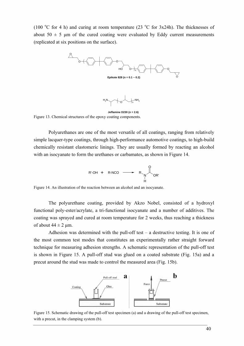
40
(100 oC for 4 h) and curing at room temperature (23 oC for 3x24h). The thicknesses of about 50 ± 5 μm of the cured coating were evaluated by Eddy current measurements (replicated at six positions on the surface).
O
OH O O
O
O
O
n
NH2 ONH2n
Epikote 828 (n = 0.1 ~ 0.2)
Jeffamine D230 (n = 2.6) Figure 13. Chemical structures of the epoxy coating components.
Polyurethanes are one of the most versatile of all coatings, ranging from relatively simple lacquer-type coatings, through high-performance automotive coatings, to high-build chemically resistant elastomeric linings. They are usually formed by reacting an alcohol with an isocyanate to form the urethanes or carbamates, as shown in Figure 14.
NR
HOR'
O
R'-OH + R-NCO
Figure 14. An illustration of the reaction between an alcohol and an isocyanate.
The polyurethane coating, provided by Akzo Nobel, consisted of a hydroxyl functional poly-ester/acrylate, a tri-functional isocyanate and a number of additives. The coating was sprayed and cured at room temperature for 2 weeks, thus reaching a thickness of about 44 ± 2 μm.
Adhesion was determined with the pull-off test – a destructive testing. It is one of the most common test modes that constitutes an experimentally rather straight forward technique for measuring adhesion strengths. A schematic representation of the pull-off test is shown in Figure 15. A pull-off stud was glued on a coated substrate (Fig. 15a) and a precut around the stud was made to control the measured area (Fig. 15b).
Figure 15. Schematic drawing of the pull-off test specimen (a) and a drawing of the pull-off test specimen, with a precut, in the clamping system (b).
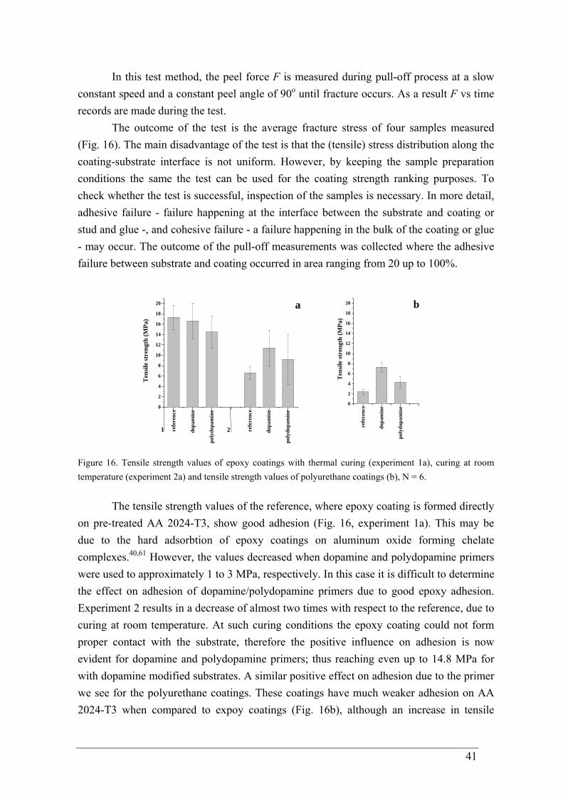
41
In this test method, the peel force F is measured during pull-off process at a slow constant speed and a constant peel angle of 90o until fracture occurs. As a result F vs time records are made during the test.
The outcome of the test is the average fracture stress of four samples measured (Fig. 16). The main disadvantage of the test is that the (tensile) stress distribution along the coating-substrate interface is not uniform. However, by keeping the sample preparation conditions the same the test can be used for the coating strength ranking purposes. To check whether the test is successful, inspection of the samples is necessary. In more detail, adhesive failure - failure happening at the interface between the substrate and coating or stud and glue -, and cohesive failure - a failure happening in the bulk of the coating or glue - may occur. The outcome of the pull-off measurements was collected where the adhesive failure between substrate and coating occurred in area ranging from 20 up to 100%.
0
2
4
6
8
10
12
14
16
18
20
Ten
sile
stre
ngth
(MPa
)
poly
dopa
min
e
poly
dopa
min
e
dopa
min
e
dopa
min
e
refe
renc
e
refe
renc
e
21
a
0
2
4
6
8
10
12
14
16
18
20
Ten
sile
stre
ngth
(MPa
)
poly
dopa
min
e
dopa
min
e
refe
renc
e
b
Figure 16. Tensile strength values of epoxy coatings with thermal curing (experiment 1a), curing at room temperature (experiment 2a) and tensile strength values of polyurethane coatings (b), N = 6.
The tensile strength values of the reference, where epoxy coating is formed directly on pre-treated AA 2024-T3, show good adhesion (Fig. 16, experiment 1a). This may be due to the hard adsorbtion of epoxy coatings on aluminum oxide forming chelate complexes.40,61 However, the values decreased when dopamine and polydopamine primers were used to approximately 1 to 3 MPa, respectively. In this case it is difficult to determine the effect on adhesion of dopamine/polydopamine primers due to good epoxy adhesion. Experiment 2 results in a decrease of almost two times with respect to the reference, due to curing at room temperature. At such curing conditions the epoxy coating could not form proper contact with the substrate, therefore the positive influence on adhesion is now evident for dopamine and polydopamine primers; thus reaching even up to 14.8 MPa for with dopamine modified substrates. A similar positive effect on adhesion due to the primer we see for the polyurethane coatings. These coatings have much weaker adhesion on AA 2024-T3 when compared to expoy coatings (Fig. 16b), although an increase in tensile
42
strength values was recorded till up to three times of samples having dopamine primers and up to two times of samples having polydopamine dopamine primers.
The measured tensile strength values of the epoxy and polyurethane coatings having dopamine/polydopamine primers are high when compared with the marine mussel M. edulis average adhesion on aluminum (0.29 ± 0.11 MPa, detailed description in Chapter 1),62 that could rise due to good adhesion of epoxy and polyurethane coatings. Therefore there is a high need to design a better defined system for studies of adherence behavior of dopamine-based materials on metals.
2.4 Conclusion
Dopamine reacts spontaneously with dioxygen at alkaline pH forming melanine
type biomolecules. Moreover, when metal substrates are immersed into dopamine aqueous solutions, they can initiate dopamine oxidation from acidic environment (pH 4.0) forming catechol and quinone containing and from alkaline environment (pH 8.5) melanine-type material containing thin, adherent films of controlled thicknesses. Besides the moisture–resistant adsorption of dopamine and polydopamine, polydopamine films had developed cohesive strength through self-polymerization.
Catechols are coordinated with gold at the metal interface. Furthermore metal complexes on aluminum oxide are proposed.
The quinone functional groups present in dopamine and polydopamine films exploits as a versatile platform for secondary reactions toward nucleophiles. Covalently bound add-layers were used to determine adhesion of dopamine and polydopamine films on aluminum alloy. The resulting tensile strength values of the epoxy and polyurethane coatings having dopamine/polydopamine primers are high when compared with the marine mussel M. edulis average adhesion on aluminum (0.29 ± 0.11 MPa). This could be the result of the good adhesion of epoxy and polyurethane coatings. Therefore there is a need to design a better defined system for studies of adherence behavior of dopamine-based materials on metals.
2.5 References
1 Lee, H.; Dellatore, S. M.; Miller, W. M.; Messersmith, P. B. Science 2007, 318,
426. 2 Bernsmann, F.; Ponche, A.; Ringwald, C.; Hemmerlé, J.; Raya, J.; Bechinger, B.;
Voegel, J.-C.; Schaaf, P.; Ball, V. J. Phys. Chem. C 2009, 113, 8234. 3 Herlinger, E.; Jamesonb, R. F.; Linerta, W. J. Chem. Soc. Perkin Trans. 2 1995, 2,
259.
43
4 Barreto, W. J.; Barreto, S. R.G.; Santos, M. A.; Schimidt, R.; Paschoal, F. M.M.; Mangrich, A. S.; deOliveira, L. F.C. Journal of Inorganic Biochemistry 2001, 84, 89.
5 Lee, B. P.; Dalsin, J. L.; Messersmith, P. B. Biomacromolecules 2002, 3, 1038. 6 Leeden, M. C. Langmuir 2005, 21, 11373. 7 Clark, M. B.; Gardella, J. A.; Schultz, T. M.; Patil, D. G.; Salvati, L. Anal. Chem.
1990, 62, 949. 8 Meredith, P.; Powell, B. J.; Riesz, J.; Nighswander-Rempel, S. P.L; Pederson, M.
R.; Moore, E. G. Soft Matter 2006, 2, 37. 9 Peter, M. C.; Forsfer, H. Angnv. Chem. Ini. Ed. EngI. 1989, 28, 741. 10 Hervé, M.; Hirschinger, J.; Granger, P.; Gilard, P.; Deflandre, A.; Goetz, N.
Biochimica et Biophysica Acta 1994, 1204,19. 11 Bernsmann, F. Melanin Made by Dopamine Oxidation: Thin Films and Interactions
with Polyelectrolyte Multilayers, PhD thesis 2010, University of Strasbourg. 12 Schwartz, M. Innovations in Materials Manufacturing, Fabrication, and
Enviromental Safety 2011, Taylor and Francis Group, USA. 13 Miyake, S. Appl. Phys. Lett. 1994, 65, 980. 14 Clancy, C. M. R.; Nofsinger, J. B.; Hanks, R. K.; Simon, J. D. J. Phys. Chem. B
2000, 104, 7871. 15 Hu, J.; Xiao, X.-D.; Ogletree, D. F.; Salmeron, M. Surface Science 1995, 327, 358. 16 Cuccureddu, F.; Murphy, S.; Shvets, I.V.; Porcu, M.; Zandbergen, H.W.; Sidorov,
N.S.; Bozhko, S.I. Surface Science 2010, 604, 1294. 17 Ishida, T.; Tsuneda, S.; Nishida, N.; Hara, M.; Sasabe, H.; Knoll, W. Langmuir
1997, 13, 4638. 18 Malitesta, C.; Losito, I.; Sabbatini, L.; Zambonin, P.G. Journal of Electron
Spectroscopy and Related Phenomena 1998, 97, 199. 19 Holländer, A.; Pippig, F.; Dubreuil, M.; Vangeneugden, D. Plasma Process. Polym.
2008, 5, 345. 20 Chilkoti, A.; Ratner, B. D.; Briggst, D. Chem. Mater. 1991, 3, 51. 21 Jagst, E. Surface Functional Group Characterization Using Chemical
Derivatization X-ray Photoelectron Spectroscopy (CD-XPS), PhD thesis 2011, Free University Berlin.
22 Desimoni, E.; Casella, G. I.; Morone, A.; Salvi, A. M. Surface and Interface Analysis 1990, 15, 627434.
23 Brand, J.; Snijders, P. C.; Sloof, W. G.; Terryn, H.; Wit, J. H. W. J. Phys. Chem. B 2004, 108, 6017.
24 Li, X.-G.; Huang, K.-L.; Liu, S.-Q.; Tan, N.; Chen, L.-Q. Trans. Nonferrous Met. Soc. China 2007, 17, 195.
25 Geng, S.; Zhang, S.; Onishi, H. Materials Technology 2002, 17, 234. 26 Brunner, H.; Mayer, U.; Hoffmann, H. Applied Spectroscopy 1997, 51, 209.
44
27 Mendelsohn, R.; Flach, C. R. Handbook of Vibrational Spectroscopy 2006, 1, John Wiley and Sons, 1028.
28 Porter, M. D. Anal. Chem. 1988, 60, 1143 A. 29 Hollins, P.; Pritchard, J. Progress in Surface Science 1966, 19, 276. 30 Bubert, H.; Jenett, H. Surface and Thin Film Analysis 2002, WILEY-VCH Verlag
GmbH, Germany. 31 Kosters, P. FT-IR Spectroscopy of Thin Biological Layers, PhD thesis 2000,
University of Twente. 32 Lin-Vien, D.; Colthup, N. B.; Fateley, W. G.; Grasselli, J. G. The Handbook of
Infrared and Raman Characteristic Frequencies of Organic Molecules 1991, Academic Press: San Diego, CA.
33 Jun, Y.; Zhu, X.-Y. J. Am. Chem. Soc. 2004, 126, 13224. 34 Boer, B.; Frank, M. M.; Chabal, Y. J.; Jiang, W.; Garfunkel, E.; Bao, Z.
Langmuir 2004, 20, 1539. 35 Ting II, G. G.; Acton, O.; Ma, H.; Ka, J. W.; Jen, A. K.-Y. Langmuir 2009, 25,
2140. 36 Wang, W.-C.; Wang, J.; Liao, Y.; Zhang, L.; Cao, B.; Song, G.; She, X. Journal of
Applied Polymer Science 2010, 117, 534. 37 Bredenberg, J. B.; Sarbakl, Z. J . Chem. Tech. Biotechnol. 1988, 42, 221. 38 Morris, M. D. Analytical Chem. 1975, 47, 2453. 39 Wang, X.; Jin, B.; Lin, X. Analytical Sciences 2002, 18, 931. 40 Kollek, H. Int.J. Adhesion and Adhesives 1985, 5, 75. 41 Nuzzo, R. G.; Fusco, F. A.; Allara, D. L. J. Am. Chem. Soc. 1987, 109, 2358. 42 Allara, D. L.; Baca, A.; Pryde, C. A. Macromolecules 1978, 11, 1215. 43 Sommerdijk, N. A. J. M.; Buynsters, P. J. J. A.; Akdemir, H.; Geurts, D. G.;
Pistorius, A. M. A.; Feiters, M. C.; Nolte, R. J. M.; Zwanenburg, B. Chem. Eur. J. 1998, 4, 127.
44 Mielczarski, J. A.; Mielczarski, E.; Zachwieja, J.; Casus, J. M. Langmuir 1995,11, 2787.
45 Kowalchyk, W. K.; Davis, K. L.; Morris, M. D. Spectrochim Acta 1995, 51A, 145. 46 Günzler, H.; Gremlich, H.-U. IR Spectroscopy 2002, WILEY-VCH Verlag GmbH,
Germany. 47 Brejna, P. R.; Griffiths, P. R.; Yang, J. Applied Spectroscopy 2009, 63, 396. 48 Wilson, E. B.; Decius, J. C.; Cross, P. C. Molecular Vibrations 1955, McGraw-Hill,
New York. 49 Koh, T. Y.; Greaves, S. J.; Griffith, W. P. Spectrochimica Acta 1994, 50A, 857. 50 Barreto, W. J.; Barreto, S. R. G.; Ando, R. A.; Santos, P. S.; Mauro, E. D.; Jorge, T.
Spectrochimica Acta Part A 2008, 71, 1419. 51 Lee, N.-S.; Hsieh, Y.-Z.; Paisley, R. F.; Morris, M. D. Anal. Chem. 1988, 60, 442. 52 Barreto, W.J.; Ponzoni, S.; Sassi, P. Spectrochimica Acta Part A 1999, 55, 65.
45
53 Majoube, M.; Vergoten, G. J. of Raman Spectroscopy 1992, 23, 431. 54 Gaber, B. P.; Miskowski, V.; Spiro, T. G. J Am Chem Soc 1974, 96, 6868. 55 Ooka, A. A.; Garrell, R. L. Biopolymers 2000, 57, 92. 56 Barreto, W. J.; Barreto, S. R. G.; Ponzoni, S.; Kawano, Y.; Mauro, E. D.; Magosso,
H. A.; Silva, W.P. Monatshefte für Chemie 2005, 136, 701. 57 Smith, T. Journal of Colloid and Interface Science 1980, 75, 51. 58 Lee, H.; Rho, J.; Messersmith, P. B. Adv. Mater. 2009, 21, 431. 59 He, T.; Shi, Z.L.; Fang, N.; Neoh, K.G.; Kang, E.T.; Chan, V. Biomaterials 2009,
30, 317. 60 Foyet, A.; Wu, T. H.; Ven, L.; Kodentsov, A.; With, G.; Benthem, R. Progr. in
Organic Coatings 2009, 64, 138. 61 Mercier, D.; Rouchaud, J.-C.; Barthés-Labrousse, M.-G. Applied Surface Science
2008, 254, 6495. 62 Burkett, J. R.; Wojtas, J. L.; Cloud, J. L.; Wilker, J. J. The Journal of Adhesion
2009, 85, 601.
46
CHAPTER 3
N-stearoyldopamine monolayer on gold and sapphire
In this chapter, we used the model molecule N-stearoyldopamine. This molecule contains a catechol residue that is capable of binding to metals and metal oxides1-3 and a hydrophobic alkyl-chain that gives an ability to organize molecules on surfaces using the Langmuir-Blodgett preparation technique. Attention is devoted in studying the adsorption behavior of N-stearoyldopamine onto gold and aluminum oxide in terms of monolayer characteristics. The organization of Langmuir-Blodgett monolayers on these metal substrates as suggested by previous experimental evidence with marine mussel foot proteins4,5 was observed directly and studied in detail with several surface sensitive techniques. Our study shows that within the monolayer the catechols functions as a surface anchor on gold and the alkyl-chains appear to be tilted within a monolayer, while the amide functionality stabilizes the formed film. The statements are supported with the outcomes of molecular simulations of the monolayer on gold.
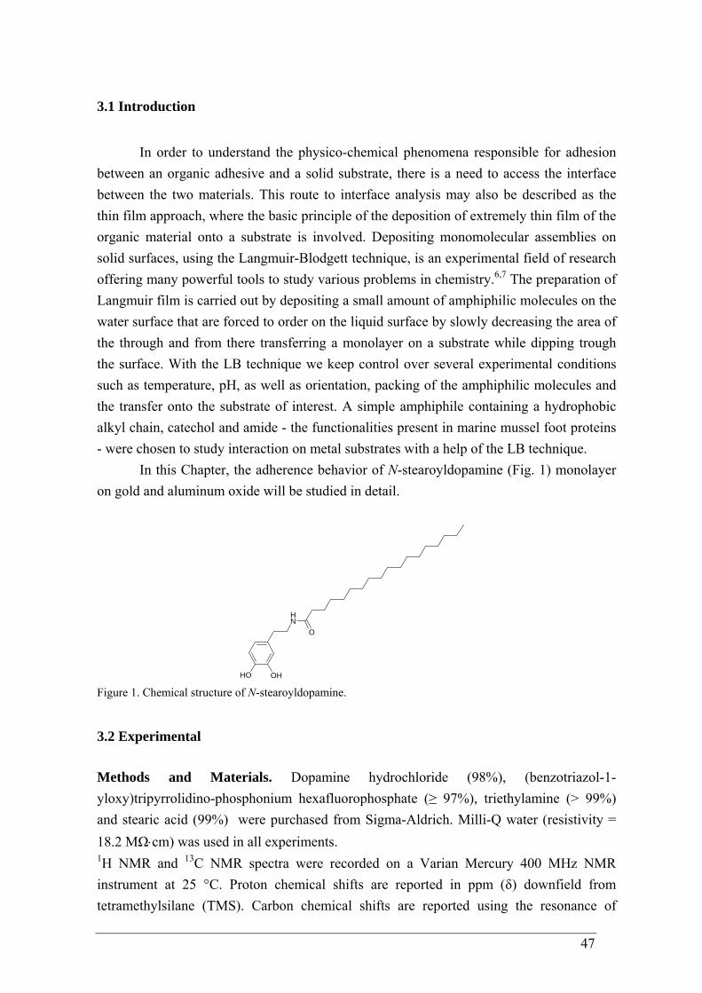
47
3.1 Introduction
In order to understand the physico-chemical phenomena responsible for adhesion
between an organic adhesive and a solid substrate, there is a need to access the interface between the two materials. This route to interface analysis may also be described as the thin film approach, where the basic principle of the deposition of extremely thin film of the organic material onto a substrate is involved. Depositing monomolecular assemblies on solid surfaces, using the Langmuir-Blodgett technique, is an experimental field of research offering many powerful tools to study various problems in chemistry.6,7 The preparation of Langmuir film is carried out by depositing a small amount of amphiphilic molecules on the water surface that are forced to order on the liquid surface by slowly decreasing the area of the through and from there transferring a monolayer on a substrate while dipping trough the surface. With the LB technique we keep control over several experimental conditions such as temperature, pH, as well as orientation, packing of the amphiphilic molecules and the transfer onto the substrate of interest. A simple amphiphile containing a hydrophobic alkyl chain, catechol and amide - the functionalities present in marine mussel foot proteins - were chosen to study interaction on metal substrates with a help of the LB technique.
In this Chapter, the adherence behavior of N-stearoyldopamine (Fig. 1) monolayer on gold and aluminum oxide will be studied in detail.
OH
NH
O
OH Figure 1. Chemical structure of N-stearoyldopamine.
3.2 Experimental Methods and Materials. Dopamine hydrochloride (98%), (benzotriazol-1-yloxy)tripyrrolidino-phosphonium hexafluorophosphate (≥ 97%), triethylamine (> 99%) and stearic acid (99%) were purchased from Sigma-Aldrich. Milli-Q water (resistivity = 18.2 MΩ⋅cm) was used in all experiments. 1H NMR and 13C NMR spectra were recorded on a Varian Mercury 400 MHz NMR instrument at 25 °C. Proton chemical shifts are reported in ppm (δ) downfield from tetramethylsilane (TMS). Carbon chemical shifts are reported using the resonance of
48
CDCl3 as internal standard. Splitting patterns are designated as singlet (s), doublet (d), double doublet (dd), triplet (t), quartet (q) and multiplet (m). Melting points were detected on a Büchi B-540 instrument. Infrared spectra were recorded on a Perkin–Elmer 1605 FT-IR spectrometer. Thermo-gravimetric analyses (TGA) were carried out using a TA Instruments Q500 Thermogravimetric Analyzer applying an oxygen stream and a heating rate of 10 K/min. The LC/MS spectra were recorded on HP/Agilent 1100 MSD spectrometer. Elemental analyses were performed with a Perkin Elmer, Series II 2400 elemental analyzer for CHNS-O. Synthesis of N-stearoyldopamine. N-stearoyldopamine was synthesized by a modified literature procedure.8 To a stirred mixture of dopamine hydrochloride (1.0 g, 5.3 mmol) in 25 mL of dry THF (benzotriazol-1-yloxy)tripyrrolidinophosphonium hexafluorophosphate (2.75 g, 5.4 mmol) and stearic acid (1.5 g, 5.3 mmol) was added. The mixture was then stirred at room temperature for 10 min and then cooled to 5 °C. A solution of EtN3 (2.23 mL, 16.0 mmol) in 5 mL of dry THF was added dropwise while stirring the mixture. The cooling-bath was then removed and the solution was stirred at room temperature for 12 h. After that the solution was evaporated and 20 mL of brine and 150 mL of diethyl ether were added to the residue. The water layer was extracted with Et2O (2 x 20 mL) and the extracts were combined, washed with a Na2CO3 solution, dried (Mg2SO4) and rotary evaporated to dryness. The residue was chromatographed using EA-CHCl3 (99:l) mixture as eluents to give 1.94 g (88%) solid residue. Crystallization from CHCl3-MeOH gave 1.33 g (61%) of a white solid, m.p. < 96-99 °C, lit. m.p. 97-98.5 °C from chloroform-methanol; 1H NMR (400 MHz, CDCl3): δ/ppm = 7.65 (s, 1H, OH), 6.80 (d, J=7.5 Hz, 1H-4’), 6.75 (d, J=1.6 Hz, 1H-5’), 6.56 (dd, J1=7.5 Hz, J2=1.6 Hz, 1H-3’), 5.90 (s, 1H, OH), 5.60 (t, J=2.5 Hz, 1H-NH), 3.48 (q, J=6.8 Hz, 2H-1’), 2.70 (t, J=7.2 Hz, 2H-2’), 2.15 (t, J=8.0 Hz, 2H-2), 1.54-1.60 (m, 2H-3), 1.20-1.32 (m, 28H-4-17), 0.88 (t, J=6.8 Hz, 3H-18); 13C NMR (400 MHz, DMSO): δ/ppm = 172.3, 145.5, 143.9, 130.7, 119.6, 116.4, 115.8, 115.7, 35.8, 35.2, 31.7, 29.5, 29.2, 25.7, 22.5, 14.4. IR: 3308, 2955, 2850, 1638, 1546, 1456, 1375, 1260, 1058, 805, 720 cm-1. TGA (O2, 5% weight loss): 246 °C. LCMS; [M+H]+ m/z 839.1 (100), 420 (82), 840 (43), 421 (21), 137 (20), 861 (19). C26H45NO3 (419.65): Calcd.: C 74.42%, H 10.81%, N 3.34%; found: C 74.39%, H 10.82%, N 3.27%. Substrates and Substrate Preparation. Mica substrates 80 x 30 mm2 (G250-1, Agar Scientific Limited, United Kingdom) were cleaved by removing a mica sheet before use. C-plane sapphire polished (< 1 nm surface roughness) wafers with (0001) surfaces and dimensions of 10 x 10 mm x 2 mm (Valley Design Corp., USA) was pretreated before film formation, i.e. sonicated (ultrasonic bath, Branson 1510) in acetone, chloroform, methanol and milli-Q water for 10 min. One side polished silicon wafers 10 x 10 mm2 (resistivity of 1–5 ohm⋅cm, 500–550 μm thick, emerging plane’s indices are 1 0 0) (Silicon Quest International, Inc., USA)) were coated with an approximately 20 nm thick layer of TiO2 (99.99% pure) (settings: 120 W RF power for 600 s at 7.10-3 mbar) and an approximately 20 nm thick layer of Au (99.99% pure) (settings: 60 W RF power for 240 s at 7.10-3 mbar)
49
by physical vapor deposition using a Moorfield minilab magnetron sputtering device. These gold substrates were used immediately after sputtering. Mica substrates covered with epitaxial gold Au(111) thin film were purchased from PHASIS, Switzerland and used as recieved. An aqueous 0.1 mM AuCl4
- solution was prepared by dissolving HAuCl4·3H2O (Sigma Aldrich) into milli-Q water. A germanium disk (99.999% pure, single crystalline) of 14 mm diameter and 2 mm thickness (Korth Kristalle GmbH, Kiel) was polished with Microcloth using 1 μm MetaDi II diamond polishing compound (both from Buehler Ltd, Lake Bluff, IL) and was degreased by immersion into 4 successive baths using subsequently: chloroform, acetone, methanol and deionized water as solvents. An additional soak in the above solvents was carried out with sonication for 5 min. Following the reported method of Brejna et al.,9 gold was deposited via galvanic displacement. The germanium substrates were finally soaked in scintillation vials containing the aqueous AuCl4
- solution for 10 min at room temperature. The germanium with the deposited gold substrate was then removed from the AuCl4
- solution and washed with copious amounts of milli-Q water. The surface was subsequently blown dry with a stream of N2. Klarite® substrates that consist of photolithographically patterned silicon wafers coated with epitaxial gold were purchased from Renishaw Diagnostics, United Kingdom and used as recieved. The modified substrates were dried at room temperature under a N2 atmosphere before the XPS, SERS, IR, AFM and CA measurements. Surface modification, Langmuir–Blodgett (LB) film deposition. All the isotherm measurements and Langmuir–Blodgett transfers were done in an integrated dust-free box at 23 °C and ambient humidity. Film interfacial compression and solid transfer was performed in a commercial double barrier Langmuir–Blodgett (LB) trough (KSV Instrument Ltd., Finland) with an initial film area of 496.5 × 150 mm. The trough was constructed from Teflon and the barriers from Delrin (polyacetal). The trough and barriers were rinsed with copious amounts of ethanol and milli-Q water prior to use. The surface pressure was measured by a Pt Wilhelmy plate with a sensitivity of 0.01 mN/m. The clean subphase was taken as the surface pressure zero reference. A monolayer was formed by spreading a solution of N-stearoyldopamine (1 mM) from CHCl3/CH3OH (19/1 v/v) on water. The monolayer was left undisturbed for 10 min to allow solvent evaporation prior to compression. Computer controlled symmetrically movable barriers were used to regulate the surface area. Film compression proceeded at a barrier speed of 3 mm/min and surface pressures were recorded during the compression process. A monolayer transfer was performed by an upstroke of the immersed substrate through the monolayer at the air-water interface. A deposition speed of 0.1 mm/min was used. Characterization. X-ray Reflectometry (XRR) measurements. X-ray measurements are performed on a Rigaku Geigerflex powder diffractometer as well on a Bruker D8 Discover diffractometer with GADDS, a two-dimensional (2D) detector system. The Rigaku powder diffractometer is a focusing beam Bragg-Bretano setup with a graphite monochromator on
50
the secondary site whereas the Bruker system is using parallel beam geometry equipped with a Göbel mirror on the primary site. The XRR patterns obtained with the Bruker system are measured with Cu radiation, wavelength 1.54184 Å, at 40 kV and 40 mA. A slit system, on the primary site as well as the secondary site, is used in combination with a scintillation counter. Furthermore, in order to obtain high (angular) resolution and sufficient scattered intensity on the XRR measurements, the slit system is used in combination with a knife edge collimator placed very close to the sample surface. Atomic Force Microscopy (AFM) imaging and analysis. Samples were imaged in air by a scanning probe microscope Ntegra Aura (NT-MDT), in tapping mode. The silicon cantilevers NSG03 (NT-MDT) with typical tip radius of about 10 nm and resonance frequency of 80 kHz were employed. All data analysis was performed with the Nova 1087 (NT-MDT). X-Ray Photoelectron Spectroscopy (XPS) measurements. Survey and high-resolution spectra were collected on a Kratos AXIS Ultra spectrometer, equipped with a monochromatic Al Kα X-ray source and a delay-line detector (DLD). Spectra were obtained using the aluminium anode (Al Kα = 1486.6 eV) operating at 150 W and a takeoff angle of 0°, defined as the angle between the photoelectron detector and the surface normal. A pass energy of 160 eV for wide scan and 40 eV for region scan was used. The background pressure was 2 x 10⁻⁹ mbar. All XPS spectra were referenced to the aliphatic hydrocarbon component of the C1s signal at 284.5 eV. Fourier Transformation-Infrared (FT-IR) Spectroscopy. IR spectra were recorded using Varian 3100 FT-IR spectrometer with DTGS detector. The spectra of adsorbates were recorded with a resolution of 4 cm-1 and 50 scans. Infrared-Reflection Absorption Spectroscopy (IRRAS). A Varian 670 FTIR spectrometer, equipped with a liquid nitrogen cooled mercury cadmium telluride (MCT) detector, purged with nitrogen was used. The spectra of adsorbates were recorded with a resolution of 4 cm-1 and co-adding 4000 scans. A Harrick Seagull accessory was used; the reflection angle of incidence for gold substrate was 85o and 80o for sapphire. P-polarized radiation was obtained using a rotable wire grid polarizer. Raman and Surface Enhanced Raman Spectroscopy (SERS) measurements. The Raman and SER spectra were obtained with a Dilor Labram spectrometer, equipped with a Millennia II doubled HeNe laser having a excitation wavelength of 632.8 nm and operated at a power 10 mW, 600 grooves/mm holographic grating and a 1024 x 256 pixel CCD camera. Spectra were recorded in the region 400–4000 cm-1. Depth profiles, i.e. spectral intensity as a function of the distance of the sampled spot to the surface, were taken by manually adjusting the distance between sample and objective. The recording time for a single spectrum is around 60 seconds. A 100x magnification, (Olympus, numerical aperture 0.8) ultra-long working distance objective was used. The Labram was equipped with a built-in camera, enabling the visualization of the reflected laser light from the inner
51
side of the glass plate. The focal depth zero was determined by minimizing the spot size of this reflected light. Contact Angle (CA) Goniometry. Static contact angle measurements were performed on a video-based semi-automatic contact angle meter (DataPhysics OCA 30 Instrument Gm, Germany) at 22 oC and 42 % relative humidity, using milli-Q water as a probe liquid. Six drops were measured on each sample. Density functional theory (DFT) calculations. Theoretical calculations were carried out using the ab initio program package Gaussian09.10 Optimized geometries of the molecules and the vibrational frequencies at their respective optimized geometries were computed by DFT calculations. The B3LYP (Becke three hybrid exchange,11 Lee-Yang-Parr correlation12), along with the Pople split valence diffused and polarized basis set 6-31++G(d,p) were utilized in the calculation for the free molecule. It is known that the 6-31++G(d,p) basis set in conjunction with the B3LYP functional is sufficient for the calculation of geometric parameters and harmonic frequencies of dopamine-like molecules which have a substituted phenyl ring. For the molecule-gold model complexes, geometry optimization using the basis set LanL2DZ13 was chosen, which makes use of the ECP pseudopotentials for gold atoms. The theoretically estimated vibration frequencies of free molecules obtained from the B3LYP/6-31++G(d,p) level of calculations were scaled by a factor of 0.975, whereas the gold-molecule surface complex models obtained from the B3LYP/Lanl2DZ level of calculations were scaled by a scaling factor of 0.96. Cartesian displacements and calculated (B3LYP/6-31++G(d,p)) vibrational modes of the molecule have been displayed using the GaussView 5.0 software.14 Molecular dynamics simulations were carried out with the Nosé-Hoover thermostat using the Verlet integrator with a time step of 1 fs and a temperature of 300 K. The electrostatic contributions were calculated with the Particle Mesh Ewald approach (PME) using a 1.2 Å grid. For the Lennard-Jones interactions a cut-off at 11 Å was used with a smooth switching-off starting at 10 Å. For each system, energy minimization was performed first. Then, an equilibration NVT run was performed for 1 ns. Finally, a NVT production run of 100 ps was carried out to generate the data used for the analysis. All the molecular dynamics have been performed with GROMACS. Visual Molecular Dynamics (VMD) was used for the visualization of the molecular configurations.15
3.3 Results and Discussion
3.3.1 Monolayer formation using the Langmuir-Blodgett technique
In order to investigate the contribution of the catechol functionality in adhesion on metal substrates, the Langmuir-Blodgett (LB) technique was considered. This technique provides an efficient approach to fabricate organized monomolecular films at air-water interface. Thus we get control over the order, orientation and spacing of the organic functional groups. Finally the monolayer is transferred to a substrate of interest by dipping.
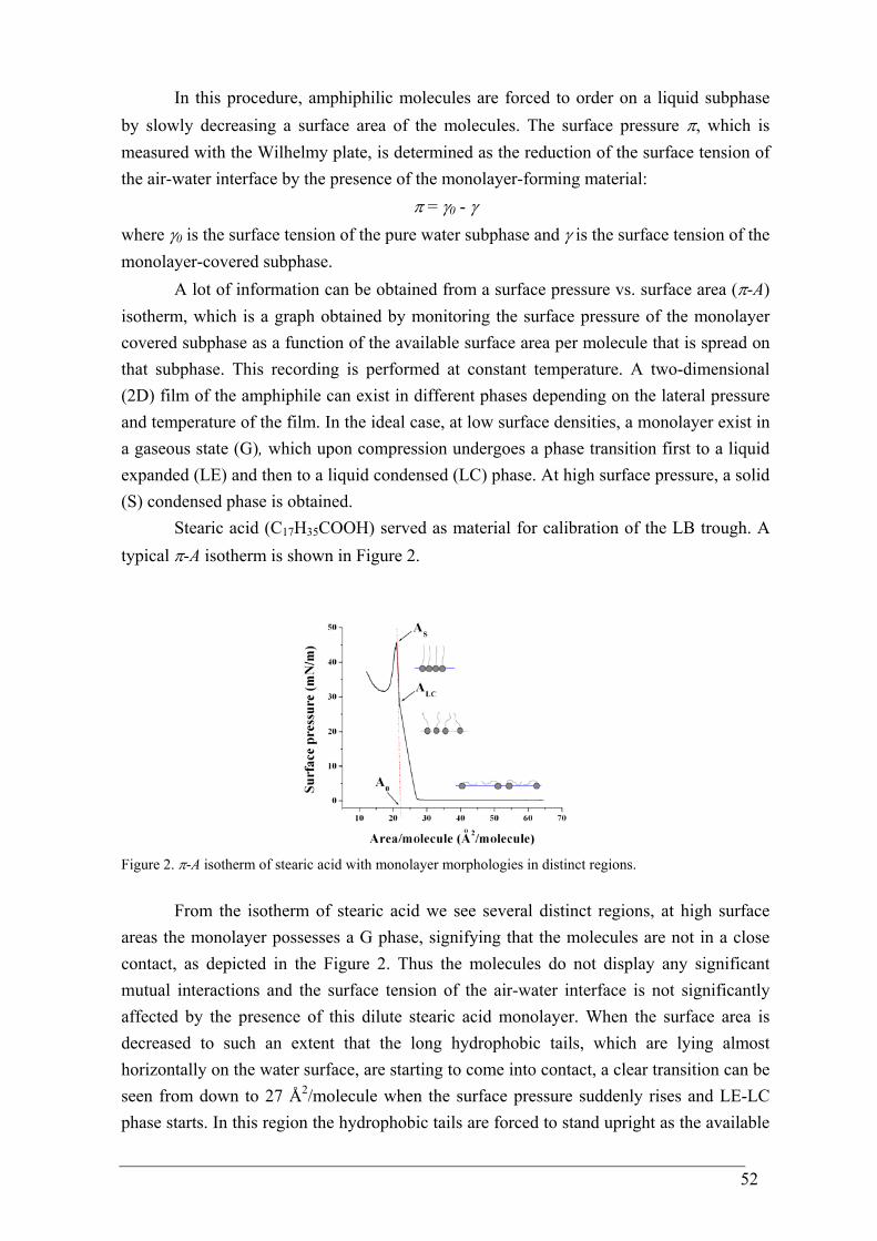
52
In this procedure, amphiphilic molecules are forced to order on a liquid subphase by slowly decreasing a surface area of the molecules. The surface pressure π, which is measured with the Wilhelmy plate, is determined as the reduction of the surface tension of the air-water interface by the presence of the monolayer-forming material:
π = γ0 - γ where γ0 is the surface tension of the pure water subphase and γ is the surface tension of the monolayer-covered subphase.
A lot of information can be obtained from a surface pressure vs. surface area (π-A) isotherm, which is a graph obtained by monitoring the surface pressure of the monolayer covered subphase as a function of the available surface area per molecule that is spread on that subphase. This recording is performed at constant temperature. A two-dimensional (2D) film of the amphiphile can exist in different phases depending on the lateral pressure and temperature of the film. In the ideal case, at low surface densities, a monolayer exist in a gaseous state (G), which upon compression undergoes a phase transition first to a liquid expanded (LE) and then to a liquid condensed (LC) phase. At high surface pressure, a solid (S) condensed phase is obtained.
Stearic acid (C17H35COOH) served as material for calibration of the LB trough. A typical π-A isotherm is shown in Figure 2.
Figure 2. π-A isotherm of stearic acid with monolayer morphologies in distinct regions.
From the isotherm of stearic acid we see several distinct regions, at high surface areas the monolayer possesses a G phase, signifying that the molecules are not in a close contact, as depicted in the Figure 2. Thus the molecules do not display any significant mutual interactions and the surface tension of the air-water interface is not significantly affected by the presence of this dilute stearic acid monolayer. When the surface area is decreased to such an extent that the long hydrophobic tails, which are lying almost horizontally on the water surface, are starting to come into contact, a clear transition can be seen from down to 27 Å2/molecule when the surface pressure suddenly rises and LE-LC phase starts. In this region the hydrophobic tails are forced to stand upright as the available
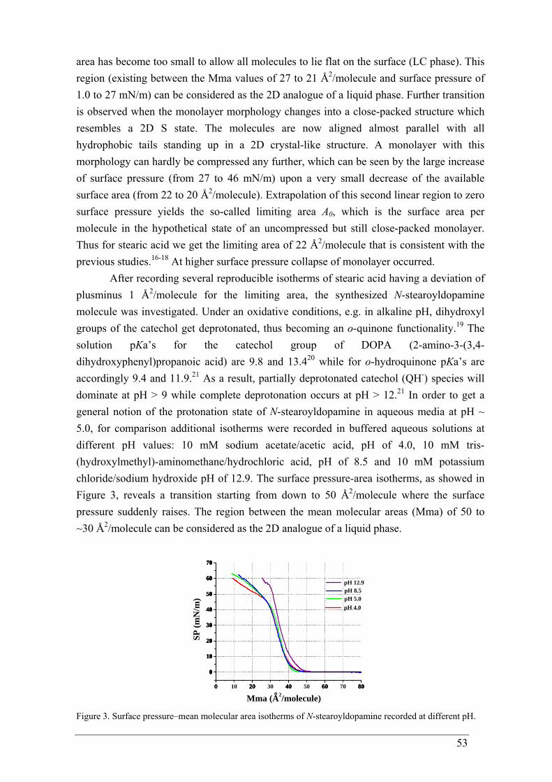
53
area has become too small to allow all molecules to lie flat on the surface (LC phase). This region (existing between the Mma values of 27 to 21 Å2/molecule and surface pressure of 1.0 to 27 mN/m) can be considered as the 2D analogue of a liquid phase. Further transition is observed when the monolayer morphology changes into a close-packed structure which resembles a 2D S state. The molecules are now aligned almost parallel with all hydrophobic tails standing up in a 2D crystal-like structure. A monolayer with this morphology can hardly be compressed any further, which can be seen by the large increase of surface pressure (from 27 to 46 mN/m) upon a very small decrease of the available surface area (from 22 to 20 Å2/molecule). Extrapolation of this second linear region to zero surface pressure yields the so-called limiting area A0, which is the surface area per molecule in the hypothetical state of an uncompressed but still close-packed monolayer. Thus for stearic acid we get the limiting area of 22 Å2/molecule that is consistent with the previous studies.16-18 At higher surface pressure collapse of monolayer occurred.
After recording several reproducible isotherms of stearic acid having a deviation of plusminus 1 Å2/molecule for the limiting area, the synthesized N-stearoyldopamine molecule was investigated. Under an oxidative conditions, e.g. in alkaline pH, dihydroxyl groups of the catechol get deprotonated, thus becoming an o-quinone functionality.19 The solution pKa’s for the catechol group of DOPA (2-amino-3-(3,4-dihydroxyphenyl)propanoic acid) are 9.8 and 13.420 while for o-hydroquinone pKa’s are accordingly 9.4 and 11.9.21 As a result, partially deprotonated catechol (QH-) species will dominate at pH > 9 while complete deprotonation occurs at pH > 12.21 In order to get a general notion of the protonation state of N-stearoyldopamine in aqueous media at pH ~ 5.0, for comparison additional isotherms were recorded in buffered aqueous solutions at different pH values: 10 mM sodium acetate/acetic acid, pH of 4.0, 10 mM tris-(hydroxylmethyl)-aminomethane/hydrochloric acid, pH of 8.5 and 10 mM potassium chloride/sodium hydroxide pH of 12.9. The surface pressure-area isotherms, as showed in Figure 3, reveals a transition starting from down to 50 Å2/molecule where the surface pressure suddenly raises. The region between the mean molecular areas (Mma) of 50 to ~30 Å2/molecule can be considered as the 2D analogue of a liquid phase.
0 20 40 60 80
0
10
20
30
40
50
60
70
0 10 20 30 40 50 60 70 80
0
10
20
30
40
50
60
70
0 20 40 60 80
0
10
20
30
40
50
60
70
0 20 40 60 80
0
10
20
30
40
50
60
70
oMma (A2/molecule)
pH 4.0
SP (m
N/m
) pH 5.0
pH 12.9 pH 8.5
Figure 3. Surface pressure–mean molecular area isotherms of N-stearoyldopamine recorded at different pH.
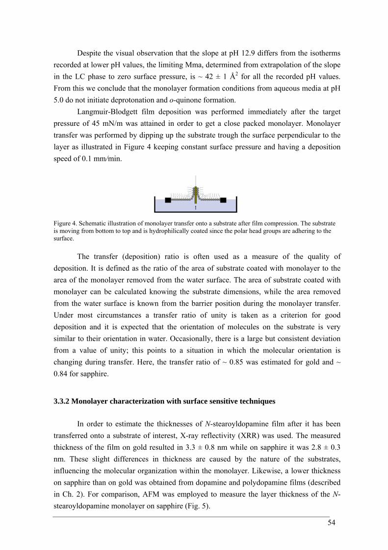
54
Despite the visual observation that the slope at pH 12.9 differs from the isotherms recorded at lower pH values, the limiting Mma, determined from extrapolation of the slope in the LC phase to zero surface pressure, is ~ 42 ± 1 Å2 for all the recorded pH values. From this we conclude that the monolayer formation conditions from aqueous media at pH 5.0 do not initiate deprotonation and o-quinone formation.
Langmuir-Blodgett film deposition was performed immediately after the target pressure of 45 mN/m was attained in order to get a close packed monolayer. Monolayer transfer was performed by dipping up the substrate trough the surface perpendicular to the layer as illustrated in Figure 4 keeping constant surface pressure and having a deposition speed of 0.1 mm/min.
Figure 4. Schematic illustration of monolayer transfer onto a substrate after film compression. The substrate is moving from bottom to top and is hydrophilically coated since the polar head groups are adhering to the surface.
The transfer (deposition) ratio is often used as a measure of the quality of deposition. It is defined as the ratio of the area of substrate coated with monolayer to the area of the monolayer removed from the water surface. The area of substrate coated with monolayer can be calculated knowing the substrate dimensions, while the area removed from the water surface is known from the barrier position during the monolayer transfer. Under most circumstances a transfer ratio of unity is taken as a criterion for good deposition and it is expected that the orientation of molecules on the substrate is very similar to their orientation in water. Occasionally, there is a large but consistent deviation from a value of unity; this points to a situation in which the molecular orientation is changing during transfer. Here, the transfer ratio of ~ 0.85 was estimated for gold and ~ 0.84 for sapphire.
3.3.2 Monolayer characterization with surface sensitive techniques
In order to estimate the thicknesses of N-stearoyldopamine film after it has been transferred onto a substrate of interest, X-ray reflectivity (XRR) was used. The measured thickness of the film on gold resulted in 3.3 ± 0.8 nm while on sapphire it was 2.8 ± 0.3 nm. These slight differences in thickness are caused by the nature of the substrates, influencing the molecular organization within the monolayer. Likewise, a lower thickness on sapphire than on gold was obtained from dopamine and polydopamine films (described in Ch. 2). For comparison, AFM was employed to measure the layer thickness of the N-stearoyldopamine monolayer on sapphire (Fig. 5).
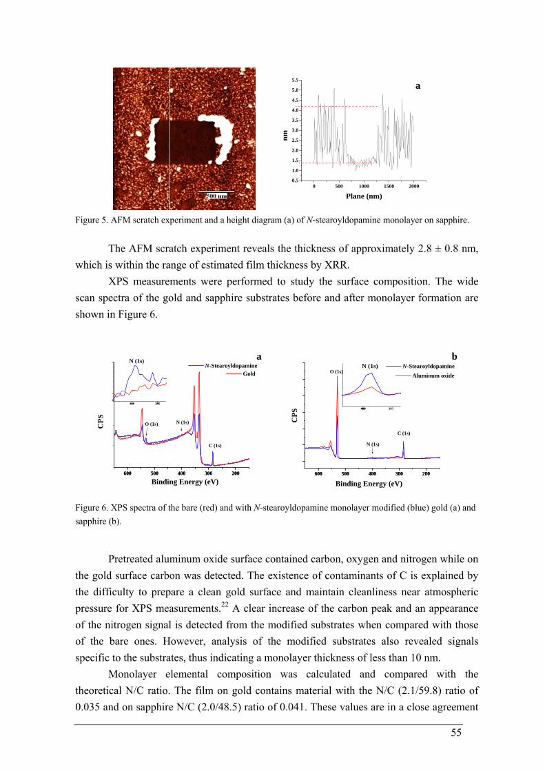
55
0 500 1000 1500 20000.5
1.0
1.5
2.0
2.5
3.0
3.5
4.0
4.5
5.0
5.5a
nm
Plane (nm)
Figure 5. AFM scratch experiment and a height diagram (a) of N-stearoyldopamine monolayer on sapphire.
The AFM scratch experiment reveals the thickness of approximately 2.8 ± 0.8 nm, which is within the range of estimated film thickness by XRR.
XPS measurements were performed to study the surface composition. The wide scan spectra of the gold and sapphire substrates before and after monolayer formation are shown in Figure 6.
600 500 400 300 200600 500 400 300 200
400 395400 395
N (1s)
CPS
Binding Energy (eV)
N-Stearoyldopaminea
C (1s)
N (1s)O (1s)
Gold
600 500 400 300 200600 500 400 300 200
CPS
Aluminum oxide
b
N (1s)
O (1s)
C (1s)
400 395400
N (1s)
Binding Energy (eV)
N-Stearoyldopamine
Figure 6. XPS spectra of the bare (red) and with N-stearoyldopamine monolayer modified (blue) gold (a) and sapphire (b).
Pretreated aluminum oxide surface contained carbon, oxygen and nitrogen while on the gold surface carbon was detected. The existence of contaminants of C is explained by the difficulty to prepare a clean gold surface and maintain cleanliness near atmospheric pressure for XPS measurements.22 A clear increase of the carbon peak and an appearance of the nitrogen signal is detected from the modified substrates when compared with those of the bare ones. However, analysis of the modified substrates also revealed signals specific to the substrates, thus indicating a monolayer thickness of less than 10 nm.
Monolayer elemental composition was calculated and compared with the theoretical N/C ratio. The film on gold contains material with the N/C (2.1/59.8) ratio of 0.035 and on sapphire N/C (2.0/48.5) ratio of 0.041. These values are in a close agreement
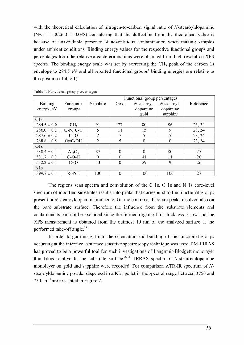
56
with the theoretical calculation of nitrogen-to-carbon signal ratio of N-stearoyldopamine (N/C = 1.0/26.0 = 0.038) considering that the deflection from the theoretical value is because of unavoidable presence of adventitious contamination when making samples under ambient conditions. Binding energy values for the respective functional groups and percentages from the relative area determinations were obtained from high resolution XPS spectra. The binding energy scale was set by correcting the CHx peak of the carbon 1s envelope to 284.5 eV and all reported functional groups’ binding energies are relative to this position (Table 1). Table 1. Functional group percentages. Functional group percentages
Binding energy, eV
Functional groups
Sapphire Gold N-stearoyl-dopamine
gold
N-stearoyl-dopamine sapphire
Reference
C1s 284.5 ± 0.0 CHx 91 77 80 86 23, 24 286.0 ± 0.2 C-N, C-O 5 11 15 9 23, 24 287.6 ± 0.2 C=O 2 7 5 5 23, 24 288.8 ± 0.5 O=C-OH 2 5 0 0 23, 24 O1s 530.4 ± 0.1 Al2O3 87 0 0 80 25 531.7 ± 0.2 C-O-H 0 0 41 11 26 532.2 ± 0.1 C=O 13 0 59 9 26 N1s 399.7 ± 0.1 R2-NH 100 0 100 100 27
The regions scan spectra and convolution of the C 1s, O 1s and N 1s core-level spectrum of modified substrates results into peaks that correspond to the functional groups present in N-stearoyldopamine molecule. On the contrary, there are peaks resolved also on the bare substrate surface. Therefore the influence from the substrate elements and contaminants can not be excluded since the formed organic film thickness is low and the XPS measurement is obtained from the outmost 10 nm of the analyzed surface at the performed take-off angle.28
In order to gain insight into the orientation and bonding of the functional groups occurring at the interface, a surface sensitive spectroscopy technique was used. PM-IRRAS has proved to be a powerful tool for such investigations of Langmuir-Blodgett monolayer thin films relative to the substrate surface.29,30 IRRAS spectra of N-stearoyldopamine monolayer on gold and sapphire were recorded. For comparison ATR-IR spectrum of N-stearoyldopamine powder dispersed in a KBr pellet in the spectral range between 3750 and 750 cm-1 are presented in Figure 7.

57
3500 3000 1500 10003500 3000 1500 1000
2964
1298
82187
391010
90
1257
137814
17
2852
2920
284829
18 a
b
Abs
orba
nce
(a. u
.)
Wavenumber, cm-1
1180
1464
1504
1551
161416
433283
3363
1111
3355
787
816
86295
3
1227
1260
1369
143314
71
1531
1549
1613
1637
3298
3471
3500 3000 1500 10003500 3000 1500 1000
2964
285029
2228
742934Abs
orba
nce
(a. u
.)
Wavenumber, cm-1
b
a
Gold substrate Sapphire substrate
Figure 7. ATR-IR (a) and IRRA spectrum (b) of N-stearoyldopamine on gold and sapphire.
The ATR-IR spectrum of N-stearoyldopamine shows relatively strong and broad bands in the 3000-3500 cm-1 region, resulting form the role of intermolecular hydrogen bonds existing in the molecules. The bands are assigned to the O-H and N-H stretching vibrations.31 The major band assignments are summarized in table 2. In contrary to IRRAS measurements on sapphire the broad peak region have not been detected relative to those in the bulk spectrum, reflecting the imposition of the surface selection rules. Table 2. ATR-IR and IRRA spectra, peak frequency (cm-1) and assignment.
ATR-IR N-
stearoyldopamine
IRRAS N-
stearoyldopamine on gold
IRRAS N-
stearoyldopamine on sapphire
Assignments and
references
3471 3355 3298 2918 2848 1637 1613 1549 1531 1471 1433 1369 1260 1227 1180 1111 868 816
- 3363 3283 2920 2852 1643 1614 1551 1504 1464 1417 1378 1257 1236 - 1090 873 821
- - - 2934 2870 - - - - - - - - - - - - -
O-H O-H str.31 N-H str. 31 CH2 asymm. str. CH2 symm. str. Amide I (νC=O) Arom. C=C str.32 Amide II (δN-H) Arom. C=C str.32 Arom. C=C str.32 CH2 asymm. def. 31
δO-H 32
νC-O 32
Amide III (νC-N) in-plane δC-H
32 in-plane δC-H
32 Ring “breathing” 31 Ring “breathing” 31
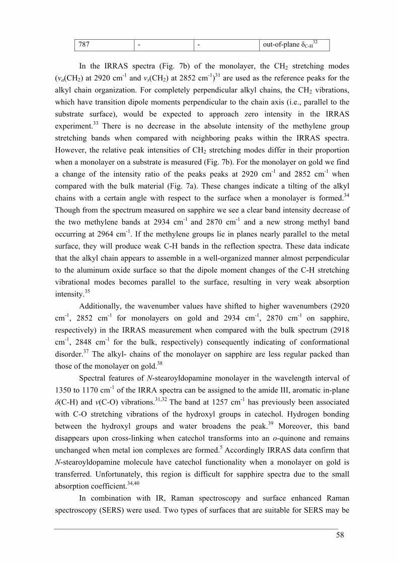
58
787 - - out-of-plane δC-H32
In the IRRAS spectra (Fig. 7b) of the monolayer, the CH2 stretching modes (νa(CH2) at 2920 cm-1 and νs(CH2) at 2852 cm-1)31 are used as the reference peaks for the alkyl chain organization. For completely perpendicular alkyl chains, the CH2 vibrations, which have transition dipole moments perpendicular to the chain axis (i.e., parallel to the substrate surface), would be expected to approach zero intensity in the IRRAS experiment.33 There is no decrease in the absolute intensity of the methylene group stretching bands when compared with neighboring peaks within the IRRAS spectra. However, the relative peak intensities of CH2 stretching modes differ in their proportion when a monolayer on a substrate is measured (Fig. 7b). For the monolayer on gold we find a change of the intensity ratio of the peaks peaks at 2920 cm-1 and 2852 cm-1 when compared with the bulk material (Fig. 7a). These changes indicate a tilting of the alkyl chains with a certain angle with respect to the surface when a monolayer is formed.34 Though from the spectrum measured on sapphire we see a clear band intensity decrease of the two methylene bands at 2934 cm-1 and 2870 cm-1 and a new strong methyl band occurring at 2964 cm-1. If the methylene groups lie in planes nearly parallel to the metal surface, they will produce weak C-H bands in the reflection spectra. These data indicate that the alkyl chain appears to assemble in a well-organized manner almost perpendicular to the aluminum oxide surface so that the dipole moment changes of the C-H stretching vibrational modes becomes parallel to the surface, resulting in very weak absorption intensity.35
Additionally, the wavenumber values have shifted to higher wavenumbers (2920 cm-1, 2852 cm-1 for monolayers on gold and 2934 cm-1, 2870 cm-1 on sapphire, respectively) in the IRRAS measurement when compared with the bulk spectrum (2918 cm-1, 2848 cm-1 for the bulk, respectively) consequently indicating of conformational disorder.37 The alkyl- chains of the monolayer on sapphire are less regular packed than those of the monolayer on gold.38
Spectral features of N-stearoyldopamine monolayer in the wavelength interval of 1350 to 1170 cm-1 of the IRRA spectra can be assigned to the amide III, aromatic in-plane δ(C-H) and ν(C-O) vibrations.31,32 The band at 1257 cm-1 has previously been associated with C-O stretching vibrations of the hydroxyl groups in catechol. Hydrogen bonding between the hydroxyl groups and water broadens the peak.39 Moreover, this band disappears upon cross-linking when catechol transforms into an o-quinone and remains unchanged when metal ion complexes are formed.5 Accordingly IRRAS data confirm that N-stearoyldopamine molecule have catechol functionality when a monolayer on gold is transferred. Unfortunately, this region is difficult for sapphire spectra due to the small absorption coefficient.34,40
In combination with IR, Raman spectroscopy and surface enhanced Raman spectroscopy (SERS) were used. Two types of surfaces that are suitable for SERS may be
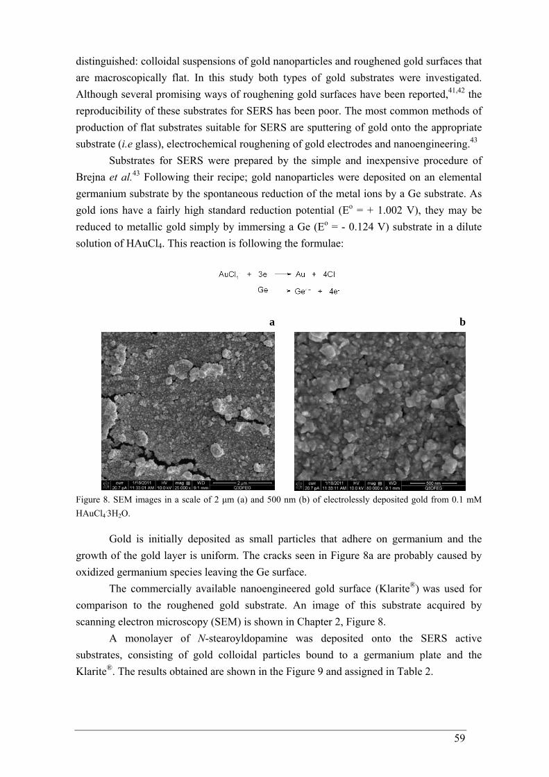
59
distinguished: colloidal suspensions of gold nanoparticles and roughened gold surfaces that are macroscopically flat. In this study both types of gold substrates were investigated. Although several promising ways of roughening gold surfaces have been reported,41,42 the reproducibility of these substrates for SERS has been poor. The most common methods of production of flat substrates suitable for SERS are sputtering of gold onto the appropriate substrate (i.e glass), electrochemical roughening of gold electrodes and nanoengineering.43
Substrates for SERS were prepared by the simple and inexpensive procedure of Brejna et al.43 Following their recipe; gold nanoparticles were deposited on an elemental germanium substrate by the spontaneous reduction of the metal ions by a Ge substrate. As gold ions have a fairly high standard reduction potential (Eo = + 1.002 V), they may be reduced to metallic gold simply by immersing a Ge (Eo = - 0.124 V) substrate in a dilute solution of HAuCl4. This reaction is following the formulae:
a b
Figure 8. SEM images in a scale of 2 μm (a) and 500 nm (b) of electrolessly deposited gold from 0.1 mM HAuCl4
.3H2O.
Gold is initially deposited as small particles that adhere on germanium and the growth of the gold layer is uniform. The cracks seen in Figure 8a are probably caused by oxidized germanium species leaving the Ge surface.
The commercially available nanoengineered gold surface (Klarite®) was used for comparison to the roughened gold substrate. An image of this substrate acquired by scanning electron microscopy (SEM) is shown in Chapter 2, Figure 8.
A monolayer of N-stearoyldopamine was deposited onto the SERS active substrates, consisting of gold colloidal particles bound to a germanium plate and the Klarite®. The results obtained are shown in the Figure 9 and assigned in Table 2.
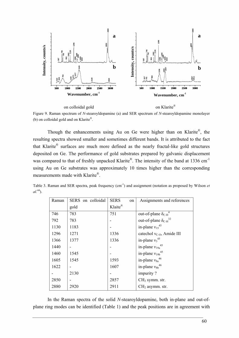
60
500 1000 1500 2000 2500 3000500 1000 1500 2000 2500 300021
30
2920
1545
1377
1271
118378
3
604
523
Inte
nsity
, cou
nts/
s
b
2880
2850
1622160514
601440
1366
1296
1130
1062
953
79274
6
587
Wavenumber, cm-1
a
500 1000 1500 2000 2500 3000500 1000 1500 2000 2500 3000
1593
2857617
751 98
2
1607
1336
2911In
tens
ity, c
ount
s/s
Wavenumber, cm-1
b
162214
60
2850
2880
587
746
792
953
1062 11
30
1296
1366
1440
1605
a
on colloidal gold on Klarite® Figure 9. Raman spectrum of N-stearoyldopamine (a) and SER spectrum of N-stearoyldopamine monolayer (b) on colloidal gold and on Klarite®.
Though the enhancements using Au on Ge were higher than on Klarite®, the resulting spectra showed smaller and sometimes different bands. It is attributed to the fact that Klarite® surfaces are much more defined as the nearly fractal-like gold structures deposited on Ge. The performance of gold substrates prepared by galvanic displacement was compared to that of freshly unpacked Klarite®. The intensity of the band at 1336 cm-1 using Au on Ge substrates was approximately 10 times higher than the corresponding measurements made with Klarite®. Table 3. Raman and SER spectra, peak frequency (cm-1) and assignment (notation as proposed by Wilson et al.44).
Raman SERS on colloidal gold
SERS on Klaite®
Assignments and references
746 792 1130 1296 1366 1440 1460 1605 1622 - 2850 2880
783 783 1183 1271 1377 - 1545 1545 - 2130 - 2920
751 - - 1336 1336 - - 1593 1607 - 2857 2911
out-of-plane δC-H4
out-of-plane δC-H32
in-plane ν1545
catechol νC-O, Amide III in-plane ν3
45 in-plane ν19a
45 in-plane ν19b
45 in-plane ν8a
46 in-plane ν8b
46 impurity ? CH2 symm. str. CH2 asymm. str.
In the Raman spectra of the solid N-stearoyldopamine, both in-plane and out-of-plane ring modes can be identified (Table 1) and the peak positions are in agreement with

61
those as calculated by DFT and the previously reported ones.4,45-47 The strong band at 1296 cm-1 is assigned to the amide III band (mainly C-N stretching coupled with N-H bending)48 and a catechol C-O stretch49 that is an important indicator of catechol coordination to the surface.4,46 Furthermore the ν8a in-plane ring stretching mode is responsive to coordination of the catecholic oxygen atoms with the surface.4,47
In the SER spectra the intensities of the in-plane ring modes are significantly enhanced relative to those of the out-of-plane ring modes. The downshift of the ν8a in-plane ring stretching mode of the SER spectra as compared to the conventional Raman spectra reveals that the catecholic oxygen atoms are coordinated to the gold surface.4 According to the surface selection rule of Moskovits50 and Creighton51, in a SERS experiment the vibrations having larger components of polarizability in the direction normal to the surface will be enhanced. Relative intensity comparisons between the Raman and SER spectra suggest a more perpendicular than parallel orientation of the o-diphenol derivative when it is coordinated to the gold.4
In principle, catechol rings may coordinate to noble metal surfaces via the hydroxyl groups or/and the benzene ring. For instance, a study of Gao et al. reports flat attachment of benzene to a gold surface via π electrons.52 The flat orientation of the benzene ring is proven by the strong enhancement of the out-of-plane vibration. The authors also find a flat coordination of the halobenzenes, whereas for nitrobenzene and benzonitrile they find a "hanging" orientation. Benzene and benzene-d6 also take a flat orientation on silver, as Moscowitz et al. have shown.53 The same orientation has been described for the hydroxybenzoic acids on silver by Fleger et al.,54 and for the o-aminobenzoic by Suh and Moscowitz.55 On the contrary, phenolmercaptide on Ag is reported to be perpendicular oriented.56 p-Thiocresol and benzylamine lie flat on copper surfaces,57 whereas the n-Docosyl homologue of hemicyanine (4-[4-(dimethylamino)styryl]-1-docosylpyridinium bromide (DCHC) and 2-(docosylamino)-5-nitropyridine (DCANP), applied as Langmuir-Blodgett films, show a perpendicularly orientation of the benzene ring.58 However, another report shows that catechol is standing up on silver.59
To get the right impression of the vibrational situation, the geometries have been optimized and the vibrational spectra of benzene, catechol, dopamine and N-stearoyldopamine using density functional theory (DFT) were calculated in collaboration with dr. Günter Hoffmann (TU/e). The situation is visualized in Figure 10 of the examples of the calculated (DFT, B3LYP/6-311++G**) vibrations of 4-methylcatechol.
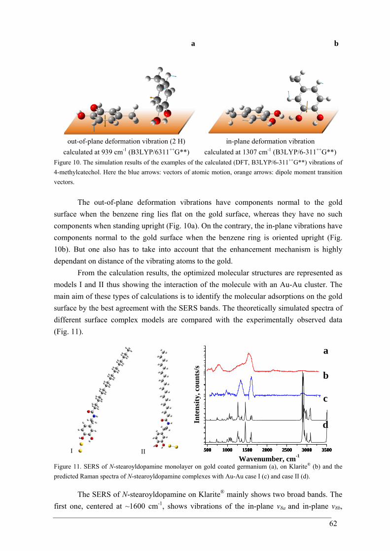
62
a b
out-of-plane deformation vibration (2 H) calculated at 939 cm-1 (B3LYP/6311++G**)
in-plane deformation vibration calculated at 1307 cm-1 (B3LYP/6-311++G**)
Figure 10. The simulation results of the examples of the calculated (DFT, B3LYP/6-311++G**) vibrations of 4-methylcatechol. Here the blue arrows: vectors of atomic motion, orange arrows: dipole moment transition vectors.
The out-of-plane deformation vibrations have components normal to the gold surface when the benzene ring lies flat on the gold surface, whereas they have no such components when standing upright (Fig. 10a). On the contrary, the in-plane vibrations have components normal to the gold surface when the benzene ring is oriented upright (Fig. 10b). But one also has to take into account that the enhancement mechanism is highly dependant on distance of the vibrating atoms to the gold.
From the calculation results, the optimized molecular structures are represented as models I and II thus showing the interaction of the molecule with an Au-Au cluster. The main aim of these types of calculations is to identify the molecular adsorptions on the gold surface by the best agreement with the SERS bands. The theoretically simulated spectra of different surface complex models are compared with the experimentally observed data (Fig. 11).
I II 500 1000 1500 2000 2500 3000 3500500 1000 1500 2000 2500 3000 3500500 1000 1500 2000 2500 3000 3500500 1000 1500 2000 2500 3000 3500
Wavenumber, cm-1
b
Inte
nsity
, cou
nts/
s
a
c
d
Figure 11. SERS of N-stearoyldopamine monolayer on gold coated germanium (a), on Klarite® (b) and the predicted Raman spectra of N-stearoyldopamine complexes with Au-Au case I (c) and case II (d).
The SERS of N-stearoyldopamine on Klarite® mainly shows two broad bands. The
first one, centered at ~1600 cm-1, shows vibrations of the in-plane ν8a and in-plane ν8b,
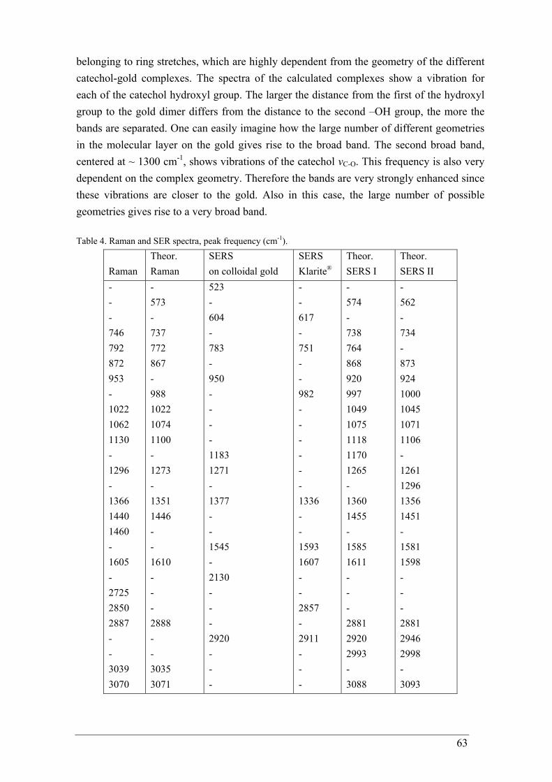
63
belonging to ring stretches, which are highly dependent from the geometry of the different catechol-gold complexes. The spectra of the calculated complexes show a vibration for each of the catechol hydroxyl group. The larger the distance from the first of the hydroxyl group to the gold dimer differs from the distance to the second –OH group, the more the bands are separated. One can easily imagine how the large number of different geometries in the molecular layer on the gold gives rise to the broad band. The second broad band, centered at ~ 1300 cm-1, shows vibrations of the catechol νC-O. This frequency is also very dependent on the complex geometry. Therefore the bands are very strongly enhanced since these vibrations are closer to the gold. Also in this case, the large number of possible geometries gives rise to a very broad band. Table 4. Raman and SER spectra, peak frequency (cm-1).
Raman
Theor. Raman
SERS on colloidal gold
SERS Klarite®
Theor. SERS I
Theor. SERS II
- - - 746 792 872 953 - 1022 1062 1130 - 1296 - 1366 1440 1460 - 1605 - 2725 2850 2887 - - 3039 3070
- 573 - 737 772 867 - 988 1022 1074 1100 - 1273 - 1351 1446 - - 1610 - - - 2888 - - 3035 3071
523 - 604 - 783 - 950 - - - - 1183 1271 - 1377 - - 1545 - 2130 - - - 2920 - - -
- - 617 - 751 - - 982 - - - - - - 1336 - - 1593 1607 - - 2857 - 2911 - - -
- 574 - 738 764 868 920 997 1049 1075 1118 1170 1265 - 1360 1455 - 1585 1611 - - - 2881 2920 2993 - 3088
- 562 - 734 - 873 924 1000 1045 1071 1106 - 1261 1296 1356 1451 - 1581 1598 - - - 2881 2946 2998 - 3093
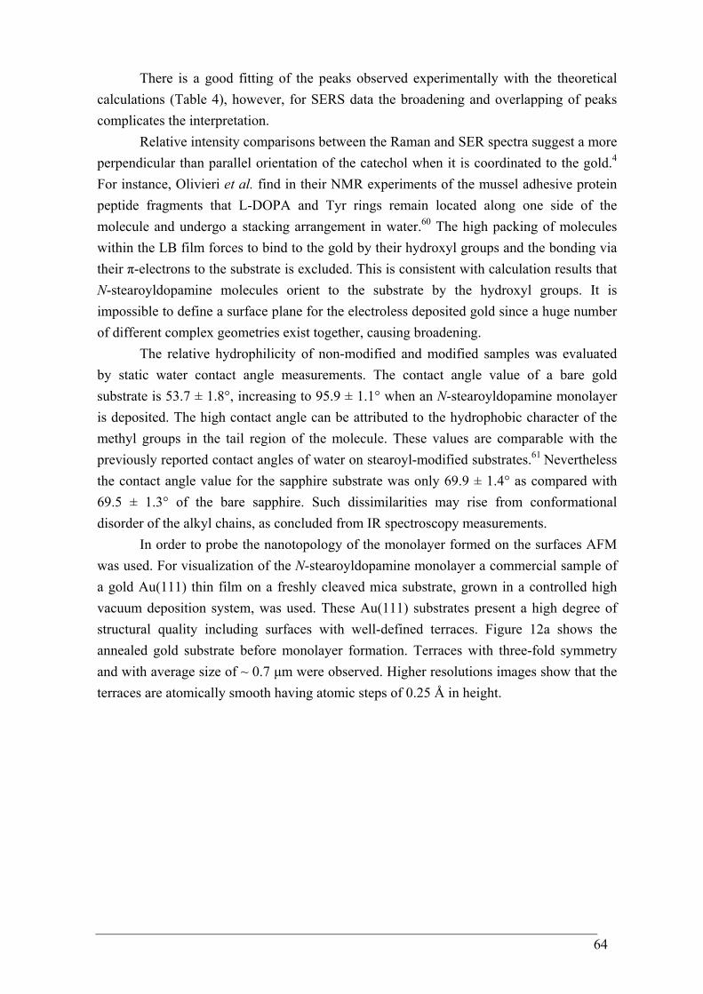
64
There is a good fitting of the peaks observed experimentally with the theoretical calculations (Table 4), however, for SERS data the broadening and overlapping of peaks complicates the interpretation.
Relative intensity comparisons between the Raman and SER spectra suggest a more perpendicular than parallel orientation of the catechol when it is coordinated to the gold.4 For instance, Olivieri et al. find in their NMR experiments of the mussel adhesive protein peptide fragments that L-DOPA and Tyr rings remain located along one side of the molecule and undergo a stacking arrangement in water.60 The high packing of molecules within the LB film forces to bind to the gold by their hydroxyl groups and the bonding via their π-electrons to the substrate is excluded. This is consistent with calculation results that N-stearoyldopamine molecules orient to the substrate by the hydroxyl groups. It is impossible to define a surface plane for the electroless deposited gold since a huge number of different complex geometries exist together, causing broadening.
The relative hydrophilicity of non-modified and modified samples was evaluated by static water contact angle measurements. The contact angle value of a bare gold substrate is 53.7 ± 1.8°, increasing to 95.9 ± 1.1° when an N-stearoyldopamine monolayer is deposited. The high contact angle can be attributed to the hydrophobic character of the methyl groups in the tail region of the molecule. These values are comparable with the previously reported contact angles of water on stearoyl-modified substrates.61 Nevertheless the contact angle value for the sapphire substrate was only 69.9 ± 1.4° as compared with 69.5 ± 1.3° of the bare sapphire. Such dissimilarities may rise from conformational disorder of the alkyl chains, as concluded from IR spectroscopy measurements.
In order to probe the nanotopology of the monolayer formed on the surfaces AFM was used. For visualization of the N-stearoyldopamine monolayer a commercial sample of a gold Au(111) thin film on a freshly cleaved mica substrate, grown in a controlled high vacuum deposition system, was used. These Au(111) substrates present a high degree of structural quality including surfaces with well-defined terraces. Figure 12a shows the annealed gold substrate before monolayer formation. Terraces with three-fold symmetry and with average size of ~ 0.7 μm were observed. Higher resolutions images show that the terraces are atomically smooth having atomic steps of 0.25 Å in height.
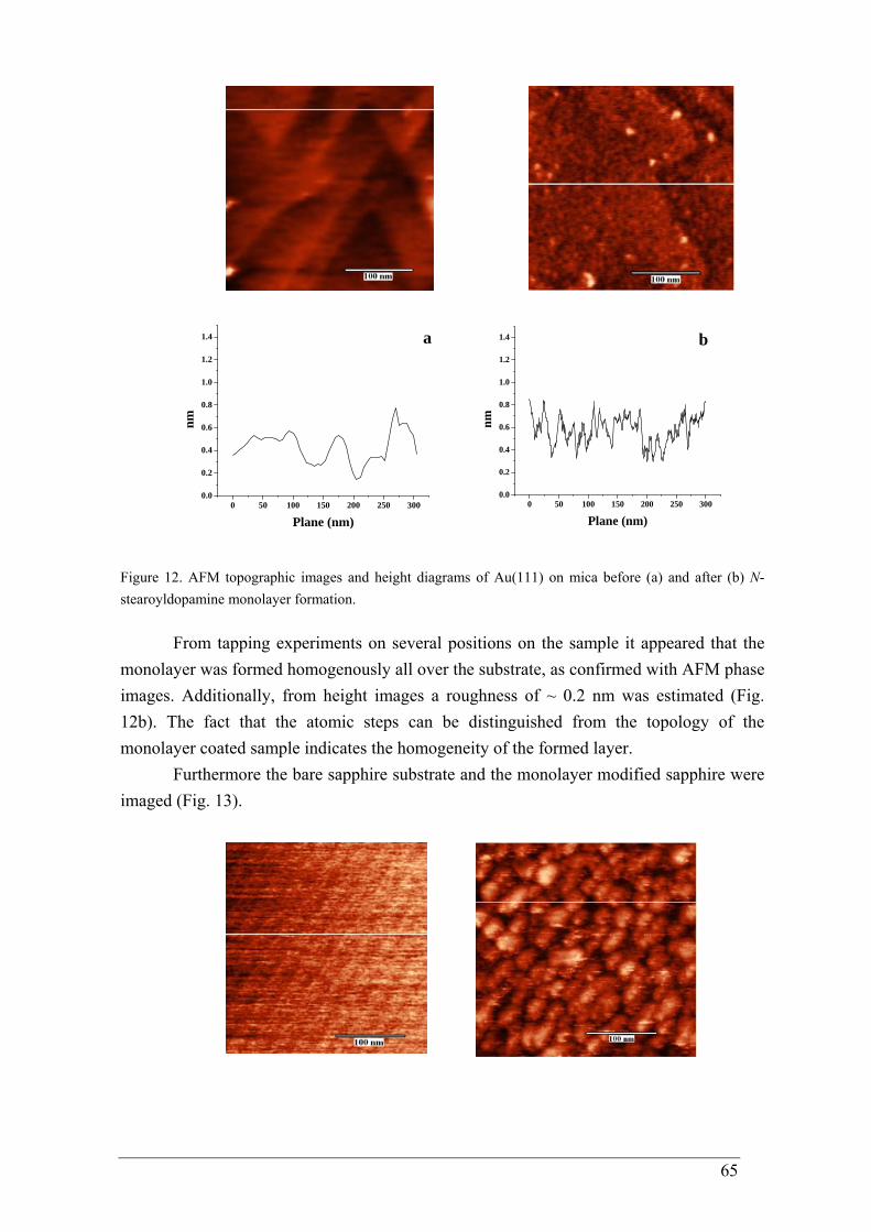
65
0 50 100 150 200 250 3000.0
0.2
0.4
0.6
0.8
1.0
1.2
1.4
nm
Plane (nm)
a
0 50 100 150 200 250 3000.0
0.2
0.4
0.6
0.8
1.0
1.2
1.4 b
nm
Plane (nm)
Figure 12. AFM topographic images and height diagrams of Au(111) on mica before (a) and after (b) N-stearoyldopamine monolayer formation.
From tapping experiments on several positions on the sample it appeared that the
monolayer was formed homogenously all over the substrate, as confirmed with AFM phase images. Additionally, from height images a roughness of ~ 0.2 nm was estimated (Fig. 12b). The fact that the atomic steps can be distinguished from the topology of the monolayer coated sample indicates the homogeneity of the formed layer.
Furthermore the bare sapphire substrate and the monolayer modified sapphire were imaged (Fig. 13).
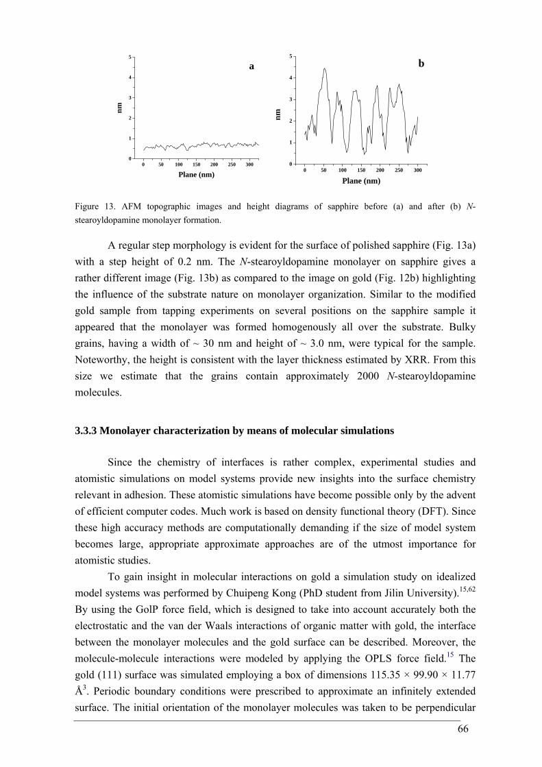
66
0 50 100 150 200 250 3000
1
2
3
4
5
nm
Plane (nm)
a
0 50 100 150 200 250 300
0
1
2
3
4
5
nm
Plane (nm)
b
Figure 13. AFM topographic images and height diagrams of sapphire before (a) and after (b) N-stearoyldopamine monolayer formation.
A regular step morphology is evident for the surface of polished sapphire (Fig. 13a) with a step height of 0.2 nm. The N-stearoyldopamine monolayer on sapphire gives a rather different image (Fig. 13b) as compared to the image on gold (Fig. 12b) highlighting the influence of the substrate nature on monolayer organization. Similar to the modified gold sample from tapping experiments on several positions on the sapphire sample it appeared that the monolayer was formed homogenously all over the substrate. Bulky grains, having a width of ~ 30 nm and height of ~ 3.0 nm, were typical for the sample. Noteworthy, the height is consistent with the layer thickness estimated by XRR. From this size we estimate that the grains contain approximately 2000 N-stearoyldopamine molecules.
3.3.3 Monolayer characterization by means of molecular simulations
Since the chemistry of interfaces is rather complex, experimental studies and atomistic simulations on model systems provide new insights into the surface chemistry relevant in adhesion. These atomistic simulations have become possible only by the advent of efficient computer codes. Much work is based on density functional theory (DFT). Since these high accuracy methods are computationally demanding if the size of model system becomes large, appropriate approximate approaches are of the utmost importance for atomistic studies.
To gain insight in molecular interactions on gold a simulation study on idealized model systems was performed by Chuipeng Kong (PhD student from Jilin University).15,62 By using the GolP force field, which is designed to take into account accurately both the electrostatic and the van der Waals interactions of organic matter with gold, the interface between the monolayer molecules and the gold surface can be described. Moreover, the molecule-molecule interactions were modeled by applying the OPLS force field.15 The gold (111) surface was simulated employing a box of dimensions 115.35 × 99.90 × 11.77 Å3. Periodic boundary conditions were prescribed to approximate an infinitely extended surface. The initial orientation of the monolayer molecules was taken to be perpendicular
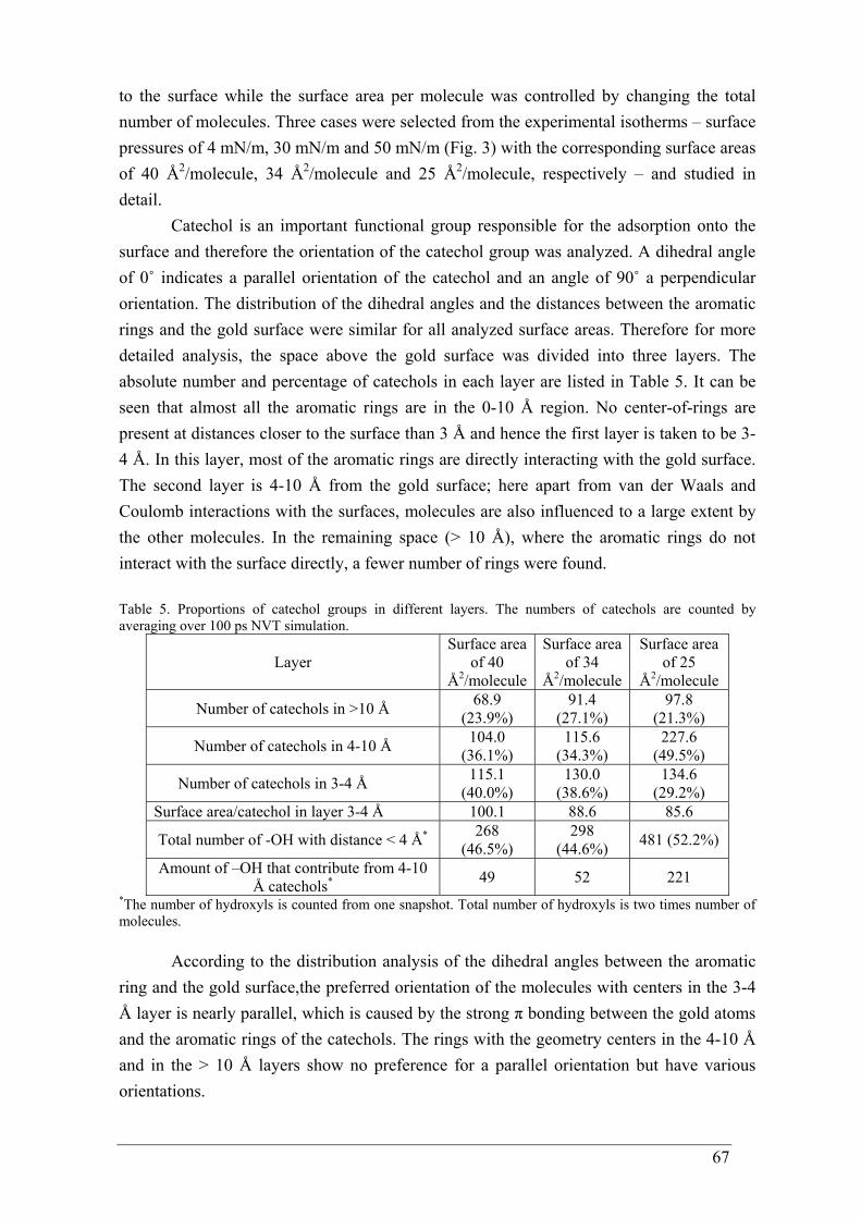
67
to the surface while the surface area per molecule was controlled by changing the total number of molecules. Three cases were selected from the experimental isotherms – surface pressures of 4 mN/m, 30 mN/m and 50 mN/m (Fig. 3) with the corresponding surface areas of 40 Å2/molecule, 34 Å2/molecule and 25 Å2/molecule, respectively – and studied in detail.
Catechol is an important functional group responsible for the adsorption onto the surface and therefore the orientation of the catechol group was analyzed. A dihedral angle of 0˚ indicates a parallel orientation of the catechol and an angle of 90˚ a perpendicular orientation. The distribution of the dihedral angles and the distances between the aromatic rings and the gold surface were similar for all analyzed surface areas. Therefore for more detailed analysis, the space above the gold surface was divided into three layers. The absolute number and percentage of catechols in each layer are listed in Table 5. It can be seen that almost all the aromatic rings are in the 0-10 Å region. No center-of-rings are present at distances closer to the surface than 3 Å and hence the first layer is taken to be 3-4 Å. In this layer, most of the aromatic rings are directly interacting with the gold surface. The second layer is 4-10 Å from the gold surface; here apart from van der Waals and Coulomb interactions with the surfaces, molecules are also influenced to a large extent by the other molecules. In the remaining space (> 10 Å), where the aromatic rings do not interact with the surface directly, a fewer number of rings were found. Table 5. Proportions of catechol groups in different layers. The numbers of catechols are counted by averaging over 100 ps NVT simulation.
Layer Surface area
of 40 Å2/molecule
Surface area of 34
Å2/molecule
Surface area of 25
Å2/molecule
Number of catechols in >10 Å 68.9 (23.9%)
91.4 (27.1%)
97.8 (21.3%)
Number of catechols in 4-10 Å 104.0 (36.1%)
115.6 (34.3%)
227.6 (49.5%)
Number of catechols in 3-4 Å 115.1 (40.0%)
130.0 (38.6%)
134.6 (29.2%)
Surface area/catechol in layer 3-4 Å 100.1 88.6 85.6
Total number of -OH with distance < 4 Å* 268 (46.5%)
298 (44.6%) 481 (52.2%)
Amount of –OH that contribute from 4-10 Å catechols* 49 52 221
*The number of hydroxyls is counted from one snapshot. Total number of hydroxyls is two times number of molecules.
According to the distribution analysis of the dihedral angles between the aromatic ring and the gold surface,the preferred orientation of the molecules with centers in the 3-4 Å layer is nearly parallel, which is caused by the strong π bonding between the gold atoms and the aromatic rings of the catechols. The rings with the geometry centers in the 4-10 Å and in the > 10 Å layers show no preference for a parallel orientation but have various orientations.
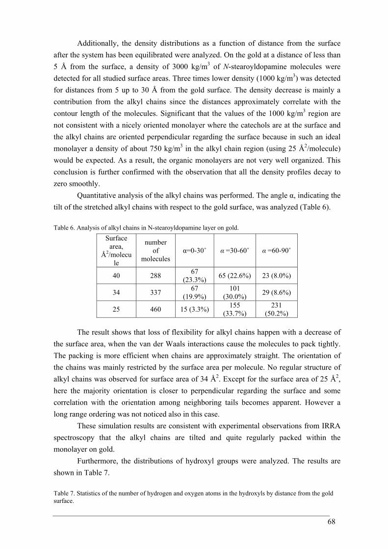
68
Additionally, the density distributions as a function of distance from the surface after the system has been equilibrated were analyzed. On the gold at a distance of less than 5 Å from the surface, a density of 3000 kg/m3 of N-stearoyldopamine molecules were detected for all studied surface areas. Three times lower density (1000 kg/m3) was detected for distances from 5 up to 30 Å from the gold surface. The density decrease is mainly a contribution from the alkyl chains since the distances approximately correlate with the contour length of the molecules. Significant that the values of the 1000 kg/m3 region are not consistent with a nicely oriented monolayer where the catechols are at the surface and the alkyl chains are oriented perpendicular regarding the surface because in such an ideal monolayer a density of about 750 kg/m3 in the alkyl chain region (using 25 Å2/molecule) would be expected. As a result, the organic monolayers are not very well organized. This conclusion is further confirmed with the observation that all the density profiles decay to zero smoothly.
Quantitative analysis of the alkyl chains was performed. The angle α, indicating the tilt of the stretched alkyl chains with respect to the gold surface, was analyzed (Table 6). Table 6. Analysis of alkyl chains in N-stearoyldopamine layer on gold.
Surface area,
Å2/molecule
number of
molecules α=0-30˚ α =30-60˚ α =60-90˚
40 288 67 (23.3%) 65 (22.6%) 23 (8.0%)
34 337 67 (19.9%)
101 (30.0%) 29 (8.6%)
25 460 15 (3.3%) 155 (33.7%)
231 (50.2%)
The result shows that loss of flexibility for alkyl chains happen with a decrease of the surface area, when the van der Waals interactions cause the molecules to pack tightly. The packing is more efficient when chains are approximately straight. The orientation of the chains was mainly restricted by the surface area per molecule. No regular structure of alkyl chains was observed for surface area of 34 Å2. Except for the surface area of 25 Å2, here the majority orientation is closer to perpendicular regarding the surface and some correlation with the orientation among neighboring tails becomes apparent. However a long range ordering was not noticed also in this case.
These simulation results are consistent with experimental observations from IRRA spectroscopy that the alkyl chains are tilted and quite regularly packed within the monolayer on gold.
Furthermore, the distributions of hydroxyl groups were analyzed. The results are shown in Table 7. Table 7. Statistics of the number of hydrogen and oxygen atoms in the hydroxyls by distance from the gold surface.
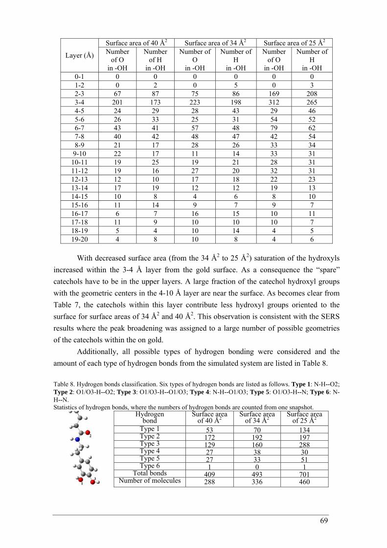
69
Surface area of 40 Å2 Surface area of 34 Å2 Surface area of 25 Å2
Layer (Å) Number of O
in -OH
Number of H
in -OH
Number of O
in -OH
Number of H
in -OH
Number of O
in -OH
Number of H
in -OH 0-1 0 0 0 0 0 0 1-2 0 2 0 5 0 3 2-3 67 87 75 86 169 208 3-4 201 173 223 198 312 265 4-5 24 29 28 43 29 46 5-6 26 33 25 31 54 52 6-7 43 41 57 48 79 62 7-8 40 42 48 47 42 54 8-9 21 17 28 26 33 34
9-10 22 17 11 14 33 31 10-11 19 25 19 21 28 31 11-12 19 16 27 20 32 31 12-13 12 10 17 18 22 23 13-14 17 19 12 12 19 13 14-15 10 8 4 6 8 10 15-16 11 14 9 7 9 7 16-17 6 7 16 15 10 11 17-18 11 9 10 10 10 7 18-19 5 4 10 14 4 5 19-20 4 8 10 8 4 6
With decreased surface area (from the 34 Å2 to 25 Å2) saturation of the hydroxyls increased within the 3-4 Å layer from the gold surface. As a consequence the “spare” catechols have to be in the upper layers. A large fraction of the catechol hydroxyl groups with the geometric centers in the 4-10 Å layer are near the surface. As becomes clear from Table 7, the catechols within this layer contribute less hydroxyl groups oriented to the surface for surface areas of 34 Å2 and 40 Å2. This observation is consistent with the SERS results where the peak broadening was assigned to a large number of possible geometries of the catechols within the on gold.
Additionally, all possible types of hydrogen bonding were considered and the amount of each type of hydrogen bonds from the simulated system are listed in Table 8. Table 8. Hydrogen bonds classification. Six types of hydrogen bonds are listed as follows. Type 1: N-H--O2; Type 2: O1/O3-H--O2; Type 3: O1/O3-H--O1/O3; Type 4: N-H--O1/O3; Type 5: O1/O3-H--N; Type 6: N-H--N. Statistics of hydrogen bonds, where the numbers of hydrogen bonds are counted from one snapshot.
Hydrogen bond
Surface area of 40 Å2
Surface area of 34 Å2
Surface area of 25 Å2
Type 1 53 70 134 Type 2 172 192 197 Type 3 129 160 288 Type 4 27 38 30 Type 5 27 33 51 Type 6 1 0 1
Total bonds 409 493 701 Number of molecules 288 336 460
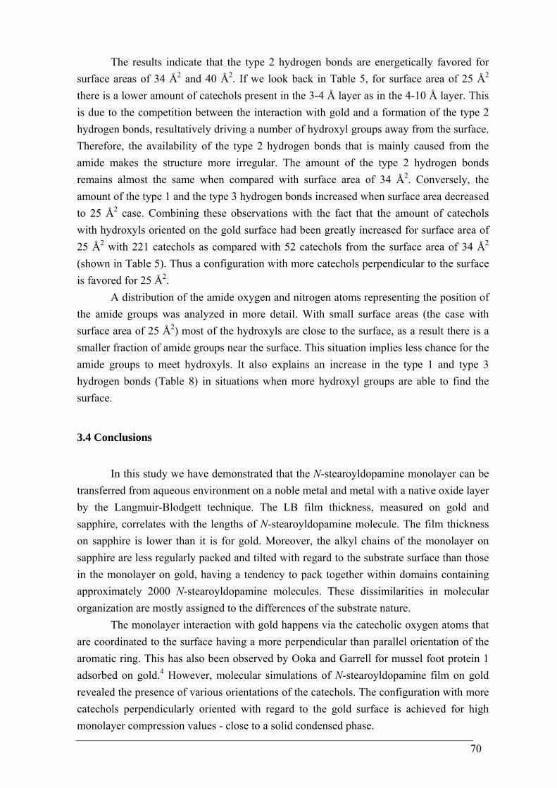
70
The results indicate that the type 2 hydrogen bonds are energetically favored for surface areas of 34 Å2 and 40 Å2. If we look back in Table 5, for surface area of 25 Å2
there is a lower amount of catechols present in the 3-4 Å layer as in the 4-10 Å layer. This is due to the competition between the interaction with gold and a formation of the type 2 hydrogen bonds, resultatively driving a number of hydroxyl groups away from the surface. Therefore, the availability of the type 2 hydrogen bonds that is mainly caused from the amide makes the structure more irregular. The amount of the type 2 hydrogen bonds remains almost the same when compared with surface area of 34 Å2. Conversely, the amount of the type 1 and the type 3 hydrogen bonds increased when surface area decreased to 25 Å2 case. Combining these observations with the fact that the amount of catechols with hydroxyls oriented on the gold surface had been greatly increased for surface area of 25 Å2 with 221 catechols as compared with 52 catechols from the surface area of 34 Å2
(shown in Table 5). Thus a configuration with more catechols perpendicular to the surface is favored for 25 Å2.
A distribution of the amide oxygen and nitrogen atoms representing the position of the amide groups was analyzed in more detail. With small surface areas (the case with surface area of 25 Å2) most of the hydroxyls are close to the surface, as a result there is a smaller fraction of amide groups near the surface. This situation implies less chance for the amide groups to meet hydroxyls. It also explains an increase in the type 1 and type 3 hydrogen bonds (Table 8) in situations when more hydroxyl groups are able to find the surface.
3.4 Conclusions
In this study we have demonstrated that the N-stearoyldopamine monolayer can be
transferred from aqueous environment on a noble metal and metal with a native oxide layer by the Langmuir-Blodgett technique. The LB film thickness, measured on gold and sapphire, correlates with the lengths of N-stearoyldopamine molecule. The film thickness on sapphire is lower than it is for gold. Moreover, the alkyl chains of the monolayer on sapphire are less regularly packed and tilted with regard to the substrate surface than those in the monolayer on gold, having a tendency to pack together within domains containing approximately 2000 N-stearoyldopamine molecules. These dissimilarities in molecular organization are mostly assigned to the differences of the substrate nature.
The monolayer interaction with gold happens via the catecholic oxygen atoms that are coordinated to the surface having a more perpendicular than parallel orientation of the aromatic ring. This has also been observed by Ooka and Garrell for mussel foot protein 1 adsorbed on gold.4 However, molecular simulations of N-stearoyldopamine film on gold revealed the presence of various orientations of the catechols. The configuration with more catechols perpendicularly oriented with regard to the gold surface is achieved for high monolayer compression values - close to a solid condensed phase.
71
Moreover, a large amount of hydrogen bonding, caused by amide group, was calculated. With decreasing surface areas more of the hydroxyl groups are able to orient to the gold surface.
3.5 References
1 Lin, Q.; Gourdon, D.; Sun, C.; Holten-Andersen, N.; Anderson, T. H.; Waite, J. H.;
Israelachvili, J. N. PNAS 2007, 104, 3782. 2 Lee, H.; Scherer, N. F.; Messersmith, P. B. Proc. Natl. Acad. Sci. USA 2006, 29,
12999. 3 Waite, J. H. Int. J. Biol. Macromol. 1990, 12, 139. 4 Ooka, A. A.; Garrell, R. L. Biopolymers 2000, 57, 92. 5 Fant, C.; Hedlund, J.; Höök, F.; Berglin, M.; Fridell, E.; Elwing, H. The Journal of
Adhesion 2010, 86, 25. 6 Zasadzinski, J. A.; Viswanthan, R.; Madsen, L.; Garnaes, J.; Schwartz, D. K.
Science 1994, 263, 1726. 7 Dubois, L. H.; Nuzzo, R. G. Annu. Rev. Phys. Chem. 1992, 43, 437. 8 Czamocki, Z.; Matuszewska, M. P.; Matuszewska, I. Organic preparations and
procedures int. 1998, 30, 699. 9 Porter, Jr. L. A.; Choi, H. C.; Ribbe, A. E.; Buriak, J. M. Nano Letters 2002, 2,
1067. 10 Gaussian 09, Revision A.02; Frisch, M. J.; Trucks, G. W.; Schlegel, H. B.;
Scuseria, G. E.; Robb, M. A.; Cheeseman, J. R.; Scalmani, G.; Barone, V.; Mennucci, B.; Petersson, G. A.; Nakatsuji, H.; Caricato, M.; Li, X.; Hratchian, H. P.; Izmaylov, A. F.; Bloino, J.; Zheng, G.; Sonnenberg, J. L.; Hada, M.; Ehara, M.; Toyota, K.; Fukuda, R.; Hasegawa, J.; Ishida, M.; Nakajima, T.; Honda, Y.; Kitao, O.; Nakai, H.; Vreven, T.; Montgomery, Jr. J. A.; Peralta, J. E.; Ogliaro, F.; Bearpark, M.; Heyd, J. J.; Brothers, E.; Kudin, K. N.; Staroverov, V. N.; Kobayashi, R.; Normand, J.; Raghavachari, K.; Rendell, A.; Burant, J. C.; Iyengar, S. S.; Tomasi, J.; Cossi, M.; Rega, N.; Millam, J. M.; Klene, M.; Knox, J. E.; Cross, J. B.; Bakken, V.; Adamo, C.; Jaramillo, J.; Gomperts, R.; Stratmann, R. E.; Yazyev, O.; Austin, A. J.; Cammi, R.; Pomelli, C.; Ochterski, J. W.; Martin, R. L.; Morokuma, K.; Zakrzewski, V. G.; Voth, G. A.; Salvador, P.; Dannenberg, J. J.; Dapprich, S.; Daniels, A. D.; Farkas, O.; Foresman, J. B.; Ortiz, J. V.; Cioslowski, J.; Fox, D. J. Gaussian, Inc., Wallingford CT 2009.
11 Becke, A. D. J. Chem. Phys. 1993, 98, 5648. 12 Lee, C.; Yang, W.; Parr, R. G. Phys. Rev. B 1988, 37, 785. 13 Frisch, M. J.; Pople, J. A.; Binkley, J. S. J. Chem. Phys. 1984, 80, 3265. 14 Rībena, D.; Hoffmann, G. G.; With, G. To be published. 15 Kong, C-P.; Peters, E. A. J. F.; Zhang, H-X.; With, G. To be published.
72
16 Hann, R. A.; Kathirgamanathan, P. Phil. Trans. R. Soc. Lond. A 1990, 330, 141. 17 El-hefian, E. A.; Misran, M.; Yahaya, A. H. Maejo Int. J. Sci. Technol. 2009, 3,
277. 18 Hwang, M.-J.; Kim, K. Langmuir 1999, 15, 3563. 19 Yu, M.; Hwang, J.; Deming, T. J. J. Am. Chem. Soc. 1999, 121, 5825. 20 Pettit, L. Pure Appl. Chem. 1984, 56, 247. 21 Brooksby, P. A.; Schiel, D. R.; Abell, A. D. Langmuir 2008, 24, 9074. 22 Ishida, T.; Tsuneda, S.; Nishida, N.; Hara, M.; Sasabe, H.; Knoll, W. Langmuir
1997, 13, 4638. 23 Desimoni, E.; Casella, G. I.; Morone, A.; Salvi, A. M. Surface and Interface
Analysis 1990, 15, 627434. 24 Bernsmann, F.; Ponche, A.; Ringwald, C.; Hemmerlé, J.; Raya, J.; Bechinger, B.;
Voegel, J.-C.; Schaaf, P.; Ball, V. J. Phys. Chem. C 2009, 113, 8234. 25 Brand, J.; Snijders, P. C.; Sloof, W. G.; Terryn, H.; Wit, J. H. W. J. Phys. Chem. B
2004, 108, 6017. 26 Li, X.-G.; Huang, K.-L.; Liu, S.-Q.; Tan, N.; Chen, L.-Q. Trans. Nonferrous Met.
Soc. China 2007, 17, 195. 27 Clark, M. B.; Gardella, J. A.; Schultz, T. M.; Patil, D. G.; Salvati, L. Anal. Chem.
1990, 62, 949. 28 Geng, S.; Zhang, S.; Onishi, H. Materials Technology 2002, 17, 234. 29 Allara, D. L.; Swalen, J. D. J. Phys. Chem. 1982, 86, 2700. 30 Naselli, C.; Rabolt, J. F.; Swalen, J. D. J. Phys. Chem. 1985, 82, 2136. 31 Lin-Vien, D.; Colthup, N. B.; Fateley, W. G.; Grasselli, J. G. The Handbook of
Infrared and Raman Characteristic Frequencies of Organic Molecules 1991, Academic Press: San Diego, CA.
32 Bredenberg, B-son. J; Sarbak, Z. J. Chem. Tech. Biotechnol. 1988, 42, 221. 33 Allara, D. L.; Nuzzo, R. G. Langmuir 1985, 1, 45. 34 Brunner, H.; Mayer, U.; Hoffmann, H. Applied Spectroscopy 1997, 51, 209. 35 Yang, B. D.; Wakamatsu, T. Surf. And Interf. Analysis 1996, 24, 803. 36 Tillman, N.; Ulman, A.; Schildkraut, J. S.; Penner, T. L. J. Am. Chem. Soc. 1988,
110, 6136. 37 Nuzzo, R. G.; Fusco, F. A.; Allara, D. L. J. Am. Chem. Soc. 1987, 109, 2358. 38 Ting II, G. G.; Acton, O.; Ma, H.; Ka, J. W.; Jen, A. K.-Y. Langmuir 2009, 25,
2140. 39 Barth, A. Prog. in Biophys. and Molecular Biology 2000, 74, 141. 40 Kosters, P. FT-IR spectroscopy of Thin Biological Layers, PhD thesis 2004,
University of Twente. 41 Brejna, P. R.; Griffiths, P. R.; Yang, J. Applied Spectroscopy 2009, 63, 396. 42 Sackmann, M.; Materny, A. J. Raman Spectrosc. 2006, 37, 305. 43 Brejna, P. R.; Griffiths, P. R.; Yang, J. Applied Spectroscopy 2009, 63, 396.
73
44 Wilson, E. B.; Decius, J. C.; Cross, P. C. Molecular Vibrations 1955, McGraw-Hill, New York.
45 Salama, S.; Stong J. D.; Neilands, J. B.; Spiro, T. G. Biochemistry 1978, 17, 3781. 46 Lee, N.-S.; Hsieh, Y.-Z.; Paisley, R. F.; Morris, M. D. Anal Chem 1988, 60, 442. 47 Kowalchyk, W. K.; Davis, K. L.; Morris, M. D. Spectrochim Acta 1995, 51A, 145. 48 Thomas, Jr. G. J.; Prescott, B. Biopolymers 1987, 26, 921. 49 Gaber, B. P.; Miskowski, V.; Spiro, T. G. J Am Chem Soc 1974, 96, 6868. 50 Moskovits, M. J. Chem. Phys. 1982, 77, 4408. 51 Creighton, J. A. Surf. Sci. 1983, 124, 209. 52 Gao, P.; Weaver, M.J. J. Phys. Chem. 1985, 89, 5040. 53 Moskovits, M.; DiLella, D. P. J. Chem. Phys. 1980, 73, 6068. 54 Fleger, Y.; Mastai, Y.; Rosenbluh, M.; Dressler, D. H. J. Raman Spectr. 2009, 40,
1572. 55 Suh, J. S.; Moskovits, M. J. Am. Chem. Soc. 1986, 108, 4711. 56 Xue, G.; Ma, M.; Zhang, J.; Lu, Y. J. of Colloid and lnterface Science 1992, 150, 1. 57 Xue, G.; Zhang, J. Applied Spectroscopy 1991, 45, 760. 58 Muniz-Miranda, M.; Puggelli, M.; Ricceri, R.; Gabrielli, G. Langmuir 1996,12,
4417. 59 Koh, T. Y.; Greaves, S. J.; Griffith, W. P. Spectrochimica Acta A 1994, 50, 857. 60 Olivieri, M. P.; Wollman, R. M.; Alderfer, J. L. J. Peptide Res. 1997, 50, 436442. 61 Ou, J.; Wang, J.; Liu, S.; Zhou, J.; Yang, S. J. Phys. Chem. C 2009, 113, 20429. 62 Kong, C-P.; Rībena, D.; Peters, E. A. J. F.; Zhang, H-X.; With, G. To be published.
74
CHAPTER 4
4-stearylcatechol monolayer on gold and sapphire The investigation in catechol interaction with a metal surface was continued with an other model molecule where the amide functionality is excluded. Monolayers of 4-stearylcatechol have been transferred from the air - water interface onto gold and aluminum oxide substrates using the Langmuir-Blodgett technique. The organization of the monolayers on a molecular scale was examined by several surface sensitive techniques and compared with the data for N-stearoyldopamine monolayers. Our study shows that, similar to N-stearoyldopamine monolayers, the catechols function as a surface anchor on gold and the alkyl-chains appear to be tilted within a monolayer. Although the alkyl- chains are tilted within the monolayer for both molecules, the irregularity of the 4-stearylcatechol film on gold leads to domain formation that is caused by the absence of the amide functionality. On the contrary, from the molecular simulations it appeared that for both types of molecules parallel orientations of the catechols with the gold are also present. However, hydrogen bonds formed between the amide functionality and the catechol hydroxyl groups have a profound influence on the structure and regularity on the adsorbed layer.
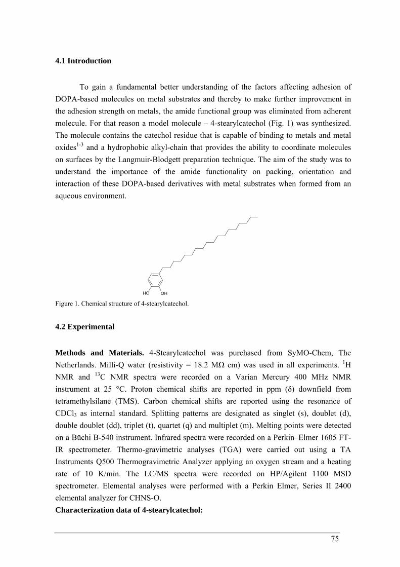
75
4.1 Introduction
To gain a fundamental better understanding of the factors affecting adhesion of
DOPA-based molecules on metal substrates and thereby to make further improvement in the adhesion strength on metals, the amide functional group was eliminated from adherent molecule. For that reason a model molecule – 4-stearylcatechol (Fig. 1) was synthesized. The molecule contains the catechol residue that is capable of binding to metals and metal oxides1-3 and a hydrophobic alkyl-chain that provides the ability to coordinate molecules on surfaces by the Langmuir-Blodgett preparation technique. The aim of the study was to understand the importance of the amide functionality on packing, orientation and interaction of these DOPA-based derivatives with metal substrates when formed from an aqueous environment.
OH OH Figure 1. Chemical structure of 4-stearylcatechol.
4.2 Experimental
Methods and Materials. 4-Stearylcatechol was purchased from SyMO-Chem, The Netherlands. Milli-Q water (resistivity = 18.2 MΩ cm) was used in all experiments. 1H NMR and 13C NMR spectra were recorded on a Varian Mercury 400 MHz NMR instrument at 25 °C. Proton chemical shifts are reported in ppm (δ) downfield from tetramethylsilane (TMS). Carbon chemical shifts are reported using the resonance of CDCl3 as internal standard. Splitting patterns are designated as singlet (s), doublet (d), double doublet (dd), triplet (t), quartet (q) and multiplet (m). Melting points were detected on a Büchi B-540 instrument. Infrared spectra were recorded on a Perkin–Elmer 1605 FT-IR spectrometer. Thermo-gravimetric analyses (TGA) were carried out using a TA Instruments Q500 Thermogravimetric Analyzer applying an oxygen stream and a heating rate of 10 K/min. The LC/MS spectra were recorded on HP/Agilent 1100 MSD spectrometer. Elemental analyses were performed with a Perkin Elmer, Series II 2400 elemental analyzer for CHNS-O. Characterization data of 4-stearylcatechol:
76
Brown solid; m.p. < 89-93 °C, 1H NMR (400 MHz, CDCl3): δ/ppm = 7.21 (s, 1H, OH), 6.76 (d, J = 8.0 Hz, 1H-2’), 6.69 (d, J = 2.0 Hz, 1H-3’), 6.61 (dd, J1 = 8.0 Hz, J2 = 2.0 Hz, 1H-1’), 2.80 (s, 1H, OH), 2.48 (t, J = 7.0 Hz, 2H-1), 1.55 (t, J = 7.0 Hz, 2H-2), 1.18-1.34 (m, 30H-3, 4, 5, 6, 7, 8, 9, 10, 11, 12, 13, 14, 15, 16, 17), 0.88 (t, J = 7.0 Hz, 3H-18); 13C NMR (400 MHz, CDCl3): δ/ppm = 143.95, 141.86, 135.50, 120.09, 115.31, 114.96, 35.27, 31.90, 31.67, 29.67, 29.63, 29.62, 29.53, 29.34, 29.29, 22.66, 14.05. IR: 3482, 3354, 2918, 2849, 1601, 1522, 1472, 1441, 1356, 1282, 1273, 1265, 1260, 1184, 1117, 951, 866, 812, 787 cm-1. TGA (O2, 5% weight loss): 224 °C. C24H42O2 (362.60): Calcd.: C 79.50%, H 11.68%; found: C 79.49%, H 11.70%. Substrates and Substrate Preparation. Mica substrates 80 x 30 mm2 (G250-1, Agar Scientific Limited, United Kingdom) were cleaved by removing a mica sheet before use. C-plane sapphire polished (< 1 nm surface roughness) wafers with (0001) surfaces and dimensions of 10 x 10 mm x 2 mm (Valley Design Corp., USA) was pretreated before film formation, i.e. sonicated (ultrasonic bath, Branson 1510) in acetone, chloroform, methanol and milli-Q water for 10 min. One side polished silicon wafers 10 x 10 mm2 (resistivity of 1–5 ohm⋅cm, 500–550 μm thick, emerging plane’s indices are 1 0 0) (Silicon Quest International, Inc., USA)) were coated with an approximately 20 nm thick layer of TiO2 (99.99% pure) (settings: 120 W RF power for 600 s at 7.10-3 mbar) and an approximately 20 nm thick layer of Au (99.99% pure) (settings: 60 W RF power for 240 s at 7.10-3 mbar) by physical vapor deposition using a Moorfield minilab magnetron sputtering device. These gold substrates were used immediately after sputtering. Mica substrates covered with epitaxial gold Au(111) thin film were purchased from PHASIS, Switzerland and used as recieved. An aqueous 0.1 mM AuCl4
- solution was prepared by dissolving HAuCl4·3H2O (Sigma Aldrich) into milli-Q water. A germanium disk (99.999% pure, single crystalline) of 14 mm diameter and 2 mm thickness (Korth Kristalle GmbH, Kiel) was polished with Microcloth using 1 μm MetaDi II diamond polishing compound (both from Buehler Ltd, Lake Bluff, IL) and was degreased by immersion into 4 successive baths using subsequently: chloroform, acetone, methanol and deionized water as solvents. An additional soak in the above solvents was carried out with sonication for 5 min. Following the reported method of Brejna et al.,4 gold was deposited via galvanic displacement. The germanium substrates were finally soaked in scintillation vials containing the aqueous AuCl4
- solution for 10 min at room temperature. The germanium with the deposited gold substrate was then removed from the AuCl4
- solution and washed with copious amounts of milli-Q water. The surface was subsequently blown dry with a stream of N2. Klarite® substrates that consist of photolithographically patterned silicon wafers coated with epitaxial gold were purchased from Renishaw Diagnostics, United Kingdom and used as recieved. The modified substrates were dried at room temperature under a N2 atmosphere before the XPS, SERS, IR, AFM and CA measurements.
77
Surface modification, Langmuir–Blodgett (LB) film deposition. All the isotherm measurements and Langmuir–Blodgett transfers were done in an integrated dust-free box at 23 °C and ambient humidity. Film interfacial compression and solid transfer was performed in a commercial double barrier Langmuir–Blodgett (LB) trough (KSV Instrument Ltd., Finland) with an initial film area of 496.5 × 150 mm. The trough was constructed from Teflon and the barriers from Delrin (polyacetal). The trough and barriers were rinsed with copious amounts of ethanol and milli-Q water prior to use. The surface pressure was measured by a Pt Wilhelmy plate with a sensitivity of 0.01 mN/m. The clean subphase was taken as the surface pressure zero reference. A monolayer was formed by spreading a solution of 4-stearylcatechol (1 mM) from CHCl3/CH3OH (19/1 v/v) on water. The monolayer was left undisturbed for 10 min to allow solvent evaporation prior to compression. Computer controlled symmetrically movable barriers were used to regulate the surface area. Film compression proceeded at a barrier speed of 3 mm/min and surface pressures were recorded during the compression process. A monolayer transfer was performed by an upstroke of the immersed substrate through the monolayer at the air-water interface. A deposition speed of 0.1 mm/min was used. Characterization. X-ray Reflectometry (XRR) measurements. X-ray measurements are performed on a Rigaku Geigerflex powder diffractometer as well on a Bruker D8 Discover diffractometer with GADDS, a two-dimensional (2D) detector system. The Rigaku powder diffractometer is a focusing beam Bragg-Bretano setup with a graphite monochromator on the secondary site whereas the Bruker system is using parallel beam geometry equipped with a Göbel mirror on the primary site. The XRR patterns obtained with the Bruker system are measured with Cu radiation, wavelength 1.54184 Å, at 40 kV and 40 mA. A slit system, on the primary site as well as the secondary site, is used in combination with a scintillation counter. Furthermore, in order to obtain high (angular) resolution and sufficient scattered intensity on the XRR measurements, the slit system is used in combination with a knife edge collimator placed very close to the sample surface. X-Ray Photoelectron Spectroscopy (XPS) measurements. Survey and high-resolution spectra were collected on a Kratos AXIS Ultra spectrometer, equipped with a monochromatic Al Kα X-ray source and a delay-line detector (DLD). Spectra were obtained using the aluminium anode (Al Kα = 1486.6 eV) operating at 150 W and a takeoff angle of 0°, defined as the angle between the photoelectron detector and the surface normal. A pass energy of 160 eV for wide scan and 40 eV for region scan was used. The background pressure was 2 x 10⁻⁹ mbar. All XPS spectra were referenced to the aliphatic hydrocarbon component of the C1s signal at 284.5 eV. Fourier Transformation-Infrared (FT-IR) Spectroscopy. IR spectra were recorded using Varian 3100 FT-IR spectrometer with DTGS detector. The spectra of adsorbates were recorded with a resolution of 4 cm-1 and 50 scans. Infrared-Reflection Absorption Spectroscopy (IRRAS). A Varian 670 FTIR spectrometer, equipped with a liquid nitrogen cooled mercury cadmium telluride (MCT) detector, purged with nitrogen was used. The
78
spectra of adsorbates were recorded with a resolution of 4 cm-1 and co-adding 4000 scans. A Harrick Seagull accessory was used; the reflection angle of incidence for gold substrate was 85o and 80o for sapphire. P-polarized radiation was obtained using a rotable wire grid polarizer. Raman and Surface Enhanced Raman Spectroscopy (SERS) measurements. The Raman and SER spectra were obtained with a Dilor Labram spectrometer, equipped with a Millennia II doubled HeNe laser having a excitation wavelength of 632.8 nm and operated at a power 10 mW, 600 grooves/mm holographic grating and a 1024 x 256 pixel CCD camera. Spectra were recorded in the region 400–4000 cm-1. Depth profiles, i.e. spectral intensity as a function of the distance of the sampled spot to the surface, were taken by manually adjusting the distance between sample and objective. The recording time for a single spectrum is around 60 seconds. A 100x magnification, (Olympus, numerical aperture 0.8) ultra-long working distance objective was used. The Labram was equipped with a built-in camera, enabling the visualization of the reflected laser light from the inner side of the glass plate. The focal depth zero was determined by minimizing the spot size of this reflected light. Contact Angle (CA) Goniometry. Static contact angle measurements were performed on a video-based semi-automatic contact angle meter (DataPhysics OCA 30 Instrument Gm, Germany) at 22 oC and 42 % relative humidity, using milli-Q water as a probe liquid. Six drops were measured on each sample. Atomic Force Microscopy (AFM) imaging and analysis. Samples were imaged in air by a scanning probe microscope Ntegra Aura (NT-MDT), in tapping mode. The silicon cantilevers NSG03 (NT-MDT) with typical tip radius of about 10 nm and resonance frequency of 80 kHz were employed. All data analysis was performed with the Nova 1087 (NT-MDT). Density functional theory (DFT) calculations. Theoretical calculations were carried out using the ab initio program package Gaussian09.5 Optimized geometries of the molecules and the vibrational frequencies at their respective optimized geometries were computed by DFT calculations. The B3LYP (Becke three hybrid exchange,6 Lee-Yang-Parr correlation7), along with the Pople split valence diffused and polarized basis set 6-31++G(d,p) were utilized in the calculation for the free molecule. It is known that the 6-31++G(d,p) basis set in conjunction with the B3LYP functional is sufficient for the calculation of geometric parameters and harmonic frequencies of dopamine-like like molecules which have a substituted phenyl ring. For the molecule-gold model complexes, geometry optimization using the basis set LanL2DZ8 was chosen, which makes use of the ECP pseudopotentials for gold atoms. The theoretically estimated vibration frequencies of free molecules obtained from the B3LYP/6-31++G(d,p) level of calculations were scaled by a factor of 0.975, whereas the gold-molecule surface complex models obtained from the B3LYP/Lanl2DZ level of calculations were scaled by a scaling factor of 0.96. Cartesian
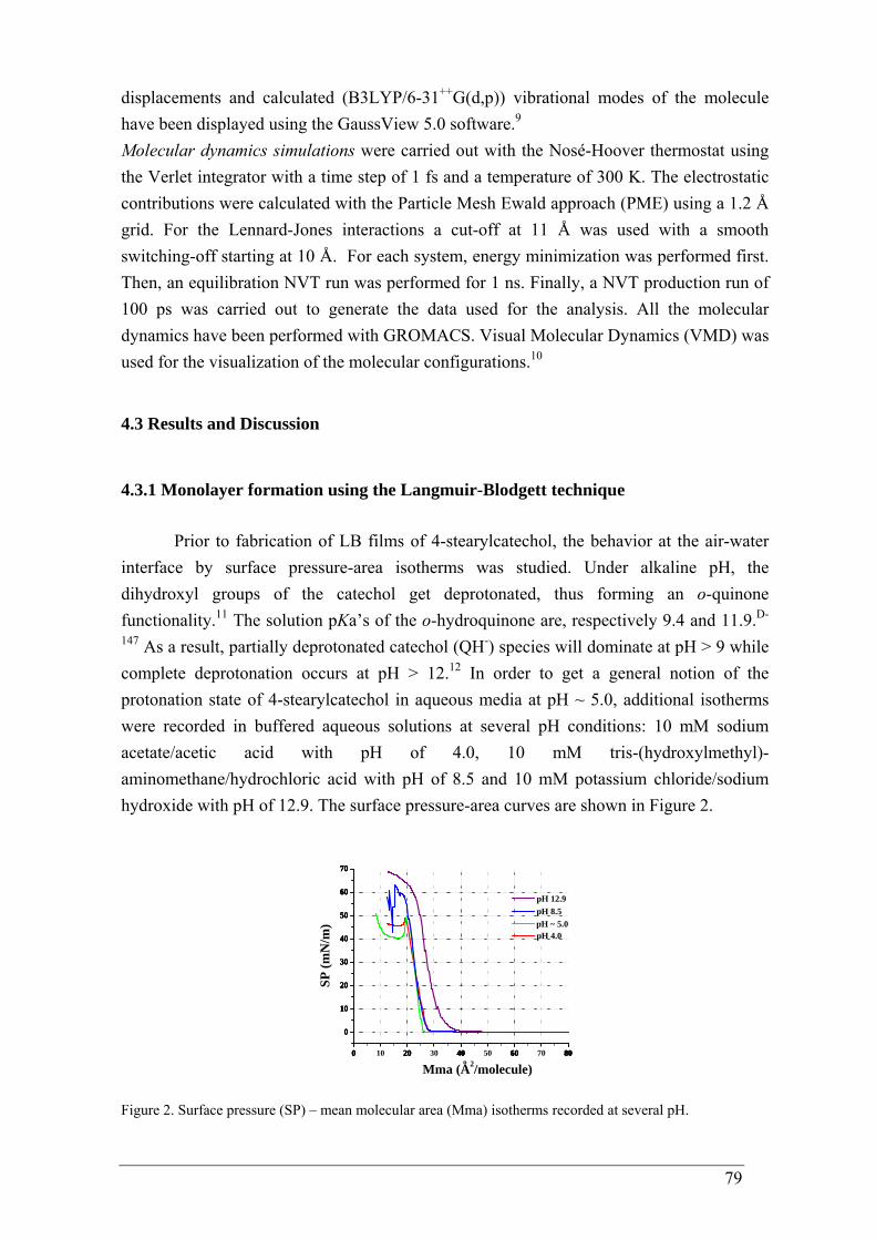
79
displacements and calculated (B3LYP/6-31++G(d,p)) vibrational modes of the molecule have been displayed using the GaussView 5.0 software.9 Molecular dynamics simulations were carried out with the Nosé-Hoover thermostat using the Verlet integrator with a time step of 1 fs and a temperature of 300 K. The electrostatic contributions were calculated with the Particle Mesh Ewald approach (PME) using a 1.2 Å grid. For the Lennard-Jones interactions a cut-off at 11 Å was used with a smooth switching-off starting at 10 Å. For each system, energy minimization was performed first. Then, an equilibration NVT run was performed for 1 ns. Finally, a NVT production run of 100 ps was carried out to generate the data used for the analysis. All the molecular dynamics have been performed with GROMACS. Visual Molecular Dynamics (VMD) was used for the visualization of the molecular configurations.10
4.3 Results and Discussion
4.3.1 Monolayer formation using the Langmuir-Blodgett technique
Prior to fabrication of LB films of 4-stearylcatechol, the behavior at the air-water interface by surface pressure-area isotherms was studied. Under alkaline pH, the dihydroxyl groups of the catechol get deprotonated, thus forming an o-quinone functionality.11 The solution pKa’s of the o-hydroquinone are, respectively 9.4 and 11.9.D-
147 As a result, partially deprotonated catechol (QH-) species will dominate at pH > 9 while complete deprotonation occurs at pH > 12.12 In order to get a general notion of the protonation state of 4-stearylcatechol in aqueous media at pH ~ 5.0, additional isotherms were recorded in buffered aqueous solutions at several pH conditions: 10 mM sodium acetate/acetic acid with pH of 4.0, 10 mM tris-(hydroxylmethyl)-aminomethane/hydrochloric acid with pH of 8.5 and 10 mM potassium chloride/sodium hydroxide with pH of 12.9. The surface pressure-area curves are shown in Figure 2.
0 20 40 60 80
0
10
20
30
40
50
60
70
0 10 20 30 40 50 60 70 80
0
10
20
30
40
50
60
70
0 20 40 60 80
0
10
20
30
40
50
60
70
0 20 40 60 80
0
10
20
30
40
50
60
70
pH 4.0 pH ~ 5.0
SP (m
N/m
)
Mma (A2/molecule)
pH 8.5
o
pH 12.9
Figure 2. Surface pressure (SP) – mean molecular area (Mma) isotherms recorded at several pH.
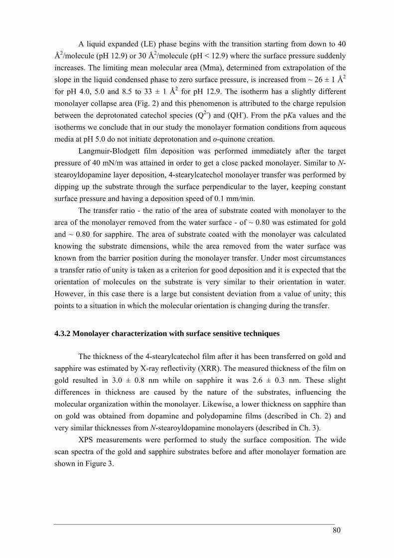
80
A liquid expanded (LE) phase begins with the transition starting from down to 40 Å2/molecule (pH 12.9) or 30 Å2/molecule (pH < 12.9) where the surface pressure suddenly increases. The limiting mean molecular area (Mma), determined from extrapolation of the slope in the liquid condensed phase to zero surface pressure, is increased from ~ 26 ± 1 Å2 for pH 4.0, 5.0 and 8.5 to 33 ± 1 Å2 for pH 12.9. The isotherm has a slightly different monolayer collapse area (Fig. 2) and this phenomenon is attributed to the charge repulsion between the deprotonated catechol species (Q2-) and (QH-). From the pKa values and the isotherms we conclude that in our study the monolayer formation conditions from aqueous media at pH 5.0 do not initiate deprotonation and o-quinone creation.
Langmuir-Blodgett film deposition was performed immediately after the target pressure of 40 mN/m was attained in order to get a close packed monolayer. Similar to N-stearoyldopamine layer deposition, 4-stearylcatechol monolayer transfer was performed by dipping up the substrate through the surface perpendicular to the layer, keeping constant surface pressure and having a deposition speed of 0.1 mm/min.
The transfer ratio - the ratio of the area of substrate coated with monolayer to the area of the monolayer removed from the water surface - of ~ 0.80 was estimated for gold and ~ 0.80 for sapphire. The area of substrate coated with the monolayer was calculated knowing the substrate dimensions, while the area removed from the water surface was known from the barrier position during the monolayer transfer. Under most circumstances a transfer ratio of unity is taken as a criterion for good deposition and it is expected that the orientation of molecules on the substrate is very similar to their orientation in water. However, in this case there is a large but consistent deviation from a value of unity; this points to a situation in which the molecular orientation is changing during the transfer.
4.3.2 Monolayer characterization with surface sensitive techniques
The thickness of the 4-stearylcatechol film after it has been transferred on gold and sapphire was estimated by X-ray reflectivity (XRR). The measured thickness of the film on gold resulted in 3.0 ± 0.8 nm while on sapphire it was 2.6 ± 0.3 nm. These slight differences in thickness are caused by the nature of the substrates, influencing the molecular organization within the monolayer. Likewise, a lower thickness on sapphire than on gold was obtained from dopamine and polydopamine films (described in Ch. 2) and very similar thicknesses from N-stearoyldopamine monolayers (described in Ch. 3).
XPS measurements were performed to study the surface composition. The wide scan spectra of the gold and sapphire substrates before and after monolayer formation are shown in Figure 3.
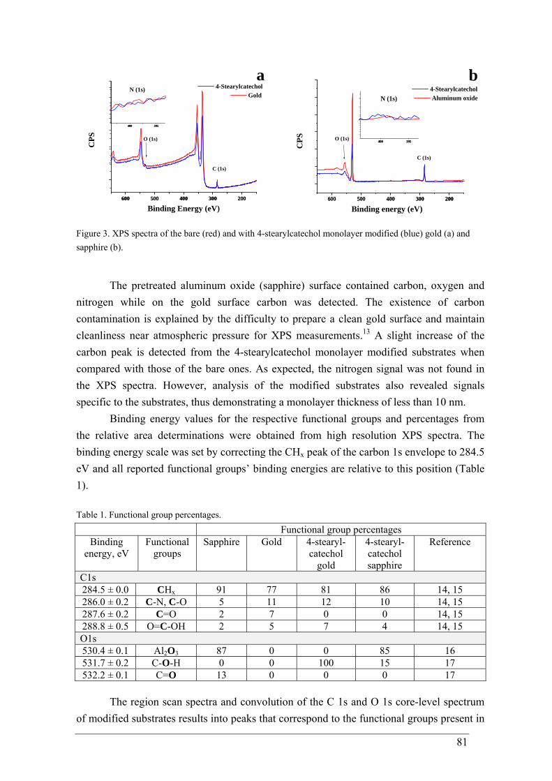
81
600 500 400 300 200600 500 400 300 200
400 395400 395
N (1s)C
PS Gold
a
O (1s)
C (1s)
Binding Energy (eV)
4-Stearylcatechol
600 500 400 300 200600 500 400 300 200
Aluminum oxide
b
400 395400 395
N (1s)
O (1s)
C (1s)
CPS
Binding energy (eV)
4-Stearylcatechol
Figure 3. XPS spectra of the bare (red) and with 4-stearylcatechol monolayer modified (blue) gold (a) and sapphire (b).
The pretreated aluminum oxide (sapphire) surface contained carbon, oxygen and nitrogen while on the gold surface carbon was detected. The existence of carbon contamination is explained by the difficulty to prepare a clean gold surface and maintain cleanliness near atmospheric pressure for XPS measurements.13 A slight increase of the carbon peak is detected from the 4-stearylcatechol monolayer modified substrates when compared with those of the bare ones. As expected, the nitrogen signal was not found in the XPS spectra. However, analysis of the modified substrates also revealed signals specific to the substrates, thus demonstrating a monolayer thickness of less than 10 nm.
Binding energy values for the respective functional groups and percentages from the relative area determinations were obtained from high resolution XPS spectra. The binding energy scale was set by correcting the CHx peak of the carbon 1s envelope to 284.5 eV and all reported functional groups’ binding energies are relative to this position (Table 1). Table 1. Functional group percentages. Functional group percentages
Binding energy, eV
Functional groups
Sapphire Gold 4-stearyl-catechol
gold
4-stearyl-catechol sapphire
Reference
C1s 284.5 ± 0.0 CHx 91 77 81 86 14, 15 286.0 ± 0.2 C-N, C-O 5 11 12 10 14, 15 287.6 ± 0.2 C=O 2 7 0 0 14, 15 288.8 ± 0.5 O=C-OH 2 5 7 4 14, 15 O1s 530.4 ± 0.1 Al2O3 87 0 0 85 16 531.7 ± 0.2 C-O-H 0 0 100 15 17 532.2 ± 0.1 C=O 13 0 0 0 17
The region scan spectra and convolution of the C 1s and O 1s core-level spectrum of modified substrates results into peaks that correspond to the functional groups present in
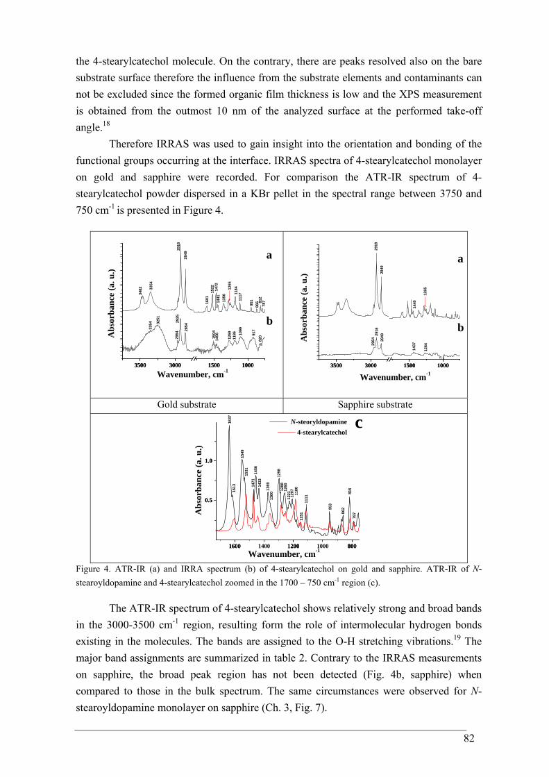
82
the 4-stearylcatechol molecule. On the contrary, there are peaks resolved also on the bare substrate surface therefore the influence from the substrate elements and contaminants can not be excluded since the formed organic film thickness is low and the XPS measurement is obtained from the outmost 10 nm of the analyzed surface at the performed take-off angle.18
Therefore IRRAS was used to gain insight into the orientation and bonding of the functional groups occurring at the interface. IRRAS spectra of 4-stearylcatechol monolayer on gold and sapphire were recorded. For comparison the ATR-IR spectrum of 4-stearylcatechol powder dispersed in a KBr pellet in the spectral range between 3750 and 750 cm-1 is presented in Figure 4.
3500 3000 1500 10003500 3000 1500 1000
2964
82091
7
1099
1186
1269
145615
04
3251
3354 b
a
Abs
orba
nce
(a. u
.)
Wavenumber, cm-1
111711
84
78781
286
6951
1265
1356
1441
1472
1522
1601
3354
3482
2854
2925
2849
2918
3500 3000 1500 10003500 3000 1500 1000
1440
1265
b
a
2964
1437
1264
2849
2918
284929
16
Abs
orba
nce
(a. u
.)
Wavenumber, cm-1
Gold substrate Sapphire substrate
1600 1400 1200 1000 800
0.5
1.0
1600 1200 800
0.5
1.0
787
816
86295
31111
1151
1180
1207
1221
1260
1288
1298
1360
136914
3314
56
Wavenumber, cm-1
N-steoryldopamine c
1471
1531
1549
1613
1637
Abs
orba
nce
(a. u
.)
4-stearylcatechol
Figure 4. ATR-IR (a) and IRRA spectrum (b) of 4-stearylcatechol on gold and sapphire. ATR-IR of N-stearoyldopamine and 4-stearylcatechol zoomed in the 1700 – 750 cm-1 region (c).
The ATR-IR spectrum of 4-stearylcatechol shows relatively strong and broad bands in the 3000-3500 cm-1 region, resulting form the role of intermolecular hydrogen bonds existing in the molecules. The bands are assigned to the O-H stretching vibrations.19 The major band assignments are summarized in table 2. Contrary to the IRRAS measurements on sapphire, the broad peak region has not been detected (Fig. 4b, sapphire) when compared to those in the bulk spectrum. The same circumstances were observed for N-stearoyldopamine monolayer on sapphire (Ch. 3, Fig. 7).
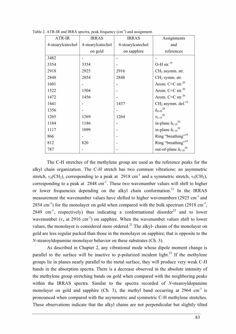
83
Table 2. ATR-IR and IRRA spectra, peak frequency (cm-1) and assignment.
ATR-IR 4-stearylcatechol
IRRAS 4-stearylcatechol
on gold
IRRAS 4-stearylcatechol
on sapphire
Assignments and
references 3482 3354 2918 2848 1601 1522 1472 1441 1356 1265 1184 1117 866 812 787
- 3354 2925 2854 - 1504 1456 - - 1269 1186 1099 - 820 -
- - 2916 2848 - - - 1437 - 1264 - - - - -
- O-H str.19 CH2 asymm. str. CH2 symm. str. Arom. C=C str.20 Arom. C=C str.20 Arom. C=C str.20 CH2 asymm. def.19
δO-H20
νC-O20
in-plane δC-H20
in-plane δC-H20
Ring “breathing”19 Ring “breathing”19 out-of-plane δC-H
20
The C-H stretches of the methylene group are used as the reference peaks for the alkyl chain organization. The C-H stretch has two common vibrations: an asymmetric stretch, νa(CH2), corresponding to a peak at 2918 cm-1 and a symmetric stretch, νs(CH2), corresponding to a peak at 2848 cm-1. These two wavenumber values will shift to higher or lower frequencies depending on the alkyl chain conformation.21 In the IRRAS measurement the wavenumber values have shifted to higher wavenumbers (2925 cm-1 and 2854 cm-1) for the monolayer on gold when compared with the bulk spectrum (2918 cm-1, 2849 cm-1, respectively) thus indicating a conformational disorder22 and to lower wavenumber (νa at 2916 cm-1) on sapphire. When the wavenumber values shift to lower values, the monolayer is considered more ordered.21 The alkyl- chains of the monolayer on gold are less regular packed than those in the monolayer on sapphire; that is opposite to the N-stearoyldopamine monolayer behavior on these substrates (Ch. 3).
As described in Chapter 2, any vibrational mode whose dipole moment change is parallel to the surface will be inactive to p-polarized incident light.23 If the methylene groups lie in planes nearly parallel to the metal surface, they will produce very weak C-H bands in the absorption spectra. There is a decrease observed in the absolute intensity of the methylene group stretching bands on gold when compared with the neighboring peaks within the IRRAS spectra. Similar to the spectra recorded of N-stearoyldopamine monolayer on gold and sapphire (Ch. 3), the methyl band occurring at 2964 cm-1 is pronounced when compared with the asymmetric and symmetric C-H methylene stretches. These observations indicate that the alkyl chains are not perpendicular but slightly tilted
84
with regard to the surface when a monolayer is formed.24 They are more tilted within the monolayer on gold than on sapphire.
Spectral features of the monolayer in the wavelength interval of 1350 to 1170 cm-1 of the IRRA spectra can be assigned to the aromatic in-plane δ(C-H) and ν(C-O) vibrations.19,20 The band at 1265 cm-1 has previously been associated with C-O stretching vibrations of the hydroxyl groups in catechol. Hydrogen bonding between the hydroxyl groups and water broadens the peak.25 Moreover, this band disappears upon cross-linking when catechol transforms into an o-quinone and remains unchanged when metal ion complexes are formed.26 Accordingly the IRRAS data confirm that when a monolayer is transferred onto gold 4-stearylcatechol molecule has catechol functionality. The 1265 cm-1 band had also been detected in IRRAS on sapphire. The presence of the C-O stretch and the lack of a stretch for free phenolic O-H from the 3000-3500 cm-1 region can be assigned to a bidentante bonding mode.21 The chemisortion of catechol at the TiO2 surface has previously been discussed in terms of a multisite surface complexation model.27 It is reported that catechol adsorbs at the TiO2 (anatase)/aqueous solution interface, forming inner-sphere surface complexes by which adsorbed catecholate anions bind to surface titanium ions in a process that involves the substitution of surface coordinated -OH (or water molecules). 21,27 Similarly the –OH region was not observed for N-stearoyldopamine monolayer on sapphire; however, the C-O stretch was not observed as well. Therefore it is not clear weather there is a different binding at the interface for N-stearoyldopamine molecules when compared with 4-stearylcatechol molecules or the low absorption intensities are attributable to the small absorption coefficient for sapphire.
In combination with IR, Raman spectroscopy and surface enhanced Raman spectroscopy (SERS) were used. Similar to the SERS studies on N-stearoyldopamine monolayer on gold (Chapter 3), two types of gold substrates were used: colloidal suspensions of gold nanoparticles deposited on an elemental germanium substrate and roughened gold surfaces that are macroscopically flat – the commercially available nanoengineered gold surface - Klarite® (details are given in Chapter 3). The results are shown in Figure 5 and assigned in Table 2.
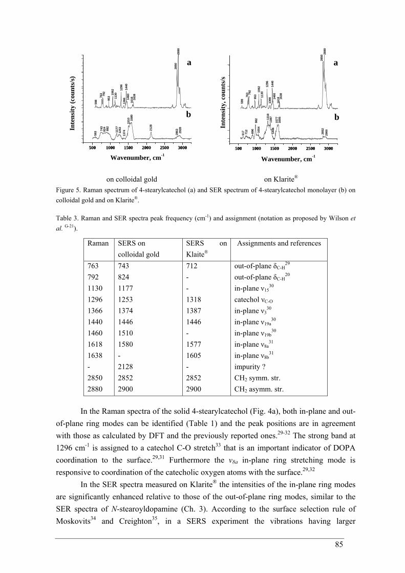
85
500 1000 1500 2000 2500 3000500 1000 1500 2000 2500 3000
2928
286321
28
1580
1510
137412
5311
77892
824
743
583
598
Inte
nsity
(cou
nts/
s)
Wavenumber, cm-1
b
2880
2850
1638
161814
6014
4013
6612
96
113010
6295
3792
763
a
500 1000 1500 2000 2500 3000500 1000 1500 2000 2500 3000
161814
60
617
712
982
1387
1605
2900
598
763 79
2
953
1062
1130
1296
1366
1440
1638
2880
2850
a
1577
2852
1446
896
1054
1318
Inte
nsity
, cou
nts/
s
Wavenumber, cm-1
b
on colloidal gold on Klarite®
Figure 5. Raman spectrum of 4-stearylcatechol (a) and SER spectrum of 4-stearylcatechol monolayer (b) on colloidal gold and on Klarite®. Table 3. Raman and SER spectra peak frequency (cm-1) and assignment (notation as proposed by Wilson et al. G-21).
Raman SERS on colloidal gold
SERS on Klaite®
Assignments and references
763 792 1130 1296 1366 1440 1460 1618 1638 - 2850 2880
743 824 1177 1253 1374 1446 1510 1580 - 2128 2852 2900
712 - - 1318 1387 1446 - 1577 1605 - 2852 2900
out-of-plane δC-H29
out-of-plane δC-H20
in-plane ν1530
catechol νC-O in-plane ν3
30 in-plane ν19a
30 in-plane ν19b
30 in-plane ν8a
31 in-plane ν8b
31 impurity ? CH2 symm. str. CH2 asymm. str.
In the Raman spectra of the solid 4-stearylcatechol (Fig. 4a), both in-plane and out-of-plane ring modes can be identified (Table 1) and the peak positions are in agreement with those as calculated by DFT and the previously reported ones.29-32 The strong band at 1296 cm-1 is assigned to a catechol C-O stretch33 that is an important indicator of DOPA coordination to the surface.29,31 Furthermore the ν8a in-plane ring stretching mode is responsive to coordination of the catecholic oxygen atoms with the surface.29,32
In the SER spectra measured on Klarite® the intensities of the in-plane ring modes are significantly enhanced relative to those of the out-of-plane ring modes, similar to the SER spectra of N-stearoyldopamine (Ch. 3). According to the surface selection rule of Moskovits34 and Creighton35, in a SERS experiment the vibrations having larger
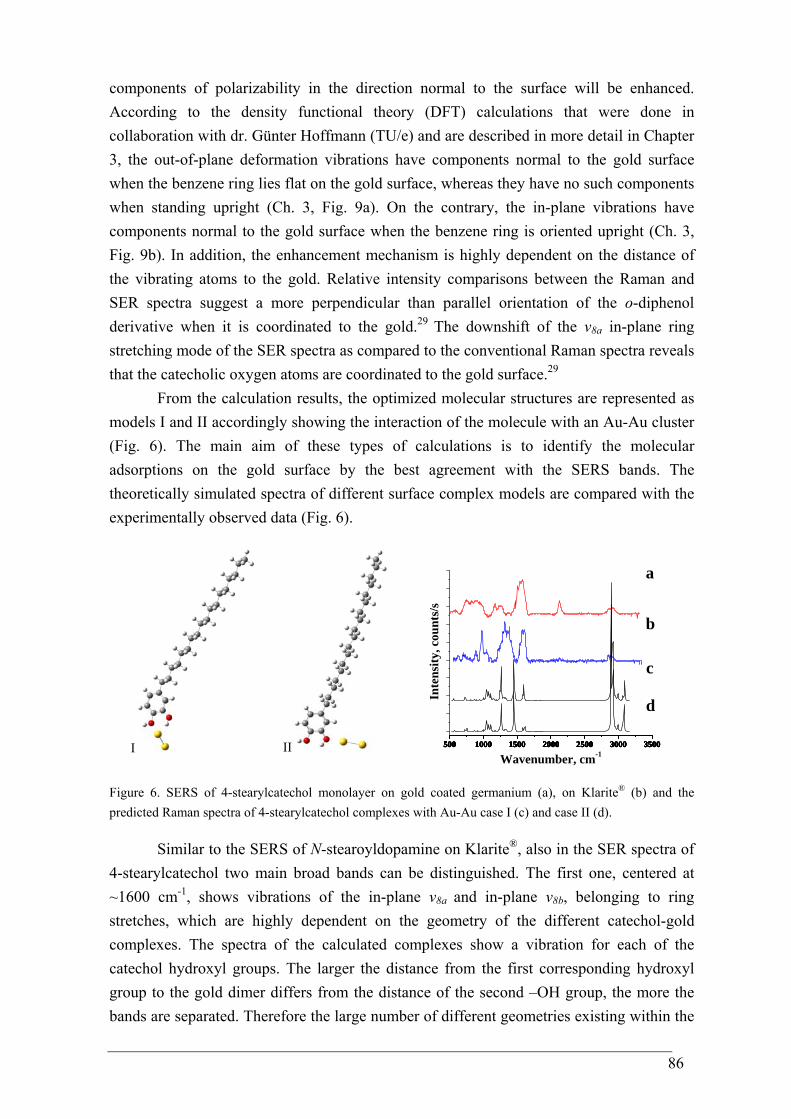
86
components of polarizability in the direction normal to the surface will be enhanced. According to the density functional theory (DFT) calculations that were done in collaboration with dr. Günter Hoffmann (TU/e) and are described in more detail in Chapter 3, the out-of-plane deformation vibrations have components normal to the gold surface when the benzene ring lies flat on the gold surface, whereas they have no such components when standing upright (Ch. 3, Fig. 9a). On the contrary, the in-plane vibrations have components normal to the gold surface when the benzene ring is oriented upright (Ch. 3, Fig. 9b). In addition, the enhancement mechanism is highly dependent on the distance of the vibrating atoms to the gold. Relative intensity comparisons between the Raman and SER spectra suggest a more perpendicular than parallel orientation of the o-diphenol derivative when it is coordinated to the gold.29 The downshift of the ν8a in-plane ring stretching mode of the SER spectra as compared to the conventional Raman spectra reveals that the catecholic oxygen atoms are coordinated to the gold surface.29
From the calculation results, the optimized molecular structures are represented as models I and II accordingly showing the interaction of the molecule with an Au-Au cluster (Fig. 6). The main aim of these types of calculations is to identify the molecular adsorptions on the gold surface by the best agreement with the SERS bands. The theoretically simulated spectra of different surface complex models are compared with the experimentally observed data (Fig. 6).
I II 500 1000 1500 2000 2500 3000 3500500 1000 1500 2000 2500 3000 3500500 1000 1500 2000 2500 3000 3500500 1000 1500 2000 2500 3000 3500500 1000 1500 2000 2500 3000 3500500 1000 1500 2000 2500 3000 3500
b
a
Inte
nsity
, cou
nts/
s
Wavenumber, cm-1
c
d
Figure 6. SERS of 4-stearylcatechol monolayer on gold coated germanium (a), on Klarite® (b) and the predicted Raman spectra of 4-stearylcatechol complexes with Au-Au case I (c) and case II (d).
Similar to the SERS of N-stearoyldopamine on Klarite®, also in the SER spectra of
4-stearylcatechol two main broad bands can be distinguished. The first one, centered at ~1600 cm-1, shows vibrations of the in-plane ν8a
and in-plane ν8b, belonging to ring stretches, which are highly dependent on the geometry of the different catechol-gold complexes. The spectra of the calculated complexes show a vibration for each of the catechol hydroxyl groups. The larger the distance from the first corresponding hydroxyl group to the gold dimer differs from the distance of the second –OH group, the more the bands are separated. Therefore the large number of different geometries existing within the
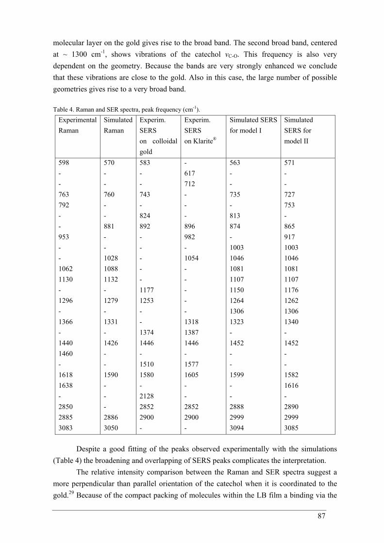
87
molecular layer on the gold gives rise to the broad band. The second broad band, centered at ~ 1300 cm-1, shows vibrations of the catechol νC-O. This frequency is also very dependent on the geometry. Because the bands are very strongly enhanced we conclude that these vibrations are close to the gold. Also in this case, the large number of possible geometries gives rise to a very broad band. Table 4. Raman and SER spectra, peak frequency (cm-1).
Experimental Raman
Simulated Raman
Experim. SERS on colloidal gold
Experim. SERS on Klarite®
Simulated SERS for model I
Simulated SERS for model II
598 - - 763 792 - - 953 - - 1062 1130 - 1296 - 1366 - 1440 1460 - 1618 1638 - 2850 2885 3083
570 - - 760 - - 881 - - 1028 1088 1132 - 1279 - 1331 - 1426 - - 1590 - - - 2886 3050
583 - - 743 - 824 892 - - - - - 1177 1253 - - 1374 1446 - 1510 1580 - 2128 2852 2900 -
- 617 712 - - - 896 982 - 1054 - - - - - 1318 1387 1446 - 1577 1605 - - 2852 2900 -
563 - - 735 - 813 874 - 1003 1046 1081 1107 1150 1264 1306 1323 - 1452 - - 1599 - - 2888 2999 3094
571 - - 727 753 - 865 917 1003 1046 1081 1107 1176 1262 1306 1340 - 1452 - - 1582 1616 - 2890 2999 3085
Despite a good fitting of the peaks observed experimentally with the simulations (Table 4) the broadening and overlapping of SERS peaks complicates the interpretation.
The relative intensity comparison between the Raman and SER spectra suggest a more perpendicular than parallel orientation of the catechol when it is coordinated to the gold.29 Because of the compact packing of molecules within the LB film a binding via the
88
hydroxyl groups to the gold occurs and the bonding via their π-electrons to the substrate is prohibited. This is consistent with calculation results that the 4-stearylcatechol molecules orient to the substrate by the hydroxyl groups and with previous studies of N-stearoyldopamine monolayer on gold (Ch. 3). It is impossible to define a surface plane for the electroless deposited gold since a huge number of different complex geometries exist together, causing broadening.
The changes in surface wetting properties related to the monolayer deposition were investigated measuring the contact angles with water. The static water contact angle of a bare gold substrate was determined to be 53.7 ± 1.8°, increasing to 98.7 ± 1.3° when a 4-stearylcatechol monolayer was deposited. The increased contact angle can be attributed to the hydrophobic character of the hydrocarbon chains of the deposited molecules. These values are comparable with those previously described for N-stearoyldopamine monolayer on gold (Ch. 3) and in the literature reported water contact angle values of a monolayer on hafnium oxide (HfO2), where a self-assembled monolayer of 4-stearylcatechol was formed from tetrahydrofuran (THF).21 This gives the impression that there is no influence of the amide functionality on contact angle value caused by a dense packing of the molecules. A much larger increase of the static water contact angle was detected when sapphire substrates were used. The angle on a bare aluminum oxide substrate is 69.5 ± 1.3°, but on 4-stearylcatechol monolayer modified sapphire samples it increased to 115.5 ± 1.1°. This high contact angle is comparable with the values obtained for well-ordered hydrophobic self-assembled monolayers of octadecylphosphate on aluminum oxide and octadecyltrichlorosilane on silicon.36,37
In order to probe the nanotopology of the monolayer AFM was used. For visualization of the 4-stearylcatechol monolayer a commercial sample of a gold (111) thin film on freshly cleaved mica substrate, grown in a controlled high vacuum deposition system, was used. These Au(111) substrates present a high degree of structural quality including surfaces with well-defined terraces (Ch. 3, Fig. 11a).
Topographical images of the 4-stearylcatechol modified Au(111) surface are shown in Figure 7. For these films a roughness of ~ 0.2 nm was determined, similar to the roughness of N-stearoyldopamine monolayer on gold (Ch. 3, Fig. 11).
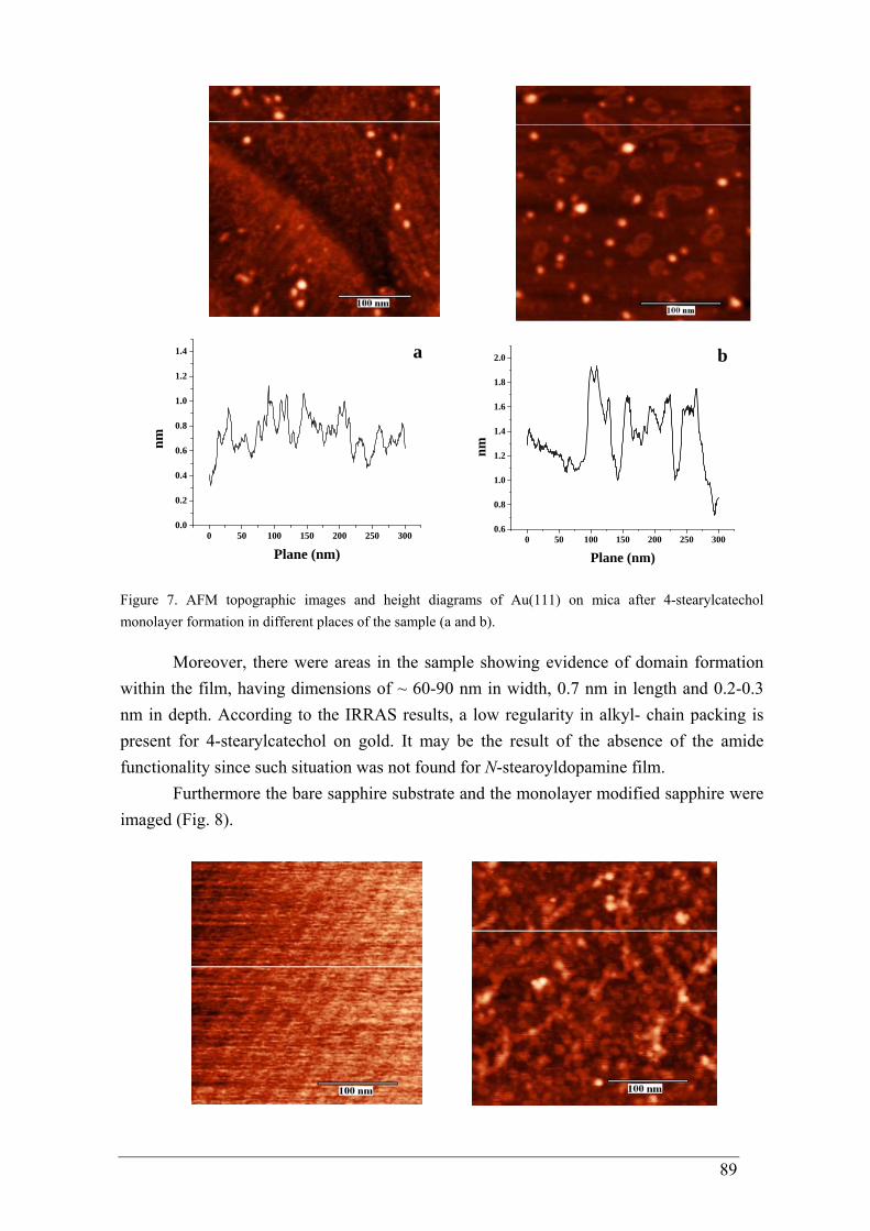
89
0 50 100 150 200 250 3000.0
0.2
0.4
0.6
0.8
1.0
1.2
1.4
nm
Plane (nm)
a
0 50 100 150 200 250 3000.6
0.8
1.0
1.2
1.4
1.6
1.8
2.0 b
nm
Plane (nm)
Figure 7. AFM topographic images and height diagrams of Au(111) on mica after 4-stearylcatechol monolayer formation in different places of the sample (a and b).
Moreover, there were areas in the sample showing evidence of domain formation within the film, having dimensions of ~ 60-90 nm in width, 0.7 nm in length and 0.2-0.3 nm in depth. According to the IRRAS results, a low regularity in alkyl- chain packing is present for 4-stearylcatechol on gold. It may be the result of the absence of the amide functionality since such situation was not found for N-stearoyldopamine film.
Furthermore the bare sapphire substrate and the monolayer modified sapphire were imaged (Fig. 8).
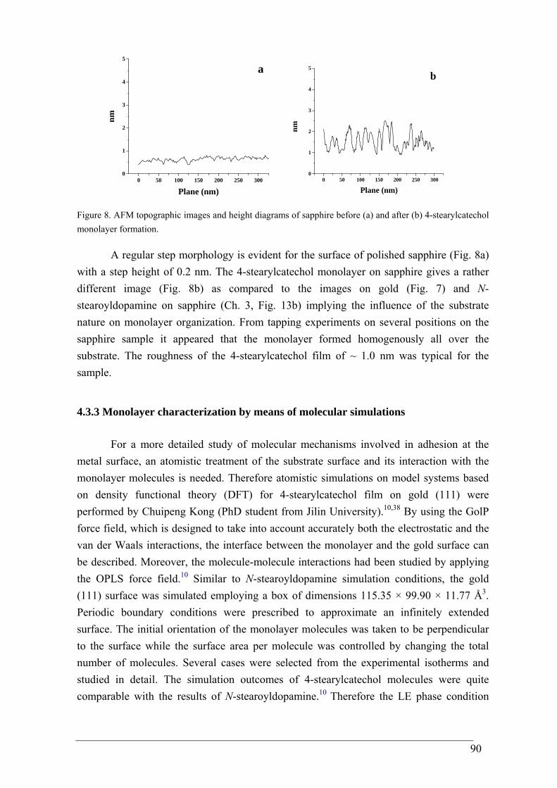
90
0 50 100 150 200 250 3000
1
2
3
4
5
nm
Plane (nm)
a
0 50 100 150 200 250 3000
1
2
3
4
5
nm
Plane (nm)
b
Figure 8. AFM topographic images and height diagrams of sapphire before (a) and after (b) 4-stearylcatechol monolayer formation.
A regular step morphology is evident for the surface of polished sapphire (Fig. 8a) with a step height of 0.2 nm. The 4-stearylcatechol monolayer on sapphire gives a rather different image (Fig. 8b) as compared to the images on gold (Fig. 7) and N-stearoyldopamine on sapphire (Ch. 3, Fig. 13b) implying the influence of the substrate nature on monolayer organization. From tapping experiments on several positions on the sapphire sample it appeared that the monolayer formed homogenously all over the substrate. The roughness of the 4-stearylcatechol film of ~ 1.0 nm was typical for the sample.
4.3.3 Monolayer characterization by means of molecular simulations
For a more detailed study of molecular mechanisms involved in adhesion at the metal surface, an atomistic treatment of the substrate surface and its interaction with the monolayer molecules is needed. Therefore atomistic simulations on model systems based on density functional theory (DFT) for 4-stearylcatechol film on gold (111) were performed by Chuipeng Kong (PhD student from Jilin University).10,38 By using the GolP force field, which is designed to take into account accurately both the electrostatic and the van der Waals interactions, the interface between the monolayer and the gold surface can be described. Moreover, the molecule-molecule interactions had been studied by applying the OPLS force field.10 Similar to N-stearoyldopamine simulation conditions, the gold (111) surface was simulated employing a box of dimensions 115.35 × 99.90 × 11.77 Å3. Periodic boundary conditions were prescribed to approximate an infinitely extended surface. The initial orientation of the monolayer molecules was taken to be perpendicular to the surface while the surface area per molecule was controlled by changing the total number of molecules. Several cases were selected from the experimental isotherms and studied in detail. The simulation outcomes of 4-stearylcatechol molecules were quite comparable with the results of N-stearoyldopamine.10 Therefore the LE phase condition
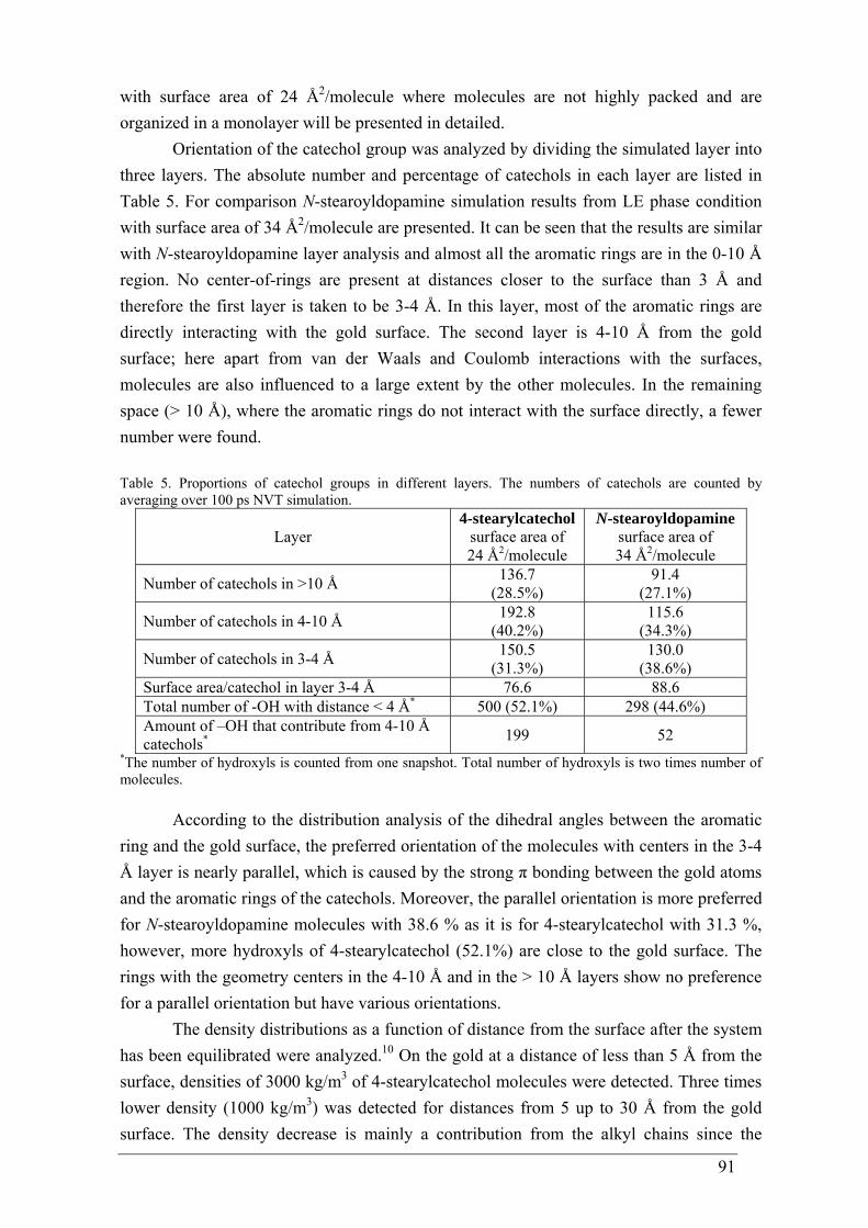
91
with surface area of 24 Å2/molecule where molecules are not highly packed and are organized in a monolayer will be presented in detailed.
Orientation of the catechol group was analyzed by dividing the simulated layer into three layers. The absolute number and percentage of catechols in each layer are listed in Table 5. For comparison N-stearoyldopamine simulation results from LE phase condition with surface area of 34 Å2/molecule are presented. It can be seen that the results are similar with N-stearoyldopamine layer analysis and almost all the aromatic rings are in the 0-10 Å region. No center-of-rings are present at distances closer to the surface than 3 Å and therefore the first layer is taken to be 3-4 Å. In this layer, most of the aromatic rings are directly interacting with the gold surface. The second layer is 4-10 Å from the gold surface; here apart from van der Waals and Coulomb interactions with the surfaces, molecules are also influenced to a large extent by the other molecules. In the remaining space (> 10 Å), where the aromatic rings do not interact with the surface directly, a fewer number were found. Table 5. Proportions of catechol groups in different layers. The numbers of catechols are counted by averaging over 100 ps NVT simulation.
Layer 4-stearylcatechol
surface area of 24 Å2/molecule
N-stearoyldopamine surface area of 34 Å2/molecule
Number of catechols in >10 Å 136.7 (28.5%)
91.4 (27.1%)
Number of catechols in 4-10 Å 192.8 (40.2%)
115.6 (34.3%)
Number of catechols in 3-4 Å 150.5 (31.3%)
130.0 (38.6%)
Surface area/catechol in layer 3-4 Å 76.6 88.6 Total number of -OH with distance < 4 Å* 500 (52.1%) 298 (44.6%) Amount of –OH that contribute from 4-10 Å catechols* 199 52
*The number of hydroxyls is counted from one snapshot. Total number of hydroxyls is two times number of molecules.
According to the distribution analysis of the dihedral angles between the aromatic ring and the gold surface, the preferred orientation of the molecules with centers in the 3-4 Å layer is nearly parallel, which is caused by the strong π bonding between the gold atoms and the aromatic rings of the catechols. Moreover, the parallel orientation is more preferred for N-stearoyldopamine molecules with 38.6 % as it is for 4-stearylcatechol with 31.3 %, however, more hydroxyls of 4-stearylcatechol (52.1%) are close to the gold surface. The rings with the geometry centers in the 4-10 Å and in the > 10 Å layers show no preference for a parallel orientation but have various orientations.
The density distributions as a function of distance from the surface after the system has been equilibrated were analyzed.10 On the gold at a distance of less than 5 Å from the surface, densities of 3000 kg/m3 of 4-stearylcatechol molecules were detected. Three times lower density (1000 kg/m3) was detected for distances from 5 up to 30 Å from the gold surface. The density decrease is mainly a contribution from the alkyl chains since the
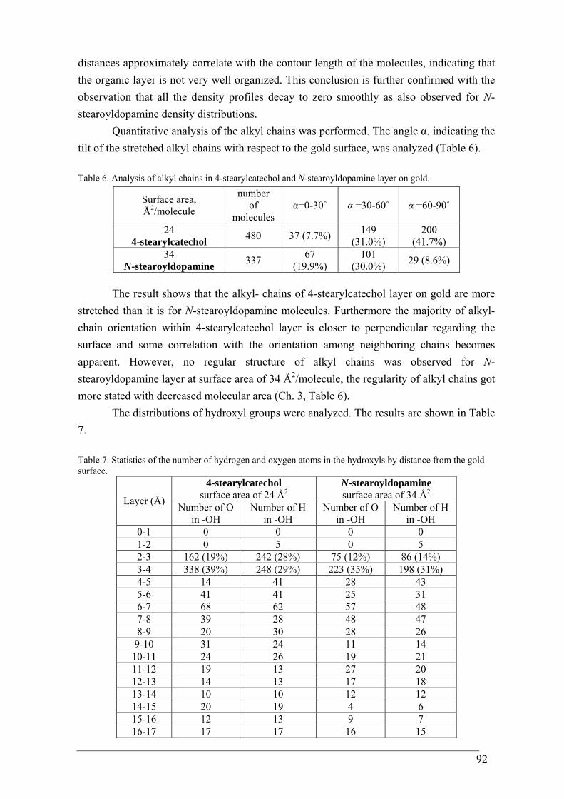
92
distances approximately correlate with the contour length of the molecules, indicating that the organic layer is not very well organized. This conclusion is further confirmed with the observation that all the density profiles decay to zero smoothly as also observed for N-stearoyldopamine density distributions.
Quantitative analysis of the alkyl chains was performed. The angle α, indicating the tilt of the stretched alkyl chains with respect to the gold surface, was analyzed (Table 6). Table 6. Analysis of alkyl chains in 4-stearylcatechol and N-stearoyldopamine layer on gold.
Surface area, Å2/molecule
number of
molecules α=0-30˚ α =30-60˚ α =60-90˚
24 4-stearylcatechol 480 37 (7.7%) 149
(31.0%) 200
(41.7%) 34
N-stearoyldopamine 337 67 (19.9%)
101 (30.0%) 29 (8.6%)
The result shows that the alkyl- chains of 4-stearylcatechol layer on gold are more stretched than it is for N-stearoyldopamine molecules. Furthermore the majority of alkyl- chain orientation within 4-stearylcatechol layer is closer to perpendicular regarding the surface and some correlation with the orientation among neighboring chains becomes apparent. However, no regular structure of alkyl chains was observed for N-stearoyldopamine layer at surface area of 34 Å2/molecule, the regularity of alkyl chains got more stated with decreased molecular area (Ch. 3, Table 6).
The distributions of hydroxyl groups were analyzed. The results are shown in Table 7. Table 7. Statistics of the number of hydrogen and oxygen atoms in the hydroxyls by distance from the gold surface.
4-stearylcatechol surface area of 24 Å2
N-stearoyldopamine surface area of 34 Å2 Layer (Å) Number of O
in -OH Number of H
in -OH Number of O
in -OH Number of H
in -OH 0-1 0 0 0 0 1-2 0 5 0 5 2-3 162 (19%) 242 (28%) 75 (12%) 86 (14%) 3-4 338 (39%) 248 (29%) 223 (35%) 198 (31%) 4-5 14 41 28 43 5-6 41 41 25 31 6-7 68 62 57 48 7-8 39 28 48 47 8-9 20 30 28 26
9-10 31 24 11 14 10-11 24 26 19 21 11-12 19 13 27 20 12-13 14 13 17 18 13-14 10 10 12 12 14-15 20 19 4 6 15-16 12 13 9 7 16-17 17 17 16 15
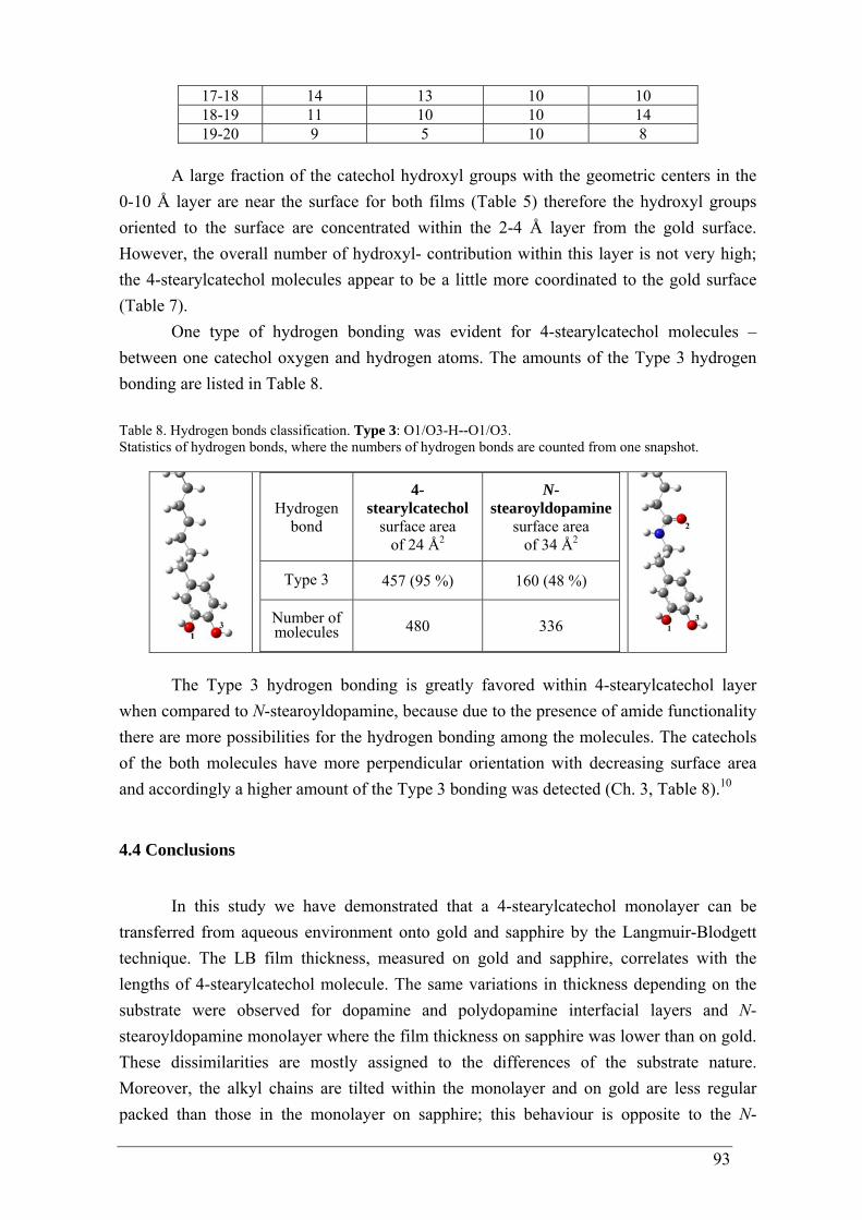
93
17-18 14 13 10 10 18-19 11 10 10 14 19-20 9 5 10 8
A large fraction of the catechol hydroxyl groups with the geometric centers in the 0-10 Å layer are near the surface for both films (Table 5) therefore the hydroxyl groups oriented to the surface are concentrated within the 2-4 Å layer from the gold surface. However, the overall number of hydroxyl- contribution within this layer is not very high; the 4-stearylcatechol molecules appear to be a little more coordinated to the gold surface (Table 7).
One type of hydrogen bonding was evident for 4-stearylcatechol molecules – between one catechol oxygen and hydrogen atoms. The amounts of the Type 3 hydrogen bonding are listed in Table 8. Table 8. Hydrogen bonds classification. Type 3: O1/O3-H--O1/O3. Statistics of hydrogen bonds, where the numbers of hydrogen bonds are counted from one snapshot.
Hydrogen bond
4-stearylcatechol
surface area of 24 Å2
N-stearoyldopamine
surface area of 34 Å2
Type 3 457 (95 %) 160 (48 %)
Number of molecules 480 336
The Type 3 hydrogen bonding is greatly favored within 4-stearylcatechol layer when compared to N-stearoyldopamine, because due to the presence of amide functionality there are more possibilities for the hydrogen bonding among the molecules. The catechols of the both molecules have more perpendicular orientation with decreasing surface area and accordingly a higher amount of the Type 3 bonding was detected (Ch. 3, Table 8).10
4.4 Conclusions
In this study we have demonstrated that a 4-stearylcatechol monolayer can be
transferred from aqueous environment onto gold and sapphire by the Langmuir-Blodgett technique. The LB film thickness, measured on gold and sapphire, correlates with the lengths of 4-stearylcatechol molecule. The same variations in thickness depending on the substrate were observed for dopamine and polydopamine interfacial layers and N-stearoyldopamine monolayer where the film thickness on sapphire was lower than on gold. These dissimilarities are mostly assigned to the differences of the substrate nature. Moreover, the alkyl chains are tilted within the monolayer and on gold are less regular packed than those in the monolayer on sapphire; this behaviour is opposite to the N-
94
stearoyldopamine monolayer behavior on these substrates. The irregularity of the film on gold leads to domain formation that is probably caused by the absence of the amide functionality.
The monolayer interaction with gold happens via the catecholic oxygen atoms that are coordinated to the surface having a more perpendicular than parallel orientation of the aromatic ring. This has also been observed by Ooka and Garrell for mussel foot protein 1 adsorbed on gold29 and N-stearoyldopamine studies on gold. However, molecular simulation results of the 4-stearylcatechol film on gold were rather similar to the ones of N-stearoyldopamine; it revealed deficiency of large amount of hydrogen bonding, attributable to the absence of the amide group - the catechols have various orientations at the gold surface being not well organized that is consistent with the experimental observations. Nevertheless it appears that the catechol hydroxyl- groups of the 4-stearylcatechol film are a little more coordinated to the gold surface and the alkyl- chains are more stretched with an orientation closer to perpendicular than it was detected for the N-stearoyldopamine film.
4.5 References
1 Lin, Q.; Gourdon, D.; Sun, C.; Holten-Andersen, N.; Anderson, T. H.; Waite, J. H.; Israelachvili, J. N. PNAS 2007, 104, 3782.
2 Lee, H.; Scherer, N. F.; Messersmith, P. B. Proc. Natl. Acad. Sci. USA 2006, 29, 12999.
3 Waite, J. H. Int. J. Biol. Macromol. 1990, 12, 139. 4 Porter, Jr. L. A.; Choi, H. C.; Ribbe, A. E.; Buriak, J. M. Nano Letters 2002, 2,
1067. 5 Gaussian 09, Revision A.02; Frisch, M. J.; Trucks, G. W.; Schlegel, H. B.;
Scuseria, G. E.; Robb, M. A.; Cheeseman, J. R.; Scalmani, G.; Barone, V.; Mennucci, B.; Petersson, G. A.; Nakatsuji, H.; Caricato, M.; Li, X.; Hratchian, H. P.; Izmaylov, A. F.; Bloino, J.; Zheng, G.; Sonnenberg, J. L.; Hada, M.; Ehara, M.; Toyota, K.; Fukuda, R.; Hasegawa, J.; Ishida, M.; Nakajima, T.; Honda, Y.; Kitao, O.; Nakai, H.; Vreven, T.; Montgomery, Jr. J. A.; Peralta, J. E.; Ogliaro, F.; Bearpark, M.; Heyd, J. J.; Brothers, E.; Kudin, K. N.; Staroverov, V. N.; Kobayashi, R.; Normand, J.; Raghavachari, K.; Rendell, A.; Burant, J. C.; Iyengar, S. S.; Tomasi, J.; Cossi, M.; Rega, N.; Millam, J. M.; Klene, M.; Knox, J. E.; Cross, J. B.; Bakken, V.; Adamo, C.; Jaramillo, J.; Gomperts, R.; Stratmann, R. E.; Yazyev, O.; Austin, A. J.; Cammi, R.; Pomelli, C.; Ochterski, J. W.; Martin, R. L.; Morokuma, K.; Zakrzewski, V. G.; Voth, G. A.; Salvador, P.; Dannenberg, J. J.; Dapprich, S.; Daniels, A. D.; Farkas, O.; Foresman, J. B.; Ortiz, J. V.; Cioslowski, J.; Fox, D. J. Gaussian, Inc., Wallingford CT 2009.
6 Becke, A. D. J. Chem. Phys. 1993, 98, 5648. 7 Lee, C.; Yang, W.; Parr, R. G. Phys. Rev. B 1988, 37, 785. 8 Frisch, M. J.; Pople, J. A.; Binkley, J. S. J. Chem. Phys. 1984, 80, 3265.
95
9 Rībena, D.; Hoffmann, G. G.; With, G. To be published. 10 Kong, C-P.; Peters, E. A. J. F.; Zhang, H-X.; With, G. To be published. 11 Yu, M.; Hwang, J.; Deming, T. J. J. Am. Chem. Soc. 1999, 121, 5825. 12 Brooksby, P. A.; Schiel, D. R.; Abell, A. D. Langmuir 2008, 24, 9074. 13 Ishida, T.; Tsuneda, S.; Nishida, N.; Hara, M.; Sasabe, H.; Knoll, W. Langmuir
1997, 13, 4638. 14 Desimoni, E.; Casella, G. I.; Morone, A.; Salvi, A. M. Surface and Interface
Analysis 1990, 15, 627434. 15 Bernsmann, F.; Ponche, A.; Ringwald, C.; Hemmerlé, J.; Raya, J.; Bechinger, B.;
Voegel, J.-C.; Schaaf, P.; Ball, V. J. Phys. Chem. C 2009, 113, 8234. 16 Brand, J.; Snijders, P. C.; Sloof, W. G.; Terryn, H.; Wit, J. H. W. J. Phys. Chem. B
2004, 108, 6017. 17 Li, X.-G.; Huang, K.-L.; Liu, S.-Q.; Tan, N.; Chen, L.-Q. Trans. Nonferrous Met.
Soc. China 2007, 17, 195. 18 Geng, S.; Zhang, S.; Onishi, H. Materials Technology 2002, 17, 234. 19 Lin-Vien, D.; Colthup, N. B.; Fateley, W. G.; Grasselli, J. G. The Handbook of
Infrared and Raman Characteristic Frequencies of Organic Molecules 1991, Academic Press: San Diego, CA.
20 Bredenberg, B-son. J; Sarbak, Z. J. Chem. Tech. Biotechnol. 1988, 42, 221. 21 Ting II, G. G.; Acton, O.; Ma, H.; Ka, J. W.; Jen, A. K.-Y. Langmuir 2009, 25,
2140. 22 Nuzzo, R. G.; Fusco, F. A.; Allara, D. L. J. Am. Chem. Soc. 1987, 109, 2358. 23 Allara, D. L.; Nuzzo, R. G. Langmuir 1985, 1, 45. 24 Yang, B. D.; Wakamatsu, T. Surf. And Interf. Analysis 1996, 24, 803. 25 Barth, A. Prog. in Biophys. and Molecular Biology 2000, 74, 141. 26 Fant, C.; Hedlund, J.; Höök, F.; Berglin, M.; Fridell, E.; Elwing, H. The Journal of
Adhesion 2010, 86, 25. 27 Rodríguez, R.; Blesa, M. A.; Regazzoni, A. E. J. of Colloid and Interface Science
1996, 177, 122. 28 Wilson, E. B.; Decius, J. C.; Cross, P. C. Molecular Vibrations 1955, McGraw-
Hill, New York. 29 Ooka, A. A.; Garrell, R. L. Biopolymers 2000, 57, 92. 30 Salama, S.; Stong J. D.; Neilands, J. B.; Spiro, T. G. Biochemistry 1978, 17, 3781. 31 Lee, N.-S.; Hsieh, Y.-Z.; Paisley, R. F.; Morris, M. D. Anal Chem 1988, 60, 442. 32 Kowalchyk, W. K.; Davis, K. L.; Morris, M. D. Spectrochim Acta 1995, 51A, 145. 33 Gaber, B. P.; Miskowski, V.; Spiro, T. G. J Am Chem Soc 1974, 96, 6868. 34 Moskovits, M. J. Chem. Phys. 1982, 77, 4408. 35 Creighton, J. A. Surf. Sci. 1983, 124, 209. 36 Hahner, G.; Hofer, R.; Klingenfuss, I. Langmuir 2001, 17, 7047. 37 Mirij, S. A. Surf. Interface Analysis 2006, 38, 158.

96
38 Kong, C-P.; Rībena, D.; Peters, E. A. J. F.; With, G.; Zhang, H-X. To be published.
97
CHAPTER 5
Adhesion on AA-2024-T In this chapter attempts are made to quantify the adhesion strength of the anchoring catechols on metal oxide. A covalently bound top-coating was used to determine the adhesion of an N-linoleoyldopamine primer on an aluminum alloy. Several application conditions were tested for the top-coating and the primer. The resulting tensile strength values of the top-coating having an N-linoleoyldopamine monolayer primer showed an obvious improvement in adhesion whereas a negative impact on adhesion occurred when the primer was not applied in a uniform and controlled manner by the Langmuir-Blodgett technique.
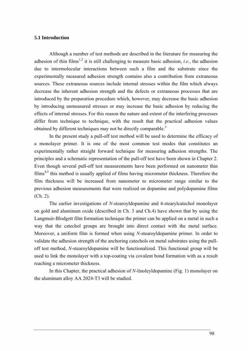
98
5.1 Introduction
Although a number of test methods are described in the literature for measuring the
adhesion of thin films1,2 it is still challenging to measure basic adhesion, i.e., the adhesion due to intermolecular interactions between such a film and the substrate since the experimentally measured adhesion strength contains also a contribution from extraneous sources. These extraneous sources include internal stresses within the film which always decrease the inherent adhesion strength and the defects or extraneous processes that are introduced by the preparation procedure which, however, may decrease the basic adhesion by introducing unmeasured stresses or may increase the basic adhesion by reducing the effects of internal stresses. For this reason the nature and extent of the interfering processes differ from technique to technique, with the result that the practical adhesion values obtained by different techniques may not be directly comparable.3
In the present study a pull-off test method will be used to determine the efficacy of a monolayer primer. It is one of the most common test modes that constitutes an experimentally rather straight forward technique for measuring adhesion strengths. The principles and a schematic representation of the pull-off test have been shown in Chapter 2. Even though several pull-off test measurements have been performed on nanometer thin films4,5 this method is usually applied of films having micrometer thickness. Therefore the film thickness will be increased from nanometer to micrometer range similar to the previous adhesion measurements that were realized on dopamine and polydopamine films (Ch. 2).
The earlier investigations of N-stearoyldopamine and 4-stearylcatechol monolayer on gold and aluminum oxide (described in Ch. 3 and Ch.4) have shown that by using the Langmuir-Blodgett film formation technique the primer can be applied on a metal in such a way that the catechol groups are brought into direct contact with the metal surface. Moreover, a uniform film is formed when using N-stearoyldopamine primer. In order to validate the adhesion strength of the anchoring catechols on metal substrates using the pull-off test method, N-stearoyldopamine will be functionalized. This functional group will be used to link the monolayer with a top-coating via covalent bond formation with as a result reaching a micrometer thickness.
In this Chapter, the practical adhesion of N-linoleyldopamine (Fig. 1) monolayer on the aluminum alloy AA 2024-T3 will be studied.
99
OHOH
NH
O
Figure 1. Chemical structure of N-linoleyldopamine.
5.2 Experimental
Methods and Materials. Dopamine hydrochloride (98%), (benzotriazol-1-yloxy)tripyrrolidino-phosphonium hexafluorophosphate (≥ 97%), triethylamine (> 99%) and linoleic acid (99%) were purchased from Sigma-Aldrich. Milli-Q water (resistivity = 18.2 MΩ⋅cm) was used in all experiments. Irgacure 819, a phenylbis(2,4,6-trimethylbenzoyl)phosphine oxide photo initiator, was purchased from Ciba Specialty Chemicals. A 3-functional thiol, Trimethylolpropane tris(3-mercaptopropionate), was purchased from Sigma-Aldrich. Poly(cyclo-hexane carbonate) - synthetized by copolymerization of cyclohexene oxide and carbon dioxide - was provided by dr. R. J. Sabong (TU/e, the Netherlands). 1H NMR and 13C NMR spectra were recorded on a Varian Mercury 400 MHz NMR instrument at 25 °C. Proton chemical shifts are reported in ppm (δ) downfield from tetramethylsilane (TMS). Carbon chemical shifts are reported using the resonance of CDCl3 as internal standard. Splitting patterns are designated as singlet (s), doublet (d), double doublet (dd), triplet (t), quartet (q) and multiplet (m). Melting points were detected on a Büchi B-540 instrument. Infrared spectra were recorded on a Perkin–Elmer 1605 FT-IR spectrometer. Thermo-gravimetric analyses (TGA) were carried out using a TA Instruments Q500 Thermogravimetric Analyzer applying an oxygen stream and a heating rate of 10 K/min. The LC/MS spectra were recorded on HP/Agilent 1100 MSD spectrometer. Elemental analyses were performed with a Perkin Elmer, Series II 2400 elemental analyzer for CHNS-O. Synthesis of N-linoleoyldopamine. N-linoleoyldopamine was synthesized by a modified literature procedure.6 To a stirred mixture of dopamine hydrochloride (1.0 g, 5.3 mmol) in 25 mL of dry THF (benzotriazol-1-yloxy)tripyrrolidinophosphonium hexafluorophosphate (2.75 g, 5.4 mmol) and cis-9,cis-12-octadecadienoic acid (2.95 g, 10.5 mmol) was added. The mixture was then stirred at room temperature for 10 min and then cooled to 5 °C. A solution of EtN3 (2.23 mL, 16.0 mmol) in 5 mL of dry THF was added dropwise while stirring the mixture. The cooling bath was then removed and the solution was stirred at
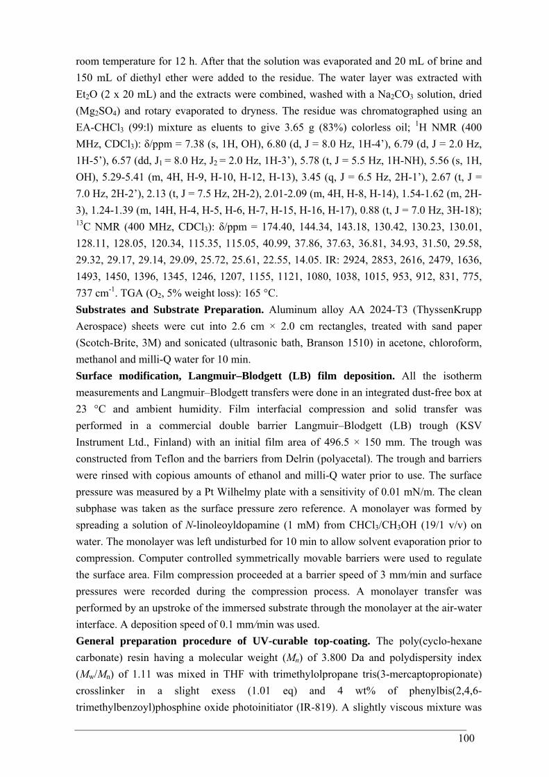
100
room temperature for 12 h. After that the solution was evaporated and 20 mL of brine and 150 mL of diethyl ether were added to the residue. The water layer was extracted with Et2O (2 x 20 mL) and the extracts were combined, washed with a Na2CO3 solution, dried (Mg2SO4) and rotary evaporated to dryness. The residue was chromatographed using an EA-CHCl3 (99:l) mixture as eluents to give 3.65 g (83%) colorless oil; 1H NMR (400 MHz, CDCl3): δ/ppm = 7.38 (s, 1H, OH), 6.80 (d, J = 8.0 Hz, 1H-4’), 6.79 (d, J = 2.0 Hz, 1H-5’), 6.57 (dd, J1 = 8.0 Hz, J2 = 2.0 Hz, 1H-3’), 5.78 (t, J = 5.5 Hz, 1H-NH), 5.56 (s, 1H, OH), 5.29-5.41 (m, 4H, H-9, H-10, H-12, H-13), 3.45 (q, J = 6.5 Hz, 2H-1’), 2.67 (t, J = 7.0 Hz, 2H-2’), 2.13 (t, J = 7.5 Hz, 2H-2), 2.01-2.09 (m, 4H, H-8, H-14), 1.54-1.62 (m, 2H-3), 1.24-1.39 (m, 14H, H-4, H-5, H-6, H-7, H-15, H-16, H-17), 0.88 (t, J = 7.0 Hz, 3H-18); 13C NMR (400 MHz, CDCl3): δ/ppm = 174.40, 144.34, 143.18, 130.42, 130.23, 130.01, 128.11, 128.05, 120.34, 115.35, 115.05, 40.99, 37.86, 37.63, 36.81, 34.93, 31.50, 29.58, 29.32, 29.17, 29.14, 29.09, 25.72, 25.61, 22.55, 14.05. IR: 2924, 2853, 2616, 2479, 1636, 1493, 1450, 1396, 1345, 1246, 1207, 1155, 1121, 1080, 1038, 1015, 953, 912, 831, 775, 737 cm-1. TGA (O2, 5% weight loss): 165 °C. Substrates and Substrate Preparation. Aluminum alloy AA 2024-T3 (ThyssenKrupp Aerospace) sheets were cut into 2.6 cm × 2.0 cm rectangles, treated with sand paper (Scotch-Brite, 3M) and sonicated (ultrasonic bath, Branson 1510) in acetone, chloroform, methanol and milli-Q water for 10 min. Surface modification, Langmuir–Blodgett (LB) film deposition. All the isotherm measurements and Langmuir–Blodgett transfers were done in an integrated dust-free box at 23 °C and ambient humidity. Film interfacial compression and solid transfer was performed in a commercial double barrier Langmuir–Blodgett (LB) trough (KSV Instrument Ltd., Finland) with an initial film area of 496.5 × 150 mm. The trough was constructed from Teflon and the barriers from Delrin (polyacetal). The trough and barriers were rinsed with copious amounts of ethanol and milli-Q water prior to use. The surface pressure was measured by a Pt Wilhelmy plate with a sensitivity of 0.01 mN/m. The clean subphase was taken as the surface pressure zero reference. A monolayer was formed by spreading a solution of N-linoleoyldopamine (1 mM) from CHCl3/CH3OH (19/1 v/v) on water. The monolayer was left undisturbed for 10 min to allow solvent evaporation prior to compression. Computer controlled symmetrically movable barriers were used to regulate the surface area. Film compression proceeded at a barrier speed of 3 mm/min and surface pressures were recorded during the compression process. A monolayer transfer was performed by an upstroke of the immersed substrate through the monolayer at the air-water interface. A deposition speed of 0.1 mm/min was used. General preparation procedure of UV-curable top-coating. The poly(cyclo-hexane carbonate) resin having a molecular weight (Mn) of 3.800 Da and polydispersity index (Mw/Mn) of 1.11 was mixed in THF with trimethylolpropane tris(3-mercaptopropionate) crosslinker in a slight exess (1.01 eq) and 4 wt% of phenylbis(2,4,6-trimethylbenzoyl)phosphine oxide photoinitiator (IR-819). A slightly viscous mixture was
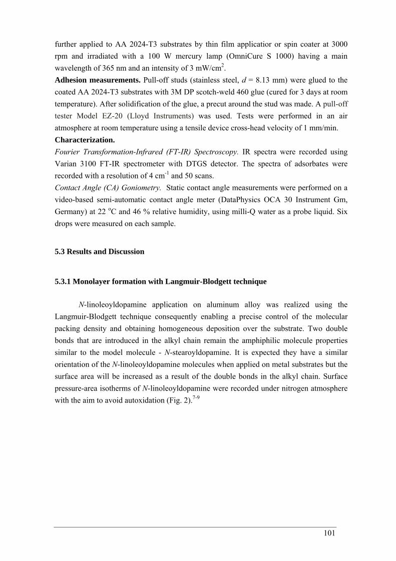
101
further applied to AA 2024-T3 substrates by thin film applicatior or spin coater at 3000 rpm and irradiated with a 100 W mercury lamp (OmniCure S 1000) having a main wavelength of 365 nm and an intensity of 3 mW/cm2. Adhesion measurements. Pull-off studs (stainless steel, d = 8.13 mm) were glued to the coated AA 2024-T3 substrates with 3M DP scotch-weld 460 glue (cured for 3 days at room temperature). After solidification of the glue, a precut around the stud was made. A pull-off tester Model EZ-20 (Lloyd Instruments) was used. Tests were performed in an air atmosphere at room temperature using a tensile device cross-head velocity of 1 mm/min. Characterization. Fourier Transformation-Infrared (FT-IR) Spectroscopy. IR spectra were recorded using Varian 3100 FT-IR spectrometer with DTGS detector. The spectra of adsorbates were recorded with a resolution of 4 cm-1 and 50 scans. Contact Angle (CA) Goniometry. Static contact angle measurements were performed on a video-based semi-automatic contact angle meter (DataPhysics OCA 30 Instrument Gm, Germany) at 22 oC and 46 % relative humidity, using milli-Q water as a probe liquid. Six drops were measured on each sample.
5.3 Results and Discussion
5.3.1 Monolayer formation with Langmuir-Blodgett technique
N-linoleoyldopamine application on aluminum alloy was realized using the Langmuir-Blodgett technique consequently enabling a precise control of the molecular packing density and obtaining homogeneous deposition over the substrate. Two double bonds that are introduced in the alkyl chain remain the amphiphilic molecule properties similar to the model molecule - N-stearoyldopamine. It is expected they have a similar orientation of the N-linoleoyldopamine molecules when applied on metal substrates but the surface area will be increased as a result of the double bonds in the alkyl chain. Surface pressure-area isotherms of N-linoleoyldopamine were recorded under nitrogen atmosphere with the aim to avoid autoxidation (Fig. 2).7-9
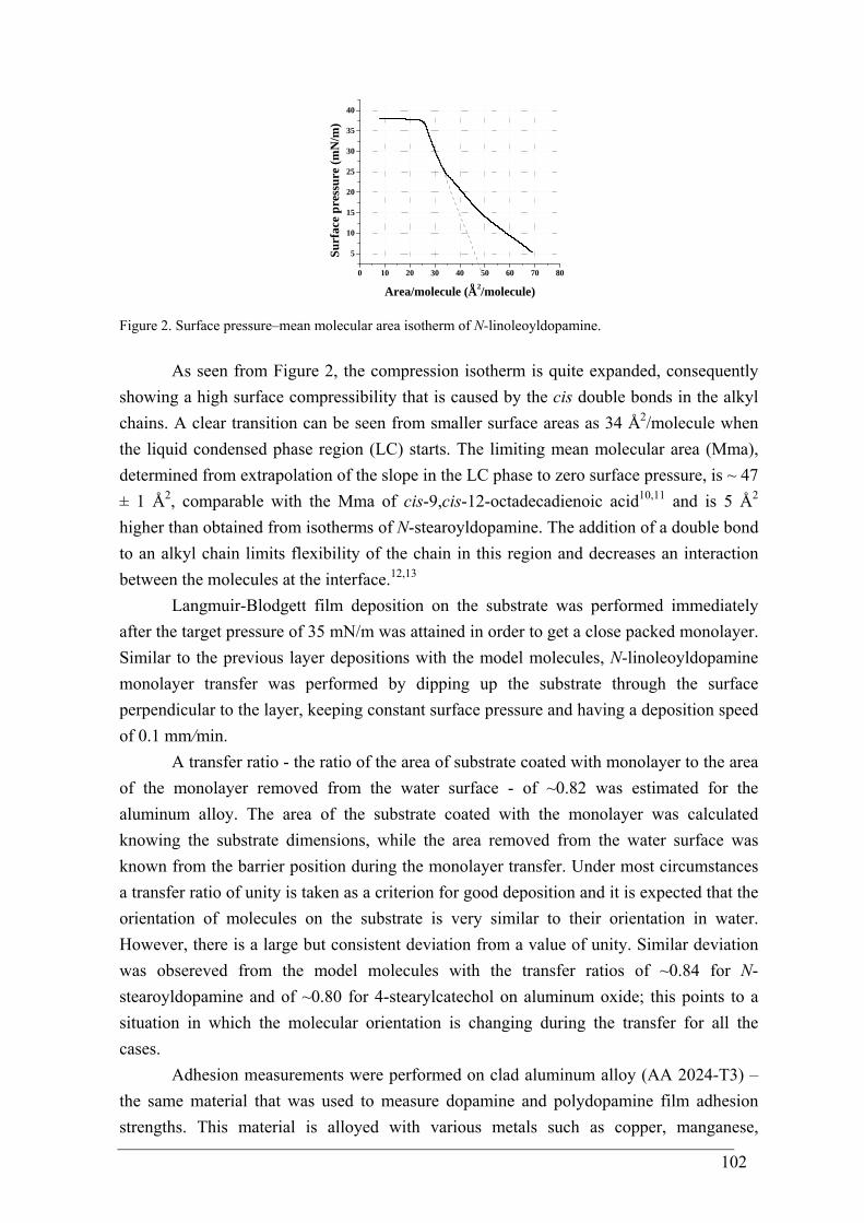
102
0 10 20 30 40 50 60 70 80
5
10
15
20
25
30
35
40
o
Surf
ace
pres
sure
(mN
/m)
Area/molecule (A2/molecule)
Figure 2. Surface pressure–mean molecular area isotherm of N-linoleoyldopamine.
As seen from Figure 2, the compression isotherm is quite expanded, consequently showing a high surface compressibility that is caused by the cis double bonds in the alkyl chains. A clear transition can be seen from smaller surface areas as 34 Å2/molecule when the liquid condensed phase region (LC) starts. The limiting mean molecular area (Mma), determined from extrapolation of the slope in the LC phase to zero surface pressure, is ~ 47 ± 1 Å2, comparable with the Mma of cis-9,cis-12-octadecadienoic acid10,11 and is 5 Å2
higher than obtained from isotherms of N-stearoyldopamine. The addition of a double bond to an alkyl chain limits flexibility of the chain in this region and decreases an interaction between the molecules at the interface.12,13
Langmuir-Blodgett film deposition on the substrate was performed immediately after the target pressure of 35 mN/m was attained in order to get a close packed monolayer. Similar to the previous layer depositions with the model molecules, N-linoleoyldopamine monolayer transfer was performed by dipping up the substrate through the surface perpendicular to the layer, keeping constant surface pressure and having a deposition speed of 0.1 mm/min.
A transfer ratio - the ratio of the area of substrate coated with monolayer to the area of the monolayer removed from the water surface - of ~0.82 was estimated for the aluminum alloy. The area of the substrate coated with the monolayer was calculated knowing the substrate dimensions, while the area removed from the water surface was known from the barrier position during the monolayer transfer. Under most circumstances a transfer ratio of unity is taken as a criterion for good deposition and it is expected that the orientation of molecules on the substrate is very similar to their orientation in water. However, there is a large but consistent deviation from a value of unity. Similar deviation was obsereved from the model molecules with the transfer ratios of ~0.84 for N-stearoyldopamine and of ~0.80 for 4-stearylcatechol on aluminum oxide; this points to a situation in which the molecular orientation is changing during the transfer for all the cases.
Adhesion measurements were performed on clad aluminum alloy (AA 2024-T3) – the same material that was used to measure dopamine and polydopamine film adhesion strengths. This material is alloyed with various metals such as copper, manganese,
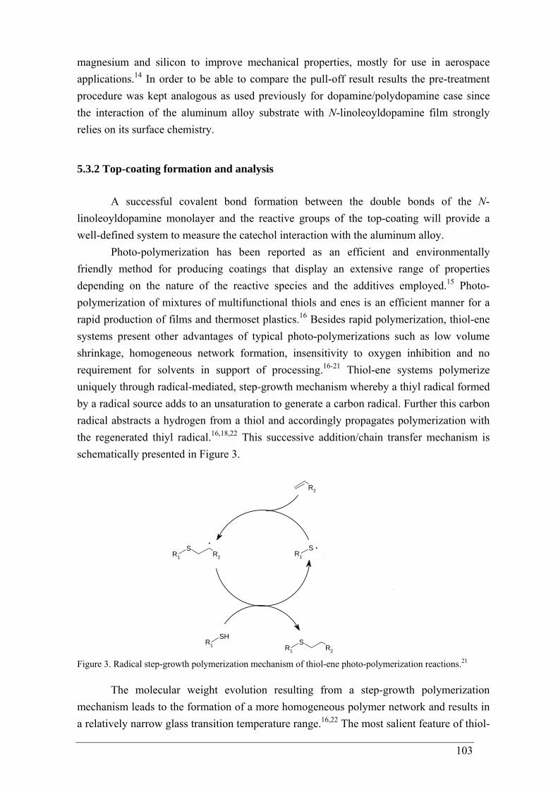
103
magnesium and silicon to improve mechanical properties, mostly for use in aerospace applications.14 In order to be able to compare the pull-off result results the pre-treatment procedure was kept analogous as used previously for dopamine/polydopamine case since the interaction of the aluminum alloy substrate with N-linoleoyldopamine film strongly relies on its surface chemistry.
5.3.2 Top-coating formation and analysis
A successful covalent bond formation between the double bonds of the N-linoleoyldopamine monolayer and the reactive groups of the top-coating will provide a well-defined system to measure the catechol interaction with the aluminum alloy.
Photo-polymerization has been reported as an efficient and environmentally friendly method for producing coatings that display an extensive range of properties depending on the nature of the reactive species and the additives employed.15 Photo-polymerization of mixtures of multifunctional thiols and enes is an efficient manner for a rapid production of films and thermoset plastics.16 Besides rapid polymerization, thiol-ene systems present other advantages of typical photo-polymerizations such as low volume shrinkage, homogeneous network formation, insensitivity to oxygen inhibition and no requirement for solvents in support of processing.16-21 Thiol-ene systems polymerize uniquely through radical-mediated, step-growth mechanism whereby a thiyl radical formed by a radical source adds to an unsaturation to generate a carbon radical. Further this carbon radical abstracts a hydrogen from a thiol and accordingly propagates polymerization with the regenerated thiyl radical.16,18,22 This successive addition/chain transfer mechanism is schematically presented in Figure 3.
R2
R1
SR2 R1
S
R1
SHR1
SR2
..
Figure 3. Radical step-growth polymerization mechanism of thiol-ene photo-polymerization reactions.21
The molecular weight evolution resulting from a step-growth polymerization mechanism leads to the formation of a more homogeneous polymer network and results in a relatively narrow glass transition temperature range.16,22 The most salient feature of thiol-
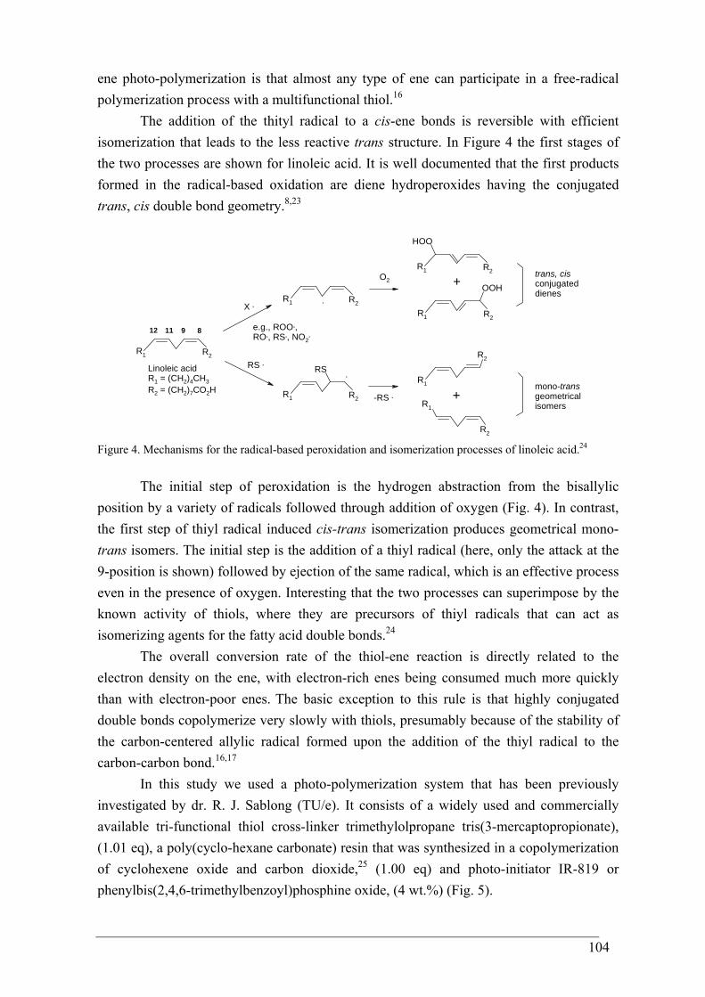
104
ene photo-polymerization is that almost any type of ene can participate in a free-radical polymerization process with a multifunctional thiol.16
The addition of the thityl radical to a cis-ene bonds is reversible with efficient isomerization that leads to the less reactive trans structure. In Figure 4 the first stages of the two processes are shown for linoleic acid. It is well documented that the first products formed in the radical-based oxidation are diene hydroperoxides having the conjugated trans, cis double bond geometry.8,23
R2R1
R2R1
R2R1
RS
R2R1
HOO
R2R1
OOH
R1
R2
R1
R2
891112
Linoleic acidR1 = (CH2)4CH3R2 = (CH2)7CO2H
X .
e.g., ROO.,RO., RS., NO2
.
RS .
.
O2
.
+
+-RS .
trans, cisconjugateddienes
mono-transgeometricalisomers
Figure 4. Mechanisms for the radical-based peroxidation and isomerization processes of linoleic acid.24
The initial step of peroxidation is the hydrogen abstraction from the bisallylic position by a variety of radicals followed through addition of oxygen (Fig. 4). In contrast, the first step of thiyl radical induced cis-trans isomerization produces geometrical mono-trans isomers. The initial step is the addition of a thiyl radical (here, only the attack at the 9-position is shown) followed by ejection of the same radical, which is an effective process even in the presence of oxygen. Interesting that the two processes can superimpose by the known activity of thiols, where they are precursors of thiyl radicals that can act as isomerizing agents for the fatty acid double bonds.24
The overall conversion rate of the thiol-ene reaction is directly related to the electron density on the ene, with electron-rich enes being consumed much more quickly than with electron-poor enes. The basic exception to this rule is that highly conjugated double bonds copolymerize very slowly with thiols, presumably because of the stability of the carbon-centered allylic radical formed upon the addition of the thiyl radical to the carbon-carbon bond.16,17
In this study we used a photo-polymerization system that has been previously investigated by dr. R. J. Sablong (TU/e). It consists of a widely used and commercially available tri-functional thiol cross-linker trimethylolpropane tris(3-mercaptopropionate), (1.01 eq), a poly(cyclo-hexane carbonate) resin that was synthesized in a copolymerization of cyclohexene oxide and carbon dioxide,25 (1.00 eq) and photo-initiator IR-819 or phenylbis(2,4,6-trimethylbenzoyl)phosphine oxide, (4 wt.%) (Fig. 5).
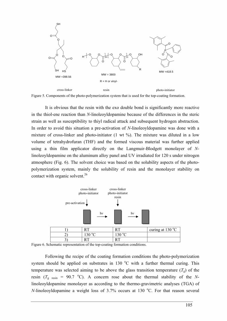
105
O OH
O
O O O
O
OH
R
m
n
P
O
OO
O
O OO O
O
SH
SHSHMW = 3800
MW =418.5
MW =398.56R = H or vinyl-
cross-linker resin photo-initiator Figure 5. Components of the photo-polymerization system that is used for the top-coating formation.
It is obvious that the resin with the exo double bond is significantly more reactive in the thiol-ene reaction than N-linoleoyldopamine because of the differences in the steric strain as well as susceptibility to thiyl radical attack and subsequent hydrogen abstraction. In order to avoid this situation a pre-activation of N-linoleoyldopamine was done with a mixture of cross-linker and photo-initiator (1 wt %). The mixture was diluted in a low volume of tetrahydrofuran (THF) and the formed viscous material was further applied using a thin film applicator directly on the Langmuir-Blodgett monolayer of N-linoleoyldopamine on the aluminum alloy panel and UV irradiated for 120 s under nitrogen atmosphere (Fig. 6). The solvent choice was based on the solubility aspects of the photo-polymerization system, mainly the solubility of resin and the monolayer stability on contact with organic solvent.26
1) RT RT curing at 130 oC 2) 130 oC 130 oC 3) RT RT
Figure 6. Schematic representation of the top-coating formation conditions.
Following the recipe of the coating formation conditions the photo-polymerization system should be applied on substrates in 130 oC with a further thermal curing. This temperature was selected aiming to be above the glass transition temperature (Tg) of the resin (Tg resin = 90.7 oC). A concern rose about the thermal stability of the N-linoleoyldopamine monolayer as according to the thermo-gravimetric analyses (TGA) of N-linoleoyldopamine a weight loss of 3.7% occurs at 130 oC. For that reason several
pre-activation
cross-linker photo-initiator
hν hν
cross-linker photo-initiator
resin
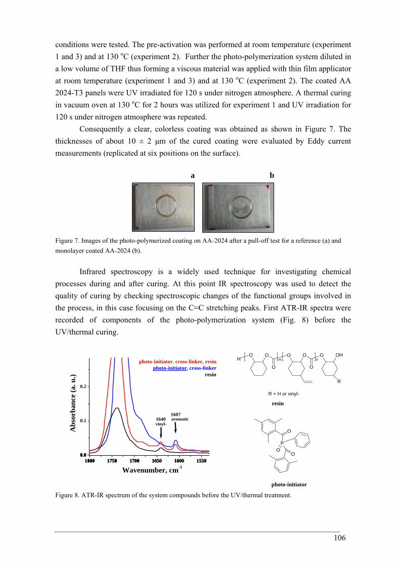
106
conditions were tested. The pre-activation was performed at room temperature (experiment 1 and 3) and at 130 oC (experiment 2). Further the photo-polymerization system diluted in a low volume of THF thus forming a viscous material was applied with thin film applicator at room temperature (experiment 1 and 3) and at 130 oC (experiment 2). The coated AA 2024-T3 panels were UV irradiated for 120 s under nitrogen atmosphere. A thermal curing in vacuum oven at 130 oC for 2 hours was utilized for experiment 1 and UV irradiation for 120 s under nitrogen atmosphere was repeated.
Consequently a clear, colorless coating was obtained as shown in Figure 7. The thicknesses of about 10 ± 2 μm of the cured coating were evaluated by Eddy current measurements (replicated at six positions on the surface).
a b
Figure 7. Images of the photo-polymerized coating on AA-2024 after a pull-off test for a reference (a) and monolayer coated AA-2024 (b).
Infrared spectroscopy is a widely used technique for investigating chemical processes during and after curing. At this point IR spectroscopy was used to detect the quality of curing by checking spectroscopic changes of the functional groups involved in the process, in this case focusing on the C=C stretching peaks. First ATR-IR spectra were recorded of components of the photo-polymerization system (Fig. 8) before the UV/thermal curing.
1800 1750 1700 1650 1600 15500.0
0.1
0.2
1800 1750 1700 1650 1600 15500.0
1800 1750 1700 1650 1600 15500.0
1800 1750 1700 1650 1600 15500.0
vinyl-aromatic16401607
photo-initiator, cross-linker, resinphoto-initiator, cross-linker
Abs
orba
nce
(a. u
.)
Wavenumber, cm-1
resin
O OH
O
O O O
O
OH
R
m
n
P
O
OO
R = H or vinyl-
resin
photo-initiator Figure 8. ATR-IR spectrum of the system compounds before the UV/thermal treatment.
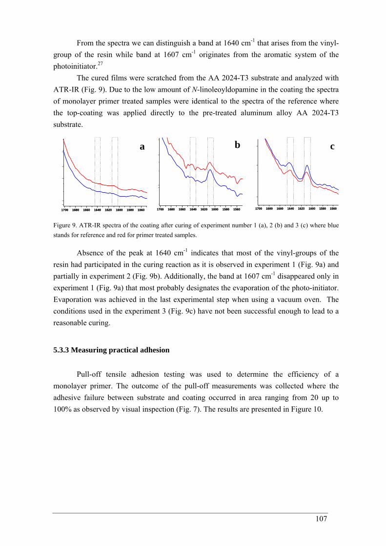
107
From the spectra we can distinguish a band at 1640 cm-1 that arises from the vinyl- group of the resin while band at 1607 cm-1 originates from the aromatic system of the photoinitiator.27
The cured films were scratched from the AA 2024-T3 substrate and analyzed with ATR-IR (Fig. 9). Due to the low amount of N-linoleoyldopamine in the coating the spectra of monolayer primer treated samples were identical to the spectra of the reference where the top-coating was applied directly to the pre-treated aluminum alloy AA 2024-T3 substrate.
1700 1680 1660 1640 1620 1600 1580 15601700 1680 1660 1640 1620 1600 1580 1560
a
1700 1680 1660 1640 1620 1600 1580 15601700 1680 1660 1640 1620 1600 1580 1560
b
1700 1680 1660 1640 1620 1600 1580 15601700 1680 1660 1640 1620 1600 1580 1560
c
Figure 9. ATR-IR spectra of the coating after curing of experiment number 1 (a), 2 (b) and 3 (c) where blue stands for reference and red for primer treated samples.
Absence of the peak at 1640 cm-1 indicates that most of the vinyl-groups of the resin had participated in the curing reaction as it is observed in experiment 1 (Fig. 9a) and partially in experiment 2 (Fig. 9b). Additionally, the band at 1607 cm-1 disappeared only in experiment 1 (Fig. 9a) that most probably designates the evaporation of the photo-initiator. Evaporation was achieved in the last experimental step when using a vacuum oven. The conditions used in the experiment 3 (Fig. 9c) have not been successful enough to lead to a reasonable curing.
5.3.3 Measuring practical adhesion
Pull-off tensile adhesion testing was used to determine the efficiency of a monolayer primer. The outcome of the pull-off measurements was collected where the adhesive failure between substrate and coating occurred in area ranging from 20 up to 100% as observed by visual inspection (Fig. 7). The results are presented in Figure 10.
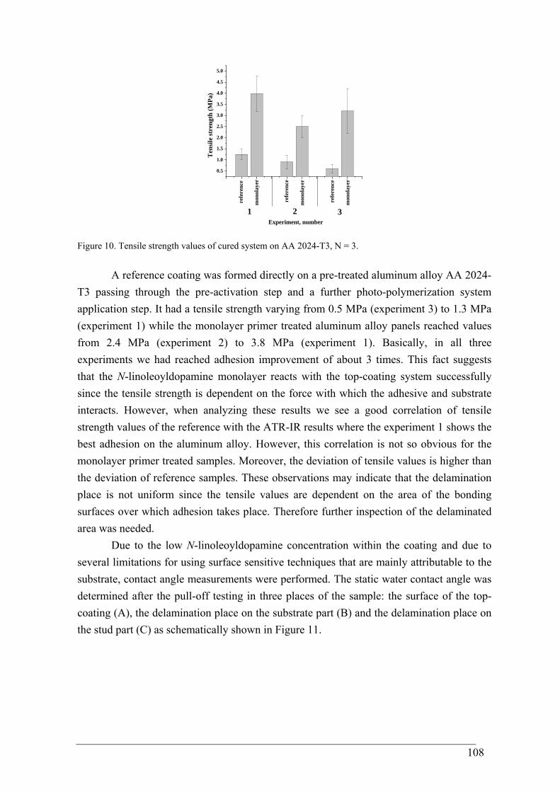
108
0.5
1.0
1.5
2.0
2.5
3.0
3.5
4.0
4.5
5.0
Experiment, number321
mon
olay
er
mon
olay
er
refe
renc
e
refe
renc
e
mon
olay
er
refe
renc
e
Ten
sile
stre
ngth
(MPa
)
Figure 10. Tensile strength values of cured system on AA 2024-T3, N = 3.
A reference coating was formed directly on a pre-treated aluminum alloy AA 2024-T3 passing through the pre-activation step and a further photo-polymerization system application step. It had a tensile strength varying from 0.5 MPa (experiment 3) to 1.3 MPa (experiment 1) while the monolayer primer treated aluminum alloy panels reached values from 2.4 MPa (experiment 2) to 3.8 MPa (experiment 1). Basically, in all three experiments we had reached adhesion improvement of about 3 times. This fact suggests that the N-linoleoyldopamine monolayer reacts with the top-coating system successfully since the tensile strength is dependent on the force with which the adhesive and substrate interacts. However, when analyzing these results we see a good correlation of tensile strength values of the reference with the ATR-IR results where the experiment 1 shows the best adhesion on the aluminum alloy. However, this correlation is not so obvious for the monolayer primer treated samples. Moreover, the deviation of tensile values is higher than the deviation of reference samples. These observations may indicate that the delamination place is not uniform since the tensile values are dependent on the area of the bonding surfaces over which adhesion takes place. Therefore further inspection of the delaminated area was needed.
Due to the low N-linoleoyldopamine concentration within the coating and due to several limitations for using surface sensitive techniques that are mainly attributable to the substrate, contact angle measurements were performed. The static water contact angle was determined after the pull-off testing in three places of the sample: the surface of the top-coating (A), the delamination place on the substrate part (B) and the delamination place on the stud part (C) as schematically shown in Figure 11.
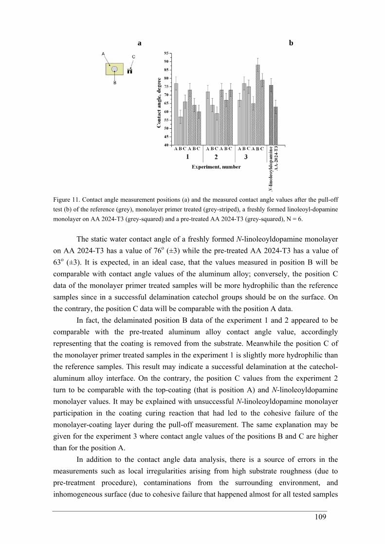
109
a
b
Figure 11. Contact angle measurement positions (a) and the measured contact angle values after the pull-off test (b) of the reference (grey), monolayer primer treated (grey-striped), a freshly formed linoleoyl-dopamine monolayer on AA 2024-T3 (grey-squared) and a pre-treated AA 2024-T3 (grey-squared), N = 6.
The static water contact angle of a freshly formed N-linoleoyldopamine monolayer on AA 2024-T3 has a value of 76o (±3) while the pre-treated AA 2024-T3 has a value of 63o (±3). It is expected, in an ideal case, that the values measured in position B will be comparable with contact angle values of the aluminum alloy; conversely, the position C data of the monolayer primer treated samples will be more hydrophilic than the reference samples since in a successful delamination catechol groups should be on the surface. On the contrary, the position C data will be comparable with the position A data.
In fact, the delaminated position B data of the experiment 1 and 2 appeared to be comparable with the pre-treated aluminum alloy contact angle value, accordingly representing that the coating is removed from the substrate. Meanwhile the position C of the monolayer primer treated samples in the experiment 1 is slightly more hydrophilic than the reference samples. This result may indicate a successful delamination at the catechol-aluminum alloy interface. On the contrary, the position C values from the experiment 2 turn to be comparable with the top-coating (that is position A) and N-linoleoyldopamine monolayer values. It may be explained with unsuccessful N-linoleoyldopamine monolayer participation in the coating curing reaction that had led to the cohesive failure of the monolayer-coating layer during the pull-off measurement. The same explanation may be given for the experiment 3 where contact angle values of the positions B and C are higher than for the position A.
In addition to the contact angle data analysis, there is a source of errors in the measurements such as local irregularities arising from high substrate roughness (due to pre-treatment procedure), contaminations from the surrounding environment, and inhomogeneous surface (due to cohesive failure that happened almost for all tested samples
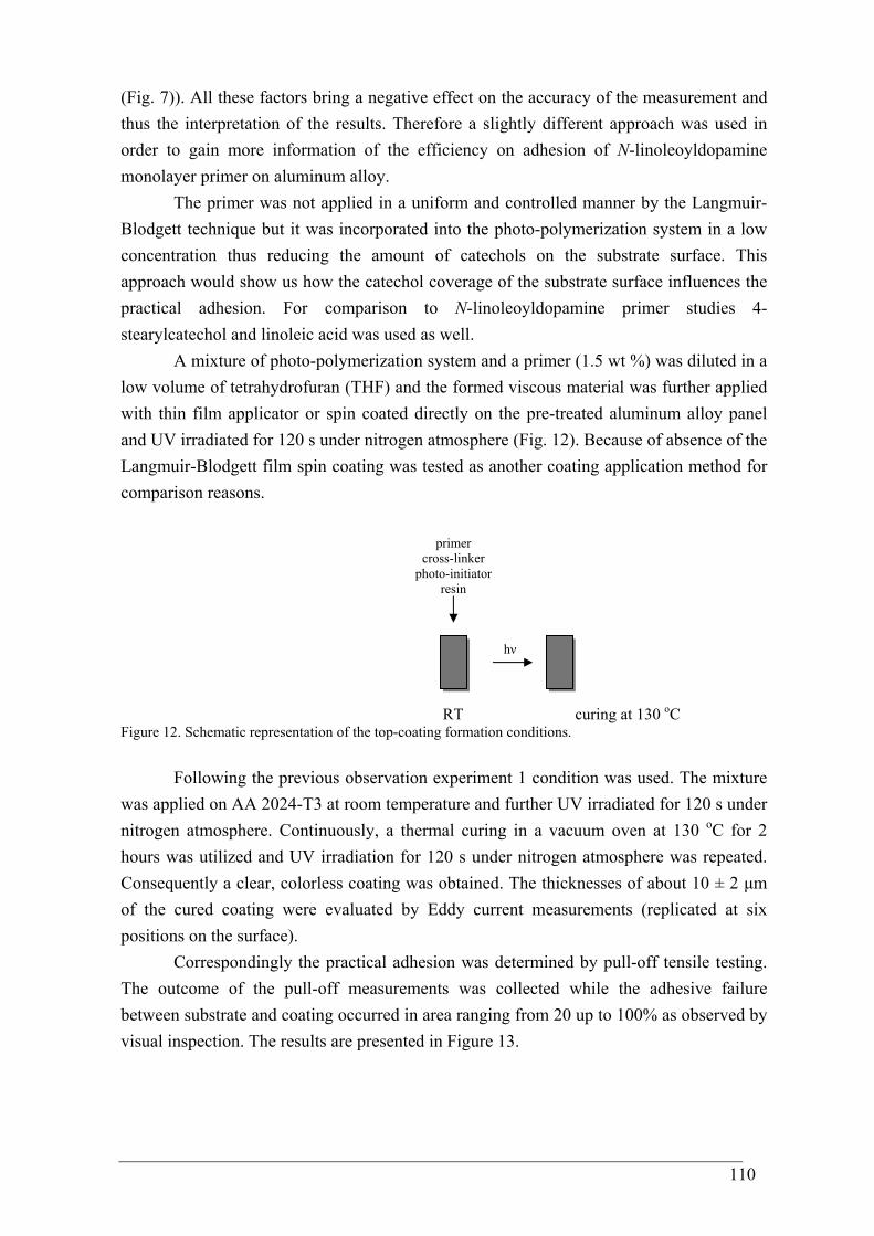
110
(Fig. 7)). All these factors bring a negative effect on the accuracy of the measurement and thus the interpretation of the results. Therefore a slightly different approach was used in order to gain more information of the efficiency on adhesion of N-linoleoyldopamine monolayer primer on aluminum alloy.
The primer was not applied in a uniform and controlled manner by the Langmuir-Blodgett technique but it was incorporated into the photo-polymerization system in a low concentration thus reducing the amount of catechols on the substrate surface. This approach would show us how the catechol coverage of the substrate surface influences the practical adhesion. For comparison to N-linoleoyldopamine primer studies 4-stearylcatechol and linoleic acid was used as well.
A mixture of photo-polymerization system and a primer (1.5 wt %) was diluted in a low volume of tetrahydrofuran (THF) and the formed viscous material was further applied with thin film applicator or spin coated directly on the pre-treated aluminum alloy panel and UV irradiated for 120 s under nitrogen atmosphere (Fig. 12). Because of absence of the Langmuir-Blodgett film spin coating was tested as another coating application method for comparison reasons.
RT curing at 130 oC
Figure 12. Schematic representation of the top-coating formation conditions.
Following the previous observation experiment 1 condition was used. The mixture was applied on AA 2024-T3 at room temperature and further UV irradiated for 120 s under nitrogen atmosphere. Continuously, a thermal curing in a vacuum oven at 130 oC for 2 hours was utilized and UV irradiation for 120 s under nitrogen atmosphere was repeated. Consequently a clear, colorless coating was obtained. The thicknesses of about 10 ± 2 μm of the cured coating were evaluated by Eddy current measurements (replicated at six positions on the surface).
Correspondingly the practical adhesion was determined by pull-off tensile testing. The outcome of the pull-off measurements was collected while the adhesive failure between substrate and coating occurred in area ranging from 20 up to 100% as observed by visual inspection. The results are presented in Figure 13.
hν
primer cross-linker
photo-initiator resin
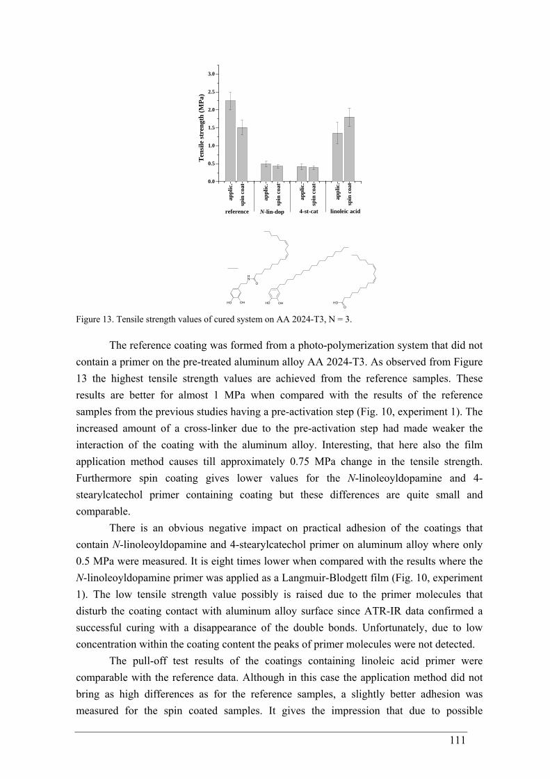
111
0.0
0.5
1.0
1.5
2.0
2.5
3.0
appl
ic.
appl
ic.
appl
ic.
linoleic acid4-st-catN-lin-dop
spin
coa
t
spin
coa
t
spin
coa
t
spin
coa
t
appl
ic.
reference
Ten
sile
stre
ngth
(MPa
)
OHOH
NH
O
OHO
OH OH
Figure 13. Tensile strength values of cured system on AA 2024-T3, N = 3.
The reference coating was formed from a photo-polymerization system that did not contain a primer on the pre-treated aluminum alloy AA 2024-T3. As observed from Figure 13 the highest tensile strength values are achieved from the reference samples. These results are better for almost 1 MPa when compared with the results of the reference samples from the previous studies having a pre-activation step (Fig. 10, experiment 1). The increased amount of a cross-linker due to the pre-activation step had made weaker the interaction of the coating with the aluminum alloy. Interesting, that here also the film application method causes till approximately 0.75 MPa change in the tensile strength. Furthermore spin coating gives lower values for the N-linoleoyldopamine and 4-stearylcatechol primer containing coating but these differences are quite small and comparable.
There is an obvious negative impact on practical adhesion of the coatings that contain N-linoleoyldopamine and 4-stearylcatechol primer on aluminum alloy where only 0.5 MPa were measured. It is eight times lower when compared with the results where the N-linoleoyldopamine primer was applied as a Langmuir-Blodgett film (Fig. 10, experiment 1). The low tensile strength value possibly is raised due to the primer molecules that disturb the coating contact with aluminum alloy surface since ATR-IR data confirmed a successful curing with a disappearance of the double bonds. Unfortunately, due to low concentration within the coating content the peaks of primer molecules were not detected.
The pull-off test results of the coatings containing linoleic acid primer were comparable with the reference data. Although in this case the application method did not bring as high differences as for the reference samples, a slightly better adhesion was measured for the spin coated samples. It gives the impression that due to possible
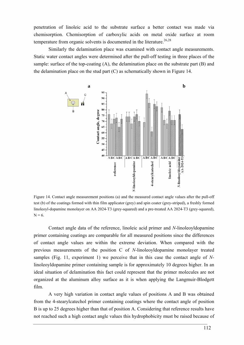
112
penetration of linoleic acid to the substrate surface a better contact was made via chemisorption. Chemisorption of carboxylic acids on metal oxide surface at room temperature from organic solvents is documented in the literature.26,28
Similarly the delamination place was examined with contact angle measurements. Static water contact angles were determined after the pull-off testing in three places of the sample: surface of the top-coating (A), the delamination place on the substrate part (B) and the delamination place on the stud part (C) as schematically shown in Figure 14.
a
b
Figure 14. Contact angle measurement positions (a) and the measured contact angle values after the pull-off test (b) of the coatings formed with thin film applicator (grey) and spin coater (grey-striped), a freshly formed linoleoyl-dopamine monolayer on AA 2024-T3 (grey-squared) and a pre-treated AA 2024-T3 (grey-squared), N = 6.
Contact angle data of the reference, linoleic acid primer and N-linoleoyldopamine primer containing coatings are comparable for all measured positions since the differences of contact angle values are within the extreme deviation. When compared with the previous measurements of the position C of N-linoleoyldopamine monolayer treated samples (Fig. 11, experiment 1) we perceive that in this case the contact angle of N-linoleoyldopamine primer containing sample is for approximately 10 degrees higher. In an ideal situation of delamination this fact could represent that the primer molecules are not organized at the aluminum alloy surface as it is when applying the Langmuir-Blodgett film.
A very high variation in contact angle values of positions A and B was obtained from the 4-stearylcatechol primer containing coatings where the contact angle of position B is up to 25 degrees higher than that of position A. Considering that reference results have not reached such a high contact angle values this hydrophobicity must be raised because of
113
the alkyl-chains of 4-stearylcatechol primer. Most probably the primer molecules concentrated at the coating-aluminum alloy interface and due to partial delamination of the coating from the metal surface (the positions B and C are comparable) the contact angles are highly hydrophobic. This observation brings our attention back to the pull-off results that did not show any difference in adhesion between N-linoleoyldopamine primer and 4-stearylcatechol primer containing coatings (Fig. 13) and to the pull-off results where the N-linoleoyldopamine primer was applied as a Langmuir-Blodgett film (Fig. 10), consequently pointing out that the Langmuir-Blodgett technique had successfully organized catechols to the metal surface. Moreover, the measured tensile strength values of the N-linoleoyldopamine primer treated coatings (Fig. 10, experiment 1) are 13 times higher as compared with the marine mussel M. edulis average adhesion on aluminum (0.29 ± 0.11 MPa, detailed description in Chapter 1).29
Previous studies of marine mussel foot proteins have proposed that the adhesion depends on DOPA,30,31 and created synthetic analogs have generally confirmed that the higher the DOPA content, the stronger the adhesion.32-34 It is announced that both natural and synthetic adhesives containing DOPA show good adhesion properties.35,36 The molecular level adhesion resulted in outstanding bonding between the substrate surface and the catechol. For instance, a single DOPA molecule was found to bind reversibly to TiO2 surfaces with a dissociation force of 805 pN in the presence of water.37 To put 805 pN in perspective, single molecule atomic force microscopy experiments revealed a dissociation force of 2000 pN for the silicon-carbon covalent bond38 and approximately 10-20 pN for the unzipping of DNA hydrogen bond pairs.39 Therefore, the catechol-titania interaction of 805 pN is quite strong and robust in aqueous environments.40 Recently an upscale from the single molecule adhesion to micro-level has been reported. Comparison of our work with some of these studies provides correlations. Yu et al. performed tensile strength measurements of poly(L-lysine4⋅HBr-L-DOPA1) and showed the adhesive strength of 4.3 ± 0.7 MPa on aluminum oxide.32 Our results here, on aluminum alloy, show a very similar average value of 4.0 ± 0.8 MPa (Fig. 10, experiment 1). Another example is provided by a synthetic adhesive based on dopamine containing methacrylate copolymer p(DMA-co-MMA) that was used as a primer to bond medical methacrylate type bone cement to aluminum oxide. A tensile strength of 20.9 ± 3.7 MPa was measured.41 This high tensile strength value will keep a motivation in the scientific society to work on the development of new synthetic adhesives that exhibit similar wet adhesive capabilities as mussel proteins and to continue to examine the main chemical interactions of which DOPA appears to be capable.
5.4 Conclusions
In this study we have demonstrated that a N-linoleoyldopamine monolayer can be
transferred from aqueous environment on a metal with a native oxide layer by the
114
Langmuir-Blodgett technique. The unsaturation in the alkyl chain exploits as a platform for secondary reactions.
The resulting tensile strength values of the top-coating having N-linoleoyldopamine monolayer primer are about 3 times higher as compared with the reference and about 13 times higher as compared with marine mussel M. edulis average adhesion on aluminum oxide. Conversely, a negative impact on adhesion occurs when the primer is not applied in a uniform and controlled manner by the Langmuir-Blodgett technique, thus having an 8 times lower tensile strength as compared with the results where N-linoleoyldopamine primer was applied as a Langmuir-Blodgett film.
According to the results of this work and the literature data, synthetic adhesives inspired by mussel adhesive proteins can be used successfully to improve the adhesive properties between polymers and metals.
5.5 References
1 Mittal, K. L. Adhesion Measurement of Thin Films, Thick Films and Bulk Coatings
1978, American Society for Testing and Materials, Philadelphia. 2 Mittal, K. L. Adhesion Measurement of Films and Coatings 1995, VSP, Utrecht. 3 Mittal, K. L. Electrocomponent Science and Technology 1976, 3, 21. 4 Assaban, A.; Gillet, M. J. of Adhesion Science and Technology 1999, 13, 871. 5 Frederick, J. R.; Ludema, K. C. J. Appl. Phys. 1964, 35, 256. 6 Czamocki, Z.; Matuszewska, M. P.; Matuszewska, I. Organic preparations and
procedures int. 1998, 30, 699. 7 Tallman, K. A.; Rector, C. L. ; Porter, N. A. J. Am. Chem. Soc. 2009, 131, 5635. 8 Tallman, K. A.; Roschek, Jr., B.; Porter, N. A. J. Am. Chem. Soc. 2004, 126, 9240. 9 Porter, N. A.; Caldwell, S. E.; Mills, K. A. Lipids 1995, 30, 277. 10 Tomoaia-Cotisel, M.; Zsako, J.; Mocanu, A.; Lupea, M.; Chifu, E. J. Colloid
Interface Sci. 1987, 117, 464. 11 Peltonen, J. P. K .; Rosenholm, J. B. Thin Solid Films 1989, 179, 543. 12 Heikkila, R. E.; Kwong, C. N.; Cornell, D. G., J. Lipid Res. 1970, 11, 190. 13 Seelig, A.; Seelig, J. Biochemistry 1977, 16, 45. 14 Chidambaram, D.; Clayton, C. R.; Halada, G. P.; Kendig, M. W. J. of Electrochem.
Soc. B 2004, 151, 605. 15 Roper, T. M.; Guymon, C. A.; Jönsson, E. S.; Hoyle, C. E. J. Polymer Science:
Part A: Polymer Chemistry 2004, 42, 6283. 16 Hoyle, C. E.; Lee, T. Y.; Roper, T. J. Polymer Science, Part A: Polymer Chemistry
2004, 42, 5301. 17 Morgan, C. R.; Magnotta, F.; Ketley, A. D. J. Polym. Science. Polym. Chem. Ed.
1977, 15, 627.
115
18 Cramer, N. B.; Bowman, C. N. J. Polymer Science, Part A: Polymer Chemistry 2001, 39, 3311.
19 Qin, L.; Wicks, D. A.; Hoyle, C. E. J. Polymer Science, Part A: Polymer Chemistry 2007, 45, 5103.
20 Carioscia, J. A.; Schneidewind, L.; O’Brien, C.; Ely, R.; Feeser, C.; Cramer, N. B.; Bowman, C. N. J. Polymer Science, Part A: Polymer Chemistry 2007, 45, 5686.
21 Hoyle, C. E.; Lowe, A. B.; Bowman, C. N. Chemical Society Reviews 2010, 39,1355.
22 Nair, D. P.; Cramer, N. B.; Scott, T. F.; Bowman, C. N.; Shandas, R. Polymer 2010, 51, 4383.
23 Liu, W.; Yin, H.; Akazawa, Y. O.; Yoshida, Y.; Niki, E.; Porter, N. A. Chem. Res. Toxicol. 2010, 23, 986.
24 Huijser, S.; HosseiniNejad, E.; Sablong, R. ; Jong, C.; Koning, C. E.; Duchateau, R. Macromolecules 2011, 44, 1132.
25 Koning, C.; Wildeson, J.; Parton, R.; Plum, B.; Steeman, P.; Darensbourg, D. J. Polymer 2001, 42, 3995.
26 Ting II, G. G.; Acton, O.; Ma, H.; Ka, J. W.; Jen, A. K.-Y. Langmuir 2009, 25, 2140.
27 Lin-Vien, D.; Colthup, N. B.; Fateley, W. G.; Grasselli, J. G. The Handbook of Infrared and Raman Characteristic Frequencies of Organic Molecules 1991, Academic Press: San Diego, CA.
28 Hall, J. T.; Hansma, P. K. Surface Science 1978, 76, 61. 29 Burkett, J. R.; Wojtas, J. L.; Cloud, J. L.; Wilker, J. J. The Journal of Adhesion
2009, 85, 601. 30 Waite, J.H. Integr. comp. biol. 2002, 42,1172. 31 Deming, T. J. Curr Opin Chem Biol. 1999, 3, 100. 32 Yu, M.; Deming, T. J. Macromolecules 1998, 31, 4739. 33 Lee, B. P.; Chao, C.-Y.; Nunalee, F. N.; Motan, E.; Shull, K. R.; Messersmith, P. B.
Macromolecules 2006, 39, 1740. 34 Yu, M.; Hwang, J.; Deming, T. J. J. Am. Chem. Soc. 1999, 121, 5825. 35 Waite, H.; Tanzer, M. L. Science 1981, 212, 1038. 36 Lee, H.; Dellatore, S. M.; Miller, W. M.; Messersmith, P. B. Science 2007, 318,
426. 37 Lee, H.; Scherer, N. F.; Messersmith, P. B. Proc. Natl. Acad. Sci. USA 2006, 29,
12999. 38 Grandbois, M.; Beyer, M.; Rief, M.; Clausen-Schaumann, H.; Gaub, H. E. Science
1999, 283, 5408. 39 Rief, M.; Clausen-Schaumann, H.; Gaub, H. E. Nat. Struct. Bio. 1999, 6, 346. 40 Hamming, L. M.; Fan, X. W.; Messersmith, P. B.; Brinson, L. C. Compos Sci
Technol. 2008, 68, 2042.

116
41 Stepuk, A.; Halter, J. G.; Schaetz, A.; Grass, R. N.; Stark, W. J. Chem. Commun. 2012, 48, 6238.
117
Conclusions and outlook
In the present PhD thesis the behaviour of dopamine-based promoters on metals
were investigated and one approach of determing practical adhesion had been developed. From the research several issues arose that need further investigations. Therefore apart from the main contributions of this thesis I would like to point out these open issues that can be the subject of future research. With interaction related issue:
Monolayer studies revealed that both model molecules, N-stearoyldopamine and 4-stearylcatechol, when transferred from an aqueous environment on a noble metal and metal with a native oxide layer by the Langmuir-Blodgett technique, formed films with similar variations in thickness depending from the substrate nature. The film thickness on sapphire is lower than for gold. Monolayer interaction with gold happens via the catecholic oxygen atoms that are coordinated to the surface having a more perpendicular than parallel orientation of the aromatic ring. This has also been observed by Ooka and Garrell for mussel foot protein adsorbed on gold. Since the chemistry of interfaces is rather complex, experimental studies and atomistic simulations on model systems can provide insights into the surface chemistry. Molecular simulation results on gold revealed the presence of various orientations of the catechols at the gold surface. This situation points out the need to use complementary, i.e. other surface sensitive techniques, e.g. X-ray Absorption Near Edge Spectroscopy (XANES). With this technique thin organic films on surfaces can be studied and the orientation of organic material by varying the X-Ray incidence angle can be determined. Another promising technique that is rather new is Tip-enhanced Raman spectroscopy (TERS) - a combination of two well-known techniques: atomic force microscopy (AFM) and Raman spectroscopy. AFM provides high spatial resolution and Raman spectroscopy enables chemical analysis of the sample. During this PhD research TERS depth profiling measurements were performed on N-stearoyldopamine monolayers. Our first experiments showed a large distance-related change in the TER spectra of the film, which was transferred onto a glass substrate.1 Due to the complicated nature of the Raman spectra, interpretation of the obtained results is not straightforward and has to be accompanied by high quality ab initio calculations.
The alkyl chains of N-stearoyldopamine monolayer on sapphire are less regularly packed and tilted with regard to the substrate surface than those in the monolayer on gold, having a tendency to pack together within domains containing approximately 2000 N-stearoyldopamine molecules. In contrast, the alkyl chains are tilted within the 4-stearylcatechol monolayer and on gold are less regular packed than those in the monolayer on sapphire. The irregularity of the film on gold leads to domain formation that is probably caused by the absence of the amide functionality. Molecular simulation results revealed that due to to the absence of the amide group catechols have various orientations at the gold surface and are not well organized. Nevertheless it appears that the catechol hydroxyl-
118
groups of the 4-stearylcatechol film are somewhat more coordinated to the gold surface and the alkyl- chains are more stretched with an orientation closer to perpendicular than was detected for the N-stearoyldopamine film. With adhesion strength related issue:
N-linoleoyldopamine monolayers transferred from an aqueous environment on an aluminum alloy by the Langmuir-Blodgett technique was exploited as a platform for secondary reactions since covalent bond formation between the double bonds of the N-linoleoyldopamine monolayer and the reactive groups of the top-coating provided a well-defined system to measure the catechol interaction with the aluminum alloy. The resulting tensile strength values of the top-coating having a N-linoleoyldopamine monolayer primer are about 3 times higher as compared with the reference and about 13 times higher as compared with marine mussel M. edulis average adhesion on aluminum oxide. Conversely, a negative impact on adhesion occurs when the primer is not applied in a uniform and controlled manner by the Langmuir-Blodgett technique. In that case it has an 8 times lower tensile strength as compared with the results where N-linoleoyldopamine primer was applied as a Langmuir-Blodgett film.
For comparison dopamine and polydopamine coatings also have been used. The quinone functional groups present in dopamine and polydopamine films are exploited as a versatile platform for secondary reactions. Covalently bound add-layers were used to determine the adhesion of dopamine and polydopamine films on aluminum alloy. The resulting tensile strength values of the epoxy and polyurethane coatings having dopamine/polydopamine primers are high – up to 50 times when compared with the marine mussel M. edulis average adhesion on aluminum (0.29 ± 0.11 MPa). This could be the result of the good adhesion of epoxy and polyurethane coatings.
Adhesion measurements of thin films are difficult and there are many factors influencing the results. Excluding the measurement errors I would like to point out such factors as the secondary reaction efficiency, primer (dopamine/polydopamine/monolayer) interaction at the metal interface and metal pre-treatment conditions. This PhD work presents one approach of determing practical adhesion; nevertheless, there are many issues open to further research. The primers can be functionalized and used for secondary reactions thus rapidly increasing the applications. It would also be interesting to explore the wet-adhesion capabilities of the primers as well as the adhesion on other metals.
According to the results of this work and the literature data, synthetic adhesives inspired by mussel adhesive proteins can be used successfully to improve the adhesive properties between polymers and metals.
References
1 Hoffmann, G. G.; Xue, L.; Loos, J.; With, G. Macromol. Symp. 2011, 305, 26.
119
Summary
Increasingly in science inspiration is drawn from Nature that provides many
outstanding examples of adhesive strategies. One of them is the adhesion of marine organisms, which present strong binding to virtually all inorganic and organic surfaces in aqueous environments in which most synthetic adhesives function poorly. The attachment occurs by secreting adhesive proteins that harden and cross-link in situ. Previous studies of these functionally unique proteins have revealed the presence of an unusual amino acid, 3,4-dihydroxy-L-phenylalanine (DOPA), which is responsible for the cohesive and adhesive strength of this natural glue and gives to marine organisms the ability to bond with numerous substrates including glass, Teflon, wood, concrete, plastics, metals, biological cell lines, bone, teeth, and others. Although the precise mechanism for assembly of the proteins is not understood, the obtained knowledge broadens biomimetic strategies for synthesizing new practical adhesives.
In this PhD thesis our efforts to understand the adherence behavior of dopamine-based promoters on metals are described. Firstly a simple DOPA mimics – dopamine was used to coat metal substrates and the properties of dopamine and polydopamine coatings on metals were studied in detail. Dip coating from aqueous dopamine hydrochloride solutions at pH 4 and pH 8 was performed on gold (having nor or hardly any native oxide) and aluminium (having a native aluminium oxide layer). The formed coatings were analyzed with surface sensitive techniques dealing with coating composition and the oxidation state, as well as with mechanical measurements characterising the adhesion of coatings. The outcomes of these studies emphasized a need to design a better defined system.
As a result two model molecules, N-stearoyldopamine and 4-stearylcatechol, were synthesized. These molecules contain a catechol residue that is capable of binding to metals and metal oxides and a hydrophobic alkyl-chain that gives an ability to organize molecules on surfaces using the Langmuir-Blodgett preparation technique. By using these catechol-containing molecules the role of amide functionality on packing, orientation and interaction with the metal surface could be compared. Attention was devoted in studying the adsorption behavior onto gold and aluminum oxide in terms of monolayer characteristics. The organization of Langmuir-Blodgett monolayers on a molecular scale was examined by several surface sensitive techniques. The experimental work was supported with molecular dynamic simulations on gold in order to get a more complete understanding of the monolayer organization near the interface. Our study shows that within the monolayer the catechols functions as a surface anchor on gold and the alkyl-chains appear to be tilted within the monolayer. Moreover, the irregularity of the 4-stearylcatechol film on gold leads to domain formation that is caused by the absence of the amide functionality. On the contrary, from the molecular simulations it appeared that for both types of molecules parallel orientations of the catechols with the gold are also present.
120
However, hydrogen bonds formed between the amide functionality and the catechol hydroxyl groups have a profound influence on the regularity on the adsorbed layer.
Attempts were made to quantify the adhesion strength of the anchoring catechols on metal oxide. A covalently bound top-coating was used to determine the adhesion of an N-linoleoyldopamine primer on an aluminum alloy. Several application conditions were tested for the top-coating and the primer. The resulting tensile strength values of the top-coating having an N-linoleoyldopamine monolayer primer formed by the Langmuir-Blodgett technique showed an obvious improvement in adhesion whereas a negative impact on adhesion occurred when the primer was not applied in a uniform and controlled manner. According to the results of this work and the literature data, synthetic adhesives inspired by mussel adhesive proteins can be used successfully to improve the adhesive properties between polymers and metals. Consequently, the outcomes of the research will give an input in the development of useful synthetic polymer adhesives that exhibit similar wet adhesive capabilities as mussel foot proteins.
121
Samenvatting
In de wetenschap wordt in toenemende mate inspiratie gehaald uit de natuur,
waarin uitstekende voorbeelden van adhesiestrategieën te vinden zijn. Eén daarvan is de adhesie van zeeorganismen, die een sterke hechting aan vrijwel alle anorganische en organische oppervlakken vertonen in waterige milieus, waarin de meeste synthetische lijmen slecht functioneren. De aanhechting vindt plaats door de afscheiding van adhesieve eiwitten, die uitharden en vernetten in situ. Eerdere studies naar deze functioneel unieke eiwitten hebben de aanwezigheid van een ongewoon aminozuur onthuld, 3,4-dihydroxy-L-fenylalanine (DOPA), dat verantwoordelijk is voor de cohesieve en adhesieve sterke van deze natuurlijke lijm en zeeorganismen het vermogen geeft te hechten aan talrijke oppervlakken, waaronder glas, Teflon, hout, beton, kunststoffen, metalen, biologische cellijnen, bot, tanden en dergelijke. Ondanks dat het exacte mechanisme van de assemblage van de eiwitten niet volledig begrepen wordt, biedt de verworven kennis ruimte aan biomimetische strategieën voor de synthese van nieuwe toepasbare lijmen.
In dit proefschrift worden onze inspanningen beschreven om het adhesiegedrag van op dopamine gebaseerde promotoren aan metalen te begrijpen. Eerst werd een eenvoudig afgeleide van de DOPA – dopamine – gebruikt om metaaloppervlakken te coaten en de eigenschappen van de dopamine- en polydopaminecoatings op metalen werden tot in detail bestudeerd. Dipcoating van waterige oplossingen van dopaminehydrochloride werd uitgevoerd bij pH 4 en pH 8 op goud (van nature zonder of vrijwel zonder oxidelaag) en aluminium (met een natuurlijke aluminiumoxidelaag). De gevormde coatings werden geanalyseerd met oppervlaktesensitieve technieken om de coatingcompositie en de oxidatietoestand te bepalen, evenals met mechanische metingen om de adhesie van de coatings te karakteriseren. De resultaten van deze studies benadrukten de noodzaak om een beter gedefinieerd systeem te ontwerpen.
Derhalve werden twee modelmoleculen gesynthetiseerd, N-stearoyldopamine en 4-stearylcatechol. Deze moleculen bevatten een catecholgroep met het vermogen om aan metalen en metaaloxiden te binden en een hydrofobe alkylketen met het vermogen om moleculen op oppervlakken te organiseren, gebruikmakend van de Langmuir-Blodgett-preparatietechniek. Door gebruik te maken van deze catecholfunctionele moleculen kon de rol van de amidefunctionaliteit in pakking, oriëntatie en interactie met het metaaloppervlak onderzocht worden. Aandacht werd geschonken aan het bestuderen van het adsorptiegedrag aan goud en aluminiumoxide in termen van monolaageigenschappen. De organisatie van Langmuir-Blodgett-monolagen werd onderzocht op een moleculaire schaal met verschillende oppervlaktesensitieve technieken. Het experimentele werk werd ondersteund door moleculaire dynamische simulaties op goud om een completer begrip van de monolaagorganisatie aan het grensvlak te krijgen. Ons onderzoek laat zien dat, in de monolaag, de catecholgroepen als oppervlaktehechters aan goud functioneren en dat de alkylketens in de monolaag gekanteld zijn. De onregelmatigheid van de 4-
122
stearylcatechollaag op goud leidt bovendien tot domein formatie die veroorzaakt worden door de afwezigheid van de amidefunctionaliteit. Daarentegen bleek uit de moleculaire simulaties dat voor beide typen moleculen ook parallelle oriëntaties van de catecholgroepen met het goud aanwezig zijn. Waterstofbruggen die zich tussen de amidefunctionaliteiten en de hydroxylgroepen in de catecholgroepen vormen, hebben niettemin een diepgaande invloed op de regulariteit van de geadsorbeerde laag.
Gepoogd werd om de adhesiesterkte van de hechtende catecholgroepen aan metaaloxiden te kwantificeren. Een covalent gebonden toplaag werd gebruikt om de adhesie van een grondlaag van N-linoleoyldopamine aan een aluminiumlegering te bepalen. Verschillende applicatiecondities voor de toplaag en de grondlaag werden getest. De resulterende waarden voor de trekvastheid van de toplaag met een monogrondlaag van N-linoleoyldopamine, gevormd met de Langmuir-Blodgett-techniek, lieten een duidelijke verbetering in adhesie zien, terwijl een negatieve invloed op de adhesie resulteerde wanneer de grondlaag niet in een uniforme en gecontroleerde manier werd aangebracht. Zoals blijkt uit de resultaten van dit werk en gegevens uit de literatuur, kunnen synthetische lijmen geïnspireerd door de adhesieve eiwitten van mosselen gebruikt worden om de hechtingseigenschappen tussen polymeren en metalen succesvol te verbeteren. De resultaten van dit onderzoek zullen daarom een impuls geven aan de ontwikkeling van toepasbare polymeerlijmen die vergelijkbare natte adhesiecapaciteiten als de mosseleiwitten vertonen.
123
Kopsavilkums
Arvien vairāk zinātnē iedvesma tiek ņemta no dabas, kas piedāvā daudzas izcilas
adhēziju stratēģijas. Tā piemēram, daži jūras organismi spēj veidot spēcīgu kontaktu ar faktiski visām neorganiskām un organiskām virsmām ūdens vidē, kur vairums sintētiskās līmes darbojas vāji. Kontakts tiek veidots ar proteīnu sekrēta palīdzību, kas in-situ sacietē un veido starpķēdes. Agrākie pētījumi noradīja uz netipiskas amino skābes, 3,4-dihidroksi-L-fenilalanīna (DOPA) klātbūtni šajos funkcionāli unikālajos proteīnos, kas veic kohezīvas un adhezīvas īpašības un tādejādi šie jūras organismi spēj veidot spēcīgu kontaktu ar daudziem materiāliem ieskaitot stiklu, teflonu, koku, betonu, plastmasu, metālu, bioloģiskās šūnas, kaulu, zobus un citus materiālus. Lai gan precīzs šo proteīnu izkārtojuma mehānisms nav zināms, iegūtās zināšanas var tikt izmantotas jaunu adhezīvu materiālu izstrādei.
Šajā promocijas darbā ir aprakstīti mūsu centieni izprast dopamīna tipa molekulu adhēziju ar metālu. Sākotnēji DOPA analogs - dopamīns tika izmantots, lai pārklātu metāla substrātus un dopamīna, kā arī polidopamīna pārklājuma īpašības tika sīkāk pētītas. Iemērcot metāla substrātus dopamīna hidrohlorīda ūdens šķīdumā pie pH 4 un pH 8, pārklājumi tika veikti uz zeltu un alumīniju. Veidotie pārklājumi tālāk tika analizēti ar virsmas jutīgam tehnikām, tādejādi pievēršot uzmanību pārklājuma sastāvam un oksidācijas pakāpei. Adhēzija tika noteikta veicot mehāniskus mērījumus. Iegūtie rezultāti noradīja uz nepieciešamību izstrādāt labāk definētu sistēmu.
Rezultātā divas modeļmolekulas, N-stearoildopamīns un 4-stearilkatehols, tika sintezētas. Šīs molekulas satur katehola daļu, kas ir spējīga saistīties ar metāliem un metāla oksīdiem, kā arī hidrofobo alkil- daļu, kura ļauj organizēt molekulas uz virsmām ar Langmuir-Blodgett sagatavošanas tehniku. Salīdzinot šīs katehola saturošās molekulas tām esot izvietotām monoslānī uz metāla virsmas, amīda funkcionālas grupas ietekme uz molekulu blīvējumu, orientāciju un savstarpējo mijiedarbību tādejādi tika noteikta. Uzmanība tika veltīta adsorbcijai uz zeltu un alumīnija oksīdu. Langmuir-Blodgett monoslāņa organizācija molekulārā skalā tika noteikta ar dažādām virsmas jutīgām tehnikām. Eksperimentālais darbs tika papildināts ar molekulāro dinamikas simulāciju uz zeltu, tādejādi iegūstot vispārīgu izpratni par monoslāņa organizāciju uz metāla virsmas. Mūsu pētījumi rada, ka kateholi darbojas kā virsmas koordinējošā daļa ar zelta virsmu un alkil- ķēdes monoslānī ir noliektas. Turklāt 4-stearilkatehola slāņa neregularitāte uz zelta virsmas noved pie domēnu veidošanos, kas ir amīda funkcionālās grupas neesamības rezultāts. Toties molekulāro simulāciju rezultāti noradīja, ka abām modeļmolekulām ir iespējama arī paralēla kateholu orientācija ar zelta virsmu. Liela ietekme uz adsorbētā slāņa regularitāti ir arī ūdeņraža saitēm, kas veidojas starp amīdu funkcionālajam grupām un kateholu hidroksilgrupām.
Kovalenti saistot N-linoleoildopamīna pamatslāni, kas veidots uz alumīnija oksīda virsmas, ar virsējā-pārklājuma palīdzību tika mēģināts noteikt kateholu adhēzijas stiprību

124
ar metāla oksīda virsmu. Dažādi pamatslāņa un virsējā-pārklājuma veidošanas veidi tika testēti. Tā rezultējošā stiepes izturība virsējam-pārklājumam, kura N-linoleoildopamīna pamatslānis tika veidots ar Langmuir-Blodgett sagatavošanas tehniku, uzradīja nepārprotamu adhēzijas uzlabojumu, toties negatīva ietekme uz adhēziju tika novērota, kad pamatslānis netika izveidots vienmērīgi un organizēti uz metāla virsmas. Saskaņā ar šī darba rezultātiem un literatūras datiem, sintētiskās līmes, kuras veidotas iedvesmojoties no šiem jūras gliemju adhēzijas proteīniem, var tikt veiksmīgi izmantotas adhezīvo īpašību uzlabošanā starp polimēriem un metāliem. Tādejādi šī darba rezultāti dos ieguldījumu noderīgu sintētisko polimēru līmju attīstībā, kam piemīt adhēzijas spējas ūdens vidē, līdzīgi kā tas ir šiem jūras gliemju adhēziju proteīniem.
125
Acknowledgements
This PhD project would not have been possible without the help and support of many people. Therefore, I would like to dedicate the last pages of this book to thank all these people who directly or indirectly contributed to this work and its completion. First of all, I would like to thank my promoter prof.dr. Gijsbertus de With for the opportunity to work in his group on this interesting topic. Thanks for all your support and guidance, and for the possibilities and the freedom to perform the research. My gratitude also goes to my copromoter dr. Nico Sommerdijk for his quick response to my questions and readiness to share his knowledge by giving very useful advices and inputs, that helped me to get over couple of scientific problems I had. I owe many thanks to my copromoter Mr Leo van der Ven for the useful suggestions and support during the project review and DPI meetings. I would like to thank the members of the doctorate committee prof.dr. G. de With, dr. N.A.J.M. Sommerdijk, prof.dr. C. Jérôme, prof.dr. G.J. Vancso, prof.dr.ir. R.A.J. Janssen and prof.dr. R.A.T.M. van Benthem for reading and approving my thesis. I am looking forward to defend my work to you. Prof.dr. J. Meuldijk I thank you for taking the chairman tasks. The Dutch Polymer Institute is kindly acknowledged for their financial support and for providing insights into various research fields during the cluster and annual meetings. I had the pleasure to gain insight in molecular interactions on gold when collaborating with Chuipeng Kong (PhD student from Jilin University). I am delighted that Chuipeng had been interested in doing a simulation study on my molecules. Dear Chuipeng, it was very informative to talk to you and thanks a lot for being always available for my questions. I wish you success with your PhD thesis and career! Regarding simulation study process I am also very thankful to dr. Günter Hoffmann and dr. Frank Peters for their assistance. My most sincere gratitude goes to dr. Rafael Sablong for providing me with the photo-polymerization system that was used for adhesion testing in Chapter 5. Dear Rafael, I highly appreciate your contribution to my dissertation and wish you all the best with your research! Many thanks are given to the AFM-experts: dr. Alexander Alekseev, Marco Hendrix and Willem van der Linden for performing AFM measurements. Your knowledge and
126
experience was very valuable when dealing with monolayer thin films. I also wish to thank Otto van Asselen for sharing his experience regarding IRRA spectroscopy. It was an appreciation for me to meet you and to gain some knowledge from you. I would like to thank Gilbère Mannie for performing XPS measurements, Maria Eriksson for TGA and Ralf Bovee for EA. Definitely TU/e is a fantastic university with great professionals and scientists. I keep wondering how I can fully convey my appreciation for those who were helpful, opened in sharing their experience, giving good advices/instructions and supporting me. Special thanks go to Christoph, Pit, Pauline, Carin, Bas, Tonny, Jan, Mindaugas, Elham, Cees, Martijn, Andreas, Wouter, Yogesh and many, many others. I hope to meet you again somewhere in this small world. Furthermore, I want to express my appreciation to the SMG group for their company and very useful assistance during these years. Thanks to Imanda, Sasha, Jos L., Paul, Huub, Niek, Catarina, Camille, Indu, Marcos, Mark, Beryl, Svetlana, Wouter, Hans, Katherine, Thanos, Adolphe, Judit, Baris, Vladimir, Fabio, Ela, Laura, Tina, Claudia, Katya, Isabelle, Maarten, Heiner, Delei, Qingling, Gökhan, Niels, Beulah, Koen, Marcel, Kirill, Oana, Cem, Karthikeyan, Hesam, Aroa and all the former and present members of SMG. Thanks to my great office-mates: Archan, Ziwei, Pedro, Jos. I would like to wish SMG to be a strong group in the coming years and success to all of you in your research and work! I want to thank my dear friends in Eindhoven Konstantinos, Julia and Eleonora. Thank you for your company and for making my days here more joyful. Vēlos pateikt paldies arī visiem skolotājiem, pasniedzējiem un kolēģiem par dotajām zināšanām un dalīšanos pieredzē. Iegūtās iemaņas man noteikti noderēja šī darba īstenošanā. Īpašs paldies maniem dārgajiem vecākiem, māsai, omammai un draugiem par atbalstu. Τέλος, η θερμότερη ευγνωμοσύνη πηγαίνει στον σύζυγό μου, Κωνσταντίνος. Κωνσταντίνε, σ΄ ευχαριστώ πολύ για την υποστήριξή σου, συμπαράσταση, υπομονή, αγάπη και κατανόηση. Αυτή η διατριβή είναι αφιερωμένη σε σένα με όλη μου την αγάπη.
127
List of publications
Rībena, D.; Alekseev, A.; van Asselen, O.; Mannie, G. J. A.; Hendrix, M. M. R. M.; van der Ven, L. G. J.; Sommerdijk, N. A. J. M.; de With, G. “The significance of amide functionality on DOPA-based monolayers on gold”. Submitted. Kong, C-P; Rībena, D.; Peters, E.; Zang, H-X; de With, G. “Understanding the Langmuir Monolayers of N-stearoyldopamine and 4-stearyl-catechol: A molecular dynamics study”. Submitted. Rībena, D.; Hoffmann, G. G.; de With, G. “A SERS Study on Monolayers of Model Compounds for the Adhesive Proteins of Mussels”. To be submitted.
128
Curriculum Vitae
Dina Rībena obtained her Bachelor (BSc) of Natural Sciences degree in Chemistry at the Riga Technical University (Latvia) in 2006. Her Master (MSc) of Engineer Sciences in Chemical Engineering was gained in collaboration within Riga Technical University (Latvia) and Royal Institute of Technology (Sweden). In 2008 she graduated with cum laude and started a PhD project in Eindhoven University of Technology, the Netherlands. The most important results of this research on dopamine modification of interfaces between polymers and metals are described in this thesis.


![Radiation Modification of Natural Polymers - Hacettepe · matrix, plant growth stimulator ... CHITOSAN Food processing, ... Topic: Radiation Modification of Natural Polymers [13]](https://static.fdocuments.us/doc/165x107/5ac116db7f8b9a1c768c7345/radiation-modification-of-natural-polymers-plant-growth-stimulator-chitosan.jpg)