Surface modification of polymers for biocompatibility via...
23
Surface modification of polymers for biocompatibility via exposure to extreme ultraviolet radiation Inam Ul Ahad, 1,2 Andrzej Bartnik, 1 Henryk Fiedorowicz, 1 Jerzy Kostecki, 1 Barbara Korczyc, 1 Tomasz Ciach, 3 Dermot Brabazon 2 1 Institute of Optoelectronics, Military University of Technology, 00–908 Warsaw, Poland 2 Advanced Processing Technology Research Centre, School of Mechanical and Manufacturing Engineering, Faculty of Engineering and Computing, Dublin City University, Dublin 9, Ireland 3 Department of Biotechnology and Bioprocess Engineering, Warsaw University of Technology Ul, Wary nskiego 1 , 00–645 Warsaw, Poland Abstract Polymeric biomaterials are being widely used for the treatment of various traumata, diseases and defects in human beings due to ease in their synthesis. As biomaterials have direct interaction with the extracellular environment in the biological world, biocompatibility is a topic of great significance. The introduction or enhancement of biocompatibility in certain polymers is still a challenge to overcome. Polymer biocompatibility can be controlled by surface modification Various physical and chemical methods (e.g., chemical and plasma treatment, ion implantation, and ultraviolet irradiation etc.) are in use or being developed for the modification of polymer surfaces. However an important limitation in their employment is the alteration of bulk material. Different surface and bulk properties of biomaterials are often desirable for biomedical applications. Because extreme ultraviolet (EUV) radiation penetration is quite limited even in low density mediums, it could be possible to use it for surface modification without influencing the bulk material. This article reviews the degree of biocompatibility of different polymeric biomaterials being currently employed in various biomedical applications, the surface properties required to be modified for biocompatibility control, plasma and laser ablation based surface modification techniques, and research studies indicating possible use of EUV for enhancing biocompatibility. Key Words: extreme ultraviolet, polymer processing, biocompatibility, biomaterials, surface modification techniques. How to cite this article: Ahad IU, Bartnik A, Fiedorowicz H, Kostecki J, Korczyc B, Ciach T, Brabazon D. 2014. Surface modification of polymers for biocompatibility via exposure to extreme ultraviolet radiation. J Biomed Mater Res Part A 2014:102A:3298–3310. INTRODUCTION The fundamental requirement of a material to be used as biomaterials is its ability to receive an appropriate host response. This response depends on how similar the implant behaves as compared to the real organ. Such requirement is commonly termed as biocompatibility. A material must achieve three fundamental aspects of biocompatibility to be employed as biomaterial in a patient. Biochemical compatibility is the principal aspect which reveals that the foreign material should not induce toxicity, irritation, allergy or carcinogenicity in the host. Second, the material should have a strong bio-adhesive quality. Adhesion of biomaterial
Transcript of Surface modification of polymers for biocompatibility via...

Surface modification of polymers for biocompatibility via
exposure to extreme ultraviolet radiation
Inam Ul Ahad,1,2
Andrzej Bartnik,1
Henryk Fiedorowicz,1
Jerzy Kostecki,1
Barbara Korczyc,1
Tomasz Ciach,3
Dermot Brabazon2
1 Institute of Optoelectronics, Military University of Technology, 00–908 Warsaw, Poland
2 Advanced Processing Technology Research Centre, School of Mechanical and Manufacturing Engineering,
Faculty of Engineering and Computing, Dublin City University, Dublin 9, Ireland 3Department of Biotechnology and Bioprocess Engineering, Warsaw University of Technology Ul, Wary
nskiego 1 , 00–645 Warsaw, Poland
Abstract
Polymeric biomaterials are being widely used for the treatment of various traumata, diseases
and defects in human beings due to ease in their synthesis. As biomaterials have direct
interaction with the extracellular environment in the biological world, biocompatibility is a
topic of great significance. The introduction or enhancement of biocompatibility in certain
polymers is still a challenge to overcome. Polymer biocompatibility can be controlled by
surface modification Various physical and chemical methods (e.g., chemical and plasma
treatment, ion implantation, and ultraviolet irradiation etc.) are in use or being developed for
the modification of polymer surfaces. However an important limitation in their employment
is the alteration of bulk material. Different surface and bulk properties of biomaterials are
often desirable for biomedical applications. Because extreme ultraviolet (EUV) radiation
penetration is quite limited even in low density mediums, it could be possible to use it for
surface modification without influencing the bulk material. This article reviews the degree of
biocompatibility of different polymeric biomaterials being currently employed in various
biomedical applications, the surface properties required to be modified for biocompatibility
control, plasma and laser ablation based surface modification techniques, and research studies
indicating possible use of EUV for enhancing biocompatibility.
Key Words: extreme ultraviolet, polymer processing, biocompatibility, biomaterials, surface
modification techniques.
How to cite this article: Ahad IU, Bartnik A, Fiedorowicz H, Kostecki J, Korczyc B, Ciach
T, Brabazon D. 2014. Surface modification of polymers for biocompatibility via exposure to
extreme ultraviolet radiation. J Biomed Mater Res Part A 2014:102A:3298–3310.
INTRODUCTION
The fundamental requirement of a material to be used as biomaterials is its ability to receive
an appropriate host response. This response depends on how similar the implant behaves as
compared to the real organ. Such requirement is commonly termed as biocompatibility. A
material must achieve three fundamental aspects of biocompatibility to be employed as
biomaterial in a patient. Biochemical compatibility is the principal aspect which reveals that
the foreign material should not induce toxicity, irritation, allergy or carcinogenicity in the
host. Second, the material should have a strong bio-adhesive quality. Adhesion of biomaterial
must be specific to a particular type of cells or tissues which depends upon the application. A
good adhesive contact between the implant and surrounding tissues must be established so
that biomaterial performs efficiently.
Lastly the biomaterial should possess similar biomechanical properties as those of its
surrounding tissues and organ for which it is replaced. It is quite worthy to note that all these
properties are application dependent. Biomaterials with a low level of biocompatibility
readily induce different infections within the patients. Organic polymers are considered as
important materials in various biomedical applications ranging from conventional cell growth
to the construction of hybrid tissues and artificial organs. Synthetic and naturally occurring
polymers have become important elements in new strategies for producing engineered
tissues. Several classes of polymers are now employed in biomedical applications, including
situations in which the polymer remains in intimate contact with cells and tissues for
prolonged periods.1–4
Control of the degree of biocompatibility in biomaterials is still a
challenge to overcome within the health care industry as these polymers very often do not
possess the surface properties needed for various applications.
Therefore their surface needs to be refined to the microstructure level to obtain better
performance in biomedical applications in terms of biocompatibility.4
Various chemical and
physical methods for modification of the polymer surfaces have been developed and are
currently being studied, including chemical and plasma treatment, ion implantation, and UV-
irradiation. Yet no surface modification technique is unanimously accepted since these
methods are often associated with undesirable side effects. One of them is the degradation of
the internal bulk of the material. Biomaterials have very precise requirements that derive
from the mechanical performance of the bulk properties. These provisions can be categorized
informally into three main groups including mechanical performance, mechanical durability,
and physical properties. In total hip replacement surgeries for example, the biomaterial used
for constructing a prosthetic implant must be mechanically strong and rigid.
In case of mitral valve replacement, the leaflet of valve must be flexible and tough; otherwise
it will cause hindrance in blood flow. Synthetic vascular graft material requires very specific
modulus properties in order to behave similar to real vascular soft tissue when implanted
within the body such that the walls of the artery or vein pulsate in a similar manner to real
tissue. In case of porous membranes (e.g., in dialysis), the membrane material should have
high young’s modulus and low yield strain though flexible and strong. For articular cartilage
substitute the requirements are totally opposite to that of porous membranes. If the bulk
material properties are modified inadequately and the material became stiff during UV or
other surface modification techniques, then the risk of restenosis or thrombosis originating
from these regions became unacceptably high. Similarly surface modification techniques may
spoil the refraction and clarity of bulk material of intraocular lens, making them inefficient.
Extreme ultraviolet radiation is high-energy ultraviolet radiation, having photons with
energies ranging from about 10 eV up to 124 eV (corresponding to wavelengths of 124– 10
nm, respectively). Degradation of bulk material can be avoided by using short wavelength
radiation in the extreme ultraviolet (EUV) range that is absorbed within a very thin (<100 nm)
layer of the polymer for surface modification.
The following section, Polymeric Biomaterials and Their Bioincompatibility section, of
this article presents important polymeric biomaterials which are used in the health care
industry that require biocompatibility control. In Surface Properties for Biocompatibility
Control section the important surface properties of biomaterials are discussed. Because
biomaterials and host interact with each other in various ways, different surface properties of
biomaterials influence this coexistence. These surface properties are used to determine the
qualifications of a material to be used as biomaterial. A number of surface properties along
with their contribution in interaction of host and biomaterial are also discussed. In Surface
Modification section of this article a review of the plasma and laser ablation based surface
modification techniques are discussed. Recently a laser-plasma based EUV source dedicated
for polymer processing and surface modification has been built in Institute of Optoelectronics
(IOE) at Military University of Technology ( MUT ) Warsaw.5
Only a few studies have been
conducted utilizing this source for surface modification and biocompatibility control. Strong
indications of the applicability of this source to be used for polymer processing and surface
modification for biocompatibility control are presented.
POLYMERIC BIOMATERIALS AND THEIR BIO-INCOMPATIBILITY
Biomaterials are specially selected, structured and designed to interact with biological world
for treatment of various traumata, diseases, and defects. The most important requirement of a
biomaterial is to remain in intimate contact with host tissues without producing any harmful
effect (biocompatibility). Biocompatibility is a general term used to describe the suitability of
a material for exposure to the body or bodily fluids. It is the ability of a material to perform
with an appropriate response in a specific application and it dependents on the particular
application or biological conditions. A material will be considered as biocompatible (in a
specific application) if it allows the body to function without any complications such as
allergic reactions or other adverse side effects. Lack of biocompatibility can result in
disruption of the normal healing processes and additional complications like inflammation,
cytotoxicity, cell disruption, skin irritation, thrombosis and so forth.
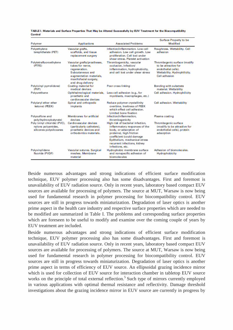
Important polymers used as biomaterials widely in the health care industry are presented
below along with their applications and associated problems due to lack of biocompatibility
control.
Polyethylene terephthalate
Polyethylene terephthalate (PET) has a wide range of applications in biomedical engineering.
Particularly in tissue engineering, PET is used for vascular grafts and scaffolds for tissue
regeneration. Moreover PET is extensively used for tissue replacement surgery.3,4
Polyamide
fabrics are also used for the same applications due to similar characteristics. Nevertheless
both polymers exhibit various problems due to lack of integration with the host environment.
The most common use of PET grafts is in bypass surgeries. The most common problem
encountered by any vascular graft is infection due to lack of antibacterial properties. Most of
these polymeric grafts are manufactured in knitted format and often result in occlusion or
distal embolization. Because of flow of blood through grafts, erosion into adjacent structures
is also common. Infection due to bad host response may also cause formation of true or false
aneurysm which may cause sudden death.6,7
Because of the smooth surface, PET depicts low
cell adhesion in a host which results in weak cell attachment. The weakness in cell attachment
consequently affects the cell growth, proliferation, and differentiation. Moreover cell loss also
occurs under increased shear stress due to decreased roughness. Roughness on surface of PET
can be introduced or increased through various procedures, however they also alter the bulk
material and the desired mechanical characteristics of PET are ultimately lost. PET
demonstrates low water wettability which causes various problems upon contact with blood
plasma (e.g., platelet activation).8,9
Use of an autologous vein graft may avoid all these
complications however they are often not available or sometimes not suitable for the
particular case.
Polytetrafluoroethylene (PTFE)
In the stenotic arteries bypass autologous vein grafts are preferred to use but in case of
unavailability, polytetrafluoroethylene (PTFE) is commonly used. Beside health care
applications, PTFE also has various uses in different industries due to its low friction
characteristic. In addition to the stenotic arteries vascular grafts, PTFE is also used for general
construction of vascular prostheses, tubes for nerve regeneration, subcutaneous augmentation
materials, and in maxillofacial surgery.1,10,11
Various complications and hazards are affiliated
with this polymer as its surface is thrombogenic which leads to vascular occlusion.1,12
To
avoid complications, the surfaces of polymers should be attractive for endothelial cells which
are not thrombogenic in nature, thus thrombogenicity can be avoided resulting in an increase
of biocompatibility of these vascular prostheses. Drug delivery is another important
application of polymers in biomedical engineering. Particularly for protein drug delivery,
ability to absorb water, open swollen structure and biodegradability is required in polymers so
that protein can be efficiently loaded.13
Poly(vinyl pyrrolidone) (PVP)
For some artificial organs (e.g., artificial heart) nonbiodegradable materials are required with
excellent biocompatibility and nonantigenicity.14
Holding ability to absorb water without
dissolving in host environment is crucial for such applications. Hydrogel polymers are
prominently used materials which are highly absorbent and can contain huge amounts of
water in their composition (99.9%). They possess similar properties as those of soft tissues
though in some cases they may be toxic or nonbiocompatible. Beside fabrication of artificial
organs, they are also used for scaffold material for tissue engineering, cell encapsulation, and
intelligent cell culture substrates. For introducing biocompatibility or optimizing the degree
of biocompatibility, polymeric coatings are applied on medical device surfaces.15
Poly (vinyl
pyrrolidone) (PVP) is a common hydrogel coating material for medical devices due to its in-
adhesive response towards bacteria. Moreover this polymer provides a smooth surface which
has low friction to extracellular fluids. Yet PVP binding with substrate is not stable for long
periods of time, thus cannot be used for long term implants. Because of the high level of
biocompatibility of PVP, it can be extremely beneficial for long term implants if crosslinking
is improved, consequently their stability with substrate will be higher. Different groups have
applied various approaches to optimize crosslinking. Though an important limitation exists
with stable hydrogel in that they lose the desired mechanical properties which are required for
certain applications like heart valves and artificial heart.16
This limitation is application
dependent as for breast implants for example, different mechanical properties compared to
those for vascular grafts or heart valves are required. Therefore the requirements of
crosslinking density are application dependent.
Polyurethane ( PU )
Polyurethane is a synthetic rubber use in diverse biomedical applications including
ophthalmological materials, prosthetics and cardiovascular devices.17
Particularly in
prosthetic devices they portray less cell adhesion and may require to have moderate surface
hydrophilicity in order to be compatible for various cell types (e.g., myoblasts, macrophages,
etc.).18
Poly(aryl ether ether ketone) ( PEEK )
Because of the thermoplastic nature, strong mechanical and chemical properties offered by
poly(aryl ether ether ketone) (PEEK) makes it valuable for medical implants. The degree of
robustness of this polymer is truly exploited in various industries. In biomedical engineering
spinal and orthopedic implants are usually constructed by PEEK. They are relatively inert and
have been proved to be biocompatible. It is a good replacement of metallic biomaterials in the
prosthesis manufacturing. Important concerns in the application of PEEK as a biomaterial
include water absorption which reduces polymer crystallinity and inertness of PEEK which
affects cell adhesion resulting in limited bone fixation. The versatile nature of PEEK can be
extended via surface modification for novel implant applications.19 , 20
Polysulfone and poly(hydroxybutyrate)
These polymers are used as membranes in various biomedical artificial devices. In principle
these polymers exhibit extremely low biocompatibility, yet they are valuable to be used as
membranes in the biological world. To increase the biocompatibility, plasma coatings are
often deposited on their surfaces.21
Other polymers
Poly (vinyl chloride) (PVC), nylons polyamides, and silicones polysiloxanes are prominent
polymers used for manufacturing of cardiovascular devices (particularly catheters), prosthetic
devices and orthodontics materials. Problems associated with these polymers used in the
biological worlds include their low biocompatibility in terms of high risk of bacterial
infection, inflammatory responses of the body, and adsorption of proteins. High friction
coefficient leads to damage of the epithelium or it may result in the excessive mechanical
shear stresses. These problems cause various long term side effects such as recurrent
infections and kidney inflammation and so forth.
SURFACE PROPERTIES FOR BIOCOMPATIBILITY CONTROL The basic factors
that govern the biocompatibility of biomaterials are incompletely understood. No single test
is sufficient to characterize the material on the basis of the biocompatibility. A variety of tests
are necessary to determine the degree of the biocompatibility. These tests depend on the
surface property to be investigated, implant class, application, and most importantly the host
cell type to be in contact with the biomaterial. The response between the host and the
biomaterial is not unidirectional. It is important to note that the routine of this mechanism is
not unique even for any particular application. A range of natural phenomena occur due to
interaction of the host and the biomaterial. Therefore a range of in vitro and in vivo tests,
which characterize the surface properties and the chemical structure of the polymers that
influence their biocompatibility have been developed and are routinely implanted.
In this section, most important surface characteristics of the polymers are discussed which
can be modified in order to gain the control over the degree of the biocompatibility.
Surface morphology
The surface morphology of biomaterials determines the interactions occurring at the interface
site of the host and biomaterial. Crystallinity is a major morphological characteristic of
materials that influence host response. Moreover low dielectric constant, low refractive index,
and high optical transparency along with good mechanical and thermal stability are the
important properties required in bio-MEMS, blood-contact devices and cell culture substrates.
Chemical structure and functional groups
The harmful response by the host, like damaged cells or irritants which cause inflammation
and homogenization can be avoided by making the biomaterial attractive for endothelial cells
(improved endothelialization).6,22,23
Similarly for various applications, proliferation and cell
adhesion towards a particular host cell types are required. To make the polymer surface
attractive for the particular cell type, polymeric biomaterials surfaces are often functionalized
through various modification techniques by trapping or dopping reactive substances on the
surface.23,24
The chemical composition is therefore important to recognize the presence of
externally introduced functional groups for particular applications and also to determine the
presence of any toxic substance (element or compound) that may be present within the
material.
Interfacial free energy
For biologically and mechanically stable solid (biomaterial)liquid (blood/extracellular fluid)
interface a low solid–biological fluid interfacial free energy of the order 1–3 dyne cm21
is
required.25
Although the interfacial free energy primarily depends upon the interface layer
thickness, it also depend on certain variables such as temperature, friction, and pressure of the
biological fluids. These variables in the biological environment are ever-changing within a
specific range.24
This thermodynamic quantity contributes to the adsorption of blood
components onto the guest biomaterial.
The blood component adsorption thus can be control by interfacial free energy.
Wettability
It is evident that polymeric surfaces possess quite low wettability.26
The wetting of a surface
by a liquid is affected by the roughness of the surface. In practice it is shown that both the
chemical properties (heterogeneity) and the physical properties (surface roughness, shape,
and particle size) of the surface influence its wetting behavior.27
Wettability of the
biomaterials can be optimized to limit the contact friction between the host and the implant.28
Regulating the wettability influences the protein adsorption and biocontact properties.29
Wettability is also related to surface energy. Low surface energy polymers depict poor
wettability.30
Good wettability is required in some biomaterials for deposition of functional
groups onto the surface. Such modified polymers can be used as a substrate material used for
cell
cultures.26 , 31
Hydrophobicity
Particularly for biodegradable applications, the biomaterials to be used should have increased
hydrophobicity so that they may dissolve in water. For tissue repairing scaffolds,
biodegradable biomaterials are preferred in research projects for special experiments. Such
biomaterials are now employed in recent commercial applications. The proliferation and
differentiation of cells can be biologically altered by control of the surface hydrophobicity
and charge of culture substrates.32
Hydrophilicity
Protein fouling has been a major problem for biomaterials in general often making them
nonbiocompatible. The aggregation of proteins results in their adsorption onto the surface of
the biomaterials. Ultimately thrombus formation due to protein fouling causes not only
resistance in the extracellular fluid flow but also the surface chemistry of the host alters.
Particularly in the membrane surfaces, increased hydrophilicity helps to suppress protein
fouling.33 – 35
Cytotoxicity
Toxic behavior investigation is mandatory in order to assess any material to be used in
biomedical engineering applications. The cytotoxicity experiments determine whether the
material depicts toxic behavior while in contact with the general or particular cell lines. The
test is generally done in a laboratory using standard/relevant cell lines and the cells are seeded
on the materials. As far as the experimental evaluation of biocompatibility is concerned, the
cytotoxicity tests are widely cited as the primary assessment of biocompatibility.
Adhesion
For the design of biomaterials, adhesion is an important factor to be optimized for the
compatible cell–material interactions. Therefore it is important to control the cell growth on
implant surfaces in order to characterize the biomaterials for biocompatibility.36–38
In the
biological world, “Bioadhesion” represents both bacterial and cell adhesion. Bacterial
adhesion on biomaterial surfaces could be fatal as it results in evasion or inhibition of
immune response of the host. However the requirement of cell adhesion on implant is both
application dependent and cell-type dependent. Typically in vascular grafts, increased
adhesion is desirable for better cell attachment, proliferation, and spreading.39
Moreover
metallization of polymers requires strong adhesion between the polymer and the metal for
medical applications.26
As an indirect paradigm, for vascular prostheses, the inner surface is
in direct exposure to the endothelial cells and therefore it must be attractive to such cells. This
is achieved by a protein coating on the polymer surfaces. However strong adhesion is
required to hold the protein coating onto the polymer surfaces.11
On the contrary for some applications platelet adhesion with polymeric implant has to be
minimized in order to avoid thrombus formation.22,40,41
In the artificial heart valves,
thromboembolism is the prominent complication which hosts experience, eventually leading
to blockage in the blood flow.42,43
To increase the uptime of such prosthetics, the implants
should be treated to minimize thrombous regeneration.
SURFACE MODIFICATION
Surface modification to alter a wide range of characteristics of surfaces is an expanding field
enchanting the researchers and industries equally. Particularly for biomedical engineering
applications the assorted mechanisms of host and biomaterial interaction require explicit
surface characteristics in order to avoid any deleterious effects. Employment of polymers as
biomaterials in the healthcare industry is well established due to the ease in the production of
versatile polymers which hold required physical and chemical properties. Because surface
properties of polymers determine their biological performance during interaction with the
host, biocompatibility control through surface modification is an inevitable step in the
production process. Different bulk and surface properties are crucial for biomaterials in the
biomedical engineering applications. It is however not possible to well define these properties
during a single stage fabrication process. The common practice is to provide a special
treatment following the fabrication of the biomaterials to modify surface properties to the
desired level. Eventually decoupled bulk and surface properties are attained. A combination
of two or more physical and/or chemical treatments can assure such modifications. In the
following section a brief overview of plasma based and laser ablation based surface
modification techniques used to control the degree of the biocompatibility are presented.
Thenceforth initial research studies regarding a new technique of surface modification by
extreme ultraviolet ( EUV ) are discussed with indications of possible application in the
control of the degree of biocompatibility.
Plasma-enhanced chemical vapor deposition
Plasma-enhanced chemical vapor deposition (PECVD) is a chemical process in which gas
vapors from plasma deposit on the surface of the sample being treated. Plasma deposition is
quite often used in manufacturing of semiconductor devices particularly those with
temperature sensitive structures. PECVD has vast applications ranging from semiconductor
technology,44–47
molecular sieve membranes for gas separation48
and packaging barrier
films.49,50
However surface modification by PECVD for biomedical engineering applications
is limited to silicon based membranes and substrates,51
steel,52
and alloys.53,54
Polymer
processing for biocompatibility control by PECVD is quite restricted due to non-uniformity
and formation of by-products. Nevertheless a few studies demonstrated preparation of
diamond-like carbon (DLC) coated films by PECVD with an improved degree of
biocompatibility.55 – 57
Reactive ion etching
Reactive-ion etching (RIE) is the main plasma etching technology used for fabrication of
microstructures. The etching mechanism in RIE is a result of chemical etching which takes
place due to chemical reaction between the sample (wafer or film) and gas atoms forming a
molecule to be removed from the substrate. Negligible amount of physical etching is also
involved. RIE is primarily used in the semiconductor industry for the fabrication of the
integrated circuits (IC).58
As RIE is typically used for pattern transfer, prominent
applications in biomedical engineering can be found in the fabrication of membranes,
microelectrode arrays (MEA), and microelectromechanical systems ( MEMS ) for biosensors
and lab-on-a-chip (LOC). Ultrananocrystalline diamond (UNCD) membranes with 100 and
200 nm diameter pores (high porosity [50%]) were fabricated using reactive ion etching.59
Such membranes mimic natural filtration system thus can be used for wide applications in
biomedical engineering. Micro-patterns are introduced in poly (dimethylsiloxane) (PDMS)
silicone elastomer using customized RIE technique for fabrication of elastic multielectrode
array for surface stimulation of the spinal cord.60
However there are associated undesirable
RIE effects which influence the micropatterning. Most prominent is the implantation of
impurities during the chemical process of etching such as hydrogen diffusion. There is risk of
pattern damage due to the presence of the energetic ions or radiation. Because of the chemical
interactions loss of the doping agent which is aimed to be inserted into the sample is another
major problem. Most undesirable factors can be eliminated or limited but post processing is
required. 58
Plasma immersion ion implantation
Plasma immersion ion implantation (PIII) is used to insert impurity into the substrate by
extracting accelerated ions from the plasma and directing them towards the sample. PIII has
been used to improve antibacterial properties of polymers. Polyvinyl chloride (PVC) which is
one of the most produced plastic has been coated with triclosan and bronopol and doped with
argon using plasma treatment. Improvement in antibacterial properties against S. aureus and
E. coli is demonstrated by biocompatibility tests though such surface modification.61
Argon
and oxygen immersed on surfaces of polycarbonate and polytetrafluoroethylene using PIII
respectively. Oxygen enrichment resulted in hydrophocitiy of surface which offer higher
affinity for human cell attachment.62
PIII used to treat polyethylene terephthalate surface by
acetylene to control the degree of hemocompatibility with pronounced effect on bacteria
adhesion.62
PIII can also be successfully employed for surface modification of the bio-implant
alloys with the doping of nitrogen and phosphorus.63
Nevertheless in the long run, the
antibacterial property introduced or enhanced by PIII is reduced significantly due to
interactions in the biological world.62
Ultraviolet radiation surface modification
The ultraviolet radiation in the range from 126 to 222 nm wavelength can be well absorbed by
organic materials. The ultraviolet laser light from excimer lamps can be used to irradiate the
sample with enough energy to disrupt the molecular bonds on the surface. The disruption of
molecular bonds causes a number of photo-physical, thermal and photochemical processes.64–
68 This influence is not limited only to surface layer of the material but it also alters the bulk
properties. The use of the excimer laser for surface modification to provide enhanced
biocompatibility is quite an old technique.66,69–71
Laurens et al. irradiated polycarbonate (PC)
and polyether-etherketone (PEEK) with different UV wavelengths even below the ablation
threshold and demonstrate increased wettability after treatment.31
Because different types of cell from the host (the patient) interact with the polymer
surface, it is quite beneficial to introduce particular functional (reactive) groups on the
interface site of the biomaterial. Such type of material functionalization adjusts the surface
characteristics of the biomaterial and can provide attractive sites for the attachment of the
particular cell types. In this way, the biomaterial can be designed with favorable adhesive
properties for particular tissues. Consequently inflammatory and toxic responses can be
avoided. Tidwell et al. tabulated the effect of different functional groups on the proliferation
of bovine aortic endothelial cells. It has been demonstrated in the study that the growth rate of
these cells significantly improved in the presence of different chemical functionalities.72
Functional groups deposition onto the polymer surfaces is quite a delicate process as the
polymer surfaces are treated in a way to remain nontoxic while in contact with particular cell
types. Therefore no toxic materials should be induced during the modification process.
UV surface modification has been considered to control the degree of biocompatibility for
various polymers. Heitz et al. irradiated polytetrafluoroethylene (PTFE) with UV light of a
Xe2-excimer lamp at 172 nm wavelength. The polymer was treated in an ammonia
atmosphere. Some samples were grafted with amino acid alanine after being treated by UV. It
was observed in the study that UV irradiated PTFE foils depict higher optical absorbance,
exhibit strong fluorescence and increased wettability. Consequently rat aortic smooth muscle
cells (SMC), mouse fibroblasts (3T3 cells) and human umbilical vein endothelial cells adhere
more to such UV irradiated polymer samples as compare to untreated samples and exhibit
good proliferation.73
Gumpenberger from the same group performed further investigations on
UV irradiated PTFE and observed formation of new chemical groups on treated polymer
surfaces and demonstrated statistically higher proliferation rates and elevated adhesion on
smooth muscle cells and fibroblasts.11
Prolonged process timings (up to 30 min UV
irradiation) were used in these studies. Uchida et al. confirmed increased hydrophilicity of
poly(ethylene terephthalate) (PET) film upon UV treatment.9
Doi et al. introduced
microporosity in polyurethane (PU)-based vascular prosthesis through computer-aided
excimer laser (KrF) ablation technique. This small caliber graft was expected to exhibit
enhanced in vivo transmural tissue proliferation.74
This anticipation is further confirmed by
the same group for polyurethane grafts.75
UV light was used to enhance the interaction
between DNA molecules and the plasma polymer chains.76
Several other biocompatibility
control studies through UV irradiation can be found elsewhere.3,29
Surface functionalization
of amorphous carbon films can be used for various biomedical engineering applications. UV
laser assisted micro-structuring of hydrogenated amorphous carbon thin films result in
formation of carboxyl groups at the surface which leads to improved wettability of water,
polar and dispersive liquids.77
It is quite worthy to note that rather than polymers, UV micro-
patterning is more suitable for control of the degree of biocompatibility of metallic
alloys.78 – 80
Extreme ultraviolet (EUV) radiation surface modification
A basic requirement for a technique to be acceptable for the surface modification to provide
control over the degree of the biocompatibility is that the bulk properties are retained during
treatment. Photo (chemical) processes employed conventionally use short wavelength
radiation (UV) with photon energies sufficient to break the chemical bonds on the polymer
surface. These photons are capable of penetrating deep inside the polymer. In some cases
penetration depth of these radiations is up to 500 mm which ultimately alter bulk properties.81
Similarly in the case of plasma based techniques, degradation of sample lattice and bulk
material was reported. Yet for application of polymers in biomedical engineering, surface and
bulk properties must be decoupled as nearly for all applications in the biomedical domain,
different surface and bulk properties of biomaterials are required.
Extreme ultraviolet (EUV) radiation is high-energy ultraviolet radiation, having photons
with energies from about 10 eV up to 124 eV (corresponding to wavelengths from 124 to 10
nm, respectively). Two important factors encourage the employment of EUV for surface
modification of polymers. The most significant is the corresponding photon energy which is
capable of breaking more molecular bonds at the upper polymeric surface as compared to
excimer lamps or excimer lasers. Smooth ablation of polymers by a laser plasma based EUV
source is well established.82
Second since the EUV radiations are highly absorbable even in
low dense medium, their penetration depth is very limited (<100 nm in the upper layer of
polymers).83
The range of wavelength and penetration depth offered by EUV photons make
them possible to write small patterns on the surface layers of polymers.

The EUV radiation may produce by plasma based sources or by synchrotron sources. In
plasma based sources, EUV can be produced either by laser irradiation or by gas discharges.
In synchrotron sources (SR), ultrarelativistic charge particles accelerated through magnets
emits EUV radiation. In the laser-plasma based sources, a hightemperature plasma is
generated by the interaction of high power laser pulses with a solid target. However,
production of debris during laser interaction with matter target is a huge limitation. This
problem has been solved by introducing gas target instead of a solid target.
Laser ablation leans on the nature of the material and its ability to absorb energy.
Therefore the wavelength of the ablation laser should have a minimum absorption depth.
Laser ablation rate hence primarily depends upon the laser wavelength and the pulse length.
Therefore the ablation rate relies upon amount of total energy delivered in one shot and
optimal spectral distribution. These two factors cause the main reason of the huge difference
between the ablation rate of SR sources and laser-plasma based sources. SR sources have
long pulse length and have low-peak-power while high energy laser-plasma sources are able
to produce ultra-short pulses with high peak-power. As a matter of fact, single photon from
both sources carries enough energy to break any chemical bond for direct photo-etching.
However the photons from two sources interact with material distinctively due to following
factors:
Total energy in one shot. As the peak intensity (PI) is calculated by peak-power in a given
unit area (focal spot), low peak power SR source produce low-peak-intensity irradiation. As a
result of this irradiation only a small area (focal spot) with a very thin surface layer obtain
energy for quite a slow ablation process. On the contrary, in case of laserplasma source, high
peak intensity irradiation causes a huge number of simultaneously occurring radiation
induced photoetching proceedings. The absorption length of the radiation is relatively long in
this case as compare to a SR source. The energy which is absorbed in the surface layer is also
able to overheat the material. The evaporated (or sublimated) materials blow off into the
vacuum. In this way fast ablation rate is achieved by a compact laser-plasma source which
results in the construction of microstructures up to few microns in depth at material surface.
Optimal spectral distribution. As explained above, laser ablation depends upon the ability of
a material to absorb energy. This means that the absorption length of the radiation in the
materials should be comparable to the thickness of the material being desorbed. Strictly
speaking, for a given radiation energy range (power density) there must be an optimal spectral
distribution for efficient photo-etching. With specific critical energy and wavelength, SR
sources do not fit in this criterion. However studies by single shot highpeak-power laser-
plasma source demonstrated the optimal spectral distribution.
Considering these two factors, a compact laser plasma source demonstrates fast laser
ablation rate as that of synchrotron sources. Moreover due to the limited number of SR
sources and high cost of such facilities, various laser-plasma based EUV sources by various
groups have been proposed and considered for various applications in science and
technology. A laser-plasma based EUV source is specially built which is dedicated to
polymer processing and surface modification at MUT, Warsaw.5
Recent studies from this
EUV source show that EUV micro-patterning produce micro- and nano-structures on polymer
surfaces and consequently the surface properties of polymeric biomaterials modified in order
to attain control over the degree of the biocompatibility. Moreover photochemical processes
(by introducing extra gas during EUV irradiation on the sample) result in the formation of
specific functional groups at the polymer surface which could be useful to get good adhesion
and proliferation of particular cell types.
Our group has been involved in the treatment of inorganic and organic materials by laser
plasma based EUV source to identify ablation and micro structuring for a long period.84–89
Polytetrafluoroethylene (PTFE) surface has been modified with this technique to produce a
surface with a high aspect ratio cross sectional profile which indicates that this method can be
used as a polymer surface modification technique for biomedical applications. Scanning
electron microscopy (SEM) has been employed for investigation the effects of EUV micro-
patterning on different polymers using this source.82,90
Surfaces of poly ( methyl
methacrylate) (PMMA) and fluorinated ethylene propylene (FEP) were modified in two EUV
spectral ranges (using Zr and Al filters). This study provided an understanding about the
effect of EUV intensity on the surface modification. It was observed that irradiating filtered
EUV produced different surface alterations on the macro- and nanoscale.91
Changes in surface morphology and chemical structure by EUV irradiation were
investigated in detail on polyethylene terephthalate (PET).92
EUV radiation in the wavelength
range of 9–70 nm was exposed for duration of 1–120 s at 10 Hz repetition rate. With
increased number of irradiating pulses, it was observed that a ripple like microstructure
changed to a wall-like pattern which is similar to pattern attain by UV laser PET ablation as
demonstrated by Arenholz et al. in 1991.93
Although UV and EUV photons interact with
polymers in quite different ways, the resemblance between the wall type micropatterns
induced by the UV and EUV irradiation is highly significant as it establishes ground to
explore the EUV surface modification technique for the biocompatibility control. To further
investigate the patterns of structures which appeared on the polymer surfaces upon EUV
exposure, polycarbonate (PC) foils were treated in a similar way as PET foils by our group
using a 10-Hz laser plasma EUV source. This EUV source is unique in the world as it possess
auxiliary gas puff valve which enables the user to introduce extra gas during EUV exposure
within the interaction region.
The additional supplied gas can be excited and ionized by EUV and can result in further
refinement of control over the photon energy deposition onto the sample near-surface and
hence influencing the morphological changes on the treated sample surface. Various
experimental schemes were employed for polymer irradiation depending upon laser pulses.
The samples were also irradiated with increasing number of pulses to visualize the effect of
EUV intensity on morphological alterations. The number of pulses depends upon the
sensitivity of polymer towards EUV radiation. The experimental setup details have been
reported previously.92
EUV treated and pure polycarbonate (PC) surface areas were investigated by Scanning
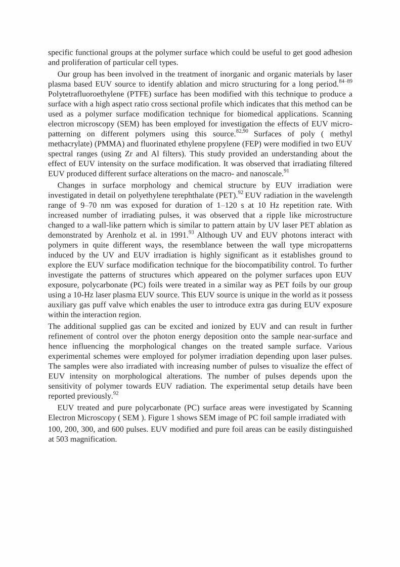
Electron Microscopy ( SEM ). Figure 1 shows SEM image of PC foil sample irradiated with
100, 200, 300, and 600 pulses. EUV modified and pure foil areas can be easily distinguished
at 503 magnification.
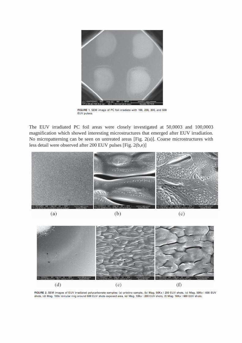
The EUV irradiated PC foil areas were closely investigated at 50,0003 and 100,0003
magnification which showed interesting microstructures that emerged after EUV irradiation.
No micropatterning can be seen on untreated areas [Fig. 2(a)]. Coarse microstructures with
less detail were observed after 200 EUV pulses [Fig. 2(b,e)]
however more refined and detailed wall-like structures were observed in areas irradiated
by 600 EUV pulses [Fig. 2( c,f )]. An interesting ring structure was also found to surround the
EUV exposed area as shown in Figure 2( d ).
As described earlier, modifications in chemical structure and the presence of functional
groups on the polymer surfaces change the behavior of the polymeric biomaterial in activities
like cell adhesion, cell growth, and toxicity. Therefore it is crucial to determine the alterations
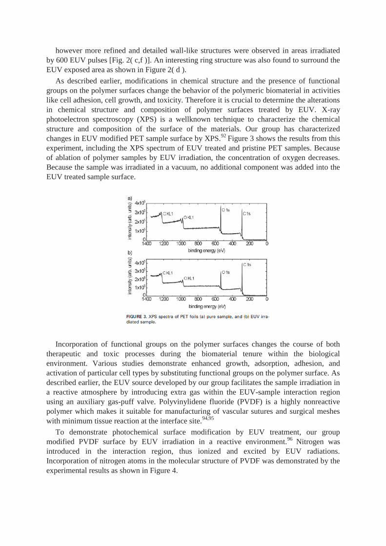
in chemical structure and composition of polymer surfaces treated by EUV. X-ray
photoelectron spectroscopy (XPS) is a wellknown technique to characterize the chemical
structure and composition of the surface of the materials. Our group has characterized
changes in EUV modified PET sample surface by XPS.92
Figure 3 shows the results from this
experiment, including the XPS spectrum of EUV treated and pristine PET samples. Because
of ablation of polymer samples by EUV irradiation, the concentration of oxygen decreases.
Because the sample was irradiated in a vacuum, no additional component was added into the
EUV treated sample surface.
Incorporation of functional groups on the polymer surfaces changes the course of both
therapeutic and toxic processes during the biomaterial tenure within the biological
environment. Various studies demonstrate enhanced growth, adsorption, adhesion, and
activation of particular cell types by substituting functional groups on the polymer surface. As
described earlier, the EUV source developed by our group facilitates the sample irradiation in
a reactive atmosphere by introducing extra gas within the EUV-sample interaction region
using an auxiliary gas-puff valve. Polyvinylidene fluoride (PVDF) is a highly nonreactive
polymer which makes it suitable for manufacturing of vascular sutures and surgical meshes
with minimum tissue reaction at the interface site.94,95
To demonstrate photochemical surface modification by EUV treatment, our group
modified PVDF surface by EUV irradiation in a reactive environment.96
Nitrogen was
introduced in the interaction region, thus ionized and excited by EUV radiations.
Incorporation of nitrogen atoms in the molecular structure of PVDF was demonstrated by the
experimental results as shown in Figure 4.
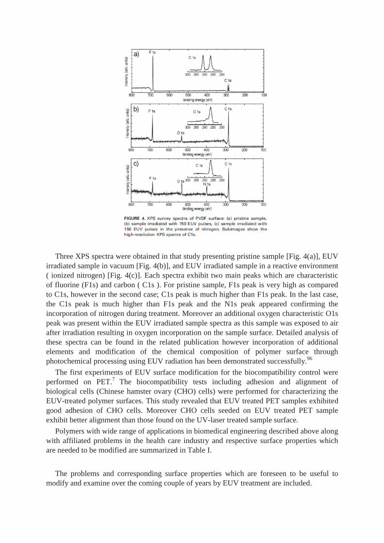
Three XPS spectra were obtained in that study presenting pristine sample [Fig. 4(a)], EUV
irradiated sample in vacuum [Fig. 4(b)], and EUV irradiated sample in a reactive environment
( ionized nitrogen) [Fig. 4(c)]. Each spectra exhibit two main peaks which are characteristic
of fluorine (F1s) and carbon ( C1s ). For pristine sample, F1s peak is very high as compared
to C1s, however in the second case; C1s peak is much higher than F1s peak. In the last case,
the C1s peak is much higher than F1s peak and the N1s peak appeared confirming the
incorporation of nitrogen during treatment. Moreover an additional oxygen characteristic O1s
peak was present within the EUV irradiated sample spectra as this sample was exposed to air
after irradiation resulting in oxygen incorporation on the sample surface. Detailed analysis of
these spectra can be found in the related publication however incorporation of additional
elements and modification of the chemical composition of polymer surface through
photochemical processing using EUV radiation has been demonstrated successfully.96
The first experiments of EUV surface modification for the biocompatibility control were
performed on PET.7
The biocompatibility tests including adhesion and alignment of
biological cells (Chinese hamster ovary (CHO) cells) were performed for characterizing the
EUV-treated polymer surfaces. This study revealed that EUV treated PET samples exhibited
good adhesion of CHO cells. Moreover CHO cells seeded on EUV treated PET sample
exhibit better alignment than those found on the UV-laser treated sample surface.
Polymers with wide range of applications in biomedical engineering described above along
with affiliated problems in the health care industry and respective surface properties which
are needed to be modified are summarized in Table I.
The problems and corresponding surface properties which are foreseen to be useful to
modify and examine over the coming couple of years by EUV treatment are included.
Beside numerous advantages and strong indications of efficient surface modification
technique, EUV polymer processing also has some disadvantages. First and foremost is
unavailability of EUV radiation source. Only in recent years, laboratory based compact EUV
sources are available for processing of polymers. The source at MUT, Warsaw is now being
used for fundamental research in polymer processing for biocompatibility control. EUV
sources are still in progress towards miniaturization. Degradation of laser optics is another
prime aspect in the health care industry and respective surface properties which are needed to
be modified are summarized in Table I. The problems and corresponding surface properties
which are foreseen to be useful to modify and examine over the coming couple of years by
EUV treatment are included.
Beside numerous advantages and strong indications of efficient surface modification
technique, EUV polymer processing also has some disadvantages. First and foremost is
unavailability of EUV radiation source. Only in recent years, laboratory based compact EUV
sources are available for processing of polymers. The source at MUT, Warsaw is now being
used for fundamental research in polymer processing for biocompatibility control. EUV
sources are still in progress towards miniaturization. Degradation of laser optics is another
prime aspect in terms of efficiency of EUV source. An ellipsoidal grazing incidence mirror
which is used for collection of EUV source for interaction chamber in tabletop EUV source
works on the principle of total external reflection.5
Such type of mirrors currently employed
in various applications with optimal thermal resistance and reflectivity. Damage threshold
investigations about the grazing incidence mirror in EUV source are currently in progress by
some groups.97
Damaged grazing incidence mirror result in uncontrolled EUV flux ultimately
influencing EUV ablation process and micro-structuring of treated polymers.97
CONCLUSION
Despite being a multi-billion dollar industry, control over the degree of biocompatibility is
still an important research and development challenge facing the biomaterials research and
industrial communities. To overcome this challenge it is required that the processes which
occur at the interface site between the biomaterial and the host do not induce any deleterious
effects such as chronic inflammatory response or formation of unusual tissues. Therefore the
importance of biomaterials with appropriate surface properties is evident. At the same time,
specific bulk properties are essential, particularly mechanical properties for biomaterials in
order to perform particular tasks in the biological world. It is evident that designing of
biomaterials which fulfill both needs is quite difficult. A common approach is to fabricate
biomaterials with adequate bulk properties, followed by modification of surface properties
through various treatments. This procedure leads to development of a final material with
decoupled bulk and surface properties. However there is no surface modification technique
unanimously accepted to control the degree of the biocompatibility for polymers. Plasma and
laser ablation techniques have been employed for surface modification of polymers however
they induce alterations in bulk material due to deep penetration in to the material.
To avoid such problems, extreme ultraviolet (EUV) radiation has been successfully
employed for surface modifications with a few polymer types. Most importantly EUV micro-
patterning produced similar wall-like structures on polymer surfaces as those created by the
UV irradiation. The discovery of this breakthrough sets up new trails for the exploitation of
EUV micro-patterning to allow the control over the degree of biocompatibility. The changes
in the physical structure of the surface provide for appropriate physical properties for the
interface site between host and biomaterial. Moreover crosslinking of functional groups by
introducing extra gas during EUV exposure to the polymers could significantly improve
biocompatibility for particular biological cell types. To date only a few studies carried out for
surface modification of polymers by EUV irradiation. In only one study biocompatibility tests
were performed on EUV-treated polymers. These studies yield promising indications towards
employment of EUV radiation for enhanced biocompatibility in polymers. Exploitation of
specific wavelengths within the EUV spectrum for biomedical engineering applications is a
vast vacant area belongs to polymer physics, chemistry, and biotechnology yet to be explored.
Such studies however require multidisciplinary teams from medicine, engineering, and
material science.
ACKNOWLEDGMENTS
With the support of the Erasmus Mundus program of the European Union. With support from
the 7th Framework Program’s Laserlab Europe project (Nr 284464).
REFERENCES
1. Jagur-Grodzinski J. Biomedical application of functional polymers. React Funct Polym
1999;39:99–138.
2. Griffith LG. Polymeric biomaterials. Acta Mater 2000;48:263–277.
3. Seal BL, Otero TC, Panitch A. Polymeric biomaterials for tissue and organ regeneration.
Mater Sci Eng 2001;34:147–230.
4. Hadjizadeh A, Ajji A, Bureau MN. Preparation and characterization of NaOH treated
micro-fibrous polyethylene terephthalate nonwovens for biomedical application. J Mech
Behav Biomed Mater
2010;3:574–583.
5. Bartnik A, Fiedorowicz H, Jarocki R, Kostecki J, Szczurek M, Wachulak PW. Laser-
plasma EUV source dedicated for surface processing of polymers. Nucl Instrum Methods
Phys Res Sect A 2011;647:125–131.
6. Andrews KD, Feugier P, Black RA, Hunt JA. Vascular prostheses: Performance related to
cell-shear responses. J Surg Res 2008;149: 39–46.
7. Reisinger B, Fahrner M, Frischauf I, Yakunin S, Svorcik V, Fiedorowicz H, Bartnik A,
Romanin C, Heitz J. EUV micropatterning for biocompatibility control of PET. Appl Phys
A Mater Sci Process 2010;100:511–516.
8. Cenni E, Arciola CR, Ciapetti G, Granchi D, Savarino L, Stea S, Cavedagna D, Curti T,
Falsone G, Pizzoferrato A. Platelet and coagulation factor variations induced in vitro by
polyethylene terephthalate (Dacron) coated with pyrolytic carbon. Biomaterials
1995;16:973–976.
9. Uchida E, Uyama Y, Ikada Y. Surface graft polymerization of acrylamide onto
poly(ethylene terephthalate) film by UV irradiation. J Polym Sci A Polym Chem
1989;27:527–537.
10. Moczulska M, Bitar M, SwieRszkowski W, Bruinink A. Biological characterization of
woven fabric using two- and threedimensional cell cultures. J Biomed Mater Res
2012;100:882–893.
11. Gumpenberger T, Heitz J, Bauerle€ D, Kahr H, Graz I, Romanin C, Svorcik V, Leisch F.
Adhesion and proliferation of human endothelial cells on photochemically modified
polytetrafluoroethylene. Biomaterials 2003;24:5139–5144.
12. Lopez JA, Chen J. Pathophysiology of venous thrombosis. Thromb Res 2009;123:S30 –S
34.
13. Nederberg F, Atthoff B, Bowden T, Welch K, Stromme M, Hilborn J. Biodegradable
ionomers for the loading and release of proteins: Formation, characterization, mechanism,
and consequence of water uptake. Am Chem Soc 2008;977:250–266.
14. Patel A, Mequanint K. Novel physically crosslinked polyurethaneblock-poly(vinyl
pyrrolidone) hydrogel biomaterials. Macromol Biosci 2007;7:727–737.
15. Peppas NA, Hilt JZ, Khademhosseini A, Langer R. Hydrogels in biology and medicine:
From molecular principles to bionanotechnology. Adv Mater 2006;18:1345–1360.
16. Butruk B, Trzaskowski M, Ciach T. Fabrication of biocompatible hydrogel coatings for
implantable medical devices using Fentontype reaction. Mater Sci Eng C 2012;32:1601–
1609.
17. Yoda R. Elastomers for biomedical applications. J Biomater Sci Polym Ed 1998;9:561–
626.
18. Lin DT, Young TH, Fang Y. Studies on the effect of surface properties on the
biocompatibility of polyurethane membranes. Biomaterials 2001;22:1521–1529.
19. Kurtz S, Devine J. PEEK biomaterials in trauma, orthopedic, and spinal implants.
Biomaterials 2007;28:4845–4869.
20. Chu P, Chen J, Wang L, Huang N. Plasma-surface modification of biomaterials. Mater Sci
Eng 2002;36:143–206.
21. Rihova B. Biocompatibility of biomaterials: Hemocompatibility, immunocompatibility
and biocompatibility of solid polymeric materials and soluble targetable polymeric
carriers. Adv Drug Deliv Rev 1996;21:157–176.
22. McGuigan AP, Sefton MV. The influence of biomaterials on endothelial cell
thrombogenicity. Biomaterials 2007;28:2547–2571.
23. Ravi S, Chaikof E. Biomaterials for vascular tissue engineering. Regener Med 2010;5:1–
21.
24. Wang YX, Robertson JL, Spillman WB, Claus O. Effects of the chemical structure and the
surface properties of polymeric biomaterials on their biocompatibility. Pharm Res
2004;21:1362–1373.
25. Yue Z, Wen F, Gao S, Ang MY, Pallathadka PK, Liu L, Yu H. Preparation of three-
dimensional interconnected macroporous cellulosic hydrogels for soft tissue engineering.
Biomaterials 2010;31: 8141–8152.
26. Li W, Charters R, Luther-Davies B, Mar L. Significant improvement of adhesion between
gold thin films and a polymer. Appl Surf Sci 2004;233:227–233.
27. Nagy M, Skvarla J. Evaluating the wetting (surface polarity) and roughness of PET foils
surface degraded by incipient alkaline hydrolysis. Ann Faculty Eng Hunedoara Int J Eng
2011;9: 235–240.
28. Kazmierska K, Szwast M, Ciach T. Determination of urethral catheter surface lubricity. J
Mater Sci Mater Med 2008;19:2301–2306.
29. Vladkova TG. Surface engineered polymeric biomaterials with improved biocontact
properties. Int J Polym Sci 2010;2010:1–22.
30. Yang GH, Kang ET, Neoh KG, Zhang Y, Tan KL. Electroless deposition of copper on
polyimide films modified by surface graft copolymerization with nitrogen-containing vinyl
monomers. Colloid Polym Sci 2001;279:745–753.
31. Laurens P, Ould Bouali M, Meducin F, Sadras B. Characterization of modifications of
polymer surfaces after excimer laser treatments below the ablation threshold. Appl Surf
Sci 2000;154–155: 211–216.
32. Tabata Y. Biomaterials design of culture substrates for cell. Inflammation Regener
2011;31:137–145.
33. Dahe GJ, Kadam SS, Sabale SS, Kadam DP, Sarkate LB, Bellare JR. In vivo evaluation of
the biocompatibility of surface modified hemodialysis polysulfone hollow fibers in rat.
PloS One 2011;6:
1 – 9.
34. Sashiwa H, Aiba S. Chemically modified chitin and chitosan as biomaterials. Prog Polym
Sci 2004;29:887–908.
35. Huang XJ, Xu ZK, Huang XD, Wang ZG, Yao K. Biomimetic surface modification on
polyacrylonitrile-based asymmetric membranes via direct formation of phospholipid
moieties. Polymer 2006;47:3141–3149.
36. Keselowsky BG, Collard DM, Garcıa AJ. Surface chemistry modulates focal adhesion
composition and signaling through changes in integrin binding. Biomaterials
2004;25:5947–5954.
37. Ding Z, Chen J, Gao S, Chang J, Zhang J, Kang ET. Immobilization of chitosan onto poly-
L-lactic acid film surface by plasma graft polymerization to control the morphology of
fibroblast and liver cells. Biomaterials 2004;25:1059–1067.
38. Desmet T, Morent R, De Geyter N, Leys C, Schacht E, Dubruel P. Nonthermal plasma
technology as a versatile strategy for polymeric biomaterials surface modification: A
review. Biomacromolecules 2009;10:2351–2378.
39. Chen M, Zamora P, Som P. Cell attachment and biocompatibility of
polytetrafluoroethylene (PTFE) treated with glow-discharge plasma of mixed ammonia
and oxygen. J Biomater Sci Polym Ed
2003;14:917–935.
40. Lindhout T, Blezer R, Maassen C, Heijnen V, Reutelingsperger CPM. Platelet
procoagulant surface as an essential parameter for the in vitro evaluation of the blood
compatibility of polymers. J Mater Sci Mater Med 1995;6:367–372.
41. Dadsetan M, Mirzadeh H, Sharifi-Sanjani N, Salehian P. IR laser surface modification of
polyethylene terephthalate as biomaterial. Process Fabr Adv Mater VIII 2000;9:221–229.
42. Goodman SL, Tweden KS, Albrecht RM. Platelet interaction with pyrolytic carbon heart-
valve leaflets. J Biomed Mater Res 1996;32:249–258.
43. Favia P, d’Agostino R. Plasma treatments and plasma deposition of polymers for
biomedical applications. Surf Coat Technol 1998; 98:1102–1106.
44. Banerjee D, Mukherjee S, Chattopadhyay KK. Controlling the surface topology and hence
the hydrophobicity of amorphous carbon thin films. Carbon 2005;48:1025–1031.
45. Santana G, Fandino~ J, Ortiz A, Alonso JC. Low temperature–low hydrogen content
silicon nitrides thin films deposited by PECVD using dichlorosilane and ammonia
mixtures. J Non-Crystalline Solids 2005;351:922–928.
46. Cianci E, Schina A, Minotti A, Quaresima S, Foglietti V. Dual frequency PECVD silicon
nitride for fabrication of CMUTs’ membranes. Sensors Actuators A 2006;127:80–87.
47. Sarro PM, deBoer CR, Korkmaz E, Laros JMW. Low-stress PECVD SiC thin films for IC-
compatible microstructures. Sens Actuators A 1998;67:175–180.
48. Kafrouni W, Rouessac V, Julbe A, Durand J. Synthesis of PECVD a-SiCXNY:H
membranes as molecular sieves for small gas separation. J Membr Sci 2009;329:130–137.
49. Madocks J, Rewhinkle J, Barton L. Packaging barrier films deposited on PET by PECVD
using a new high density plasma source. Mater Sci Eng B 2005;119:268–273.
50. Anma H, Yoshimoto Y, Warashina M, Hatanaka Y. Low temperature deposition of SiC
thin films on polymer surface by plasma CVD. Appl Surf Sci 2001;175/176:484–489.
51. Choi W, Gangadharan S. Structural study of plasma enhanced chemical vapour deposited
silicon carbide films. Mater Sci Eng B 2000;75:174–176.
52. Prasad GR, Daniels S, Cameron DC, Mcnamara BP, Tully E, Kennedy RO. PECVD of
biocompatible coatings on 316L stainless steel. Surf Coat Technol 2005;200:1031–1035.
53. Li M, Cheng Y, Zheng YF, Zhang X, Xi TF, Wei SC. Plasma enhanced chemical vapor
deposited silicon coatings on Mg alloy for biomedical application. Surf Coat Technol
2013;228:262–265.
54. Polcar T, Vitu T, Cvrcek L, Novak R, Vyskocil J, Cavaleiro A. Tribological behaviour of
nanostructured Ti-C:H coatings for biomedical applications. Solid State Sci
2009;11:1757–1761.
55. Bendavid A, Martin PJ, Randeniya L, Amin MS. The properties of fluorine containing
diamond-like carbon films prepared by plasma-enhanced chemical vapour deposition.
Diamond Relat Mater 2009;18:66–71.
56. Ahmed MH, Byrne JA, McLaughlin J. Evaluation of glycine adsorption on diamond like
carbon (DLC) and fluorinated DLC deposited by plasma-enhanced chemical vapour
deposition (PECVD). Surf Coat Technol 2012;209:8–14.
57. Cui F, Li D. A review of investigations on biocompatibility of diamond-like carbon and
carbon nitride films. Surf Coat Technol 2000;131:481–487.
58. Jansen H, Gardeniers H, Boer MD, Elwenspoek M, Fluitman J. A survey on the reactive
ion etching of silicon in microtechnology. J Micromech Microeng 1996;6:14–28.
59. Makarova O, Divan R, Moldovan N, Rosenmann D, Tang CM. Nanoporous
ultrananocrystalline diamond membranes. J Vacuum Sci Technol B Microelectron
Nanometer Struct 2010;28:C6 P42– C6P47.
60. Meacham K, Giuly R, Guo L. A lithographically-patterned, elastic multi-electrode array
for surface stimulation of the spinal cord. Biomed Microdev 2008;10:259–269.
61. Zhang W, Chu PK, Ji J, Zhang Y, Liu X, Fu RKY, Ha PCT, Yan Q. Plasma surface
modification of poly vinyl chloride for improvement of antibacterial properties.
Biomaterials 2006;27:44–51.
62. Bazaka K, Jacob MV, Crawford RJ, Ivanova EP. Plasma-assisted surface modification of
organic biopolymers to prevent bacterial attachment. Acta Biomater 2011;7:2015–2028.
63. Chu PK. Enhancement of surface properties of biomaterials using plasma-based
technologies. Surf Coat Technol 2007;201:8076 – 8082.
64. Kawamura Y, Toyoda K, Namba S. Effective deep ultraviolet photoetching of polymethyl
methacrylate by an excimer laser. Appl Phys Lett 1982;40:374–375.
65. Srinivasan V, Mayne-Banton R. Self-developing photoetching of poly(ethylene
terephthalate) films by far ultraviolet excimer laser radiation. Appl Phys Lett
1982;41:576–578.
66. Kuper€ M, Stuke S. Ablation of polytetrafluoroethylene ( Teflon ) with femtosecond UV
excimer laser pulses. Appl Phys Lett 1988 ;
54:4–6.
67. Bityurin N, Arnold N, Luk’yanchuk B, Bauerle€ D. Bulk model of laser ablation of
polymers. Appl Surf Sci 1998;127–129:164–170.
68. Lippert T. Laser application of polymers. Adv Polym Sci 2004;168: 51–246.
69. Okazaki M, Ohmae H, Hino T. Insolubilization of apatite-collagen composites by UV
irradiation. Biomaterials 1989;10:564–568.
70. Okazakl M, Ohmae H, Takahashi J, Kimura H, Sakuda M. Insolubilized properties of UV-
irradiated C03 apatite-collagen composites. Biomaterials 1990;11:568–572.
71. Matsuda K, Inoue T. Novel photoreactive surface modification technology for fabricated
devices. ASAIO Trans—Am Soc Artif Intern 1990;36:M161 –M 164.
72. Tidwell CD, Ertel SI, Ratner BD, Tarasevich BJ, Atre S, Allara DL. Endothelial cell
growth and protein adsorption on terminally functionalized, self-assembled monolayers of
alkanethiolates on gold. Langmuir 1997;13:3404–3413.
73. Heitz J, Svorcık V, Bacakov a L, Rockova K, Ratajova E, Gumpenberger T, Bauerle€ D,
Dvorankov a B, Kahr H, Graz I, Romanin C. Cell adhesion on polytetrafluoroethylene
modified by UV-irradiation in an ammonia atmosphere. J Biomed Mater Res
2003;67:130–137.
74. Doi K, Nakayama Y, Matsuda T. Novel compliant and tissuepermeable microporous
polyurethane vascular prosthesis fabricated using an excimer laser ablation technique. J
Biomed Mater Res 1996;31:27–33.
75. Masuda S, Doi K, Satoh S, Oka T, Matsuda T. Vascular endothelial growth factor
enhances vascularization in microporous small caliber polyurethane grafts. ASAIO J
1997;43:M530 –M 534.
76. Zhihong Z. Surface modification by plasma polymerization and application of plasma
polymers as biomaterials. Doctoral dissertation, Johannes Gutenberg-Universitat€ Mainz,
Dec. 2003.
77. Pfleging W, Kohler R, Torge M, Trouillet V, Danneil F, Stuber€ M. Control of wettability
of hydrogenated amorphous carbon thin films by laser-assisted micro- and
nanostructuring. Appl Surf Sci 2011;257:7907–7912.
78. Gallardo-Moreno AM, Pacha-Olivenza MA, Saldana~ L, Perez- Giraldo C, Bruque JM,
Vilaboa N, Gonzalez-Mart ın ML. In vitro biocompatibility and bacterial adhesion of
physico-chemically modified Ti6Al4V surface by means of UV irradiation. Acta Biomater
2009;5:181–192.
79. Rumeng W, Chenglin CH, Yanfen L, Lihong Y, Yaopu P, Yinsheng D, Pinghua L, Chu
PK. Surface modification and biocompatibility of NiTi shape memory alloy treated with
advanced oxidation in UV/H2O2 photocatalytic system. Rare Metal Mater Eng
2008;37:2027–2030.
80. Mendez-Vilas A, Gonzalez-Mart ın A, Pacha-Olivenza ML, Perez- Giraldo MA, Saldana~
C, Vilaboa L, Gallardo-Moreno N. UV light irradiation of Ti6Al4V: Antibacterial impact
and biocompatibility in vitro. 8th World Biomaterials Congress 2008;4:1943.
81. Murthy NS, Prabhu RD, Martin JJ, Zhou L, Headrick RL. Selfassembled and etched cones
on laser ablated polymer surfaces. J Appl Phys 2006;100:023538/1–023538/12.
82. Bartnik A, Fiedorowicz H, Jarocki R, Kostecki J, Szczurek A, Szczurek M. Ablation and
surface modifications of PMMA using a laser-plasma EUV source. Appl Phys B
2009;96:727–730.
83. Bartnik A, Fiedorowicz H, Jarocki R, Kostecki J, Szczurek M, Chernyayeva O, Sobczak
JW. EUV-induced physico-chemical changes in near-surface layers of polymers. J
Electron Spectrosc Relat Phenom 2011;184:270–275.
84. Bartnik A, Fiedorowicz H, Jarocki R, Kostecki J, Szczurek M, Havlikova R, Pina L, Sv
eda L, Inneman A. Response of inorganic materials to laser—Plasma EUV radiation
focused with a lobster eye collector. Damage to VUV, and X-ray optics. Proc SPIE 2007;
6586:6580A/1–6580A/9.
85. Fiedorowicz H, Bartnik A, Jakubczak K, Jarocki R, Juha L. Application of laser plasma
soft X-ray and EUV sources in micro- and nanotechnology. Laser Technology VIII:
Applications of lasers. Proc SPIE 2007;6598:65980G.
86. Fiedorowicz H, Bartnik A, Jakubczak K, Jarocki R, Kostecki J, Pina L, Rakowski R,
Szczurek A, Szczurek M. Micro- and nanoprocessing of organic polymers using a compact
laser plasma EUV source equipped with EUV optical systems. Ultrafast X-ray sources and
detectors. Proc SPIE 2007;6703:67030C.
87. Bartnik A, Fiedorowicz H, Jarocki R, Kostecki J, Rakowski R, Szczurek A, Szczurek M.
Applications of laser plasma EUV source based on a gas puff target. AIP Conf Proc
2008;993:379–382.
88. Bartnik A, Fiedorowicz H, Jarocki R, Juha L, Kostecki J, Rakowski R, Szczurek M.
Micromachining of organic polymers by X-ray photo-etching using a 10Hz laser-plasma
radiation source. Microelectron Eng 2005;78/79:452–456.
89. Bartnik A, Fiedorowicz H, Jarocki R, Juha L, Kostecki J, Rakowski R, Szczurek M.
Strong temperature effect on X-ray photo-etching of polytetrafluoroethylene using a 10 Hz
laserplasma radiation source based on a gas puff target. Appl Phys B 2006;82:529–532.
90. Bartnik A, Fiedorowicz H, Jarocki R, Kostecki J, Rakowski R, Szczurek M. Surface
changes of solids under intense EUV irradiation using a laser-plasma source 2009.
Damage to VUV, EUV, and X-ray optics II. Proc SPIE 2009;7361:73610C/1–73610C/11.
91. Bartnik A, Fiedorowicz H, Jarocki R, Kostecki J, Szczurek M. PMMA and FEP surface
modifications induced with EUV pulses in two selected wavelength ranges. Appl Phys A
Mater Sci Process
2009;98:61–65.
92. Bartnik A, Fiedorowicz H, Jarocki R, Kostecki J, Szczurek M, Bilinski A, Chernyayeva O,
Sobczak JW. Physical and chemical modifications of PET surface using a laser-plasma
EUV source. Appl Phys A Mater Sci Process 2010;99:831–836.
93. Arenholz E, Svorcik V, Kefer T, Heitz J, Biiuerle D. Structure formation in UV-laser
ablated poly-ethylene-terephthalate ( PET ). Appl Phys A Solids Surf 1991;331:330–331.
94. Laroche G, Marois Y, Guidoin R, King MW, Martin L, How T, Douville Y.
Polyvinylidene fluoride (PVDF) as a biomaterial: From polymeric raw material to
monofilament vascular suture. J Biomed Mater Res 1995;29:1525–1536.
95. Klinge U, Klosterhalfen B, Ottinger AP, Junge K, Schumpelick V. PVDF as a new
polymer for the construction of surgical meshes. Biomaterials 2002;23:3487–3493.
96. Bartnik A, Lisowski W, Sobczak J, Wachulak P, Budner B, Korczyc B, Fiedorowicz H.
Simultaneous treatment of polymer surface by EUV radiation and ionized nitrogen. Appl
Phys A Mater Sci Process 2012;109:39–43.
97. Barkusky F, Bayer A, Doring€ S, Grossmann P, Mann K. Damage threshold
measurements on EUV optics using focused radiation from a table-top laser produced
plasma source. Opt Express
2010;18:4346–4355.


![Radiation Modification of Natural Polymers · Topic: Radiation Modification of Natural Polymers [17] Rayon is produced from viscose, a polymer made from cellulosic materials such](https://static.fdocuments.us/doc/165x107/5fdbffe9c7c6af3e01446787/radiation-modification-of-natural-topic-radiation-modification-of-natural-polymers.jpg)