Do individual cells have a life history? The Cell Cycle.
36
-
Upload
lucy-powers -
Category
Documents
-
view
218 -
download
2
Transcript of Do individual cells have a life history? The Cell Cycle.


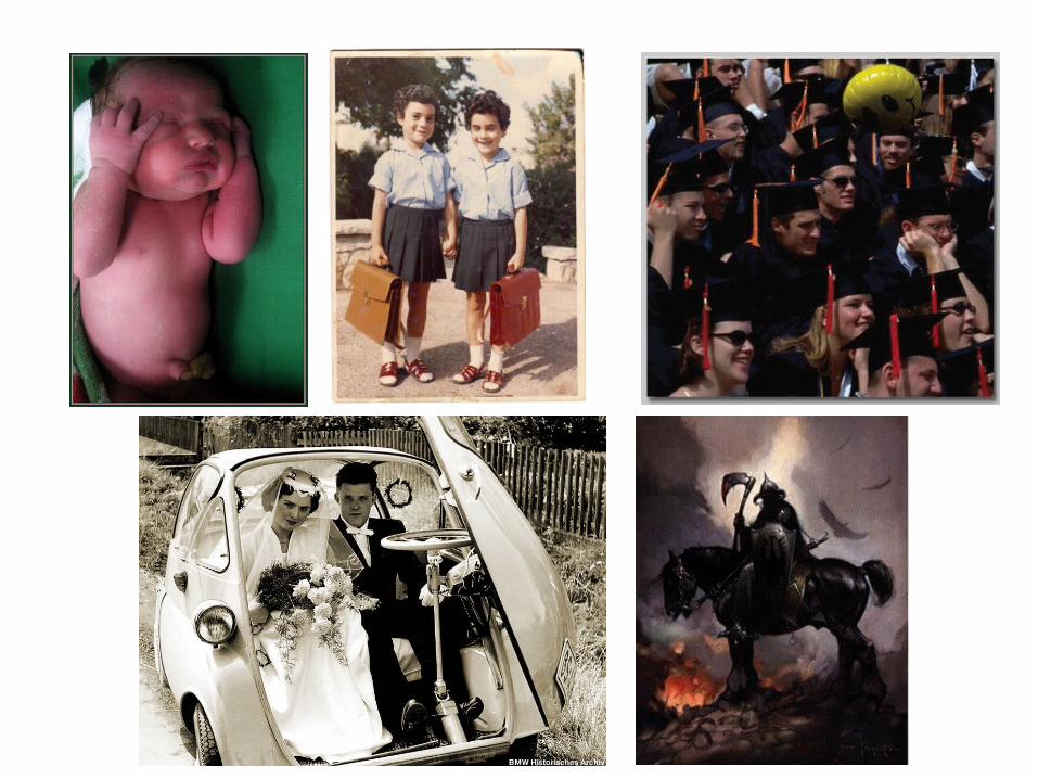

Do individual cells have a life history?

The Cell Cycle
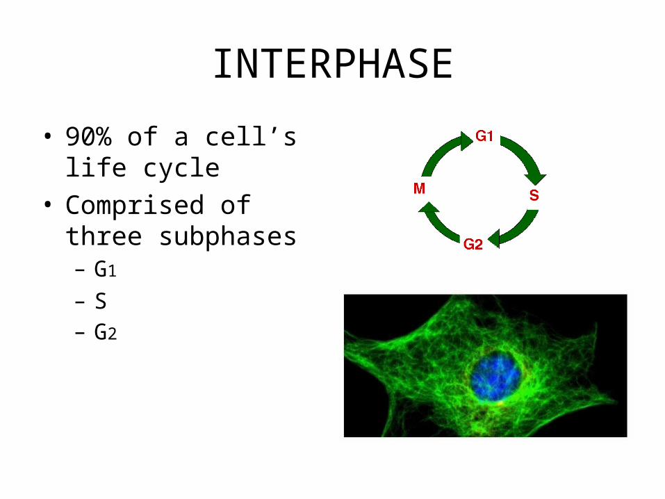
INTERPHASE
• 90% of a cell’s life cycle
• Comprised of three subphases– G1
– S– G2
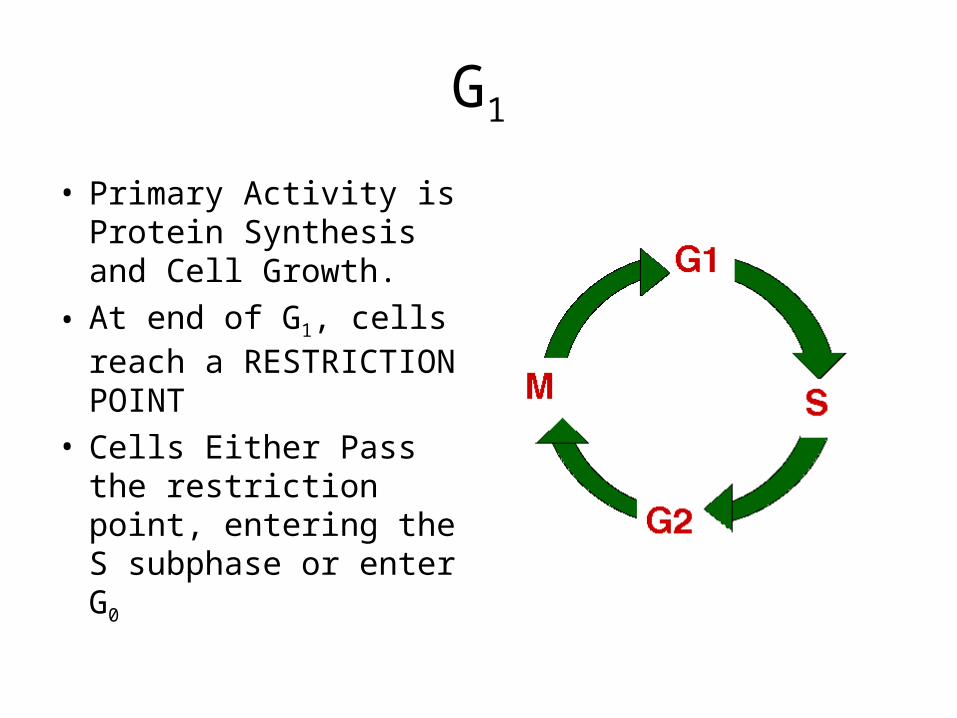
G1
• Primary Activity is Protein Synthesis and Cell Growth.
• At end of G1, cells reach a RESTRICTION POINT
• Cells Either Pass the restriction point, entering the S subphase or enter G0

WHY DO CELLS EVER HAVE TO DIVIDE?

Surface Area To Volume
• As the cell increases in size, the volume (cytoplasm) of the cell grows faster than the surface area
• With more cell volume to feed, cell is required to do more diffusion, but can’t speed up RATE of diffusion
• Cell also cannot excrete wastes via diffusion effectively

G0
• If cells do not pass the restriction point at the end of G1, they enter G0
• Cells in G0 will never divide again
• Most somatic (body) cells are in G0

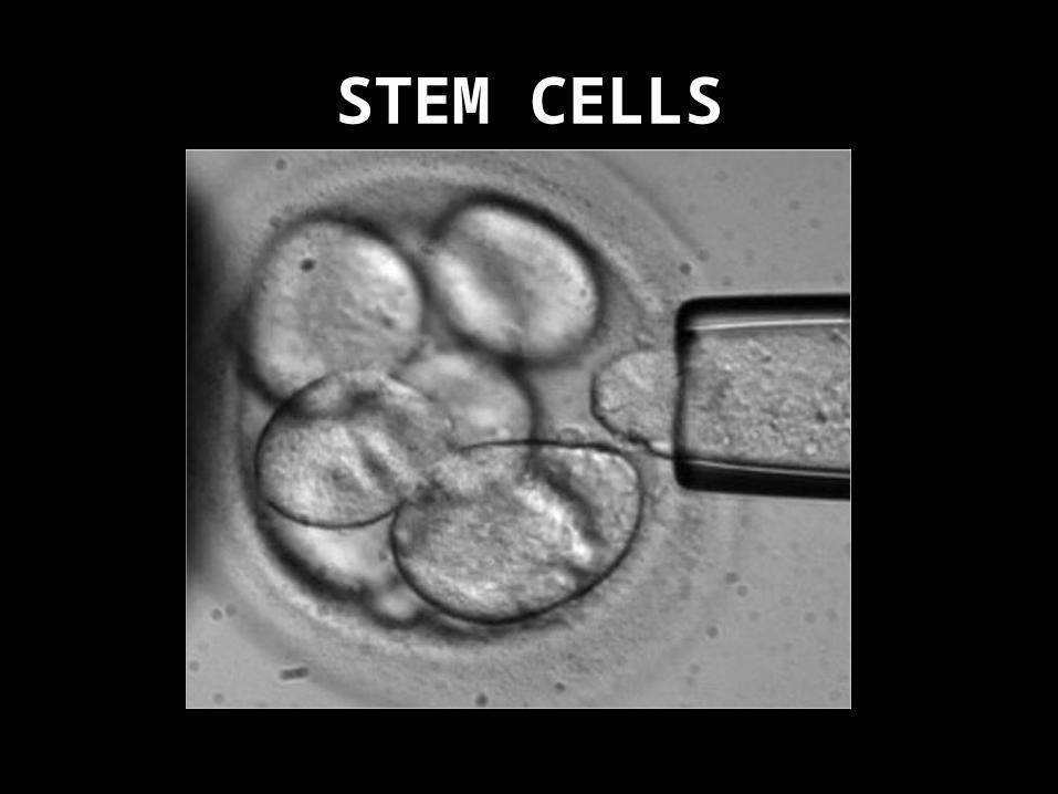
STEM CELLS

STEM CELLS


S Phase
• Cells that DO pass the restriction point enter the S phase
• In S phase, DNA is replicated
• Control over S phase regulated by the enzyme kinase

G2
• At the end of S phase, cells re-enter a period of protein synthesis and organelle development

M Phase = Mitosis
• NOTE: NOT A PART OF INTERPHASE
• If cells pass the restriction point, they must undergo S, G2 and finally, the division of the genetic material known as MITOSIS


Mitosis (M Phase)
• To ensure a high surface area to volume ratio (and to reduce the need for more DNA), the cell undergoes mitotic division
• Mitosis is comprised of four subphases; prophase, metaphase anaphase and telophase


Early Prophase
• chromatin begins to coil and condense to form chromosomes
• each chromosome appears to have two strands (each containing a single DNA molecule)
• each strand is called a chromatid
• each chromatid is attached to its sister chromatid at the centromere

Late Prophase
• the nuclear envelope and nucleolus disappear
• in cytoplasm, the spindle apparatus forms
• eventually the spindle guides the separation of sister chromatids into the two daughter cells

METAPHASE
• spindle grows and forms attachments to the chromosomes at the centromeres
• chromosomes move to an equatorial plate which is formed along the midline between the poles
• chromosomes are at their most condensed state


ANAPHASE• centromeres divide to create
two chromosomes instead of a pair of attached chromatids
• spindle fibers shorten and the sister chromosomes are drawn to the opposite poles of the cell
• poles of the spindle apparatus are pushed apart as the cell elongates
• anaphase results in the exact division of genetic information to each daughter cell

TELOPHASE
• nuclear envelopes reassemble and surround each set of daughter chromosomes
• nucleoli reappear inside the newly formed nuclei
• chromosomes recondense in the daughter cells to become chromatin

CYTOKINESIS
• in animal cell, a furrow appears around the cell that eventually pinches the cell into two new cells
• in plants, a cell plate forms between the two daughter nuclei as the cell wall divides the cell

The length of the cell cycle varies on the species

Length of cell cycle also depends on the cell function WITHIN an organism

Variances In Mitosis
• At any given time, different parts of an organism may be in different parts of the cell cycle
• Different organs, tissues and structures in the body also perform mitosis at different rates

Lab: Comparitive Cell Cycles In Allium cepa

Comparative Cell Cycles in Allium cepa
• Purpose: To compare the relative proportion of time required to complete the cell cycle in two different regions of an organism’s anatomy
• Two zones of root growth in Allium cepa– Zone of elongation– Zone of maturation

How do the lengths of the various parts of the cell cycle (including mitotic subphases)
compare? Why are they different?

Protocol
• For each lab pair, one lab partner will assess the zone of elongation, one will assess the zone of maturation
• While the other partner tallies the results, the other partner should view the required region of the slide and classify each of the cells in the field of view based on its place in the cell cycle
• Continue until 100 cells have been assessed• Move the slide to the zone of maturation and
switch roles with your partner. Repeat

Analysis
• Based on your collected results, construct two pie charts for the percentage of time spent in each of the cell cycle phases (interphase, prophase, metaphase, anaphase and telophase).
• In a paragraph, contrast the reasons behind the differences. Use the following terms: G1, G2, S, G0, zone of elongation, zone of maturation, mitosis, anaphase, telophase, metaphase, prophase, cytokinesis,
• Explain the differences regarding the function of each subphase and the actions in each of the subphases