DNA Replication Accessory Proteinsdnareplication.cshl.edu/content/free/chapters/17_hubscher.pdf ·...
19
17 DNA Replication Accessory Proteins Ulrich Hubscher, Giovanni Maga, and Vladimir N. Podust Department of Veterinary Biochemistry University of Zurich-lrchel CH-8057 Zurich, Switzerland INTRODUCTION DNA replication requires the concerted action of many enzymes, as well as other protein and non-protein cofactors. The DNA, in preparation for DNA synthesis, has to become single-strand to serve as a template for the replicative DNA polymerases (pols). It is this form of the DNA that is especially prone to damage of any kind. Nature has provided a set of proteins that support the replicative pols in performing processive, ac- curate, and rapid DNA synthesis. Furthermore, such proteins also prevent damage to the transient single-strand (ss) DNA. These proteins are called DNA replication accessory proteins. The three best known are the proliferating cell nuclear antigen (PCNA), replication factor C (RF-C), and replication protein A (RP-A). In this chapter, we focus on these three protein classes and compare them to their selected counterparts in eukaryotic viruses. Additional replication proteins that also assist the proper function of pols, such as the 3 -5 exonuclease, DNA primase, RNase H, 5 -3 ‘ exonuclease, DNA helicases, DNA ligases, and DNA topoisomerases, are covered in various other chapters. Early Discovery of Replication Accessory Proteins in Prokaryotes by Genetics and Defined In Vitro Replication Systems Fifteen years ago it was realized that bacteriophages of Escherichia coli provide a window to understand the cellular events of DNA replication (Kornberg and Baker 1992). By using ssDNA from 4x174, G4, and M13 as model replicons, the requirements for a ssDNA-binding protein (SSB) and a DNA synthesis complex were identified. The latter includes the pol I11 holoenzyme, which consists of many polypeptides besides the pol I11 core. For example, it includes the homodimeric subunit and the hetero- pentameric y complex (containing the y, 6, 6‘, x, and Q subunits) (for review, see Kuriyan and O’Donnell 1993). Well-established genetics DNA Replication in Eukaryoric Cells 8 1996 Cold Spring Harbor Laboratory Press 0-87969-459-9/96 $5 + .OO 525
Transcript of DNA Replication Accessory Proteinsdnareplication.cshl.edu/content/free/chapters/17_hubscher.pdf ·...

17 DNA Replication Accessory Proteins
Ulrich Hubscher, Giovanni Maga, and Vladimir N. Podust Department of Veterinary Biochemistry University of Zurich-lrchel CH-8057 Zurich, Switzerland
INTRODUCTION
DNA replication requires the concerted action of many enzymes, as well as other protein and non-protein cofactors. The DNA, in preparation for DNA synthesis, has to become single-strand to serve as a template for the replicative DNA polymerases (pols). It is this form of the DNA that is especially prone to damage of any kind. Nature has provided a set of proteins that support the replicative pols in performing processive, ac- curate, and rapid DNA synthesis. Furthermore, such proteins also prevent damage to the transient single-strand (ss) DNA. These proteins are called DNA replication accessory proteins. The three best known are the proliferating cell nuclear antigen (PCNA), replication factor C (RF-C), and replication protein A (RP-A). In this chapter, we focus on these three protein classes and compare them to their selected counterparts in eukaryotic viruses. Additional replication proteins that also assist the proper function of pols, such as the 3 -5 exonuclease, DNA primase, RNase H, 5 -3 ‘ exonuclease, DNA helicases, DNA ligases, and DNA topoisomerases, are covered in various other chapters.
Early Discovery of Replication Accessory Proteins in Prokaryotes by Genetics and Defined In Vitro Replication Systems
Fifteen years ago it was realized that bacteriophages of Escherichia coli provide a window to understand the cellular events of DNA replication (Kornberg and Baker 1992). By using ssDNA from 4x174, G4, and M13 as model replicons, the requirements for a ssDNA-binding protein (SSB) and a DNA synthesis complex were identified. The latter includes the pol I11 holoenzyme, which consists of many polypeptides besides the pol I11 core. For example, it includes the homodimeric subunit and the hetero- pentameric y complex (containing the y, 6, 6 ‘ , x, and Q subunits) (for review, see Kuriyan and O’Donnell 1993). Well-established genetics
DNA Replication in Eukaryoric Cells 8 1996 Cold Spring Harbor Laboratory Press 0-87969-459-9/96 $5 + .OO 525

526 U. Hubscher, G. Maga, and V.N. Podust
facilitated the cloning of all genes and expression of the polypeptides in bacteria. This allowed the study of the prokaryotic replication machinery in detail (see, e.g., Stukenberg et a]. 1994). Similarly in bacteriophage T4, the gp45 and gp44/62 complex share analogous functions to the f3 subunit and y complex, respectively (see, e.g., Young et al. 1992; Kuriyan and O’Donnelll993).
Discovery of Replication Accessory Proteins in Eukaryotes Thanks to Defined In Vitro Replication Systems
In eukaryotes, a protein was identified that increased the processivity of pol-6 (Tan et a]. 1986). It was later identified in the SV40 in vitro replication system as PCNA (Prelich et al. 1987). The same in vitro replication system brought insight into the requirement of an SSB called HSSB (Wobbe et al. 1987), replication factor A (Fairman and Stillman 1988) or RP-A (Wold and Kelly 1988), the latter name now adopted for this protein. Soon thereafter, a factor that could coordinate the synthesis of leading and lagging strands during SV40 DNA replication was identi- fied and called RF-C (Tsurimoto and Stillman 1989). All three proteins have been found to be functional analogs of prokaryotic accessory proteins (see Table 1).
Why Are Accessory Proteins Required in DNA Replication?
DNA replication accessory proteins provide particular functions that are mandatory for replicative pols. Such functions include the recruitment of particular pols when needed, the facilitation of pol binding to the primer terminus, the increase in pol processivity, the prevention of non- productive binding of the pol to ssDNA, the release of the pol after DNA synthesis, and the bridging of pol interactions to other replication proteins. Thus, it is not surprising that these proteins are universally found in nature (Table 1).
REPLICATION ACCESSORY PROTEINS
PCNA
PCNA is the most extensively studied cellular DNA replication acces- sory protein (Table 2). It was originally discovered as a cell-cycle- regulated nuclear protein whose rate of synthesis correlates with the proliferative state of normal cells and tissues, and later as a processivity

Tabl
e 1 E
ukar
yotic
and
prok
aryo
tic D
NA
repl
icat
ion
acce
ssor
y pr
otei
ns
Org
anis
m
euka
ryot
e Fu
nctio
nal c
ompo
nent
E.
col
i ba
cter
ioph
age
T4
(yea
st to
man
) H
SV
aden
ovir
us
AdD
BPa
UL
42
Proc
essiv
ity fa
ctor
or
p su
buni
t gp
45
PCN
A
slidi
ng c
lam
p 0
z
Cla
mp
load
er, b
race
y
com
plex
gp
44-g
p62
com
plex
R
F-C
?
? pr
otei
n, or
mat
chm
aker
D
SS
B
SSB
SS
B (E
. col
i)
RP-
A
ICP8
b A
dDBP
P o_
5' 2
5
Oth
er D
NA
rep
licat
ion
acce
ssor
y pro
tein
s inc
lude
3 ' -
5 ' e
xonu
clea
se, D
NA
pri
mas
e, R
Nas
e H
, 5 ' -
3 ' e
xonu
clea
se, D
NA
hel
icas
es, a
nd D
NA
liga
ses.
They
ar
e rev
iew
ed in
oth
er ch
apte
rs in
this
book
. aT
he A
dDBP
incr
ease
s the
stra
nd-d
ispla
cem
ent a
ctiv
ity o
f the
ade
novi
rus-
enco
ded
DN
A p
olym
eras
e. bI
CP8
can
also
enha
nce p
roce
ssiv
ity.
3 R I
s s 2 E.
0
cn
3
0)

528 U. Hubscher, G. Maga, and V.N. Podust
TabZe 2 Proliferating cell nuclear antigen Number of amino acids Molecular size (kD) Stoke's radius (A) 36.5,d 40e PI 4.Sd Sedimentation coefficient (S) 5d9e Structure in solution ring-shaped homotrimer Interaction with pol-6 and pol-&
260:-' 365,' 275k 29,a-i 40,' 31
RF-C cyclins A, B, D; CDC2, CDKs 2,4,5; p21
CDK-inhibitor; Gadd45 sliding clamp, increases primer binding and processivity of pol4 and pol-&
stimulates ATPase activity of RF-C, and forms together with RF-C and ATP the primer recognition complex
Functions
regulation of DNA replication (?) 'D. melanogaster (Yamaguchi et al. 1990); brat (Matsumoto et al. 1987); 'calf (Tan et al. 1986);
dhuman (Almendral et al. 1987); 'S. cerevisiae (Bauer and Burgers 1990); fS. pombe (Waseem et al. 1992); gbaculovirus (O'Reilly et al. 1989); hOxytricha sativa (Suzuka et al. 1989, 1991); 'D. carota (Hata et al. 1992); kPlasmodium falciparum (Kilbey et al. 1993).
factor for pol-6 (for review, see Hiibscher and Spadari 1994). The gene encoding this protein has been cloned from a variety of sources, includ- ing yeast, human, and plants, and shows a highly conserved structure (Table 2). Compared to human PCNA, the amino acid sequence similarity is 32% for lower eukaryotes (yeast and protozoa), 64% for plant, and 70% for Drosophila rnelanogaster. PCNA is a highly acidic protein encoded by a single gene in all the organisms tested, with the one exception of Dracus carota, in which two distinct genes encoding two different forms of PCNA were isolated. The human gene has been mapped to chromosome 20, and two pseudogenes have been localized on chromosomes X and 6. The structure of the genomic clone of human PCNA is known: It comprises six exons, five introns, and a short 3 ' - untranslated region. Introns 1 and 4 have been implicated in the negative regulation of transcription of the PCNA gene (Chang et al. 1990; Alder et al. 1992). The promoter sequence contains binding sites for several transcription factors such as Spl and E2F, suggesting that the expression of the PCNA gene may be tightly regulated (Jones et al. 1988; Mudryj et al. 1990).
The extreme structural conservation of PCNA is also seen at a func- tional level. With synthetic oligonucleotides or poly(dA)/oligo(dT) as

DNA Replication Accessory Proteins 529
DNA templates, PCNA increases the productive binding to a primer and the processivity of pol-6. Yeast PCNA interacts with mammalian pol-6, and mammalian PCNA interacts with yeast pol-6 (Bauer and Burgers 1988). PCNA was also shown to stimulate the primer binding and primer elongation reactions catalyzed by pol-& on similar templates (Maga and Hiibscher 1995). If, however, a singly primed M13 DNA was used as a model for a natural template, at least two other auxiliary proteins, RF-C and RP-A, are required. It has been shown that PCNA forms a complex together with RF-C at the primer in an ATP-dependent manner (Lee and Hunvitz 1990; Burgers 1991; Tsurimoto and Stillman 1991; Podust et al. 1992). This primer recognition complex can efficiently recruit po l4 or pol-& (but not pol-a) to the primer terminus. These functional experi- ments, together with the observation that PCNA is essential for coor- dinated leading- and lagging-strand synthesis during in vitro SV40 DNA replication (Prelich and Stillman 1988), clearly indicate a role for PCNA in DNA replication. PCNA has also been shown to participate in DNA excision repair and to be required for the gap-filling step (Shivji et al. 1992). Recent results showed that PCNA physically interacts with Gadd45, a protein that stimulates DNA excision repair and inhibits entry into S phase in mammalian cells, thus providing a new link between PCNA and the DNA repair pathway (Smith et al. 1994).
PCNA itself does not bind to DNA. It has been shown that PCNA can be loaded onto DNA in two ways: (1) enzymatically, by RF-C and ATP, and (2) topologically, in an ATP-independent manner (Burgers and Yoder 1993). Despite less than 5% of amino-acid-sequence similarity, PCNA is functionally equivalent to two prokaryotic proteins known to function as auxiliary factors in DNA replication: the p subunit of pol I11 holoenzyme of E. coli and the product of the gene 45 of the bac- teriophage T4 (Kuriyan and O’Donnell 1993). Yeast PCNA has recently been crystallized, and its structure has been determined (Krishna et al. 1994). PCNA has been shown to be a homotrimer with a closed circular structure that can encircle duplex DNA with a minimum of specific inter- actions, similar to the dimeric p subunit of E. coli pol 111. The current model for the function of PCNA suggests that this protein acts as a slid- ing clamp that allows pols to move rapidly along the DNA while remain- ing topologically bound to it.
Recent results have indicated that PCNA can interact with the cell- cycle-regulatory machinery. Isolation of kinase complexes containing cyclins A, B, and D, associated with the cyclin-dependent kinases CDC2, CDK2, CDK4, or CDK5, showed that a complex of PCNA and the cyclin-dependent kinase inhibitor p21 associates with each cyclin/CDK

530 U. Hubscher, G. Maga, and V.N. Podust
dimer to form quaternary complexes (Xiong et al. 1992; Zhang et al. 1993). Furthermore, p21 has been shown to inhibit PCNA-dependent DNA replication in the absence of cyclins/CDKs, through a direct protein-protein interaction with PCNA (Flores-Rozas et al. 1994; Li et al. 1994; Waga et al. 1994). It is known that the major tumor suppressor protein p53 regulates the expression of p21. In addition, it has recently been shown that p53 is also implicated in the regulation of the expression of the PCNA gene (Jackson et al. 1994; Yamaguchi et al. 1994), suggest- ing that, during p53-mediated suppression of cell proliferation, p21 and PCNA may be involved in coordination of the cell-cycle progression, DNA replication, and DNA repair.
R F-C
RF-C as an essential replication factor had first been isolated from hu- man 293 cells in the in vitro SV40 DNA replication system (Tsurimoto and Stillman 1989). This protein was later purified from HeLa cells (Lee et al. 1991a), yeast (Yoder and Burgers 1991; Fien and Stillman 1992), and calf thymus (Podust et al. 1992) by using a complementation assay that enabled pol-6 to replicate a primed ssDNA in the presence of PCNA and RP-A. RF-C binds preferentially to template-primer junctions on the DNA with ATP or ATPyS stimulating binding of RF-C to DNA (Lee et al. 1991a; Tsurimoto and Stillman 1991). Footprinting experiments revealed that RF-C bound at a template-primer junction covers 12 bases of ssDNA, whereas on the double-strand (ds) DNA it binds 8 bases of the template strand and 15 bases of the primer strand (Tsurimoto and Stillman 1991). RF-C possesses a DNA-stimulated ATPase activity (Tsurimoto and Stillman 1990; Yoder and Burgers 1991; Lee et al. 1991a; Podust et al. 1992). In the absence of PCNA, ssDNA stimulates the ATPase, whereas primer-template junctions have little or no effect on the activity. PCNA increases ATP hydrolysis by RF-C on multiple primed ssDNA, whereas on unprimed ssDNA, only a marginal effect was detected. These data suggested that PCNA causes the RF-C ATPase ac- tivity to become dependent on a primer terminus. All eight nucleoside triphosphates could be hydrolyzed by RF-C with preference for dATP and ATP. K , values for ATP in the ATPase assay were 42 p~ for human and 15 p~ for yeast RF-C, respectively. ATPyS and AMP-PNP are com- petitive inhibitors of the ATPase of yeast RF-C with Ki values of 1.8 p~ and 130 p ~ , respectively.
RF-C itself had little or no effect on DNA synthesis by pols a, 6, and E. In contrast, the conjunction of RF-C with PCNA, RP-A, and ATP

DNA Replication Accessory Proteins 531
strongly inhibited pol-a and stimulated pols 6 and E. Isolation of stable pol 6 and E complexes, formed with DNA in the presence of ATP, RF-C, and PCNA (called holoenzymes), indicated that pols 6 and E directly in- teract with these auxiliary proteins (Lee and Hunvitz 1990; Lee et al. 1991b; Burgers 1991; Podust et al. 1992).
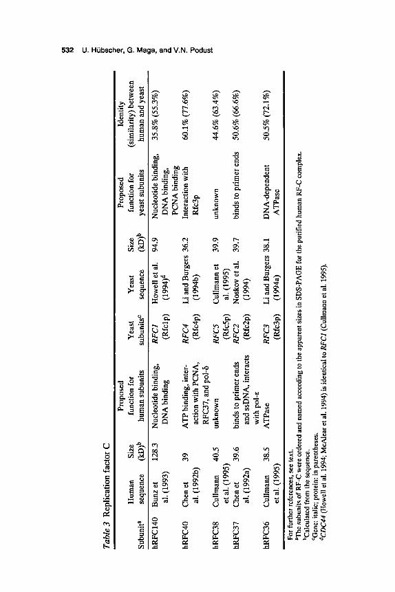
RF-C is a multiprotein complex composed of five subunits, including one large subunit and four small subunits (Table 3). All subunits are en- coded by different genes, and all show an extensive amino-acid-sequence homology in the middle part of the protein (O’Donnell et al. 1993; Bunz et al. 1993; Li and Burgers 1994b). All the genes encoding the subunits of yeast RF-C have been shown to be essential (Howell et al. 1994; Li and Burgers 1994a,b; Noskov et al. 1994; Cullmann et al. 1995). The large subunit of human RF-C could be cross-linked to DNA (Tsurimoto and Stillman 1991), and the DNA-binding properties of this subunit might also be expected from primary sequence data (Bunz et al. 1993; Howell et al. 1994). Interestingly, this subunit was able to be cloned from human and murine cDNA libraries by virtue of its dsDNA-binding properties (Burbelo et al. 1993; Lu and Riegel 1994; Luckow et al. 1994). In yeast, the interaction with PCNA was attributed to the large RF-C subunit (McAlear et al. 1994) and in human, to one of the small subunits, hRFC40 (Chen et al. 1992b; Pan et al. 1993). hRFC40 and hRFC37 interacted directly with pol-6 and pol-&, respectively, as well as with each other (Pan et al. 1993). Yeast Rfc3p and Rfc4p yielded a stable complex upon coexpression in E. coli (Li and Burgers 1994b). Finally, hRFC37 and its yeast homolog Rfc2p showed specific primer end bind- ing (Pan et al. 1993; Noskov et al. 1994), whereas hRFC36 and Rfc3p displayed a DNA-dependent ATPase activity (Li and Burgers 1994a; H. Flores-Rozas et al., pers. comm.).
RF-C is a protein that enzymatically loads PCNA onto DNA. Biochemical studies of this reaction might be generalized by the follow- ing model (Fig. 1) (Lee and Hunvitz 1990; Burgers 1991; Stillman 1994; Podust et al. 1995): RF-C first binds to DNA in the presence of ATP. PCNA then binds to the RF-CDNA complex, forming an unstable inter- mediate complex. Upon hydrolysis of ATP by RF-C, the proteinDNA intermediate undergoes a conformational change resulting in a stable complex (the PCNA clamp). Current data suggest that RF-C remains as a part of the complex after PCNA loading (Podust et al. 1995). The clamp can interact with pol-6 and pol-&, resulting in the formation of the cor- responding holoenzymes that can efficiently replicate single-strand DNA. Preferential binding of RF-C to the template primer, together with the footprinting data, suggested that assembly of the RF-C/PCNA com-
8 != N
Tab
le 3
Rep
licat
ion
fact
or C
Pr
opos
ed
Prop
osed
Id
entit
y H
uman
Si
ze
func
tion
for
Yea
st
Yea
st Si
ze
func
tion
for
(sim
ilarit
y) be
twee
n Su
buni
ta
sequ
ence
(I
cD)~
hu
man
sub
units
su
buni
tsC
se
quen
ce
(IcD
)~
yeas
t sub
units
ho
man
and
yea
st
hRFC
140
Bun
z et
12
8.3
Nuc
leot
ide b
indi
ng,
RF
Cl
How
ell e
t a].
94.9
N
ucle
otid
e bin
ding
, 35
.8%
(55
.3%
) a]
. (19
93)
DN
A b
indi
ng
(Rfc
lp)
(199
4)d
DN
A b
indi
ng,
PCN
A b
indi
ng
hRFC
40
Che
net
39
ATP
bin
ding
, int
er-
RFC
4 Li
and
Bur
gers
36.
2 In
tera
ctio
n with
60
.1%
(77.
6%)
a]. (
1992
b)
actio
n w
ith P
CN
A,
(Rfc
4p)
(199
4b)
Rfc
3p
RFC
37, a
nd p
ol4
hRFC
38
Cul
lman
n 40
.5
unkn
own
RFCS
C
ullm
ann
et
39.9
un
know
n 44
.6%
(63
.4%
)
hRFC
37
Che
n et
39
.6
bind
s to
prim
er e
nds
RFCZ
N
osko
v et
a].
39.7
bi
nds t
o pr
imer
end
s 50
.6%
(66.
6%)
et a
l. (1
995)
(R
fcSp
) a]
. (19
95)
a]. (
1992
a)
and
ssD
NA
, int
erac
ts
(Rfc
2p)
(199
4)
with
pol
-^
hRFC
36
Cul
lman
n 38
.5
ATP
ase
RFC3
Li
and
Bur
gers
38.
1 D
NA
-dep
ende
nt
50.5
% (7
2.1%
) et
a]. (
1995
) (R
fc3p
) (1
994a
) A
TPas
e
For
furt
her r
efer
ence
s, se
e te
xt.
Th
e su
buni
ts of
RF-
C w
ere
orde
red
and
nam
ed a
ccor
ding
to th
e ap
pare
nt si
zes i
n SD
S-PA
GE
for t
he p
urifi
ed h
uman
RF-
C c
ompl
ex.
bCal
cula
ted f
rom
the
sequ
ence
. ‘G
ene:
ita
lic; p
rote
in: i
n pa
rent
hese
s. dC
DC
44 (H
owel
l et a
l. 19
94; M
cAle
ar et
al.
1994
) is i
dent
ical
to R
FC
l (C
ullm
ann
et a
l. 19
95).
I
c;
c)
5 P J P
5 z -u
0 c n
52

DNA Replication Accessory Proteins 533
ATP RF-C heteropentarner
PCNA
DNA
I
PCNA clamo . - . .. . -. -. . ADP + Pi
Figure 1 Model for the assembly of the RF-C/PCNA clamp. The heteropentameric RF-C first loads to dsDNA in the presence of ATP. PCNA then binds to the RF-C/DNA complex forming an unstable intermediate. This complex can slide along dsDNA until it encounters a template-primer junction. A 3' -OH group appears to be required for hydrolysis of ATP by the RF-C ATPase activity and for the conversion of the RF-C/PCNA intermediate into a catalytically competent PCNA clamp. The so-formed clamp is able to track along dsDNA. For details see text and citations therein.
plex occurred at the 3'-OH end of the primer. Recent studies showed that the assembly of the primary RF-C/F'CNA complex does not require 3 ' -OH ends and occurs directly on dsDNA, whose topological structure and sequence do not give restriction for loading. The primary salt- sensitive RF-C/PCNA complex was suggested to slide along DNA to the 3 ' -OH end of a primer, where the final conversion of the intermediate complex to the catalytically competent PCNA clamp takes place (Podust et al. 1995). Using different approaches, it has been shown that the RF-
534 U. Hubscher, G. Maga, and V.N. Podust
C/PCNA complex is able to track along the dsDNA (Podust et al. 1994, 1995; Tinker et al. 1994).
RP-A
RP-A is an essential protein that participates in DNA replication, DNA repair, and homologous DNA recombination (Table 4) (for review, see Hiibscher and Spadari 1994). Various functional aspects of this protein are also covered in other chapters (see, e.g., Weisshart and Fanning; Brush and Kelly; Friedberg and Wood; Stillman; Borowiec; Newlon; Blow and Chong; all this volume). The protein was first discovered in human cells in the in vitro SV40 replication system (Wobbe et al. 1987) and has a heterotrimeric structure with polypeptides of molecular weights of 70K (called RP-Al), 32-34K (called RP-A2), and 11-14K (called RP-A3). All three subunits of RP-A are essential for viability of the cell (Brill and Stillman 1991) and have functions in DNA replication (Wobbe et al. 1987; Fairman and Stillman 1988; Wold and Kelly 1988), DNA repair (nucleotide excision repair) (Coverley et al. 1991), and homologous DNA recombination (Longhese et al. 1994).
The trimeric RP-A protein has many functional tasks. It is an SSB, partially sensitive to the base content with a preference for thymine-rich stretches in the SV40 origin of replication and has a low affinity to dsDNA (Kim et al. 1992). The binding to ssDNA appears to occur in two modes, since complexes covering 8-10 nucleotides and 30 nucleotides were identified (Blackwell and Borowiec 1994). RP-A assists the SV40 large T antigen in unwinding of the viral origin of replication (Wold and Kelly 1988; Kenny et al. 1989), binds to pol-a:primase (Dornreiter et al. 1992), and appears to suppress nonspecific priming events by the DNA primase activity of pol-a:primase (Collins and Kelly 1991). Furthermore, an important role was postulated in primosome assembly (Melendy and Stillman 1993). RP-A directly interacts with the DNA-binding protein XPA, which is known to be involved in the damage-recognition step of the nucleotide excision repair pathway (Matsnda et al. 1995). Binding to the tumor suppressor protein p53 and to the transcriptional activators VP16 and GALA suggested a role in transcription as well (He et al. 1993; Li and Botchan 1993). Finally, RP-A stimulated the four different DNA helicases A, B, C, and D from calf thymus in a species-specific way (Thommes et al. 1992) and copurified with DNA helicase F (Georgaki et al. 1994).
What is known about the individual subunits? Human RP-A1 is lo- cated on chromosome 17q13.3 (Umbricht et al. 1994), is responsible for

DNA Replication Accessory Proteins 535
Table 4 Replication protein A replication factor A (RF-A) Other names
Structure
Involvement in DNA transactions
Properties of the heterotrimeric RP-A
Properties of RP-A1
Properties of RP-A2
Properties of RP-A3
human ssDNA-binding protein (HSSB) heterotrimer of 70 kD (RP-Al), 32-34 kD
DNA replication homologous DNA recombination nucleotide excision repair transcription? partially sequence-dependent SSB, binds in two
low affinity to dsDNA assists SV40 T antigen in unwinding of the SV40 origin of replication
interacts with pol-a:primase suppresses nonspecific priming events functions in primosome assembly participates in DNA elongation binds to p53, V16, and GALA essential gene located on human chromosome 17q13.3 responsible for binding to ssDNA unwinding of DNA at the origin of SV40 replication
essential gene located on human chromosome lp35 phosphorylated by a DNA-activated protein kinase hyperphosphorylated after treatment with ionizing
phosphorylation stimulates nonspecific DNA
phosphorylation not required for in vitro replication binds to XP-A protein essential gene located on chromosome 7p22 possible role in assembly of RP-A
(RP-A2), and 11-14 kD (RP-A3)
different modes
radiation and UV light
unwinding
For references, see text.
binding to ssDNA, and assists unwinding of the origin of SV40 replica- tion by viral T antigen (Kenny et al. 1990). Furthermore, RP-A1 unwinds dsDNA nonspecifically (Georgaki and Hiibscher 1993). Human RP-A2 is located on chromosome lp35 (Umbricht et al. 1994). This subunit is phosphorylated in a cell-cycle-dependent manner on Ser-23 and Ser-29

536 U. Hubscher, G. Maga, and V.N. Podust
(Din et al. 1990). Hyperphosphorylation of this subunit occurs after treat- ment of the cell with UV light (Carty et al. 1994) and ionizing radiation (Liu and Weaver 1993). Phosphorylation in the two serines mentioned is not required for in vitro replication (Henricksen and Wold 1994). Phosphorylation of RP-A2 is catalyzed in cyclin-A-activated extracts by a cdkkyclin A complex and by a DNA-dependent protein kinase (Brush et al. 1994; Pan et al. 1994). Finally, the sequence-independent unwind- ing of DNA by RP-A1 is stimulated after phosphorylation of RP-A2 in vitro (Georgaki and Hubscher 1993). It is not clear at which step of the heterotrimer assembly the phosphorylation of RP-A2 occurs in vivo (see also Fig. 2). Human RP-A3 is located on chromosome 7p22 (Umbricht et al. 1994). Little is known about its function. A role in the assembly of the heterotrimeric RP-A has been suggested (Fig. 2) (Henricksen et al. 1994; Stigger et al. 1994).
Viral Counterparts of Eukaryotic Accessory Proteins
The two model systems of adenovirus and herpes simplex virus type 1 (HSV-1) rely on their own SSBs for their replication. The adenovirus DNA-binding protein, Ad-DBP, is described in more detail by Hay (this volume). It is a 59-kD zinc metalloprotein and has a triple function in DNA replication (Table 1): (1) binding to ssDNA, (2) helping the viral pol to perform strand-displacement DNA synthesis, and (3) helping in the initiation of DNA replication of the adenovirus. The recently reported crystal structure of the Ad-DBP nucleic-acid-binding domain allows in- sight into functional details of this protein (Tucker et al. 1994).
ICP8 from HSV-1 is the SSB and is a zinc metalloprotein with a molecular mass of 128 kD (for more details, see Challberg, this volume). The protein is essential for DNA replication, probably by acting as a typical SSB, and may play a key role in initiation of DNA replication by virtue of its helix-destabilizing properties and by its interaction with the initiating DNA helicase UL9 (Boehmer et al. 1994). Finally, ICP8 stimu- lates the cognate pol, the UL30 gene product (Ruyechan and Weir 1984).
The UL42 protein from HSV-1 is a processivity enhancing factor for the herpetic pol (UL30) and forms a complex with it (Hernandez and Lehman 1990; for more details, see Challberg, this volume). The com- plex of the pol, the UL42 processivity factor, the ICP8 protein, and the herpes simplex-encoded he1icase:primase could be identified. It is shown to replicate dsDNA by a rolling-circle mechanism (Skaliter and Lehman 1994).

DNA Replication Accessory Proteins 537
Phosphorylation in vivo
RP-A heterotrimer
+ B- RP-A2 RP-A3 (P32) (P l l )
RP-A1 (P70)
Figure 2 Model for the assembly of the RP-A trimer. RP-A2 and RP-A3 first form a complex, which provides the proper conformation for binding of the large RP-A1 subunit, resulting in a functionally active heterotrimeric RP-A form. (Adapted from Henricksen et al. 1994.)
CONCLUSIONS
The three DNA replication accessory proteins, PCNA, RF-C, and RP-A, possess a variety of essential functions in DNA replication. Their roles provide the replication machinery with physiological properties that al- low the achievement of a coordinated action and of an accurate and fast polymerization mode. The functional conservation from bacteria to man is not surprising, due to their universal tasks in DNA replication. Since all subunits of PCNA, RF-C, and RP-A have been cloned and over- expressed, more insights into their biological functions in the DNA replication process will emerge soon.
ACKNOWLEDGMENTS
The work carried out in the authors’ laboratory has continously been sup- ported by the Swiss National Science Foundation. G.M. is supported by the Bundesamt fur Bildung und Wissenschaft, which sponsors the Swiss part of a EU Human Capital and Mobility Program. We thank B. Stillman and J. Hurwitz for providing us with unpublished data.
REFERENCES
Alder, H., M. Yoshinouchi, M.B. Prystowsky, P. Appasamy, and R. Baserga. 1992. A conserved region in intron 1 negatively regulates the expression of the PCNA gene. Nucleic Acids Res. 20: 1769-1775.
538 U. Hubscher, G. Maga, and V.N. Podust
Almendral, J.M., D. Huebsch, P.A. Blundell, H. Macdonald-Bravo, and R. Bravo. 1987. Cloning and sequence of the human nuclear protein cyclin: Homology with DNA- binding proteins. Proc. Natl. Acad. Sci. 84: 1575-1579.
Bauer, G.A. and P.M.J. Burgers. 1988. The yeast analog of mammalian cyclin/proliferating cell nuclear antigen interacts with mammalian DNA polymerase 6. Proc. Natl. Acad. Sci. 85: 7506-7510.
-. 1990. Molecular cloning, structure and expression of the yeast proliferating cell nuclear antigen. Nucleic Acids Res. 18: 261-265.
Blackwell, L.J. and J.A. Borowiec. 1994. Human replication protein A binds single- stranded DNA in two distinct complexes. Mol. Cell. Biol. 14: 3993-4001.
Boehmer, P.E., M.C. Craigie, N.D. Stow, and I.R. Lehman. 1994. Association of origin binding protein and single strand DNA-binding protein, ICP8, during herpes simplex virus type 1 DNA replication in vivo. J. Biol. Chem. 269: 29329-29334.
Brill, S.J. and B. Stillman. 1991. Replication factor-A from Saccharomyces cerevisiae is encoded by three essential genes coordinately expressed at S phase. Genes Dev. 5:
Brush, G.S., C.W. Anderson, and T.J. Kelly. 1994. The DNA-activated protein kinase is required for the phosphorylation of replication protein A during simian virus 40 DNA replication. Proc. Natl. Acad. Sci. 91: 12520-12524.
Bunz, F., R. Kobayashi, and B. Stillman. 1993. cDNAs encoding the large subunit of hu- man replication factor C. Proc. Natl. Acad. Sci. 90: 11014-11018.
Burbelo, P.D., A. Utani, Z.-Q. Pan, and Y. Yamada. 1993. Cloning of the large subunit of activator 1 (replication factor C) reveals homology with bacterial DNA ligases. Proc. Natl. Acad. Sci. 90: 11543-11547.
Burgers, P.M.J. 1991. Saccharomyces cerevisiae replication factor C. 11. Formation and activity of complexes with the proliferating cell nuclear antigen and with DNA polymerases 6 and E. J. Biol. Chem. 266: 22698-22706.
Burgers, P.M.J. and B.L. Yoder. 1993. ATP-independent loading of the proliferating cell nuclear antigen requires DNA ends. J . Biol. Chem. 268: 19923-19926.
Carty, M.P., M. Zernik-Kobak, S. McGrath, and K. Dixon. 1994. UV light-induced DNA synthesis arrest in HeLa cells is associated with changes in phosphorylation of human single-stranded DNA-binding protein. EMBO J. 13: 21 14-2123.
Chang, C.-D., L. Ottavio, S. Travali, K.E. Lipson, and R. Baserga. 1990. Transcriptional and posttranscriptional regulation of the proliferating cell nuclear antigen gene. Mol. Cell. Biol. 10: 3289-3296.
Chen, M., Z.-Q. Pan, and J. Hurwitz. 1992a. Studies of the cloned 37-kDa subunit of ac- tivator 1 (replication factor C) of HeLa cells. Proc. Natl. Acad. Sci. 89: 5211-5215.
. 1992b. Sequence and expression in Escherichia coli of the 40-kDa subunit of activator 1 (replication factor C) of HeLa cells. Proc. Natl. Acad. Sci. 89: 2516-2520.
Collins, K.L. and T.J. Kelly. 1991. Effects of T antigen and replication protein A on the initiation of DNA synthesis by DNA polymerase a-primase. Mol. Cell. Biol. 11:
Coverley, D., M.K. Kenny, M. Mum, W.D. Rupp, D.P. Lane, and R.D. Wood. 1991. Re- quirement for the replication protein SSB in human DNA excision repair. Nature 349:
Cullmann, G., K. Fien, and B. Stillman. 1995. Characterization of the five replication fac-
Din, S.-U., S.J. Brill, M.P. Fairman, and B. Stillman. 1990. Cell-cycle regulated
1589- 1600.
2 108-21 15.
538-541.
tor C genes of Saccharomyces cerevisiae. Mol. Cell. Biol. 15: 4661-4671.

DNA Replication Accessory Proteins 539
phosphorylation of DNA replication factor A from human and yeast cells. Genes Dev.
Dornreiter, I., L.F. Erdile, I.U. Gilbert, D. von Winkler, T.J. Kelly, and E. Fanning. 1992. Interaction of DNA polymerase a-primase with cellular replication protein A and SV40 T antigen. EMBO J. 11: 769-776.
Fairman, M.P. and B. Stillman. 1988. Cellular factors required for multiple stages of SV40 DNA replication in vitro. EMBO J. 7: 1211-1218.
Fien, K. and B. Stillman. 1992. Identification of replication factor C from Saccharomyces cerevisiae: A component of the leading-strand DNA replication complex. Mol. Cell. Biol. 12: 155-163.
Flores-Rozas, H., Z. Kelman, F.B. Dean, Z.-Q. Pan, J.W. Harper, S.J. Elledge, M. O’Donnell, and J. Hurwitz. 1994. Cdk-interacting protein 1 directly binds with proliferating cell nuclear antigen and inhibits DNA replication catalyzed by the DNA polymerase 6 holoenzyme. Proc. Natl. Acad. Sci. 91: 8655-8659.
Georgaki, A. and U. Hiibscher. 1993. DNA unwinding by replication protein A is a prop- erty of the 70 kDa subunit and is facilitated by phosphorylation of the 32 kDa subunit. Nucleic Acids Res. 21: 3659-3565.
Georgaki, A., N. Tuteja, B. Sturzenegger, and U. Hiibscher. 1994. Calf thymus DNA helicase F, a replication protein A copurifying enzyme. Nucleic Acids Res. 22:
Hata, S., H. Kouchi, Y. Tanaka, E. Minami, T. Matsumoto, 1. Suzuka, and J. Hashimoto. 1992. Identification of carrot cDNA clones encoding a second putative proliferating cell-nuclear antigen, DNA polymerase 6 auxiliary protein. Eur. J. Biochem. 203:
He, Z., B.T. Brinton, J. Greenblatt, J.A. Hassel, and C.J. Ingles. 1993. The transactivator proteins VP16 and GAL4 bind replication factor A. Cell 73: 1223-1232.
Henricksen, L.A. and M.S. Wold. 1994. Replication protein A mutants lacking phosphorylation sites for p34cdc2 kinase support DNA replication. J. Biol. Chem. 269:
Henricksen, L.A., C.B. Umbricht, and M.S. Wold. 1994. Recombinant replication protein A: Expression, complex formation, and functional characterization. J. Biol. Chem. 269:
Hernandez, T.R. and I.R. Lehman. 1990. Functional interaction between herpes simplex- 1 DNA polymerase and UL42 protein. J. Biol. Chem. 265: 11227-11232.
Howell, E.A., M.A. McAlear, D. Rose, and C. Holm. 1994. CDC44: A putative nucleotide-binding protein required for cell cycle progession that has homology to sub- units of replication factor C. Mol. Cell. Biol. 14: 255-267.
Hiibscher, U. and S. Spadari. 1994. DNA replication and chemotherapy. Physiol. Rev. 74:
Jackson, D.A., A.B. Hassan, R.J. Errington, and P.R. Cook. 1994. Sites in human nuclei where damage induced by ultraviolet light is repaired: Localization relative to tran- scription sites and concentrations of proliferating cell nuclear antigen and the tumor suppressor protein, p53. J. Cell Sci. 107: 1753-1760.
Jones, N.C., P.W. Rigby, and E.B. Ziff.1988. Trans-acting protein factors and the regula- tion of eukaryotic transcription: Lessons from studies on DNA tumor viruses. Genes Dev. 2: 267-281.
Kenny, M.K., S.-H. Lee, and J. Hurwitz. 1989. Multiple functions of human single- strand-DNA binding protein in simian virus 40 DNA replication: Single-strand stabi-
4: 968-977.
1128-1134.
367-371.
24203-24208.
11121-11 132.
25 9-304.

540 U. Hubscher, G. Maga, and V.N. Podust
lization and stimulation of DNA polymerases a and 6. Proc. Natl. Acad. Sci. 86:
Kenny, M.K., U. Schlegel, H. Furneau, and J. Hurwitz. 1990. The role of human single- stranded DNA binding protein and its individual subunits in simian virus 40 DNA replication. J. Biol. Chem. 265: 7693-7700.
Kilbey, B.J., I. Fraser, S. McAleese, M. Goman, and R.G. Ridley. 1993. Molecular char- acterisation and stage-specific expression of proliferating cell nuclear antigen (PCNA) from the malarial parasite, Plasmodium falciparum. Nucleic Acids Res. 21: 239-243.
Kim, C., R.O. Snyder, and M.S. Wold. 1992. Binding properties of replication protein A from human and yeast cells. Mol. Cell. Biol. 12: 3050-3059.
Kornberg, A. and T.A. Baker. 1992. DNA replication. W.H. Freeman, New York. Krishna, T.S.R., X.-P. Kong, S. Gary, P.M.J. Burgers, and J. Kuriyan. 1994. Crystal
structure of the eukaryotic DNA polymerase processivity factor PCNA. Cell 79:
Kuriyan, J. and M. O’Donnell. 1993. Sliding clamps of DNA polymerases. J. Mol. Bid .
Lee, S.-H. and J. Hurwitz. 1990. Mechanism of elongation of primed DNA by DNA polymerase 6, proliferating cell nuclear antigen, and activator 1. Proc. Natl. Acad. Sci.
Lee, S.-H., A.D. Kwong, Z.-Q. Pan, and J. Hurwitz. 1991a. Studies on the activator 1 protein complex, an accessory factor for proliferating cell nuclear antigen-dependent DNA polymerase 6. J. Biol. Chem. 266: 594-602.
Lee, S.-H., Z.-Q. Pan, A.D. Kwong, P.M.J. Burgers, and J. Hurwitz. 1991b. Synthesis of DNA by DNA polymerase E in vitro. J. Biol. Chem. 266: 22707-22717.
Li, R. and M.R. Botchan. 1993. The acidic transcriptional activation domains of VP16 and p53 bind the cellular replication protein A and stimulate in vitro BPV-1 DNA replication. Cell 73: 1207-1221.
Li, R., S. Waga, G.J. Hannon, D. Beach, and B. Stillman. 1994. Differential effects by the p21 CDK inhibitor on PCNA-dependent DNA replication and repair. Nature 371: 354-357.
Li, X. and P.M.J. Burgers. 1994a. Molecular cloning and expression of the Sac- charomyces cerevisiae RFC3 gene, an essential component of replication factor C. Proc. Natl. Acad. Sci. 91: 868-872.
-. 1994b. Cloning and characterization of the essential Saccharomyces cerevisiae RFC4 gene encoding the 37-kDa subunit of replication factor C. J. Biol. Chem. 269:
Liu, V.F. and D.T. Weaver. 1993. The ionizing radiation-induced replication protein A phosphorylation response differs between ataxia telangiectasia and normal human cells. Mol. Cell. Biol. 13: 7222-7231.
Longhese, M.P., P. Plevani, and G. Lucchini. 1994. Replication factor A is required in vivo for DNA replication, repair, and recombination. Mol. Cell. Biol. 14: 7884-7890.
Lu, Y. and A.T. Riegel. 1994. The human DNA-binding protein, PO-GA, is homologous to the large subunit of mouse replication factor C: Regulation by alternate 3 ‘ process- ing of mRNA. Gene 145: 261-265.
Luckow, B., F. Bunz, B. Stillman, P. Lichter, and G. Schiitz. 1994. Cloning, expression, and chromosomal localization of the 140-kilodalton subunit of replication factor C from mice and humans. Mol. Cell. Biol. 14: 1626-1634.
Maga, G. and U. Hiibscher. 1995. DNA polymerase E interacts with proliferating cell
9757-9761.
1233-1243.
234: 915-925.
87: 5672-5676.
21 880-2 1884.

DNA Replication Accessory Proteins 541
nuclear antigen in primer recognition and elongation. Biochemistry 34: 891-901. Matsuda, T., M. Saijo, I. Kuraoka, T. Kobayashi, Y. Nakatsu, A. Nagai, T. Enjoji, C.
Masutani, K. Sugasawa, F. Hanaoka, A. Yasui, and K. Tanaka. 1995. DNA repair protein XPA binds replication protein A (RPA). J. Biol. Chem. 270: 4152-4157.
Matsumoto, K., T. Moriuchi, T. Koji, and P.K. Nakane. 1987. Molecular cloning of cDNA coding for rat proliferating cell nuclear antigen (PCNA)/cyclin. E M U 0 J. 6:
McAlear, M.A., E.A. Howell, K.K. Espenshade, and C. Holm. 1994. Proliferating cell nuclear antigen (po130) mutations suppress cdc44 mutations and identify potential regions of interaction between the two encoded proteins. Mol. Cell. Biol. 14:
Melendy, T. and B. Stillman. 1993. An interaction between replication protein A and SV40 T antigen appears essential for primosome assembly during SV40 DNA replica- tion. J. Biol. Chem. 268: 3389-3395.
Mudryj, M., S.W. Hiebert, and J.R. Nevins. 1990. A role for the adenovirus inducible E2F transcription factor in a proliferation dependent signal transduction pathway.
Noskov, V., S. Maki, Y. Kawasaki, S.-H. Leem, B.-I. Ono, H. Araki, Y. Pavlov, and A. Sugino. 1994. The RFC2 gene encoding a subunit of replication factor C of Sac- charomyces cerevisiae. Nucleic Acids Res. 22: 1527-1 535.
O’Donnell, M., R. Onrust, F.B. Dean, M. Chen, and J. Hurwitz. 1993. Homology in ac- cessory proteins of replicative polymerases-E. coli to humans. Nucleic Acids Res. 21:
O’Reilly, D.R., A.M. Crawford, and L.K. Miller. 1989. Viral proliferating cell nuclear antigen. Nature 337: 606.
Pan, Z.-Q., M. Chen, and J. Hurwitz. 1993. The subunits of activator 1 (replication factor C) carry out multiple functions essential for proliferating cell nuclear antigen- dependent DNA synthesis. Proc. Natl. Acad. Sci. 90: 6-10.
Pan, Z.-Q., A.A. Amin, E. Gibbs, H. Niu, and J. Hurwitz. 1994. Phosphorylation of the p34 subunit of human single-stranded-DNA-binding protein in cyclin A-activated G, extracts is catalyzed by cdk-cyclin A complex and DNA-dependent protein kinase. Proc. Natl. Acad. Sci. 91: 8343-8347.
Podust, L.M., V.N. Podust, C. Floth, and U. Hiibscher. 1994. Assembly of DNA polymerase 6 and E holoenzymes depends on the geometry of the DNA template. Nucleic Acids Res. 22: 2970-2975.
Podust, L.M., V.N. Podust, J.M. Sogo, and U. Hiibscher. 1995. Mammalian DNA polymerase auxiliary proteins: Analysis of replication factor C-catalyzed proliferating cell nuclear antigen loading onto circular double-stranded DNA. Mol. Cell. Biol. 34:
Podust, V.N., A. Georgaki, B. Strack, and U. Hiibscher. 1992. Calf thymus RF-C as an essential component for DNA polymerase 6 and E holoenzymes function. Nucleic Acids Rex 20: 4159-4165.
Prelich, G. and B. Stillman. 1988. Coordinated leading and lagging strand synthesis dur- ing SV40 DNA replication in vitro requires PCNA. Cell 53: 117-126.
Prelich, G., M. Kostura, D.R. Marshak, M.B. Mathews, and B. Stillman. 1987. The cell- cycle regulated proliferating cell nuclear antigen is required for SV40 DNA replication in vitro. Nature 326: 471-475.
Ruyechan, W.T. and A.C. Weir. 1984. Interaction with nucleic acids and stimulation of
637-642.
4390-4397.
E M U 0 J. 9: 2179-2184.
1-3.
3072-3081.

542 U. Hubscher, G. Maga, and V.N. Podust
the viral DNA polymerase by the herpes simplex type 1 major DNA-binding protein. J. Virol. 52: 727-733.
Shivji, M.K.K., M.K. Kenny, and R.D. Wood. 1992. Proliferating cell nuclear antigen is required for DNA excision repair. Cell 69: 367-374
Skaliter, R. and I.R. Lehman. 1994. Rolling circle DNA replication in vitro by a complex of herpes simplex virus type 1-encoded enzymes. Proc. Nutl. Acud. Sci. 91:
Smith, M.L., I.-T. Chen, Q. Zhan, I. Bae, C.-Y. Chen, T.M. Gilmer, M.B. Kastan, P.M. O’Connor, and A.J. Fornace, Jr. 1994. Interaction of the p53-regulated protein Gadd45 with proliferating cell nuclear antigen. Science 266: 1376-1380.
Stigger, E., F.B. Dean, J. Hurwitz, and S.-K. Lee. 1994. Reconstitution of functional hu- man single-stranded DNA-binding protein from individual subunits expressed by recombinant baculoviruses. Proc. Nutl. Acud. Sci. 91: 579-583.
10665-10669.
Stillman, B. 1994. Smart machines at the DNA replication fork. Cell 78: 725-728. Stukenberg, P.T., J. Turner, and M. O’Donnell. 1994. An explanation for lagging strand
DNA replication: Polymerase hopping among DNA sliding clamps. Cell 78: 877-887. Suzuka, I., S. Hata, M. Matsuoka, S. Kosugi, and T. Hashimoto. 1991. Highly conserved
structure of proliferating cell nuclear antigen (DNA polymerase of auxiliary protein) gene in plants. Eur. J . Biochem. 195: 571-575.
Suzuka, I., H. Daidoji, M. Matsuoka, K.4. Kadowaki, Y. Takasaki, P.K. Nakane, and T. Moriuchi. 1989. Gene for proliferating-cell nuclear antigen (DNA polymerase 6 auxil- iary protein) is present in both mammalian and higher plant genomes. Proc. Nutl. Acud. Sci. 86: 3189-3193.
Tan, C.-K., C. Castillo, A.G. So, and K.L. Downey. 1986. An auxiliary proteir, for DNA polymerase 6 from fetal calf thymus.J. Biol. Chem. 261: 12310-12316.
Thommes, P., E. Ferrari, R. Jessberger, and U. Hubscher. 1992. Four different DNA helicases from calf thymus. J. Biol. Chem. 267: 6063-6073.
Tinker, R.L., G.A. Kassavetis, and E.P. Geiduschek. 1994. Detecting the ability of viral, bacterial and eukaryotic replication proteins to track along DNA. EMBO J. 13:
Tsurimoto, T. and B. Stillman. 1989. Purification of a cellular replication factor, RF-C, that is required for coordinated synthesis of leading and lagging strands during simian virus 40 DNA replication in vitro. Mol. Cell. Biol. 9: 609-619.
-. 1990. Functions of replication factor C and proliferating cell nuclear antigen: Functional similarity of DNA polymerase accessory proteins from human cells and bacteriophage T4. Proc. Nutl. Acud. Sci. 87: 1023-1027.
. 1991. Replication factors required for SV40 DNA replication in vitro. I. DNA structure-specific recognition of a primer-template junction by eukaryotic DNA polymerases and their accessory proteins. J. Biol. Chem. 266: 1950-1960.
Tucker, P.A., D. Tsernoglou, A.D. Tucker, F.E.J. Coenjaerts, H. Lennders, and P.C. van der Vliet. 1994. Crystal structure of the adenovirus DNA binding protein reveals a hook-on model for cooperative DNA binding. EMBO J. 13: 2994-3002.
Umbricht, C.B., C.A. Griffin, A.L. Hawkins, K.H. Grzeschik, P. O’Connell, R. Leach, E.D. Green, and T.J. Kelly. 1994. High-resolution genomic mapping of the three hu- man replication protein A genes (RPAI, RPA2, and RPA3). Genomics 20: 249-257.
Waga, S., G.J. Hannon, D. Beach, and B. Stillman. 1994. The p21 inhibitor of cyclin- dependent kinases controls DNA replication by interaction with PCNA. Nature 369:
5330-5337.
574-578.

DNA Replication Accessory Proteins 543
Waseem, N.H., K. Labib, P. Nurse, and D.P. Lane. 1992. Isolation and analysis of the fis- sion yeast gene encoding DNA polymerase 6 accessory protein PCNA. EMBO J. 11:
Wobbe, C.R., L. Weissbach, J.A. Boroviec, F.B. Dean, Y. Murkami, P. Bullock, and J. Hurwitz. 1987. Replication of simian virus 40 origin-containing DNA in vitro with purified proteins. Proc. Natl. Acad. Sci. 84: 1834-1838.
Wold, M.S. and T. Kelly. 1988. Purification and characterization of replication protein A, a cellular protein required for in vitro replication of simian virus 40 DNA. Proc. Natl. Acad. Sci. 85: 2523-2527.
Xiong, Y., H. Zhang, and D. Beach. 1992. D type cyclins associate with multiple protein kinases and the DNA replication and repair factor PCNA. Cell 71: 505-514.
Yamaguchi, M., Y. Hayashi, S. Matsuoka, T. Takahashi, and A. Matsukage. 1994. Dif- ferential effect of p53 on the promoters of mouse DNA polymerase p gene and proliferating cell nuclear antigen gene. Eur. J . Biochem. 221: 227-237.
Yamaguchi, M., N. Yasuyoshi, T. Moriuchi, F. Hirose, C.-C. Hui, Y. Suzuky, and A. Matsukage. 1990. Drosophila proliferating cell nuclear antigen (cyclin) gene: Struc- ture, expression during development and specific binding of homeodomain proteins to its 5 ’ -flanking region. Mol. Cell. Biol. 10: 872-879.
Yoder, B.L. and P.M. Burgers. 1991. Saccharomyces cerevisiae replication factor C. I. Purification and characterization of its ATPase activity. J. Biol. Chem. 266:
Young, M.C, M.K. Reddy, and P.H. von Hippel. 1992. Structure and function of bac-
Zhang, H., Y. Xiong, and D. Beach. 1993. Proliferating cell nuclear antigen and p21 are
51 11-51 20.
22689-22697.
teriophage T4 DNA polymerase holoenzyme. Biochemistry 31: 8675-8690.
components of multiple cell cycle kinase complexes. Mol. Biol. Cell 4: 897-906.