DM Dr Osama Mahmoud.pdf
27
EndoC1ine .1Il- DiAbETES MELLITUS o Diabetes mellitus (OM) is a clinical syndrome characterized by chronic hyperglycaemia due to absolute or relative insulin deficiency or insulin resistance, or both leading to disturbance of metabolism of carbohydrate, protein, fat, water and electrolytes. !J DM is the leading cause of chronic renal failure, lower limb amputations and adult blindness. Prevalence of DM: Prevalence rates for type 1 DM are about 0.3%, it accounts for about 10% of D.M. prevalence rates for type 2 DM are 3-5% and 10-15% after age of 50 years, it accounts for about 85-90% of DM. Diagnosis: (Normally fasting plasma glucose < 110 mg/dL and post prandial < 140 mg/dL) Criteria for the diagnosis of DM include: 1- Presence of the classic symptoms of D.M. e.g. polyuria, polydipsia, rapid weight loss + random plasma sugar -"-200 mg/dl. 2- Fasting plasma glucose -"- 126 mgl dl & post - prandial -"- 200 mg/dl on more than one occasion. 3- Random plasma glucose -"- 200 mg/dl (glucose measured in plasma is 10% reater than that of whole blood. Impaired glucose tolerance ClGD & impaired fasting glucose ClFG) . • Fasting < 126 mg/dL with post prandial value z 140 and < 200 mg/dL. • Impaired glucose fasting (fasting plasma glucose> 110 and < 126 .• • IGT and IFG refer to intermediate states between normal glucose tolerance and D.M and a ear to be risk factors for t e 2 O.M. Classification of Diabetes Mellitus 1- Type I diabetes (~cell destruction --> absolute insulin deficiency) (A) Immune - mediated (B) Idiopathic 11-Type 2 diabetes (Insulin resistance with relative insulin deficiency) 111-Other specific types of diabetes: (A) Genetic defects of ~ cell function causing maturity onset diabetes of the young (MOOY) and its types (see later). (B) Genetic defects in insulin action: • Insulin resistance (Type A see later) . • Lipodystrophy syndromes.
-
Upload
raouf-rafat-soliman -
Category
Documents
-
view
336 -
download
8
Transcript of DM Dr Osama Mahmoud.pdf

EndoC1ine .1Il-DiAbETES MELLITUS
o Diabetes mellitus (OM) is a clinical syndrome characterized by chronic hyperglycaemia
due to absolute or relative insulin deficiency or insulin resistance, or both leading to
disturbance of metabolism of carbohydrate, protein, fat, water and electrolytes.
!J DM is the leading cause of chronic renal failure, lower limb amputations and adultblindness.
Prevalence of DM:Prevalence rates for type 1 DM are about 0.3%, it accounts for about 10% of D.M.prevalence rates for type 2 DM are 3-5% and 10-15% after age of 50 years, itaccounts for about 85-90% of DM.
Diagnosis: (Normally fasting plasma glucose < 110 mg/dL and post prandial < 140mg/dL)
Criteria for the diagnosis of DM include:
1- Presence of the classic symptoms of D.M. e.g. polyuria, polydipsia, rapidweight loss + random plasma sugar -"-200 mg/dl.
2- Fasting plasma glucose -"- 126 mgl dl & post - prandial -"-200 mg/dl onmore than one occasion.
3- Random plasma glucose -"- 200 mg/dl (glucose measured in plasma is10% reater than that of whole blood.
Impaired glucose tolerance ClGD & impaired fasting glucose ClFG). • Fasting < 126 mg/dL with post prandial value z 140 and < 200 mg/dL.
• Impaired glucose fasting (fasting plasma glucose> 110 and < 126 .•• IGT and IFG refer to intermediate states between normal glucose
tolerance and D.M and a ear to be risk factors for t e 2 O.M.
Classification of Diabetes Mellitus1- Type I diabetes (~cell destruction --> absolute insulin deficiency)
(A) Immune - mediated
(B) Idiopathic
11-Type 2 diabetes (Insulin resistance with relative insulin deficiency)
111-Other specific types of diabetes:(A) Genetic defects of ~ cell function causing maturity onset diabetes of the young
(MOOY) and its types (see later).
(B) Genetic defects in insulin action:
• Insulin resistance (Type A see later) .
• Lipodystrophy syndromes.

Erulocrine ill...(C) Pancreatic diseases:
* Pancreatitis
• Cystic fibrosis
(D) Endocrinopathies:
• AcromegaJly
• Pheochromocytoma
(E) Drugs:
• Glucocorticoids - beta blockers - thiazides - thyroxine - diazoxide.
(F) Other genetic syndromes sometimes associated with diabetes:
• Down's syndrome • Klinefelter's syndrome
• Turner's syndrome • Didmoad's syndrome
• Friedreich's ataxia • Huntington'S chorea.
• Pancreatectomy
• Haemochromatosis
• Cushing's· syndrome
• Hyperthyroidism
IV- Gestational diabetes mellitus (GDM)
Type lA DM (immune mediated):• Type 1A OM develops as a result of the synergistic effects of genetic,
environmental and immunologic factors that ultimately destroy the
pancreatic beta cells.
• Features of diabetes do not become evident until a majority of beta cells are
destroyed (about 80%). At this point, residual functional beta cells still exist
but are insufficient in number to maintain glucose tolerance, then transitionfrom glucose intolerance to frank diabetes will occur.
After the initial clinical presentation oftype 1A OM, a (Honeymoon) phase may occur during
which glyce";'ic control Is achieved with modest doses of insulin or, rarely, insulin is not
needed. However, this fleeting phase of endogenbus insulin production from residual beta
cells disappears as the autoimmune process destroys the remaining beta cells, and the
individual becomes corirpletely insulin deficient.
• There is little or no endogenous insulin, so it is insulin dependant.
• Because of marked hypoinsulinemia, patients are usually presented with
acute symptoms and complications: e.g. polyuria, polydipsia and
ketoacidosis with Or without precipitating factor (ketoacidosis prone).
• The age at onset is usually < 30 years, particularly childhood andadolescence, it may occur at any age !?
60

EndOCrine~
Etiology' (The following abnormalities in both the humoral and cellular arms of theimmune system have been identified) in type 1A OM.
Autoimmune reaction which may be provoked by viral infection!? 7Destruction of Beta cells.
• Anti-islet cell antibodies (ICA) are present in >75 % of cases where
pancreatic islet molecules targeted by the autoimmune process leading to
ICA which include antibodies to insulin and GAD (glutamic .acid
decarboxylase). Also T Iymphoeytes are aclivated and proliferate when
stimulated with islet proteins. Beta cells also seem to be susceptible to the
toxic effects of TNF and Ill.
• This autoimmune hypothesis may explain the HLA association and also
how the cyclosporine therapy prolongs Beta-eells survival.
pathology;
• The pancreatic islets are infiltrated with Iymphoeytes (in a process termed
insulitis). After all beta cells are destroyed, the inflammatory process
abates, the islets become atrophic and immunologic markers disappear. So
antibodies may be detected in some individuals several years prior to the
onset of diabetes.
Typ. 2 D.M.: (HIDDM)
':Ii
• It is much more common than type I, the age of onset: > 40 years.
• It is mainly due to peripheral insulin resistanoe along ~ impaired insulin'secretion and excessive hepatic glucose production.
• Because insulin J. is not marked, ketoacidosis usually occurs in presence ofprecipitatin(l factor. e.g. infections, myocardial infarction or sullJEllY.
• Some patients with Type Umay progress to Type I (late onset 100M !1).
Patients with type /I D.M may be:
~~!~~~:--- These patients may respond to
dietary therapy alone ±hypoglycemic drugs.
- Some patients show insulinresistance.
"-...:.- 'Qtiese~~~~~~:'~Obesity leads to insulin resistance,so insulin level is high. Thesepatients need dietary therapy +hypoglycemic drugs.
61

Eruiocrine ill-Obesity, particularly visceral or central is very common in type 2 OM. Adipocytes
secrete (Ieptin, TNF, free fatty acids, resistin and adiponectin) that modulate insulin
secretion, insulin action and body llieig~t and may contribute to the insulin resistance.
Markers of inflammation such as \L6 and c-reactive protein are often elevated in type
20.M.
Patholp9Y:
• The pancrease contains islet amyloid deposition (amylin) which isco-secrted with insulin.
The terms insulin dependent O.M (lODM) arid non insulin dependent OM (NIOOM)are obsolete, because many ihdividU~ls with type 2 OM eventually require insulin so, the use
. of the term NIOOM leads to a constderable contusion.
Also age is not a criterion in the classification system, although type 1 OM mostlydevelops before age of. 30, it is estimated. that between 5-10% of individuals who developOM after age of 30 haye type IA OM. " .
It is known that type"2 OM more ',typically 'devetdps with increaslnq age, but it al~ooccurs in children, particularly in obese adolescents,
Maturity anset diabetes al the. yaung (MODY)It is an autosomal disease, Types:
• (MOOY 1): Mutation in Hepatocyte nuclear transcription factor (HNF4a). Pre,sents afteradolescence progressive rnanaqed by oral agents or insulin.
• (MOOY 2): Mutation in glucokinase (glucose sensor). There is mild hyperglycemia frombirth, stable and treated by diet alone. •
• (MOOY 3): Mutation in hepatic nuclear factor 1a (HNFa1). It is like type 1 but presentsduring adolescence.
• (MOOY 4): Mutation in insulin promotor factor 1 (lpF1). It is rare, presentation before25 years is unsual.
• (MOOY 5): Mutation in hepatic nuclear factor 1~ (HNF1~). It is rare, early onset diabetes.Renal cysts, proteinuria and renal failure can occur.
Gest.atianal D.M(It is a glucose intolerance state first .detected during pregnancy andlimited to pregnancy)
It develops in 4% of pregnancies especially in the 3rd trimester.
Beta cell reserve is apparently inadequate for the increased insulin requirement.
Giucose level return to normai after few wks after labour, 30-60% develop O.M5-15 years later.
EndoC1ille Ill.-Diagnosis of gestational diabetes (fasting blood sugar> 126 mg/dL or byoral glucose to tolerance test as bellow) ,
Patients should be screened at 25 weeks of gestation with 50 gm glucose load,diabetes is suggested if the plasma glucqse level 1 hour after glucose ingestion ~140-150, this should be followed by a full 3 hour oral glucose tolerance test(OGTI).
Criteria for gestational D.M by 3 hour OGTI after an oral glucose load of 100 gmare fasting ~105, 1 hour ~ 190, 2 hour ~ 165 and 3 hour ~ 145 mgldl plasma,
Inheritance af D.M1- TyDe I O.M
• A child of diabetic father has a chance of developing D.M 2,5-5% than withdiabetic mother 1,25-2,5%.
• If one child in a family has type 1 DM each sibling has 5% risk of developing D.M.
• Identical twin of a patient with type 1 DM has 30-350/. chance of developing thedisease.
2- TYRe 2 OM• Identical twins of a patient with type 2 DM have a greater than 90% chance of
developing diabetes and about 25% of other patients have a first degree relative
with type 2 DM. These data suggest genetic component.
Type 1 Type 2 'Age Usually < 30 yrs Usually:> 40 yrs I
Ketosis Common (ketosis prone) Uncommon IBody weight Non obese (lean patients) Obese 50 - 90 %
Insulin .l. markedly Moderate .l. or insulin resistance'(iinsulin) F' . I
HL.Aassociation HLA DR3,4 No association .'
+ veF.H. Uncommon Common IAssociated + ve e.g. thyroiditis -ve Iautoimmune diseaseC-peptide Disappearance of c-peptide C peptide persists.
Treatment with Always necessary Usually nor required except afterinsulin failure of 'oral hypoqlycemlcs
Level of Insulin and other hormones in O.MType I D.M ~ marked H of insulin.Type II D.M ~ insulin level vary I or T (insulin resistance).i Glucagon ~ glycogenolysis ~ T glucose.
Epinephrine I ,cortisol i ,and G.H it during stress (anti insulin hormones).
63

1!
Endoaine iIJImportant terms:• Potential O.M. -7 persons with normal GTI who have increased risk of developing DM
for genetic reasons e.g a first degree relative with OM.
• Latent O.M.: Discovered by hyperglycemia following steroid GTI I?
• Chemical O.M.: Accidental discovery of ii blood sugar without clinical symptoms.
• Stress hyperglycemia: Episodes of hyperglycaemia during severe illness e.g acutegastroenteritis, pneumonia, strokes or myocardial infarction.
• Overt O.M.: (Clinical D.M.) = symptoms of D.M.+ i blood glucose.
Metaholic disturhances in D.M:• Normally insulin tends to .j blood glucose through:
1· Increasing the cell membrane permeability to glucose except (brain).
2· Stimulation of conversion of glucose into glycogen.
3· Inhibiting gluconeogenesis.
• In D.M. insulin deficiency or resistance ..• hyperglycemia, if blood sugarexceeds renal threshold for glucose (180 mg %) this willlead to glucosuria.
As a result of defective glucose utilization, fatty acids are releasedfrom adipose tissue ..• deposited in the liver ..• fatty liver.
Part of fatty acids transformed into acetoacetic acid and beta hydroxybutyric acid (Ketone bodies) which become source of energy but ifaccumulate in blood ..• Ketosis -7 Ketoacidosis (J- PH) -7 Coma
In severe cases, protein synthesis become inhibited & arninoacids areconverted into ..• glucose ..• -ve nitrogen balance & muscle wasting.
Glucose toxicity: i.e Hyperglycemia impairs the function of ~-cells and the action ofinsultn on peripheral tissue --7 further rise in serum glucose levels.
Causes an,d pathogenesis of diahetic complications:1. Polyol pathway, where glucose is reduced to sorbitol by aldole reductase.
T Sorbitol -7 e of ATPase activity & myoinositol depletion. Also it might exertosmotic effects that could lead to cell injury. This will affect nerves, lens, kidney &blood vessels.
2. Glycosylalion of proteins & collagens, this will affect Hb, plasma proteins & lensproteins, blood vessel walls, lipids & nucleic acids with formation of advancedglycosylation end products (AGE) with their undesired effects.
3. Glycosylated collagen becomes less soluble, this may lead to increase thicknessof basement membrane of capillaries with narrowing of their lumens affecting theretinal blood vessels, renal glomeruli and vasa norvorum. Also the glycosylationwill affects renal tubules & peripheral nerves.
Other mechanisms of vascular complications of D.M:J- RBCs deform ability. } So diabetics are very liable to vasculari Platelet· aggregation. complications e.g cerebrovascular andJ.. Fibrinolysis, cardiovascular diseases.Hvnerlinidemia and hvnertension.
64

-rI
I
Endocrine ~
Clinical Picture1- Acute presentation:
Polyuria (due to osmotic diuresis).Polydipsia (thirst) due to loss of fluid.Weakness or fatigue.Vulvovaginitis or pruritis.Polyphagia as l insulin -7 inability of glucose to enter the satiety centre.Weight loss (due to fluid depletion and breakdown of fat and proteins).
I Ketoacidosis may occur if the above symptoms are not recognized.
2- Sub-acute presentation:
Blurring of vision due to glucose induced changes in refraction, pruritisvulvae (candida infection), in addition to polyuria and weight loss.
3- Complications as a presenting fEature· (see later),
Staph skin infection.Neuropathy.Impotence.Coronacy heart disease.Retinopathy.Urinary tract infection.
4- Asymptomatic diabetes:Glycosuria or hyperglycemia discovered during routine Investigations.
• Nocturnal eneuresis may signal the onset of D.M. in children.
• Pruritis vulvae & vaginitis signal the onset of D.M. in adult female.
• Diabetes should be suspected in.
Obese patient with -ve F.H. of D.M.
Patient with peripheral polyneuropathy.
Female patient with large babies, polyhydramnios.or unexplained fetal death.
~omplications of n.M.11- Skin complications:
• Fungal infection (mucocutaneous candidiasis).
• Bacterial infection ~ recurrent furuncles & carbuncles.
• Neuropathic foot ulcers (painless. planter).
• Xanthomas (yellow papules or nodules of the skin i.e. lipid deposition dueto hyperlipidemia).
6,---------

---I
I
I
EndoCline ~
Specific diabetic dermatoses1- Necrobiosis lipoidica
Patch of erythema over the shin of tibia which becomes yellowish,atrophic and may ulcerate.
2- Diabetic dermopathy (pigmented pretibial papules)Begins as an erythematous area and evolves into area of circularpigmentation.
3- Diffuse granuloma annulareSmall papules that often turn into rings on the dorsum of hands andfeet. They are slightly erythematous and become dusky with healing.
Acanthosis nigricans is sometimes a feature of severe insulin resistance and accompanying diabetes
2- Ocular complications:Lids 7 styes - xanthelasma (papules of lipid deposition confined to eyelids).Iris -7 new vessel formation (rubeosis iridis).
• Lens 7 cataract, also the lens may be affected by reversible osmoticchanges in patient with acute hyperglycemia 7 blurring of vision.External ocular palsy especially of the sixth nerve.Diabetic retinopathy:- Background retinopathy.- Pre- roliferative retina ath
- Diabetic maculopathy.- Proliferative retino ath
Diabetic RetinopathyD.M causes increase thickness of capillary basement membrane with
increased permeability of the retinal capillaries. Aneurysmal dilatation may occur insome vessels while others become occluded, these changes are first detectable byfluorescein angiography. Chronic retinal hypoxia stimulates production of vascularendothelial growth factor causing new vessel formation and increased vascularpermeability causing exudative damage.
Background Retinopathy( r capillary permeability)
- Microangiopathies.- Haemorrhages .. Exudate rich in lipids & proteins
hard exudates .. Diabetic maculopathy, it may lead to blindness in absence of proliferation, there
is macular edema ~ macular damage.- Pre roliferative retina ath with carton wall sots and venous beadin .
Proliferative Retinopathy(Hypoxia or ischemia of retina)
- Neovascularization due to retinal ischemia.- Vitreous haemorrhage., Retinal detachment.
3- Neurological complications: see neurology (diabetic neuropathy)• The vascular hypothesis postulates occlusion of the vasa nervorum as the
primary cause.• Also it may be due to accumulation of sorbitol within schwann cells. The earliest
functional change in diabetic nerves is delayed nerve conduction velocity.• Schwann cell injury, axonal damage and myelin degeneration will occur.
66
Endocrine ~
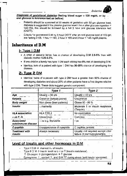
4- Vascular c••mplicalions:1. Microvascular disease (e.g. Nephropathy - Retinopathy· Neuropathy).2. N1acrovasciJlardisease (Atherosclerosis) 7 Stroke, TlAs.
7 Coronary heart disease.7 Peripherai ischaemia (e.g LL).
3. Gangrene of the foot (diabetic foot) 7 may occur with palpable dorsalis pedisartery indicating microangiopathy.
4. Angina - myocardial infarction may be painless due to neuropathy.
-(,: Risk factors for rnacrovascular complications in O.M- Duration - Increasing age - Hypertension- Hyperinsulinism - Hyperlipidemia - Proteinuria
1< Insulin resistance ...• Hyper insulinlsrn, this leading to dyslipidemia (1' VLOL,.J; HOL) see later. '
5- Renal complications: (see nephrology):1. Urinary tract infection, pyelonephritis which may lead to (papillary necrosis)
2. Glomerular disease (Diabetic nephropathy).= Diabetic glomerulosclerosis 7 Nephrotic $.
Diabetic glomerulosclerosis may be diffuse or nodular. The laller is called asKimmelstiel Wilson syndrome.Microscopy of diabetic nephropathy:
Increased mesangial matrix.
Hyalinization of aflrent arteriole.
• Thick basement membrane.
- Hyalinization of eflrent arteriole.
&- Gastro-intestinal complications:• Change in the bowel habits (autonomic neuropathy)
Constipation. - Diarrhea.• Gastroparesis with gastric dyspepsia.
7- Hepatic complications:1- Fatty infiltration in type II D.M. 2- Glycogen infiltration in type I D.M.
Liver and DM as above plus: ""• NASH may occur in diabetics (see liver).• . Auto immune hepatitis may be associated with type 1 A diabetes.• ,Haemochromatosis -) D.M.• tiver cirrhosis -) Insulin resistance.• Selection of oral hypolycemics in cases of liver cirrhosis (see later).
B~Genitourinary complications:• Male: Impotence due to autonomic neuropathy and vascular complications.
• Female: Vulvovaginal infections, dyspareunia.
• Autonomic neuropathy may lead to cystopathy with inability to sense a fullbladder and failure to void completely with post-void residual increase -7
hesitancy, incontinence and recurrent urinary tract infections, diagnosis bycystometry and urodynamic studies.
67
EndoCline ~
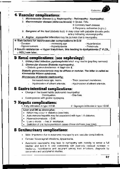
9- Diabetic fool:• Amputations in diabetes can be delayed or prevented by patient education and
medical supervision.
• Ischaemia, infection and neuropathy combine to produce tissue necrosis.
• It is important to distinguish between the ischaemic and the neuropathia foot.
~ Ischaemia Neuropathy
Symptoms - Claudication· Usually painless
Rest pain - Painful neuropathy may occur
f I'!spection Trophic changes High arch
Clawing of toes ji Palpation Cold Warm IPulseless Pulse is present.
•.Ulceration Painful Painless\I Heel and.toes Plantar i
Mangement of diabetic footInfection: early effective antibiotic therapy.
Ischaemia: The feet are assessed with doppler ultrasound, the occludedvessels can be treated by pass surgery or angioplasty.
10- Diabetic infections:Diabetics have a greater frequency and severity of infections due to abnormalities In
cell mediated immunity and phagocytic function associated with hyperglycemic and
diminished vascularization, also hyperglycemia aids the colonization and growth of a
variety of organisms e.g candida and other fungal species.
• Skin -7 Staph infections, mucocutaneous candidiasis.
• Gall bladder 7 Emphysematous cholecystitis.
• Urinacy tract -7 Pyelonephritis, perinephric abscess and emphysematous
pyelonephritis.
• Lung -7 Tuberculosis.
-7 Staph, pneumococcal or gm -ve bacterial pneumonia.
• Rhinocerebral mucormycosis is a rare fungal infection.
68

--.--Endocrine ~
11· Coma = (Acute diabetic complications):
Non ketotic'hyperosmolar coma
a. Lactic acid coma (severe acidosis)It occurs as a result of accumulation of lactic acid in blood (> 5 mmol/L)
Patients present with a severe metabolic acidosis without significanthyperglycemia or ketosis This may occur in diabetic patients under biguanidestherapy with high doses.
High risk with biguanides therapy with advanced liver or renal dysfunction.
Treatment by rehydration + infusion of isotonic bicarbonate 1.26%.
b. Hyperosmolar coma: (It is characteristic of uncontrolled type 2 OM)
Marked rise of blood glucose (> 600 mg/ell). There is minimal insulin levelwhich is not enough to e hyperqlycernia but enough to e ketogenesis in the liver.This will lead to hyperglycemia without significant ketosis, hyperosmolarity> 320 mOsmiL -7 dehydration, stupor and coma. The hyperosmolar state maypredispose to stroke or myocardial infarction.
Common precipitating factors include: consumption of glucose rich fluid, steroidtherapy.
Plasma osmolarity =? [Na +KmPn/LJ "-PlasmaGlucoSe ~g1dl .".' . "-' 18
Normall it is 285·300 mOsmlL, Value > 320 mosrrvt.« h erosmolari
C. Hypoglycemic coma:It results from big doses of Insulin with uncorresponding amount of diet (Insulinshock). It usually occurs in patients under Insulin therapy, also it can be causedby oral hyoglycemics (see hypoglycemia).
d. Diabetic ketoacidosis (DKA):Diabetic ketoacidosis is the hall mark of type 10M. Its main causes are:• Undiagnosed diabetes.
• Stress of intercurrent H1ness.
• Interruption of insulin therapy.
r.!!t~9.!\\!!.~.~!~In the absence of insulin, hepatic glucose production accelerates, andperipheral uptake by tissues such as muscles is reduced.
High glucose levels lead to osmotic diuresis with loss of fluid andelectrolytes leading to dehydration.
Plasma osmolarity rises and renal perfusion falls.
Absence of glucose utilization leading to lipolysis -7 elevation of free tattyacids in blood, this will lead to conversion of fatty acids into ~etone bodies in
69

EndoC1ine ill.-rnitochodrta of the liver (ketogenesis) giving acetone, acetoacetate and ~hydroxy butyrate.
Accumulation of ketone bodies produces metabolic acidosis.
The excess ketones are excreted in the urine, also appear in breath. Ketonebodies -7 vomiting -7 further fluid loss.
Respiratory compensation for the acidosis ~ hyperventilation (air hunger).
Progressive dehydration impairs renal excretion of hydrogen ions andketones aggravating the acidosis.
G!i!\i.CJ~LP.i~~J1.r.~.9L~Il!,~!l~ic.~!llQ!l,~.i~.Q§i.§\Hyperventilation (kussmaul breathing).Nausea, vomiting.Deterioration of level of consiousness.Dry skin (dehydration).Superimposed infection.Sometimes acute abdomen may occur.
!.!!Y!l.~ljg!l\\Q!!§.:1· High blood glucose, acidosis.2- Ketonaemia or heavy ketonuria.3- Acidosis.
Complications of DKA• Cerebral edema due to rapid
reduction of blood glucoseand use of hypotonic saline.
. Acute respiratory distress $• Thromboembolism
'Disseminated intravascularcoagulation.Acute circulatory failure.
• Serious Infections e.gmucormycosis.
, Hypoglycemic coma Ketotic coma11- Petient Follow the treatment Neglect the-treatment I
2- Onset Acute Gradual ,3-Pulse Tachycardia + good volume Weak & rapid i
4- Blood pressure High (1 C.A) Low (dehydration + acidosis)'
i5- Skin Sweaty, pale Dry &·inelastic
6- Breath Normal Acetone odour ~7- Pupils Dilated Not dilated
8- R~spiration Nonmal -. Acidotic
9- Tongue . Normal Dry (under surface of the tonque)
10- Urine No sugar +ve Sugar & acetone.
11- IV glucosiJ. . • Rapid recovery ifearly , No effect •I'1;[- Coma Irritable . Not irritable I
13- Temperature Normal, hypothenmia may Subnormaloccur
I~ Acute diabetic complications = Diabetic comas & Infections IChronic diabetic complications = Diabetic triopathy & atherosclerosis
Q Electrolyte deficiency in D."'1
• Hyponatremia with uncontrolled diabetes and DKA leading to lethargy and weakness.
• Hypomagnesemia ----? muscle spasms and tetany.
• Hypophosphatemia -t Osteomalacia and muscle weakness.
70

Endocrine
Invest· tions of a case of DiabetesJ.- Urine for glycosuria: (qualitative rather than quantitative)
Dipstick methods:Glucose is detected in urine in cases of D.M, renal glycosuria, alimentary
glycosuria (gastrectomy, thyrotoxicosis), lactosuria and in the presence of reducingagents in urine e.g. vitamin C and aspirin.
2- Urine for Ketonuria'
Qualitative detection of ketones bodies can be done by "Nitroprusside test ",dipstick test is available.
Heavy ketonuria can inhibit some dipstick tests for glucose.
3= Blood sugar tests:
Fasting: NonnalIv 75 • 115 mg/d\Fasting" 126 mg/dl is diagnostic.
If < 126 mg/dl in suspected patient orai glucose tolerance test can bedone.
Twohours post - prandial blood sugar: nonnally < 140 mg/dlIt usually returns to normal after 2 hours (after intake of 75 gm glucose).
If " 200 mg/dl, the diagnosis is confirmed.
!lan~om blood sugar ~ 200 is also di~gI).Q§ti£
(lral Glucose toleranre test (OGTI);It is performed only in borderline or suspected cases, diagnosis ofimpaired glucose tolerance and diagnosis of gestational diabetes.
Patient should take normal sufficient diet for at least 3 days before thetest.
Method:
1- Fasting venous sample of plasma sugar is determined.
,I
2- Bladder is then emptied.
3- 75 gm glucose are then taken orally for adult and 100 gm"for pregnantfemale
j.

EndoC1ine ~4. Samples of venous blood & urine are tested for glucose: at zero, 1/2
hour, 1 hour, 2 hours and 3 hours.
• Normal glucose tolerance is considered when fasting is <126 mgldl and PP. < 140 mg with no value between zerotime & 2 hours exceeding 200 mgldl (i.e. peak < 200 mg/dl).All urine samples are -ve for glucose.
• Because of diffioulties in interpreting & the lack of standardsrelated to aging, it is replaced generally by fastinghyperglycemia, post - prandial hyperglycemia and randomehyperglycemia as means of diaqnosis of D.M.
• Abnormalities in OGTT: (see also DD of glycosuria)
Renal glycosuria.Alimentary glycosu·ria.Impaired glucose tolerance.Flat response i.e. (the difference between the peak leveland fasting < 20-25 mgldl) e.g. malabsorption, insuiinomaand adrenal hypofunction.
4- Glycosylated Hb:
It is produced by a reaction between glucose & the terminal valine of theB-chain of the hemoglobin molecule.
Normally, it is 4-8 % of the total Hb.
In diabetics it reaches about 20%, so it is Ii in diabetics with chroniohyperglycemia; it refleots their metabolic control over the preceding 6weeks.Glyoosylated plasma proteins (fructosamine) may also be measured "San index of control. Glycosylated albumin is the major component,fructosamine measurement related to glycaemic control over thepreceding 1-3 weeks.
5- Self glucose monitoring: (home blood glucose monitoringby finger orick)
Capillary blood glucose measurerments performed by the patientsthemselves.
It is useful in patients in whom tight metabolic control is required.
Patients asked to take regular profiles (e.g. four daily samples on twodays eaoh week) and to note these in a record book.
Blood is taken from the side of a finger not from the tip whioh is denselyinnervated.
6- Hyperketonemia
Serum levels of aoetoacetate, acetone, and Beta hydroxybutyrate oan be done.
Ail lnvestlqatlons for complications of C.M should-be done e.g urine analysis, kidneyfunction tests, serum. electrolytes, blood pH. fundus examination," duplex seane jerlower limb arteries and CTIMRI for brain. . .
72

Endocrine ~
Differential Diagnosis of glucosuria (melituria)
1- Renal glycosuria:
- Due to H renal threshold of glucose, so at some point in the OGTT there isglycosuria inspite of the fact that plasma glucose below renal threshold.
- This is usually a hereditary tubular defect.
2- L8g - storage curve:
i.e. (alimentary glycosuria) due to rapid absorption e.g. after gastrectomy(late dumping syndrome) & thyrotoxicosis. There is sharp early rise in plasmaglucose> 200 mg/dl + glycosuria, but P.P. is below fasting level.
3- Transient hyperglycemia & glycosuria:
May occur during stress.
4- Reducing substances in urine: (false +ve test)
As salicylates, chloral hydrate.
Renal threshold of glucose Is 180 mgldl.Renal glycosuria is a benign asymptomatic condition where glucoseappears in urine despite a normal blood level ot glucose.As many as 50 % of pregnant women normally have demonstrable
. glucose in urine during the 3'" & 4th months but in late weeks ofre nanc lactose rna be resent lactosuria).
frreatment of Diabetes mellituslInsulin
• Insulin is a polypeptide, formed of 2 chains (A,B) linked by disulfide bonds.
• It is synthesized as preproinsulin which is then converted into proinsulin. Thelater is hydrolyzed into insulin & C peptide which are secreted in equimolaramount.
• C peptide measurement gives a better index of pancreatic B-cell function thanperipheral insulin.
• Insulin secretion is:
Increased by
• Glucose, sulfonylurea
• Amino acids (AA) specially arginine
Decreased by
• Somatostatin, beta blockers
• Thiazides.
• The main sites of inactivation are the liver & kidney.
73

Endocrine til.-Actions of insulin
fA- Rapid (tr.a!2sport i!."-~ctS)B- E!adua!jan!!!!..olic !!!e.=tsLI Entery of glucose, AA, K into the CarbohYdratescells
Glucose uptake by the brain is
obligatory and is not dependent on
insulin, other tissue such as
muscies and fats are facultative Fat
glucose consumers so insulin
facilitates glucose uptake by these
tissues.
Mechanism of action of insulin
IGlycogen storage.
IPeripheral Utilization of glucose.
'\'Gluconeogenesis.
e Lipogenesis.
,\, Ketogenesis by the liver.
Protein
I AA transport to the cell.
It has a protein sparing effect.
• The insulin receptor consists of two a subunits which include the bindingsites for insulin and two p subunits which traverse the cell membrane.
• When insulin binds 10 the a subunits this triggers tyrosine kinase activity ofthe p subunits, ieading to migration of a glucose transporter (GLUT,) to thecell surface ~ increase glucose transport into the cell.
Preparations of insulin
• Insulin was discovered in 1921.
• Bovine and porcine insulin have been the main stays of therapy until theintroduction of synthetic human insulin.
• Beef insulin differs from human insulin by three amino acids so inducesantibody formation, whereas pork insulin differs by one amino acid so it isrelatively non immunogenic
• Human insulin is now available from 2 sources.
a- Semisynthetic Le. chemical substitution of alanine of porcineinsulin by threonine.
b- Total synthesis of A&B chains separately by recombinant DNA.
• Conventional insulin preparations (bovine, porcine) contain potentiallyantigenic components. New procedures have been devised to preparepurer preparations. e.g. single mono component (M.C) insulin.
• Soluble insulin can be formulated with protamine or zinc to retard its action.
74

Type
IA- Rapidlv acting or soluble
Iclear solution)
It is also called crystalline insulinor regular insulin.
I It is given by S.C., I.M, I.VI injection.
Is- Intermediate actingIsophan insulin (cloudy
I solution).
It is called NPH (neutralprotamine hagedorn).I It is given by S.C injection only.
IC- Long acting
Protamine zinc insulin (PZI).
it is given by S.C injection only.
I
Endocrine ~
Trade names Action
- Insulin Actrapid.
- Hurnulin R.
Each 1 ml = 20 units.
Onset: 30 min.
Peak: 2-4 hrs.
Duration: 6-8 hrs.
Insulin NPH.
Humulin N.
Monotard.
Lente.
Each 1 ml = 40 units.
Onset: 2 hrs.
Peak: 5-10 hrs.
Duration: 16-24 hrs.
Insulin ultra-Iente.
Insulin ultratard.
Humulin L.
Each 1 ml = 40 units.
Onset: 6 hrs.
Peak: 10-20 hrs.
Duration: 24-28 hrs. IN.B Recently most insulin preparations are in the form of 100 unit/1 mi.
Insulin mixtures
• Premixed combination of 30% soluble with 70% NPH is the most widely used(humulin mixtard) or initard (50i50) are acceptable and convenient forpatients.
• Zinc and protamine insulin can be mixed in the syringe with soluble insulinimmediately prior to injection.
Indications of insluin:
1- Type 10M.
2- D.M not adequately controlled with diet and oral agent (type 2 OM).
3- Hyperglycemic ketoacidosis, hyperosmolar coma.
4- Critical episodes in type 2 OM e.g. operation, infection, ischemia, trauma&pregnancy.
5- Insulin test of hypothalamic hypophyseal adrenal axis.
6- Hyperkalemia.
7- Insulin stimulation test for GH assessment.
-Insulin analogues:
• Insulin lispro (Human log) is a regular insulin analogue which dissociates muchmore rapidly and thus enter the circulation more rapidly than soluble insulin.

Endocrine ~Special problems in D.M:
1- Infection: There is increased demand for insulin. Also, regular insulinshould be used.
2- Pregnancy & labour:• Oral antidiabetic are contraindicated.• Single dose insulin is changed into multiple doses.
3- Surgery: ( t insulin demand)• Preoperatively: shift to regular insulin.• Postoperatively: continue insulin therapy.
Complications of insulin therapy:
1- Hypoglycemia is the most common complication (see hypoglycemia)
2-lnsulin allergy:Local reaction as pruritic erythematous indurated lesion.Angioedema, anaphylaxis
Allergy can be avoided by changing the source of insulin.Antihistaminic & topical steroids are helpful.
3- Insulin resistance (insulin antibodies) with dyslipidemia andhypertension. The insulin requirement is increased up to 200unites/d or more.
4- Weight gain:- Patients who are non compliant are predisposed to weight gain
with insulin therapy (insulin makes you feel hungry).
5- Pseudo insulin resistance (Somogyi phenomenon);
- Occurs in patients over treated with insulin -7 hypoglycemia withrelease of anti insulin hormones -7 rebound hyperglycemia.
Treatment: reduction of insulin dose and dietary control.
6- Insulin lipodystrophies:Atrophy or hypertrophy of S.C. fatty tissue at the site of insulin injection.
7· Peripheral edema due to salt and water retension.Insulin resistance: It is a clinical condition characterized by increased serum insulin levelwith high or normal blood glucose with development of the metabolic $ or syndrome x (see later).
Causes:Surgery, infections, acromegaly, Cushing's syndrome, acanthosis nigricans andpolycystic ovary syndromeInsulin antibodies (prereceptor resistance), obesity (receptor resistance),failure to activate receptor tyrosine kinase (post receptor resistance).
Treatment:Change to human preparations, treatment of the cause, immunosuppression bycorticosteroids in cases of insulin antibodies, weight reduction.Manifestations of the metabolic syndrome or syndrome x includehyperinsulinemia, hypertension. Dyslipidemia (-1- HDL and T VLDL), centralobesity, type 2 DM or impaired glucose tolerance (IGT) or impaired fastingaluGose and accelerated cardia vascular disease.
76

Endocrine
Dawn phenomenon:Early morning rise in plasma gl~cose requirinq increased amounts of insulin.' It isindependent of Somogyi mechanism. The nocturnal surge of gr,Owth hormone release maybe a factor.
Oral anlidiabelil;SThey are particularly used in type 2 OM without ketosis when an initial trials on dietalone has failed to control symptoms and hyperglycemia.
Thev are 2 groups: a. SUlfonylurea. b. Biguanides.
A- Sulfonylurea
Mechanism of action:1- Stimulate insulin release from ~ cells (insulin secretagogues) through
closure of ATP-sensitive potassium channels on the 8-cell membrane;this will promote calcium influx, leading to insulin release.
2- They increase insulin sensitivity in peripheral tissues!?3- They reduce the hepatic release of glucose.
Indications:t- NIOOM.2- Tolbutamide test to diagnose insulinoma.3- Chlorpropamide in treatment of nephrogenic diabetes·insipidus.
MYj)!]~_I!l_'!.~ti!!!!_~~t- Hypoglycemia. 2- Alcohol intolerance, allergy.3- Chlorpropamide causes 7 hyponatremia.4- GIT u set, cholestatic laundice.
Sulfonylureas shouldbe used with care in patients with liver disease, andonly those primarily excreted by the liver should be given to patients withrenal lmpairrnent.'
B- Biguanides
M.~.~.h.,!!!j~!!!.91l!~ti9.!E1- J.Absorption of glucose from the gut.
2-tAnaerobic metabolism of glucose to lactate.3- Tlnsutin sensitivity (upregulation of insulin receptors). i.e they are insulin
sensitizers.4- J. Hepatic production of glucose by inhibiting gluconeogenesis.
IndicatioI.!§.;t- As a supplement to a sulfonylurea when this with dietary advice fail to
control blood sugar.2- Over weight diabetics.
3- To + insulin requirement where insulin resistance is not due to antibodies.
77

Endoc);", Ill.-Adverse effects:
1- Lactic acidosis may occur especially in patients with severe hepaticor renal disease.
2- GI upset: Anorexia, vomiting, epigastric discomfort and diarrhea.
I Unlike sulphonylureas, biguanides do not induce hypoglycemia in non diabetic fndividuals.
Preparations & doses
Drug Trade name Dose Half life(mg/d) (hours)
i 1- Sulfonylureas: I1st generation:- Chlorpropamide Pamidine (100 mg, 250 mg)'tab 100-500 36
- Tolbutamide Diamot (500 mg) tab 500-3000 42nd generation: (lessdrug interactions)
- Glibenclamide Euglucone or Doanil (5 mg) tab 2.5-15 12 I- Gliclazide Diamicron (80 mg) tab 40-240 10
,- Glipizide Minidiab (5 mg) tab 2.5-30 3.5
-Glimepiride Amaryl (1mg, 2mg, 3mg) tab. 1-8It's duration of action is 24 hr
2- Biguanides:- Metformin Cidophage or Glucophage 850-2250 5
I (850 mo. 500 mol' tab
- Glibenclamide: long half life, renal excretion, avoid in renal impairment. It is prone toinduce severe hypoglycemia. It should be avoided in elderly.
- Gliclazide: fairly long half life, mainly metabolized by liver, can be used in renalimpairment. It cause few side effects.' .
- Chlorpropamide: long acting, renal excretion, avoid in renal impairment.- Tolbutamide: short half life preferred in old age, metabolized by liver, can be used in
renal Impairment.- Glimepiride has low incidence of hypoglycemia and can be administrated once daily.
Other recent oral hypoglycemic drugs1- Glucosidase inhibitors e.g. Acarbose (Glucobay) 50 mg tab TDS
• These drugs inhibit the enzymes involved in the breakdown ofcarbohydrate in the intestine ~ t glucose absorption.
• They will reduce the postprandial rise in blood glucose. It can beused in patients with liver disease.
Side effects: - Abdominal discomfort, flatulence and diarrhea.2- Repaglinide, (Novonorm)
• It stimulates insulin production at meal times, 0.5 mg or 1 mgbefore meal times.
3- Piog/itazone (Glustin) 15-30 mg/D one dose, it reduces insulinresistance i.e insulin sensitizer, also itreduces hepatic glucose production byinhibiting gluconeogenesis.
78

EndoC1ine ~
Treatment of type 1 diabetes
• Caloric recommendations are 36 kcaVkg for males and 34 kcaVkg for females.
• Carbohydrate contents is 50-60%, (4 Kcal/gm), proteins 15%, (4 Kcal/gm) fats30-35% (9 KcaVgm) of total energy intake. Polyunsaturated fats are preferred.Salt restriction for hypertensive diabetics.
• The minimal proteins requirement for good maturation is 0.9 g/kg.
• The use of non-nutritive sweeteners e. as artame is useful.
Intervention~ It! prediabetic stage (+ve islet's Abs) to prevent type fA DM!?
• Neonatal and early infancy cow milk deprivation.
• Immune suppression by cyclosporine or azathioprine.~ ."
• AntioxIdant."'
The Side effects and risk of long term immunosuppression are felt to be greater than the riskof diabetes.
Insulin therapy
The usual required insulin dose in most insulin deticient diabetics is about 0.5
to 0.8 unit per kg/D, it can be given by the different following methods (with trial
and error!?) with initial daily dose 0.3 units/kg/D. Sometimes the required
insulin is higher.
A- Conventional iJ!~.J!!i!!J!.t!\.~JffiWe can give one or two injections/day of intermediate acting insulinwith or without the addition of small doses of regular insulin.
Adults of normal weight may be started on 15-20 units/day, obesepatients may be started on 25-30 units/day. Changes should be nomore than 5-10 units/step.
Single insulin injection provides adequate control in patients withresidual insulin secretion.
Poorly controlled patients should be placed on twice daily insulininjections with about two thirds of the total insulin given beforebreakfast and the remainder before supper using mixtard insulin asbelow.
Example: Most patients on twice daily insulin injections treated with a mixture of
intermediate and regUlar insulin e.g. 20 units NPH plus 10 units of regular before
breakfast and 10 units of NPH plus 5 units of regular before supper using
mlxtard insulin (70/30 each 1 ml = 40 or 100 units).
79

E1UioCline til.-B- The multiple subcutaneous insulin inj~jjon tet;.hnigue:
Administration of intermediate or long acting insulin in the evening assingle dose together with regUlar insulin prior to each meal.
One approach is to give 25% of the daily dose as intermediate insulinwith the other 75% given as regular insulin divided such that 40, 30and 30 percent is given 30 minute before breakfast, lunch and supperrespectively.
The introduction of (pen injection) devices has made this approachmuch more acceptable to patients.
C- Infusion devices:CSII (continuous subcutaneous insulin infusion); insulin isdelivered by a small pump strapped around the waist.
Insulin is delivered at a basal rate continuously throughout the dayvia a needle in the subcutaneous tissue of abdominal wall.
Mealtime doses are delivered when the patient touches a button onthe side of the pump.
D- Pancrease /Islet transplantation:Because of long term Immunosuppression pancrease/islettransplantation is at present an option for only a select group ofpatients, mainly for type 1 O.M requiring renal transplantation, this isalso more effective in preventing nephropathy in the grafted kidney.
Treatment of type 2 diahetes
Piet: (similar to diet in type' OM but I?l
• The majority of type 2 OM, patients are obese, the main goai of diet therapy istherefore weight loss.
• In thin type 2 OM, patients calories should not be restricted.
• The diet of aN patients with type 2 OM should be limited in fat & cholesterol.
Prevention of type 2 PM
It is indicated in individuals with a strong family history of OM or those withimpaired glucose tolerance (IGT) or impaired fasting glucose.
• Diet control and exercise to maintain normal body mass index (BMI).
• Metformin is helpfui in prevention or delaying of O.M!?
• Ramipril (ACE) and pravastatin (cholesterol lowering) are also helpful !?
Oral antidiabetic drugs:
• In patients with type 2 D.M with no successful metabolic control with dietarytherapy alone, the next therapeutic step is oral antidiabetic + diet.
80

EndoCTine ill.-Insulin; Some patients with type 2 DM require insulin therapy.
• Acceptable metabolic control may not be achieved from the start with oralantidiabetic (1 ry failure), or a patient who initially responded to oral agentmay with time fail to respond (2ry failure).
• The use of insulin in type 2 diabetes is similar to that described in type Idiabetes, except that because of the presence of insulin resistance, higherdoses may be needed.
• Because of some residual endogenous insulin secretion, a single daily doseof an intermediate insulin may be enough.
Treatment Pathway for D.N.:
The ideal goals for glycemil: control:• Fasting plasma glucose (90-130 mg/dL).• Peak post prandial plasma glucose < 180 mg/dL.• Glvcosylated Hb < 7%.
8J

I lI
Endocrine ~
Management 01 diabetic ketoacidosis:i Insulin therapy Fluid therapy K therapy by infusion
(crystalline)~. Start I.V insulin,
5u/h.
• When blood sugar<250 mg/dL reduceinsulin \0 1-4 u/h.
• Start I.V 0.9% saline1 liter in 30 minutes.
• Then 0.5 liter 0.9%saline in 30 minutes.
• Then 0.5 liter 0.9%saline in 1 hour.
• Then 0.5 liter 0.9%saline in 2 hours.
• Then change toglucose 5% 0.5liter/2h when bloodsugar < 250 mg/dL.
• There is total body K deficit, althoughthe initial levels may not be low due toacidosis. I
• Insulin therapy leads to K influx into thecell, so K therapy must be started with Iinitiation of insulin therapy, '!is follows:
- If plasma I< > 5 S meqll give no K. I- If K 3 5-5 5 give 20 meq for each
liter of infused fluids IIf K < 3.5 give 40 meq for each hterlof Infused fluids. .
Notes• The insulin regimen can be started by 10-20 u I.M followed by 5 u/h I.M.
• Average fluid deficit = 6 liters
- 3 L for extra-cellular compartment, replaced by saline.
- 3 L for intracellular compartment, replaced by glucose.
• The patient may need urinary catheter (if no urine is passed after 2 hr),nasogastric tube (if drowsy), antibiotic (if infection is likely).
• If plasma Na > 155 meqlL give saline 0.45 % rather than 0.9% until Na falls to 140meqlL.
• If PH < 7 give 300-500 ml 1.26% sodium bicarbonate over 30 minutes.
• Monitor blood glucose, Na, K, pulse, blood pressure, urine output, respiration,plasma osmolarity and PH.
• Continue with the above regimen until fluid deficit is replaced, ketonuria abolishedand adequate oral intake can be started.
Treatment 01 hyperosmolar coma (nan ketotic):The therapy for the hyperosmolar coma is very similar to that for OKA, with
the administrations of insulin, fluids & K.
This coma is complicated sometimes with: Mol - stroke - infection -pulmonary emboli.
Small dose of insulin is required to prevent rapid lowering of blood sugar, toprevent brain edema (desequilibrium syndrome).
Low molecular weight heparin may be required to prevent thromboticcomplications.
82
Brittle diabetesThis term is used to describe unpredictable fluctuations of blood glucose with recurrentepisodes of hyperglycaemia with or without ketoacidosis and/or recurrent hypoglycaemicepisodes.
Causes of recurrent hupoglucaemja:• Over treatment with insulin.
• Low renal thereshold for glucose.
• Endocrine causes e.g pituitary or adrenal insufficiency.• Gastroparesis due to autonomic neuropathy leading to mismatch between the time of
absorption and the peak of insulin action.
• Renal failure.• Uncooperative, unintelligent patient.
Causes of recurrent ketoacidosis;• Inappropriate insulin combinations.• Intercurrent illness e.g unsuspected infections.• Unknown aetiology.
Causes of recurrent hyperglycemia:• As ketoacidosis plus somogyi and dawn phenomena.
Factitious disorders and malingering behaviour may be also responsible for-blood glucosef.Iuctuations. .
Management of brittle djabetes(1) Hospitalization, careful evaluation of the patient.
(2) Treatment of cause.
(3) Use crystalline Insulin at regular Interval e.g every 6 hours.
(4) Insulin pump.
HypaglycemiaDefinition and diagnosis of hypoglycemia is based on the presence of a triad calledwhipple's triad:
1) A low plasma glucose concentration « 45-50 mg/dl).
2) Symptoms consistent with hypoglycemia.
3) The improvement of these symptoms following an increase in plasma glucose.
Glucose thresholds for hypoglycemia induced symptoms and physiologic responsesvary widely so, whipple's traid is important for diagnosis.
83

EndoCline ~
Causes of hypoglycemia:
Fasting Hypoglycemia• Insulinoma, hepatoma.
• Critical illness e.g extensive hepatic dysfunction, chronic renalfailure, malnutrition or anorexia nervosa.
• Drugs: Insulin, oral hypoglycemics (it may be factitious).
• Hormonal deficiency: (GH, Epinephrine, Cortisol).
Postprandial (reactive) hyp.emia:• Alimentary hypoglycemia (reactive) e.g dumping syndrome after
gastric surgery (see GIT)
• Functional hypoglycemia.
• Reactive hypoglycemia of diabetes, sometimes occurs with earlydiabetes (late but excessive release of insulin after a carbohydratediet).
• Galactosemia, fructose intolerance, ethanol induced.
Important CaUSES of Hupoglycemia and approach to the patients
1) Insulinoma:• Relatively rare tumors between age of 40-70 years.
• Diagnosis by low glucose level « 45 mgldl) in presence of highplasma insulin (6 U/ml or more).
• It is also helpful to measure plasma C peptide (high).
• CT scan, celiac or superior mesenteric arteriography for diagnosis.Treatment:
- Surgical removal.- Medical to prevent insulin release e.g by diazoxide, octreotide.
2) Insuli.!!J!Jerapy'• The presence of hypoglycemia, high insulin levels and low C peptide
indicate excess exogenous insulin.
~ral hypoglycemics• Common with long acting drugs.
• This occurs especially in patients with renal & hepatic diseases.
• Insulin level is high, C peptide is high like insulinoma, so search for ahypoglycemic agent in blood or urine.
• Observed in patients after gastrectomy (alimentary hypoglycemia)this is due to rapid glucose absorption with excessive insulin release,glucose metabolized rapidly but insulin levels remain high resulting inhypoglycemia 1-2 hours postprandially.
84

Endocrine IlIl.-• Idiopathic postprandial hypoglycemia.
Clinical presentation of Hypoglycemia·
1) Secondary to catecholamine release (Adrenergic) this occurs with rapiddecrease in glucose level:
- Sweating
- Tremors
- Anxiety
- Hunger
- Tachycardia.
Treatment of severe hypoglycemia;• Initial treatment of a confused or comatosed patient with severe hypoglycemia
is to infuse a bolus of 50 ml of 50%. Glucagon 1mg 1M or S.C can be' used.
• Then give continuous infusion of 10% of glucose at a rate sufficient to keep theglucose level greater than 100 mg/dl.
• In many situations especially following long acting insulin or oral hypoglycemicdrugs, the hypoglycemia will last for an extended period of time, so continuethe treatment with close observation.
I Mild to moderate c~se$ -can by treated by oral glucose or. sucre'se 0"100 mi. of sweet drink,
Q. Causes of Hypoglycemia in patients taking insulin or sulphonylurea ?• Missed, delayed or inadequate diet. • Unexpected or unusual exercise• Errors in doses or schedule • Poorly designed insulin regimen.• Gastroparesis (autonomic neuropathy) .• Malabsorption or dumping.• Unrecognized other endocrine disorder e.g Addison's disease• Factitious.
85