Disorders of Perception Things that can go wrong with visual perception that cannot be characterized...
33
Disorders of Perception Things that can go wrong with visual perception that cannot be characterized as simple absolute visual field defects. Extra- striate cortex, ‘visual association cortex’, ‘higher order’ visual deficits. Remember: visual perception is all about analyzing different aspects of an image, not just getting a picture into the head! There are multiple parallel systems that analyze
-
Upload
virginia-price -
Category
Documents
-
view
217 -
download
3
Transcript of Disorders of Perception Things that can go wrong with visual perception that cannot be characterized...

Disorders of PerceptionThings that can go wrong with visual perception that cannot be characterized as simple absolute visual field defects. Extra-striate cortex, ‘visual association cortex’, ‘higher order’ visual deficits.
Remember: visual perception is all about analyzing different aspects of an image, not just getting a picture into the head! There are multiple parallel systems that analyze different aspects of a visual scene.
Primary section objective: know the definitions.

Visual Object Agnosia
• The impairment of object recognition in the presence of relatively intact elementary visual perception, memory, and general intellectual function.
Farah & Feinberg, 1997

Agnosia
• Apperceptive: lack of recognition because of impaired (higher order) visual perception.
• Associative: lack of recognition because of lack of association of percept with its meaning.
Lissauer 1890 per Farah & Feinberg, 1997

Apperceptive Agnosia
• Basic acuity and visual fields preserved:– Good acuity,– Brightness discrimination,– Color vision, and– Other elementary visual capabilities
• However, cannot distinguish between or recognize different visual shapes
• Cannot copy (draw) shapes

Apperceptive Agnosia
• Apperceptive agnosias often occur after carbon monoxide poisoning or other diffuse toxic effects.
• Subtypes include: dorsal and ventral simultanagnosia, and visual hemi-neglect.
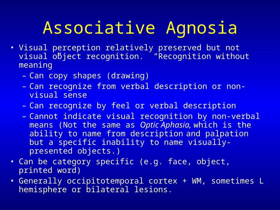
Associative Agnosia• Visual perception relatively preserved but not visual object
recognition. “Recognition without meaning”– Can copy shapes (drawing)– Can recognize from verbal description or non-visual
sense– Can recognize by feel or verbal description– Cannot indicate visual recognition by non-verbal means
(Not the same as Optic Aphasia, which is the ability to name from description and palpation but a specific inability to name visually-presented objects.)
• Can be category specific (e.g. face, object, printed word)• Generally occipitotemporal cortex + WM, sometimes L
hemisphere or bilateral lesions.

Simultanagnosia• Impairment in perception of multielement or
multipart visual displays. (e.g. piecemeal description of a complex picture. May fail to interpret overall scene.) – Dorsal: Cannot attend to >1 part of object. May act
blind as they bump into objects that are close to each other. Have trouble counting.
• Bilateral Posterior Parietal lesions.
– Ventral: Recognition of simple shapes not obviously impaired. (letter by letter reading). Don’t bump into objects i.e. don’t act blind. Can count, so can actually ‘see’ more than one object. But don’t see the whole picture in a scene, and can’t identify more than one object at a time.
• L inferior temporo-occipital cortex

Perceptual Categorization Deficit
• (Warrington and Taylor, 1973)• Subjects have trouble matching 3D objects
across differences in viewing conditions (changes in lighting, perspective).
• But no obvious problem in normal life: require changes in lighting etc. that even normals find challenging (Farah 1999). More a deficit in ‘visual problem solving’?– Poorly localizing, usually. R inferior parietal lobe.

Prosopagnosia
• “The man who mistook his wife for a hat”• Προςοπόν (prosopon) = face, agnosia = non-
knowledge. • Usually aware of deficit.• Sometimes called “Face blindness”• Rely on non-facial cues (voice, gait, clothing)• Recognize that they are looking at a face.• Can discriminate some qualities (gender,
race, ~age, emotional expression!)

Prosopagnosia• Superior temporal sulcus has strong connections with
the amygdala. Injury can cause a severe deficit in comprehension of facial expressions and gaze directions. Adolphs et al. 1994, Young et al., 1995
• Can be secondary to PCA infarct encroaching on medial cortex of occipital and temporal lobes and the inferior longitudinal fasciculus (i.e., occipital to temporal projections).
• Can be associated with Alzheimer’s (and much else).• Most strongly associated with the fusiform gyrus: in
particular the fusiform face area.


Central Achromatopsia• Can perceive borders defined by color differences that classic
photoreceptor color-blind people cannot (Ishihara test).• Pure form is very rare (requires specific bilateral lesions in
ventro-medial occipital lobe)• A defect in color perception caused by an acquired cerebral
lesion.– Reduced hue discrimination– Deficient color constancy– Cannot order colors in a test
• Colors are either all gray, or “washed out”, “dirty”, or “faded”• Problems in imagining colors are reported (Oliver Sachs,
colorblind painter)• Also known as color agnosia

Balint Syndrome• Simultanagnosia (independent of object size or
visual periphery.) • Spatial disorientation: inability to orient, localize,
and reach for objects = “optic ataxia”• Oculomotor disturbances: fixation, saccade
initiation and accuracy, smooth-pursuit. “optic apraxia” = inability to voluntarily move eyes to a new position
• Damage to posterior superior watershed areas (Brodmann 7 + 19). Multiple strokes, Alzheimer’s, tumors, other injury.

Misidentification Syndromes• Patient incorrectly identifies and reduplicates
persons, places, objects, or events.• Capgras Σ: multiple persons (incl. Family)
replaced by impostors.• Fregoli Σ: a familiar person is impersonating a
stranger.• Intermetamorphosis: Persons known to the
patient have exchanged identities.• Delusion of subjective doubles: the patient
themselves have been replaced.

Capgras Syndrome• Also called Capgras Delusion.• Named after French psychiatrist Joseph Capgras, wrote
a paper about a woman who was convinced that “doubles” had replaced people she knew.
• It has been proposed that face recognition has both conscious and unconscious parts. Idea that people can recognize the faces, but they associate them with strangers (don’t show appropriate emotional reactions to faces they recognize). Visual processing defect has to be in addition to other cognitive problems (schizophrenia). Not just cognitive: cognitive + visual processing.
• (some prosopagnosics show opposite: no recognition but appropriate affect, i.e. emotional response).

Fregoli Syndrome• Also Fregoli Delusion• Named after Italian actor and
quick-change artist Leopoldo Fregoli
• Belief that different people are a single person in disguise.
• Like Capgras, thought to be a problem with normal face recognition.
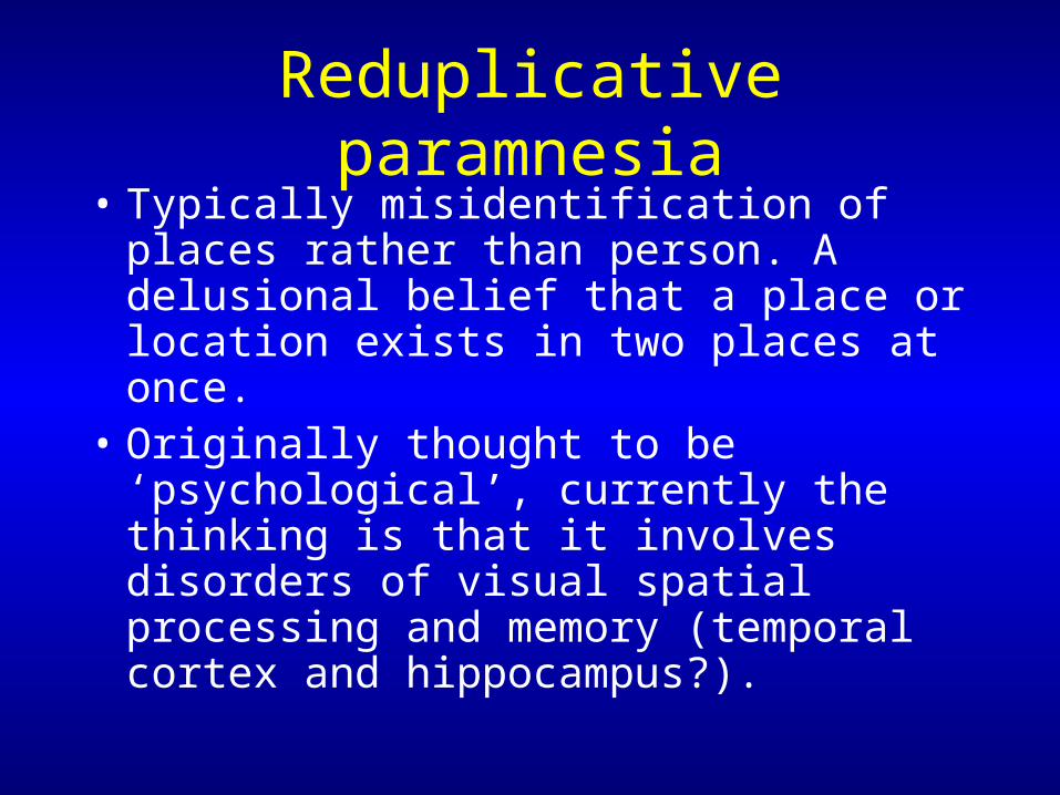
Reduplicative paramnesia• Typically misidentification of places
rather than person. A delusional belief that a place or location exists in two places at once.
• Originally thought to be ‘psychological’, currently the thinking is that it involves disorders of visual spatial processing and memory (temporal cortex and hippocampus?).

The following excerpt illustrates some of the core features of the delusion. The patient had suffered a head injury after a fall in his home. The impact had caused a fractured skull and frontal lobe damage to both sides (although more pronounced on the right) owing to the formation of intracerebral hematomas:
“A few days after admission to the Neurobehavioural Center, orientation for time was intact, he could give details of the accident (as related to him by others), could remember his doctors' names and could learn new information and retain it indefinitely. He exhibited, however, a distinct abnormality of orientation for place. While he quickly learned and remembered that he was at the Jamaica Plain Veterans Hospital (also known as the Boston Veterans Administration Hospital), he insisted that the hospital was located in Taunton, Massachusetts, his home town. Under close questioning, he acknowledged that Jamaica Plain was part of Boston and admitted it would be strange for there to be two Jamaica Plain Veterans Hospitals. Nonetheless, he insisted that he was presently hospitalized in a branch of the Jamaica Plain Veterans Hospital located in Taunton. At one time he stated that the hospital was located in the spare bedroom of his house.” Benson et al 1976

Reduplicative paramnesia (RP), a syndrome named by Pick in 1903 to describe a specific and limited disturbance of memory, is characterized by a subjective certainty that a familiar place or person has been duplicated.1 Most often seen in posttraumatic brain injuries, this syndrome has been described in a variety of neurologic conditions, including strokes, intracerebral hemorrhages, tumors, dementias, encephalopathies,and various psychiatric conditions.2 The classic and most commonlydescribed neuroanatomical localization is the combination of bilateral frontal lobe and right hemisphere lesions.3,4 Although many theories have been advanced to explain how these lesions could produce this syndrome, the specific pathophysiology is unknown. We report a patient who provides anatomical and neuropsychological support for the theory that RP is a syndrome that may develop in a vulnerable brain by a lesion in the ventral visual stream, disrupting communication between the visual cortex and both visual processing areas in the inferior temporal lobe and visual memory in the nondominant parahippocampal region.
From Budson et al., 2000.

Hemi-Neglect• Cannot notice or pay attention to visual
stimuli on one hemifield. Can’t even imagine that half the world is even there! Not even black…
• Usually it’s the left visual hemifield that is neglected (“Eyes Right”), due to a right parietal lesion. (The opposite almost never happens: the visual system no longer symmetrical at this level?).
• Relatively common
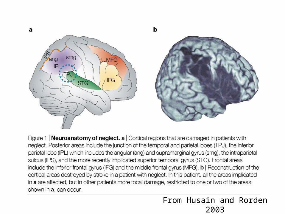
From Husain and Rorden 2003


Riddoch PhenomenonAfter George Riddoch, a British neurologist who first described the syndrome in 1917. Also called “statokinetic dissociation”. This is the preservation of the perception of motion in an otherwise blind hemifield after a visual cortical (occipital) lesion.The objects have no form or color, only motion can be perceived. Subjects describe the motion as being like “a black shadow moving on a black background” (so not really blindsight, where there is no conscious perception at all). Possibly caused by direct activation of MT via alternate pathways (maybe LGN to MT direct, or superior colliculus to MT), or by small ‘islands’ of remaining functionality in visual cortex.

Visual AtaxiaPatients with homonymous hemianopia due to occipital lobe lesions may experience loss of balance due to altered sensory input I.e. not a problem with cerebellum vestibular system etc. Possible sensation of falling towards the blind hemifield.

AkinetopsiaLoss of perception of motion with preservation of the perception of other modalities of vision (color, shape, etc). Damage to the human homolog of MT/V5 (“human MT” sometimes called MT+ or just MT).
Akinetopsia was mentioned in the 2006 episode of House titled "Son of Coma Guy." Dr. House diagnosed the patient by flashing the lights of the hospital room which caused the patient to have a seizure. The patient also could not catch a bag of chips and was startled when Dr. House moved closer to him as the motion was undetected until the motion ceased (wikipedia).

Visual AllesthesiaA disorder of visual perception where the visual fields appear to be tilted, rotated or flipped.

Anosognosia is a condition in which a person who suffers disability due to brain injury seems unaware of or denies the existence of his or her handicap. This may include unawareness of quite dramatic impairments, such as blindness or paralysis. It was first named by neurologist Joseph Babinski in 1914, although relatively little has been discovered about the cause of the condition since its initial identification. The word comes from the Greek words "nosos" disease and "gnosis" knowledge. (wikipedia)

Anton-Babinski syndrome, more frequently known as Anton's blindness, or Anton’s Syndrome, is a symptom of brain damage occurring in the occipital lobe. People who suffer from it are "cortically blind," but affirm, often quite adamantly and in the face of clear evidence of their blindness, that they are capable of seeing. Failure to see is dismissed by the sufferer through confabulation. It is mostly seen following a stroke, but may also be seen after head injury. It's believed that the cause is damage to two areas; the portion of the brain responsible for eyesight, and the portion responsible for detecting the presence of vision. (wikipedia)
Hence, Anton’s Syndrome includes anosognosia. Has also been reported to occur with optic nerve problems if frontal lobe damage also present.

The Brain is Complicated
• Not all visual problems live in the eye • Not all visual problems involve explicit visual
field defects or loss of acuity.• This just scratches the surface: neurology is a
big field.• Moose bites can be very nasty.