Digital capture, design, and manufacturing of an extraoral ... · CLINICAL REPORT Digital capture,...
4
CLINICAL REPORT Digital capture, design, and manufacturing of an extraoral device for a clarinet player with Bell’s palsy Cynthia Aita-Holmes, DMD, a Peter Liacouras, PhD, b William O. Wilson, Jr, DDS, MS, c and Gerald T. Grant, DMD, MS d Lyme disease is a debilitating infection that may cause a variety of mild to crippling symptoms. Dental practitioners must be aware of the systemic effects of this often multiorgan disorder. Its clinical man- ifestations may include facial and dental pain, facial nerve palsy, headache, temporomandibular joint pain, and masticatory muscle pain. 1 Lyme disease is presently the most common tick-associated illness in the United States. 2 Neuroborreliosis is the most common secondary mani- festation of Lyme disease, and Bell’s palsy is the most common presentation of neuroborreliosis. 3 Unilateral and bilateral facial paralysis may occur in up to 11% of patients with Lyme disease. 4 Bell’s palsy can be very debilitating both esthetically and functionally, especially when it af- fects a professional musician’s ability to play a wind in- strument. Although most patients recover well from Bell’s palsy, up to 30% have a poor outcome, with persistent facial weakness, psychological difficulties, and facial pain. 5 CLINICAL REPORT A 47-year-old man who played clarinet on active duty with the US Army band was referred to the maxillofacial prosthetics clinic for evaluation. He had a diagnosis of Bell’s palsy as a complication of Lyme disease infection he had acquired a few months before his visit. His facial paralysis precluded him from playing the clarinet because he was unable to create an efficient embouchure. The embouchure is the most important connection to the instrument for any wind instrument player. It serves as the seal for the air stream and is the definitive adjustment for the quality of the tone, pitch, timbre, and response of the instrument. 6 To restore his ability to form an appropriate embouchure, the patient fabricated a device, which improved his ability to play his instrument. Because he obtained less than ideal results at home, he consulted with us to see if we could provide a better solution. An examination of the patient revealed that when he attempted to play his clarinet, he was unable to create a seal around the mouthpiece and his right cheek would puff out (Fig. 1). He described the device he had fabri- cated and discussed its deficiencies. With this informa- tion, a chairside device was fabricated to create a seal around the clarinet mouthpiece and apply pressure to his cheek on the affected side. This device was fabricated with light-polymerizing resin (Triad; Dentsply Intl) and Disclaimer: The views expressed in this article are those of the authors and do not necessarily reflect the official policy or position of the Department of the Navy, Army, Department of Defense, nor the U.S. Government. We certify that all individuals who qualify as authors have been listed; each has participated in the conception and design of this work, the analysis of data, the writing of the document, and the approval of the submission of this version; that the document represents valid work; that if we used information derived from another source, we obtained all necessary approvals to use it and made appropriate acknowledgements in the document; and that each takes public responsibility for it. a Lieutenant Colonel, Dental Corps, United States Army, Naval Postgraduate Dental School, Bethesda, Md. b Director of Services, Department of Radiology, 3D Medical Applications Center, Walter Reed National Military Medical Center, Bethesda, Md. c Commander, Dental Corps, Maxillofacial Prosthetics, Naval Postgraduate Dental School, Bethesda, Md. d Captain, Dental Corps, United States Navy, Walter Reed National Military Medical Center, Bethesda, Md. ABSTRACT An extraoral device was fabricated to assist a clarinet player with Bell’s palsy. The device was fabricated by using stereophotogrammetry, digital design, and additive manufacturing technologies. (J Prosthet Dent 2015;114:297-300) THE JOURNAL OF PROSTHETIC DENTISTRY 297
Transcript of Digital capture, design, and manufacturing of an extraoral ... · CLINICAL REPORT Digital capture,...

CLINICAL REPORT
Disclaimer: TArmy, Deparand design othat if we useand that eacaLieutenant CbDirector of ScCommanderdCaptain, De
THE JOURNA
Digital capture, design, and manufacturing of an extraoraldevice for a clarinet player with Bell’s palsy
Cynthia Aita-Holmes, DMD,a Peter Liacouras, PhD,b William O. Wilson, Jr, DDS, MS,c andGerald T. Grant, DMD, MSd
ABSTRACTAn extraoral device was fabricated to assist a clarinet player with Bell’s palsy. The device wasfabricated by using stereophotogrammetry, digital design, and additive manufacturingtechnologies. (J Prosthet Dent 2015;114:297-300)
Lyme disease is a debilitatinginfection that may cause avariety of mild to cripplingsymptoms. Dental practitionersmust be aware of the systemic
effects of this often multiorgan disorder. Its clinical man-ifestations may include facial and dental pain, facial nervepalsy, headache, temporomandibular joint pain, andmasticatory muscle pain.1 Lyme disease is presently themost common tick-associated illness in the United States.2Neuroborreliosis is the most common secondary mani-festation of Lyme disease, and Bell’s palsy is the mostcommon presentation of neuroborreliosis.3 Unilateral andbilateral facial paralysis may occur in up to 11% of patientswith Lyme disease.4 Bell’s palsy can be very debilitatingboth esthetically and functionally, especially when it af-fects a professional musician’s ability to play a wind in-strument. Although most patients recover well from Bell’spalsy, up to 30% have a poor outcome, with persistentfacial weakness, psychological difficulties, and facial pain.5
CLINICAL REPORT
A 47-year-old man who played clarinet on active dutywith the US Army band was referred to the maxillofacialprosthetics clinic for evaluation. He had a diagnosis ofBell’s palsy as a complication of Lyme disease infection
he views expressed in this article are those of the authors and do not nectment of Defense, nor the U.S. Government. We certify that all individualsf this work, the analysis of data, the writing of the document, and the appd information derived from another source, we obtained all necessary apph takes public responsibility for it.olonel, Dental Corps, United States Army, Naval Postgraduate Dental Schervices, Department of Radiology, 3D Medical Applications Center, Walter, Dental Corps, Maxillofacial Prosthetics, Naval Postgraduate Dental Schoontal Corps, United States Navy, Walter Reed National Military Medical Cen
L OF PROSTHETIC DENTISTRY
he had acquired a few months before his visit. His facialparalysis precluded him from playing the clarinet becausehe was unable to create an efficient embouchure. Theembouchure is the most important connection to theinstrument for any wind instrument player. It serves asthe seal for the air stream and is the definitive adjustmentfor the quality of the tone, pitch, timbre, and responseof the instrument.6 To restore his ability to form anappropriate embouchure, the patient fabricated a device,which improved his ability to play his instrument.Because he obtained less than ideal results at home, heconsulted with us to see if we could provide a bettersolution.
An examination of the patient revealed that when heattempted to play his clarinet, he was unable to create aseal around the mouthpiece and his right cheek wouldpuff out (Fig. 1). He described the device he had fabri-cated and discussed its deficiencies. With this informa-tion, a chairside device was fabricated to create a sealaround the clarinet mouthpiece and apply pressure to hischeek on the affected side. This device was fabricatedwith light-polymerizing resin (Triad; Dentsply Intl) and
essarily reflect the official policy or position of the Department of the Navy,who qualify as authors have been listed; each has participated in the conceptionroval of the submission of this version; that the document represents valid work;rovals to use it and made appropriate acknowledgements in the document;
ool, Bethesda, Md.Reed National Military Medical Center, Bethesda, Md.l, Bethesda, Md.ter, Bethesda, Md.
297
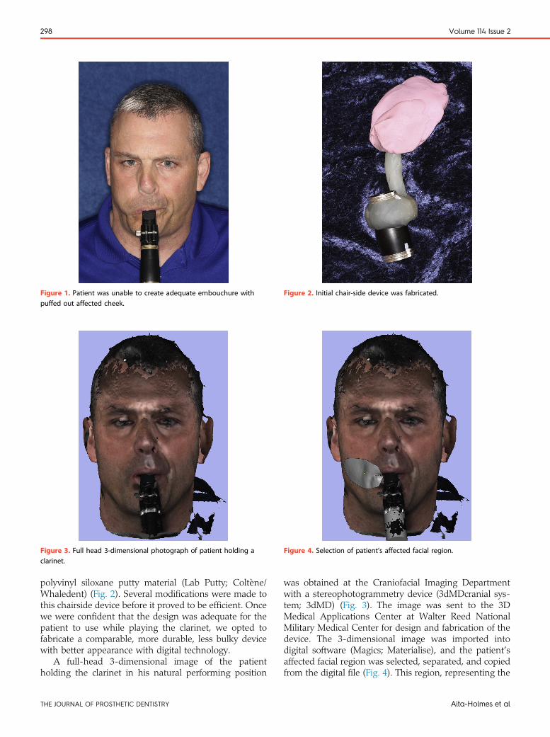
Figure 1. Patient was unable to create adequate embouchure withpuffed out affected cheek.
Figure 2. Initial chair-side device was fabricated.
Figure 3. Full head 3-dimensional photograph of patient holding aclarinet.
Figure 4. Selection of patient’s affected facial region.
298 Volume 114 Issue 2
polyvinyl siloxane putty material (Lab Putty; Coltène/Whaledent) (Fig. 2). Several modifications were made tothis chairside device before it proved to be efficient. Oncewe were confident that the design was adequate for thepatient to use while playing the clarinet, we opted tofabricate a comparable, more durable, less bulky devicewith better appearance with digital technology.
A full-head 3-dimensional image of the patientholding the clarinet in his natural performing position
THE JOURNAL OF PROSTHETIC DENTISTRY
was obtained at the Craniofacial Imaging Departmentwith a stereophotogrammetry device (3dMDcranial sys-tem; 3dMD) (Fig. 3). The image was sent to the 3DMedical Applications Center at Walter Reed NationalMilitary Medical Center for design and fabrication of thedevice. The 3-dimensional image was imported intodigital software (Magics; Materialise), and the patient’saffected facial region was selected, separated, and copiedfrom the digital file (Fig. 4). This region, representing the
Aita-Holmes et al

Figure 5. Digital model of proposed device. Figure 6. Definitive device fabricated in titanium alloy with additivemanufacturing process.
Figure 7. Definitive device attached to clarinet in functional performingposition.
August 2015 299
affected area, was then extruded away from the face toproduce a solid part section that would serve to applypressure on the patient’s right cheek. A clamp wasdesigned to go around the clarinet shaft (Solidworks;Dassault Systems), and a freeform bar was added toconnect the solid cheek section to the clamp (FreeformModeling Plus; 3D Systems) (Fig. 5). The completed partwas then additively manufactured with electron beammelting technology (Arcam AB) from titanium alloy. Themetal was finished, and the inside of the clamp and thefacial contact area were covered in silicone (Silipos;Niagara).
The patient was seen in the maxillofacial clinic asecond time for a clinical evaluation of the manufacturedtitanium device. The device was attached to the clarinetshaft in the appropriate position for optimal pressureapplication. The device helped the patient create a betterembouchure, although it was determined that it could beimproved. Polyvinyl siloxane putty material (Lab Putty;Coltène/Whaledent) was added to the titanium device toincrease the amount of pressure generated on the cheekand to extend its medial portion toward the commissureof the mouth. These modifications were tested and foundto improve the quality of the sound generated. Mea-surements were obtained with a periodontal probe todetermine the thickness of the added putty to quantifythe extent to which the digital model of the device shouldbe altered. Modifications were made in the digital soft-ware, and a new device was printed (Fig 6).
The definitive device was delivered, and its effec-tiveness was tested while the patient played the clarinet(Fig. 7). The patient was pleased with this device and waseager to begin practicing to maintain his skills during hisslow recovery from Bell’s palsy.
Aita-Holmes et al
DISCUSSION
Because of this patient’s lengthy recovery from Bell’spalsy and the nature of his primary occupation, a solutionthat would allow him to play his clarinet despite his facialparalysis was imperative. A digital designing andmanufacturing treatment approach was chosen for thispatient because conventional facial moulage impressiontechniques would not have been practical, a facialmoulage would generate distorted facial tissue contoursand would not capture an accurate relative position of the
THE JOURNAL OF PROSTHETIC DENTISTRY

300 Volume 114 Issue 2
clarinet mouthpiece, and due to the long lever needed toattach the device to the clarinet, high forces would begenerated; subsequently, the selection of a very strongmaterial was imperative. Therefore, the original proto-type fabricated in resin would not have withstood theforces to be applied over several months. Also, conven-tional dental casting techniques would not have beentime- or cost-efficient methods of creating an effectivedevice.
The method used to fabricate the device presentedseveral advantages. The digital imaging technique pro-vided an accurate capture of the soft tissue contours andof their relative position with the clarinet mouthpiece.Digital design allowed for easy and cost-efficientfabrication and modification of the device. Additivemanufacturing presented the option of producing a de-vice in an acceptable material that could withstand thehigh forces that would be applied to the device overseveral months.
SUMMARY
Patients with Bell’s palsy face debilitating esthetic andfunctional consequences. For the patient presented inthis report, this disease presented a major problembecause it prevented him from functioning in his primaryoccupation.
The application of digital technologies in the fabrica-tion of the device proved to be a quick and cost efficientmethod of solving this patient’s dilemma. The procedure
THE JOURNAL OF PROSTHETIC DENTISTRY
presented involved 3 straightforward visits at the maxil-lofacial prosthetics clinic and resulted in a functionaldevice that allowed the patient to continue in his occu-pation during his slow recovery. The use of additivemanufacturing technologies is becoming widespread inthe medical field. Dental professionals must stay abreastof this quickly evolving technology to ensure that the besttreatment possible is provided. Maxillofacial prostho-dontists frequently meet with patients presenting withunusual situations. For these patients, nontraditionalmethods of treatment must be considered to expand thechances for successful outcomes.
REFERENCES
1. Heir GM, Fein LA. Lyme disease: considerations for dentistry. J Orofac Pain1996;10:74-86.
2. Callister SM, Schell RF, Lovrich SD. Lyme disease assay which detects killedBorrelia burgdorferi. J Clin Microbiol 1991;29:1773-6.
3. Struthers S. Bell’s palsy and Lyme disease. Br J Gen Pract 2009;59:779.4. Clark JR, Carlson RD, Sasaki CT, Pachner AR, Steere AC. Facial paralysis in
Lyme disease. Laryngoscope 1985;95:1341-5.5. Davenport RJ. Bell’s palsy: new evidence provides a definitive drug therapy
strategy. Br J Gen Pract 2009;59:569-70.6. Mossblad G. Overcoming Bell’s palsy. Part Idthe first 4 days. Saxophone
Journal Sept/Oct 2002. Available at: http://www.dornpub.com/SaxjPDF/BellsPalsyPart1.pdf. Accessed April 9, 2014.
Corresponding author:Dr Gerald T. GrantWalter Reed National Military Medical Center8901 Wisconsin AveBethesda, MD 20889Email: [email protected]
Copyright © 2015 by the Editorial Council for The Journal of Prosthetic Dentistry.
Aita-Holmes et al