Difficulties in swallowing and eating following acquired ... · ment by the Coombes approach”)....
98
PhD thesis Difficulties in swallowing and eating following acquired brain injury ‐ From a professional and a patient perspective Annette Kjærsgaard The Institute of Public Health and Hammel Neurorehabilitation and Research Centre Faculty of Health Sciences University of Southern Denmark 2013
Transcript of Difficulties in swallowing and eating following acquired ... · ment by the Coombes approach”)....

PhD thesis
Difficulties in swallowing and eating following
acquired brain injury
‐ From a professional and a patient perspective
Annette Kjærsgaard
The Institute of Public Health and
Hammel Neurorehabilitation and Research Centre
Faculty of Health Sciences
University of Southern Denmark
2013

ii
This PhD study was carried out during my employment at the Research Initiative for Activity Stud‐
ies and Occupational Therapy, Institute of Public Health, University of Southern Denmark and at
the Neurorehabilitation and Research Centre in Hammel, Denmark in the period from October
2008 to September 2012.
ISBN 978‐87‐92646‐65‐1
Main supervisor
Bengt H. Sjölund, Professor, MD, DMSc, Research Initiative in Rehabilitation, the Institute of Pub‐
lic Health, Faculty of Health Sciences, University of Southern Denmark, Odense, Denmark.
Co‐supervisors
Tove Borg, Senior Researcher, PhD, OT, Hammel Neurorehabilitation and Research Centre, Ham‐
mel, Denmark.
Hanne Kaae Kristensen, PhD, OT, Department of Rehabilitation, Odense University Hospital,
Odense, Denmark (supervisor since 2011).
Co‐worker
Lars Hedemann Nielsen, MD, Consultant Anaesthetist, Hammel Neuorehabilitation and Research
Centre and the Neurointensive Step‐down Unit at the Intensive Unit, Regional Hospital of Silke‐
borg, Silkeborg, Denmark.
Evaluation committee:
Olle Ekberg, Professor, MD, PhD, Department of Diagnostic Radiology, Malmö University Hospital,
Malmö, Sweden.
Mette Holst, Head of Clinical Nutrition Research, PhD, MKS, RN, Department of Medical Gastroen‐
terology, Centre for Nutrition and Bowel Disease, Aalborg Hospital, Aalborg, Denmark.
Lis Wagner, Professor, DrPH, RN, Research Unit of Nursing, Institute of Clinical Research, Univer‐
sity of Southern Denmark, Odense, Denmark (chairman).
Annette Kjærsgaard, MSc, OT
Hammel Neurorehabilitation and Research Centre, Voldbyvej 15, DK‐8340 Hammel, Denmark
[email protected] , Phone: +45 7841 9068 or mobile phone +45 2081 8652

i
Preface
"Eating, apparently a biological matter is actually profoundly social. What we eat, where
we get it, how it is prepared, when we eat and with whom, what it means to us – all these
depend on social arrangements” (DeVault 1991) (page 35)
First of all, I would like to take you on a little, but for me long journey leading up to this thesis. I
have, since I graduated as an occupational therapist (OT) in 1989, worked within rehabilitation of
persons with severe acquired brain injury (ABI). First, I worked as a leading OT at the “Genoptræn‐
ingscentret Lunden” in Varde. In 1990, I met a young man who had been involved in a motorbike
accident. He was diagnosed as in a persistent vegetative state three months after his accident and
was admitted from the neurological ward to a nursing home, where I met him nine month after
his injury. He still had a nasal feeding tube and had just got to taste a little yoghurt. His parents
wanted him to live in another place, and they visited “Lunden”. During the conversation with his
parents, he was sitting in his wheelchair beside us. I had placed a piece of chocolate in front of
him. Suddenly, without our notice, he had, with his uncoordinated movements, put a piece of
chocolate in his mouth and we could hear his cheerful sounds and see his excited facial expres‐
sions.
Why is this story so important to me? Because it became very obvious that eating is very im‐
portant to both the person and the relatives, and I realised that I did not know how to systemati‐
cally assess and treat him. At that time I did not realise that there was a close relation between
eating and drinking and all the infections and pneumonias that the residents at “Lunden” had. I
could not find anyone in Denmark, who was able to supervise and teach me, so I just did my best!
On my further professional journey, searching for knowledge and skills about assessment and
treatment of difficulties in swallowing and eating, I met a lot of very important and inspiring per‐
sons. I will especially name two. Karen Nielsen, the leading therapist at “Therapiezentrum Bur‐
gau” (TZB), Germany, who gave me the opportunity to work at an early rehabilitation unit at TZB,
where I was introduced to both the rehabilitation approaches of “Gespürter Interaktionstherapie”
by Fèlicie Affolter and Facial‐Oral Tract Therapy (F.O.T.T.) by Kay Coombes. Later I met and got to
know the speech and language therapist Kay Coombes, United Kingdom. In 2002, I became the
first Nordic F.O.T.T. instructor. In 2005 I wrote the book “Ansigt, mund og svælg – Undersøgelse
og behandling efter Coombes konceptet” (”Face, mouth and oral tract – assessment and treat‐

ii
ment by the Coombes approach”). This book is based on a holistic approach to assessment and
treatment of the face, mouth and oral tract and the enclosed charts are now systematically used
in Danish clinical neurorehabilitation practice and at the OT basic education programmes.
Since 2000, I have worked at Hammel Neurocenter, where I started as a project OT responsi‐
ble for a development project “Undersøgelse af synkeproblemer hos senhjerneskade patienter”
(“Assessment of swallowing problems in brain injured patients”). The increased need for evi‐
dence‐based medicine became more apparent and at that time, I got the opportunity to upgrade
my clinical skills with academic skills. In 2006, I graduated as a Master of Science in Occupational
Therapy (MScOT) at the University of Lund, Sweden, and as part of my master thesis I searched
for evidence of the assessment and treatment charts from my book.
I began my PhD journey in 2008. As you can see I have had many years of clinical OT experi‐
ence with the assessment and treatment of difficulties in swallowing and eating following ABI, so
my position as a researcher is not neutral. On the other hand, my clinical skills and knowledge
have aided me in formulating hypotheses and collecting and understanding my research data.

iii
Financial and other conflicts of interest
This study was supported financially by the University of Southern Denmark (Health, Man and
Society, The Research Initiative for Activity Studies and Occupational Therapy and the Faculty of
Health Science), The Danish Association of Occupational Therapy (FF 1/10‐9 and FF1/11‐2) and
The Region Hospital Hammel Neurocenter. The author is a certified F.O.T.T. instructor and is
regularly conducting G/F.O.T.T. courses. The author has no personal financial interest in any
commercial company or institution directly or indirectly related to this thesis.

iv
List of abbreviations
ABI Acquired Brain Injury
BDI Berliner Dysphagia Index
BMI Body Mass Index
CPG Central Pattern Generator
FEES Fiberoptic Endoscopic Evaluation of Swallowing
FEESST Fiberoptic Endoscopic Evaluation of Swallowing with Sensory Testing
FIM Functional Independence Measure
(FIM = FIM change from admission to discharge from IRP)
FOIS Functional‐Oral Intake Scale
F.O.T.T. Facial‐Oral Tract Therapy
GCS Glasgow Coma Scale
HR Hazard Ration
ICF International Classification of Functioning
IRP Inpatient Rehabilitation Programme
LOS Length Of Stay
MBS Modified Barium Swallow
OT Occupational Therapist or therapy
PAS Penetration‐Aspiration Scale
PEG Percutaneous Endoscopic Gastrostomy
RCT Randomized Controlled Trial
RLAS Ranchos Los Amigos Scale
(RLAS= RLAS change from admission to discharge from IRP)
SWAL‐CARE Swallowing Quality of Care Questionnaire
SWAL‐QOL Swallowing Quality of Life Questionnaire
TBI Traumatic Brain Injury
UES Upper Esophageal Sphincter
UTI Urinary Tract Infections
VFS Video Flouroscopy Swallowing
VSE Videofluorographic Swallowing Evaluation
WHO World Health Organization

v
Definitions
Acquired brain injury
Acquired brain injury is defined as damage to the brain that occurs after birth, and which is not
related to congenital disorders, developmental disabilities, or processes that progressively dam‐
age the brain (Toronto Acquired Brain Injury Network 2011).
Adaptation
Adaptation is defined as the process by which a person maintains a useful relationship to the en‐
vironment (Coelho and others 1974).
Dysphagia
Dysphagia is understood as oropharyngeal dysphagia, which is defined as difficulties in ingestion,
swallowing, eating and drinking using subcategories in the International Classification of Function‐
ing, Disability and Health (ICF) (WHO 2001).
Inpatient rehabilitation programme
The inpatient rehabilitation programme is an intensive, hospital‐based, interdisciplinary rehabili‐
tation unit, here at a national neurorehabilitation centre. Interventions were based on 24‐hour
assessment and treatment approaches (Graham and others 2009; Affolter and others 2009; Han‐
sen and Jakobsen 2010).

vi
Summary in English
Stroke and traumatic brain injury (TBI) are the main causes of acquired brain injury (ABI). In 2009,
there were an estimated 12,500 cases of hospitalisations from stroke and an estimated 9,500
cases of hospitalisations from TBI in Denmark. Many of these patients need rehabilitation. Neu‐
rorehabilitation in Denmark is undergoing development and specialisation. In this process, diffi‐
culties in swallowing, eating and drinking (dysphagia) in patients with ABI have increasingly come
to our attention. In foreign studies the incidence of dysphagia is given as 27 to 61% among brain‐
injured patients in neurorehabilitation. Dysphagia may result in lack of oral intake and conse‐
quently in malnutrition, dehydration and complications like aspiration, pneumonia and choking at
worst. Dysphagia may result in prolonged rehabilitation and inappropriate transfers between in‐
tensive care and rehabilitation units.
The overall objective of this thesis is to evaluate difficulties in swallowing and eating follow‐
ing ABI in relation to rehabilitation from both a professional and a patient perspective. The thesis
consists of two sub studies described in three papers. Study I is a randomized controlled trial re‐
ported in two papers, and Study II is a qualitative case study with multiple‐cases described in Pa‐
per III.
Study I (Paper I) evaluates whether patients assessed for initiation of oral intake by clinical
assessment (Facial‐Oral Tract Therapy (F.O.T.T.) had a greater risk of developing aspiration pneu‐
monia during neurorehabilitation than patients assessed by instrumental assessment (Fiberoptic
Endoscopic Evaluation of Swallowing (FEES)). 679 patients with acquired brain injury were
screened for possible participation in the trial, and 138 patients were randomized between June
2009 and April 2011. No significant diagnosis‐related or demographic differences between the
groups were found. 119 patients (62 F.O.T.T./57 FEES) were included in the analysis of the pri‐
mary outcome. Four patients assessed by F.O.T.T. and 12 by FEES were diagnosed with pneumo‐
nia (p=0.03). Excluding six patients with pneumonia before initiating oral intake and three pa‐
tients, who did not fulfil the primary outcome criteria for aspiration pneumonia, left seven pa‐
tients for analysis. Four of them developed aspiration pneumonia within 10 days after initiating
oral intake; one patient evaluated by F.O.T.T. and three patients by FEES.
Study I (Paper II) investigates whether there is a difference in time for initiation of oral intake
and time to total oral intake when initiation of oral intake is based on F.O.T.T. or on FEES, and
how other factors may influence the time to initiation. Paper II includes a calculation of the inci‐

vii
dence of dysphagia in this study. The incidence of dysphagia in neurorehabilitation was 47 %.
There was no difference in time for initiation or recovery of total oral intake during inpatient re‐
habilitation whether using F.O.T.T. or FEES. For 42% of the patients oral intake had been initiated
on admission and 92% at discharge. 2.5% of the patients recovered total oral intake within the
first 24 hours of admission, and 37% had recovered total oral intake before discharge. Within 62
days (F.O.T.T.) and 54 days (FEES) 25 % of the patients recovered total oral intake. The possibility
of achieving this depends on a low level of consciousness and physical function, age, length of
stay and number of dysphagia interventions.
Study II (Paper III) explores and interprets the patient perspective two to 18 months after
discharge from neurorehabilitation. It explores how reduced functions of swallowing and oral
intake influence the experience of food and meals as well as everyday life after the injury, and
how the patients experience the neurorehabilitation approach to dysphagia. In this study six of
the 119 patients from Study I were interviewed. A comparative analysis was used to analyse data.
Five main themes resulted from the analysis: Individual psychological characteristics, swallowing
and digestion, eating and drinking, communication and meals as well as rehabilitation during hos‐
pitalisation and after discharge. Processes of change were interpreted as adaptation processes.
Three important subthemes emerged: 1) Tube feeding; 2) Difficulties in swallowing associated
with meals involving social gathering and; 3) Neurorehabilitation approach to swallowing and eat‐
ing.
It can be concluded that dysphagia occurs in nearly half the patients admitted to neuroreha‐
bilitation in Denmark. It has been demonstrated that a non‐instrumental approach like F.O.T.T. to
assess swallowing disorders in patients with ABI may be as effective in predicting safe swallowing
(safe in terms of no or minimal aspiration) as an instrumental approach like FEES. The qualitative
study shows that the patients’ ability to adapt to the difficulties in swallowing and eating depends
on the phases of their illness trajectory. The (even temporarily) reduced or lost swallowing and/or
eating function is unexpected and difficult for the patient and causes strong emotional reactions
even 18 months after the injury. The findings also show new knowledge of clinical interest; it is
e.g. demonstrated that it is possible to adapt and develop new structures for valuable activities
associated with swallowing and eating.
Keywords: Dysphagia, Neurorehabilitation, Facial‐Oral Tract Therapy

viii
Sammenfatning på dansk
Apopleksi og traumatisk hjerneskade er de største årsager til erhvervet hjerneskade. Der var i
2009 ca. 12.500 indlæggelsesforløb pga. apopleksi og ca. 9.500 indlæggelsesforløb pga. trauma‐
tisk hjerneskade i Danmark. Mange af disse patienter har behov for rehabilitering. I Danmark ud‐
vikles og specialiseres neurorehabilitering. Herunder er man i stigende grad blevet opmærksom
på problemer med at synke, spise og drikke (dysfagi) hos personer med erhvervet hjerneskade.
Hyppigheden af dysfagi angives i udenlandske studier fra 27 til 61 % blandt hjerneskadede perso‐
ner, som indgår i neurorehabilitering. Dysfagi kan resultere i, at man spiser og drikker for lidt og
dermed kommer til at lide af under‐ eller fejlernæring, væskemangel samt komplikationer som fejl‐
synkning, udvikling af lungebetændelse og i værste fald kvælning. Dysfagi kan medføre en forlæn‐
gelse af rehabiliteringen og uhensigtsmæssige overflytninger mellem intensivt‐ og rehabiliterings‐
afsnit.
Det overordnede formål med denne afhandling er at undersøge problemer med at synke og
spise i forbindelse med neurorehabilitering, både i et professionelt og et patientperspektiv.
Afhandlingen består af to delstudier beskrevet i tre artikler. Studie I er et randomiseret kon‐
trolleret studie, som rapporteres i to artikler. Studie II er et kvalitativt casestudie med seks cases,
som er beskrevet i artikel III.
Studie I (artikel I) undersøger, om patienter, der vurderes at kunne initiere oralt indtag ved
hjælp af klinisk undersøgelse (Facial‐Oral Tract Therapy (F.O.T.T.)), har større risiko for udvikling af
lungebetændelse på grund af fejlsynkning i forbindelse med neurorehabilitering end for patienter,
der vurderes ved hjælp af instrumentel undersøgelse (Fiberoptic Endoscopic Evaluation of Swal‐
lowing (FEES)). I undersøgelsen er 679 patienter med erhvervet hjerneskade screenet for mulig
deltagelse, og 138 patienter blev randomiseret i tiden fra juni 2009 til april 2011. Der var ingen
signifikante diagnoserelaterede eller demografiske forskelle mellem grupperne. I analysen indgik
119 patienter (62 F.O.T.T./ 57 FEES). Fire patienter undersøgt ved hjælp af F.O.T.T. og 12 ved
hjælp af FEES blev diagnosticeret med lungebetændelse (p=0.03). Seks af disse patienter blev eks‐
kluderet, da de udviklede lungebetændelse før initiering af oralt indtag, og tre patienter blev eks‐
kluderet, da de ikke opfyldte det primære outcome kriterium for lungebetændelse på grund af
fejlsynkning, hvilket efterlod syv patienter til analysen. Fire af disse udviklede lungebetændelse
inden for 10 dage efter initiering af oralt indtag; en patient evalueret ved hjælp af F.O.T.T. og tre
patienter ved hjælp af FEES.

ix
Studie I (artikel II) afdækker, om der er forskel i tidspunkt for initiering af oralt indtag og ti‐
den til totalt oralt indtag, når initiering af oralt indtag sker ved hjælp af F.O.T.T. eller FEES, og
hvordan andre faktorer kan indvirke på dette. Desuden indeholder artikel 2 en beregning af hyp‐
pigheden af dysfagi i dette studie. Hyppigheden af dysfagi i forbindelse med neurorehabilitering
var 47 %. Der var ingen forskel i tid til initiering eller generhvervelse af total oralt indtag under
indlæggelsen, uanset om der blev anvendt F.O.T.T. eller FEES. Oralt indtag var initieret for 42 % af
patienterne ved indlæggelsen og for 92 % ved udskrivelsen. Totalt oralt indtag var opnået for 2,5
% af patienterne inden for det første døgn efter indlæggelsen, og 37 % var på totalt oralt indtag
inden udskrivelse. Inden for 62 dage (F.O.T.T.) og 54 dage (FEES) opnåede 25 % total oralt indtag.
Muligheden for at opnå dette var bl.a. afhængig af lavt bevidsthedsniveau og fysisk funktionsni‐
veau, alder, indlæggelsestid og antal behandlinger for dysfagi.
Studie II (artikel III) udforsker og fortolker patientperspektivet to til 18 måneder efter udskri‐
velse fra neurorehabilitering. Her undersøges, hvordan funktionsnedsættelser omkring synkning
og oralt indtag af mad og drikke påvirker oplevelsen af mad og måltider og hverdagslivet efter
skaden, samt hvordan neurorehabiliteringsindsatsen i relation til dette opleves. I studiet inter‐
viewes seks af de 119 patienter fra Studie I. En komparativ indholdsanalyse blev anvendt til at
analysere data. Fem hovedtemaer fremkom i analysen: Individuelle psykologiske egenskaber,
synkning og fordøjelse, spise og drikke, kommunikation og måltider samt rehabilitering under
indlæggelse og efter udskrivelse. Forandringsprocesser blev fortolket som adaptationsprocesser.
Tre betydningsfulde undertemaer fremkom: 1) Mad og drikke ved hjælp af sonde; 2) Synkepro‐
blemer i relation til måltider med socialt samvær samt; 3) Neurorehabiliteringstilgangen vedrø‐
rende synkning og spisning.
Det kan konkluderes, at dysfagi forekommer hos næsten halvdelen af de patienter, der ind‐
lægges til neurorehabilitering i Danmark. Det er blevet påvist, at en ikke‐instrumentel tilgang som
F.O.T.T. til undersøgelse af synkeproblemer hos patienter med erhvervede hjerneskader kan være
lige så effektiv til at forudsige sikker synkefunktion (sikker hvad angår ingen eller minimal fejl‐
synkning) som en instrumentel tilgang som FEES. Det kvalitative studie viser, at det at leve med
synke‐ og spiseproblemer afhænger af, i hvilken fase af sygdomsforløbet patienterne befinder sig.
Den (selv midlertidigt) nedsatte eller tabte synke‐ og/eller spisefunktion er uventet, svær og
fremkalder stærke følelsesmæssige reaktioner selv 18 måneder efter skaden. I resultaterne frem‐
kommer således ny viden af klinisk interesse, hvor det bl.a. påvises, at det er muligt at adaptere
og udvikle nye strukturer for værdifulde aktiviteter relateret til synkning og spisning.
Nøgleord: Dysfagi, Neurorehabilitering, Facial‐Oral Tract Therapy
x
List of publications
The thesis is based on two linked studies (Study I and II) with results reported in the following
three papers. In the text they will be referred to as Paper I, Paper II and Paper III. The papers are
attached to the thesis as Paper I, II and III.
Paper I Kjaersgaard A, Nielsen LH, Sjolund BH: Randomized trial of two swallowing assess‐
ment approaches: Facial‐Oral Tract Therapy vs. Fiberoptic Endoscopic Evaluation of
Swallowing.
Submitted
Paper II Kjaersgaard A, Nielsen LH, Sjolund BH: When does oral intake take place in inpa‐
tient rehabilitation following acquired brain injury? A randomized study of two as‐
sessment approaches (Facial‐Oral Tract Therapy vs. Fiberoptic Endoscopic Evalua‐
tion of Swallowing).
Submitted
Paper III Kjaersgaard A, Kristensen HK, Borg T: Clinical implications of lived experience and
adaptation to reduced abilities to swallowing and eating following acquired brain
injury.
Submitted

Table of contents
Preface .............................................................................................................................................i Financial and other conflicts of interest ........................................................................................iii List of abbreviations.......................................................................................................................iv Definitions.......................................................................................................................................v Summary in English........................................................................................................................ vi Sammenfatning på dansk.............................................................................................................viii List of publications ..........................................................................................................................x
1. INTRODUCTION ......................................................................................................................4
2. BACKGROUND ........................................................................................................................6 2.1 Difficulties in ingestion, swallowing, eating and drinking........................................................ 6
2.1.1 Difficulties in ingestion and swallowing........................................................................................... 6 2.1.2 Difficulties in eating and drinking .................................................................................................... 7
2.2 The meaning of eating, food and meals .................................................................................. 8 2.2.1 Theories about the mouth and eating ............................................................................................. 8 2.2.2 Food and meals................................................................................................................................ 9
2.3 Swallowing ............................................................................................................................... 9 2.3.1 Neural control of the tongue and swallowing ............................................................................... 10 2.3.2 Normal physiology of swallowing .................................................................................................. 11 2.3.3 Pre‐oral phase................................................................................................................................ 12
2.4 Rehabilitation of persons with acquired brain injury (ABI).................................................... 13
3. AIM AND OBJECTIVES ...........................................................................................................15 3.1 General aim............................................................................................................................ 15
3.1.1 Specific aims .................................................................................................................................. 15
4. CONCEPTUAL FRAMES ..........................................................................................................16 4.1 Scientific frames and methodological considerations ........................................................... 16 4.2 Rehabilitation and the International Classification of Functioning, Disability and Health (ICF)
..................................................................................................................................................... 16 4.3 Rehabilitation as a process related to adaptation................................................................. 18
5. METHODS .............................................................................................................................20 5.1 Study design ........................................................................................................................... 20 5.2 Participants ............................................................................................................................ 22 5.3 Randomization ....................................................................................................................... 22 5.4 Inpatient neurorehabilitation setting .................................................................................... 23 5.5 Intervention approaches........................................................................................................ 23
5.5.1 Facial‐Oral Tract Therapy (F.O.T.T.) ............................................................................................... 23 5.5.2 Fiberoptic Endoscopic Evaluation of Swallowing (FEES) ................................................................ 27
5.6 Data collection and outcome measures ................................................................................ 29 5.6.1 Aspiration pneumonia ................................................................................................................... 29 5.6.2 Functional Oral Intake Scale (FOIS)................................................................................................ 30 5.6.3 Penetration Aspiration Scale (PAS) ................................................................................................ 31 5.6.4 Berliner Dysphagia Index (BDI) ...................................................................................................... 32

2
5.6.5 Level of modified consistencies of food and liquid........................................................................ 32 5.6.6 Functional Independence Measure (FIM) .................................................................................. 32 5.6.7 Ranchos Los Amigos Scale (RLAS) .................................................................................................. 33 5.6.8 Other quantitative, functional outcome measures ....................................................................... 33 5.6.9 Semi‐structured interviews............................................................................................................ 33
5.7 Data analysis .......................................................................................................................... 37 5.7.1 Data analysis paper I ...................................................................................................................... 37 5.7.2 Data analysis paper II ..................................................................................................................... 37 5.7.3 Data analysis paper III .................................................................................................................... 38
6. ETHICAL CONSIDERATIONS ...................................................................................................39
7. RESULTS................................................................................................................................40 7.1 Paper I .................................................................................................................................... 42
7.1.1 Incidence of pneumonia ................................................................................................................ 42 7.2 Paper II ................................................................................................................................... 43
7.2.1 Incidence of dysphagia .................................................................................................................. 43 7.2.2 Initiation of oral intake .................................................................................................................. 43 7.2.3 Reaching total oral intake .............................................................................................................. 44 7.2.4 Factors of importance for reaching total oral intake..................................................................... 44
7.3 Paper III .................................................................................................................................. 45 7.3.1 Individual psychological assets ...................................................................................................... 45 7.3.2 Swallowing and ingestion .............................................................................................................. 45 7.3.3 Eating and drinking ........................................................................................................................ 46 7.3.4 Communication and meals ............................................................................................................ 46 7.3.5 Rehabilitation of swallowing and eating........................................................................................ 47
8. DISCUSSION..........................................................................................................................49 8.1 Main results and insights across the two studies .................................................................. 50
8.1.1 Does dysphagia matter? ................................................................................................................ 50 8.1.2 Are both F.O.T.T. and FEES necessary?.......................................................................................... 50 8.1.3 The best swallowing protocol? ...................................................................................................... 52 8.1.4 Reduced consciousness in relation to swallowing and eating ....................................................... 52 8.1.5 Other factors influencing initiation of oral intake and reaching total oral intake ......................... 53 8.1.6 Feeding by tube and first oral intake ............................................................................................. 54 8.1.7 Swallowing difficulties and meals with social interactions............................................................ 55 8.1.8 Inpatient rehabilitation approach concerning swallowing and eating .......................................... 56
8.2 Conceptual framework .......................................................................................................... 57 8.2.1 The International Classification of Functioning, Disability and Health .......................................... 57 8.2.2 Theories about adaptation ............................................................................................................ 57
8.3 Methodological considerations and limitations .................................................................... 60 8.3.1 Methodological considerations ..................................................................................................... 60 8.3.2 Limitations ..................................................................................................................................... 64
8.4 Implications for clinical practice ............................................................................................ 67
9. CONCLUSIONS AND FURTHER STUDIES .................................................................................69 9.1 Main conclusions ................................................................................................................... 69 9.2 Further studies ....................................................................................................................... 70
10. ACKNOWLEDGEMENTS .......................................................................................................72
11. REFERENCES........................................................................................................................74

3
FIGURES IN THE THESIS
Figure 1 Thesis process map (p. 21) Figure 2 Flowchart for Study I (p. 40) Figure 3 Conceptual model of Study II (p. 48)
TABLES IN THE THESIS
Table 1 An overview of the approaches, scales and interviews used as data collection methods in the two studies two studies (p. 29) Table 2 Functional Oral Intake Scale (FOIS) (p. 30) Table 3 The Penetration‐Aspiration Scale (PAS) (p. 31) Table 4 Domains in the semi‐structured interview guide (p. 36) Table 5 The age (years) and numbers of participants included in the studies (p. 41)
4
1. INTRODUCTION
The main purpose of this thesis is to explore and evaluate the difficulties in swallowing and eating
following acquired brain injury (ABI) during an inpatient rehabilitation programme (IRP), from a
professional and a patient perspective.
ABI typically produces a potentially wide range of impairments affecting the physical, neuro‐
cognitive and/or psychological functioning (Teasell and others 2007).
Dysphagia may result in lack of oral intake, in malnutrition and dehydration, and in fre‐
quent/prolonged periods with infections that are responsible for a deteriorated and prolonged
rehabilitation process (Perry and Love 2001; Westergren 2006). The condition is often caused by
unrecognised aspiration of e.g. saliva or food (Langmore and others 1998). The incidence of clini‐
cally diagnosed dysphagia is stated to be from 27% to 93% among ABI patients in IRP (Winstein
1983; Hansen and others 2008a; Falsetti and others 2009).
Pneumonia is a major aspiration‐related pulmonary complication in patients with ABI (Pilitsis
and Rengachary 2001). Aspiration pneumonia is a main cause of early death in stroke, if the pa‐
tients are not screened and treated for dysphagia (Smithard and others 1996; Perry and Love
2001; Ramsey and others 2003; Westergren 2006). Minimising the risks and episodes of aspira‐
tion, maximising nutritional intake, and providing the patient with the most appropriate alterna‐
tive to oral feeding will help to facilitate the maximal recovery potential for these individuals
(Mackay and others 1999b).
Ideally it should be possible to assess the difficulties in swallowing and eating using a three‐
tiered assessment cascade: screening, bedside evaluation (clinical assessment) and objective
measurement (instrumental assessments) (Farrell and O'Neill 1999). This thesis will focus on the
clinical assessment Facial‐Oral Tract Therapy (F.O.T.T.) and the instrumental assessment Fiberop‐
tic Endoscopic Evaluation of Swallowing (FEES). There is a wide range of multidisciplinary rehabili‐
tation interventions, but the majority of the interventions are only supported by limited evidence
(Cullen and others 2007). In Denmark neurorehabilitation is still developing and specialising
(National Board of Health 2011), and there is increased attention to difficulties in swallowing and
eating (dysphagia) and to the intervention approaches like F.O.T.T. and FEES.

5
Oropharyngeal dysphagia is typically managed by a multidisciplinary team with speech‐language
pathologists being primarily responsible for management (Cichero 2006); however, in Denmark this
role is upheld by occupational therapists (OT) (Kjaersgaard and Langhorn 2007).
Takahata et al (Takahata and others 2011) conclude that early initiation of oral feeding after
sufficient preparation may safely improve the clinical outcomes of intracerebral hemorrhage pa‐
tients, in terms of survival, the incidence of chest infection, the LOS and swallowing function.
Formisano et al (Formisano and others 2004) conclude that oral feeding appears to be an accurate
prognostic index of the final outcome in severe traumatic brain injury (TBI). However, despite the
significance of dysphagia following ABI, limited data are available regarding the natural history of
swallowing disorders or on the prognosis and outcomes in this population.
This thesis aims to provide new knowledge of both scientific and clinical relevance about in‐
patient neurorehabilitation, specifically of the difficulties in swallowing and eating following ABI
during inpatient neurorehabilitation described from both a professional and a patient perspective.

6
2. BACKGROUND
2.1 Difficulties in ingestion, swallowing, eating and drinking
2.1.1 Difficulties in ingestion and swallowing
Dysphagia in this thesis is understood as oropharyngeal dysphagia, which is defined as difficulties
in ingestion, swallowing, eating and drinking using subcategories in ICF (WHO 2001).
"Dysphagia" is derived from the Greek, dys—meaning disordered, and phagein—meaning to
eat (Winstein 1983; Groher and Crary 2010). Dysphagia is often defined as the medical term for
the symptom of difficulty in swallowing (Wikipedia 2012). Dysphagia affects the most cardinal of
human functions, the ability to eat and drink (McHorney and others 2000). Dysphagia is common
in patients with neurological disorders (Bakheit 2001). Neurogenic dysphagia may cause dehydra‐
tion, malnutrition, aspiration and long periods with fever, can contribute to a less optimal and
increased duration of rehabilitation and lead to feelings of shame, dependency and other nega‐
tive experiences (Jacobsson and others 2000; Bakheit 2001; Carlsson and others 2004).
Dysphagia is not a primary medical diagnosis but rather a dysfunction or symptom of underly‐
ing disease and is therefore described more often by its clinical characteristics (signs) (Groher and
Crary 2010). The risk of developing aspiration pneumonia cannot be accurately predicted from
any single clinical sign or symptom. Dysphagia, when defined broadly, can include the perceptual
and cognitive awareness of the eating situation and the physiological responses to the smell of
food (Leopold and Kagel 1996). Dysphagia can also interrupt the eating pleasure (Buchholz 1996).
Tanner (Tanner 2003) purposes this definition of dysphagia: “Impairment of emotional, cognitive,
sensory, and/or motor acts involved with transferring a substance from the mouth to the stom‐
ach, resulting in failure to maintain hydration and nutrition, and posing a risk of choking and aspi‐
ration” (page 70).
Groher (Groher and Crary 2010) suggests that a swallowing disorder should be distinguished
from a feeding disorder. A feeding disorder is the impairment in the process of food transport
outside the alimentary system. A feeding disorder is usually the result of weakness or incoordina‐
tion in the hand or arm used to move the food from the plate to the mouth.

7
Salassa (Salassa 1999) said that swallowing should not be confused with eating. Eating or oral
nutrition requires three components: volition, preparation, and swallowing. Volition is the con‐
scious mental ability or will to perform the act. Preparation is the physical ability to prepare nour‐
ishment and then take it into the mouth.
“When dysphagia is examined broadly, it is clearly /…… / one that has potential activ‐
ity/participation limitations and psychosocial consequences for the person” (Threats 2007). There
is a clear‐cut need to optimise a brief initial examination that employs several key signs and symp‐
toms to accurately detect patients with possible unsafe swallows and who therefore need more
extensive testing (AHRQ 1999). There is a consensus that an interdisciplinary team approach is
essential for the optimal management of patients with neurogenic dysphagia (White and others
2008; Karkos and others 2009). The treatment of difficulties in swallowing and eating often takes
two parallel courses: compensations to allow the patients to eat at least some food orally without
aspirating and rehabilitative exercises to build strength and coordination so that the patients no
longer need the compensations and can return to full oral intake (Logemann 2008; Gonzalez‐
Fernandez and Daniels 2008).
Evaluation of the effect of therapy in oropharyngeal dysphagia fits into this growing interest.
In a systematic review (Speyer and others 2010) Speyer et al conclude that in general, statistically
significant positive therapy effects are found. However, the number of papers is rather small and
many of these effect studies have diverse methodological problems. Furthermore, the conclusions
of most studies cannot be generalised easily or compared to one another because of the diversity
in subject characteristics, therapies, and assessment instruments. Therefore, when trying to de‐
termine whether swallowing therapy in general is effective, one may conclude that no single an‐
swer can be given. Speyer et al also conclude that many questions about the effects of therapy in
oropharyngeal dysphagia remain unanswered. Although some positive significant outcome stud‐
ies have been published, there is a need for further research using RCTs.
2.1.2 Difficulties in eating and drinking
The ability to swallow and eat without difficulty provides satiety and pleasure and is one of the
most important aspects of social life (Gustafsson and Tibbling 1991). People, who have difficulties
in swallowing and eating, are experiencing major limitations in their daily lives. Beyond physical
difficulties they can also experience cognitive and/or social problems. Their striving for control are
based on strategies as being careful when eating (Medin and others 2010b). They avoid getting
out among others and isolate themselves and lose one of the most important things in human
context: Sharing meals with relatives and others, which reduces a person's quality of life signifi‐
cantly (Grahn 1996; Elferich 2001; Ekberg and others 2002). Eating and related activities were

8
clearly important aspects of life for stroke survivors, socially and psychologically, as well as func‐
tionally (Perry and McLaren 2003). It is important for the treating clinicians to be aware of psycho‐
logical issues, to address them according to the patients’ clinical recovery, and to consider the
interplay between psychological and biomedical consequences (Martino and others 2010).
2.2 The meaning of eating, food and meals
Swallowing and eating are necessities of life and are often taken for granted. They are compli‐
cated processes based on physical and cognitive skills (Kumlien and Axelsson 2002; Johansson and
Johansson 2009). Eating is not just nutrient supply but also entails socialising and pleasurable ex‐
periences for healthy people, and meals are often the focus for celebrations with family members
and friends (Stringer 1999; Johansson and Johansson 2009). Those who eat and drink together are
by this very act tied to one another by a bond of friendship and mutual obligation (Smith 1894).
“Eating, apparently a biological matter is actually profoundly social. What we eat, where we get it,
how it is prepared, when we eat and with whom, what it means to us – all these depend on social
arrangements” (DeVault 1991) (page 35). “This (the transformation of the meal into a sociological
issue) gives birth to the rules regulating eating and drinking, rules that do not, however, concern
food as a substance, but the form of its consumption” (Simmel 1984).
2.2.1 Theories about the mouth and eating
The sociologist Pasi Falk (Falk 1994) describes that the mouth is central in the process of eating.
“The mouth is the central character in the story outlining the corporeality of (modern) consump‐
tion, not only due to its role as the primal organ of consumption but also due to its expressive
functions, as an organ of speech” (page 7). “The mouth is the most controlled opening of the
body, with regard to the influx (eating) but also concerning the “sublimated” outflux of speech”
(page 14). In other words, the mouth is the place where the expression and experience meet. An‐
gelella (Angelella 2009) describes in her PhD thesis about “Alimentary modernism” that there
have been a few theorists who have posited the interpenetration of body and world in acts of
eating (page 8). She postulates that according to Merleau‐Ponty (Merleau‐Ponty 1962), and op‐
posed to Bakhtin (Bakhtin 1984), “we cannot say that a person eats and, in eating incorporates
the world into himself; it is rather through eating that he comes into being” (page 10). Bakthin is
one theorist, who has posited the interpenetration of body and world in acts of eating, and he
describes that “eating and drinking is one of the most significant manifestations of the grotesque
body” (page 281).
9
2.2.2 Food and meals
“Food and drink are essential to human life, not just as nourishment, but also as carriers of mean‐
ing and significance” (Jenkins 1999). One of the culinary most important functions is to demon‐
strate the community and the distance between social groups within a society. “Food is also part
of our identity that we carry with us, without necessarily being aware of it” (Bourdieu 1992). The
food is surrounded by rules of what is good and evil, what is healthy and unhealthy, right and rea‐
sonably as right and wrong. The food has symbolic meanings and conceptual content, it refers to
something more e.g. to sex, sexuality and family (Holm 1998). Jenkins (Jenkins 1999) found that
Danes are more concerned about eating and drinking as a social event than about what we eat
and drink, and how food and drink are produced.
The meal is an important mean to maintain, establish and develop social contacts (Buchholz
1996). Meals unite people, and it is a recurring, regular and daily activity of living, which empha‐
sises everyday communities in families and among colleagues. In many cultures meals are a
framework for the indication of special occasions or selected communities in social life (Holm
2003). Meals are also social events which bring family members together and which give the indi‐
vidual the opportunity to experience himself as linked to others. The meal symbolises the family
as a social unit, and the food served during the meal is thus a material carrier of the community
(Holm 2003). Participation in meals and meals are part of everyday life, an ordinary and familiar
thing we do every day. It is an activity in which people participate throughout their lives. Other
activities are encapsulated in such meals e.g. shopping, cooking, serving and cleaning up
(Bundgaard 2005).
2.3 Swallowing
Effective swallowing is an essential part of life and is performed thousands of times per day, often
without conscious consideration (Barritt and Smithard 2009). For most people, swallowing or de‐
glutition is a normal and effortless task, but despite its ease, it is a complex and dynamic sensori‐
motor event involving volitional and involuntary movements of the lips, tongue, and floor of the
mouth, soft palate, pharynx, larynx, oesophagus and respiratory muscles. 26 pairs of muscles and
five cranial nerves are involved (Ertekin 2002; Mistry and Hamdy 2008; Matsuo and Palmer 2008).
Swallowing describes a complex function in which food and liquid are transported from the oral
region to the stomach in what appears as a well‐coordinated function (Miller 2008). Understand‐
ing the normal physiology and pathophysiology of eating and swallowing is fundamental to evalu‐
ate and treat disorders of eating and swallowing, and to develop dysphagia rehabilitation pro‐
grammes (Matsuo and Palmer 2008).

10
2.3.1 Neural control of the tongue and swallowing
The tongue in mammals has important motor and sensory functions. Besides exploratory and ma‐
nipulating functions it is essential for suckling, swallowing and vocalisation. Bilateral supranuclear
innervation of the hypoglossal nucleus and other bulbar nuclei may have afforded an evolutionary
survival value to animals. It had been long believed that the cortical representation of tongue mo‐
tor control is symmetrical in the two hemispheres (Umapathi and others 2000).
Swallowing is a complex motor and sensory activity that depends on a hierarchical interaction
between the cerebral cortex, the brain stem swallowing centre, and cranial nerves (Mistry and
Hamdy 2008). Coordination of swallowing depends on the integrity of sensory pathways from the
tongue, mouth, pharynx and larynx (cranial nerves V, VII, IX, X) and coordinated voluntary and
reflex contractions involving cranial nerves V, VII, and X‐XII (Wiles 1991). The main centre for
swallowing control is located in the brain stem – called the Central Pattern Generator (CPG) ‐ and
has two main functions: 1) the triggering and timing of the swallowing pattern and 2) the control
of the motor neurons involved in swallowing (Gonzalez‐Fernandez and Daniels 2008). The CPG is
located in the upper medullary and pontine areas of the brain and is bilaterally distributed within
the reticular formation. The CPG represents the first level of swallowing control.
The second level is the sub‐cortical structures, such as the basal ganglia, hypothalamus,
amygdala, and tegmental area of the midbrain. Evidence shows that the swallowing musculature
is bilaterally controlled (Ertekin and Aydogdu 2003). Hamdy et al (Hamdy and others 1996)
showed that muscles involved in human swallowing appear to be represented bilaterally on the
pre‐central cortex, in discrete topographic areas, which display interhemispheric asymmetry, in‐
dependent of handedness. Lateralisation to the right hemisphere tends to be greater than that in
the left hemisphere. Insular cortex is found to lateralise to the right hemisphere in right‐handed
subjects for voluntary saliva swallows. It has also been reported that reflexive or automatic swal‐
lows are represented in the primary sensorimotor cortex and in several other common cortical
regions (Ertekin and Aydogdu 2003).
Stroke affecting the hemisphere with the dominant swallowing projection results in dys‐
phagia and clinical recovery has been correlated with compensatory changes in the previous non‐
dominant unaffected hemisphere. This asymmetric bilaterality may explain why up to half of
stroke patients are dysphagic and why many will regain a safe swallow over a comparatively short
period (Singh and Hamdy 2006).

11
2.3.2 Normal physiology of swallowing
Over the past 20 years, research on the physiology of swallowing has confirmed that the oro‐
pharyngeal swallowing process can be modulated, both volitionally and in response to different
sensory stimuli.
The swallowing function can be defined with regard to either its clinical or neurophysiological
basis. From the clinical point of view, voluntary swallow occurs when a human has a desire to eat
or drink such as during mealtime and while awake and aware. Spontaneous swallow is the result
of accumulated saliva and/or food remnants in the mouth. It occurs mostly without the person
being aware, such as between meals and during sleep. Voluntary swallow is part of eating behav‐
iour, while spontaneous swallow is a type of protective reflex action. It is important to emphasise
that although the initiation of voluntary swallow is planned, its pharyngeal phase is a reflex
(Ertekin 2011).
Understanding the normal physiology and pathophysiology of eating and swallowing is fun‐
damental for evaluating and treating the difficulties in swallowing and eating (Matsuo and Palmer
2008). Functional swallowing occurs as a result of a series of purposeful movements that allow
transport of food and liquid from the mouth into the oesophagus. Since the airway and the
“foodway” effectively share a common path in the mouth and pharynx, an elaborate mechanism
exists to separate the two during swallowing thus preventing airway penetration by swallowed
material: at the same time breathing and speech are necessarily arrested (Wiles 1991).
The normal swallow in humans is generally conceptualised as occurring in different phases
(Daniels and Huckabee 2008). Normal swallowing is often divided (artificial construct) into three
phases: 1) oral phase divided into a preparatory part, with preparation of food for propulsion to
the pharynx, and an oral propulsive part, where the food is pushed by the tongue through the
pharynx, (2) pharyngeal phase, with specific movements to transport the bolus to the upper oe‐
sophageal sphincter (UES), and (3) oesophageal phase, where the bolus is propelled through the
oesophagus and lower oesophageal sphincter to the stomach (Logemann 1998; Matsuo and
Palmer 2008). The oral phase (oral preparatory and oral propulsive part) is mostly under voluntary
control (Palmer and others 2007; Ertekin 2011). Once oral propulsion occurs, the following proc‐
esses are a series of spontaneous movements designed to transport the food and protect the air‐
way (Gonzalez‐Fernandez and Daniels 2008).
The traditional definition of swallowing includes all events once nutrition is placed in the
mouth, the oral preparation, and the transfer of nutrition from mouth to stomach. Defined as
such, swallowing consists of one voluntary phases (oral phase (oral preparatory and oral propul‐
sive)) and two involuntary phases (pharyngeal and oesophageal) (Salassa 1999).
12
2.3.3 Preoral phase
A model of ingestion considering both pre‐swallowing and swallowing functions has been de‐
scribed by Leopold & Kagel (Leopold and Kagel 1997), while the traditional definition of normal
swallowing does not consider external factors as attention, eating behaviour, and feeding
method, which may also have an impact on swallowing efficiency and safety (Daniels and Huck‐
abee 2008). The F.O.T.T. approach also adds an additional phase to the described three phases of
the normal swallow in humans, which is the pre‐oral phase, including factors influencing swallow‐
ing, before the food gets into the mouth (Hansen and Jakobsen 2010). Since 1976 Coombes
(Coombes 2008a; Coombes 2011) has emphasised the significance of the pre‐oral phase in normal
eating. It is a state of readiness for eating.
It is important to promote this state of readiness in those with eating difficulties before they
begin to eat. For example, they should be seated in an appropriate way, with an opportunity to
prepare for the presentation of food by seeing it, smelling it and by tactile contact with the table
(spontaneous or guided touching), the cutlery and handling the food or holding a cup with assis‐
tance as required. Leopold & Kagel (Leopold and Kagel 1983; Leopold and Kagel 1997) support
this paradigm and they call it the Pre‐oral (Anticipatory) Stage: Interstage Relationships and de‐
scribe it as a useful paradigm, particularly in neurogenic populations, to modify the next stage of
ingestion, the oral‐preparatory stage. During the pre‐oral stage of ingestion, the visual and olfac‐
tory qualities of food excite salivation which mechanically assists bolus preparation, transfer, and
transport.
The normal pre‐oral phase is essentially the state of sensori‐motor “readiness”. It involves
preparation and transport of food to the mouth, anticipatory saliva production and possibly swal‐
lowing, in response to smelling the food, or seeing it. Preparation includes anticipation of the
meal, coordination of the movements of the eyes, arms, and hands together with the movements
of the trunk, head, and jaw. The spontaneous “postural background” allows for an optimal rela‐
tion of head, shoulders and trunk, promoting a stable foundation for manual dexterity, eye‐hand
coo‐ordination, arm movement and co‐ordinated jaw opening (Coombes 2001; Hansen and Ja‐
kobsen 2010).
These important operations “set the scene” for the oral phase which comprises bolus forma‐
tion and transport, lubricated with saliva, to the back of the mouth so that it can be delivered into
the pharynx (throat). Each phase influences the subsequent phases of normal swallowing. Thus,
the incoordination of the oral phase affects the timing and co‐ordination of the pharyngeal phase,
even when the pharyngeal reflex remains intact (Coombes 2008b). The pre‐oral phase includes a

13
lot of therapeutic possibilities of involving the person in the daily activity of eating and drinking
(Gratz 2002; Kjærsgaard 2005a).
2.4 Rehabilitation of persons with acquired brain injury (ABI)
Stroke and traumatic brain injury (TBI) are the main causes of ABI. According to the Danish Na‐
tional Patient Registry, Denmark had about 12,500 cases of hospitalisation from stroke in 2009
and about 9,500 cases of hospitalisation from TBI and other forms of ABI. Many of these people
need rehabilitation (National Board of Health 2011). Rehabilitation of patients with severe ABI is a
sub‐speciality within neurorehabilitation (Chua and others 2007). ABI is an umbrella term, en‐
compassing a wide spectrum of brain injuries that generally include traumatic and non‐traumatic
aetiologies such as cerebral concussion, brain contusions, subarachnoid haemorrhages or other
“acquired” problems such as hypoxia. The definition of ABI in this study is employed by the To‐
ronto Acquired Brain Injury Network (Toronto Acquired Brain Injury Network 2011), in which ABI
is defined as “damage to the brain that occurs after birth and which is not related to congenital
disorders, developmental disabilities, or processes that progressively damage the brain”. Causes
of ABI include (but are not limited to) such as hypoxia, illness, infection, stroke, substance abuse,
toxic exposure, trauma, and tumour. ABI may cause temporary or permanent impairment in such
areas as cognitive, emotional, metabolic, motor, perceptual motor and/or sensory brain functions
(Turner‐Stokes and others 2005; Wiseman‐Hakes and others 2010).
A severe brain injury is considered if the initial Glasgow Coma Score (GCS) is 8 or lower (Rimel
and others 1979; Rimel and others 1982). GCS is a neurological scale that aims to give a reliable,
objective way of recording the conscious state of a person for initial as well as subsequent as‐
sessment. A patient is assessed against the criteria of the scale after six hours following head
trauma, the lowest possible GCS (the sum) is 3 (deep coma or death), while the highest is 15 (fully
awake person) (Teasdale and Jennett 1974; Jennett and Teasdale 1977). In Denmark, GCS is a
standard score in all patients with TBI and other severe ABIs, but is not a standard score within
the stroke population.
In a Cochrane review from 2003 of multi‐disciplinary rehabilitation of ABI the authors con‐
cluded that problems following ABI vary; different services are required to suit the needs of pa‐
tients with different problems. Patients admitted acutely to hospital with moderate to severe ABI
should be routinely followed up to assess their need for rehabilitation. Intensive intervention ap‐
pears to lead to earlier gains. The balance between intensity and cost‐effectiveness has yet to be
determined. Patients discharged from IRP should have access to outpatient or community‐based

14
services appropriate to their needs. Even those with milder ABI benefit from follow‐up, and ap‐
propriate information and advice.
Another systematic review of rehabilitation of ABI from 2007 (Teasell and others 2007) found
that only 28% of the interventional studies were RCTs. Over half of the 275 interventional studies
were single group interventions, pointing to the need for studies of improved methodological
quality into ABI rehabilitation. In a review of the efficacy of ABI rehabilitation from 2007 (Cullen
and others 2007) the findings show that the majority of interventions were only supported by
limited evidence, and the conclusion was that there is a need for studies of improved methodo‐
logical quality into ABI rehabilitation. The growing evidence suggests that ABI rehabilitation and
research should be guided by a philosophy that focuses on: restoration, compensation, function
and participation in all aspects of daily life (Wiseman‐Hakes and others 2010).

15
3. AIM AND OBJECTIVES
3.1 General aim
Based on the described background, the overall aim of this thesis was to explore and evaluate the
difficulties in swallowing and eating during inpatient neurorehabilitation – from a professional
and a patient perspective.
The overall aim was achieved by addressing three specific aims:
3.1.1 Specific aims
The specific aims for each of the included papers were:
To examine whether patients assessed for initiation of oral intake by F.O.T.T. had a greater
risk of developing of pneumonia during neurorehabilitation than patients assessed by FEES
(Paper I).
To investigate if there is a difference in time to recovery of functional oral intake before dis‐
charge from an inpatient neurorehabilitation programme for patients with ABI assessed using
F.O.T.T. and for patients evaluated by FEES,
o if other factors than the assessment approaches, measurable in the clinical setting,
influenced the time to recovery,
o and to calculate the incidence of dysphagia (Paper II).
To explore and interpret how persons with ABI experience and adapt to reduced abilities to
swallowing and eating ‐ and clinical implications (Paper III).
16
4. CONCEPTUAL FRAMES
4.1 Scientific frames and methodological considerations
The different aims guided the scientific frames and methods for each study. The research meth‐
ods included in this thesis are both quantitative and qualitative. Study I was performed within the
natural science tradition focusing on testing objective theories by examining the relationship
among variables (Creswell 2009b). Study II was performed within a phenomenological (Merleau‐
Ponty 1962)‐hermeneutic (Gadamer 2004) science tradition focusing on exploring, understanding
and interpreting the meaning that individuals or groups ascribe to a social or human problem
(Creswell 2009b). The explorative approach to development of this knowledge was used, because
existing knowledge in this field is very limited, and because learning about the impact of biomedi‐
cal and psychological consequences of dysphagia, from a patient perspective provides a deeper
understanding of what is important to the patient (Olson 2001; Martino and others 2010)
4.2 Rehabilitation and the International Classification of Functioning,
Disability and Health (ICF)
Rehabilitation is a complex health intervention undertaken in a complex environment (Shiell and
others 2008). “Rehabilitation” is taken to be a process and not a treatment or specific action
(Wade 2005). Rehabilitation aims to alter activities and participation; it does not necessarily aim
to return a person to some pre‐existing or socially “normal” state (Wade and others 2010). Reha‐
bilitation is set in a complex system, so the relationship between any particular action or change
and change in other domains is nonlinear (Shiell and others 2008). Rehabilitation is a multidisci‐
plinary health care activity (Wade 2005). According to the World Report on Disability, rehabilita‐
tion is "a set of measures that assist individuals who experience, or are likely to experience, dis‐
ability to achieve and maintain optimal functioning in interaction with their environments” (World
Health Organization 2011). The Convention of the Rights of Persons with Disabilities, in its article
26 calls for "appropriate measures /‐/ to enable persons with disabilities to attain and maintain
their maximum independence, full physical, mental, social and vocational ability, and full inclusion
and participation in all aspects of life" (United Nation 2006).

17
To me, the ICF is not a theoretical model and I agree with Whyte (Whyte 2008) describing
that “ICF contains seeds of a unified theory, but is not a theoretical model. ICF is fundamentally a
taxonomic system of human functioning with hints of theory of the enablement and disablement
process”. In this thesis ICF was used as framework for understanding the meaning and the com‐
plexity of having difficulties in swallowing and eating following ABI. The ICF is based on a bio‐
psychosocial model of functioning and disability, a model which integrates components of health
into a unified and coherent view. The model sets out and maps out the relationships between six
components of health (Appendix 1): the Health Condition, Body Functions and Structures, Activity,
Participation, Environmental Factors and Personal Factors (WHO 2001; Geyh and others 2011).
Dysphagia was in this thesis defined as difficulties in ingestion, swallowing, eating and
drinking.
In Paper I focus was on difficulties in ingestion and swallowing (impairments), in Paper II on eat‐
ing and drinking (activity limitations) and in Paper III on ingestion, swallowing, eating and drinking
(activity limitations and participation restrictions) and personal factors as individual psychological
assets.
The keywords (underscored) are defined from the World Health Organization’s The Interna‐
tional Classification of Functioning, Disability and Health (ICF) (WHO 2001).
b510 Ingestion functions are related to taking in and manipulating solids or liquids through
the mouth into the body. Inclusions: functions of sucking, chewing and biting, manipulating
food in the mouth, salivation, swallowing, burping, regurgitation, spitting and vomiting; im‐
pairments such as dysphagia, aspiration of food, aerophagia, excessive salivation, drooling
and insufficient salivation.
b5105 Swallowing is clearing the food and drink through the oral cavity, pharynx and oe‐
sophagus into the stomach at an appropriate rate and speed.
d550 Eating is carrying out the coordinated tasks and actions of eating food that has been
served, bringing it to the mouth and consuming it in culturally acceptable ways, cutting or
breaking food into pieces, opening bottles and cans, using eating implements, having meals,
feasting or dining.
d560 Drinking is taking hold of a drink, bringing it to the mouth, and consuming the drink in
culturally acceptable ways, mixing, stirring and pouring liquids for drinking, opening bottles

18
and cans, drinking through a straw or drinking running water such as from a tap or a spring;
feeding from the breast.
ICF does not contain a classification of Personal Factors, but characterises it as follows: “Personal
Factors are the particular background of an individual’s life and living, and comprise features of
the individual that are not part of a health condition or health state. These factors may include
gender, race, age, other health conditions, fitness, lifestyle, habits, upbringing, coping styles, so‐
cial background, education, profession, past and current experience (past life events and concur‐
rent events), overall behaviour pattern and character style, individual psychological assets and
other characteristics, all or any of which may play a role in disability at any level” (Geyh and oth‐
ers 2011).
The traditional medical definition of dysphagia as difficulty of swallowing (Wikipedia 2012), is
included in the ICF dimension of body functions and anatomy with ingestion and swallowing func‐
tions (difficulty in oral, pharyngeal and oesophageal phase), which contains definitions of oral
intake of food and swallowing function. In this study the understanding of dysphagia was ex‐
panded with the ICF definitions of eating and drinking (pre‐oral phase), which is included in the
ICF dimension of activity and participation. In this thesis oesophageal dysphagia was excluded.
4.3 Rehabilitation as a process related to adaptation
Adaptation at an interpersonal level, with the influence of both personal (individual adaptation)
and environmental (environmental adaptation) and human behaviour exerts an influence on each
other over time. In Study II the patients’ processes of changes over time were understood theo‐
retically as adaptation.
Adaptation is an essential concept in rehabilitation and has various definitions (Van Dijk 2004;
Eriksson and others 2006). Adaptation is defined as the process by which a person maintains a
useful relationship to the environment (Coelho and others 1974). The process of adaptation is not
seen as linear, but as back and forth endeavours that will entail periods of regression and subse‐
quent progression.
A theoretical framework might help the practice of rehabilitation to select relevant variables
for measurement, and subsequently make interpretations of the measurement outcomes that are
relevant for this practice (Van Dijk 2004). Van Dijk defines the aims of rehabilitation as a process
related to adaptation, or, framed differently, maintaining or regaining meaningfulness. Rehabilita‐
tion is considered both as a process of adaptation and as assistance in that process. The aim of
rehabilitation as assistance could then be considered as reinforcing the person’s resources and

19
enriching his or her environment in order to maintain or regain meaningfulness (Van Dijk 2000).
Fugel‐Meyer (Fugel‐Meyer and Fugl‐Meyer 1988) describe that the primary task of rehabilitation
after brain injury is to restore function and to turn residual disability to ability as much as possi‐
ble, and he based this paradigm on the concept that health means ability to experience satisfac‐
tion of life. Understanding the aim of rehabilitation is to mobilise the resources of individuals with
impairment(s) so that, by having realistic goals, they may achieve optimal life satisfaction (Van
Dijk 2004).
The Spencer et al. (Spencer and others 1996) in his concept of adaptation focuses on changes
in life narratives and provides insight into what happens when chapters end and begin in a per‐
son’s life story. Two aspects of this concept are particularly relevant to the examination of major
life changes. First, adaptation is an interactive process that occurs between an organism and its
environment. Second, adaptation is an inherently cumulative process in which the past shapes the
future. Spencer et al. describe three premises for the adaptive repertoire, which includes: the
environment, the person, and the processes of change.
Moreover, King (King 1978) describes four basic characteristics of the individual adaptive
process: 1) Dependent upon the individual having a positive and active role 2) Occurs only when it
is evoked by the specific environmental demands of needs, tasks and goals 3) Is most efficiently
organised below the level of consciousness, with conscious attention being directed to objects or
tasks 4) It is self‐reinforcing, with each successful adaptation serving as a stimulus for tackling the
next more complex environmental challenge.
The aim of using theories of adaptation in this thesis was to explore, understand and inter‐
pret the person’s level of adaptation or acceptance of lost functional skills related to swallowing
and eating, right after the injury and at the time of interview. Central elements were the patient’s
experiences of interdisciplinary neurorehabilitation approaches concerning the assessment and
treatment of difficulties in swallowing, eating and drinking following ABI and the adaptation to
daily living with social relationships involving food and liquid.

20
5. METHODS
Different quantitative instruments were used to evaluate the difficulties in swallowing and eating
following ABI. Moreover, the persons’ individual experience of difficulties in swallowing and eat‐
ing was explored by using semi‐structured, qualitative interviews.
5.1 Study design
The thesis was designed as two separate studies described in three papers.
Study I was a randomized controlled trial, consisting of two papers (Paper I and II). The study
was designed as a prospective randomized controlled trial. The basis of the power calculation was
the estimated risk of aspirations during neurorehabilitation, since it was not possible to find any
specific data regarding aspiration pneumonia. It was assumed that there is a 20% higher risk of
aspiration in the group assessed using F.O.T.T. than in the group using FEES (Lim and others 2001;
Leder and Espinosa 2002). With a significance level of 5% and a strength of 80%, the sample size
was calculated by a power calculus, showing that each group had to include 59 subjects for rejec‐
tion of the null hypothesis. The study was therefore designed to include 118 subjects.
Study II was a multiple‐case study described in one paper (Paper III). See Figure 1 on the fol‐
lowing page.

21
Figure 1: Thesis process map
22
5.2 Participants
Patients with ABI, defined as stroke, subarachnoid haemorrhage, TBI, anoxia and other acute neu‐
rological disorders were enrolled consecutively in Study I. The enrolment was performed between
June 2009 and April 2011.
The inclusion criteria in Study I included anamnestic information on swallowing difficulties
from the acute hospital (need for a feeding tube or modified consistencies of food or liquid), sta‐
ble vital functions and personal or surrogate consent. The exclusion criteria included full oral in‐
take present on admission without the need for feeding tube or modified texture of food and liq‐
uids, previously known dysphagia, cancer diagnosis, pneumonia present on admission, tracheo‐
stomy tube present on admission, or under 18 years of age.
In Study II, six persons with ABI were purposefully selected from the larger Study I. The inclu‐
sion criteria were: 1) Person with ABI enrolled in the study mentioned above 2) has or has had a
feeding tube 3) was able to understand the interview question and express/describe their experi‐
ence in Danish (Functional Independence Measure (FIM) score 5‐7 at item: Expression and Mem‐
ory present on admission or at discharge from neurorehabilitation). The participants were se‐
lected, with help from the local, clinical dysphagia expert, working in each unit at Hammel Neuro‐
center, using purposeful sampling (Creswell 2009a) to make sure that the persons included in the
study showed variation according to age, gender and severity in swallowing and eating difficulties.
The participants gave their verbal and written informed consent to participate and were guaran‐
teed confidentiality. Participation was voluntary, and participants could withdraw from the study
at any time.
5.3 Randomization
An administrator not involved in the study, produced blocks of opaque sequentially numbered
sealed envelopes containing the randomization information (F.O.T.T or FEES) from an independ‐
ently computer‐generated randomization list, produced by a hospital pharmacy. The randomiza‐
tion was performed in blocks of 20. The patients were asked to participate in the study via the
nurse in charge of admitting patients to the centre.
The patients or their relatives and the patients’ general practitioner or medical public health
officer received the oral and written information about the study from the treating occupational
therapist (OT) within 24‐48 hours. After having obtained the consent, either informed or surro‐
gate, two leading staff members were responsible for the allocation of patients by opening the
next sealed envelope and using the information therein. The OTs did not participate in the in‐

23
strumental assessment. Similarly, the co‐authors and the data analyst took no part in the data collec‐
tion or assignment of patients.
5.4 Inpatient neurorehabilitation setting
The Region Hospital Hammel Neurocenter is an intensive, hospital‐based, interdisciplinary na‐
tional neurorehabilitation centre. Interventions are based on 24/7 assessment and treatment ap‐
proaches, which emphasises early sensory‐motor stimulation to facilitate the activities of daily
living (Graham and others 2009; Affolter and others 2009; Hansen and Jakobsen 2010).
At Hammel Neurocenter, dysphagia treatment is based on the treatment of the whole indi‐
vidual (Hansen and Jakobsen 2010). If the patient could not initiate oral intake at the first clinical
or instrumental assessment, it was repeated continuously as part of the dysphagia treatment ses‐
sions. The objectives of the treatment were that the patients would be able to fulfil the criteria for
initiation of oral intake (see below). The number of treatments for dysphagia was determined by
the patient’s overall condition, the severity of impairments, and the patient’s responses to the
interventions. After initiation of oral intake the treatment was individually planned and per‐
formed based on the F.O.T.T. approach.
5.5 Intervention approaches
This section contains a presentation of the two main intervention approaches (F.O.T.T. and FEES)
used in this thesis.
5.5.1 FacialOral Tract Therapy (F.O.T.T.)
The clinical assessment F.O.T.T. was used to both to assess and to treat difficulties in swallowing
and eating in the Studies I‐III.
F.O.T.T.™ (Coombes 2008a; Hansen and Jakobsen 2010) was developed by the speech and
language therapist Kay Coombes, UK and is used in many different countries and neurorehabilita‐
tion settings. Several courses are held in most parts of Europe every year (Coombes 2011).
F.O.T.T. is a neurorehabilitation approach widely used in Denmark (Kjærsgaard 2004), despite the
low number of studies addressing its effectiveness or efficacy (Seidl and others 2007).
F.O.T.T. provides a comprehensive interdisciplinary approach and a structured way of both
assessment and treatment of persons with swallowing and eating difficulties, oral hygiene, non‐
verbal communication, and speech movements caused by neurological conditions (Kjærsgaard
2005a; Hansen and Jakobsen 2010). The focus in this thesis was on the assessment part of the

24
F.O.T.T. approach. F.O.T.T. is a practical hands‐on approach originally founded on the work of Bo‐
bath (Graham and others 2009) and aims for reintegration and reorganization of the facial‐oral
functions in the context of daily living. Single impairments are not treated in isolation, but are
analysed and treated in combination with all impairments, activities and participation (Seidl and
others 2007).
Aims of therapy:
To facilitate long‐term functioning.
To prevent deformity and pain due to deterioration over time even in non‐progressive condi‐
tions (Coombes 2008b).
Principles of therapy :
Eating and drinking problems respond to treatment of the whole individual. Swallowing is
influenced by a number of factors that must be taken into account and modified as necessary
in treatment to produce improvement. Influential factors include: muscle tone and body pos‐
ture; balance and dynamic stability; the position of the carer and the way in which the food is
presented; food texture; cutlery size and material; the mealtime environment e.g. noise and
visual distractions; comfort and the time available for eating.
Emphasises the role of sensation i.e. feeling or “feedback” from the body’s position and
movement. This is more effective than telling someone how to move or eat, or rely on use of
vision.
Does not require understanding of the spoken language, and the individual is not guided by
verbal direction but by physical touch and handling. Therefore the approach is very helpful
when assessing and treating ABI patients in vegetative and minimal consciousness state.
Handling facilitates the normal muscle tone and gradually increases the tolerance of touch in
the patients where hypersensitivity makes feeding and teeth cleaning difficult.
Seeks to prevent unhelpful learning experiences, for example swallowing with the head fal‐
ling or tilted backwards. This is unsafe because it opens the airway, increasing the risk of
choking and aspiration. Moreover, the person will become accustomed to the abnormal head
position and they will find it increasingly difficult to adapt to a safer way of eating.
Importantly, F.O.T.T. avoids “forced” experience in attempts to provide nutrition and main‐
tain oral hygiene. Force‐feeding and physical restraint during teeth cleaning are symptoms of

25
desperation and are counterproductive. They are liable to result in gagging or vomiting which
can be difficult to reverse.
Careful attention is paid to the entire sequence of an activity and this includes taking into
account the way in which it is initiated. Therefore, since 1976 Coombes has emphasised the
significance of the pre‐oral phase in normal eating.
The face, mouth and hands are rich in sensation and afford enormous possibilities for sensori‐
motor learning, given appropriate input. Helpful sensory feedback from the body is the most
reliable route to improving the experience of mealtimes and promoting verbal and non‐
verbal communication (Coombes 2008a; Coombes 2008b).
F.O.T.T. in contrast to other treatment approaches
In F.O.T.T. postural control is recognised as fundamental to selective normal movement patterns
for all activities, including movements of the face and oral tract. Therefore, positioning the patient
to promote postural control is an integral part of the treatment. F.O.T.T. differs from other swal‐
lowing therapies or approaches in being an integrated treatment and assessment for swallowing,
speech, breathing and facial expressions united in one approach. Moreover, in contrast to other
treatments, F.O.T.T. uses functional activities and objects from everyday life where the therapist
provides the patient with tactile information to facilitate movements which are as normal as pos‐
sible instead of using verbal instructions mainly for exercises (Kjærsgaard 2005a; Hansen and Ja‐
kobsen 2010; Nusser‐Müller‐Busch 2011). In other behavioural therapeutic approaches, the pa‐
tient must have sufficient perceptive, cognitive, and sensory motor prerequisites to perform
strategies or manoeuvres. These strategies are designed to place specific aspects of pharyngeal
swallow physiology under voluntary control e.g. the Mendelssohn manoeuvre is designed to in‐
crease the extent and duration and width of cricopharyngeal opening, the supraglottic swallow is
designed to close the airway at the level of the true vocal folds before and during swallow and the
Chin‐Down Posture widens valleculae to prevent bolus from entering airway; narrows airway en‐
trance; pushes epiglottis posteriorly and pushes tongue base backward toward pharyngeal wall
(Logemann 1998; Logemann 1999). These strategies are focusing on airway protection, strength‐
ening of muscles, and compensation manoeuvres, whereas in F.O.T.T. the therapist will strive for
the patient to perform a movement or a movement pattern (e.g., chewing, drinking from a cup) as
normal as possible and involve the patient as much as possible, but still focusing on airway pro‐
tection (Kjærsgaard 2005a; Coombes 2008a; Hansen and Jakobsen 2010; Nusser‐Müller‐Busch
2011).
26
Clinical assessment of the mouth and oral tract
Clinical assessment of oral functions (Kjærsgaard 2005b; Kjærsgaard 2008; Hansen and Jakobsen
2010), where individual items of the F.O.T.T. approach were selected (in close cooperation with
other F.O.T.T. experts), was performed by the treating OT within 24 hours of admission. The aim
was to perform the visual and tactile assessment and to assess the prerequisites for swallowing
saliva and initiation of oral intake (Kjærsgaard 2005b; Hansen and Jakobsen 2010). The visual as‐
sessment of the oral cavity was made with a flashlight and a spatula to inspect the oral structures:
teeth, gums, lips, tongue, cheeks and soft palate, both at rest and in movement and an observa‐
tion of structures, movements, range and quality of movements. In the tactile assessment the OT
applies, via a gloved, wet small finger, a structured stimulation with tactile, rhythmic strokes of
the gums and cheeks with jaw control grip. It is repeated three times at each quarter of the
mouth. Then a three‐step touch along the tongue and lastly a firm touch at the alveolar ridge.
After each part the patient is given the opportunity to swallow (Seidl and others 2007). In the tac‐
tile assessment focus is on the responses to oral sensation and tone. In the visual and tactile as‐
sessment it is observed whether the patient swallows saliva spontaneously, frequency of swallow‐
ing and the ability to protect the airway. As a conclusion the OT evaluates the following seven
criteria: Is the patient: 1) Awake and conscious and/or can he respond to verbal communication?
2) Able to sit in an upright position with some head control? Does he: 3) have some oral transport
of saliva? 4) Have spontaneous or facilitated swallowing of saliva? 5) Cough after swallowing of
saliva? 6) Have gurgling breath sounds after swallowing of saliva? 7) Experience difficulties in
breathing after swallowing of saliva? To initiate oral intake the patient needs functional abilities,
so that the therapist can put a YES in the four first criteria and a NO in the following three criteria.
The conclusion of the evaluation was documented in a special study chart and the clinical assess‐
ment lasted 30‐60 min on average.
Treatment
The aim of the F.O.T.T. treatment is the reintegration and reorganization of the facial‐oral func‐
tions in the context of daily living. Impairments are not treated in isolation, but are analysed and
treated in combination with all impairments (Seidl and others 2007). The treatment methods in‐
clude slow, organised touch of the patient’s hands, facilitating hand‐to‐hand and hand‐to‐face
contact, together with specific oral stimulation, therapeutic oral hygiene routines, and facilitation
of swallowing. F.O.T.T. does not require that the patients are capable of following instructions.
Therefore, patients with a very low level of consciousness also receive F.O.T.T. e.g. to begin with,

27
they will be given treatment with oral stimulation and therapeutic eating (small amounts of food
given in the treatment session).
Therapeutic eating
The initiation of oral intake (therapeutic eating) is performed safely and controlled with food and
liquids of different textures and the OT prevents inappropriate patterns and enhances normal
movements (Kjærsgaard 2005a; Hansen and Jakobsen 2010). It is used to graduate food and liquid
textures in the attempt to achieve total oral intake. Modified consistencies are described later in
the section of methods. If the patient could not initiate oral intake at the first clinical or instru‐
mental assessment, it was repeated continuously as part of the dysphagia treatment sessions. The
treatment goals were that the patients would be able to meet the criteria for initiation of oral
intake, described in the clinical assessment. All OTs at the centre are all continuously trained in
F.O.T.T., and there is an OT specialist in F.O.T.T. at every ward to support colleagues in the as‐
sessment and treatment of swallowing and eating. The number of treatments for dysphagia was
determined by the patient’s overall condition, the severity of impairments, the patient’s re‐
sponses to the interventions. After initiation of oral intake treatment was individually planned and
performed based on the F.O.T.T. approach.
5.5.2 Fiberoptic Endoscopic Evaluation of Swallowing (FEES)
The instrumental assessment FEES was used, by the examining team within 24‐48 hours of admis‐
sion, to assess the prerequisites for swallowing saliva and initiation of oral intake, for the evalua‐
tion of the swallowing function and the ability to protect the airways. FEES was performed by an
interdisciplinary team consisting of a physician and an OT with formal special training or two spe‐
cially trained OTs with competency in performing FEES. Furthermore, it was possible to try out
therapeutic interventions to decide whether oral intake was safe and to make recommendations
and implement strategies allowing safe oral intake. FEES is easy to perform and repeat, and it can
be performed at the bedside and it involves few risks for the patient (Langmore 2003; Leder and
Murray 2008).
Before the process of FEES the patient was positioned with in upright, straight position of the
spine, the pelvis forward and the neck in a flexed position. The patient’s nose and mouth were
cleared of saliva, and the nasogastric feeding tubes were removed. The endoscope was passed
through the patient’s nostril and moved forward along the floor of the nose through the velo‐
pharyngeal port. The tip of the endoscope was advanced into the hypopharynx (Leder 1999). The
examining team observed via colour video monitor changes in the anatomy structures of the lar‐

28
ynx and pharynx at rest and in motion, timing and eliciting physiologic movements of the bolus
through the pharynx, the ability to protect the airways, identifying the presence and management
of oropharyngeal secretions (by light source to enhance visibility), spontaneous swallows, the
capability to clear the bolus during deglutition, residue of material in the hypopharynx, and timing
of bolus flow and laryngeal closure. If dysphagia was identified, implication of various therapeutic
interventions was performed (with the endoscope in place) to determine if postural (e.g. head
position), dietary (e.g. bolus volume and consistency), and behavioural changes (e.g. effortful
swallow or two swallows per bolus) were successful in promoting a safer and more efficient oral
intake (Leder and Murray 2008). All examinations were visualised, recorded and stored on hard
disc. The examination lasted 30 min on average.
Documentation was carried out using: the Berliner Dysphagia Index (BDI) (Seidl and others
2002), the Penetration Aspiration Scale (PAS) (Rosenbek and others 1996) and the Functional Oral
Intake Scale (FOIS) (Crary and others 2005) to document the functional level of oral intake of food
and liquid which was recommended after the FEES. The scales are described in the following sec‐
tion of methods. The findings were documented, immediately after each FEES assessment. In case
the patient could not co‐operate and/or saliva was pooling with penetration or aspiration (PAS 8),
no oral intake was initiated (FOIS 1). The treatment goal was then to increase the patient’s con‐
sciousness and/or ability to swallow saliva and a re‐FEES was always performed before initiating
oral intake. The patients were also referred for a re‐FEES, when the treating OT clinically evalu‐
ated that it was possible to increase the level oral of intake.

29
5.6 Data collection and outcome measures
In the two studies and three papers a number of approaches, scales and interviews (Table 1) were
used for assessing and collecting data concerning impairments and functioning. Thus, aspiration
pneumonia was diagnosed following specific criteria, swallowing impairments were assessed with
the FOIS (overall functional level of oral intake), the PAS (level of aspiration and/or penetration),
the BDI (anatomy and physiology) and the modified consistencies of food and liquid (specification
of level of oral intake). Cognitive impairment was assessed using the RLAS. On the activity level
FIM was used as an overall functional measure. On the participation level semi‐structured inter‐
views were used for assessment. Conversely, in the present clinical setting, radiological tech‐
niques were not available, which excluded videofluoroscopy as well as direct measurements of
brain function or structure.
Table 1: An overview of the approaches, scales and interviews used as data collection methods in the two studies
Approaches/scales/interview Study I Paper I Paper II Study II Paper III
Aspiration Pneumonia X X
Functional Oral Intake Scale X X
Penetration Aspiration Scale X X
Berliner Dysphagia Index X X
Level of modified consistencies of food and liquid
X X X
Functional Independence Measure X X X X X
Ranchos Los Amigos Scale X X X X X
Semi‐structured interviews X X
5.6.1 Aspiration pneumonia
Since it was not possible for resource reasons to send all patients for a videofluoroscopy evalua‐
tion of swallowing (with the radiology department 28 km away), we decided to use aspiration
pneumonia occurring during the time of neurorehabilitation as the primary outcome measure.
Aspiration pneumonia was diagnosed according to the definition described in the British Thoracic
Societies Guideline for Management of Community Acquired Pneumonia in Adults (Lim and others
2009) as: 1) fever (›38°C), 2) leukocytosis with neutrophilia or leucopenia or increase in C‐Reactive
Protein and 3) appearance of new infiltrative changes on chest radiograph plus detection of at
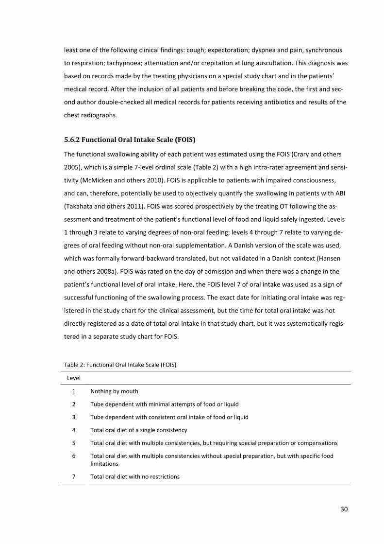
30
least one of the following clinical findings: cough; expectoration; dyspnea and pain, synchronous
to respiration; tachypnoea; attenuation and/or crepitation at lung auscultation. This diagnosis was
based on records made by the treating physicians on a special study chart and in the patients’
medical record. After the inclusion of all patients and before breaking the code, the first and sec‐
ond author double‐checked all medical records for patients receiving antibiotics and results of the
chest radiographs.
5.6.2 Functional Oral Intake Scale (FOIS)
The functional swallowing ability of each patient was estimated using the FOIS (Crary and others
2005), which is a simple 7‐level ordinal scale (Table 2) with a high intra‐rater agreement and sensi‐
tivity (McMicken and others 2010). FOIS is applicable to patients with impaired consciousness,
and can, therefore, potentially be used to objectively quantify the swallowing in patients with ABI
(Takahata and others 2011). FOIS was scored prospectively by the treating OT following the as‐
sessment and treatment of the patient’s functional level of food and liquid safely ingested. Levels
1 through 3 relate to varying degrees of non‐oral feeding; levels 4 through 7 relate to varying de‐
grees of oral feeding without non‐oral supplementation. A Danish version of the scale was used,
which was formally forward‐backward translated, but not validated in a Danish context (Hansen
and others 2008a). FOIS was rated on the day of admission and when there was a change in the
patient’s functional level of oral intake. Here, the FOIS level 7 of oral intake was used as a sign of
successful functioning of the swallowing process. The exact date for initiating oral intake was reg‐
istered in the study chart for the clinical assessment, but the time for total oral intake was not
directly registered as a date of total oral intake in that study chart, but it was systematically regis‐
tered in a separate study chart for FOIS.
Table 2: Functional Oral Intake Scale (FOIS)
Level
1 Nothing by mouth
2 Tube dependent with minimal attempts of food or liquid
3 Tube dependent with consistent oral intake of food or liquid
4 Total oral diet of a single consistency
5 Total oral diet with multiple consistencies, but requiring special preparation or compensations
6 Total oral diet with multiple consistencies without special preparation, but with specific food limitations
7 Total oral diet with no restrictions
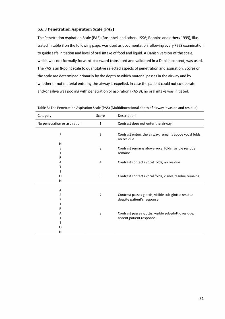
31
5.6.3 Penetration Aspiration Scale (PAS)
The Penetration Aspiration Scale (PAS) (Rosenbek and others 1996; Robbins and others 1999), illus‐
trated in table 3 on the following page, was used as documentation following every FEES examination
to guide safe initiation and level of oral intake of food and liquid. A Danish version of the scale,
which was not formally forward‐backward translated and validated in a Danish context, was used.
The PAS is an 8‐point scale to quantitative selected aspects of penetration and aspiration. Scores on
the scale are determined primarily by the depth to which material passes in the airway and by
whether or not material entering the airway is expelled. In case the patient could not co‐operate
and/or saliva was pooling with penetration or aspiration (PAS 8), no oral intake was initiated.
Table 3: The Penetration Aspiration Scale (PAS) (Multidimensional depth of airway invasion and residue)
Category Score Description
No penetration or aspiration 1 Contrast does not enter the airway
P E N E T R A T I O N
2 3 4 5
Contrast enters the airway, remains above vocal folds, no residue Contrast remains above vocal folds, visible residue remains Contrast contacts vocal folds, no residue Contrast contacts vocal folds, visible residue remains
A S P I R A T I O N
7 8
Contrast passes glottis, visible sub‐glottic residue despite patient’s response Contrast passes glottis, visible sub‐glottic residue, absent patient response

32
5.6.4 Berliner Dysphagia Index (BDI)
During the FEES examination the patient’s anatomical structures and functions in nose, pharynx and
larynx, different consistencies of food and liquid, protection of the airways and consciousness related
to oral functions and swallowing were evaluated using a Danish modified version of this protocol
(Appendix 1), which is not back‐translated and validated. The BDI (Seidl and others 2002) is a stan‐
dardised protocol to document the FEES examination and it has been examined for inter‐ and intra‐
rater reliability, which indicated that the BDI can be used to document FEES (Seidl and others 2002).
Findings from the BDI were transferred to a rating scale in order to assess the severity of the swallow‐
ing function. In study I the BDI provided a classification of the severity of swallowing and eating and
served as a documentation of FEES and made it possible to compare the different FEES examinations
during IRP. Together with the F.O.T.T. assessment, the BDI provided the treatment recommendation
for the treating OT.
5.6.5 Level of modified consistencies of food and liquid
The level of modified consistencies of food and liquid was assessed by the OT using the F.O.T.T.
approach: therapeutic eating and registered with a date in a special study chart every time there
were changes in the level of food or liquid during IRP. The date registered was the date when the
patient was able to eat or drink, the consistency in question together with different professionals
and their relatives. The levels of modified food and liquid were given according to the Complete
Danish Diet Handbook (National Board of Health and others 2012) with four levels of liquid (jelly,
honey, natural thick e.g. chocolate milk, normal thin liquid) and three levels of food (puree, souf‐
flé, soft and solid food).
5.6.6 Functional Independence Measure (FIM)
During IRP admission, every four weeks and at the discharge scores of the patients’ functional
levels of independence were rated, as a standard procedure, by the interdisciplinary team by ob‐
serving the patients’ functions.
The FIM is an 18‐item ordinal scale (Granger and others 1993), used in relation to all diag‐
noses within a rehabilitation population. It is viewed as most useful for assessment of progress
during inpatient rehabilitation. FIM scores range from one to seven: a FIM item score of
seven is categorised as "complete independence" while a score of one is "total assist" (performs
less than 25% of task). Scores falling below six require another person for supervision or assis‐
tance.

33
The FIM measures independent performance in self‐care, sphincter control, transfers, lo‐
comotion, communication, and social cognition. By adding the points for each item, the possible
total score ranges from 18 (lowest) to 126 (highest) level of independence (Wright 2000).
5.6.7 Ranchos Los Amigos Scale (RLAS)
The patients’ cognitive level of functioning in the study was rated by the interdisciplinary team
using RLAS. This is a standard procedure on admission to IRP, every four weeks during IRP and at
discharge.
RLAS is also called The Rancho Level of Cognitive Functioning Scale (LCFS) (Sander 2002) and
is one of the earlier developed scales used to assess cognitive functioning in post‐coma patients
(Hagen and others 1972). It was developed for use in the planning of treatment, tracking of re‐
covery, and classification of outcome levels. Use of the scale generates a classification of the pa‐
tient in one of eight levels, with level I (No response) and level VIII (Purposeful‐appropriate).
RLAS is tested as a reliable and valid scale (Gouvier and others 1987). For patients with severe
ABI, oral intake is related to their cognitive function and consciousness. Mackay et al. (Mackay
and others 1999b) recommend that RLAS Level IV was needed for initiation of oral intake; Level VI
was needed for total oral intake.
5.6.8 Other quantitative, functional outcome measures
Patient characteristics such as age, gender, ICD10 diagnosis, GCS in the acute stage (initial), date
of injury to calculate the time since injury, Body Mass Index (BMI) present on admission and dis‐
charge, LOS in IRP (days), duration: of oral intubation, of mechanical ventilation, of tracheostomy
tube and of feeding tube and the amount of coded dysphagia interventions (number of interven‐
tions concerning stimulation of the oral cavity, oral tract, and face in the Danish intervention
documentation system (National Board of Health 2012)) were all documented from the medical
records by the treating OT.
5.6.9 Semistructured interviews
Semi‐structured interviews (Kvale 2007; Kvale and Brinkmann 2009; Tanggaard and Brinkmann
2010) were carried out in the qualitative Study II in order to capture the participants’ perspectives
and priorities of their experiences of difficulties in swallowing and eating following ABI .
A semi‐structured interview attempts to understand themes of the lived everyday world from
the subjects’ own perspective. It comes close to an everyday conversation, but as a professional
interview it has a purpose and involves a specific approach and technique; it is semi‐structured –
it is neither an open everyday conversation nor a closed questionnaire (Kvale 2007; Kvale and

34
Brinkmann 2009). The semi‐structured interview was chosen because the pre‐defined research
questions guided the interview, and the semi‐structured open interview questions also made it
possible to depart from them and at the same time open to the participants’ own reflections and
perspectives.
Kvale’s and Brinkmann’s (Kvale 2007; Kvale and Brinkmann 2009) seven stages (thematizing,
designing, interviewing, transcribing, analyzing, verifying and report) of an interview inquiry was
used for systematic planning of Study II. The key questions when planning an interview investiga‐
tion concern the why (clarifying the purpose of the study), the what (obtaining pre‐knowledge of
the subject matter to be investigated) and the how of the interview (becoming familiar with dif‐
ferent techniques of interviewing and analyzing, and deciding, which to apply in order to obtain
the intended knowledge (Kvale 2007; Kvale and Brinkmann 2009). The thematization of “the
what” requires preliminary and detailed literature studies (Tanggaard and Brinkmann 2010). In
Study II the stage of thematizing consisted of both a detailed literature search and my pre‐
understanding. As hermeneutics (Gadamer 2004) has demonstrated it is only possible to under‐
stand the world through a pre‐understanding (Tanggaard and Brinkmann 2010) and familiarity
with the content of an investigation is not only obtained through literature and theoretical studies
(Kvale and Brinkmann 2009). In Study II, many years of experience as a clinician, treating patients
with difficulties in swallowing and eating provided me, as the interviewer, with extensive knowl‐
edge and pre‐understanding of the local language, the daily routines, and the power structures
during neurorehabilitation.
The literature search showed that in the past two decades, noteworthy advances have been
made in measuring the physiologic outcomes of dysphagia, including measurement of duration of
structure and bolus movements, stasis, and penetration–aspiration. However, there was a paucity
of data on health outcomes from the patients’ perspective, such as quality of life and patient satis‐
faction. In a study of McHorney et al (McHorney and others 2000) they found that a patient‐
based, dysphagia specific outcomes tool was needed to enhance information on treatment varia‐
tions and treatment effectiveness. The conceptual foundation and the item generation process
for the SWAL‐QOL and SWAL‐CARE were obtained by qualitative data on dysphagia quality‐of‐life
and quality‐of‐care health outcomes from dysphagic patients and their caregivers participating in
focus groups. Using the focus group data, they wrote a large number of questionnaire items and
constructed a standardized, disease‐specific questionnaire (the SWAL‐QOL) that measures dys‐
phagia patients’ experiences of health outcomes).
35
Preparation for interview
Two months before the interview, as preparation for the interview, the participants received an
information letter with the informed consent form and a questionnaire. The questionnaire was
used as preparation to help the participants to focus on their difficulties in swallowing and eating
(and not the whole process of rehabilitation) during the interview. The questionnaire was a Dan‐
ish translation and modification of the SWAL‐QOL (McHorney and others 2000) and the SWAL‐
CARE (McHorney and others 2002) (Appendix 3). The two patient‐centred outcome tools were
developed by well‐known researchers within dysphagia e.g. McHorney A.C., Rosenbek J.C., Rob‐
bins J, Logemann J.A. for dysphagia researchers and clinicians. The questionnaire was used as
preparation for the interview and as inspiration for themes in the semi‐structured interview guide
(Appendix 4), but it was also stressed that the coming interview should focus on the reflections
and perspectives of the participant. The modification process of the questionnaire was supervised
by a senior researcher, and the questionnaire was translated by a professional. However, the Dan‐
ish version of the questionnaire (Appendix 3) is not back‐translated and validated.
The interview guide
The semi‐structured interviews were conducted according to an interview guide (Appendix 4). The
semi‐structured interview guide was inspired by topics from the SWAL‐QOL and SWAL‐CARE
(McHorney and others 2002) (Table 4 on the following page) with suggested questions, but not
constructed as a questionnaire with closed questions. The semi‐structured interview guide was
developed with brief and simple types of interview questions that intended to open the reflec‐
tions and perspectives of the participant.
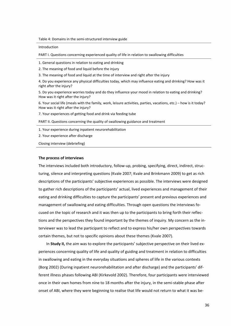
36
Table 4: Domains in the semi‐structured interview guide
Introduction
PART I. Questions concerning experienced quality of life in relation to swallowing difficulties
1. General questions in relation to eating and drinking
2. The meaning of food and liquid before the injury
3. The meaning of food and liquid at the time of interview and right after the injury
4. Do you experience any physical difficulties today, which may influence eating and drinking? How was it right after the injury?
5. Do you experience worries today and do they influence your mood in relation to eating and drinking? How was it right after the injury?
6. Your social life (meals with the family, work, leisure activities, parties, vacations, etc.) – how is it today? How was it right after the injury?
7. Your experiences of getting food and drink via feeding tube
PART II. Questions concerning the quality of swallowing guidance and treatment
1. Your experience during inpatient neurorehabilitation
2. Your experience after discharge
Closing interview (debriefing)
The process of interviews
The interviews included both introductory, follow‐up, probing, specifying, direct, indirect, struc‐
turing, silence and interpreting questions (Kvale 2007; Kvale and Brinkmann 2009) to get as rich
descriptions of the participants’ subjective experiences as possible. The interviews were designed
to gather rich descriptions of the participants’ actual, lived experiences and management of their
eating and drinking difficulties to capture the participants’ present and previous experiences and
management of swallowing and eating difficulties. Through open questions the interviews fo‐
cused on the topic of research and it was then up to the participants to bring forth their reflec‐
tions and the perspectives they found important by the themes of inquiry. My concern as the in‐
terviewer was to lead the participant to reflect and to express his/her own perspectives towards
certain themes, but not to specific opinions about these themes (Kvale 2007).
In Study II, the aim was to explore the participants’ subjective perspective on their lived ex‐
periences concerning quality of life and quality of guiding and treatment in relation to difficulties
in swallowing and eating in the everyday situations and spheres of life in the various contexts
(Borg 2002) (During inpatient neurorehabilitation and after discharge) and the participants’ dif‐
ferent illness phases following ABI (Kirkevold 2002). Therefore, four participants were interviewed
once in their own homes from nine to 18 months after the injury, in the semi‐stable phase after
onset of ABI, where they were beginning to realise that life would not return to what it was be‐

37
fore (Kirkevold 2002). The last two participants were interviewed twice, and got the same ques‐
tions twice, in the continued rehabilitation phase after onset of ABI, which is a phase of constant
change, although as time passes, changes occur more slowly and gradually (Kirkevold 2002). The
interviews took place in their rooms in the ward (two months after injury) and about one month
after discharge in their own home/room at a nursing home (four months after injury).
The interviews, which lasted between 30‐60 minutes, were tape‐recorded and fully tran‐
scribed verbatim. The transcribed data were then de‐identified and fictive names were created
for each participant to maintain and respect their confidentiality. Reflective notes (Creswell JW
2009) were taken after the interview to enhance the understanding of the participants’ described
lived experiences during the analysis of the interviews. The reflective notes and the results from
the Danish translated and modified SWAL‐QOL and SWAL‐CARE questionnaire were not further
analysed.
5.7 Data analysis
Several data analyses were conducted in the two studies included in this thesis.
5.7.1 Data analysis paper I
The patients were grouped into five main categories according to their ICD10 diagnosis: stroke
(Cerebral infarction, brain stem infarction and haemorrhage); subarachnoid haemorrhage; trau‐
matic brain injury; anoxia and other. All data were analyzed using STATA/SE 11 for Windows. The
analysis was undertaken by the authors who were blinded to the randomization, and all data
were analyzed before dividing the list into two groups. Patient demographic variables were sum‐
marised by median, minimum and maximum values for continuous variables, and by count and
percentage for categorical variables. The Student’s t‐test was used to compare normally distrib‐
uted data and the Wilcoxon signed‐rank test was used to compare non‐normally distributed vari‐
ables.
5.7.2 Data analysis paper II
Data were analysed using STATA/SE 11 and SAS 9.1. A statistical consultant assisted in the analy‐
sis. Patient demographic variables were summarised by median, minimum and maximum values
for continuous variables, and by count and percentage for categorical variables. We compared
FOIS scores present on admission and discharge by the Wilcoxon signed‐rank test. Within the in‐
tervention groups, the probability distributions of time from admission to initiation and full oral
intake were estimated by Kaplan‐Meier curves and compared by the log‐rank test. Moreover, the

38
comparisons were adjusted for the influence of predictor variables using the multiple Cox propor‐
tional hazards model (31), where the effects of predictor variables were expressed by hazard ra‐
tios and their 95%‐confidence intervals (CI). The chance of reaching total oral intake during IRP
was compared between the intervention groups and assessed for the influence of predictor vari‐
ables by logistic regression. As general statistical rules of thumb dictate a limit to the number of
predictor variables (Vittinghoff and McCulloch 2007) to ensure valid results, several sets of predic‐
tor variables was considered. The Hosmer‐Lemeshow test (Hosmer and Lemeshow 2000) was
used to assess the goodness of fit of the logistic regression model for each set of predictor vari‐
ables.
5.7.3 Data analysis paper III
An ongoing process involving continuous reflection (Creswell 2009a) was used for data analysis.
The eight interviews were read separately and as a whole, and thoroughly several times by the
first author to obtain an overall understanding of each person and the data. The words and
phrases dealing with different aspects of experiences related to the domains in the semi‐
structured interview guide were coded by the author of this thesis. The coding process was per‐
formed using a freeware computer program “Open Code” (Dahlgren and others 2004). The codes
in each interview were then compared to identify similarities and differences, and subsequently
codes that reflected similar aspects were generated into main and sub‐themes. The ICF (WHO
2001) domains were afterwards used as a conceptual framework to organise the themes. ICF
made it possible to structure the perspective of the body, the individual and society to describe
the bio‐psycho‐social complexity of swallowing, eating, communication and meals.

39
6. ETHICAL CONSIDERATIONS
Study I was approved by the Regional Committee on Biomedical Research Ethics in Denmark (Reg.
No.: 20090013) and the Danish Data Protection Agency (Reg. No: 2007‐58‐0010). The patients or
their relatives or peers received the oral and written information about the study from the treat‐
ing OT within 24‐48 hours of admission. Information was given that participation in the study was
voluntary and that participants had the right to end their participation at any time. All participants
or relatives or peers as well as the patient’s general practitioner or medical public health officer
gave surrogate consent before randomization and enrolment in the study.
In Study II the treating OT took the first contact and introduced the study. The next contact
was made by the author of this thesis, two months before and repeated the week before the in‐
terview. Two months before the interview the participants received an information letter with an
informed consent form and a questionnaire (Appendix 3). The participants gave their verbal and
written informed consent to participate and were guaranteed confidentiality. Participation was
voluntary, and participants could withdraw from the study at any time. Data were considered
confidential in line with the legislation of The Act on Processing of Personal Data (Act No. 429 of
31 May 2000) issued by Danish Data Protection Agency, who also gave their permission to the
study (Jr. nr. 1‐16‐02‐5‐09). The Central Denmark Region Committees on Biomedical Research
Ethics wrote on 27 September 2010 (Reg. nr. 20395) that “The interview study is not directly re‐
lated to the first study and should be regarded as an independent study. It follows the guideline of
the Law Committee of the interview studies equated with surveys. This means that interviews
only have to be notified to the Committee, if they include human biological material, which is not
the case from your description, and you can therefore conduct this study without further feed‐
back from the Committee (Act No. 402 of 28. May 2003)”.

40
7. RESULTS
Between June 2009 and April 2011, 679 potentially eligible patients were screened for inclusion in
Study I. The flowchart (Figure 2) below illustrates the included and excluded patients in Study I.
Figure 2: Flowchart for Study I
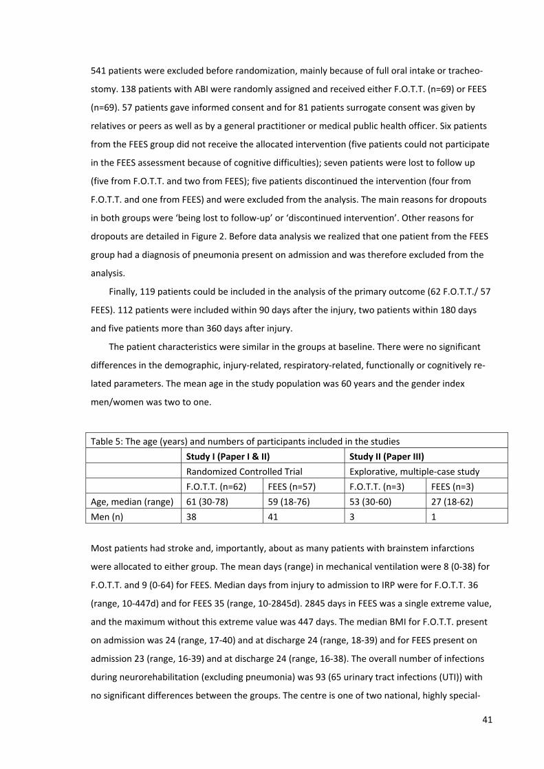
41
541 patients were excluded before randomization, mainly because of full oral intake or tracheo‐
stomy. 138 patients with ABI were randomly assigned and received either F.O.T.T. (n=69) or FEES
(n=69). 57 patients gave informed consent and for 81 patients surrogate consent was given by
relatives or peers as well as by a general practitioner or medical public health officer. Six patients
from the FEES group did not receive the allocated intervention (five patients could not participate
in the FEES assessment because of cognitive difficulties); seven patients were lost to follow up
(five from F.O.T.T. and two from FEES); five patients discontinued the intervention (four from
F.O.T.T. and one from FEES) and were excluded from the analysis. The main reasons for dropouts
in both groups were ‘being lost to follow‐up’ or ‘discontinued intervention’. Other reasons for
dropouts are detailed in Figure 2. Before data analysis we realized that one patient from the FEES
group had a diagnosis of pneumonia present on admission and was therefore excluded from the
analysis.
Finally, 119 patients could be included in the analysis of the primary outcome (62 F.O.T.T./ 57
FEES). 112 patients were included within 90 days after the injury, two patients within 180 days
and five patients more than 360 days after injury.
The patient characteristics were similar in the groups at baseline. There were no significant
differences in the demographic, injury‐related, respiratory‐related, functionally or cognitively re‐
lated parameters. The mean age in the study population was 60 years and the gender index
men/women was two to one.
Table 5: The age (years) and numbers of participants included in the studies
Study I (Paper I & II) Study II (Paper III)
Randomized Controlled Trial Explorative, multiple‐case study
F.O.T.T. (n=62) FEES (n=57) F.O.T.T. (n=3) FEES (n=3)
Age, median (range) 61 (30‐78) 59 (18‐76) 53 (30‐60) 27 (18‐62)
Men (n) 38 41 3 1
Most patients had stroke and, importantly, about as many patients with brainstem infarctions
were allocated to either group. The mean days (range) in mechanical ventilation were 8 (0‐38) for
F.O.T.T. and 9 (0‐64) for FEES. Median days from injury to admission to IRP were for F.O.T.T. 36
(range, 10‐447d) and for FEES 35 (range, 10‐2845d). 2845 days in FEES was a single extreme value,
and the maximum without this extreme value was 447 days. The median BMI for F.O.T.T. present
on admission was 24 (range, 17‐40) and at discharge 24 (range, 18‐39) and for FEES present on
admission 23 (range, 16‐39) and at discharge 24 (range, 16‐38). The overall number of infections
during neurorehabilitation (excluding pneumonia) was 93 (65 urinary tract infections (UTI)) with
no significant differences between the groups. The centre is one of two national, highly special‐

42
ized neurorehabilitation centres in Denmark. In this study 42% of the patients were transferred to
further IRP in regional neurorehabilitation units. Twenty‐six percent were discharged to nursing
homes or sheltered accommodation with municipal rehabilitation service, 20% were discharged
to their own home for further municipal outpatient rehabilitation, and the remaining 12% were
discharged to other hospital departments for further medical treatment.
In Study II, six persons with ABI were recruited from Study I. There were no dropouts in
Study II.
7.1 Paper I
The aim of Paper I was to test the hypothesis that patients with ABI (stroke, subarachnoid hemor‐
rhage, TBI, anoxia etc.), who are evaluated for initiation of oral intake using only the assessment
component of F.O.T.T., have a greater risk of developing of pneumonia during neurorehabilitation
than those who are evaluated using FEES.
The results will be described in brief. Please refer to Paper I for additional details e.g. tables
and figures to support the results.
7.1.1 Incidence of pneumonia
The overall incidence of pneumonia was 13.5% (16/119 patients) during the study period. Four of
the 62 patients in the F.O.T.T. group and 12 of 57 in the FEES group were diagnosed with pneu‐
monia difference significant at p=0.03 by Fishers exact test. The number of days from admission
to neurorehabilitation to pneumonia ranged from 0 to 60. Six patients in the FEES group but none
in the F.O.T.T. group developed pneumonia before initiation of oral intake and had to be excluded
from further analysis. Three of these six patients were unable to initiate oral intake during the
period of neurorehabilitation.
The remaining 10 patients (4 F.O.T.T./ 6 FEES) developed aspiration pneumonia three to 49
days after initiation of oral intake. Of these 10 patients, five developed aspiration pneumonia
three to 10 days (1 F.O.T.T./ 4 FEES) after initiation of oral intake and five patients after 32 to 49
days (3 F.O.T.T./ 2 FEES). Moreover, the diagnosis of pneumonia in three of these patients was
not in accordance with our the primary outcome definition of aspiration pneumonia (Lim and
others 2009) two FEES patients did not have new infiltrative changes on chest radiography and
one patient assessed by F.O.T.T. was not evaluated by chest radiography. Thus, seven patients
remained for analysis. Of these three of the 62 patients were assessed by F.O.T.T. and four of the
57 patients were assessed by FEES.

43
There was no significant difference in the number of dysphagia interventions between the two
groups, or between patients with pneumonia before or after oral initiation.
7.2 Paper II
The aim of Paper II was to investigate if there was a difference in time to recovery of functional
oral intake in inpatient neurorehabilitation of patients with ABI assessed using F.O.T.T. and FEES,
respectively, and if other factors influenced the time to recovery, and the calculation of incidence
of dysphagia.
The results will be described in brief. Please refer to Paper II for additional details e.g. tables
and figures to support the results.
7.2.1 Incidence of dysphagia
Between June 2009 and April 2011, 679 ABI patients were admitted to inpatient neurorehabilita‐
tion and 320 of 679 patients (47%) had clinically diagnosed swallowing difficulties (dysphagia)
according to the clinical criteria used at the centre, where dysphagia is defined as difficulties in
the pre‐oral, oral, pharyngeal and/or oesophageal phase of normal swallowing.
7.2.2 Initiation of oral intake
A total of 50 of 119 patients (42%) (29 F.O.T.T./ 21 FEES; p=0.35) patients were initiated for oral
intake on admission, while 109 of 119 patients (92%) ( 59 F.O.T.T./ 50 FEES; p=0.35) were initiated
before discharge. All patients who were initiated on admission maintained their status during IRP.
For the 59 of 119 patients initiated during IRP, the median number of days after injury until initia‐
tion was 39 (range, 10‐447) and from admission until initiation 1 day (range, 0‐65). Comparison of
the Kaplan‐Meier curves between groups (log‐rank test) showed no significant difference in the
time to initiation of oral intake. The median time until initiation in IRP was 8 days (F.O.T.T.) and 7
days (FEES), respectively. For patients on total oral intake at discharge, the median number of
days post injury onset until initiation was 32.5 (range: 10‐88), and the median number of days
from admission until initiation was 0.5 days (range: 0‐20). Adjusting the comparison of the groups
for the confounding effect of other factors did not show any significant difference in the time to
initiation of oral intake between the groups (p=0.120). The likelihood to be initiated during IRP
tended to be dependent on cognitive functioning as assessed by the RLAS score (Hazard Ration
(HR) 1.266; p=0.056). The instantaneous probability increased by 27% for each step to a higher
level of RLAS. While TBI and stroke tended to be more critical diagnoses regarding initiation com‐

44
pared to the reference group “other”, there was no statistically significant difference between the
diagnostic groups. 10 out of 69 patients` time to initiation of oral intake were right‐censored, be‐
cause they were not initiated at discharge.
7.2.3 Reaching total oral intake
Three of 119 patients (2.5%) (0 F.O.T.T./ 3 FEES; p=0.11) achieved total oral intake within 24 hours
of admission to IRP, while a total 44 of 119 patients (37%) (19 F.O.T.T./ 25 FEES; p=0.18) were on
total oral intake at discharge. Three patients who achieved total oral intake within 24 hours of
admission remained so at discharge; 41 patients went from FOIS <7 to FOIS=7 during IRP. A sur‐
vival analysis showed that the estimated mean time from injury until total oral intake was 56.0
days (range: 22‐179) and from admission until total oral intake 29.5 days (range: 0‐151). In the
Kaplan‐Meier curves, 75 out of 116 patients were right‐censored, because they were not at total
oral intake at discharge. During IRP many of the patients moved from FOIS levels 1 and 2 to FOIS
levels 5 or 6. A comparison between the groups showed no difference in the time to total oral
intake.
7.2.4 Factors of importance for reaching total oral intake
Using a Cox‐regression analysis to compare the groups adjusted for the potential influence of con‐
founding factors for the time to total oral intake did not show a statistical significant difference
between the groups (p=0.120). The time to total oral intake was, however, shown to decrease
significantly with age (HR=0.965; p=0.003); an estimated 35% decrease of probability for each 10
years of age. Also here, TBI and stroke seemed to be more critical diagnoses regarding oral intake
compared to the reference group “other”, but there was no statistically significant difference be‐
tween the diagnostic groups. Three patients achieved total oral intake within 24 hours of admis‐
sion so 116 of 119 patients could be included in the logistic regression analyses of the chance of
achieving total oral intake during IRP. Ultimately, 44 patients were on total oral intake at dis‐
charge from IRP. Three sets of predictor variables were chosen for the analysis. Set 1 data in‐
cluded group, age and diagnostic groups (basic variables), degree of independence (FIM‐change)
and cognitive functioning (RLAS‐change). The findings showed that the probability of reaching
total oral intake decreased significantly with age (p=0.0200) and increased significantly with FIM ‐
change (p<0.0001). Set 2 data included group, age and diagnostic groups (basic variables), number
of coded dysphagia interventions and LOS (treatment variables). The findings showed that the
probability of reaching total oral intake decreased significantly with age (p=0.0031) and number
of coded dysphagia interventions (p<0.0001), but increased by LOS (p=0.0013). Set 3 was made
from a combination of set 1 and 2 and data included diagnostic groups, degree of independence

45
(FIM‐admission), cognitive functioning (RLAS‐admission), number of coded dysphagia interven‐
tions and LOS (p=0.0013). The findings showed that the probability of reaching total oral intake
decreased significantly with number of coded dysphagia interventions (p=0.0049), but increased
with LOS (p=0.0251). These different analyses contain 18 statistical tests, i.e. the expected num‐
ber of false significances was 0.9.
7.3 Paper III
The aim of Paper III was to explore and interpret how persons with ABI experienced and adapted
to reduced abilities to swallowing and eating ‐ and clinical implications.
The findings from the semi‐structured interviews will be described in brief under the five
main themes that emerged from the analysis: individual psychological assets, swallowing and in‐
gestion, eating and drinking, communication and meals, and rehabilitation of swallowing and eat‐
ing. Please refer to Paper III for additional details and quotations to support the findings.
7.3.1 Individual psychological assets
Three sub‐themes were identified. The first was general health status. Most of the participants
described that they were healthy and seldom ill. They could do anything now, activities had be‐
come more time consuming, and they got tired very fast. The second theme was functional level
of swallowing and eating. Three of them could swallow and eat as before the injury, there were
no problems and no need for further rehabilitation after discharge. One participant felt that the
swallowing difficulties came on top of other difficulties, like he was “twice affected” right after
the injury. One of the participants with swallowing difficulties after discharge said that eating did
not take up more energy, but progress was very slow and this was very annoying. But he expected
that swallowing would get better, even that he was still coughing. One participant had got a feed‐
ing tube again after discharge, and started to doubt whether it would ever be better. The final
theme was participants’ treatment goals. Their treatment goals were expressed very differently.
Some did not feel that they had ever had any swallowing and eating difficulties after the injury.
One expressed getting to walk was first priority and getting to eat was a side issue. Others said
that the main treatment goal during neurorehabilitation was to regain normal oral intake and in
company with others.
7.3.2 Swallowing and ingestion
Four sub‐themes were identified. The first theme was symptoms choking, coughing, voice clearing
and saliva. Some did not remember any of these symptoms. Others had a lot of saliva, right after

46
the injury and was both clearing the voice and coughing a lot, and sometimes had to leave the
dinning room at the ward. The second theme was worries about swallowing, choking and pneu‐
monia. Most of the participants had not been afraid of aspiration and worried about pneumonia.
Others were worried right after the injury. But one was, at the last interview, still thinking a lot
about pneumonia, and that it was the swallowing difficulty that caused all his pneumonias. The
third theme was hunger. The participants did not feel hungry right after the injury. The last thing
they were thinking about was food and the taste was lacking. The last theme was mood and fa‐
tigue. Right after the injury they were all tired and needed a lot of sleep. Some expressed not be‐
ing able to eat was just a side issue. Others said that the difficulties in swallowing and eating af‐
fected their mood and took up much energy during neurorehabilitation, but at the time of the
interview, it did not affect most of them any more.
7.3.3 Eating and drinking
Four sub‐themes were identified. The first theme was meaning of food. Most of the participants
described themselves as lovers of food both before the injury and at the time of the interview.
Some said that right after the injury food did not mean anything to them and others said that the
importance of food gets very clear when you cannot eat by mouth and need feeding by tube. The
second theme was feeding by tube. Half of the participants had a nasal tube and the rest had had
both a nasal and a PEG tube. About the nasal tube, one of the participants said that it was really
annoying having a tube in one’s nose, and it affected one’s mood. She was happy the day it could
be removed, although it was uncomfortable when it was pulled out. One of the participants hav‐
ing had PEG for four months said, that he did not really think much about it. It was a necessary
evil, but it did not hurt and to him it had not had any negative impact. Others described it as terri‐
ble, horrible, hated it and it was a tough time getting formula and they would rather not remem‐
ber it.
The third theme was first oral intake. The participants were yearning to get something in the
mouth after a long period of tube feeding, to get normal food and even yoghurt tasted like a
grand Christmas dinner. The final theme was modification of taste. Their taste buds had changed
since the injury. Some had also stopped smoking and they said that food tasted much better now.
7.3.4 Communication and meals
Two sub‐themes were identified. The first theme was communication. The participants just ex‐
perienced minor communication problems. One said that sometimes he had trouble to speak out
loudly, and it was more difficult getting the words out. After discharge he had the feeling that he
had lost his voice completely. The second theme was swallowing difficulties and meals with social

47
interactions. Most of the participants did not feel that their previous swallowing difficulties had
caused changes in their social lives after discharges. But they gave a lot of examples during reha‐
bilitation e.g. that his parents never ate in his presence, because they felt sorry for him. Others
said that it had affected them that they could not eat with the others on the ward. After discharge
most of them had again joined gatherings with family and friends. But one said that he did not eat
as much as at home, because he needed extra time and also tried to avoid coughing too much.
7.3.5 Rehabilitation of swallowing and eating
Two sub‐themes were identified. The first theme was inpatient neurorehabilitation approach con‐
cerning swallowing and eating. The participants expressed both positive and negative experiences
concerning: guidance from the OT, oral stimulation, mobilisation of the tongue, chewing in gauze,
modified consistencies and the FEES. The second theme was rehabilitation of swallowing and eat‐
ing after discharge. Two participants needed further rehabilitation. One said that he was still
coughing and had asked the community rehabilitation centre for OT treatment of the swallowing
function. The other had been in several places since discharge. First, in a nursing home for six
days, and then at an intensive care unit for more than one month treated for severe aspiration
pneumonia. At the time of interview he had been at a new nursing home for six days, just lying in
his bed and had not had any therapy at all. He had a new appointment for FEES at the inpatient
rehabilitation centre one month after the last interview.
The three predominating sub‐themes that emerged in the five main themes were: feeding by
tube (in swallowing and ingestion), difficulties in swallowing and meals with social interactions (in
communication and meals), and inpatient rehabilitation approach concerning swallowing and
eating (in rehabilitation of swallowing). The predominant experiences are presented in the con‐
ceptual model of Study II (Figure 3, next page), which illustrates the dynamic and reciprocal inter‐
action between the findings.

48
Figure 3: Conceptual model of Study II
49
8. DISCUSSION
The two studies included in this thesis have explored and evaluated different aspects of difficul‐
ties in swallowing and eating following ABI. Both quantitative and qualitative methods have been
used in the evaluation and exploration.
To understand the complexity of the assessment of difficulties in swallowing and eating and
the following discussion this section begins with a brief introduction to the different approaches
used.
The majority of research on dysphagia and aspiration deals with acute settings (Barquist and
others 2001; Brown and others 2011), where dysphagia is likely to be temporary (White and oth‐
ers 2008). Dysphagia and aspiration are important difficulties in many patients with ABI in inpa‐
tient settings. Early detection is crucial in a postacute stroke population and there is a need for
reassessment on admission to IRP (Heckert and others 2009). There seems to be consensus
(Farrell and O'Neill 1999; Hammond and Godstein 2006; Singh and Hamdy 2006) on the assess‐
ment of dysphagia as a three‐tiered assessment cascade of screening, bedside evaluation, and
objective measurement. This cascade has been described as the best clinical practice in the reha‐
bilitation of patients with TBI in Denmark (Langhorn and others 2008). The disadvantage is that it
is difficult to refer patients to objective measurements due to lack of availability.
Bedside screening is the first step to assess whether the patients have an impairment of swal‐
lowing. The next step is an in‐depth bedside clinical assessment. However, the accuracy of a clini‐
cal assessment has shown to be a poor predictor of pharyngeal dysphagia (Splaingard and others
1988; Linden and others 1993; O'Donoghue and Bagnall 1999). A clinical assessment may be aug‐
mented by objective measurement (instrumental assessment), such as FEES, the Video Fluoros‐
copy Swallowing (VFS) also referred to as Modified Barium Swallow (MBS), or even VSE (Vide‐
ofluorographic Swallowing Evaluation).
VFS allows the clinician to see where the swallowed material actually goes, e.g. if food or fluid
is entering the respiratory tract, and if so, how much and whether the pharyngeal or oesophageal
muscles are functioning properly (White and others 2008). For this reason VSE is regarded as be‐
ing more sensitive than clinical assessment in the diagnosis of dysphagia (Lim and others 2001;
Mayer 2004).
50
FEES is a new, but well established assessment tool in otorhinolaryngology. Scinitigraphy,
manometry, and acoustic assessment are much less commonly used (White and others 2008). An
RCT demonstrated that if dysphagic outpatients have their dietary and behavioural management
guided by the results of VFS or Fiberoptic Endoscopic Evaluation of Swallowing with Sensory Test‐
ing (FEESST), their outcomes with respect to pneumonia incidence and pneumonia‐free interval
are essentially the same (Aviv 2000). A multitude of studies have shown a high level of agreement
between the two instrumental assessments, and the use of VFS as gold standard is no longer ap‐
propriate. It is likely that both assessments will continue to be used and will be seen as comple‐
mentary tools rather than competitive ones (Langmore 2003).
The following discussion consists of three interrelated parts: the most important results from
my two studies and their relation to existing knowledge; the different conceptual frameworks in
the studies and the methodological considerations and limitations. Finally, the discussion has a
section with implications for clinical practice.
8.1 Main results and insights across the two studies
8.1.1 Does dysphagia matter?
The incidence of dysphagia in the Study I population was 47%. Dysphagia is an important issue
which needs attention within IRP. The incidence in this study is nearly 50% larger than that identi‐
fied by the TBI study by Winstein (Winstein 1983), but almost 50% less than identified in a TBI study
by Hansen (Hansen and others 2008a), and nearly identical to that reported in a stroke study by
Fasetti (Falsetti and others 2009). Although these studies were conducted in multidisciplinary IRP
settings and dysphagia was diagnosed clinically, it was not possible to directly compare our findings,
because our study population included both patients with TBI and stroke and other studies had either
or.
8.1.2 Are both F.O.T.T. and FEES necessary?
The findings in this thesis show that a non‐instrumental approach like F.O.T.T. to assess the swal‐
lowing disorders in patients with ABI may be as effective in predicting safe swallowing and suit‐
able time for the initiation of oral intake as an instrumental approach like FEES. Thereby it may be
possible to avoid costly and time‐consuming instrumental examinations. The findings show that
there is no need (in terms of predicting safe swallowing) for FEES to guide the level of oral intake
until recovery of total oral intake during inpatient neurorehabilitation.
51
This was an unexpected finding as the application of an instrumental and invasive approach
like FEES in previous studies had been considered to be more reliable than a clinical approach
(Splaingard and others 1988; Leder and Espinosa 2002). Leder et al (Leder and Espinosa 2002)
concluded that the clinical examination, when compared with FEES, underestimated the aspira‐
tion risk in patients with aspiration risk and overestimated the aspiration risk in patients who did
not exhibit aspiration risk. Their clinical swallowing examination consisted of six clinical identifiers
of aspiration risk e.g. dysphonia, dysarthria, abnormal gag reflex, abnormal volitional cough,
cough after swallow, and voice change after swallow. Leder also concluded that careful considera‐
tion of the limitations of clinical testing leads us to believe that a reliable, timely, and cost‐
effective instrumental swallow evaluation should be available for the majority of patients follow‐
ing acute stroke.
Interestingly, the findings from Study I are different from the findings in the above studies.
Despite the fact that F.O.T.T. evaluates the pre‐oral and oral phase of swallowing and FEES evalu‐
ates the pharyngeal phase of swallowing, both techniques produce the same outcome in respect
to aspiration pneumonia and resuming oral intake. The difference in our findings could be due to
our different choices of clinical identifiers. Leder’s (Leder and Espinosa 2002) six clinical identifiers
of aspiration are specific swallowing items (Daniels and others 2012). The assessment part of the
F.O.T.T. approach, used in this thesis as the clinical criterion for initiation of oral intake (see
methods), includes both swallowing and non‐swallowing items (e.g. in the pre‐oral phase with
cognition and head and body posture). Difficulties in swallowing and eating respond to the treat‐
ment of the whole individual. Swallowing is influenced by a number of factors that must be taken
into account and modified as necessary in treatment. Trapl et al (Trapl and others 2007) examined
50 consecutive patients with acute stroke and suspected dysphagia, admitted to an acute stroke
unit. They found the non‐invasive, non‐instrumental, standardised bedside screening to be the
most reliable compared to FEES to assess the risk and severity of aspiration. Similarly, in studies in
acute settings, the addition of a FEES examination did not change the incidence of aspiration or
post‐extubation pneumonia (Barquist and others 2001; Brown and others 2011).
The findings in this thesis underline that a thorough clinical assessment like F.O.T.T. should
precede any instrumental procedure. The instrumental assessment of swallowing is only part of
the cascade of assessments of swallowing (Groher and Crary 2010). Our findings may implicate
that a clinical assessment like F.O.T.T. should be the primary assessment, while it contains both an
assessment and a treatment part and is easy to perform, and FEES may be performed in addition
to F.O.T.T., to guide the planning of treatment. The advantage of FEES is the visualization of the
pharyngeal phase of swallowing (before and after swallow), which is a very difficult phase to as‐

52
sess using a clinical assessment like F.O.T.T. But clinicians must be aware that FEES is just a snap‐
shot of the difficulties in swallowing and eating and not a whole picture.
8.1.3 The best swallowing protocol?
Daniels et al (Daniels and others 2012) conclude in their systematic review of valid items for
screening dysphagia risk in stroke patients that the best combination of non‐swallowing and swal‐
lowing items as well as the best swallowing protocol remain unclear. With reference to the con‐
clusion from Daniels et al, the findings from this thesis provide important new knowledge for the
development of a future valid swallowing protocol with the best combination of swallowing and
non‐swallowing items.
Our findings imply that a non‐instrumental based approach like F.O.T.T. in the assessment
part consists of both swallowing and non‐swallowing items, which may be as effective in predict‐
ing safe swallowing (safe in terms of no or minimal aspiration) as an instrumental approach like
FEES. Furthermore, our findings emphasise that a swallowing protocol has to deal with the whole
process of eating and not just the difficulties in swallowing, as supported in a systematic review
by Westergren (Westergren 2006). Westergren concludes that the best nursing practice for de‐
tecting eating difficulties includes as the first step the Standardized Bedside Swallowing Assess‐
ment (SSA) to detect dysphagia (strong evidence). As the second step an observation should be
made of eating including ingestion, deglutition and energy (moderate evidence).
8.1.4 Reduced consciousness in relation to swallowing and eating
In F.O.T.T. the patient does not need to understand verbal communication but he/she needs a
cognitive and conscious functioning corresponding to RLAS level IV (confused, agitated response)
for initiation of oral intake (Mackay and others 1999a). On the other hand, Leder et al (Leder and
others 2009) reported if the patient is able to answer orientation questions and follow single‐step
verbal commands, this is an indication that he/she could eat without aspiration. An interpretation
of their findings would be that the patients should not initiate any oral intake before they are able
to understand verbal communication and this is far from the F.O.T.T. practice used in a Danish
neurorehabilitation context (Kjærsgaard 2005b) and evaluated in Study I. This part of the assess‐
ment is based on results from a previous study by Mackay et al (Mackay and others 1999b) which
stated that the initiation of oral feeding required RLAS level IV. A study by Brady et al (Brady and
others 2006) indicated that it is safe to provide therapeutic oral feedings to patients functioning
at RLAS III if the patient: 1) does not demonstrate aspiration or aspiration is eliminated with vol‐
ume or consistency modification by a baseline instrumental swallowing examination, and 2) is
53
given close supervision during oral feeding. So perhaps it is possible to initiate therapeutic oral
feeding a little earlier than is our current practice.
Our findings in Study I showed that a lower frequency of patients with a higher level of cogni‐
tive skills recovered to total oral intake during IRP compared to the study of Brady et al (Brady and
others 2009). Our findings may be caused by the fact that many of the patients in Study I were
discharged to regional rehabilitation centres without reaching total oral intake, but if follow‐up
had been performed after regional IRP, our results might have been different.
Some of the patients in Study I randomized for FEES were excluded because they could not
co‐operate. For some of the patients with reduced consciousness it was very provoking to get the
endoscope into the nose and some of them were fighting, and they were therefore excluded. In
fact, Brady et al (Brady and others 2006) found that only a VSE or FEES could provide the neces‐
sary information to determine if oral feeding is safe for a patient with disordered consciousness.
Brady et al (Brady and others 2009) have maintained that individuals with disordered conscious‐
ness can participate in an instrumental assessment of swallowing, which is contrary to our experi‐
ence.
8.1.5 Other factors influencing initiation of oral intake and reaching total oral intake
In the present study care was taken to assess those functional factors that are possible to collect
from many patients in a clinical setting like Hammel Neurocenter. Hence, videofluoroscopy or
direct measurements of brain function or structure were neither possible nor included here.
However, it was possible to make a coarse characterization of the central nervous system lesions
from patient record data (Paper I, Table 1B) that shows a fairly even distribution between the
assessment groups (F.O.T.T./FEES) of brainstem injuries, being especially prone to cause dys‐
phagia. Nevertheless, the location of brain injury may be a confounding factor when attempts
were made to analyse for the importance of external factors (Paper II).
Our findings showed that the likelihood to be initiated during IRP tended to be dependent on
the cognitive functioning as assessed by the RLAS score. The probability increased by 27% for each
step to a higher level of RLAS. The findings in Study I were similar to the findings by Terre &
Mearin (Terre and Mearin 2007) showing that the cognitive function evaluated by RLAS is the best
prognostic factor of oro‐pharyngeal dysphagia. The time to reaching total oral intake was shown
as a minor decrease significantly with age.
Factors such as intervention group (F.O.T.T. or FEES) did not influence the probability of
reaching total oral intake during IRP. Adjustment for more functional‐related variables like
changes in the functional independence (FIM‐change) had a significant influence. A high FIM
change score is an expression of good overall functional progress and increases the likelihood of

54
reaching total oral intake during IRP. The fact that one of 18 items in the FIM score is the item
“Eating and drinking” may of course influence this to some degree. Adjustment for age revealed
that an increase in age was a negative predictor of reaching oral intake during IRP. This finding is
in accordance with other studies (Mann and others 1999; Ickenstein and others 2003; Hamidon
and others 2006; Oto and others 2009) which found an increase in age to be the most powerful
predictive factor of dysphagia outcome.
Another analysis adjusting treatment‐related variables also shows that the probability of
reaching total oral intake decreases significantly with an increase in age. The statistically signifi‐
cant effect of the number of dysphagia interventions is paradoxical in that the probability of
reaching total oral intake decreased with the number of interventions received. However, this
number may be a substitute for the patient’s condition i.e. many coded dysphagia interventions
may reflect that the patient has a low level of functioning, required a lot of support and had a
poor prognosis in terms of reaching total oral intake.
That therapy is indeed meaningful may be behind the significance of the LOS, where longer
stays in IRP increased the patients’ probability of reaching total oral intake, which is similar to the
results of Krieger et al (Krieger and others 2010), who showed that individuals with longer LOS
were more likely to advance to three meals daily and have their feeding tube removed.
The last analysis combining the functional and treatment‐related variables showed similar re‐
sults to the first, which could obviously be due to the fact that the patients who received the least
interventions were the patients´ with the highest functional abilities, even though the last analysis
taking the patients low level of functioning into account by adjusting the treatment‐related (FIM‐
admission and RLAS‐admission) variables present on admission, does not change the strength
(predictive significance) of coded dysphagia interventions and LOS.
8.1.6 Feeding by tube and first oral intake
Our findings in Study II showed that even though feeding by tube seems to solve the participants’
swallowing and ingestion difficulties at the level of body functions and structure, it seems to have
had a great impact on the participants’ level of activity and participation and their personal fac‐
tors. Our findings specified feeding by tube as an important psychological impairment to be aware
of as a clinician. If the participants had a nasal tube and a period of inpatient rehabilitation < 3
months, they could be classified as having had acute dysphagia (Martino and others 2010). At the
time of the interview they did not experience any consequences of having had dysphagia, even
though they had had a low initial GCS, long time in the intensive ward with 2‐3 weeks in a me‐
chanical ventilator, but with no episodes of acute pneumonia.

55
The picture was different if the participants had had both nasal and PEG tube and a period of
inpatient rehabilitation for > 3 months including episodes of acute pneumonia, they could then be
classified as having or having had chronic dysphagia (Martino and others 2010).
Our findings were similar to Martino et al(Martino and others 2010), where they found dis‐
parities in how acute (dysphagia < 3 months) and chronic (dysphagia > 3 months) persons priori‐
tised the psychological impairment consequences of having dysphagia.
Our findings also corresponded with a study of Rogers et al (Rogers and others 2007) where
persons with PEGs reported poor quality of life with significant deficits in all quality of life do‐
mains e.g. family life, intimate relationships, social activities, and hobbies, compared with persons
not using PEG. In our study, the need for having a feeding tube seemed to leave negative experi‐
ences, and for some participants it has had a negative impact on the person’s participation in so‐
cial gatherings and quality of life. Especially for participants with chronic dysphagia and a long
time with a PEG, the experiences were something they wanted to forget. Some of them thought
that the tube delayed the initiation of oral intake. Even participants needing a nasal feeding tube
for a short time expressed negative experiences about it.
During the time with tube feeding the participants in Study II were yearning for normal food
and most of them remembered their first oral intake. Doolittle (Doolittle 1992) finds the first oc‐
casions when resuming activities to be important events: the participants speak of the first time
they accomplish e.g. the first cup of tea without thickener. The participants in Study II had similar
experiences e.g. getting the first taste of yoghurt after the ABI was like having a grand Christmas
dinner.
Our findings showed that the time with tube feeding had a negative but not persistent impact
on the person’s quality of life. After withdrawal of the feeding tube, things often seemed to nor‐
malise, and the person used different strategies to be able to participate in social gatherings in‐
cluding food and drink as before the injury, did for example not take liquid together with solid
food and did not go to a restaurant. Although they did not get a social life as before the injury,
they got an acceptable and meaningful new life, and the time with tube feeding seemed like “a
closed chapter”.
8.1.7 Swallowing difficulties and meals with social interactions
Some of the participants in Study II described that they were asked to eat in their room together
with a helper and that they felt being “kept under surveillance” and kept outside the community
of the patients. They were longing to be able to eat together with the other patients in the ward.
They knew that it was because they had to concentrate on swallowing and eating and that they

56
would be distracted by the other patients, but still it was difficult for them to accept, because
they wanted to be part of the social activity that the meal makes up in the ward.
Medin et al.(Medin and others 2010b) highlight the complexity of having eating difficulties af‐
ter stroke. Aspects related to the participants’ striving for control are based on strategies of being
careful when eating, avoiding social activities, needing the help of others and analysing the con‐
sequences of eating different foods to eat safely and properly. The participants in this study were
aware of eating properly, when they attended for example a dinner. So if something got stuck in
the throat, they started coughing and had to leave the table, because other people should not be
bothered by their coughing.
Like in our study Medin et al (Medin and others 2010b) find that in striving for control to eat
safely and properly, some persons avoid activities which they used to perform when eating or in
which they participated before their stroke. In our study three participants avoided drinking liquid
together with food, when visiting family and did not talk and eat at the same time, so they did not
eat as much as when they were eating at home. The reason for not eating that much with unfa‐
miliar people could be fear of coughing and sense of shame for not eating properly.
8.1.8 Inpatient rehabilitation approach concerning swallowing and eating
The participants in Study II spoke positively about the guidance they had received during inpa‐
tient rehabilitation. They expressed confidence in and satisfaction with the treatment, though
sometimes they did not understand the goal of the treatment and why the treatment was
needed. The predominant experiences concerning the treatment of swallowing and eating were
oral stimulation and mobilisation of the tongue. It is important to emphasise that the mouth was
a very intimate area to many participants and that oral stimulation with the direct contact of the
therapist’s finger at the participants’ for example gums and tongue went beyond some of the par‐
ticipants usual limits. The mouth is central in the process of eating. Falk (Falk 1994) described the
mouth is the most controlled opening of the body with regard to the influx (eating) but also con‐
cerning the “sublimated” outflux of speech. The mouth is the place where expression and experi‐
ence meet.
All participants talked about the various ways of handling their situation, which could be un‐
derstood as adaptation. Therefore the theories of adaptation were applied in the discussion of
the individual losses of function as the theories provide ways of enhancing the knowledge of in‐
side perspectives of what happens when disabilities change the conditions of everyday life.
57
8.2 Conceptual framework
8.2.1 The International Classification of Functioning, Disability and Health
Rehabilitation is a complex multidisciplinary, health care intervention undertaken in a complex
environment (Turner‐Stokes and others 2005; Shiell and others 2008). Using the ICF framework in
this thesis made it possible to understand and interpret the meaning and the complexity of having
difficulties in swallowing and eating following ABI. The ICF made it easier to evaluate and explore
the whole process of eating. The use of the ICF framework made it possible to look at the person’s
difficulties in ingestion, swallowing, eating and drinking in relation to the six components of
health: health condition, body functions and structure, activity, participation, environmental and
personal factors.
When you deal with dysphagia, both as a patient and as a professional, you have to be aware
of the importance of the whole process of eating and not just safe swallowing. So if I should argue
against other authors’ (Groher and Puntil‐Sheltman 2010) description of difficulties in swallowing
and eating, I will argue that a swallowing disorder should always be looked at both as a swallow‐
ing and a feeding disorder. As a clinician you have to look at the whole person in relation to his or
her complex environment (Martino and others 2010; Medin and others 2010a).
The use of ICF as a conceptual framework in this thesis made it possible to justify and
strengthen the whole rehabilitation process of the difficulties in swallowing and eating from an
OT perspective and emphasise the rehabilitation focus on the level of activity and participation
and not just on the level of body functions and structures. Of all the disorders that OTs evaluate
and treat, the difficulties in swallowing and eating are the most medical in a traditional sense of a
medical disorder being one that could potentially result in death, but it is also a very important
issue related to the patient’s quality of life, the ability to eat safely together with others (DeVault
1991; Jenkins 1999; Johansson and Johansson 2009).
8.2.2 Theories about adaptation
The ICF, if fully elaborated into a theoretical model, will tell us what will happen to a person’s am‐
bulation if we change his/her level of strength, but it will not tell us how to change the person’s
level of change (Whyte 2008). Whyte (Whyte 2008) asked: “Can we hope for a unified theory of
rehabilitation?” The answer is: “Do not think so”. In this thesis theories of adaptation were used
to complement the ICF. The focus on adaptation was at the intrapersonal level (King 1978;
Spencer and others 1996; Schultz and Schkade 1997) at which the adaptation to an ever‐changing

58
environment with challenges of loss of abilities is complex and can be a long process, particularly
when coping with major changes in a person’s life (Jonsson and others 1999).
This process of adaptation for the participants in Study II is illustrated in the conceptual
model of the main themes in this thesis, see Figure 3, which corresponds with Spencer’s (Spencer
and others 1996) concept of adaptation and the domains in the ICF framework.
The findings in thesis stressed that an ABI is a major, dramatic change and a process where
the chapters in the participant’s life story end and begin, and that the rehabilitation following ABI
was an inherent, cumulative and prolonged process, which required a positive approach of the
person with ABI. Brands et al (Brands and others 2012) describe in their clinical messages that
adaptation to brain injury is an interactive and iterative process and that a serial model does not
illustrate the complex process of adaptation in brain injury well. The aim of Study II was not to
develop a model to illustrate the complex process of adaptation to the difficulties in swallowing
and eating following ABI, but just to explore, understand and interpret the difficulties as proc‐
esses of change. Our findings could be explicit using Spencer’s description of the interactive proc‐
ess of adaptation that occurred between an organism and its environment.
Most of the participants experienced during time a difficult, but successful adaptation to a
daily life with eating and drinking together with other people. The participants had or tried to get
a positive and active role in their own lives even though not all of them could participate physi‐
cally. Some of them did what “was expected from them” and others tried out themselves, not
waiting for professional guidance. The participants were interacting with the specific environ‐
mental demands, and after discharge they found their way back to meals with social interaction
with family and friends, even with some swallowing difficulties. Most of the participants experi‐
enced a process where they ensured individual control of swallowing saliva, initiated oral intake,
had their feeding tube discontinued and became able to eat and drink all consistencies together
with others. If there were still swallowing difficulties the conscious attention was not on the diffi‐
culty, but directed to the meal and to avoid coughing.
The adaptation strategies all served as stimuli for tackling the next more complex environ‐
mental challenge. This could be clarified and confirmed by using King’s description of the individ‐
ual adaptation as a behavioural adjustment made to ensure individual survival and self‐
actualisation. It was possible to identify King’s (King 1978) four basic characteristics in the partici‐
pants’ individual adaptive process (see background section). Our findings provided knowledge
about the person’s ability to adapt to different conditions or environments involving eating with
others, even though there had been severe difficulties right after the injury, which could be un‐
derstood theoretically by using the understandings of adaptation described by both Spencer
59
(Spencer and others 1996) and King (King 1978) in the interpretation and discussion of the find‐
ings.
The findings emphasise that the persons with swallowing difficulties were satisfied with the
guidance, even though they did not always understand the purpose of the treatment they took
part in during IRP. Using the F.O.T.T. approach, the focus of attention was on meaningful activities
and not exercises (Hansen and Jakobsen 2010), and according to King (King 1978) it was possible
for the person to leave the organization of the sensory input and motor output to the subcortical
centres (not conscious actions) where it was handled most efficiently and adaptively. The charac‐
teristic of F.O.T.T. is similar to King’s (King 1978) description of the characteristic of OT and adap‐
tation that there is always a double motivation: first, the motivation of the activity itself, and then
the second motivation, recovering from illness, maintaining health, preventing disability.
The process of adaptation contained a relearning‐to‐eat process, a process of dealing with
several losses and a process of adapting to different aspects of dependency. Our findings are simi‐
lar to the finding by Hoogerdijk et al in a TBI population (Hoogerdijk and others 2011), where they
found that the adaptation process is a long‐term learning process that continues after IRP. At the
time of the interviews the participants were in different phases of their adaptation to a daily life
with swallowing and eating following ABI, some were still in IRP and others were living at home.
Our findings indicate that severe difficulties in swallowing present on admission relate to a pro‐
longing need for rehabilitation and time of recovery compared to the time frame in the illness
trajectory.
Using Kirkevold’s model of stroke illness trajectory (Kirkevold 2002) to interpret and discuss
the time frame for the adaptation process provided new knowledge about the prolonging time
frame of recovery and points out the participant’s experiences in the different phases of the ill‐
ness trajectory following ABI. The illness trajectory (Kirkevold 2002) divides the first year after
stroke into four phases, trajectory onset, initial rehabilitation, continued rehabilitation and semi‐
stable phase. The participant’s experiences of living with difficulties in swallowing and eating were
depending on the phase of the person’s illness trajectory. The participants interviewed in the ini‐
tial rehabilitation phase experienced a good deal of discomfort, including fear of aspiration and
pneumonia and uncertainty in relation to eating although they were fairly independent in daily
living. The time frame of recovery was prolonging as the participants discovered that the symp‐
toms took longer to disappear regardless of intense work on their part. They were still hoping for
continued rehabilitation of swallowing in order to continue the recovery process. The participants
interviewed in the semi‐stable phase of rehabilitation six to 18 months after injury, experienced
that they had “normalised” eating, the meaning of food was the same as before the injury, they

60
had reached a point in their illness trajectory, where they did not worry about their possible swal‐
lowing and eating difficulties.
Perry and McLaren (Perry and McLaren 2003) find that eating‐related activities are both an
integral component of the rehabilitation process and markers of the relative “normality” of life six
months after stroke, but in our findings the process of adaptation was prolonged compared to
their study, the participants in our study were going on with life while adapting to the long‐term
effects of ABI and resuming valued activities, which is the ultimate goal of OT (King 1978) as well
as in rehabilitation (Fugel‐Meyer and Fugl‐Meyer 1988) in general.
8.3 Methodological considerations and limitations
8.3.1 Methodological considerations
Study I
The most powerful experimental technique for assessing the effectiveness of an intervention
(Robbins and others 2004; Whyte 2007) was chosen for Study I. The advantage in Study I was its
prospective study design, which asks a question and looks forward. The study was designed be‐
fore any information was collected. Its outcome was assessed relative to the intervention (F.O.T.T.
or FEES). The RCT has long been held up to the “golden standard” of research evidence and is by
some regarded as the only golden standard in research related to interventions (Kersten and oth‐
ers 2010); on the other hand, such designs are perhaps the most ethically challenging approach to
research and with research subjects who cannot provide their own informed consent, the ethical
complexities mount (Whyte 2007). The complexity of the rehabilitation intervention makes it dif‐
ficult to describe an intervention and to identify the specific features being studied or those being
effective, which in turn leads to difficulties in controlling for bias arising from expectations in the
patient or treating team. In this thesis it was possible to define more than one “primary out‐
come”; the proximate goal in biomedical research is often pathophysiological which was possible
in Paper I with aspiration pneumonia during IRP. Whereas in Paper II it became more complicated
to measure the time to recovery of total oral intake during IRP, because IRP is usually at the levels
of whole body function, activities, and social participation; follow‐up periods are much longer;
and relationships between other influencing factors are non‐linear (Wade and others 2010).
One of the disadvantages of the prospective RCT design in Study I was the time used to carry
out the study and the data collection period was nearly two years. Although we achieved the
numbers of the power calculation there was still a potential failure of a Type II error (Kirkwood
61
and Sterne 2003) e.g. there was a risk of a documentation in the patients medical record conclud‐
ing that an aspiration pneumonia was not present when it was.
Furthermore, it was difficult to monitor people for a long time and our goal was 90 days after
admission to IRP, but this was not possible, so we chose the time window from admission to dis‐
charge from IRP. A lot of the studies (Winstein 1983; Hansen and others 2008a; Hansen and oth‐
ers 2008b; Krieger and others 2010), with which we compare our data, are retrospective studies,
pose a question and look back. They use information that has usually been collected for reasons
other than research, such as administrative data and medical records with a lower data quality.
Another methodological consideration was about the difficulty in describing the F.O.T.T. as‐
sessment part in Study I, where individual items of the F.O.T.T. approach were selected (in close
cooperation with other F.O.T.T. experts). The assessment part of F.O.T.T. has not earlier been
validated and published, and a validation process was not part of this PhD thesis. It was not easy
to describe the different clinical F.O.T.T. interventions and to separate assessment from treat‐
ment. Usually the F.O.T.T. approach is described, taught and practiced as a process of evaluation
and treatment, which starts with an analysis of the patient’s problems and then continues to the
next steps to set goals, select tasks, initiate treatment, evaluate treatment and then a new analy‐
sis etc. The intervention implies a circular rather than a linear process. So measuring treatment
effects in rehabilitation research differs from biomedical research in several ways.
Study II
Questions about credibility, applicability, consistency and neutrality (Ohman 2005; Curtin and
Fossey 2007; Creswell 2009a; Brinkmann and Tanggaard 2010) were in focus when evaluating the
scientific rigour in Study II. To contribute to the credibility of the study (Curtin and Fossey 2007) a
triangulation with data from Study I (data from medical files) and interviews was performed to‐
gether with a peer debriefing with two skilled researchers involved in the analysis and interpreta‐
tion of the data and a member checking the transcripts of the interviews.
Applicability relates to the concept of transferability (Ohman 2005). Given the qualitative de‐
sign of Study II, our findings were not intended to be generalized globally (Kvale and Brinkmann
2009; Creswell 2009a), but analytical generalization (Kvale 2007; Kvale and Brinkmann 2009) may
be drawn from the interviews regardless of sampling and mode of analysis. Analytical generaliza‐
tion rests upon rich contextual descriptions and includes the researcher’s argumentation for the
transferability of the interview findings to other subjects and situations, as well as the readers’
generalizations from the paper. However, with regard to transferability (Malterud 2009; Brink‐
mann and Tanggaard 2010) there are reasons to believe that the variety of experiences of difficul‐
ties in swallowing and eating in different phases of recovery following ABI is not unique to the

62
participants of this study. Thus, it is possible that similar results could be found in other persons
with ABI. The transparency in a rich description of the setting, the steps in the data recording and
findings were in view when writing the study (Curtin and Fossey 2007) and may increase the
transferability of the study.
Consistency or dependability relates to the ability of the researcher in being flexible and able
to change the perspective in accordance with the emerging process (Ohman 2005). The inter‐
viewer developed different strategies to mitigate the challenges of interviewing persons with ABI
e.g. preparation, tolerance of extraneous stimuli and patterns of fatigue, selection of interview
questions, debriefing to facilitate the participants’ description of their experiences and to obtain
their full and active participation (Paterson and Scott‐Findlay 2002). Readers of this thesis should
from the rich, thick description of the participants and setting provided, be able to determine if
the findings can be transferred to their own contexts. The aim was to interview the person with
ABI and not any proxy, as other studies conclude that proxy responses should be used with cau‐
tion for questions about social activities and degree of satisfaction with participation (Dawson and
others 2005; Hart and others 2010). It is important to emphasise that they were all able to ver‐
bally articulate their experiences in a reflective and meaningful manner (Carlsson and others
2007).
Neutrality is viewed slightly differently in qualitative methodology than in quantitative re‐
search. We are all coloured by previous knowledge, experiences and hypotheses, and it is impor‐
tant that we, as researchers, reflect upon our previous understanding (Ohman 2005). During the
whole study period, I was very aware of my role as the researcher/practitioner and that I was the
instrument through which data were gathered, analysed and interpreted. I have earlier worked as
a developmental OT at the centre (not as a practitioner), and during the PhD period I was not part
of the clinical rehabilitation. Nevertheless, the influence of my professional status might have
influenced the research process both positively and negatively (Conneeley 2002). The participants
had knowledge of my position as a PhD student at the centre and of my professional identity as
former practitioner as an OT. My pre‐understanding and experiences provided a common under‐
standing between the participants and me as the researcher and they assumed my familiarity
with the rehabilitation context which had been a significant part of their recent experience.
During some of the interviews direct questions were asked and advice and directions were
requested. Obviously, in this situation I was regarded as the “expert”, having professional knowl‐
edge (Cotterill and Letherby 1994). These issues at times caused conflict for me in my role as a
researcher. This was sometimes resolved by advising the participants to contact the relevant pro‐
fessional in the community, or if I could be helpful, we took the questions after the interview. I

63
was aware that my role was not that of a counsellor (Cotterill and Letherby 1994; Conneeley
2002).
Study I and II
The use of both quantitative and qualitative methods in a single research project has been a sub‐
ject of considerable controversy and still remains a relatively uncommon practice in the study of
health and illness (Clarke 2009). The combination of both a quantitative (Study I) and a qualitative
(Study II) method in this thesis was found to be very useful and a major strength. Although the
thesis was not designed within the mixed methods terminology, the use of different research
techniques in parallel or sequential studies to inform and complement each other could be re‐
ferred to this method (Creswell 2009b). For the mixed methods researcher, the study will take
extra time because of the need to collect and analyse both quantitative and qualitative data
(Creswell 2009b). These designs imply that researchers have a wide range of methodological skills
and being an “expert” in both quantitative and qualitative methods may be unrealistic (Rauscher
and Greenfield 2009). A triangulation of quantitative and qualitative methods may strengthen the
credibility (Creswell 2009a) and has been suggested as an appropriate approach to be able to cap‐
ture a more comprehensive understanding of subjective well‐being following stroke (Clarke 2003;
Clarke 2009). Mixed methods are especially well suited to study the complex processes of dis‐
ablement in the WHO’s ICF (WHO 2001), namely the dynamic relationships between physical and
psychosocial contextual factors that influence recovery (Jette 2006). In other words, the combina‐
tion of methods in this thesis has hopefully increased the understanding of the difficulties in swal‐
lowing and eating following ABI.
As final methodological considerations, it had, for me as a PhD student, been a great benefit
that I had worked at Hammel Neurocenter for eight years ahead of the PhD. I had longstanding
clinical knowledge of the difficulties described in this thesis and exhaustive knowledge of the or‐
ganization and the large group of OTs in clinical practice, which made this thesis possible. On the
other hand, my neutrality could be considered, because of my longstanding clinical knowledge
and my certification as a F.O.T.T. Instructor, where I am conducting approximately four (one
week) G/F.O.T.T. courses yearly. But as I declare in the section of the financial and other conflicts
of interest, I do not have any personal financial interest other than conducting courses in F.O.T.T..
With the RCT design, it was not possible to influence the results, because as the person handling
outcome data, I was not aware of a patient’s allocation until the code of the randomization was
broken.

64
8.3.2 Limitations
Study I
This study has some limitations. In spite of our power analysis, the incidence of aspiration pneu‐
monia was low, which may cause a type II error, i.e. give too little sensitivity. Furthermore, the
inhomogeneity of the neurological diagnoses among the present patients may influence the inci‐
dence of dysphagia, making interpretation difficult. However, as is evident from Table 1B in Paper
I, the diagnoses have a reasonably symmetrical distribution between the two groups.
It should also be noted that the efficacy or effectiveness of F.O.T.T. as a treatment interven‐
tion is unknown and not scientifically evaluated. However, F.O.T.T. is already an accepted stan‐
dard intervention in Danish neurorehabilitation. The fact that FEES is an instrumental procedure
may indicate a higher threshold for its use in everyday clinical practice. If so, not using it may raise
the possibility that undetected aspiration pneumonias could become more common.
Furthermore, there is a remaining uncertainty whether the frequency of silent aspiration was
different between the two groups, since the relation between aspiration and pneumonia is not
absolute. This is a consequence of the selection of a chest radiograph verified aspiration pneumo‐
nia as the primary outcome, rather than aspiration detected by videofluoroscopy, which was not
available for economic and organizational reasons.
Five of the 119 patients were included more than 12 months after injury, with a more chronic
dysphagia problem and these patients could have been excluded from the study. The intervals
between some cases of pneumonia and oral intake were considerable, which called for alternative
interpretations. We investigated the co‐morbidity factor smoking, which did not influence the
development of pneumonia. The relation between other co‐morbidity factors as e.g. history of
stroke, Chronic Obstructive Pulmonary Disease needs to be addressed in future prospective stud‐
ies of patients with ABI. In Paper II there were many censored cases in the analyses of time to
total oral intake, which leaves a rather small material to be analysed, with the risk of a type II er‐
ror.
It has also been difficult to perform research in this study population because of the difficul‐
ties in getting an approval of Study I from the Ethical Committee, because I also wanted to include
patients who were not able to sign an informed consent, but in Denmark we need surrogate con‐
sent from both relatives and the patient’s general practitioner or the medical public health officer
before inclusion and randomization. Another factor that made inclusion difficult was that many of
the patients did not have any relatives to sign the surrogate consent form, resulting in exclusion
of some patients, which could have interfered with the external validity.

65
The aim of Study I was to investigate the highly complex IRP of difficulties in swallowing and
eating during daily, clinical, inpatient neurorehabilitation practice. I was concerned to carry out an
RCT in IRP, because of the above‐mentioned ethical challenge that 50% of the patients could not
get a FEES examination. Hammel Neurocenter got the possibility for FEES already in 2002. In the
beginning of Study I it was difficult for the clinicians to rely on their clinical assessment, if the pa‐
tient was randomized to F.O.T.T. Sasaki & Leder (Sasaki and Leder 2009) have commented on the
Danish retrospective study by Hansen et al (Hansen and others 2008a) “that no statement can be
made about the benefit of F.O.T.T. to recovery of oral or swallowing function because there was
no control group. Future research using a prospective, randomized design with both an experi‐
mental and a control group needs to investigate the impact of rehabilitation of swallowing”. The
majority of the interventions with ABI rehabilitation are supported by limited evidence (Cullen
and others 2007), and there are very few RCT studies including OT‐related interventions (Gillen
2010). Two RCT studies with relatively small numbers of patients were found with dysphagia in‐
tervention related to ABI (Hamdy and others 2003; Terre and Mearin 2012). However, we suc‐
ceeded in carrying out an RCT in a clinical setting including 119 patients with a lot of effort from
the OTs in clinical practice and found evidence and were able to state that the patients with ABI
could benefit from the dysphagia‐related OT interventions in the F.O.T.T. approach.
Another limitation in Study I was the use of some not validated instruments. The Danish ver‐
sion of the BDI (Appendix 2), A Danish version of PAS are not forward‐backward translated and
validated in a Danish context and the Danish version of FOIS (Hansen and others 2008a) has not
been validated in a Danish context.
In Study I it could be considered as an intervention bias that it was not the same OT, with the
same knowledge and skills who had been assessing and treating all the patients, but that was im‐
possible when collecting data in a daily clinical practice. There might have been a difference in
how F.O.T.T. and FEES were carried out among the two groups. Another bias might have been
that there was no blinding of the participants and interventions. It was, however, an advantage
that the author and co‐authors, as the data analysts assessing the primary outcome, were blinded
to the treatment allocation (Lewis and Warlow 2004).
Study II
A limitation in Study II was that the participants were selected from the same rehabilitation facility
and a larger RCT Study I, so they represent a selected group of people with ABI during or after
neurorehabilitation rather than the ABI population in general. In transferring the results, it is also
important to consider the limitation that the participants had a moderate or severe ABI, and they
were all able to articulate their experiences in a reflective and meaningful manner. Only three of

66
them had physical impairments at the time of the interview, but all them have had severe difficul‐
ties in swallowing, eating and drinking during the time of IRP. Hopefully, they can be the voices of
people with ABI with e.g. cognitive and communication difficulties in addition to their ABI
(Carlsson and others 2007).
Nevertheless, it is important to consider that inclusion of only six participants might be a limi‐
tation of the study. Inclusion of new participants until a point of saturation, where further inter‐
views yield little new information (Kvale and Brinkmann 2009), might have added additional diffi‐
culties and consequences than those found here. Achieving saturation was not possible within the
time and resources of this PhD thesis. Four of the participants were interviewed only once, which
could have been a weakness, as repeated interviews (Brinkmann and Tanggaard 2010) might have
produced more information and increased the trustworthiness of the statements given by the
participants.
The SWAL‐QOL and CARE are not well‐known and the Danish version used in this thesis (Ap‐
pendix 3) is not forward‐backward translated and validated in a Danish context, but it is validated
in its original language English (McHorney and others 2002), Dutch (Bogaardt and others 2009)
and Swedish (Finizia and others 2012). It is mostly validated in patients with head and neck cancer
(Rinkel and others 2009). It is a relevant tool enabling data collection on both the patient perspec‐
tive of difficulties in swallowing and eating and the related care received and the responsiveness
to processes of change. It is, however, difficult to directly implement it in a Danish neurorehabili‐
tation context. Such a tool needs to be completed by both patients and/or their relatives; other‐
wise it will not capture the experiences of patients with severe ABI. The contents also need to be
extended with questions about having or having had a feeding tube.
67
8.4 Implications for clinical practice
Effective swallowing, food and drink are essential parts of human life. The ability to swallow and
eat without difficulty provides satiety and pleasure and is one of the most important aspects of
social life. The complexity of having difficulties in swallowing and eating following ABI places spe‐
cial demands on the healthcare professionals, who are taking care of the patients with ABI and
their relatives. Both specialized professional knowledge and understanding of the patient’s per‐
spective are required.
In order to provide the best available assessment and treatment of the difficulties in swallow‐
ing and eating during inpatient neurorehabilitation, the implications for clinical practice are:
A clinical non‐instrumental assessment like the F.O.T.T. approach and an instrumental as‐
sessment like FEES are of equal clinical importance to prevent aspiration pneumonia.
Since F.O.T.T. is a simple, readily available and repeatable assessment that can provide a 24/
picture, it is recommended as a standard procedure.
The chance of recovery to total oral intake before discharge is found to depend on age and
FIM score changes, length of stay and the number of dysphagia interventions, which implies
that additional considerations in clinical practice have to be given to those factors identified
as predictive for the initiation of and recovery to total oral intake to achieve optimal out‐
come.
Feeding by tube provides an impact on the quality of life and is an experience that the person
with ABI prefers to repress. Clinical practice has to be aware of not prolonging the use of PEG
and how important the first oral intake is.
Initiation of oral intake is the main treatment goal when the person with ABI has no oral func‐
tions and first oral intake provides strong impressions.
The mouth is a very intimate area, but at the same time an area that needs professional at‐
tention in relation to treatment of swallowing and eating difficulties. Therapists treat for ex‐
ample reduced sensibility and motor functions in the tongue and other structures in the oral
cavity to enable or improve the person’s abilities to swallow and eat. Therefore, it is impor‐
tant for the therapist to understand that the mouth is an intimate area for the patient, but at
the same time an area that may require professional attention. Hence it is important for clini‐
cal practice to use meaningful activities related to treatment of the difficulties in swallowing
and eating, and when using exercises like oral stimulation it is very important to inform the
patient either verbally or tactilely about the purpose of the stimulation. Alternatively, the
68
therapist may use another strategy like the daily activity mouth hygiene, where it is possible
to reach the same treatment goal as using oral stimulation.
Eating together with other patients during inpatient neurorehabilitation is an important is‐
sue. Therefore it is important for the clinician not just to focus on safe swallowing, while the
person eats and drinks the right consistencies in a quiet room in the ward with a proper head
and body position. The person also needs to be able to do the same together with other peo‐
ple, which need to be included in the treatment planning along with the focus on swallowing
safety, so that the patient is not discharged without trying out these, very important activi‐
ties.
It is important that the therapist stimulates and guides the individual’s process of adaptation
and provides the right local environment, which the person understands, to allow him to per‐
form both competently and appropriately, so he is able to make adaptive transitions and ap‐
plications of his repertoire to new circumstances. Interpretation of the participants’ experi‐
ences in relation to the concept of adaptation contributes insight to the strategies employed
by ABI survivors. It prompts the therapists to think not only about major life changes as
something that occur solely within the individual person but as a change in the relationship
between the person and his environment over time, which may involve changes in both enti‐
ties and interaction between them. This information is valuable for both treatment planning,
common goal‐setting and enabling the therapist in supporting our clients to maintain conti‐
nuity and a coherent life story in times of change.

69
9. CONCLUSIONS AND FURTHER STUDIES
9.1 Main conclusions
Based on the results from the two studies the following main conclusions were drawn:
Dysphagia is frequent in patients with ABI in inpatient neurorehabilitation.
A non‐instrumental based approach like F.O.T.T. to assess swallowing disorders in patients
with ABI may be as effective in predicting safe swallowing (safe in terms of no or minimal as‐
piration) and suitable time for initiation of oral intake as an instrumental approach like FEES.
Thereby it may be possible to avoid costly and time‐consuming instrumental examinations.
There is no need (in terms of predicting safe swallowing) for FEES to guide the level of oral
intake until recovery of total oral intake during inpatient neurorehabilitation.
The chance of recovery to total oral intake before discharge is found to depend on age and
FIM score changes, LOS and the number of dysphagia interventions.
Living with difficulties in swallowing and eating following ABI varies depending on the differ‐
ent phases of the illness trajectory.
The reduced or lost ability to swallow and eat, even temporarily, is unexpected, difficult and
arouse strong emotional reactions even 18 months after the injury.
The participants experienced that it was possible to adapt and to develop new strategies for
valued activities related to swallowing and eating, as they strived to live their everyday life.

70
9.2 Further studies
Future research should include a validation of the F.O.T.T. assessment of mouth and oral tract,
used in this thesis. Other studies of relevance include interventional RCT studies of F.O.T.T.
against other standard treatment approaches with studies of swallowing and eating disabilities in
IRP, as well as evaluation of long‐term outcomes of persons with ABI. Therefore, a future RCT
study related to F.O.T.T. could be an efficacy study of the oral stimulation routine. Another inter‐
vention study related to F.O.T.T. could be tongue motor training, which has also been shown to be
a very strong peripheral sensory stimulation (Martin 2009). Both tongue motor training (Davies
1994) and oral stimulation routine (Kjærsgaard 2005a) are very well defined F.O.T.T. interventions
that are possible to isolate from the more holistic approach, which makes it easier to investigate
e.g. an RCT against other standard interventions.
Finding reliable and validated outcome measures within rehabilitation of difficulties in swal‐
lowing and eating of patients with ABI (both stroke, TBI and other severe ABI) is still a challenge. A
suggestion for future research is to evaluate the FOIS in relation to documentation of modified
consistencies of food and liquid to evaluate if the level of modified consistencies provides a more
reliable and clinically relevant description of the patient’s level of functioning than FOIS.
A holistic perspective is needed in future studies of dysphagia in persons with ABI, research
should include not just safe and efficient swallowing, but also the whole eating process and the
person’s whole life situation, in order to develop theories and to test them in clinical settings.
Based on these theories and exploration of the complex eating process, more sensitive instru‐
ments and methods have to be developed in a European or even better in a Scandinavian neu‐
rorehabilitation context. A translation and validation of The SWAL‐QOL and CARE questionnaire
into Danish and modifying it to inpatient neurorehabilitation of patients with ABI would be one
way to collect more qualitative data from a patient’s perspective.
When evaluating the symptom dysphagia, physiological measures for protection of the air‐
way are important to use in combination with self‐report instruments representing the patient’s
perspective (Finizia and others 2012). Using self‐report instruments within IRP need a focus on the
persons taking nutrition non‐orally (feeding by tube) for a long period of time (McHorney and
others 2002) and should consider the average reading level needed to understand a particular
Patient‐related outcome questionnaire when administering it to a patient or his/her proxy (Zraick
and others 2012; Finizia and others 2012).
The mouth and its importance for the person with ABI would be interesting to explore in fur‐
ther studies within a humanistic health research paradigm. The correlation of this phenomenon
and the impact of e.g. the therapeutic intervention oral stimulation are suggested as subjects for

71
investigation. The mouth ‐ the oral structures and sensory/motor functions are an important area
of concern in rehabilitation of swallowing and eating, but it is for the patient a very intimate area
and therefore the therapist often triggers some very basic and human reactions that we may not
be quite aware of.
Another area of concern within the humanistic health research paradigm that needs investi‐
gation is the importance of the social life around meals at the ward, which should be seen both
from a patient and a professional perspective.

72
10. ACKNOWLEDGEMENTS
I would like to express my gratitude to the numerous of patients, colleagues, supervisors, hospital
managers, friends and family, who made this journey possible and have contributed, supported,
inspired and always believed in me throughout the whole study.
First I would like to thank all the 119 patients who participated in the randomized controlled
trial and special thanks to the six of you, who later participated in the qualitative study, where you
patiently completed all the questionnaires and shared your thoughts and experiences of swallow‐
ing difficulties.
I thank my supervisors, who deserve special thanks. First my main supervisor, Professor Bengt
H. Sjölund who always guided and encouraged me with constructive criticism and comments. You
were always there when I needed your support! As you said: “Du kan alltid slå en signal”, which
was not just in theory it was also in practice. Furthermore, I am very grateful for the support and
guidance I received from my three co‐workers. First I would like to thank my colleague MD Lars
Hedemann Nielsen for your engaged and encouraging support. You have supported me in a posi‐
tive way and provided constructive criticism throughout the whole study. Other special and warm
thanks to my two co‐supervisors and OT colleagues PhD Tove Borg and PhD Hanne Kaae Kris‐
tensen for your engagement, supervision and positive participation in the qualitative part of the
study.
Other special thanks to my fellow PhD students in the Research Initiative for Activity Studies
and Occupational Therapy at the University of Southern Denmark, where I have benefited from
the network of senior researchers as well as fellow PhD students.
I am particularly grateful for the fantastic support and help I have received from the whole in‐
stitution of Hammel Neurocenter. It is so difficult to name you all, but I would like to express spe‐
cial thanks to the admitting team: Lene Sams, Rikke Mellergaard and Heidi Lindholt for helping
with the enrolment of patients; the leading staff at the unit for early rehabilitation: Mette Svane
and Marianne Holten‐Møller for helping with the randomization of patients; the FEES team – es‐
pecially Jim Jensen, Katje Bjerrum and Carsten Kock‐Jensen ‐ for doing all the extra FEES; the OTs
for the data collection; the F.O.T.T. supervisors for providing the clinical OTs with the information
about the study; the doctors for registration of pneumonia; the leading team of Hammel Neuro‐

73
center: Lis Kleinstrup, Vibeke Prenter and Merete Stubkjær Christensen for the financial support
and making this study possible.
In the research unit the special thanks to: research secretary Lena Bjørn for creating illustra‐
tions and assisting in collection of the informed contents from the general practitioners; statisti‐
cian Asger Roer Pedersen for bearing with all my questions, with teaching and invaluable help
organising data; building up a database, preparing of illustrations and various statistical analyses;
student assistant and OT Jesper Mortensen, for helping with the data entry; Professor Jørgen
Feldbæk Nielsen for his support; OT and cand.scient.san Henriette Stabel for reading and com‐
menting on this thesis; physiotherapist Wendy Mogensen for native proofreading of paper II; Ber‐
nice Garlick for native proofreading of paper I, and to all other colleagues in the Research Unit in
Hammel, for your fruitful discussions, feedback, support and for listening to both my frustrations
and joyful expressions.
Finally my friends and family deserve special and warm thanks for supporting and listening to
me during the whole study period. Especially thanks to my sister Inga Kjærsgaard for helping with
the data entry and professional help with language editing and proofreading of manuscripts
throughout the study.

74
11. REFERENCES
1. Affolter F, Bischofberger W, Fischer L, Hoffmann W, Linzmeier S, Ott‐Schindeler R, Pe‐schke V, Stöhr S, Strathoff S, Trares M. 2009. Erfassung der Wirksamkeit gespürter Inte‐raktions Therapie bei der Behandlung von Patienten (Detecting the effectiveness "gespür‐ter" interactions therapy in the treatment of patients). Neuro Rehabil (in German) 15(1):12‐7.
2. AHRQ. Diagnosis and Treatment of Swallowing Disorders (Dysphagia) in Acute‐Care Stroke Patients. Agency for Healthcare Research and Quality . 1999. 11‐12‐2008.
3. Angelella L. 2009. Alimentary modernism [dissertation]. University of Iowa.
4. Aviv JE. 2000. Prospective, randomized outcome study of endoscopy versus modified bar‐ium swallow in patients with dysphagia. Laryngoscope 110(4):563‐74.
5. Bakheit AM. 2001. Management of neurogenic dysphagia. Postgrad Med J 77(913):694‐9.
6. Bakhtin M. 1984. Rabelais and His World. Bloomington: Indiana University Press.
7. Barquist E, Brown M, Cohn S, Lundy D, Jackowski.J. 2001. Postextubation fiberoptic endo‐scopic evaluation of swallowing after prolonged endotracheal intubation: a randomized, prospective trial. Crit Care Med 29(9):1710‐3.
8. Barritt AW, Smithard DG. 2009. Role of cerebral cortex plasticity in the recovery of swal‐lowing function following dysphagic stroke. Dysphagia 24(1):83‐90.
9. Bogaardt HC, Speyer R, Baijens LW, Fokkens WJ. 2009. Cross‐cultural adaptation and vali‐dation of the Dutch version of SWAL‐QoL. Dysphagia 24(1):66‐70.
10. Borg T. 2002. Livsførelse i hverdagen under rehabilitering ‐ et socialpsykologisk studie (In Danish) [dissertation]. Aalborg: Institut for Sociale Forhold og Organisation.
11. Bourdieu P. 1992. Distinction. A social Critique of the judgement of Taste. London: Rou‐tedge.
12. Brady SL, Darragh M, Escobar NG, O'Neil K, Pape TL, Rao N. 2006. Persons with disorders of consciousness: are oral feedings safe/effective? Brain Inj 20(13‐14):1329‐34.
13. Brady SL, Pape TL, Darragh M, Escobar NG, Rao N. 2009. Feasibility of instrumental swal‐lowing assessments in patients with prolonged disordered consciousness while undergo‐ing inpatient rehabilitation. J Head Trauma Rehabil 24(5):384‐91.

75
14. Brands IM, Wade DT, Stapert SZ, van Heugten CM. 2012. The adaptation process follow‐ing acute onset disability: an interactive two‐dimensional approach applied to acquired brain injury. Clin Rehabil 26(9):840‐52.
15. Brinkmann S, Tanggaard L. 2010. Kvalitative metoder ‐ en grundbog (Qualitative method ‐ a textbook (in Danish)). 1. edition ed. København: Hans Reitzels Forlag.
16. Brown CV, Hejl K, Mandaville AD, Chaney PE, Stevenson G, Smith C. 2011. Swallowing dys‐function after mechanical ventilation in trauma patients. J Crit Care 26(1):108‐13.
17. Buchholz DW. 1996. What is dysphagia? Dysphagia 11(1):23‐4.
18. Bundgaard K. 2005. The meaning of Everyday meals in Living Units for Older People. Jour‐nal of Occupational Science 12(2):91‐101.
19. Carlsson E, Ehrenberg A, Ehnfors M. 2004. Stroke and eating difficulties: long‐term ex‐periences. J Clin Nurs 13(7):825‐34.
20. Carlsson E, Paterson BL, Scott‐Findlay S, Ehnfors M, Ehrenberg A. 2007. Methodological issues in interviews involving people with communication impairments after acquired brain damage. Qual Health Res 17(10):1361‐71.
21. Chua KS, Ng YS, Yap SG, Bok CW. 2007. A brief review of traumatic brain injury rehabilita‐tion. Ann Acad Med Singapore 36(1):31‐42.
22. Cichero JAU. 2006. Clinical Assessment, Cervical Auscultation, and Puls Oximetry. In: Cichero JAU, Murdoch BE, editors. Dysphagia: foundation, theory and Pratice. 1 ed. Chichester, UK: John Wiley & Sons, Ltd.; p 149‐90.
23. Clarke P. 2003. Towards a greater understanding of the experience of stroke: Integrating quantitative and qualitative methods. Journal of Aging Studies 17:171‐87.
24. Clarke P. 2009. Understanding the experience of stroke: a mixed‐method research agenda. Gerontologist 49(3):293‐302.
25. Coelho GV, Hamburg DA, Adams J. 1974. Coping and Adaptation. New York: Basic Books.
26. Conneeley AL. 2002. Methodological Issues in Qualitative Research for the Re-searcher/Practitioner. British Journal of Occupational Therapy 65(4):185‐90.
27. Coombes K. 2001. Facial Oral Tract Therapy (F.O.T.T.) (in english). In: Lipp B, Schlaegel W, Schneider A, Nielsen K, editors. 1991‐2001 ‐ Jubiläumsschrift ‐ 10 Jahre Schulungszent‐rum. Burgau, Germany: Therapiezentrum Burgau, Germany; p 48‐57.
28. Coombes K. F.O.T.T. Facial‐Oral Tract Therapy. http://www.iadh.org/pdf/2008spring.pdf Spring, 11‐12. 2008a. International Association For Disability and Oral Health. 5‐7‐2012a.
29. Coombes K. Summary of Facial Oral Tract Therapy (F.O.T.T.) in Action. 2008b. Ref Type:
Unpublished Work 30. Coombes K. F.O.T.T. http://www.arcos.org.uk/arcos‐services/fott . 2011. 31‐12‐2011.

76
31. Cotterill P, Letherby G. 1994. The person in the research. In: Burgess R, editor. Studies in qualitative methodology. London: Jai Press; p 107‐36.
32. Crary MA, Mann GD, Groher ME. 2005. Initial psychometric assessment of a functional oral intake scale for dysphagia in stroke patients. Arch Phys Med Rehabil 86(8):1516‐20.
33. Creswell JW. 2009. Qualitative Procedures. Research design. Qualitative, Quantitative, and Mixed Methods Approaches. Thousand Oaks, California 91320: SAGE Publications, Inc.; p 173‐202.
34. Creswell JW. 2009a. Qualitative Procedures. Research design. Qualitative, Quantitative, and Mixed Methods Approaches. Thousand Oaks, California 91320: SAGE Publications, Inc.; p 173‐202.
35. Creswell JW. 2009b. Research design: Qualitative, Quantitative, and Mixed Methods Ap‐proaches. 3. edition ed. Thousand Oaks, California: SAGE publications, Inc.
36. Cullen N, Chundamala J, Bayley M, Jutai J. 2007. The efficacy of acquired brain injury re‐habilitation. Brain Inj 21(2):113‐32.
37. Curtin M, Fossey E. 2007. Appraising the trustworthiness of qualitative studies: Guidelines for occupational therapists. Australian Occupational Therapy Journal 54:88‐94.
38. Dahlgren L, Emmelin M, Winkvist A. 2004. Computer programmes in qualitative research. Qualitative Methodology for International Public Health. Umeå: Umeå International School of Public Health, Umeå University; p 175‐85.
39. Daniels SK, Anderson JA, Willson PC. 2012. Valid items for screening dysphagia risk in pa‐tients with stroke: a systematic review. Stroke 43(3):892‐7.
40. Daniels SK, Huckabee ML. 2008. Dysphagia following stroke. Second printing. June 2011 ed. San Diego, CA: Plural Publishing, Inc.
41. Davies PM. 1994. Reanimating the Face and Mouth. Starting Again ‐ Early Rehabilitation After Traumatic Brain Injury Or Other Severe Brain Lesions. 1. ed ed. New York: Springer‐Verlag.
42. Dawson DR, Markowitz M, Stuss DT. 2005. Community integration status 4 years after traumatic brain injury: participant‐proxy agreement. J Head Trauma Rehabil 20(5):426‐35.
43. DeVault ML. 1991. Feeding the family: the social organization of caring as gendered work. Chicago: The University of Chicago Press.
44. Doolittle ND. 1992. The experience of recovery following lacunar stroke. Rehabil Nurs 17(3):122‐5.
45. Ekberg O, Hamdy S, Woisard V, Wuttge‐Hannig A, Ortega P. 2002. Social and psychological burden of dysphagia: its impact on diagnosis and treatment. Dysphagia 17(2):139‐46.
46. Elferich B. 2001. Beobachtungen und gedanken zur neurologischen Rehabilitation von Dysphagiepatienten (in German). Jubiläumsschrift 10 Jahre Schuulungszentrum. Burgau: Therapiezentrum Burgau.

77
47. Eriksson G, Tham K, Borg J. 2006. Occupational gaps in everyday life 1‐4 years after ac‐quired brain injury. J Rehabil Med 38(3):159‐65.
48. Ertekin C. 2002. Physiological and pathological aspects of oropharyngeal swallowing. Mov Disord 17 Suppl 2:S86‐S89.
49. Ertekin C. 2011. Voluntary versus spontaneous swallowing in man. Dysphagia 26(2):183‐92.
50. Ertekin C, Aydogdu I. 2003. Neurophysiology of swallowing. Clin Neurophysiol 114(12):2226‐44.
51. Falk P. 1994. The Consuming Body. London: SAGE Publications Ltd.
52. Falsetti P, Acciai C, Palilla R, Bosi M, Carpinteri F, Zingarelli A, Pedace C, Lenzi L. 2009. Oropharyngeal dysphagia after stroke: incidence, diagnosis, and clinical predictors in pa‐tients admitted to a neurorehabilitation unit. J Stroke Cerebrovasc Dis 18(5):329‐35.
53. Farrell Z, O'Neill D. 1999. Towards better screening and assessment of oropharyngeal swallow disorders in the general hospital. Lancet 354(9176):355‐6.
54. Finizia C, Rudberg I, Bergqvist H, Ryden A. 2012. A Cross‐sectional Validation Study of the Swedish Version of SWAL‐QOL. Dysphagia 27(3):325‐35.
55. Formisano R, Voogt RD, Buzzi MG, Vinicola V, Penta F, Peppe A, Stanzione P. 2004. Time interval of oral feeding recovery as a prognostic factor in severe traumatic brain injury. Brain Inj 18(1):103‐9.
56. Fugel‐Meyer AR, Fugl‐Meyer KS. 1988. The coping process after traumatic brain injury. Scand J Rehabil Med Suppl 17:51‐3.
57. Gadamer HG. 2004. Sandhed og metode: Grundtræk af en filosofisk hermeneutik (Wahr‐heit und Methode 1960 translated by Jørgensen A) (in Danish). Aarhus: Systime.
58. Geyh S, Peter C, Muller R, Bickenbach JE, Kostanjsek N, Ustun BT, Stucki G, Cieza A. 2011. The Personal Factors of the International Classification of Functioning, Disability and Health in the literature ‐ a systematic review and content analysis. Disabil Rehabil 33(13‐14):1089‐102.
59. Gillen G. 2010. Rehabilitation research focused on neurorehabilitation. Am J Occup Ther 64(2):341‐56.
60. Gonzalez‐Fernandez M, Daniels SK. 2008. Dysphagia in stroke and neurologic disease. Phys Med Rehabil Clin N Am 19(4):867‐88, x.
61. Gouvier WD, Blanton PD, LaPorte KK, Nepomuceno C. 1987. Reliability and validity of the Disability Rating Scale and the Levels of Cognitive Functioning Scale in monitoring recov‐ery from severe head injury. Arch Phys Med Rehabil 68(2):94‐7.
62. Graham JV, Eustace C, Brock K, Swain E, Erwin‐Carruthers S. 2009. The Bobath Concept in Contemporary Clinical Pratice. Top Stroke Rehabil 16(1):57‐68.
78
63. Grahn LT. 1996. Dysfagi: Ett vanligt men dolt handikapp (in Swedish). Läkartidningen 93(47):4249‐50.
64. Granger CV, Hamilton BB, Linacre JM, Heinemann AW, Wright BD. 1993. Performance profiles of the functional independence measure. Am J Phys Med Rehabil 72(2):84‐9.
65. Gratz C. 2002. F.O.T.T. ‐ Therapie des fazio‐oralen Traktes (in German). In: Habermann C., Kolster F., editors. Ergotherapie im Arbeitsfeld Neurologie. Stuttgart: Georg Thieme Ver‐lag; p 151‐69.
66. Groher ME, Crary MA. 2010. Dysphagia: Clinical management in Adults and Children. 1st ed. ed. Maryland Heights, Missouri: Mosby, Inc., an affiliate of Elsevier Inc.
67. Groher ME, Puntil‐Sheltman J. 2010. Dysphagia Unplugged. In: Groher ME, Crary MA, edi‐tors. Dysphagia: Clinical Management in Adults and Children. 1st ed. ed. Maryland Heights, Missouri: Mosby, Inc., an affiliate of Elsevier Inc.; p 1‐18.
68. Gustafsson B, Tibbling L. 1991. Dysphagia, An Unrecognized Handicap. Dysphagia 6:193‐9.
69. Hagen C, Malkmus D, Durham P. 1972. Levels of cognitive functioning. Downey (CA): Ran‐cho Los Amigos Hospital.
70. Hamdy S, Aziz Q, Rothwell JC, Singh KD, Barlow J, Hughes DG, Tallis RC, Thompson DG. 1996. The cortical topography of human swallowing musculature in health and disease. Nat Med 2(11):1217‐24.
71. Hamdy S, Jilani S, Price V, Parker C, Hall N, Power M. 2003. Modulation of human swal‐lowing behaviour by thermal and chemical stimulation in health and after brain injury. Neurogastroenterol Motil 15(1):69‐77.
72. Hamidon BB, Nabil I, Raymond AA. 2006. Risk factors and outcome of dysphagia after an acute ischaemic stroke. Med J Malaysia 61(5):553‐7.
73. Hammond CAS, Godstein LB. 2006. Cough and Aspiration of Food and Liquids Due to Oral‐Pharyngeal Dysphagia. CHEST 129:154S‐68S.
74. Hansen TS, Engberg AW, Larsen K. 2008a. Functional oral intake and time to reach unre‐stricted dieting for patients with traumatic brain injury. Arch Phys Med Rehabil 89(8):1556‐62.
75. Hansen TS, Jakobsen D. 2010. A decision‐algorithm defining the rehabilitation approach: 'Facial oral tract therapy'. Disabil Rehabil 32(17):1447‐60.
76. Hansen TS, Larsen K, Engberg AW. 2008b. The association of functional oral intake and pneumonia in patients with severe traumatic brain injury. Arch Phys Med Rehabil 89(11):2114‐20.
77. Hart T, Sherer M, Temkin N, Whyte J, Dikmen S, Heinemann AW, Bell K. 2010. Participant‐proxy agreement on objective and subjective aspects of societal participation following traumatic brain injury. J Head Trauma Rehabil 25(5):339‐48.
78. Heckert KD, Komaroff E, Adler U, Barrett AM. 2009. Postacute reevaluation may prevent Dysphagia‐associated morbidity. Stroke 40(4):1381‐5.

79
79. Holm L. 1998. Ernæringsoplysning og madkultur ‐ et sociologisk perspektiv (in Danish). Levnedsmiddelstyrelsens ernæringsoplysning ‐ en ekstern vurdering. Søborg: Fødevaredi‐rektoratet.
80. Holm L. 2003. Måltidet som socialt fællesskab (in Danish). In: Holm L, editor. MAD, MEN‐NESKER OG MÅLTIDER ‐ samfundsvidenskabelige perspektiver. København: Munksgaard; p 21‐34.
81. Hoogerdijk B, Runge U, Haugboelle J. 2011. The adaptation process after traumatic brain injury an individual and ongoing occupational struggle to gain a new identity. Scand J Oc‐cup Ther 18(2):122‐32.
82. Hosmer D, Lemeshow S. 2000. Applied Logistic Regression. Second Edition ed. New York: John Wiley & Sons, Inc.
83. Ickenstein GW, Kelly PJ, Furie KL, Ambrosi D, Rallis N, Goldstein R, Horick N, Stein J. 2003. Predictors of feeding gastrostomy tube removal in stroke patients with dysphagia. J Stroke Cerebrovasc Dis 12(4):169‐74.
84. Jacobsson C, Axelsson K, Osterlind PO, Norberg A. 2000. How people with stroke and healthy older people experience the eating process. J Clin Nurs 9(2):255‐64.
85. Jenkins R. 1999. Fællesspisning midt i jylland. Når det drejer sig mere om spisning og drik‐ning end om mad og drikke (in Danish). Tidskriftet Antropologi 39:37‐52.
86. Jennett B, Teasdale G. 1977. Aspects of coma after severe head injury. Lancet 1(8017):878‐81.
87. Jette AM. 2006. Toward a common language for function, disability, and health. Phys Ther 86(5):726‐34.
88. Johansson AE, Johansson U. 2009. Relatives' experiences of family members' eating diffi‐culties. Scand J Occup Ther 16(1):25‐32.
89. Jonsson AL, Moller A, Grimby G. 1999. Managing occupations in everyday life to achieve adaptation. Am J Occup Ther 53(4):353‐62.
90. Karkos PD, Papouliakos S, Karkos CD, Theochari EG. 2009. Current evaluation of the dys‐phagic patient. Hippokratia 13(3):141‐6.
91. Kersten P, Ellis‐Hill C, McPherson KM, Harrington R. 2010. Beyond the RCT ‐ understand‐ing the relationship between interventions, individuals and outcome ‐ the example of neurological rehabilitation. Disabil Rehabil 32(12):1028‐34.
92. King LJ. 1978. 1978 Eleanor Clarke Slagle Lecture: Toward a science of adaptive responses. Am J Occup Ther 32(7):429‐37.
93. Kirkevold M. 2002. The unfolding illness trajectory of stroke. Disabil Rehabil 24(17):887‐98.
94. Kirkwood B, Sterne A. 2003. Calculation of required sample size. In: Kirkwood B, Sterne A, editors. Medical statistics. Second edition ed. Massachusetts: Blackwell Science Ltd; p 413‐28.

80
95. Kjærsgaard A. 2004. Undersøgelse og behandling af dysfagi hos patienter med erhvervede svære hjerneskader ‐ Set ud fra et ergoterapeutisk perspektiv (Assessment and treatment of dysphagia in patients with severe acquired brain injury ‐ from an occupational therapy perspective) (in Danish) [dissertation]. Medicinska Fakulteten, Lunds universitet, Avd för arbetsterapi.
96. Kjærsgaard A. 2005a. Ansigt, mund og svælg ‐ Undersøgelse og behandling efter Coombes‐konceptet (Face, mouth and tract ‐ Assessment and treatment following the Coombes‐koncept) (in Danish). 1. udgave ed. København, Denmark: Munksgaard.
97. Kjærsgaard A. 2005b. Undersøgelse af mund og svælg (Assessment of mouth and tract (in Danish)). In: Kjærsgaard A, editor. Ansigt, mund og svælg ‐ Undersøgelse og behandling efter Coombes‐konceptet (Face,mouth and tract ‐ Assessment and treatment following the Coombes‐koncept). 1 ed. København, Denmark: Munksgaard; p 77‐95.
98. Kjærsgaard A. Supplerende materiale til bogen "Ansigt, mund og svælg" (PDF files with the F.O.T.T. assessment forms from the book "Face, mouth and oral tract" ‐ in Danish). http://www.gyldendal‐akademisk.dk/Books/9788777493904.aspx . 2008. København, Munksgaards forlag.
99. Kjaersgaard A, Langhorn L. 2007. [Dysphagia‐‐an overlooked problem in neurorehabilita‐tion]. Ugeskr Laeger (in Danish) 169(3):220‐3.
100. Krieger RP, Brady S, Stewart RJ, Terry A, Brady JJ. 2010. Predictors of returning to oral feedings after feeding tube placement for patients poststroke during inpatient rehabilita‐tion. Top Stroke Rehabil 17(3):197‐203.
101. Kumlien S, Axelsson K. 2002. Stroke patients in nursing homes: eating, feeding, nutrition and related care. J Clin Nurs 11(4):498‐509.
102. Kvale S. 2007. Doing Interviews. London: SAGE Publications Ltd.
103. Kvale S, Brinkmann S. 2009. InterViews ‐ Learning the craft of qualitative research inter‐viewing. 2. edition ed. Thousand Oaks, California: SAGE Publications.
104. Langhorn L, Eriksen E, Johansen M. Klinisk retningslinje for undersøgelse af dysfagi hos patienter med traumatisk hjerneskade (in Danish). http://www.auh.dk/files/Hospital/AUH/Om%20AUH/Afdelinger/Neurokirurgisk%20Afdeling%20NK/Pdffer/Evidens%20klin%20retn%20incl%20resume%20og%20referencer%20sept%202008.pdf [2]. 2008. Aarhus University Hospital. 24‐9‐2012.
105. Langmore SE. 2003. Evaluation of oropharyngeal dysphagia: which diagnostic tool is supe‐rior? Curr Opin Otolaryngol Head Neck Surg 11(6):485‐9.
106. Langmore SE, Terpenning MS, Schork A, Chen Y, Murray JT, Lopatin D, Loesche WJ. 1998. Predictors of aspiration pneumonia: how important is dysphagia? Dysphagia 13(2):69‐81.
107. Leder SB. 1999. Fiberoptic endoscopic evaluation of swallowing in patients with acute traumatic brain injury. J Head Trauma Rehabil 14(5):448‐53.
81
108. Leder SB, Espinosa JF. 2002. Aspiration risk after acute stroke: comparison of clinical ex‐amination and fiberoptic endoscopic evaluation of swallowing. Dysphagia 17(3):214‐8.
109. Leder SB, Murray JT. 2008. Fiberoptic endoscopic evaluation of swallowing. Phys Med Rehabil Clin N Am 19(4):787‐ix.
110. Leder SB, Suiter DM, Lisitano WH. 2009. Answering orientation questions and following single‐step verbal commands: effect on aspiration status. Dysphagia 24(3):290‐5.
111. Leopold NA, Kagel MC. 1983. Swallowing, ingestion and dysphagia: a reappraisal. Arch Phys Med Rehabil 64(8):371‐3.
112. Leopold NA, Kagel MC. 1996. Prepharyngeal dysphagia in Parkinson's disease. Dysphagia 11(1):14‐22.
113. Leopold NA, Kagel MC. 1997. Dysphagia‐‐ingestion or deglutition?: a proposed paradigm. Dysphagia 12(4):202‐6.
114. Lewis SC, Warlow CP. 2004. How to spot bias and other potential problems in randomised controlled trials. J Neurol Neurosurg Psychiatry 75(2):181‐7.
115. Lim SH, Lieu PK, Phua SY, Seshadri R, Venketasubramanian N, Lee SH, Choo PW. 2001. Accuracy of bedside clinical methods compared with fiberoptic endoscopic examination of swallowing (FEES) in determining the risk of aspiration in acute stroke patients. Dys‐phagia 16(1):1‐6.
116. Lim WS, Baudouin SV, George RC, Hill AT, Jamieson C, Le J, I, Macfarlane JT, Read RC, Rob‐erts HJ, Levy ML, Wani M, Woodhead MA. 2009. BTS guidelines for the management of community acquired pneumonia in adults: update 2009. Thorax 64 Suppl 3:iii1‐55.
117. Linden P, Kuhlemeier KV, Patterson C. 1993. The probability of correctly predicting sub‐glottic penetration from clinical observations. Dysphagia 8(3):170‐9.
118. Logemann JA. 1998. Evaluation and treatment of swallowing disorders. 2. ed. ed. Austin, Tex.: PRO‐ED.
119. Logemann JA. 1999. Behavioral management for oropharyngeal dysphagia. Folia Phoniatr Logop 51(4‐5):199‐212.
120. Logemann JA. 2008. Treatment of oral and pharyngeal dysphagia. Phys Med Rehabil Clin N Am 19(4):803‐16, ix.
121. Mackay LE, Morgan AS, Bernstein BA. 1999a. Factors affecting oral feeding with severe traumatic brain injury. J Head Trauma Rehabil 14(5):435‐47.
122. Mackay LE, Morgan AS, Bernstein BA. 1999b. Swallowing disorders in severe brain injury: risk factors affecting return to oral intake. Arch Phys Med Rehabil 80(4):365‐71.
123. Malterud K. 2009. Kvalitativa metoder i medicinsk forskning (in Norwegian). Upplaga 2:1 ed. Lund, Sweden: Studentlitteratur.
124. Mann G, Hankey GJ, Cameron D. 1999. Swallowing function after stroke: prognosis and prognostic factors at 6 months. Stroke 30(4):744‐8.
82
125. Martin RE. 2009. Neuroplasticity and swallowing. Dysphagia 24(2):218‐29.
126. Martino R, Beaton D, Diamant NE. 2010. Perceptions of psychological issues related to dysphagia differ in acute and chronic patients. Dysphagia 25(1):26‐34.
127. Matsuo K, Palmer JB. 2008. Anatomy and physiology of feeding and swallowing: normal and abnormal. Phys Med Rehabil Clin N Am 19(4):691‐707, vii.
128. Mayer V. 2004. The challenges of managing dysphagia in brain‐injured patients. Br J Community Nurs 9(2):67‐73.
129. McHorney CA, Bricker DE, Kramer AE, Rosenbek JC, Robbins J, Chignell KA, Logemann JA, Clarke C. 2000. The SWAL‐QOL outcomes tool for oropharyngeal dysphagia in adults: I. Conceptual foundation and item development. Dysphagia 15(3):115‐21.
130. McHorney CA, Robbins J, Lomax K, Rosenbek JC, Chignell K, Kramer AE, Bricker DE. 2002. The SWAL‐QOL and SWAL‐CARE outcomes tool for oropharyngeal dysphagia in adults: III. Documentation of reliability and validity. Dysphagia 17(2):97‐114.
131. McMicken BL, Muzzy CL, Calahan S. 2010. Retrospective ratings of 100 first time‐documented stroke patients on the Functional Oral Intake Scale. Disabil Rehabil 32(14):1163‐72.
132. Medin J, Larson J, von Arbin M, Wredling R, Tham K. 2010a. Elderly persons' experience and management of eating situations 6 months after stroke. Disabil Rehabil 32(16):1346‐53.
133. Medin J, Larson J, von Arbin M, Wredling R, Tham K. 2010b. Striving for control in eating situations after stroke. Scand J Caring Sci.
134. Merleau‐Ponty M. 1962. Phenomenology of perception. London, UK: Routhledge & Kegan Paul Ltd.
135. Miller AJ. 2008. The neurobiology of swallowing and dysphagia. Dev Disabil Res Rev 14(2):77‐86.
136. Mistry S, Hamdy S. 2008. Neural control of feeding and swallowing. Phys Med Rehabil Clin N Am 19(4):709‐viii.
137. National Board of Health. Brain Injury Rehabilitation ‐ A Health Technology Assessment; Summary. www.dacehta.dk 13(1). 2011. Copenhagen, National Board of Health. 23‐9‐2012.
138. National Board of Health. Sundhedsvæsenets KlassifikationsSystem (in Danish).
http://www.sst.dk/Indberetning%20og%20statistik/Klassifikationer/Hvad_er_SKS.aspx . 2012. 10‐8‐2012.
139. National Board of Health, Sundhedsstyrelsen, DTU Fødevarerinstituttet, Fødevarerstyrel‐
sen, Kost & Ernæringsforbundet, Foreningen af kliniske diætister. Tygge‐ og synkevenlig kost ‐ Den National Kosthåndbog (The Complete Danish Diet Handbook (In Danish)). http://www.kostforum.dk/‐/p‐49 . 2012. 7‐8‐2012.

83
140. Nusser‐Müller‐Busch R. 2011. Das F.O.T.T.‐Konzept: funktionell ‐ komplex ‐ alltagsbezogen (in German). In: Nusser‐Muller‐Busch R, editor. Die Therapie des Facio‐Oralen Trakts (in German). Berlin Heidelberg: Springer‐Verlag; p 2‐28.
141. O'Donoghue S, Bagnall A. 1999. Videofluoroscopic evaluation in the assessment of swal‐lowing disorders in paediatric and adult populations. Folia Phoniatr Logop 51(4‐5):158‐71.
142. Ohman A. 2005. Qualitative methodology for rehabilitation research. J Rehabil Med 37(5):273‐80.
143. Olson K. 2001. Using qualitative research in clinical pratice. In: Morse J, Swanson JM, Kuzel AJ, editors. The nature of qualitative evidence. Thousand Oaks, California: Sage Pub‐lications; p 259‐74.
144. Oto T, Kandori Y, Ohta T, Domen K, Koyama T. 2009. Predicting the chance of weaning dysphagic stroke patients from enteral nutrition: a multivariate logistic modelling study. Eur J Phys Rehabil Med 45(3):355‐62.
145. Palmer JB, Hiiemae KM, Matsuo K, Haishima H. 2007. Volitional control of food transport and bolus formation during feeding. Physiol Behav 91(1):66‐70.
146. Paterson B, Scott‐Findlay S. 2002. Critical issues in interviewing people with traumatic brain injury. Qual Health Res 12(3):399‐409.
147. Perry L, Love CP. 2001. Screening for dysphagia and aspiration in acute stroke: a system‐atic review. Dysphagia 16(1):7‐18.
148. Perry L, McLaren S. 2003. Eating difficulties after stroke. J Adv Nurs 43(4):360‐9.
149. Pilitsis JG, Rengachary SS. 2001. Complications of head injury. Neurol Res 23(2‐3):227‐36.
150. Ramsey DJ, Smithard DG, Kalra L. 2003. Early assessments of dysphagia and aspiration risk in acute stroke patients. Stroke 34(5):1252‐7.
151. Rauscher L, Greenfield BH. 2009. Advancements in contemporary physical therapy re‐search: use of mixed methods designs. Phys Ther 89(1):91‐100.
152. Rimel RW, Giordani B, Barth JT, Jane JA. 1982. Moderate head injury: completing the clini‐cal spectrum of brain trauma. Neurosurgery 11(3):344‐51.
153. Rimel RW, Jane JA, Edlich RF. 1979. An injury severity scale for comprehensive manage‐ment of central nervous system trauma. JACEP 8(2):64‐7.
154. Rinkel RN, Verdonck‐de Leeuw IM, Langendijk JA, van Reij EJ, Aaronson NK, Leemans CR. 2009. The psychometric and clinical validity of the SWAL‐QOL questionnaire in evaluating swallowing problems experienced by patients with oral and oropharyngeal cancer. Oral Oncol 45(8):e67‐e71.
155. Robbins J, Coyle J, Rosenbek J, Roecker E, Wood J. 1999. Differentiation of normal and abnormal airway protection during swallowing using the penetration‐aspiration scale. Dysphagia 14(4):228‐32.
84
156. Robbins J, Hind J, Logemann J. 2004. An ongoing randomized clinical trial in dysphagia. J Commun Disord 37(5):425‐35.
157. Rogers SN, Thomson R, O'Toole P, Lowe D. 2007. Patients experience with long‐term per‐cutaneous endoscopic gastrostomy feeding following primary surgery for oral and oro‐pharyngeal cancer. Oral Oncol 43(5):499‐507.
158. Rosenbek JC, Robbins JA, Roecker EB, Coyle JL, Wood JL. 1996. A penetration‐aspiration scale. Dysphagia 11(2):93‐8.
159. Salassa JR. 1999. A functional outcome swallowing scale for staging oropharyngeal dys‐phagia. Dig Dis 17(4):230‐4.
160. Sander A. The Level of Cognitive Functioning Scale. The Center for Outcome Measure‐ment in Brain Injury . 2002. 3‐8‐2012.
161. Sasaki CT, Leder SB. 2009. Comments on Selected Recent Dysphagia Literature "Functional Oral Intake and Time to Reach Unrestricted Dieting for Patients with Traumatic Brain In‐jury", Hansen TS et al. Arch Phys Med Rehabil 89:1556‐1562, 2008. Dysphagia 24:120.
162. Schultz S, Schkade J. 1997. Adaptation. In: Christiansen CH, Baum CM, editors. Enabling, Function and Well‐Being. Second Edition ed. Thorofare, USA: SLACK Incorporated; p 459‐81.
163. Seidl R, Nusser‐Müller‐Busch R, Ernst A. 2002. Evaluation eines Untersuchungsbogen zur endoskopishen Schluckuntersuchung (Evaluation of an examination document relating to swallowing assessment) (in German). Sprache ‐ Stimme ‐ Gehör (in German) 26:28‐36.
164. Seidl RO, Nusser‐Muller‐Busch R, Hollweg W, Westhofen M, Ernst A. 2007. Pilot study of a neurophysiological dysphagia therapy for neurological patients. Clin Rehabil 21(8):686‐97.
165. Shiell A, Hawe P, Gold L. 2008. Complex interventions or complex systems? Implications for health economic evaluation. BMJ 336(7656):1281‐3.
166. Simmel G. 1984. "Soziologie der Mahlzeit" (in German). Das Individuum und die Freiheit. Berlin: Wagenbach; p 205‐11.
167. Singh S, Hamdy S. 2006. Dysphagia in stroke patients. Postgrad Med J 82(968):383‐91.
168. Smith WR. 1894. First‐Fruits, Tithes, and Sacrificial Meals. Lectures on the Religion of the Semites. Adam and Charles Black; p 244‐68.
169. Smithard DG, O'Neill PA, Parks C, Morris J. 1996. Complications and outcome after acute stroke. Does dysphagia matter? Stroke 27(7):1200‐4.
170. Spencer JC, Davidson HA, White VK. 1996. Continuity and change: past experience as adaptive repertoire in occupational adaptation. Am J Occup Ther 50(7):526‐34.
171. Speyer R, Baijens L, Heijnen M, Zwijnenberg I. 2010. Effects of therapy in oropharyngeal dysphagia by speech and language therapists: a systematic review. Dysphagia 25(1):40‐65.

85
172. Splaingard ML, Hutchins B, Sulton LD, Chaudhuri G. 1988. Aspiration in rehabilitation pa‐tients: videofluoroscopy vs bedside clinical assessment. Arch Phys Med Rehabil 69(8):637‐40.
173. Stringer S. 1999. Managing dysphagia in palliative care. Professional Nurse 14(7):489‐92.
174. Takahata H, Tsutsumi K, Baba H, Nagata I, Yonekura M. 2011. Early intervention to pro‐mote oral feeding in patients with intracerebral hemorrhage: a retrospective cohort study. BMC Neurol 11:6.
175. Tanggaard L, Brinkmann S. 2010. Interviewet: Samtalen som forskningsmetode. In: Brink‐mann S, Tanggaard L, editors. Kvalitative metoder ‐ En grundbog (In Danish). København: Hans Reitzels Forlag; p 29‐53.
176. Tanner DC. 2003. Dysphagia. Forensic Aspectsof Communication Sciences and Disorders. Tucson, AZ: Lawyers & Judges Publishing Compagny, Inc; p 69‐111.
177. Teasdale G, Jennett B. 1974. Assessment of coma and impaired consciousness. A practical scale. Lancet 2(7872):81‐4.
178. Teasell R, Bayona N, Marshall S, Cullen N, Bayley M, Chundamala J, Villamere J, Mackie D, Rees L, Hartridge C, Lippert C, Hilditch M, Welch‐West P, Weiser M, Ferri C, McCabe P, McCormick A, Aubut JA, Comper P, Salter K, Van Reekum R, Collins D, Foley N, Nowak J, Jutai J, Speechley M, Hellings C, Tu L. 2007. A systematic review of the rehabilitation of moderate to severe acquired brain injuries. Brain Inj 21(2):107‐12.
179. Terre R, Mearin F. 2007. Prospective evaluation of oro‐pharyngeal dysphagia after severe traumatic brain injury. Brain Inj 21(13‐14):1411‐7.
180. Terre R, Mearin F. 2012. Effectiveness of chin‐down posture to prevent tracheal aspiration in dysphagia secondary to acquired brain injury. A videofluoroscopy study. Neurogastro‐enterol Motil 24(5):414‐9, e206.
181. Threats TT. 2007. Use of the ICF in dysphagia management. Semin Speech Lang 28(4):323‐33.
182. Toronto Acquired Brain Injury Network. Definition of Acquired Brain Injury ‐ Toronto Net‐work. http://www.abinetwork.ca/definition.htm . 2011. 5‐1‐2012.
183. Trapl M, Enderle P, Nowotny M, Teuschl Y, Matz K, Dachenhausen A, Brainin M. 2007. Dysphagia bedside screening for acute‐stroke patients: the Gugging Swallowing Screen. Stroke 38(11):2948‐52.
184. Turner‐Stokes L, Disler PB, Nair A, Wade DT. 2005. Multi‐disciplinary rehabilitation for acquired brain injury in adults of working age. Cochrane Database Syst Rev(3):CD004170.
185. Umapathi T, Venketasubramanian N, Leck KJ, Tan CB, Lee WL, Tjia H. 2000. Tongue devia‐tion in acute ischaemic stroke: a study of supranuclear twelfth cranial nerve palsy in 300 stroke patients. Cerebrovasc Dis 10(6):462‐5.
186. United Nation. Convention on the Rights of Persons with Disabilities. http://www.un.org/disabilities/convention/conventionfull.shtml . 2006. 14‐2‐2013.

86
187. Van Dijk AJ. 2000. Quality of life assessment: its integration in rehabilitation care through a model of daily living. Scand J Rehab Med 32:104‐10.
188. Van Dijk AJ. 2004. Assessment in rehabilitation: but which conceptual framework of func‐tioning? In: Barat M, Franchignoni F, editors. Assessment in Physical Medicine and Reha‐bilitation. Pavia, Italy: Advances in Rehabilitation ‐ Aggiornamenti in Medicina Riabilita‐tiva; p 1‐34.
189. Vittinghoff E, McCulloch C. 2007. Relaxing the Rule of Ten Events per Variable in Logistic and Cox Regression. Am J Epidemiol 165:710‐8.
190. Wade DT. 2005. Describing rehabilitation interventions. Clin Rehabil 19(8):811‐8.
191. Wade DT, Smeets RJ, Verbunt JA. 2010. Research in rehabilitation medicine: methodologi‐cal challenges. J Clin Epidemiol 63(7):699‐704.
192. Westergren A. 2006. Detection of eating difficulties after stroke: a systematic review. Int Nurs Rev 53(2):143‐9.
193. White GN, O'Rourke F, Ong BS, Cordato DJ, Chan DK. 2008. Dysphagia: causes, assess‐ment, treatment, and management. Geriatrics 63(5):15‐20.
194. WHO. 2001. International Classification of Functioning, Disability and Health. Geneva: WHO Libary Cataloguing‐in‐Publication Data.
195. Whyte J. 2007. Treatments to enhance recovery from the vegetative and minimally con‐scious states: ethical issues surrounding efficacy studies. Am J Phys Med Rehabil 86(2):86‐92.
196. Whyte J. 2008. A grand unified theory of rehabilitation (we wish!). The 57th John Stanley Coulter Memorial Lecture. Arch Phys Med Rehabil 89(2):203‐9.
197. Wikipedia. Dysphagia. http://en.wikipedia.org/wiki/Dysphagia . 8‐8‐2012. 15‐8‐2012.
198. Wiles CM. 1991. Neurogenic dysphagia. J Neurol Neurosurg Psychiatry 54(12):1037‐9.
199. Winstein CJ. 1983. Neurogenic dysphagia. Frequency, progression, and outcome in adults following head injury. Phys Ther 63(12):1992‐7.
200. Wiseman‐Hakes C, MacDonald S, Keightley M. 2010. Perspectives on evidence based practice in ABI rehabilitation. "Relevant Research": who decides? NeuroRehabilitation 26(4):355‐68.
201. World Health Organization. World Report on Disability. http://www.who.int/disabilities/world_report/2011/en/index.html . 2011. 14‐2‐2013.
202. Wright J. The FIM (TM). The Center for Outcome Measure in Brain Injury . 2000. 8‐7‐2011. 203. Zraick RI, Atcherson SR, Ham BK. 2012. Readability of patient‐reported outcome ques‐
tionnaires for use with persons with swallowing disorders. Dysphagia 27(3):346‐52.