Differential Importance of Glucose-Dependent Insulinotropic Polypeptide ... · Differential...
12
Differential Importance of Glucose-Dependent Insulinotropic Polypeptide vs Glucagon-Like Peptide 1 Receptor Signaling for Beta Cell Survival in Mice ADRIANO MAIDA,* TANYA HANSOTIA,* CHRISTINE LONGUET,* YUTAKA SEINO, ‡ and DANIEL J. DRUCKER* *Department of Medicine, Samuel Lunenfeld Research Institute, Mount Sinai Hospital, University of Toronto, Toronto, Ontario, Canada; and ‡ Kyoto University Graduate School of Medicine, Kyoto, Japan See editorial on page 1891. BACKGROUND & AIMS: Glucagon-like peptide 1 (GLP-1) and glucose-dependent insulinotropic polypeptide (GIP) activate pathways involved in beta cell survival and pro- liferation in vitro; we compared the relative importance of exogenous and endogenous GIP receptor (GIPR) and GLP-1 receptor (GLP-1R) activation for beta cell cytopro- tection in mice. METHODS: The effects of incretin hor- mone receptor signaling on beta cell regeneration and survival were assessed in mice following administration of streptozotocin in the absence or presence of the GIPR agonist [D-Ala 2 ]-GIP (D-GIP), the GLP-1R agonist ex- endin-4, or the dipeptidyl peptidase-4 inhibitor sitaglip- tin. Beta cell survival was assessed in Gipr / mice given streptozotocin and by gene expression profiling of RNA from islets isolated from Glp1r / and Gipr / mice. The antiapoptotic actions of sitagliptin were assessed in wild- type and dual incretin receptor knockout (DIRKO) mice. RESULTS: Administration of exendin-4 for 7 or 60 days improved blood glucose and insulin levels, reduced islet cell apoptosis, and increased pancreatic insulin content and beta cell mass. In contrast, D-GIP was less effective at improving these parameters under identical experimental conditions. Furthermore, Gipr / mice did not exhibit increased sensitivity to streptozotocin-induced diabetes. Sitagliptin reduced hemoglobin A 1c levels and increased plasma and pancreatic levels of insulin after streptozoto- cin administration to wild-type mice. Sitagliptin reduced the levels of activated caspase-3 in wild-type islets but not in beta cells from DIRKO mice. CONCLUSIONS: There are functionally important differences in the pharma- cologic and physiologic roles of incretin receptors in beta cells. GLP-1R signaling exerts more robust con- trol of beta cell survival, relative to GIPR activation or dipeptidylpeptidase-4 inhibition in mice in vivo. View this article’s video abstract at www.gastrojournal. org. T he gastrointestinal tract plays a critical role in the sensing, absorption, and disposal of ingested nutri- ents via multiple relay mechanisms that involve neural communication as well as complex endocrine systems converging on target organs regulating glucose ho- meostasis. 1 A classic role for gut endocrine cells in the facilitation of nutrient disposal is illustrated by the in- cretin effect, the augmentation of insulin secretion that occurs following enteral nutrient ingestion. Two princi- pal gut hormones account for the majority of the incretin effect: glucose-dependent insulinotropic polypeptide (GIP) and glucagon-like peptide 1 (GLP-1). GIP and GLP-1 exert their actions through structurally related G protein– coupled receptors, which exhibit consid- erable amino acid homology and utilize overlapping signal transduction pathways in islet beta cells. 2 Although GIP and GLP-1 both stimulate glucose-dependent insulin secretion, they exert different activities beyond the beta cell. GIP pro- motes lipid accretion and resistin secretion from adipocytes, leading to progressive impairment of insulin action follow- ing long-term GIP administration in rodents fed a high-fat diet. 3,4 Conversely, transient or genetic elimination of GIP action in rodents leads to reduced fat accumulation, resis- tance to diet-induced obesity, and preservation of insulin sensitivity. 5,6 In contrast, GLP-1, but not GIP, inhibits glucagon secre- tion and reduces the rate of gastric emptying. Moreover, GLP-1 induces satiety, and sustained GLP-1 receptor (GLP- 1R) activation is associated with weight loss in both pre- clinical and clinical studies. 7,8 GLP-1 enhances glucose com- petence and promotes restoration of glucose sensing in diabetic beta cells. 9 Moreover, GLP-1 stimulates beta cell replication, leading to expansion of beta cell mass in ro- dents with glucose intolerance or experimental diabetes. 10,11 GLP-1 also enhances cell survival in beta cell lines, isolated rodent and human islets, and models of beta cell apopto- Abbreviations used in this paper: AUC, area under the curve; D-GIP, [D-Ala 2 ] glucose-dependent insulinotropic polypeptide; DIRKO, dual incretin receptor knockout; DPP-4, dipeptidylpeptidase-4; EGFR, epi- dermal growth factor receptor; Ex-4, exendin-4; GIP, glucose-depen- dent insulinotropic polypeptide; GIPR, glucose-dependent insulino- tropic polypeptide receptor; GLP-1, glucagon-like peptide 1; GLP-1R, glucagon-like peptide 1 receptor; PARP, poly(adenosine diphosphate ribose) polymerase; S-GLP-1, [Ser 8 ] GLP-1(7-36)NH 2 ; STZ, streptozoto- cin. © 2009 by the AGA Institute 0016-5085/09/$36.00 doi:10.1053/j.gastro.2009.09.004 BASIC–LIVER, PANCREAS, AND BILIARY TRACT GASTROENTEROLOGY 2009;137:2146 –2157
Transcript of Differential Importance of Glucose-Dependent Insulinotropic Polypeptide ... · Differential...

DG
A
*G
BaaloGtmsoaetsfatRicaiciSpctiacbtd
Vo
Te
BA
SIC–LIV
ER,
PA
NCREA
S,A
ND
BILIA
RY
TRA
CT
GASTROENTEROLOGY 2009;137:2146–2157
ifferential Importance of Glucose-Dependent Insulinotropic Polypeptide vslucagon-Like Peptide 1 Receptor Signaling for Beta Cell Survival in Mice
DRIANO MAIDA,* TANYA HANSOTIA,* CHRISTINE LONGUET,* YUTAKA SEINO,‡ and DANIEL J. DRUCKER*
Department of Medicine, Samuel Lunenfeld Research Institute, Mount Sinai Hospital, University of Toronto, Toronto, Ontario, Canada; and ‡Kyoto University
raduate School of Medicine, Kyoto, Japanccmfcopea
retGtmlidats
tG1cpdrdGr
[iddtgrc
See editorial on page 1891.
ACKGROUND & AIMS: Glucagon-like peptide 1 (GLP-1)nd glucose-dependent insulinotropic polypeptide (GIP)ctivate pathways involved in beta cell survival and pro-iferation in vitro; we compared the relative importancef exogenous and endogenous GIP receptor (GIPR) andLP-1 receptor (GLP-1R) activation for beta cell cytopro-
ection in mice. METHODS: The effects of incretin hor-one receptor signaling on beta cell regeneration and
urvival were assessed in mice following administrationf streptozotocin in the absence or presence of the GIPRgonist [D-Ala2]-GIP (D-GIP), the GLP-1R agonist ex-ndin-4, or the dipeptidyl peptidase-4 inhibitor sitaglip-in. Beta cell survival was assessed in Gipr�/� mice giventreptozotocin and by gene expression profiling of RNArom islets isolated from Glp1r�/� and Gipr�/� mice. Thentiapoptotic actions of sitagliptin were assessed in wild-ype and dual incretin receptor knockout (DIRKO) mice.ESULTS: Administration of exendin-4 for 7 or 60 days
mproved blood glucose and insulin levels, reduced isletell apoptosis, and increased pancreatic insulin contentnd beta cell mass. In contrast, D-GIP was less effective atmproving these parameters under identical experimentalonditions. Furthermore, Gipr�/� mice did not exhibitncreased sensitivity to streptozotocin-induced diabetes.itagliptin reduced hemoglobin A1c levels and increasedlasma and pancreatic levels of insulin after streptozoto-in administration to wild-type mice. Sitagliptin reducedhe levels of activated caspase-3 in wild-type islets but notn beta cells from DIRKO mice. CONCLUSIONS: Therere functionally important differences in the pharma-ologic and physiologic roles of incretin receptors ineta cells. GLP-1R signaling exerts more robust con-
rol of beta cell survival, relative to GIPR activation oripeptidylpeptidase-4 inhibition in mice in vivo.
iew this article’s video abstract at www.gastrojournal.rg.
he gastrointestinal tract plays a critical role in thesensing, absorption, and disposal of ingested nutri-
nts via multiple relay mechanisms that involve neural
ommunication as well as complex endocrine systemsonverging on target organs regulating glucose ho-eostasis.1 A classic role for gut endocrine cells in the
acilitation of nutrient disposal is illustrated by the in-retin effect, the augmentation of insulin secretion thatccurs following enteral nutrient ingestion. Two princi-al gut hormones account for the majority of the incretinffect: glucose-dependent insulinotropic polypeptide (GIP)nd glucagon-like peptide 1 (GLP-1).
GIP and GLP-1 exert their actions through structurallyelated G protein–coupled receptors, which exhibit consid-rable amino acid homology and utilize overlapping signalransduction pathways in islet beta cells.2 Although GIP andLP-1 both stimulate glucose-dependent insulin secretion,
hey exert different activities beyond the beta cell. GIP pro-otes lipid accretion and resistin secretion from adipocytes,
eading to progressive impairment of insulin action follow-ng long-term GIP administration in rodents fed a high-fatiet.3,4 Conversely, transient or genetic elimination of GIPction in rodents leads to reduced fat accumulation, resis-ance to diet-induced obesity, and preservation of insulinensitivity.5,6
In contrast, GLP-1, but not GIP, inhibits glucagon secre-ion and reduces the rate of gastric emptying. Moreover,LP-1 induces satiety, and sustained GLP-1 receptor (GLP-R) activation is associated with weight loss in both pre-linical and clinical studies.7,8 GLP-1 enhances glucose com-etence and promotes restoration of glucose sensing iniabetic beta cells.9 Moreover, GLP-1 stimulates beta celleplication, leading to expansion of beta cell mass in ro-ents with glucose intolerance or experimental diabetes.10,11
LP-1 also enhances cell survival in beta cell lines, isolatedodent and human islets, and models of beta cell apopto-
Abbreviations used in this paper: AUC, area under the curve; D-GIP,D-Ala2] glucose-dependent insulinotropic polypeptide; DIRKO, dualncretin receptor knockout; DPP-4, dipeptidylpeptidase-4; EGFR, epi-ermal growth factor receptor; Ex-4, exendin-4; GIP, glucose-depen-ent insulinotropic polypeptide; GIPR, glucose-dependent insulino-ropic polypeptide receptor; GLP-1, glucagon-like peptide 1; GLP-1R,lucagon-like peptide 1 receptor; PARP, poly(adenosine diphosphateibose) polymerase; S-GLP-1, [Ser8] GLP-1(7-36)NH2; STZ, streptozoto-in.
© 2009 by the AGA Institute0016-5085/09/$36.00
doi:10.1053/j.gastro.2009.09.004

stb
acswfewficcsndpeeGbs
ee[7t(pr((pf(HsbN(m(abB
t
eMf1faatsWtiak1spk5m5roica1dd2taCfarbtcdisSscta
fdp
BA
SIC–L
IVER
,PA
NCREA
S,A
ND
BIL
IARY
TRA
CT
December 2009 INCRETINS AND BETA CELL SURVIVAL 2147
is.12–15 Conversely, elimination of GLP-1R signaling leadso a reduced number of large islets and enhanced suscepti-ility to beta cell apoptosis in mice.15,16
GIP also promotes beta cell proliferation and inhibitspoptosis in islet cell lines and diabetic rodents.17–19 Inontrast to the importance of endogenous GLP-1Rignaling for beta cell proliferation and survival,15,16
hether the GIP receptor (GIPR) is similarly importantor maintenance of beta cell mass remains unclear. Un-xpectedly, genetic disruption of the GIPR gene in miceas associated with increased20 or normal3 islet size,ndings inconsistent with an essential role for GIP in theontrol of beta cell growth or survival. We have nowompared the efficacy of pharmacologic activation ofingle incretin receptors using GLP-1R and GIP ago-ists versus activation of incretin receptors using theipeptidylpeptidase-4 (DPP-4) inhibitor sitagliptin toromote beta cell survival in mice. In complementaryxperiments, we assessed the importance of the endog-nous GIPR for beta cell survival in studies usingipr�/� mice. Our studies illuminate differences in theiology of incretin receptor signaling for beta cellurvival in murine islets.
Materials and MethodsPeptides and ReagentsPeptides were reconstituted in phosphate-buff-
red saline (PBS), aliquoted, and stored at �80°C. Ex-ndin-4 (Ex-4; purity, 99.9%; peptide content, 82%),D-Ala2]GIP (D-GIP; purity, 99.6%; peptide content,9%), and native human GIP (purity, 99%; peptide con-ent, 77%) were from California Peptide Research IncNapa, CA). [Ser8]GLP-1(7-36)NH2 (S-GLP-1; purity, �95%;eptide content, 81%) and native GLP-1(7-36)NH2 (pu-ity, �95%; peptide content, 75%) were from BachemTorrance, CA). Liraglutide was from Novo NordiskBagsvaerd, Denmark). Peptide doses were corrected foreptide purity and content. Tissue culture reagents wererom Invitrogen (San Diego, CA) and streptozotocinSTZ), collagenase V, Hank’s balanced salt solution and
istopaque, TRI reagent, and formalin and bovineerum albumin were from Sigma (St Louis, MO). Rab-it anti-IRS2 antisera was from Upstate (Lake Placid,Y), rabbit anti– epidermal growth factor receptor
EGFR) was from Rockland (Gilbertsville, PA), andouse anti-Hsp90 was from BD Transduction Labs
San Jose, CA). Horseradish peroxidase– conjugatednti-mouse and anti-rabbit secondary antibodies wereoth from GE Healthcare UK Ltd (Little Chalfont,uckinghamshire, England).
Animal ExperimentsAll animal experiments were performed according to
he protocols and procedures outlined by the Toronto Gen- f
ral and Mount Sinai Hospital Animal Care Committees.ice were housed (3–5 per cage) under specific pathogen-
ree conditions in microisolator cages and maintained on a2-hour light (7 AM)/dark (7 PM) cycle with free access toood and water unless otherwise noted. Male C57BL/6 miceged 7–8 weeks (Taconic Farms, Germantown, NY) werellowed to acclimatize for at least 1 week before experimen-al procedures. Unless otherwise noted, mice were fed atandard rodent diet (LM-485; Harlan Teklad, Madison,
I). Blood glucose level was measured in the morning via aail vein blood sample, taken before peptide injections, us-ng a blood glucose meter (Bayer, Toronto, Ontario, Can-da). Studies in Gipr�/�, Glp1r�/�, and dual incretin receptornockout (DIRKO) mice were conducted in mice aged0–14 weeks on a C57BL/6 genetic background as de-cribed.21 Littermates were used in all experiments for com-arisons between wild-type and single incretin receptornockout mice. For STZ studies, mice were fasted for
hours (8 AM to 1 PM) before STZ injection (50g · kg�1 · day�1) or vehicle (0.1 mol/L sodium citrate, pH
) for 5 consecutive days as described.15 C57BL/6 mice wereandomized to receive 24 nmol/kg Ex-4, 24 nmol/kg D-GIP,r PBS twice daily at approximately 8 AM and 6 PM by
ntraperitoneal injection. The doses of D-GIP and Ex-4 werehosen based on previous studies in STZ-treated mice15,22
nd on dose-response experiments (Supplementary FigureC) demonstrating comparable efficacy of specific peptideoses in reducing blood glucose levels. In experiments con-ucted using S-GLP-1 and liraglutide, twice-daily doses of4 nmol/kg and 100 �g/kg (27 nmol/kg) were used, respec-ively. Control nondiabetic mice received injections of theppropriate saline vehicle. In studies involving sitagliptin,57BL/6 mice were fed a control diet containing 10% kcal
at (D12450B; Research Diets, New Brunswick, NJ) duringcclimatization, following which mice were randomized toeceive the same diet with or without sitagliptin for 1 weekefore the onset of and during STZ administration. Inreatment A, mice on the sitagliptin diet were returned toontrol diet 3 days after the end of STZ injections toetermine the effect of transient DPP-4 inhibition on STZ-
nduced diabetes. In treatment B, mice were kept on theitagliptin-containing diet for an additional 55 days afterTZ administration. Sitagliptin, supplied by Merck Re-earch Laboratories (Rahway, NJ), was incorporated at aoncentration of 4 g drug per kilogram diet. This concen-ration of drug produced �90% inhibition of plasma DPP-4ctivity in both normal mice and STZ diabetic mice.
Glucose Tolerance TestingMice were fasted for 15 hours (6 PM to 9 AM) with
ree access to water before glucose loading (1 g/kg foriabetic mice or 1.5 g/kg for normal mice) for the intra-eritoneal or oral glucose tolerance tests. Blood samples
or glucose determinations were drawn from the tail vein
a6
mhwcs2waupwsfddpflcdaaTiTupfie
wVadsmsabimb�3tRM
tcdrar
dtcwgbTastehS
ttSvpPGMot
ssaww
cDoaw
BA
SIC–LIV
ER,
PA
NCREA
S,A
ND
BILIA
RY
TRA
CT
2148 MAIDA ET AL GASTROENTEROLOGY Vol. 137, No. 6
t �15 (15 minutes before glucose loading), 0, 10, 20, 30,0, and 120 minutes following glucose administration.
Experimental End PointsIslet apoptosis and pancreatic gene expression was
easured in pancreases from cohorts of mice killed �24ours after the last STZ injection. In mice killed severaleeks following the termination of STZ injections, gly-
ated hemoglobin level was measured in a 5-�L bloodample collected from the tail vein using the Bayer DCA000� Analyzer. Cardiac blood was collected and mixedith 10% (vol/vol) solution of 5000 KIU/mL aprotininnd 1.2 mg/mL EDTA; plasma was separated by centrif-gation at 4°C and stored at �80°C until assayed. Theancreas was rapidly excised, and 5 mm of the splenic tailas immediately homogenized in cold TRI reagent and
tored at �80°C until RNA isolation. An adjacent 5-mmragment was removed and snap frozen in liquid N2 foretermination of pancreatic insulin content. The remain-er of the pancreas was cut into 8 –10 similarly sizedieces, fixed in a 10% neutral buffered formalin solutionor 48 hours, and embedded in paraffin. Pancreatic insu-in content, plasma insulin levels, and quantification ofleaved caspase-3 immunopositivity were measured asescribed.3,22,23 For assessment of beta cell mass, pancre-tic sections were fixed and immunostained for insulinnd scanned using the ScanScope CS system (Aperioechnologies, Vista, CA) at 20� magnification.22 Digital
mages were analyzed with ScanScope software (Aperioechnologies). Beta cell mass was calculated as the prod-ct of the total cross-sectional area of beta cells/totalancreas area using the weight of the pancreas beforexation. Plasma DPP-4 activity was determined using annzymatic assay.24
Islet IsolationAfter the mice were killed using CO2, the pancreas
as inflated via the pancreatic duct with collagenase type(0.7 mg/mL in Hank’s balanced salt solution), excised,
nd digested at 37°C for 10 –15 minutes. The resultingigest was washed twice with cold Hank’s balanced saltolution (containing 0.25% wt/vol bovine serum albu-
in) and islets were separated using a Histopaque den-ity gradient. The interface-containing islets was removednd washed with Hank’s balanced salt solution plusovine serum albumin, and the islets were resuspended
n Ham’s F-10 containing 2 mmol/L L-glutamine, 10mol/L glucose, 50 �mol/L isobutylmethylxanthine, 1%
ovine serum albumin, 100 U/mL penicillin, and 100g/mL streptomycin. Following 4 hours of incubation at7°C, islets were hand picked into fresh media, washedwice in PBS, following which islets were either lysed forNA extraction using the RNeasy Micro Kit (Qiagen,
ississauga, Canada) or for Western blot analysis. dINS-1 832/3 Cell Culture and InsulinSecretionINS-1 832/3 cells were a kind gift from Dr Chris-
opher Newgard (Duke University, Raleigh, NC). Cultureonditions and insulin secretion experiments were con-ucted as described.23 Insulin secretion per well was cor-ected for total cellular protein using the bicinchoniniccid protein assay (Pierce, Rockford, IL) and expressedelative to secretion in 3 mmol/L glucose.
Western Blot AnalysisAn aliquot of islet lysate was reserved for protein
etermination using the bicinchoninic acid method, andhe remainder was boiled for 5 minutes in sample bufferontaining �-mercaptoethanol. Islet protein (10 –15 �g)as resolved by sodium dodecyl sulfate/polyacrylamideel electrophoresis, transferred to nitrocellulose mem-ranes overnight at 4° C, and blocked in 5% milk inBS-T for 1 hour before being incubated with primaryntibodies overnight according to the manufacturers’uggested dilutions. After incubation with secondary an-ibodies, bands were visualized on BioMax film usingnhanced chemiluminescence (GE Healthcare, Bucking-amshire, England) and densitometry quantified usingcion Image (Scion Corp, Frederick, MD).
Complementary DNA SynthesisFollowing RNA isolation according to the respec-
ive manufacturer’s instructions, first-strand complemen-ary DNA was synthesized from total RNA using theuperScript III reverse transcriptase synthesis system (In-itrogen, Carlsbad, CA) and random hexamers. Real-timeolymerase chain reaction was performed with the ABIrism 7900 Sequence Detection System using TaqManene Expression Assays and TaqMan Universal PCRaster Mix (Applied Biosystems, Foster City, CA). Levels
f messenger RNA (mRNA) transcripts were normalizedo peptidyl-propyl isomerase A (cyclophilin).
Statistical AnalysisResults are presented as mean � SEM. Statistical
ignificance was determined using 1-way or 2-way analy-is of variance with Bonferroni post hoc tests or t tests (asppropriate) using GraphPad Prism 4.0 (GraphPad Soft-are Inc, La Jolla, CA). Statistical significance was notedhen P � .05.
ResultsTo determine if GIPR activation promotes beta
ell survival, we treated C57BL/6 mice with the potentPP-4 –resistant analogue D-GIP25,26 or the GLP-1R ag-
nist Ex-4 as a control before and concomitant withdministration of STZ 50 mg · kg�1 · day�1 for 5 days,ith Ex-4 and D-GIP administration continuing for 55
ays after the last dose of STZ (Figure 1A). Consistent
wesdgTsa
Gh15rhrwfp(
(phwtc2
mubDlFtiEag
Fnsbiaw2maamirtgaaten3cigP1micn*tena
BA
SIC–L
IVER
,PA
NCREA
S,A
ND
BIL
IARY
TRA
CT
December 2009 INCRETINS AND BETA CELL SURVIVAL 2149
ith previous findings,25 D-GIP and native GIP werequipotent in vitro, as assessed by stimulation of insulinecretion (Supplementary Figure 1A). However, D-GIPisplayed superior glucose-lowering effects in an orallucose tolerance test in vivo (Supplementary Figure 1B).he doses of D-GIP and Ex-4 were chosen based on
imilar glucose-lowering efficacies in oral glucose toler-nce testing (Supplementary Figure 1C).
Treatment with the GLP-1R agonist Ex-4, but not theIPR agonist D-GIP, significantly reduced the extent ofyperglycemia following administration of STZ (FigureB; area under the curve [AUC] for glucose was 841 vs22 mmol/L per day for PBS- vs Ex-4 –treated mice,espectively; P � .001) as well as the level of glycatedemoglobin (7.8% vs 5.4% for PBS- vs Ex-4 –treated mice,espectively; P � .001; Figure 1C). Moreover, mice treatedith Ex-4, but not D-GIP, exhibited significantly lower
asting glucose levels as well as improved oral and intra-eritoneal glucose tolerance relative to PBS-treated mice
igure 1. Prolonged GIPR ago-ist administration does not re-ult in preserved or regeneratedeta cell mass or function follow-
ng STZ. (A) Incretin receptorgonists D-GIP, Ex-4, or PBSere administered twice daily (at4 nmol/kg per injection) com-encing 1 week before, during,
nd for 55 days following STZdministration to C57BL/6 maleice. Incretin treatment resulted
n differential improvement in (B)andom fed blood glucose levelshroughout the study and (C) inlycated hemoglobin measuredt day 60 (n � 13–14). (D) Oralnd (E) intraperitoneal glucoseolerance tests (oral glucose tol-rance test, day 31; intraperito-eal glucose tolerance test, day7) illustrated that only mice re-eiving Ex-4 treatment displayed
mproved fasting glucose andlucose tolerance relative toBS-treated controls (n � 13–4). For glucose tolerance tests,ice received last peptide/PBS
njections 16 hours before glu-ose administration (1 g/kg, de-oted by an arrow). *P � .05,*P � .01, ***P � .001 vs PBS-reated STZ group in all casesxcept in B, where asterisks de-ote differences between Ex-4–nd D-GIP–treated mice.
Figure 1D and E). After a total of 66 days of treatment o
Figure 2A), fed plasma insulin levels (Figure 2B) andancreatic insulin content (Figure 2C) were significantlyigher in mice treated with Ex-4 but not in mice treatedith D-GIP. Furthermore, although beta cell mass tended
o be higher in the D-GIP–treated group, it was signifi-antly increased only in mice treated with Ex-4 (FigureD and E).
The results of these studies show that twice-daily ad-inistration of a potent GIP analogue for �8 weeks is
nable to improve glucose homeostasis or regenerateeta cell mass following STZ administration. Because-GIP retains its ability to acutely lower blood glucose
evels shortly after STZ administration (Supplementaryigure 2), we assessed whether a brief period of D-GIPreatment exerts cytoprotective actions on the murineslet. Wild-type C57BL/6 mice were treated with eitherx-4 or D-GIP before, during, and for 2 days after thedministration of STZ, for a total of 13 days. Separateroups of mice were killed for analysis of beta cell ap-
ptosis 24 hours after the final STZ injection or moni-
t(t3.arth(
abaicussa
es
adMGlSStbstla
esreedrS
BA
SIC–LIV
ER,
PA
NCREA
S,A
ND
BILIA
RY
TRA
CT
2150 MAIDA ET AL GASTROENTEROLOGY Vol. 137, No. 6
ored for an additional 35 days after the last dose of STZFigure 3A). Transient treatment with Ex-4 attenuatedhe extent of hyperglycemia (AUC for glucose was 493 vs56 mmol/L per day for PBS vs Ex-4, respectively; P �
05; Figure 3B) and reduced levels of glycated hemoglobint day 40 (Figure 3C; 7.1% vs 6.4% for PBS vs Ex-4,espectively; P � .05). In contrast, twice-daily administra-ion of D-GIP for 13 days did not diminish the extent ofyperglycemia or reduce levels of glycated hemoglobin
Figure 3B and C).To determine whether a brief period of Ex-4 or D-GIP
dministration increased pancreatic insulin content oreta cell mass in the context of STZ administration, wessessed these parameters 33 days after the last peptidenjection. Mice treated with D-GIP did not exhibit in-reased beta cell mass or pancreatic insulin content (Fig-re 3D and E). In contrast, Ex-4 –treated mice exhibitedignificantly increased beta cell mass and pancreatic in-ulin content more than 4 weeks after cessation of Ex-4dministration (Figure 3D and E).
To delineate mechanisms underlying the differentialffects of Ex-4 versus D-GIP on beta cell mass, we as-
essed beta cell apoptosis immediately following STZ 4dministration. Both Ex-4 and D-GIP significantly re-uced levels of cleaved caspase-3 in beta cells (Figure 4A).oreover, we compared the cytoprotective action of D-IP with 2 structurally distinct DPP-4 –resistant ana-
ogues of human GLP-1: S-GLP-1 and liraglutide. Both-GLP-1 and liraglutide significantly reduced levels ofTZ-induced islet apoptosis relative to PBS-treated mice;reatment with D-GIP also reduced beta cell apoptosis,ut the difference relative to control was not statisticallyignificant (Figure 4B). Blood glucose monitoring illus-rated that mice exposed to S-GLP-1, but not D-GIP, hadower AUC random glucose levels relative to PBS-treatednimals (Supplementary Figure 3B).
We next assessed the effects of Ex-4 versus D-GIP onxpression of genes important for beta cell function andurvival (Supplementary Figure 4). While neither incretineceptor agonist produced broad changes in islet genexpression relative to STZ controls, Ex-4 –treated micexhibited significant reductions in levels of EGFR, epi-ermal growth factor, and poly(adenosine diphosphateibose) polymerase (PARP), the latter a key mediator ofTZ-induced apoptosis in mice27 (Supplementary Figure
Figure 2. Parameters of betacell function and beta cell massafter treatment with Ex-4 or D-GIP in STZ-treated mice. (A) In-cretin receptor agonists D-GIP,Ex-4, or PBS were administeredtwice daily (at 24 nmol/kg per in-jection) (see Figure 1A). Analysisof (B) plasma insulin, (C) pancre-atic insulin content, and (D andE) beta cell mass in fed micekilled at day 60 revealed thattreatment with Ex-4, but not D-GIP, was associated in improve-ments in these parameters (n �13–14). (E) Representative im-ages of insulin-immunostainedpancreases were obtained at 2�magnification. *P � .05, **P � .01vs PBS-treated STZ group.
A–C). In contrast, mice treated with D-GIP exhibited

hA(
rdrtard(ib
wMw(Wps0eccvr
FGawmotdamDaPatctgcss1..
BA
SIC–L
IVER
,PA
NCREA
S,A
ND
BIL
IARY
TRA
CT
December 2009 INCRETINS AND BETA CELL SURVIVAL 2151
igher levels of pancreatic glucagon, insulin receptor,kt, and PARP mRNA relative to the Ex-4 –treated cohort
Supplementary Figure 4A–C).We next ascertained whether DPP-4 inhibition, which
educes N-terminal degradation of endogenously pro-uced intact GIP and GLP-1, would engage survival oregenerative pathways in murine beta cells. Sitagliptinreatment for 60 days significantly reduced plasma DPP-4ctivity by 87%–95% in normal and STZ diabetic mice,espectively (Supplementary Figure 5A). Transient (13ays) or sustained (60 days) sitagliptin administration
Figure 5A) did not modify the extent of hyperglycemianduced by STZ (Figure 5B); however, levels of hemoglo-
igure 3. Ex-4, but not D-IP, confers partial protectiongainst STZ-induced diabetes inild-type mice. (A) C57BL/6ale mice were randomized tone of 4 treatment groups. Con-rol (CON) mice served as a non-iabetic vehicle control groupnd did not receive STZ; the re-aining mice were treated with-GIP or Ex-4 (each twice dailyt 24 nmol/kg per injection) orBS for 1 week before, during,nd 2 days after STZ administra-ion. (B) Random fed blood glu-ose levels were measuredhroughout the study, and (C)lycated hemoglobin, (D) betaell mass, and (E) pancreatic in-ulin content were assessed inamples taken at day 40 (n �0–11). (B) **P � .05, ***P �001 Ex-4 vs D-GIP; (C–E) *P �05 for Ex-4 vs PBS.
in A1c were significantly lower in mice receiving STZ a
ho were treated with sitagliptin for 60 days (Figure 5C).oreover, prolonged sitagliptin treatment was associatedith significantly higher levels of fed plasma insulin
Figure 5D) and pancreatic insulin content (Figure 5E).hile beta cell mass tended to be higher (Figure 5F), the
ercentage of beta cell area was significantly increased initagliptin-treated mice (0.12% � 0.01% vs 0.20% �.03%, P � .05, vehicle vs sitagliptin, respectively). Theffect of sitagliptin on beta cell mass versus beta cell areaould partially be explained by the slightly lower pan-reas weight in sitagliptin-treated animals (259 � 6 mgs 219 � 6 mg; P � .001 for STZ vs STZ plus sitagliptin,espectively). Interestingly, sitagliptin therapy was associ-
ted with significantly lower levels of cleaved caspase-3
imlSt
ksiidc(c
mSGpucsicSet
awwlAnGcat
gm2aGrcGcrinGlpmaiGia
mitDcilbcticd
pipmi
FmEdckbinCvm
BA
SIC–LIV
ER,
PA
NCREA
S,A
ND
BILIA
RY
TRA
CT
2152 MAIDA ET AL GASTROENTEROLOGY Vol. 137, No. 6
mmunopositivity within islets of STZ-treated wild-typeice (Figure 6A). In contrast, sitagliptin failed to reduce
evels of cleaved caspase-3 immunopositivity in islets ofTZ-treated DIRKO mice which harbor genetic inactiva-ion of both incretin receptors (Figure 6B).
Analysis of pancreatic gene expression profiles in miceilled at day 6, 24 hours following the final STZ injection,howed that sitagliptin treatment was associated withncreased levels of mRNA transcripts for Pdx-1 and thensulin receptor (Supplementary Figure 5B–D) and re-uced levels of glucokinase. Furthermore, sitagliptin in-reased pancreatic mRNA levels for IGF-1 and Akt-1Supplementary Figure 5B–D), genes important for betaell survival.28
Because Glp1r�/� mice exhibit increased hyperglyce-ia and reduced beta cell survival after administration of
TZ,15 we assessed the corresponding susceptibility ofipr�/� mice to STZ. Levels of blood glucose were com-arable in STZ-treated Gipr�/� versus Gipr�/� mice (Fig-re 7A and B). Moreover, fed plasma insulin levels, betaell mass, and pancreatic insulin content were reduced toimilar levels in Gipr�/� versus Gipr�/� mice after admin-stration of STZ (Figure 7C–E) and levels of activatedaspase-3 immunopositivity were similar in islets fromTZ-treated Gipr�/� versus Gipr�/� mice (Figure 7F). Tolucidate mechanisms underlying the differential sensi-
igure 4. Incretin receptor agonists reduce apoptosis in STZ-treatedice. (A, top) Male C57BL/6 mice were treated with either D-GIP orx-4 (24 nmol/kg per injection) or PBS twice daily for 7 days before anduring 5 consecutive days of STZ administration. (A, bottom) Cleavedaspase-3 positivity within insulin-positive islets was assessed in miceilled on day 6, �24 hours after the final STZ injection. (B, top andottom) In a separate cohort of mice, the efficacy of D-GIP in STZ-
nduced apoptosis was compared with S-GLP-1(given twice daily at 24mol/kg) and liraglutide (Lira, given twice daily at 27 nmol/kg) or PBS.ontrol nondiabetic mice received injections of the appropriate salineehicle. Approximately 100 islets per mouse were analyzed (n � 7–8ice); *P � .05, ***P � .001 vs PBS-treated mice.
ivity of Gipr�/� versus Glp1r�/� beta cells to STZ, we t
nalyzed basal levels of mRNA transcripts in islets fromild-type, Glp1r�/�, and Gipr�/� mice. No differencesere detected in the levels of mRNAs encoding the insu-
in receptor, insulin-like growth factor receptors 1 and 2,kt, Bcl-xL, Bcl-2, Pdx-1, GLUT2, PARP, SOD, Socs-3,uclear factor �B, and Creb in littermate wild-type,ipr�/�, and Glp1r�/� islets (Supplementary Figure 6). In
ontrast, levels of mRNA transcripts and protein for Irs-2nd Egfr were significantly reduced in Glp1r�/� relativeo Gipr�/� islets (Figure 8A–C).
DiscussionThe incretin hormones GIP and GLP-1 enhance
lucose-stimulated insulin secretion in nondiabetic ani-als and human subjects. However, in patients with typediabetes mellitus, the insulinotropic activities of GIP,
nd to a lesser extent GLP-1, are diminished, with theIP defect most noticeable during the late-phase insulin
esponse.29,30 In contrast, much less is known about theomparative cytoprotective and regenerative actions ofIP and GLP-1, because few experiments have directly
ompared the ability of these incretins to protect oregenerate beta cells in head-to-head studies. Our exper-ments using degradation-resistant incretin receptor ago-ists clearly show that sustained administration of theLP-1R agonist Ex-4 produces more robust reductions in
evels of glycemia, in association with increased levels oflasma insulin, pancreatic insulin content, and beta cellass. Moreover, although both Ex-4 and D-GIP reduced
poptosis in murine islets immediately after STZ admin-stration, transient administration of Ex-4, but not D-
IP, resulted in lower blood glucose levels and increasednsulin content and beta cell mass more than 4 weeksfter cessation of incretin therapy (Figure 3).
Treatment with D-GIP tended to increase beta cellass; in contrast to the effects of Ex-4, D-GIP did not
mprove plasma or pancreatic insulin levels or glucoseolerance (Figures 1 and 2). Because the doses of Ex-4 and-GIP used in our experiments were equipotent in glu-
ose reduction at the start of our studies, these findingsmply that activation of the GLP-1 receptor using Ex-4eads to more robust cytoprotection and enhancement ofeta cell mass than that seen following activation of theognate GIPR using D-GIP. Notably, D-GIP administra-ion did reduce levels of islet apoptosis and tended toncrease beta cell mass; hence, GIPR activation is able toouple to proliferative and antiapoptotic pathways iniabetic murine beta cells.It is important to consider several aspects of the ex-
erimental design and choice of reagents that influencenterpretation of our data. First, Ex-4 and D-GIP, whileroducing comparable degrees of acute glucoregulation,ay exhibit different pharmacokinetic properties follow-
ng sustained administration, leading to differential ac-
ivation of the GLP-1 versus GIP receptors, respectively.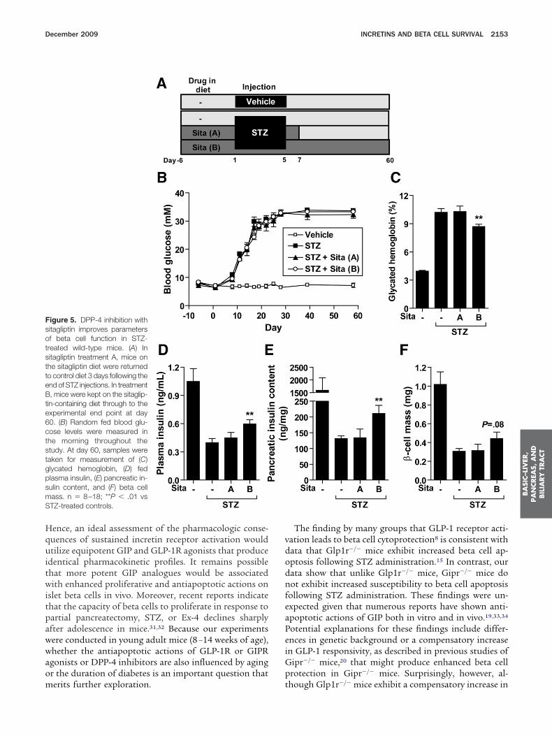
Hquitwitpawwaom
vdodnfeaPeiGp
FsotstteBte6ctstgpsmS
BA
SIC–L
IVER
,PA
NCREA
S,A
ND
BIL
IARY
TRA
CT
December 2009 INCRETINS AND BETA CELL SURVIVAL 2153
ence, an ideal assessment of the pharmacologic conse-uences of sustained incretin receptor activation wouldtilize equipotent GIP and GLP-1R agonists that produce
dentical pharmacokinetic profiles. It remains possiblehat more potent GIP analogues would be associatedith enhanced proliferative and antiapoptotic actions on
slet beta cells in vivo. Moreover, recent reports indicatehat the capacity of beta cells to proliferate in response toartial pancreatectomy, STZ, or Ex-4 declines sharplyfter adolescence in mice.31,32 Because our experimentsere conducted in young adult mice (8 –14 weeks of age),hether the antiapoptotic actions of GLP-1R or GIPRgonists or DPP-4 inhibitors are also influenced by agingr the duration of diabetes is an important question that
igure 5. DPP-4 inhibition withitagliptin improves parametersf beta cell function in STZ-reated wild-type mice. (A) Initagliptin treatment A, mice onhe sitagliptin diet were returnedo control diet 3 days following thend of STZ injections. In treatment, mice were kept on the sitaglip-
in-containing diet through to thexperimental end point at day0. (B) Random fed blood glu-ose levels were measured inhe morning throughout thetudy. At day 60, samples wereaken for measurement of (C)lycated hemoglobin, (D) fedlasma insulin, (E) pancreatic in-ulin content, and (F) beta cellass. n � 8–18; **P � .01 vsTZ-treated controls.
erits further exploration. t
The finding by many groups that GLP-1 receptor acti-ation leads to beta cell cytoprotection8 is consistent withata that Glp1r�/� mice exhibit increased beta cell ap-ptosis following STZ administration.15 In contrast, ourata show that unlike Glp1r�/� mice, Gipr�/� mice doot exhibit increased susceptibility to beta cell apoptosis
ollowing STZ administration. These findings were un-xpected given that numerous reports have shown anti-poptotic actions of GIP both in vitro and in vivo.19,33,34
otential explanations for these findings include differ-nces in genetic background or a compensatory increasen GLP-1 responsivity, as described in previous studies of
ipr�/� mice,20 that might produce enhanced beta cellrotection in Gipr�/� mice. Surprisingly, however, al-
hough Glp1r�/� mice exhibit a compensatory increase in
pab
oaGsTooovcoGfdmrtc
tuiletitg
FwocdSiai
BA
SIC–LIV
ER,
PA
NCREA
S,A
ND
BILIA
RY
TRA
CT
2154 MAIDA ET AL GASTROENTEROLOGY Vol. 137, No. 6
lasma levels of GIP and enhanced GIP insulinotropicctivity,35 these mice nonetheless remain more suscepti-le to STZ-induced diabetes.15
igure 6. DPP-4 inhibition reduces levels of islet apoptosis in STZ-treatedild-type mice but not in mice lacking incretin receptors. Separate cohortsf (A) wild-type and (B) DIRKO (Glp1r�/� Gipr�/�) mice were fed either aontrol diet or the same diet containing sitagliptin for 7 days before anduring STZ (A and B, top). Mice were killed �24 hours following the finalTZ injection for quantification of levels of cleaved caspase-3 immunopos-
tivity in beta cells (n � 5) (A and B, bottom). For quantification of isletpoptosis in normal mice, the group of mice on control diet in A was
njected with citrate buffer vehicle. *P � .05 vs STZ-treated controls.
Although levels of RNA transcripts for the majorityf cytoprotective molecules and components of thepoptotic machinery were similar in Gipr�/� versuslp1r�/� islets, levels of the EGFR and Irs-2 were
ignificantly lower in Glp1r�/� islets (Figure 8A–C).hese findings may partially explain the enhanced ap-ptotic susceptibility to apoptosis and extend previousbservations showing that GLP-1 may exert its actionsn the islet beta cell in part through EGFR transacti-ation.34,36,37 Moreover, Irs-2 has been shown to beritical for the cytoprotective and regenerative effectsf Ex-4,38 and we recently showed that Ex-4, but not D-IP, increased levels of pancreatic mRNA transcripts
or EGFR and Irs-2 in nondiabetic mice fed a high-fatiet.22 Taken together, our findings in STZ-treatedice imply that the structurally related GLP-1 and GIP
eceptors exhibit significant differences in their abilityo engage downstream molecules important for betaell survival and regeneration.
Because DPP-4 inhibitors (sitagliptin and vildaglip-in) and GLP-1R agonists (Ex-4 and liraglutide) aresed to treat type 2 diabetes mellitus, there is active
nterest in understanding whether these agents regu-ate beta cell protection and/or regeneration. Consid-rable data show antiapoptotic and proliferative ac-ions of GLP-1R agonists8,39; however, there is lessnformation on cytoprotective or regenerative proper-ies of DPP-4 inhibitors.22,40 – 44 Although random fedlycemia was not improved by sitagliptin in STZ-
Figure 7. Gipr�/� and Gipr�/�
mice exhibit similar susceptibilityto STZ-induced diabetes andbeta cell injury. (A) Fed blood glu-cose levels were monitored dur-ing and after 5 consecutive daysof STZ administration (50 mg ·kg�1 · day�1 intraperitoneally) asindicated by the arrows (n �7–16). (B) AUC for fed blood glu-cose was calculated for days16–28. At day 30, mice werekilled for determination of (C) fedplasma insulin level, (D) beta cellmass (n � 7–15), and (E) pancre-atic insulin content (n � 7–11).(F) Levels of islet apoptosis werequantified in pancreatic sectionsfrom a cohort of mice killed atday 6, �24 hours after the lastinjection of STZ (n � 5–7 mice).Cleaved caspase-3 positivitywas quantified in a minimum of30 islets per mouse pancreas.
***P � .001.
twcstimcwtt
osietocppitssp
aG1
1
1
1
1
1
Flsl 90. n
BA
SIC–L
IVER
,PA
NCREA
S,A
ND
BIL
IARY
TRA
CT
December 2009 INCRETINS AND BETA CELL SURVIVAL 2155
reated mice, circulating levels of glycated hemoglobinere reduced and pancreatic insulin content and beta
ell area and pancreatic IGF-1 and Akt mRNA tran-cripts were increased following sitagliptin administra-ion. Hence, even modest increases in levels of intactncretin hormones may enhance beta cell survival in
ice. Moreover, sitagliptin reduced the extent ofaspase-3 activation following STZ administration inild-type but not in DIRKO mice, showing that incre-
in receptors are essential transducers of the antiapop-otic actions of DPP-4 inhibitors.
In summary, our studies provide new informationn the importance of basal GIPR action for beta cellurvival and on how different mechanisms for enhanc-ng incretin receptor activation impact beta cell regen-ration in mice. Moreover, unlike the importance ofhe GLP-1 receptor for beta cell function, eliminationf endogenous GIPR signaling does not modify sus-eptibility to beta cell injury and is not associated witherturbation in levels of key signaling molecules im-ortant for beta cell growth and survival. These find-
ngs extend our understanding of the relative impor-ance and mechanisms of incretin action for beta cellurvival and may have implications for strategies de-igned to optimize beta cell growth or survival inatients with type 2 diabetes mellitus.
Supplementary Data
Note: To access the supplementary materialccompanying this article visit the online version ofastroenterology at www.gastrojournal.org, and at doi:0.1053/j.gastro.2009.09.004.
References
1. Drucker DJ. The role of gut hormones in glucose homeostasis.
igure 8. Islets from Glp-1r�/� but not Gipr�/� mice are deficient in EGittermates for either real-time polymerase chain reaction assessmenturvival. Levels of each transcript were normalized to the internal cont
evels were quantified by densitometry and normalized to levels of Hsp
J Clin Invest 2007;117:24–32.
2. Mayo KE, Miller LJ, Bataille D, et al. International Union of Phar-macology. XXXV. The glucagon receptor family. Pharmacol Rev2003;55:167–194.
3. Hansotia T, Maida A, Flock G, et al. Extrapancreatic incretinreceptors modulate glucose homeostasis, body weight, and en-ergy expenditure. J Clin Invest 2007;117:143–152.
4. Kim SJ, Nian C, McIntosh CH. Resistin is a key mediator ofglucose-dependent insulinotropic polypeptide (GIP) stimulation oflipoprotein lipase (LPL) activity in adipocytes. J Biol Chem 2007;282:34139–34147.
5. Miyawaki K, Yamada Y, Ban N, et al. Inhibition of gastric inhibitorypolypeptide signaling prevents obesity. Nat Med 2002;8:738–742.
6. McClean PL, Irwin N, Cassidy RS, et al. GIP receptor antagonismreverses obesity, insulin resistance and associated metabolicdisturbances induced in mice by prolonged consumption of highfat diet. Am J Physiol Endocrinol Metab 2007;293:E1746–E1755.
7. Deacon CF. Therapeutic strategies based on glucagon-like pep-tide 1. Diabetes 2004;53:2181–2189.
8. Drucker DJ. The biology of incretin hormones. Cell Metab 2006;3:153–165.
9. Holz GGt, Kuhtreiber WM, Habener JF. Pancreatic beta-cells arerendered glucose-competent by the insulinotropic hormone glu-cagon-like peptide-1(7-37). Nature 1993;361:362–365.
0. Stoffers DA, Kieffer TJ, Hussain MA, et al. Insulinotropic gluca-gon-like peptide-1 agonists stimulate expression of homeodo-main protein IDX-1 and increase b-cell mass in mouse pancreas.Diabetes 2000;49:741–748.
1. Xu G, Stoffers DA, Habener JF, et al. Exendin-4 stimulates bothbeta-cell replication and neogenesis, resulting in increased beta-cell mass and improved glucose tolerance in diabetic rats. Dia-betes 1999;48:2270–2276.
2. Farilla L, Hui H, Bertolotto C, et al. Glucagon-like peptide-1 pro-motes islet cell growth and inhibits apoptosis in Zucker diabeticrats. Endocrinology 2002;143:4397–4408.
3. Farilla L, Bulotta A, Hirshberg B, et al. Glucagon-like peptide 1inhibits cell apoptosis and improves glucose responsiveness offreshly isolated human islets. Endocrinology 2003;144:5149–5158.
4. Wang Q, Brubaker PL. Glucagon-like peptide-1 treatment delaysthe onset of diabetes in 8 week-old db/db mice. Diabetologia
nd IRS-2. Islets were isolated from Gipr�/� and Glp1r�/� and wild-typesal levels of (A and B) transcripts or (C) proteins involved in beta cellptidyl-propyl isomerase A (PPIA, also known as cyclophilin). Proteins� 7–8 mice per genotype. *P � .05.
FR aof barol pe
2002;45:1263–1273.

1
1
1
1
1
2
2
2
2
2
2
2
2
2
2
3
3
3
3
3
3
3
3
3
3
4
4
4
4
4
R
SSd
A
i
C
BA
SIC–LIV
ER,
PA
NCREA
S,A
ND
BILIA
RY
TRA
CT
2156 MAIDA ET AL GASTROENTEROLOGY Vol. 137, No. 6
5. Li Y, Hansotia T, Yusta B, et al. Glucagon-like peptide-1 receptorsignaling modulates beta cell apoptosis. J Biol Chem 2003;278:471–478.
6. Ling Z, Wu D, Zambre Y, et al. Glucagon-like peptide 1 receptorsignaling influences topography of islet cells in mice. VirchowsArch 2001;438:382–387.
7. Trumper A, Trumper K, Horsch D. Mechanisms of mitogenic andanti-apoptotic signaling by glucose-dependent insulinotropicpolypeptide in beta(INS-1)-cells. J Endocrinol 2002;174:233–246.
8. Trumper A, Trumper K, Trusheim H, et al. Glucose-dependentinsulinotropic polypeptide is a growth factor for beta (INS-1)cells by pleiotropic signaling. Mol Endocrinol 2001;15:1559–1570.
9. Kim SJ, Winter K, Nian C, et al. GIP stimulation of pancreaticbeta-cell survival is dependent upon phosphatidylinositol 3-ki-nase (PI3-K)/ protein kinase B (PKB) signaling, inactivation of theforkhead transcription factor Foxo1 and downregulation of baxexpression. J Biol Chem 2005;280:22297–22307.
0. Pamir N, Lynn FC, Buchan AM, et al. Glucose-dependent insuli-notropic polypeptide receptor null mice exhibit compensatorychanges in the enteroinsular axis. Am J Physiol Endocrinol Metab2003;284:E931–E939.
1. Hansotia T, Baggio LL, Delmeire D, et al. Double incretin receptorknockout (DIRKO) mice reveal an essential role for the enteroin-sular axis in transducing the glucoregulatory actions of DPP-IVinhibitors. Diabetes 2004;53:1326–1335.
2. Lamont BJ, Drucker DJ. Differential anti-diabetic efficacy of incre-tin agonists vs. DPP-4 inhibition in high fat fed mice. Diabetes2008;57:190–198.
3. Maida A, Lovshin JA, Baggio LL, et al. The glucagon-like peptide-1receptor agonist oxyntomodulin enhances {beta}-cell function butdoes not inhibit gastric emptying in mice. Endocrinology 2008;149:5670–5678.
4. Villhauer EB, Brinkman JA, Naderi GB, et al. 1-[[(3-hydroxy-1-adamantyl)amino]acetyl]-2-cyano-(S)-pyrrolidine: a potent, selec-tive, and orally bioavailable dipeptidyl peptidase IV inhibitor withantihyperglycemic properties. J Med Chem 2003;46:2774–2789.
5. Hinke SA, Gelling RW, Pederson RA, et al. Dipeptidyl peptidaseIV-resistant [D-Ala(2)]glucose-dependent insulinotropic polypep-tide (GIP) improves glucose tolerance in normal and obese dia-betic rats. Diabetes 2002;51:652–661.
6. Kuhn-Wache K, Manhart S, Hoffmann T, et al. Analogs ofglucose-dependent insulinotropic polypeptide with increaseddipeptidyl peptidase IV resistance. Adv Exp Med Biol 2000;477:187–195.
7. Pieper AA, Brat DJ, Krug DK, et al. Poly(ADP-ribose) polymerase-deficient mice are protected from streptozotocin-induced diabe-tes. Proc Natl Acad Sci U S A 1999;96:3059–3064.
8. Rhodes CJ. Type 2 diabetes—a matter of beta-cell life anddeath? Science 2005;307:380–384.
9. Vilsboll T, Krarup T, Madsbad S, et al. Defective amplification ofthe late phase insulin response to glucose by GIP in obese TypeII diabetic patients. Diabetologia 2002;45:1111–1119.
0. Vilsboll T, Knop FK, Krarup T, et al. The pathophysiology ofdiabetes involves a defective amplification of the late-phaseinsulin response to glucose by glucose-dependent insulinotropicpolypeptide-regardless of etiology and phenotype. J Clin Endocri-nol Metab 2003;88:4897–4903.
1. Rankin MM, Kushner JA. Adaptive beta cell proliferation is se-verely restricted with advanced age. Diabetes 2009;58:1365–1372.
2. Tschen SI, Dhawan S, Gurlo T, et al. Age-dependent decline inbeta cell proliferation restricts the capacity of beta cell regener-
ation in mice. Diabetes 2009;58:1312–1320. a3. Kim SJ, Nian C, Widenmaier S, et al. Glucose-dependent insuli-notropic polypeptide-mediated up-regulation of beta-cell anti-apoptotic Bcl-2 gene expression is coordinated by cyclic AMP(cAMP) response element binding protein (CREB) and cAMP-responsive CREB coactivator 2. Mol Cell Biol 2008;28:1644–1656.
4. Ehses JA, Casilla VR, Doty T, et al. Glucose-dependent insulino-tropic polypeptide promotes beta-(INS-1) cell survival via cyclicadenosine monophosphate-mediated caspase-3 inhibition andregulation of p38 mitogen-activated protein kinase. Endocrinol-ogy 2003;144:4433–4445.
5. Pederson RA, Satkunarajah M, McIntosh CH, et al. Enhancedglucose-dependent insulinotropic polypeptide secretion and insu-linotropic action in glucagon-like peptide 1 receptor �/� mice.Diabetes 1998;47:1046–1052.
6. Buteau J, Foisy S, Joly E, et al. Glucagon-like peptide 1 inducespancreatic beta-cell proliferation via transactivation of the epi-dermal growth factor receptor. Diabetes 2003;52:124–132.
7. MacDonald PE, Wang X, Xia F, et al. Antagonism of rat beta-cellvoltage-dependent K� currents by exendin 4 requires dual acti-vation of the cAMP/protein kinase A and phosphatidylinositol3-kinase signaling pathways. J Biol Chem 2003;278:52446–52453.
8. Park S, Dong X, Fisher TL, et al. Exendin-4 uses Irs2 signaling tomediate pancreatic beta cell growth and function. J Biol Chem2006;281:1159–1168.
9. Brubaker PL, Drucker DJ. Glucagon-like peptides regulate cellproliferation and apoptosis in the pancreas, gut and centralnervous system. Endocrinology 2004;145:2653–2659.
0. Ahren B, Winzell MS, Wierup N, et al. DPP-4 inhibition improvesglucose tolerance and increases insulin and GLP-1 responses togastric glucose in association with normalized islet topography inmice with beta-cell-specific overexpression of human islet amy-loid polypeptide. Regul Pept 2007;143:97–103.
1. Mu J, Woods J, Zhou YP, et al. Chronic inhibition of dipeptidylpeptidase-4 with a sitagliptin analog preserves pancreatic �-cellmass and function in a rodent model of type 2 diabetes. Diabetes2006;55:1695–1704.
2. Flock G, Baggio LL, Longuet C, et al. Incretin receptors for gluca-gon-like peptide 1 and glucose-dependent insulinotropic polypep-tide are essential for the sustained metabolic actions of vilda-gliptin in mice. Diabetes 2007;56:3006–3013.
3. Kim SJ, Nian C, Doudet DJ, et al. Inhibition of dipeptidyl pepti-dase IV with sitagliptin (MK0431) prolongs islet graft survival instreptozotocin-induced diabetic mice. Diabetes 2008;57:1331–1339.
4. Pospisilik JA, Martin J, Doty T, et al. Dipeptidyl peptidase IVinhibitor treatment stimulates beta-cell survival and islet neogen-esis in streptozotocin-induced diabetic rats. Diabetes 2003;52:741–750.
Received June 18, 2009. Accepted September 2, 2009.
eprint requestsAddress requests for reprints to: Daniel J. Drucker, MD, Mount
inai Hospital, Samuel Lunenfeld Research Institute, 60 Murraytreet, Mailbox 39, Toronto, Ontario, Canada M5T 3L9. e-mail:[email protected]; fax: (416) 361-2669.
cknowledgmentsThe authors thank Xiemin Cao for technical assistance with islet
solations.
onflicts of interestThe authors disclose the following: Dr Drucker has served as an
dvisor or consultant within the past 12 months to Amylin

PPGPNTmT
F
AHsFH
December 2009 INCRETINS AND BETA CELL SURVIVAL 2157
harmaceuticals, Arena Pharmaceuticals Inc, Arisaphharmaceuticals Inc, Eli Lilly and Company, GlaxoSmithKline,lenmark Pharmaceuticals, Hoffman-LaRoche Inc, Isisharmaceuticals Inc, Merck Research Laboratories, Metabolex Inc,ovartis Pharmaceuticals, Novo Nordisk Inc, Phenomix Inc, andransition Pharmaceuticals Inc. Neither Dr Drucker nor his familyembers hold stock directly or indirectly in any of these companies.
he remaining authors disclose no conflicts. C
undingA.M. was supported by funding from a Canadian Diabetes
ssociation Doctoral Research Award and a Canadian Institutes ofealth Research graduate scholarship. These studies wereupported in part by a grant from the Juvenile Diabetes Researchoundation (JDRF #1-2006-796) and from the Canadian Institutes ofealth Research MOP 82700. D.J.D. was supported in part by the
anada Research Chairs Program.BA
SIC–L
IVER
,PA
NCREA
S,A
ND
BIL
IARY
TRA
CT