DIFFERENCES IN FEMORAL BONE MICROSTRUCTURE BETWEEN … · bone modeling in 2.5 month-old transgenic...
4
INTRODUCTION In general, transgenic rabbits represent an alternative way to produce recombinant human proteins and enzymes. However, several problems are commonly encountered with their production, including transgene integration, stability and expression. Our previous study revealed a presence of new bone tissue type in rabbits - fibrolamellar bone tissue - in 2.5 month-old transgenic individuals with hFVIII gene (Martiniaková et al., 2005a). According to Currey (2002) fibrolamellar bone tissue is found particularly in large mammals whose bones have to grow in diameter rather quickly. It has been reported in Canis (dog), Ovis (sheep), Sus (pig), Bison (buffalo), and Bos (cattle), and also in fossil bones including Pharahippus blackburgi (primitive horse), Kannemeyeria (herbivorous mammal-like reptiles), and Brachiosaurus and Plateosaurus (extinct herbivorous Short communication DIFFERENCES IN FEMORAL BONE MICROSTRUCTURE BETWEEN 2 MONTH-OLD TRANSGENIC AND NON-TRANSGENIC RABBITS M. MARTINIAKOVÁ 1 , R. OMELKA 2 , V. SMOLÁRIKOVÁ 2 , B. GROSSKOPF 3 , P. CHRENEK 1, 4 1 Department of Zoology and Anthropology, 2 Department of Botany and Genetics, Constantine the Philosopher University, Nitra, Slovakia; 3 Institute of Zoology and Anthropology, Georg-August University, Göttingen, Germany; 4 Slovak Agricultural Research Centre, Nitra, Slovakia ABSTRACT Differences in microscopic structure of the femur between 2 month-old transgenic rabbits with hFVIII gene and the non-transgenic ones were investigated. Qualitative and quantitative histological characteristics of compact bone microstructure were evaluated. We observed the presence of fibrolamellar bone tissue only in transgenic rabbits. Measured values for area, perimeter, maximum and minimum diameter of the primary osteon vascular canals, the Haversian canals and the secondary osteons were higher in transgenic individuals. However, no any significant differences were identified between transgenic and non-transgenic rabbits. Anyway, our results confirmed a presence of new bone tissue type (fibrolamellar bone tissue) in 2 month-old transgenic rabbits. Key words: femur, transgenic rabbit, histology Correspondence: E-mail: [email protected] dinosaurs) (Mori et al., 2005). Also, we observed increased bone modeling in 2.5 month-old transgenic individuals (femoral bone length was higher in transgenic rabbits in comparison with non-transgenic ones) and also better blood supplying (area, perimeter, minimum diameter of the Haversian canals were higher in transgenic rabbits) (Martiniaková et al., 2006). It signals above mentioned results brought some new questions about microstructural differences in compact bone tissue between non-transgenic and transgenic rabbits. One of the most important is which age of rabbits the differences already appear at. Therefore, the aim of present study was to compare femoral bone microstructure in younger (2 month-old) non-transgenic in younger (2 month-old) non-transgenic versus transgenic rabbits with the hFVIII gene. We tried to find out if some changes in histological structure of the femora are also present in these rabbits. ISSN 1335-3686 20 Slovak J. Anim. Sci., 41, 2008 (1): 20 - 23 © 2008 SARC Received: October 30, 2007; Accepted: January 10, 2008
Transcript of DIFFERENCES IN FEMORAL BONE MICROSTRUCTURE BETWEEN … · bone modeling in 2.5 month-old transgenic...

INTRODUCTION
In general, transgenic rabbits represent an alternative way to produce recombinant human proteins and enzymes. However, several problems are commonly encountered with their production, including transgene integration, stability and expression. Our previous study revealed a presence of new bone tissue type in rabbits - fibrolamellar bone tissue - in 2.5 month-old transgenic individuals with hFVIII gene (Martiniaková et al., 2005a). According to Currey (2002) fibrolamellar bone tissue is found particularly in large mammals whose bones have to grow in diameter rather quickly. It has been reported in Canis (dog), Ovis (sheep), Sus (pig), Bison (buffalo), and Bos (cattle), and also in fossil bones including Pharahippus blackburgi (primitive horse), Kannemeyeria (herbivorous mammal-like reptiles), and Brachiosaurus and Plateosaurus (extinct herbivorous
Short communication
DIFFERENCES IN FEMORAL BONE MICROSTRUCTURE BETWEEN 2 MONTH-OLD TRANSGENIC AND NON-TRANSGENIC RABBITS
M. MArtInIAkOVá1, r. OMelkA2, V. SMOlárIkOVá2, B. GrOSSkOpF3, p. CHrenek1, 4
1Department of Zoology and Anthropology, 2Department of Botany and Genetics, Constantine the philosopher University, nitra, Slovakia; 3Institute of Zoology and Anthropology, Georg-August University, Göttingen, Germany; 4Slovak Agricultural research Centre, nitra, Slovakia
ABSTRACT
Differences in microscopic structure of the femur between 2 month-old transgenic rabbits with hFVIII gene and the non-transgenic ones were investigated. Qualitative and quantitative histological characteristics of compact bone microstructure were evaluated. We observed the presence of fibrolamellar bone tissue only in transgenic rabbits. Measured values for area, perimeter, maximum and minimum diameter of the primary osteon vascular canals, the Haversian canals and the secondary osteons were higher in transgenic individuals. However, no any significant differences were identified between transgenic and non-transgenic rabbits. Anyway, our results confirmed a presence of new bone tissue type (fibrolamellar bone tissue) in 2 month-old transgenic rabbits.
Key words: femur, transgenic rabbit, histology
Correspondence: e-mail: [email protected]
dinosaurs) (Mori et al., 2005). Also, we observed increased bone modeling in 2.5 month-old transgenic individuals (femoral bone length was higher in transgenic rabbits in comparison with non-transgenic ones) and also better blood supplying (area, perimeter, minimum diameter of the Haversian canals were higher in transgenic rabbits) (Martiniaková et al., 2006).
It signals above mentioned results brought some new questions about microstructural differences in compact bone tissue between non-transgenic and transgenic rabbits. One of the most important is which age of rabbits the differences already appear at. therefore, the aim of present study was to compare femoral bone microstructure in younger (2 month-old) non-transgenic in younger (2 month-old) non-transgenic versus transgenic rabbits with the hFVIII gene. We tried to find out if some changes in histological structure of the femora are also present in these rabbits.
ISSn 1335-368620
Slovak J. Anim. Sci., 41, 2008 (1): 20 - 23© 2008 SARC
received: October 30, 2007; Accepted: January 10, 2008
21
Slovak J. Anim. Sci., 41, 2008, 1
MATERIAL AND METHODS
In our experiments, fourteen clinically healthy 2 month-old rabbits were analyzed. the animals were obtained from an experimental farm of the Slovak Agricultural research Centre in nitra (Slovakia). they were housed in individual flat-deck wire cages, under a constant photoperiod of 14 h of day-light. the temperature and humidity of the building were recorded continually by means of a thermograph positioned at the same level as the cages. the rabbits were fed ad libitum with a commercial diet and water was provided ad libitum with nipple drinkers. the breeding conditions were similar to intensive industrial conditions. We used new Zealand White transgenic rabbits with the human blood clotting factor VIII gene (n=7) and the non-transgenic ones (n=7). the transgene was under transcriptional control of the whey acidic protein (WAp) promoter. transgenic versus non-transgenic individuals were identified by PCR method. total DnA was isolated from the ear issue of newborn rabbits. Conditions for PCR amplification of the hFVIII transgene were the same as reported by Chrenek et al. (2005), using primers hFVIII-F: 5’-GtA GAC AGC tGt CCA GAG GAA-3’ and hFVIII-r: 5’-GAt CtG ATT TAG TTG GCC CAT-3’, which define a 578 bp region of human FVIII cDnA.
With histological analysis, fourteen right femurs were studied. each of the bones was sectioned at the midshaft of its diaphysis. the obtained segments were macerated and degreased (Martiniaková et al., 2005b).(Martiniaková et al., 2005b). later the samples were embedded in epoxy resin Biodur (Günter von Hagens, Heidelberg, Germany). transverse thin sections (70 - 80 μm) were prepared with a sawing
microtome (Leitz 1600, Wetzlar, Germany) and affixed to glass slides with eukitt (Merck, Darmstadt, Germany). the qualitative histological characteristics of the bone tissue were determined according to the classification systems by enlow and Brown (1�56) and ricqles et al.(1�56) and ricqles et al.and ricqles et al. (1��1); the quantitative characteristics were assessedthe quantitative characteristics were assessed using the computer software Scion Image (Scion Corporation, Maryland, USA) in anterior, posterior,in anterior, posterior, medial and lateral views of thin section. Osteon size. Osteon size can vary in the views mentioned above (Skedros et al.,(Skedros et al., 2007), therefore we had to measure area, perimeter, andarea, perimeter, and the minimum and maximum diameter of primary osteon vascular canals, Haversian canals and secondary osteons in all views of thin section in order to lose difference among individuals in statistics. the t-test was used to distinguish differences in quantitative histological characteristics between transgenic and non-transgenic individuals (Statistica 4.3, 1��3).
RESULTS AND DISCUSSION
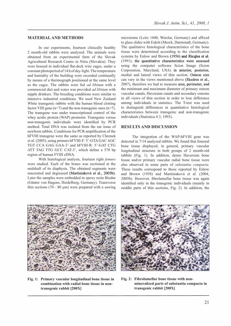
the integration of the WAp-hFVIII gene was detected in 7/14 analysed rabbits. We found that femoral bone tissue displayed, in general, primary vascular longitudinal structure in both groups of 2 month-old rabbits (Fig. 1). In addition, dense Haversian bone tissue and/or primary vascular radial bone tissue were also observed in some parts of substantia compacta. these results correspond to those reported by enlow and Brown (1�58) and Martiniaková et al. (2004, 2005b). However, fibrolamellar bone tissue was again identified only in the transgenic individuals (mainly in middle parts of thin sections, Fig. 2). In addition, the
Fig. 1: Primary vascular longitudinal bone tissue in combination with radial bone tissue in non-transgenic rabbit [200X]
Fig. 2: Fibrolamellar bone tissue with non-mineralized parts of substantia compacta in transgenic rabbit [200X]

22
Slovak J. Anim. Sci., 41, 2008, 1
fibrolamellar tissue possessed more non-mineralized parts of substantia compacta as compared to 2.5 month-old transgenic rabbits. this fact indicates that the process of mineralization might be considerably reduced in 2 month-old transgenic individuals.
In general, less number of secondary osteons was identified in 2 month-old rabbits in comparison with 2.5 month-old ones. We suppose this fact might be caused by different age of individuals. rajtová and Globočník (1978), Martiniaková et al. (2005b) mention that circumferential lamellae are substituted by the secondary osteons with increased age in mice and rabbits, respectively. Besides, Currey (2002) notes that the formation of secondary osteons tends to lead to the production of more secondary osteons during life of the individual. therefore, we observed more secondary osteons in 2.5 month-old rabbits.
All of 560 vascular canals of primary osteons, 162 Haversian canals and 162 secondary osteons of transgenic and non-transgenic rabbits were measured. the results are summarized in table 1. We found that almost all variables of the primary osteon vascular canals,
the Haversian canals and the secondary osteons disposed higher values in the transgenic individuals. However, no any significant differences were observed between 2 month-old transgenic and non-transgenic rabbits. On the other hand, we identified higher values of the Haversian canals’ variables in 2.5 month-old transgenic individuals in our previous study (Martiniaková et al., 2006) and the differences were statistically significant. We suppose that this phenomenon could be caused by a localization of many non-mineralized parts of the substantia compacta in fibrolamellar bone tissue of the transgenic rabbits. For this aim a mineralization process is slightly reduced and skeletal responses achieved through the bone modeling and remodeling are not so much increased as expected.
In conclusion, changes in femoral bone microstructure in 2 month-old transgenic versus non-transgenic rabbits were identified. Evident changes mainly in qualitative histological characteristics result especially into reduced mineralization process and bone modeling in the transgenic individuals. Although it is difficult to explain these changes, since genetic and environmental factors are taken into account, our results indicate that
Table 1: The results of quantitative histological analysis of the femurs from investigated rabbits
rabbits measured structures n variables x ± SD v (%)
transgenic vascular canals of primary osteons 280
area (μm2) 18�.58±34.46 18.18perimeter (μm) 41.86±4.08 �.74max. diameter (μm) 1�.34±2.�3 15.14min. diameter (μm) 6.28±0.�3 14.7�
non-transgenic vascular canals of primary osteons 280
area (μm2) 188.2�±42.24 22.43perimeter (μm) 41.53±4.�1 11.82max. diameter (μm) 1�.21±3.25 16.�4min. diameter (μm) 6.25±1.1� 17.73
transgenicHaversian canals 78
area (μm2) 356.75±68.46 1�.1�perimeter (μm) 55.7�±5.47 �.80max. diameter (μm) 25.32±3.10 12.25min. diameter (μm) 8.�5±1.20 13.43
non-transgenic Haversian canals 84
area (μm2) 34�.24±83.�8 24.05perimeter (μm) 54.53±6.86 12.5�max. diameter (μm) 24.55±4.34 17.66min. diameter (μm) �.04±1.22 13.45
transgenic secondary osteons 78
area (μm2) 827�.63±1234.74 14.�1perimeter (μm) 264.17±28.78 10.8�max. diameter (μm) 123.63±20.43 16.52min. diameter (μm) 44.85±8.73 1�.46
non-transgenic secondary osteons 84
area (μm2) 8124.45±114�.13 14.14perimeter (μm) 262.67±33.18 12.63max. diameter (μm) 122.60±25.25 20.60min. diameter (μm) 44.�3±8.48 18.88
n – number of measured structures, x – arithmetic mean, SD – standard deviation, v – coefficient of variation

23
Slovak J. Anim. Sci., 41, 2008, 1
they could be caused by genetic manipulations. In order to obtain more reliable results in this direction, more individuals have to be examined. It is a subject of current research. With the information about confirmed changes in histological structure of the rabbit femur for both transgenic and non-transgenic individuals we would be able to consider more precise effects of environmental and genetic factors on the bone tissue formation in several age groups of rabbits.
ACKNOWLEDGEMENT
this study was supported by the grant CGA VI/10/2007.
REFERENCES
CHrenek, p. – VASICek, D. – MAkAreVICH, A. – JUrCIk, r. - SUVeGOVA, k. - pArkAnYI, V. - BAUer, M. – rAFAY, J. – BAtOrOVA, A. – pAleYAnDA, r. K. 2005. Increased transgene integration efficiency upon microinjection of DnA into both pronuclei of rabbit embryos. In: Transgenic Res., vol. 14, 2005, p. 417-428.
CUrreY, J. D. 2002. Bones: Structure and Mechanics, 1st edition. new Jersey : princeton University press, 2002.
enlOW, D. H. – BrOWn, S. O. 1�56. A comparative histological study of fossil and recent bone tissues. part I. In: Texas J. Sci.,vol. 8, 1�56, p. 405-412.
enlOW, D. H. – BrOWn, S. O. 1�58. A comparative histological study of fossil and recent bone tissues. part III. In: Texas J. Sci., vol. 10, 1�58, p. 187-230.
MArtInIAkOVá, M. – CHrenek, p. – VOnDrákOVá, M. – OMelkA, r. – FABIŠ, M. 2004. ComparativeComparative study of rabbits and pigs compact bone tissue microscopic structure. In: J. Farm Anim. Sci., vol. 37, 2004, p. 4�-55.
MArtInIAkOVá, M. – OMelkA, r. – CHrenek, p.– OMelkA, r. – CHrenek, p. OMelkA, r. – CHrenek, p.– CHrenek, p. CHrenek, p. - RYBAN, Ľ. – PARK�NYI, �. – GR�SSK�PF, B. –– pArkánYI, V. – GrOSSkOpF, B. – pArkánYI, V. – GrOSSkOpF, B. –– GrOSSkOpF, B. – GrOSSkOpF, B. ––VOnDrákOVá, M. – BAUerOVá, M. 2005a. Changes– BAUerOVá, M. 2005a. Changes BAUerOVá, M. 2005a. Changes of femoral bone tissue microstructure in transgenic rabbits. In: Folia Biologica (Praha), vol. 51, 2005a, p. 140-144.
MArtInIAkOVá, M. – OMelkA, r. v CHrenek, p.– OMelkA, r. v CHrenek, p. OMelkA, r. v CHrenek, p. – VOnDrákOVá, M. – BAUerOVá, M. 2005b. Age- VOnDrákOVá, M. – BAUerOVá, M. 2005b. Age-– BAUerOVá, M. 2005b. Age- BAUerOVá, M. 2005b. Age-related changes in histological structure of the femur in juvenile and adult rabbits: a pilot study. In: Bull. Vet. Inst. Pulawy, vol. 4�, 2005b, p. 227-230.
MARTINIAK���, M. – �MELKA, R. – RYBAN, Ľ. –– �MELKA, R. – RYBAN, Ľ. – �MELKA, R. – RYBAN, Ľ. –– RYBAN, Ľ. – RYBAN, Ľ. –– GrOSSkOpF, B. – VOnDrákOVá, M. – BAUerOVá,– VOnDrákOVá, M. – BAUerOVá, VOnDrákOVá, M. – BAUerOVá,– BAUerOVá, BAUerOVá, M. – FABIŠ, M. – CHrenek, p. 2006. Comparative– FABIŠ, M. – CHrenek, p. 2006. Comparative FABIŠ, M. – CHrenek, p. 2006. Comparative– CHrenek, p. 2006. Comparative CHrenek, p. 2006. ComparativeComparative study of compact bone tissue microstructure between non-transgenic and transgenic rabbits with WAp-hFVIII gene construct. In: Anat. Histol. Embryol., vol. 35, 2006, p. 310-315.
MOrI, r. – kODAtA, t. – SOetA, S. – SAtO, J. – kAkInO, J. – HAMAtO, S. – tAkAkI, H. – nAItO, Y. 2005. preliminary study of histological comparison on the growth patterns of long-bone cortex in young calf, pig, and sheep. In: J. Vet. Med. Sci., vol. 67, 2005, p. 1223-122�.
RAJT���, �. – GL�B��N�K, E. 1978. Histologisches– GL�B��N�K, E. 1978. Histologisches GL�B��N�K, E. 1978. HistologischesHistologisches Studium der Alterveränderungen in der Femurcompacta bei labor-und Hausmäusen. In:In: Gegenbaurs. Morph. Jahb., vol. 124, 1�78, p. 64�-662.
rICQleS, A. J. – MeUnIer, F. J. – CAStAnet, J.– MeUnIer, F. J. – CAStAnet, J. MeUnIer, F. J. – CAStAnet, J.– CAStAnet, J. CAStAnet, J. – FrAnCIllOn-VIeIllOt, H. 1��1. Comparative FrAnCIllOn-VIeIllOt, H. 1��1. ComparativeComparative microstructure of bone. In: Hall, B. k.: Bone 3, Bone Matrix and Bone Specific Products. Boca Raton: CRC Press, 1991, p. 1-78.
SkeDrOS, J. G. – SOrenSOn, S. M. – JenSOn, n. H.– SOrenSOn, S. M. – JenSOn, n. H. SOrenSOn, S. M. – JenSOn, n. H.– JenSOn, n. H. JenSOn, n. H. 2007. Are distributions of secondary osteon variants usefulAre distributions of secondary osteon variants useful for interpreting load history in mammalian bones? In: Cells Tissues Organs, vol. 185, 2007, p. 285-307.
StAtIStICA 4.3. 1��3. electronic textbook. StatSoft Inc., 1��3.
Authors’ addresses: Dr. Monika Martiniaková, Department of Zoology and Anthropology, Constantine the Philosopher University, Nábrežie mládeže 91, 949 74 Nitra, Slovakia, phone: 0907 670 199, fax: 037/6408556; Dr. Radoslav �melka, Dr. �ladimíra Smoláriková, Department of Botany and Genetics, Constantine the Philosopher University, Nábrežie mládeže 91, 949 74 Nitra, Slovakia; Dr. Birgit Grosskopf, Institute of Zoology and Anthropology, Georg-August University, Bürgerstrasse 50, 37 073 Göttingen, Germany; Assoc. prof. Dr. peter Chrenek, Slovak Agricultural research Centre, Hlohovská 2, �4� �2 nitra


![A cementless, proximally fixed anatomic femoral stem ...bone interfaces inhibited bone ingrowth when the micro-motion exceeded 150 mm [10,11]. We hypothesized that a cementless, anatomic](https://static.fdocuments.us/doc/165x107/5e63476c137d81362d557525/a-cementless-proximally-fixed-anatomic-femoral-stem-bone-interfaces-inhibited.jpg)