Diameter Bodycorneal burn, vitreous removal/vitrectomy required, corneal injuries, or complications...
23
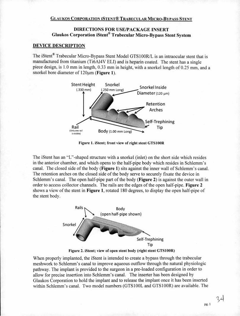
GLAUKOS CORPORATION ISTENTO TRABECULAR MICRO-BYPASS STENT DIRECTIONS FOR USE/PACKAGE INSERT Glaukos Corporation iStent® Trabecular Micro-Bypass Stent System DEVICE DESCRIPTION The iStent® Trabecular Micro-Bypass Stent Model GTS100R/L is an intraocular stent that is manufactured from titanium (Ti6AI4V ELI) and is heparin coated. The stent has a single piece design, is 1.0 mm in length, 0.33 mm in height, with a snorkel length of 0.25 mm, and a snorkel bore diameter of 120pm (Figure 1). Stent Height Snorkel . (.330 mm) (.250 mm Long) Diameter (120 im) Retention Arches Self-Trephining Rail Tip (OnIVoner.11 Body (1.00 mm Long) IlsiU.) Figure 1. iStent; front view of right stent GTS100R The iStent has an "L"-shaped structure with a snorkel (inlet) on the short side which resides in the anterior chamber, and which opens to the half-pipe body which resides in Schlemm's canal. The closed side of the body (Figure 1) sits against the inner wall of Schlemm's canal. The retention arches on the closed side of the body serve to securely fixate the device in Schlemm's canal. The open half-pipe part of the body (Figure 2) is against the outer wall in order to access collector channels. The rails are the edges of the open half-pipe. Figure 2 shows a view of the stent in Figure 1, rotated 180 degrees, to display the open half-pipe of the stent body. Rails Body (open half-pipe shown) Snorkel' Self-Trephining Tip Figure 2. iStent; view of open stent body (right stent GTS100R) When properly implanted, the iStent is intended to create a bypass through the trabecular meshwork to Schlemm's canal to improve aqueous outflow through the natural physiologic pathway. The implant is provided to the surgeon in a pre-loaded configuration in order to allow for precise insertion into Schlemm's canal. The inserter has been designed by Glaukos Corporation to hold the implant and to release the implant once it has been inserted within Schlemm's canal. Two model numbers (GTS100L and GTSIOOR) are available. The pg.1
Transcript of Diameter Bodycorneal burn, vitreous removal/vitrectomy required, corneal injuries, or complications...
GLAUKOS CORPORATION ISTENTO TRABECULAR MICRO-BYPASS STENT
DIRECTIONS FOR USE/PACKAGE INSERTGlaukos Corporation iStent® Trabecular Micro-Bypass Stent System
DEVICE DESCRIPTION
The iStent® Trabecular Micro-Bypass Stent Model GTS100R/L is an intraocular stent that ismanufactured from titanium (Ti6AI4V ELI) and is heparin coated. The stent has a singlepiece design, is 1.0 mm in length, 0.33 mm in height, with a snorkel length of 0.25 mm, and asnorkel bore diameter of 120pm (Figure 1).
Stent Height Snorkel .(.330 mm) (.250 mm Long)
Diameter (120 im)
RetentionArches
Self-TrephiningRail Tip
(OnIVoner.11 Body (1.00 mm Long)IlsiU.)
Figure 1. iStent; front view of right stent GTS100R
The iStent has an "L"-shaped structure with a snorkel (inlet) on the short side which residesin the anterior chamber, and which opens to the half-pipe body which resides in Schlemm'scanal. The closed side of the body (Figure 1) sits against the inner wall of Schlemm's canal.The retention arches on the closed side of the body serve to securely fixate the device inSchlemm's canal. The open half-pipe part of the body (Figure 2) is against the outer wall inorder to access collector channels. The rails are the edges of the open half-pipe. Figure 2shows a view of the stent in Figure 1, rotated 180 degrees, to display the open half-pipe ofthe stent body.
Rails Body(open half-pipe shown)
Snorkel'
Self-TrephiningTip
Figure 2. iStent; view of open stent body (right stent GTS100R)
When properly implanted, the iStent is intended to create a bypass through the trabecularmeshwork to Schlemm's canal to improve aqueous outflow through the natural physiologicpathway. The implant is provided to the surgeon in a pre-loaded configuration in order toallow for precise insertion into Schlemm's canal. The inserter has been designed byGlaukos Corporation to hold the implant and to release the implant once it has been insertedwithin Schlemm's canal. Two model numbers (GTS100L and GTSIOOR) are available. The
pg.1

GLAUKOS CORPORATION ISTENT@ TRABECULAR MICRO-BYPASS STENTlast digit of these model numbers (L and R) correlates to a left-flow stent and a right-flowstent. The stents are identical except the body faces opposite directions in order to facilitatenasal stent placement. Model GTS I OOL is designed for the left eye, and Model GTS I OOR isdesigned for the right eye (Table 1).
The Glaukos iStent® Trabecular Micro-Bypass Inserter Model is also available in a stand-alone configuration (GTSI00i); i.e., The Inserter Model GTS100i does not have an iStent®Trabecular Micro-Bypass Stent attached to the tip when packaged in this configuration. Theinserter is provided as a single-use, disposable device that is able to reacquire the stentintraocularly should the surgeon determine it is necessary.
TABLE 1Glaukos Corporation iStent® Trabecular Micro-Bypass Stent System
Catalogue # Description
GTS I OOL Left-flow iStent® attached to disposable inserter,designed for left eye
GTS1OOR Right-flow iStent® attached to disposable inserter,designed for right eye
GTS100i Stand-alone inserter (no stent attached)
INDICATIONS FOR USE
The iStent® Trabecular Micro-Bypass Stent Model GTS 1 OOR/L is indicated for use inconjunction with cataract surgery for the reduction of intraocular pressure (IOP) in adultpatients with mild to moderate open-angle glaucoma currently treated with ocularhypotensive medication.
CONTRAINDICATIONS
The iStent® Trabecular Micro-Bypass Stent is contraindicated under the followingcircumstances or conditions:
* In eyes with primary angle closure glaucoma, or secondary angle-closure glaucoma,including neovascular glaucoma, because the device would not be expected to work insuch situations
* In patients with retrobulbar tumor, thyroid eye disease, Sturge-Weber Syndrome or anyother type of condition that may cause elevated episcleral venous pressure
pg. 2

GLAUKOS CORPORATION ISTENT@ TRABECULAR MICRO-BYPASS STENTWARNINGS
1. The following conditions may prohibit sufficient visualization of the angle required forsafe and successful stent implantation: corneal haze, corneal opacity, or any otherconditions that may inhibit gonioscopic view in the intended implant location.
2. The surgeon should perform gonioscopy prior to taking a patient to surgery to excludecongenital anomalies of the angle, peripheral anterior synechiae (PAS), rubeosis, and anyother angle abnormalities that could lead to improper placement of the stent and pose ahazard.
3. The surgeon should inform the patient that the stent is MR-Conditional (as noted on theirPatient ID card), and if the patient needs to undergo an MRI, they should let their doctorknow they have an iStent implanted in their eye.
4. After the surgery, the surgeon should give the patient the Patient ID card (enclosed in theiStent packaging) with the appropriate information filled in, and should advise the patientto keep the card in a safe place, e.g., his or her wallet, for future reference. The surgeonshould advise the patient that this Patient ID card contains important information relatedto the iStent and that the card should be shown to their current and future health careproviders.
5. The surgeon should monitor the patient postoperatively for proper maintenance ofintraocular pressure. If intraocular pressure is not adequately maintained after surgery,the surgeon should consider an appropriate medication regimen to reduce intraocularpressure.
PRECAUTIONS
1. The safety and effectiveness of the iStent Trabecular Micro-Bypass Stent has not beenestablished as an alternative to the primary treatment of glaucoma with medications. Theeffectiveness of this device has been demonstrated only in patients with mild to moderateopen-angle glaucoma who are currently treated with ocular hypotensive medication andwho are undergoing concurrent cataract surgery for visually significant cataract.
2. The safety and effectiveness of the iStent® Trabecular Micro-Bypass Stent has not beenestablished in patients with the following circumstances or conditions which were notstudied in the pivotal trial:
* In children* In eyes with significant prior trauma* In eyes with abnormal anterior segment* In eyes with chronic inflammation* In glaucoma associated with vascular disorders* In pseudophakic patients with glaucoma* In uveitic glaucoma* In patients with prior glaucoma surgery of any type including argon laser
trabeculoplasty* In patients with medicated intraocular pressure greater than 24 mmHg* In patients with unmedicated IOP less than 22 mmHg nor greater than 36 mmHg after
"washout" of medications
pg. 3
GLAUKOS CORPORATION ISTENT@ TRABECULAR MICRO-BYPASS STENT* For implantation of more than a single stent* After complications during cataract surgery, including but not limited to, severe
corneal burn, vitreous removal/vitrectomy required, corneal injuries, or complicationsrequiring the placement of an anterior chamber IOL
* When implantation has been without concomitant cataract surgery with IOLimplantation for visually significant cataract
3. The safety and effectiveness of the iStent® Trabecular Micro-Bypass Stent has not beenestablished in patients with pseudoexfoliative glaucoma and pigmentary glaucoma,because the pivotal trial was not powered to evaluate the outcomes of these groups. Thesafety and effectiveness of the iStent® has also not been established in patients with othersecondary open-angle glaucomas.
4. MRI Conditional
Regarding the Magnetic Resonance (MR) status of the implant:a. The iStent® Trabecular Micro-Bypass Stent Model GTS I OOR/L is MR-Conditional
according to the terminology specified in the American Society for Testing andMaterials (ASTM) International, Designation: F2503-05. MR-Conditional means thatthe device is safe for use in a specified MR environment under specified conditions(see below); however, it may not be safe to use in MR environments that do notmatch these specified conditions.
b. A patient with this device can be scanned safely immediately after placement underthe following specified conditions:
i. Static magnetic field: 3-Tesla or lessii. Maximum spatial magnetic field gradient: 720-Gauss/cm or less
c. MRI-Related Heatin2, and Artifact Information:Because of the very small dimensions of the iStente, heating and artifacts will not pose aproblem or added risk.
5. ADVERSE REACTIONS
Refer to the Pivotal Clinical Trial Results section for the adverse events that occurred in thepivotal clinical trial. Additional adverse events that may be reasonably associated with theuse of the device include but are not limited to the following: anterior chamber shallowing,severe, prolonged, or persistent intraocular inflammation, aqueous misdirection, choroidaleffusion, choroidal hemorrhage, corneal decompensation, corneal injury, comealopacification, cyclodialysis cleft, damage to trabecular meshwork, hyphema, hypopyon,hypotony, hypotony maculopathy, IOL dislocation, iridodialysis, loss of vitreous, perforationof sclera, posterior capsular bag rupture, proliferative vitreoretinopathy, pupillary block,pupillary membrane formation, retinal detachment, retinal dialysis, retinal flap tears,secondary surgical intervention, including but not limited to glaucoma surgery, stentinadvertently released from inserter in eye, stent dislocation, stent not retrievable, stent notvisible with gonioscopy, stent malfunction, and vitreous hemorrhage.
pg. 4

GLAUKOS CORPORATION ISTENT@ TRABECULAR MICRO-BYPASS STENTINSTRUCTIONS FOR USE
Cataract Surgery
1. Cataract surgery with IOL implantation should be performed first followed byimplantation of the iStent.
2. The stent implantations are designed for nasal placement; therefore, surgery is to beperformed from the temporal side of the head.
3. If the angle needs to be deepened after cataract surgery for placement of the iStent, anintracameral miotic should be injected.
Stent Implantation
1. Select the model for implantation (i.e., Model GTS100L or Model GTSIOOR).2. The peel pouch containing the iStent® Trabecular Micro-Bypass Stent System should be
opened onto the sterile field. Caution: Do not use the device if the Tyvek lid has beenopened or the packaging appears damaged. In such cases, the sterility of the device maybe compromised.
3. Grasp the inserter as shown in Figure 3 with your index finger on the release button.With the release button on the inserter facing up, ensure that the orientation of the stenton the inserter is appropriate for the desired nasal implantation as shown in Figure 4a forthe Model GTS 1 OOL and in Figure 4b for the Model GTS 1 OOR.
Figure 3. Hand position on inserter
Figure 4a. Model GTSIOOL (top view) Figure 4b. Model GTSIOOR (top view)
pg. 5

GLAUKOS CORPORATION ISTENTO TRABECULAR MICRO-BYPASS STENTStent tip is inferior and points left Stent tip is inferior and points right
4. Inspect angle with a gonioprism to ensure that a good view is available at the nasalimplant location.
5. Place a gonioscope on the cornea and reposition the surgical microscope as needed tovisualize the trabecular meshwork, through the gonioprism, on the nasal side of the eye.Focus on the landmarks in the angle of the eye (Figure 5). Look up from the iris root tofind the scleral spur (white line). Then look for Schwalbe's line (white line) down fromthe cornea. The trabecular meshwork (TM) (typically a red/brown line) is between thescleral spur and Schwalbe's line. Schlemm's canal is behind the trabecular meshwork.
Figure 5. iStent Implant Site
6. Insertion of stent
a. Inject viscoelastic into the anterior chamber to assist with chamber maintenance.
b. Insert the stent (which is attached to the inserter tip) through the temporal incisionthat was used to extract the cataract and insert the intraocular lens.
c. Traverse the anterior chamber with the inserter and position the inserter tip atapproximately the pupillary margin. Place the gonioprism into the desired position(see Figure 6 for Model GTSIOOR).
Temporal Nasal
Direction of feet ofpatient
pg. 6

GLAUKOS CORPORATION ISTENTS TRABECULAR MICRO-BYPASS STENTFigure 6.
Gonioscopic View of Approach to Trabecular Meshwork (right eye)
d. Locate the trabecular meshwork, and look for bifurcated areas based on asymmetricareas of pigmentation, and select an implant location below the horizontal midline ofthe meshwork and adjacent to any pigmented areas (which could represent collectorchannels).
e. Gently slide the stent tip through the trabecular meshwork and into Schlemm's canalat the nasal position (3 to 4:00 o'clock position for the right eye; 8 to 9:00 o'clockposition for the left eye), with the tip of the implant directed inferiorly, i.e., towardsthe patient's foot: see Figure 7 for an example of Model GTS100R insertion in aright eye. Approach the trabecular meshwork at an approximate 150 angle betweenthe tip of the stent and the TM (Figure 7a). Insert the self-trephining stent tipthrough the trabecular meshwork and into Schlemm's canal (Figure 7b). A slightlifting motion may be required for insertion. The stent should be inserted so that therails are located on the back wall of Schlemm's canal and the stent body is parallel tothe iris plane (Figures 7c and 7d). Note: minimal blood reflux is a normalphysiological response to placement Schlemm's although this does not occur in allcases. canal
Schlemm's canal
Figure 7a
Figure 7c Figure 7d
Figure 7. Insertion of Stent through Trabecular MeshworkFigure 7a. Approach trabecular meshwork
Figures 7b-7d. Insertion through trabecular meshwork into Schlemm's Canal
If there is difficulty with insertion at the desired location, try inserting about 0.5 clockhour inferior (i.e., if the first attempt is at 3:00 in the right eye, move inferiorly to the3:30 position; if the first attempt is at 9:00 in the left eye, move inferiorly to the 8:30position). Continue to move inferior as needed for subsequent attempts. Note:Implanting superior to the 3:00 or 9:00 positions may prevent the tip of the devicefrom penetrating tissue due to the circular geometry of the eye.
f. Release the stent by pushing the button on the inserter. Once the stent is inSchlemm's canal, gently tap the side of the snorkel with the inserter to align the body
pg. 7

GLAUKOS CORPORATION ISTENT@ TRABECULAR MICRO-BYPASs STENTof the stent in Schlemm's canal (Figure 8). The body of the stent will not be alignedwithin Schlemm's canal without this last step.
Schlemm's canal
Figure 8.Release stent and tap side with inserter to align stent in Schlemm's canal
g. Verify that the inlet of the snorkel is visible in the anterior chamber.
h. Withdraw the inserter. A view of the implanted stent with the snorkel visible isshown below in Figure 9.
Temporal Nasal
Direction of feet ofpatient
Figure 9.Gonioscopic view of implanted stent (right eye)
7. At the end of the procedure, the following should be performed:
a. Irrigate the anterior chamber with balanced salt solution (BSS) through the cornealwound manually, or with automated irrigation/aspiration to remove viscoelastic andrefluxed blood.
b. Inflate the anterior chamber with saline solution as needed to achieve physiologicpressure.
c. Ensure that the corneal incision is sealed, and place 10-0 nylon suture if needed.
Postoperative Instructions
1. Patients should be managed postoperatively for IOP increases that may occur in the earlypostoperative period as a possible sequelae following cataract surgery in patients withglaucoma.
Retrieval of an Implanted Stent
pg- 8

GLAUKOS CORPORATION ISTENT® TRABECULAR MICRO-BYPASS STENTIf the surgeon determines that another GTSI00i inserter is required to grasp a stent (i.e., theoriginal inserter from the stent system is no longer available or not used), the ModelGTS100i inserter may be used by the surgeon as follows:1. Similar to the initial implant procedure, visualize the location of the iStent using a
goniolens.2. Enter the eye through a clear corneal incision.3. Advance to the location of the iStent, and depress the inserter button to open the inserter
jaws (Figure 10a).4. While holding down the release button, position the snorkel of the stent in the inserter
(Figure 10b), and then release the release button to grasp the snorkel of the stent (Figure10c). Once the stent is in the inserter, it can then be iniplanted as described in Step 6above, or removed from the eye. Care should be exercised when exiting the wound.
Figure 10a. Approach the stent as shown on right, and press down on the release buttonas shown on left.
Figure 10b. While holding down the release button (left), position the snorkel of the stentin the inserter (right).
Figure 10c. Release the release button (left). The stent is now grasped in the inserter (right).
Figures 10a, 10b and 10c. Steps To Reacquire an Implanted Stent
ADVERSE EVENT REPORTING
Adverse events and/or potentially sight-threatening complications that may reasonably beregarded as device related must be reported to Glaukos Corporation at:
Pg. 9
GLAUKOS CORPORATION ISTENT@ TRABECULAR MICRO-BYPASS STENT
U.S. Toll Free Phone Number: 1-800-GLAUKOS (452-8567)Alternate Phone Number: 949-367-9600Fax Number: 949-297-4540
HOW SUPPLIED
GTSIO0RJL Stent System:The stent is attached to the tip of a single-use inserter, and the system is provided sterile andnonpyrogenic in a blister tray. Each stent system is individually serialized, and the serialnumber is provided on the tray lid and unit carton. The device has been sterilized by gammaradiation.
GTS100i Inserter:The Glaukos iStentoo Trabecular Micro-Bypass Inserter Model GTS100i is a stand-aloneinserter; i.e., the Model GTS100i does not have an iStent® Trabecular Micro-Bypass Stentattached to the tip when packaged in this configuration. The GTS100i inserter is providedsterile and nonpyrogenic in a blister tray. Each inserter has a lot number which is providedon the tray lid and unit carton. The device has been sterilized by gamma radiation.
STORAGE REQUIREMENTS
The device should be stored at room temperature in the range of 15-30' C.
EXPIRATION DATE
The expiration date on the device package (tray lid) is the sterility expiration date. Inaddition, there is a sterility expiration date that is clearly indicated on the outside of the unitcarton. Sterility is assured if the tray seal is not punctured or damaged until the expirationdate. This device should not be used past the indicated sterility expiration date.
RETURN GOODS POLICY
Please contact Glaukos Corporation.
PIVOTAL CLINICAL TRIAL RESULTS
Description of the Randomized Clinical Trial
The study was a prospective, randomized, controlled, open-label, multicenter trial. A total of27 sites throughout the U.S. enrolled subjects in the randomized phase of the study. A total of240 eyes of 239 patients meeting the study eligibility criteria were randomized in a 1:1
pg. 10
GLAUKOS CORPORATION ISTENT® TRABECULAR MICRO-BYPASS STENTfashion to undergo either implantation of the iStent in conjunction with cataract surgery(treatment group) or cataract surgery without implantation of the iStent (control group).Subjects in this randomized population were treated from April 13, 2005 through June 28,2007. All subjects were followed for a period of 2 years. A total of 117 eyes of 116 subjectswere enrolled in the treatment group, and 123 subjects were enrolled in the control group. Toobtain additional safety information, the study also included a separate non-randomized armof patients to undergo iStent® implantation in conjunction with cataract surgery. A total of50 subjects were enrolled in this arm of the study (also see additional detail below in thesection entitled "Non-Randomized Cohort"). Study subjects were diagnosed with mild tomoderate open-angle glaucoma (OAG). Pseudoexfoliative and pigmentary glaucoma wereacceptable diagnoses.
Mild to moderate open-angle glaucoma was defined in the study protocol as:1. Enlarged C:D ratio consistent with glaucoma, but still 0.8, given the requirement for
early stage glaucomatous disease2. Either visual field defect or nerve abnormalities consistent with glaucoma.
In the case of visual field defect, the following criteria were to be met:* no severe nasal steps worse than 4 continuous clustered points* no more than 3 clustered points with sensitivity less than 15dB within 15 degrees from
the fixation point* no other evidence at clinical examination of moderate to advanced nerve fiber bundle
defects (i.e., Bjerrum scotoma)
In the case of nerve abnormalities consistent with glaucoma, one or more of the followingwas acceptable for diagnosis:* segmental loss of neuroretinal rim (notching)* Drance disc hemorrhage (splinter hemorrhage)* nerve fiber layer loss (as observed with an ophthalmoscope)* pseudo pit of the disc* visible laminar dots* optic nerve abnormalities determined by Heidelberg retina tomograph (HRT) confocal
scanning imaging* findings on polarimetry consistent with early glaucoma such as a wedge shaped-defect
connecting to the optic nerve head with values at or below the 5th percentile as evidencedon the deviation map, any parameter below the 5 1h percentile, or the nerve fiber indicator(NFl) >35 using GDx
* findings on optical coherence tomography (OCT) of retinal nerve fiber layer (RNFL)thickness outside of the normal range consistent with clinical evaluation of the opticnerve and RNFL
Subjects with secondary OAG were excluded, except for 4 eyes in the randomized treatmentgroup and 3 in the randomized control group with pigmentary glaucoma and 7 eyes in eachof the randomized groups with pseudoexfoliative glaucoma in the study eye based upon theprotocol inclusion and exclusion criteria. Subjects were required to have best correctedvisual acuity of 20/40 or worse with medium Brightness Acuity Tester, and clinicallysignificant cataract eligible for phacoemulsification, to qualify for the study. Subjects wererequired to be on one to three ocular hypotensive medications, with a medicated lOP of < 24
pg. 11

GLAUKOS CORPORATION ISTENT@ TRABECULAR MICRO-BYPASS STENTmmHg at screening evaluation, and with an unmedicated IOP > 22 mmHg and < 36 mmHg atbaseline visit, after washout.
Of the 116 subjects in the treatment group, 68% were 70 years of age or older at the time ofsurgery, with a mean age of 74 years. Most subjects (60%) were female, and the majority ofsubjects (71%) were Caucasian. There were equal proportions of right eyes and left eyes.Similar demographic characteristics were seen in the 123 control subjects, where 65% were70 years of age or older with a mean age of 73 years. Fifty-eight percent were female. Themajority of subjects (72%) were Caucasian, and there were equal proportions of right eyesand left eyes.
The primary effectiveness outcome was defined as IOP:5 21 mmHg without use of ocularhypotensive medication at 12 months (Intent to Treat (ITT) using Last Observation CarriedForward (LOCF)). The proportion of subjects with this outcome was compared between thetwo study groups. The secondary effectiveness outcome was defined as IOP reduction frombaseline of 20% without use of ocular hypotensive medication at 12 months (ITT usingLOCF). The proportion of subjects with this outcome was compared between the two studygroups.
Efficacy Results - Randomized Trial
Primary and Secondary Efficacy EndpointsSixty-eight percent of subjects in the treatment group (combined cataract and iStent®implantation) met the primary endpoint of IOP < 21 mm Hg with no medications at 12months (mITT' using non-responder analysis) (Table 2). In comparison, only 50% ofsubjects in the control group (cataract surgery only) met the primary endpoint. Thistreatment difference of 18% in favor of the iStent group on the primary endpoint at 12months was statistically (p = .004) and clinically significant.
I. The ITT population included 117 eyes of 16 subjects randomized to undergo iStent implantation. The modified ITT population (mITT)included 116 eyes of 116 subjects (excluded I eye from same subject).
pg. 12

GLAUKOS CORPORATION ISTENT® TRABECULAR MICRO-BYPASS STENT
TABLE 2IOP 21 MMHG WITHOUT OCULAR HYPOTENSIVE MEDICATIONS AT 12 MONTHS
Analysis Population and Cataract Surgery Cataract Surgery P-valueImputation Method with iStent® Only
N=16 N=123(%) (%) _ _
mITT Using Non-Responder Analysis 68% 50% 0.004I. Two-sided Z-iest.
The secondary endpoint in the GC-003 pivotal trial was the proportion of patients with IOPreduced 20% from baseline without medications at 12 months (mITT using non-responderanalysis). In the iStent® treatment group, 64% of subjects implanted met this endpointcompared to 47% in the cataract control group (Table 3). This treatment difference of 17%was also statistically (p = .010) and clinically significant.
TABLE 3IOP REDUCTION 20% WITHOUT OCULAR HYPOTENSIVE MEDICATIONS AT 12 MONTHS
Analysis Population and Cataract Surgery Cataract Surgery P-vluImputation Method - with iStent Only
N=l16 - N=123(%) . (%) ,
mITT Using Non-Responder Analysis 64% 47% 0.0101. Two-sided Z-test.
Safety Results - Adverse Events
Intraoperative ComplicationsIntraoperative complications specifically related to implantation of the iStent are summarizedin Table 4 for the 112 subjects in whom stent implantation was attempted. These eventsincluded iris touch (n=8), endothelium touch (n=1), intraoperative stent removal andreplacement (n=1), failure to implant stent (n=1) and stent malposition (n=l).
These data show that stent implantation was successful in the majority of cases, with onlyone report of stent implantation not completed due to poor visualization of the angle, and alow incidence of operative complications and adverse events.
TABLE 4OPERATIVE COMPLICATIONS AND ADVERSE EVENTS FROM STENT IMPLANTATION
N = 112 n (%)Iris touched by the device 8 ( 7.1%)Endothelium touched I ( 0.9%)Intraoperative stent removal and replacement 1 ( 0.9%)Failure to implant stent 1 ( 0.9%)Stent malposition 1 ( 0.9%)
Postoperative Ocular Adverse Events
pg. 13

GLAUKOS CORPORATION ISTENT@ TRABECULAR MICRO-BYPASS STENTA summary of postoperative ocular adverse events reported in the safety population duringthe randomized clinical trial is presented below. Anticipated, early postoperative eventsincluded transient events such as corneal edema, trace folds, trace striae, transient hypotonyat 5-7 hours, inflammation, epithelial defect and discomfort as expected following cataractsurgery. Iritis, anterior chamber cells and uveitis were considered separate and uniqueadverse event categories of intraocular anterior segment inflammation. The combinedincidence of these events was 2% in the treatment group (I iritis, I uveitis) vs. 6% in thecontrol group (6 iritis, I anterior chamber +1 cells requiring treatment).
One adverse event in each group was deemed by investigators to be severe. One subject inthe treatment group experienced BCVA loss ("count fingers") after suffering a stroke. Onesubject in the control group had macular traction, macular hole and macular edema treatedwith vitrectomy; this subject also had BCVA loss of 1 line at 3 months postoperative.
With the exception of adverse events specifically related to stent malposition or obstruction,adverse events occurred at a low incidence in both groups and were representative for theelderly, post-cataract surgery population evaluated in this study. Thus, there were no seriousor unanticipated safety concerns related to implantation of the iStent in conjunction withcataract surgery.
pg. 14

GLAUKOS CORPORATION ISTENT® TRABECULAR MICRO-BYPASS STENT
TABLE 5POSTOPERATIVE OCULAR ADVERSE EVENTS*
SAFETY POPULATIONAdverseEvents Cataract Surgery Cataract Surgery
with iStent® OnlyN=116 N=117
Anticipated early postoperative eventEarly postop corneal edema 9( 8%) it ( 9%)Early postop anterior chamber cells 4( 3%) 2 ( 2%)Early postop corneal abrasion 3( 3%) 2( 2%)Early postop corneal striae 2( 2%) 1 ( 1%)Early postop discomfort I ( 1%) 2( 2%)Early postop subconjunctival hemorrhage I ( 1%) 0 ( 0%)Early postop superficial punctate keratitis 0 ( 0%) 2 ( 2%)Early postop blurry vision 0( 0%) 1 ( 1%)Early postop floaters 0( 0%) 1 ( 1%)
Any BCVA loss of at least I line at or after the three month visit 8 ( 7%) 12(10%)Posterior capsular opacification 7( 6%) 12( 10%)Stent obstruction by iris, vitreous, fibrous overgrowth,fibrin, 5 ( 4%) 0 ( 0%)blood, etc.Blurry vision or visual disturbance 4( 3%) 8( 7%)Elevated lOP - other 4( 3%) 5( 4%)Stent malposition 3 ( 3%) 0( 0%)Subconjunctival hemorrhage 2( 2%) 2( 2%)Epiretinal membrane 2( 2%) 1 ( 1%)Drusen 2( 2%) 0( 0%)Iris atrophy. 2( 2%) 0( 0%)Iritis 1( 1%) 6( 5%)Conjunctival irritation due to hypotensive medication 1 ( 1%) 3 ( 3%)Disc hemorrhage I( 1%) 3( 3%)Elevated lOP requiring treatment with oral or intravenous I ( 1%) 3 ( 3%)medications or with surgical interventionAllergic conjunctivitis 1( 1%) 2( 2%)Dry eye 1( 1%) 2( 2%)Macular edema 1( 1%) 2( 2%)Cystoid macular edema 1 (I1%) 1 ( 1%)Bleeding (vitreous hemorrhage or persistent & non-preexisting I ( 1%) 0 ( 0%)hyphemaComeal edema I( 1%) 0( 0%)Transient hypotony 1( 1%) 0( 0%)Mild pain 0( 0%) 5( 4%)Foreign body sensation 0( 0%) 4 (:3%)Posterior vitreous detachment 0( 0%) 4( 3%)Rebound inflammation from tapering steroids 0 ( 0%) *2 ( 2%)Choroidal detachment 0( 0%) 1 ( 1%)Endophthalmitis 0( 0%) 1 ( 1%)
*occurring at 2% in either group, or other adverse events known to be associated with glaucoma proceduresor potential risk with stent implantation.In addition to the adverse events reported in Table 5 (i.e., adverse events that occurred at anincidence of 2% in either group), adverse events that occurred at < 2% in both groups
pg. 15

GLAUKOS CORPORATION ISTENT® TRABECULAR MICRO-BYPASS STENTincluded worsening of glaucoma and allergy to cosmetics. Adverse events that occurred at <2% in the treatment group included age-related macular degeneration, uveitis,blepharospasm, dysesthesia and/or photophobia, endo pigment, eye splash injury, eyelidbruise due to fall, metallic particle on iris, mild throbbing pain, periorbital hematoma due tofall, possible bacterial conjunctivitis, seasonal allergies, and subjconjunctival hemorrhagesecondary to aspirin. Adverse events that that occurred at < 2% in the control group includedblepharoconjunctivitis, worsening of age-related macular degeneration, anterior chamber(1+) cells at one month requiring treatment, burning due to dry eye, carotid artery disease,choroidal tubercle, and conjunctivitis.
Secondary Surgical InterventionsOne subject in the treatment group underwent trabeculoplasty (Table 6). Another subjectunderwent focal argon laser coagulation for diabetic macular edema. Stent-specificsecondary surgical interventions (Table 6) were reported in 5 randomized iStent subjects (3stent repositionings, I stent removal and replacement, 1 Nd:YAG laser iridoplasty) to resolvestent malposition or obstruction observed by investigators in the early postoperative period.
In the cataract surgery only group, two subjects underwent laser trabeculoplasty, one subjectunderwent deep sclerectomy followed by revision and laser sclerostomy five weeks later, onesubject underwent vitrectomy for macular traction, macular hole and macular edema, and onesubject underwent three separate procedures of wound resuture because of wound leak,pupilloplasty, and IOL removal and replacement.
pg. 16

GLAUKOS CORPORATION ISTENT@ TRABECULAR MICRO-BYPASS STENT
TABLE 6SECONDARY SURGICAL INTERVENTIONS - POSTOPERATIVE OCULAR ADVERSE EVENTS
SAFETY POPULATIONRandomized Group
Cataract Surgery Cataract SurgerySecondary Surgical Intervention with iStent" Only
Adverse Events N= 116 N=117n (%) n(%)
Paracentesis' 31 (27%) 34 (29%)Nd:YAG laser capsulotomy 7 ( 6%) 11 ( 9%)Stent repositioning 3 ( 3%) 0 ( 0%)Punctal cautery/punctual plugs I ( 1%) 3 ( 3%)Trabeculoplasty I ( 1%) 2 ( 2%)Nd:YAG laser iridoplasty for stent I ( 1%) 0 ( 0%)obstructionFocal argon laser photocoagulation I ( 1%) 0 ( 0%)Stent removal and replacement I ( 1%) 0 ( 0%)Deep sclerectomy/sclerostomy 0 ( 0%) 1 ( 1%)IOL removal and replacement 0 ( 0%) 1 ( 1%)LASIK 0( 0%) 1( 1%)Pupilloplasty 0( 0%) 1 ( 1%)Vitrectomy 0 ( 0%) 1 ( 1%)Wound resuture due to wound leak 0 ( 0%) I ( 1%)1. included paracentesis at the 5-7 hr exam
sopg. 17

GLAUKOS CORPORATION ISTENT® TRABECULAR MICRO-BYPASS STENT
Non-Randomized CohortAs described earlier, a non-randomized arm of the study was performed. A total of 50subjects were enrolled at 10 sites in the non-randomized phase of the study subsequent to thecompletion of enrollment of the randomized population. Subjects enrolled in this non-randomized phase underwent iStent implantation in conjunction with cataract surgery. Thepurpose of this non-randomized phase of the study was to collect safety data on additionalsubjects. The randomized and non-randomized phases of the study used identical inclusionand exclusion criteria. Both phases used the same standardized procedures, applied the samesurgical techniques, and placed the stent in the same anatomical location. Of the 50 subjectsenrolled, 46 were implanted with the iStent, 2 subjects withdrew before surgery and 2subjects were exited after surgery following unsuccessful iStent implantation. Preoperativeparameters in the non-randomized population were similar to those in the randomizedpopulation.
Results of the 46 subjects successfully implanted with the iStent (the Non-RandomizedPopulation) are presented in Tables 7- 10. Forty-four subjects completed follow-up throughMonth 24, and 2 subjects terminated from the study prior to Month 24. There were no stentsremoved or replaced during the 24-month follow-up period.
TABLE 7IOP 21 MMHG WITHOUT OCULAR HYPOTENSIVE MEDICATIONS
IOP REDUCTION 20% WITHOUT OCULAR HYPOTENSIVE MEDICATIONSNON-RANDOMIZED POPULATION AT 12 MONTHS
Non-Randomized Cataract Surgery IOP 21 mmHg IOP Reduction 2 20%with iStent@ (N = 46) without Ocular Hypotensive without Ocular Hypotensive
Medications Medications
ITT using Non-Responder Analysis 78% 72%
TABLE 8OPERATIVE COMPLICATIONS FROM STENT IMPLANTATION
SUBJECTS WITH ISTENT IMPLANTNon-Randomized Cataract Surgery with iStent@ (N = 46) n (%)Iris damage* I ( 2.2%)Stent malposition* I ( 2.2%)Ocular pain during insertion I ( 2.2%)Iris touched by the device 3 ( 6.5%)Endothelial touch I ( 2.2%)Anterior chamber collapse 1 ( 2.2%)* same eyeNote: Two subjects were discontinued before surgery, and two did not have successful stent implahtation.These four subjects are not in the populations of subjects with an iStent implant and were excluded fromcalculations in the table,
pg. 18

GLAIKOS CORPORATION ISTENT® TRABECULAR MICRO-BYPASS STENTTABLE 9
POSTOPERATIVE OCULAR ADVERSE EVENTSNON-RANDOMIZED POPULATIONAdverse Events Cataract Surgery
with iStent®N=46n(%)
Anticipated early postoperative eventEarly postop anterior chamber cells 2 ( 4%)Early postop corneal edema 2 ( 4%)Early postop anterior chamber inflammation 1 ( 2%)Early postop corneal abrasion 1 ( 2%)Early postop corneal erosion 1 ( 2%)Early postop corneal striae 1 ( 2%)Early postop pain 1( 2%)
Epiretinal membrane 4( 9%)Posterior capsular opacification 4 ( 9%)Any BCVA loss of at least I line at or after the 3 ( 7%)three month visitBlepharitis 2( 4%)Blurry vision or visual disturbance 2 ( 4%)Posterior vitreous detachment 2 ( 4%)Stent malposition 2( 4%)Stent obstruction by iris, vitreous, fibrous 2 ( 4%)overgrowth,fibrin, blood, etc.Vitreous floaters 2 ( 4%)Age related macular degeneration 1 ( 2%)Allergic conjunctivitis 1 2%)Blepharoconjunctivitis 1( 2%)Cystoid macular edema 1 (2%)Elevated lOP - other 1 (2%)Iris incarceration 1( 2%)Keratitis 1( 2%)Periorbital swelling 1( 2%)Unwanted eyelid sensation 1 ( 2%)Uveitis 1( 2%)Vitreous condensations 1 (2%)Worsening of age related macular degeneration 1 ( 2%)Worsening of glaucoma 1 ( 2%)Bleeding (vitreous hemorrhage or persistent & 0 ( 0%)non-preexisting hyphema)Comeal edema 0( 0%)Transient hypotony 0( 0%)Choroidal detachment 0( 0%)Endophthalmitis 0( 0%/)
pg. 19

GLAUKOS CORPORATION ISTENT® TRABECULAR MICRO-BYPASS STENT
TABLE 10SECONDARY SURGICAL INTERVENTIONS- POSTOPERATIVE OCULAR ADVERSE EVENTS
NON-RANDOMIZED POPULATIONNon-RandomizedCataract Surgery
Secondary Surgical Intervention with iStent®Adverse Events N= 46
n (%)Paracentesis' 12(26%)Nd:YAG laser capsulotomy 7(15%)Nd:YAG laser for stent obstruction 1 ( 2%)Iris reposition I ( 2%)Lincluded paracentesis at the 5-7 hr. exam
pg. 20
GLAUKOS CORPORATION ISTENT@ TRABECULAR MICRO-BYPASS STENT
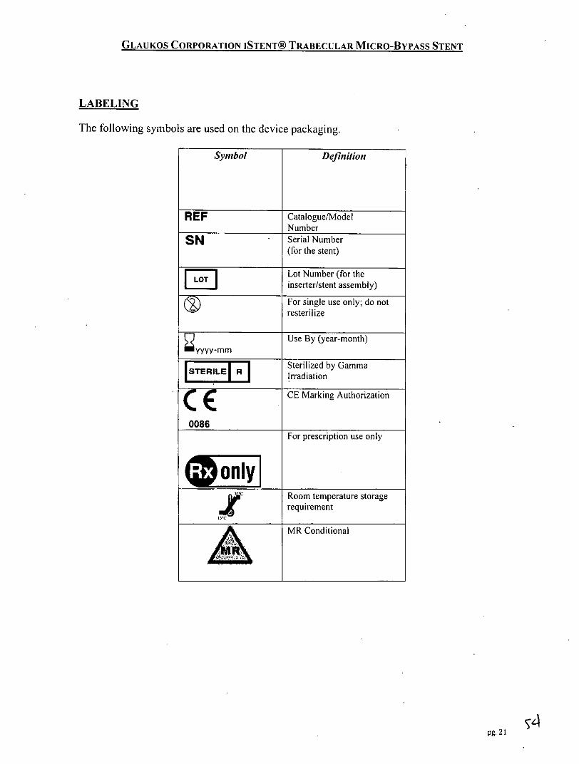
LABELING
The following symbols are used on the device packaging.
Symbol Definition
REF Catalogue/ModelNumber
SN Serial Number(for the stent)
Lot Number (for theED inserter/stent assembly)
For single use only; do notresterilize
Use By (year-month)myyyy-mm
Sterilized by GammaIrradiation( rE CE Marking Authorization
0086For prescription use only
Room temperature storagerequirementMR Conditional
pg.2

GLAUKOS CORPORATION ISTENT® TRABECULAR MICRO-BYPASS STENT
MRI INFORMATION:
The iStent® Trabecular Micro-Bypass Stent Model GTSIOOR/L is MR-Conditional accordingto the terminology specified in the American Society for Testing and Materials (ASTM)International, Designation: F2503-05. Standard Practice for Marking Medical Devices andOther Items for Safety in the Magnetic Resonance Environment. ASTM International, 100Barr Harbor Drive, PO Box C700, West Conshohocken, Pennsylvania, 2005.Non-clinical testing demonstrated that the iStent® Trabecular Micro-Bypass Stent ModelGTS I OOR/L is MR-Conditional. A patient with this device can be scanned safelyimmediately after placement under the following conditions:
* Static magnetic field: 3-Tesla or less* Maximum spatial magnetic field gradient: 720-Gauss/cm or less
MRI-Related Heating and Artifact Information* Because of the very small dimensions of the iStent, heating and artifacts will not pose a
problem or added risk.
CAUTION
Federal law restricts this device to sale by, or on the order of, a physician.
Physician training is required prior to use of the device, and consists of three main parts:* Webinar* Didactic session with Glaukos surgical representative* Observation of surgical cases by Glaukos representative until implantation proficiency is
demonstrated
Manufacturer:Glaukos Corporation26051 Merit Circle #103Laguna Hills, CA 92653Tel: 949.367.9600, Fax: 949.367.9984www.glaukos.comToll-Free: 1-800-GLAUKOS (452-8567)
iStent is a registered trademark of Glaukos Corporation,
pg. 22

GLAU Kr S'Changg Per'pecttves
PATIENT ID CARDiStent® Trabecular Micro-Bypass Stent System
Model:Place Patient ID Card Label Here
Serial No:
Implant Date: dd / mm I/vvvv OS 0 OD 0
Print Surgeon Name: A.ww.glaukos.com 45-0007 rev. 2
(DI