DIAGNOSTIC SEMINAR Myopathology of non-infectious ...
15
Pathology – Research and Practice 204 (2008) 609–623 DIAGNOSTIC SEMINAR Myopathology of non-infectious inflammatory myopathies – The current status Ekkehard Hewer a, , Hans H. Goebel a,b a Institute of Neuropathology, University Hospital Zurich, Zurich, Switzerland b Department of Neuropathology, Johannes Gutenberg University, Mainz, Germany Received 18 November 2007; accepted 4 March 2008 Abstract Besides the classical inflammatory myopathies (IM), dermatomyositis (DM), polymyositis, and inclusion body myositis, the much larger spectrum of IM includes focal and nodular myositis, granulomatous myositis, macrophagic myofasciitis, graft vs. host myositis, eosinophilic myositis, and other immune-associated conditions, some of them only recently described. In addition, paraneoplastic, statin-induced and critical illness myopathies have been considered immune-associated IM. Infectious, i.e., bacterial, viral, and parasitic IM are much less frequent in the northern hemisphere. In IM, muscle biopsy is an essential diagnostic procedure to initiate therapy. The myopathological spectrum encompasses disease-specific histopathological features, such as perifascicular atrophy in DM, non- necrotizing granulomas in sarcoid myopathy, autophagic vacuoles with tubulofilamentous inclusions in inclusion body myositis, rarely electron microscopic criteria, such as undulating tubules in endothelial cells of DM specimens, and, foremost, immunohistochemical findings. These latter features concern inflammatory infiltrates, the muscle parenchyma, the interstitial compartment, and the vasculature with varying involvement of each component in the different IM. Differences in immunohistochemical parameters among the IM, such as major histocompatibility complexes I and II, cytokines, cell adhesion molecules, different types of inflammatory cells, metalloproteinases, and complement factors procure a large gamut of data, the individual patterns of which characterize the myopathology of individual IM. r 2008 Elsevier GmbH. All rights reserved. Keywords: Inflammatory myopathies; Myopathology; Immunohistochemistry; Polymyositis; Dermatomyositis Introduction Inflammatory myopathies (IM) frequently require muscle biopsy as an essential diagnostic procedure to document inflammation as a general myopathological process or to reveal certain IM-specific additional features, such as lymphocytes within intact muscle fibers in polymyositis (PM) and inclusion body myositis (IBM), conspicuous PAS-positive macrophages in macrophagic myofasciitis, rimmed or autophagic va- cuoles with tubulofilamentous inclusions and/or intra- cellular amyloid in IBM, a perifascicular pattern of lesions or undulating tubules in dermatomyositis (DM). IM as a generic group of diseases encompass the entire age spectrum, although with different emphasis of different IM at different ages, e.g. juvenile DM or late-onset IBM. Being grossly divided into infectious and non-infectious (immune-related) conditions, IM do ARTICLE IN PRESS www.elsevier.de/prp 0344-0338/$ - see front matter r 2008 Elsevier GmbH. All rights reserved. doi:10.1016/j.prp.2008.03.006 Corresponding author. Tel.: +41 44 255 4905; fax: +41 44 255 4402. E-mail address: [email protected] (E. Hewer).
Transcript of DIAGNOSTIC SEMINAR Myopathology of non-infectious ...

ARTICLE IN PRESS
0344-0338/$ - se
doi:10.1016/j.pr
�Correspondifax: +4144 255
E-mail addre
Pathology – Research and Practice 204 (2008) 609–623
www.elsevier.de/prp
DIAGNOSTIC SEMINAR
Myopathology of non-infectious inflammatory
myopathies – The current status
Ekkehard Hewera,�, Hans H. Goebela,b
aInstitute of Neuropathology, University Hospital Zurich, Zurich, SwitzerlandbDepartment of Neuropathology, Johannes Gutenberg University, Mainz, Germany
Received 18 November 2007; accepted 4 March 2008
Abstract
Besides the classical inflammatory myopathies (IM), dermatomyositis (DM), polymyositis, and inclusion bodymyositis, the much larger spectrum of IM includes focal and nodular myositis, granulomatous myositis, macrophagicmyofasciitis, graft vs. host myositis, eosinophilic myositis, and other immune-associated conditions, some of them onlyrecently described. In addition, paraneoplastic, statin-induced and critical illness myopathies have been consideredimmune-associated IM. Infectious, i.e., bacterial, viral, and parasitic IM are much less frequent in the northernhemisphere. In IM, muscle biopsy is an essential diagnostic procedure to initiate therapy. The myopathologicalspectrum encompasses disease-specific histopathological features, such as perifascicular atrophy in DM, non-necrotizing granulomas in sarcoid myopathy, autophagic vacuoles with tubulofilamentous inclusions in inclusion bodymyositis, rarely electron microscopic criteria, such as undulating tubules in endothelial cells of DM specimens, and,foremost, immunohistochemical findings. These latter features concern inflammatory infiltrates, the muscleparenchyma, the interstitial compartment, and the vasculature with varying involvement of each component in thedifferent IM. Differences in immunohistochemical parameters among the IM, such as major histocompatibilitycomplexes I and II, cytokines, cell adhesion molecules, different types of inflammatory cells, metalloproteinases, andcomplement factors procure a large gamut of data, the individual patterns of which characterize the myopathology ofindividual IM.r 2008 Elsevier GmbH. All rights reserved.
Keywords: Inflammatory myopathies; Myopathology; Immunohistochemistry; Polymyositis; Dermatomyositis
Introduction
Inflammatory myopathies (IM) frequently requiremuscle biopsy as an essential diagnostic procedure todocument inflammation as a general myopathologicalprocess or to reveal certain IM-specific additionalfeatures, such as lymphocytes within intact muscle fibers
e front matter r 2008 Elsevier GmbH. All rights reserved.
p.2008.03.006
ng author. Tel.: +4144 255 4905;
4402.
ss: [email protected] (E. Hewer).
in polymyositis (PM) and inclusion body myositis(IBM), conspicuous PAS-positive macrophages inmacrophagic myofasciitis, rimmed or autophagic va-cuoles with tubulofilamentous inclusions and/or intra-cellular amyloid in IBM, a perifascicular pattern oflesions or undulating tubules in dermatomyositis (DM).IM as a generic group of diseases encompass the entireage spectrum, although with different emphasis ofdifferent IM at different ages, e.g. juvenile DM orlate-onset IBM. Being grossly divided into infectiousand non-infectious (immune-related) conditions, IM do

ARTICLE IN PRESS
Table 1. Immunohistochemical expression of MHC-I in
muscle tissue
On the surface of myofibers
Diffusely across regenerating myofibers
In inflammatory infiltrates
In vessel walls
Table 2. Immunohistochemistry of MHC-I expression in
neuromuscular diseases
Polymyositis
Dermatomyositis
Inclusion body myositis
Statin myopathy [37]
Duchenne muscular dystrophy [2,56]
Dysferlinopathy [13]
Limb girdle muscular dystrophy
Table 3. Markers of macrophages
CD68 General
KiMIP General
MRP14, 27E10 Early
MRP8 Intermediate
25F9 Late
Metalloproteinases
Table 4. Macrophages in myositis
Inflammatory myopathy with abundant macrophages
(IMAM)
Macrophagic myofasciitis (MMF)
Granulomatous myositis (esp. sarcoid myopathy)
Whipple disease
E. Hewer, H.H. Goebel / Pathology – Research and Practice 204 (2008) 609–623610
occur worldwide, although infectious forms are morefrequently seen in the developing world as distinctentities. However, infectious IM may certainly beencountered in ‘‘developed’’ countries, probably mostoften in conjunction with trauma and surgical proce-dures, but then, muscle biopsy is usually not adiagnostic procedure. Hence, the entire spectrum ofmyopathological diagnostic parameters and markers forIM yields different patterns during and for diagnosticworkup of IM specimens. As it is true for theoverwhelming majority of neuromuscular disorders ingeneral, autopsy studies of IM, employing moderndiagnostic myopathological techniques, have been andstill are rarely performed and, therefore, have providedlittle information to the diagnostic myopathologicalregimen, to distributional patterns of individual IM, andhave hardly ever been available to corroborate biopsy-based findings. Patients who die of IM may be fewbecause of treatability and curability, whereas patientswho die with IM may be many because still a sufficientnumber of IM are long-lasting chronic diseases. Thisautopsy-based potential of available muscle tissues hasnot yet successfully and gainfully been explored, whichaffords the opportunity for multiple, even abundantsampling of specimens from numerous different musclesto address the frequent diagnostic myopathologicalproblem of focality in IM.
Predicated on the essentiality in the diagnostic regi-men of IM of the muscle biopsy, only the armamentar-ium of modern techniques employed in myopathologymay allow a thorough and, perhaps, complete myo-pathological investigation of the biopsied muscle.Different techniques, e.g. histology, enzyme histochem-istry, electron microscopy, and immunohistochemistryhave different diagnostic values in different forms ofIM, but for each IM-suspected biopsied muscle tissue,adequate preparative conditions have to be provided fora panoply of investigations. This care for subsequentoptimal diagnostic investigations of the biopsied muscletissue commences with choosing the correct muscle forbiopsy and continues in the operating room where, uponremoval of the biopsied tissue, the proper techniqueshave immediately to be employed, i.e., freezing ofmuscle for light microscopic studies and adequatefixation of muscle for electron microscopy, and,perhaps, complementary light microscopic investiga-tions.
While the individual histological, enzyme histochem-ical, and electron microscopic methods have not beenexpanded over the past 20 years, the introduction ofimmunohistochemistry into myopathology has revolu-tionarily augmented our diagnostic armamentarium andour myopathological knowledge concerning IM. Im-munoglobulins, complement factors, cell adhesion mo-lecules, cytokines, chemokines [15], metalloproteinases[20,31], and not the least, major histocompatibility
complexes (MHC-) I and II [24] (Tables 1 and 2),subtyping of lymphocytic infiltrates, and recently ofmacrophage subpopulations (Tables 3–5) have yieldeddifferent results in different forms of IM and, thus,accorded different diagnostic connotations to individualIM. Therefore, the impact of immunohistochemistry ondiagnostic myopathology in IM will be the majorcomponent in this review, whereas myopathologicallyrelevant non-immunohistochemical techniques and find-ings will precede the subsequent canvassing of individualand groups of IM.
While non-inflammatory neuromuscular diseases lar-gely affect the muscle parenchyma, i.e., the myofibers,sometimes connective tissue, and rarely vessels, IM are

ARTICLE IN PRESS
Table 5. Different types of macrophages in inflammatory myopathies [5,44]
Type of antibody PM DM IBM GM Controlsa Controlsb
KiM1P + + + + + +
Dianova
Pan-type
27E10 17%c 37%c 24%c 6%c + o10%
BMA biomedicals Perimysial: 35%d Perimysial: 13%d
Early/acute Endomysial: 47%d Endomysial: 25%d
MRP14 14%c 19%c 6%c 6%c + o10%
BMA biomedicals Perimysial: 90%d Perimysial: 15%d
Early/acute Endomysial: 66%d Endomysial: 33%d
MRP8 21%c 26%c 31%c 15%c Not evaluated Not present
BMA biomedicals
Intermediate/subacute
25F9 10%c 17%c 7%c 47%c Not evaluated 50%
BMA biomedicals late/chronic Perimysial: 18%d Perimysial: 14%d
Endomysial: 23%d Endomysial: 24%d
PM: polymyositis; DM: dermatomyositis; IBM: inclusion body myositis; GM: granulomatous myositis.a[5] – Neurogenic atrophy and ‘‘degenerative’’ myopathies.b[44] – Duchenne muscular dystrophy.c[5] – Paraffin sections.d[44] – Frozen sections.
E. Hewer, H.H. Goebel / Pathology – Research and Practice 204 (2008) 609–623 611
marked by involvement of all skeletal muscle constitu-ents, i.e., muscle fibers, vessels of different calibers, i.e.,capillaries, arterioles, venules, connective tissue, and asan important additional component, by inflammatoryinfiltrates of heterogeneous nature. Each of thesecomponents may provide typical or atypical patternsof immunohistochemically expressed parameters indifferent IM. The abundance of applicable antibodiesmay demonstrate diagnostic overlap in different types ofIM, sometimes showing the usefulness of semi-quanti-tative information [52], the application of ‘‘inflamma-tory scores’’ [51], or the proportion or ratio of, e.g.T4 to T8 lymphocytes in different IM [53] to betterassess the diagnostic value of individual immunohisto-chemical parameters in the complex myopathology ofIM on the diagnostic path to clearly identify theindividual IM. Hence, in immunomyopathology ofIM, not only expression of antigens and their demon-stration by respective antibodies and immunohisto-chemical patterns in different IM are of importance,but also immunohistochemical profiles of all theindividual muscle tissue constituents among the manydifferent IM.
When diagnosing IM by myopathology, three generalaspects are of concern: IM with inflammatory infiltrates,IM without inflammatory infiltrates, and non-IMthough with inflammatory infiltrates, such as certainmuscular dystrophies.
Immune-mediated inflammatory myopathies
Dermatomyositis
The myopathological hallmark of DM (Fig. 1) is theperifascicular pattern of lesions. In a severely affectedspecimen, the perifascicular pattern may be replaced bya panfascicular pattern, although it may not necessarilybe present in each fascicle of the biopsied muscle tissue.Inflammatory infiltrates may be most pronounced in theperimysium extending into the individual muscle fasci-cles along the endomysium. Peripherally located musclefibers in the fascicles may undergo necrosis anddegeneration, regeneration, or atrophy. Employingmore than histological stains, this perifascicular patternmay be recognized by activated acid phosphatase, bothin interstitial cells and muscle fibers, by the presence ofenzyme histochemically partially or non-reacting‘‘ghost’’ muscle fibers, and by additional immunohisto-chemical labeling of infiltrating cells or upregulation ofproteins. This pattern implies a vascular backgroundwhich renders DM a multi-organ disease, not onlyaffecting skeletal muscle and, often, skin but, occasion-ally, also the gastrointestinal tract and the lungs [14].However, at the ultrastructural level, damage toendothelial cells of capillaries, their necrosis andsubsequent regeneration amounting first to depletionin capillaries, evidenced by numerous vascular markers,

ARTICLE IN PRESS
Fig. 1. Dermatomyositis: (A) Perifascicular involvement is marked by infiltration of the endomysium and by regenerating muscle
fibers, in contrast to the more centrally located part of the muscle fascicle, with only variation in fiber diameters and sarcolemmal
upregulation of MHC-I. (B) B-lymphocytes among muscle fibers of various sizes. (C) Ultrastructurally, a red blood cell is only
surrounded by a vascular basement membrane after degeneration of endothelial cells. (D) By electron microscopy, tubuloreticular
profiles/undulating tubules may be present in endothelial cells.
E. Hewer, H.H. Goebel / Pathology – Research and Practice 204 (2008) 609–623612
then increased angiogenesis [37], is conspicuouslyencountered by electron microscopy. Another hallmarkof DM are tubuloreticular profiles or undulating tubulesnot only within endothelial cells but also withincirculating blood lymphocytes, thus emphasizing thesystemic nature of this condition. These undulatingtubules may actually precede clinical and majorhistopathological features [19]. Undulating tubulesmay also occur in systemic lupus erythematosus, Sjogrensyndrome, and human immunodeficiency virus (HIV)infection. Another ultrastructural feature in endothelialand lymphocytic cells of DM is cylindrical confrontingcisternae [26]. While capillaries are evenly spread acrossnormal muscle fascicles with approximately one capil-lary per muscle fiber, the perifascicular lesional patterncannot exclusively be explained by capillaropathyprimarily causing DM.
Apart from labeling mural cells of vessels, foremostendothelial cells and, thereby, recognizing DM on theground of capillary depletion and renewal, abnormalpresence of the chemokine monocyte chemo-attractantprotein 1 (MCP1) [15] and the C5b9 complement ormembrane attack complex (MAC) in capillary wallsdenote DM as a microvasculopathy. The density ofcapillaries within muscle fascicles may immunohisto-chemically be documented by endothelial markers orvessel-related extracellular matrix proteins, such aslaminins or MHC-I, the latter also normally beingexpressed in vessel walls. Infiltrating lymphocyteslargely consist of B-cells and fewer CD4 helper T-
lymphocytes. Among subtypes of macrophages, late ormature macrophages marked by the late-activationmarker 25F9 seem to prevail [44]. The MHC-I issarcolemmally expressed, most markedly in the perifas-cicular area, but may also be encountered across theentire muscle parenchyma. Likewise, MHC-II is upre-gulated in DM even when inflammatory infiltrates maynot be present [24]. Of cytokines interleukin-2, tumornecrosis factors alpha and interferon gamma may beexpressed in interstitial cells, especially interleukin 4 [52].Matrix metalloproteinases (MMP) [42] or metallopro-teinases-disintegrins (ADAMS) may variedly be upre-gulated in inflammatory cells and muscle fibers, i.e.,MMP 2, 7, and 9 in the sarcolemma of atrophic fibers[47] and regenerating myofibers [12] or diffusely in smallfibers [43].
A DM-like inflammatory myopathy is inflammatorymyopathy with abundant macrophages (IMAM). Subtleimmunohistochemical differences between DM andIMAM have been outlined [8,9] in that MAC is presentin capillaries of DM but absent from IMAM, whilemacrophages in IMAM express the early inflammationmarker MRP-14. Similarities concerning infiltratingcells in both conditions are found in the expression ofCD4 and CD8 T-cells and CD20 B-lymphocytes, as wellas interleukin-10.
Necrotizing myopathy, associated with serum anti-bodies against the signal recognition particle (SRP), isalso marked by prominent capillary pathology, althoughin a more uniformly scattered fashion than the more

ARTICLE IN PRESSE. Hewer, H.H. Goebel / Pathology – Research and Practice 204 (2008) 609–623 613
patchy and perifascicular type in DM [36]. Thoseconditions show deposition of the MAC/terminalcomponent C5b9 of the complement cascade, which isalso seen in another type of necrotizing myopathymarked by so-called ‘‘pipestem capillaries’’ [23]. How-ever, the anti-SRP myopathy hardly, if ever, showsinflammatory infiltrates and a weak, incomplete, andvarying pattern of MHC-I expression on musclefibers [36].
Polymyositis
PM (Fig. 2) is an immune-mediated, often subacuteinflammatory myopathy supposedly caused by auto-aggressive inflammatory cells which, as a myopatholo-gical hallmark, enter and destroy apparently normalmuscle fibers. The cause why intact myofibers areattacked is still unknown. Upregulation of MHC-Imay even precede infiltration by inflammatory cells toattract cytotoxic T8 lymphocytes and allow their entryinto normal-looking muscle fibers. Such upregulation ofMHC-I may occur both in clinically affected and non-affected muscles [22], and the same appears to be truefor upregulation of MHC-II [22], apparently a pre-requisite to bind CD4 helper T-lymphocytes. Inflamma-tory cells, particularly lymphocytes, among which CD8lymphocytes are more numerous than CD4 cells, aredistributed across the muscle fascicles often surroundingindividual muscle fibers of normal appearance,from where their entry into these intact musclefibers originates. As part of the attack of CD8lymphocytes to intact muscle fibers, the cytolyticproteins granulysin and perforin are encountered inthese cytotoxic lymphocytes [30] as well as MPC-1 [15].B-lymphocytes are few or absent. Necrosis and regen-eration of myofibers associated with variability in size,together with endomysial fibrosis, add to the myopatho-logical pattern of PM, then resulting in a chronicmyopathy with or without inflammation which maydisappear after successful treatment, while upregulationof MHC-I and II may persist. Among macrophages,early-activation subtypes 27E10 and MRP-14 predomi-nate [44].
In addition, cell adhesion molecules are upregulatedin inflammatory and mural cells of vessels, such asintercellular cell adhesion molecule-1 (ICAM-1) andlymphocyte function-associated antigen-1 (LFA-1) al-pha and beta [51]. Of the metalloproteinase-disintegrins(ADAMs), ADAMs 17 and 19 are expressed inT-lymphocytes, both of the helper and cytotoxicsubtypes, while ADAM 8 is associated with macro-phages [20]. MMP 2, 7, and 9 are expressed in non-necrotic MHC-I-positive muscle fibers, regeneratingmuscle fibers, atrophic fibers [43,47], and endothelialcells [12], or MMP9 in inflammatory cells [31].
Inclusion body myositis
IBM (Fig. 3) is marked by the combination of twotissue patterns, firstly the inflammatory componentlargely mimicking the tissue pattern in PM, whichincludes upregulation of MHC-I and II, predominantlyCD8 cytotoxic T-cells within the infiltrates as well asinside non-necrotic muscle fibers and the upregulationof respective ADAMs proteins and, secondly, myo-pathic features, such as variation in fiber diameters,necrosis, and regeneration of muscle fibers. Moreover,there seem to be no differences in the expression ofcertain ADAMs and subtypes of macrophages betweenIBM and PM. This high similarity in immune-related components between PM and IBM may oftenrender an exact distinction of the two conditionsdifficult. Even ragged red fibers and, more often,myofibers devoid of cytochrome-C oxidase (COX)activity, representing a mitochondrial component, andpartial COX deficiency, frequently encountered in IBM,may be a rare feature in PM [6]. Furthermore, IBM maybe considered a degenerative myopathy defined byautophagic/rimmed vacuoles and aggregation of pro-teins. Rimmed vacuoles, a prominent feature in a diversenumber of neuromuscular disorders, including IBM,distal myopathies, late-onset type-II glycogenosis, neu-rogenic processes, myofibrillar myopathies (MFMs),oculopharyngeal muscular dystrophy, and others showactivation of the lysosomal marker enzyme acidphosphatase. Inclusion bodies may be seen within nucleias loosely arranged aggregates of tubulofilaments, whichactually consist of tau-containing paired helical fila-ments and, more frequently, similar and more denselypacked aggregates of tubulofilaments within the sarco-plasm, often in the vicinity of autophagic vacuoles.However, aggregation of proteins is not confined totubulofilamentous aggregates, which themselves may beencountered in a large variety of neuromusculardisorders (Table 6). The most prominent proteinaccumulating in muscle fibers in IBM is beta-amyloid,recognizable as small haphazardly deposited filamentswhich, when forming aggregates, display congophiliaenhanced by Texas red-type fluorescence microscopywhen using the Congo red stain, but also stain withcrystal violet and Thioflavin S. Many more proteins ofvery diverse nature aggregate in IBM muscle fibers, too(Table 7). IBM shares the myopathological features ofautophagy and protein aggregation with geneticallydifferent and sporadic forms of MFM, the latter alsodisplaying many different proteins [1,17], many ofwhich are encountered in both IBM and MFM.Similarly, proteins of the ubiquitin proteasome pathwayof extralysosomal protein degradation are also upregu-lated [25].
Finally, small angulated fibers are often encoun-tered in IBM muscle specimens, suggesting a subtle

ARTICLE IN PRESS
Fig. 2. Polymyositis: (A) T-lymphocytes not only appear between muscle fibers but also within an intact muscle fiber. (B) I-CAM
(interstitial cell adhesion molecule) is upregulated in the interstitium among muscle fibers. (C) In spite of little myopathology, MHC-
I is upregulated in the sarcolemma of each muscle fiber. (D) Multifocal upregulation of the lymphocyte function antigen beta
(LFAb) is apparent among muscle fibers. (E) At the ultrastructural level, a lymphocyte is situated within an intact muscle fiber, the
electron microscopic equivalent to T-lymphocytes in intact muscle fibers at the light microscopic level, as seen in Fig. 2a.
E. Hewer, H.H. Goebel / Pathology – Research and Practice 204 (2008) 609–623614
neurogenic component of denervation, while large-group atrophy and fiber type grouping followingreinnervation are absent. Such angulated atrophicmuscle fibers display increased histochemical activitiesof acid phosphatase and non-specific esterase as well asof the oxidative enzymes NADH and MAG, in the latter
two preparations often without the normal reciprocityof fiber types.
MMP 2, 7, and 9 were seen in muscle fibers,inflammatory cells, and vessel walls [47], MMP 9 ininflammatory cells [31] and MHC-I-positive non-necroticmuscle fibers [12].

ARTICLE IN PRESS
Fig. 3. Inclusion body myositis: (A) A rimmed/autophagic vacuole is present in the periphery of the central small muscle fiber,
modified Gomori trichrome stain. (B) Around vacuoles and beyond, accumulation of myotilin, a sarcomeric protein which indicates
protein aggregation often seen in inclusion body myositis. (C) The electron micrograph of a rimmed/autophagic vacuole with
lamellar debris in the subsarcolemmal region of a muscle fiber, equivalent to the rimmed/autophagic vacuole in Fig. 3a.
(D) Aggregates of filaments [F] are often seen in association with rimmed/autophagic vacuoles. (E) Higher magnification shows the
ultrastructure of these tubulofilaments. (F) Variation in fiber size, here, is also marked by several small highly atrophic muscle fibers,
one of them being a ragged red fiber, suggesting mitochondrial abnormalities, modified Gomori trichrome stain. (G) There are at
least two small COX-negative, SDH-positive (blue) muscle fibers apparent, while necrotic fibers are devoid of any enzyme activity,
combined cytochrome-C oxidate (COX)-succinic dehydrogenase (SDH) preparation.
E. Hewer, H.H. Goebel / Pathology – Research and Practice 204 (2008) 609–623 615
Macrophagic myofasciitis
The inflammatory myopathy macrophagic myofascii-tis (MMF) (Fig. 4) indicates a recently identified
condition which develops focally after injection ofaluminum-containing vaccines, even more than 10 yearsprior to biopsy [45], largely in the deltoid muscle, mostoften on the left side in adults or in the quadriceps

ARTICLE IN PRESSE. Hewer, H.H. Goebel / Pathology – Research and Practice 204 (2008) 609–623616
muscle in children, and is marked by aggregates of largemacrophages together with inflammatory lymphocytesin fascia and vicinal skeletal muscle fascicles. Theconspicuous macrophages have a PAS-intense cyto-plasm – the PAS stainability being resistant to diastasedigestion – which displays increased histochemicalactivities of acid phosphatase and non-specific esterase
Table 6. Tubulofilamentous aggregates in neuromuscular
disorders
Inclusion body myositis/inclusion body myopathy
Welander distal myopathy
Familial myopathy and periventricular leukoencephalopathy
Nonaka Japanese familial distal myopathy with rimmed
vacuoles
Dystrophic myotonia
Polyneuropathy
Sarcoidosis
Familial oculopharyngeal muscular dystrophy with distal
myopathy
Rigid spine syndrome
Acid maltase deficiency
Amyloid neuropathy
Rimmed-vacuole myopathy sparing the quadriceps
Adult-onset autosomal-dominant limb girdle muscular
dystrophy
Postpolio syndrome
Sporadic distal myopathy
Amyotrophic lateral sclerosis
Myofibrillar myopathy
Table 7. Comparative immunocytochemical results of protein agg
Proteins MM IBM
Transsarcolemmal proteins
Dystrophin 1 + –
Dystrophin 2 +
Dystrophin 3 + –
Utrophin (DRP2) +/� �/+
a-Sarcoglycan �/+ �
b-Sarcoglycan �/+ �
g-Sarcoglycan �/+ �
d-Sarcoglycan + +/�
Dysferlin + +
Merosin 80 +/� �
Merosin 300 +/� �
nNOS + +
b-Laminin +/� �
g-Laminin +/� �
Collagen 6 +/� �
Caveolin + +
a-Dystroglycan + �
b-Dystroglycan +/� �
MM: myofibrillar myopathy; IBM: inclusion body myositis; nNOS: neurona
as evidence of lysosomal upregulation. These lysosomescontain aluminum-typical spicules, the aluminum natureof which can be further documented by laser microprobemass analysis (LAMMA) [21].
Lymphocytes are largely of the T-cell type. Macro-phages which react with the common antibodies CD68and KI 1P appear, when tested for subtypes, as long-standing 25F9-type, while early-onset 27E10 macro-phages are rare or absent [48]. MHC-I is often expressedclose to the inflammatory macrophage infiltrates but isscarce in or absent from infiltrate-distant myofibers.
Muscle parenchyma may be scarcely affected in MMF,quite unlike that seen in PM, DM, and IBM, while thespectacular macrophage infiltrates may distract attentionfrom the underlying neuromuscular disorder, which hasled to the respective muscle biopsy revealing the clinicallyalmost always unsuspected MMF.
The myopathological differential diagnosis of MMFencompasses Whipple disease, affecting skeletal muscle.However, by electron microscopy, the Whipple-typicalorganisms may be documented in conjunction with thissystemic infection, while MMF is a localized process, atleast myopathologically, even in view of claims that itmay elicit immune-related generalized responsesclinically and serologically [27]. Furthermore, MMFhas to be distinguished from IMAM often seen withDM-type myopathology, and from cytophagic histiocy-tic panniculitis [4]. MMF, described without precedingvaccination [11,40] may represent IMAM rather thanMMF.
regates in MM/IBM
Proteins MM IBM
Chaperone proteins
Ubiquitin ++ +
a-B Crystallin ++ +
Heat shock protein 72/73 + +
Cytoskeletal proteins
Desmin ++ ++
Vimentin + +
Plectin + +
Sarcomeric proteins
Actin + +
a-Actinin + +
Myotilin + +
Nuclear proteins
Emerin – –
Lamin A/C – –
Others
Prion protein – –
l nitric oxide synthase (by courtesy of Dr. Alexandra Vrabie).

ARTICLE IN PRESS
Fig. 4. Macrophagic myofasciits: (A) Large bluish macrophages among muscle fibers, hematoxylin–eosin. (B) Macrophages are
strongly PAS-positive. (C) Macrophages express enhanced enzyme histochemical activity of the lysosomal marker enzyme acid
phosphatase. (D) MHC-I is only upregulated in the sarcolemma of few myofibers located close to macrophage infiltrates next to
three vessels. (E) Ultrastructurally, macrophages contain typical aluminum spicules. (F) The LAMMA technique applied to
semithin sections, containing macrophage infiltrates, identifies an aluminium peak (arrow).
E. Hewer, H.H. Goebel / Pathology – Research and Practice 204 (2008) 609–623 617
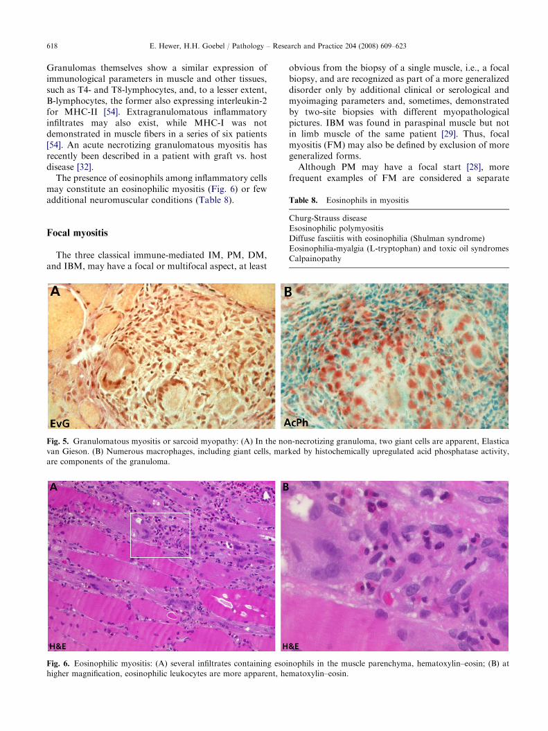
Granulomatous and eosinophilic myositis
Granulomas, i.e., focal aggregates of epithelioidhistiocytic cells, macrophages, inflammatory lympho-cytes, and occasional giant cells of the Langerhans type
within muscle parenchyma, constitute granulomatousmyositis (Fig. 5), which may be part of immune-mediated systemic disorders, such as collagenoses,sarcoidosis, in association with inflammatory boweldisease, myasthenia gravis, or infectious diseases.
ARTICLE IN PRESS
Table 8. Eosinophils in myositis
E. Hewer, H.H. Goebel / Pathology – Research and Practice 204 (2008) 609–623618
Granulomas themselves show a similar expression ofimmunological parameters in muscle and other tissues,such as T4- and T8-lymphocytes, and, to a lesser extent,B-lymphocytes, the former also expressing interleukin-2for MHC-II [54]. Extragranulomatous inflammatoryinfiltrates may also exist, while MHC-I was notdemonstrated in muscle fibers in a series of six patients[54]. An acute necrotizing granulomatous myositis hasrecently been described in a patient with graft vs. hostdisease [32].
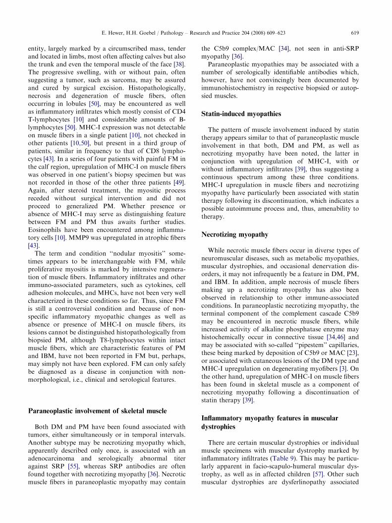
The presence of eosinophils among inflammatory cellsmay constitute an eosinophilic myositis (Fig. 6) or fewadditional neuromuscular conditions (Table 8).
Churg-Strauss disease
Esosinophilic polymyositis
Diffuse fasciitis with eosinophilia (Shulman syndrome)
Eosinophilia-myalgia (L-tryptophan) and toxic oil syndromes
Calpainopathy
Focal myositis
The three classical immune-mediated IM, PM, DM,and IBM, may have a focal or multifocal aspect, at least
Fig. 5. Granulomatous myositis or sarcoid myopathy: (A) In the no
van Gieson. (B) Numerous macrophages, including giant cells, mar
are components of the granuloma.
Fig. 6. Eosinophilic myositis: (A) several infiltrates containing esoi
higher magnification, eosinophilic leukocytes are more apparent, he
obvious from the biopsy of a single muscle, i.e., a focalbiopsy, and are recognized as part of a more generalizeddisorder only by additional clinical or serological andmyoimaging parameters and, sometimes, demonstratedby two-site biopsies with different myopathologicalpictures. IBM was found in paraspinal muscle but notin limb muscle of the same patient [29]. Thus, focalmyositis (FM) may also be defined by exclusion of moregeneralized forms.
Although PM may have a focal start [28], morefrequent examples of FM are considered a separate
n-necrotizing granuloma, two giant cells are apparent, Elastica
ked by histochemically upregulated acid phosphatase activity,
nophils in the muscle parenchyma, hematoxylin–eosin; (B) at
matoxylin–eosin.
ARTICLE IN PRESSE. Hewer, H.H. Goebel / Pathology – Research and Practice 204 (2008) 609–623 619
entity, largely marked by a circumscribed mass, tenderand located in limbs, most often affecting calves but alsothe trunk and even the temporal muscle of the face [38].The progressive swelling, with or without pain, oftensuggesting a tumor, such as sarcoma, may be assuredand cured by surgical excision. Histopathologically,necrosis and degeneration of muscle fibers, oftenoccurring in lobules [50], may be encountered as wellas inflammatory infiltrates which mostly consist of CD4T-lymphocytes [10] and considerable amounts of B-lymphocytes [50]. MHC-I expression was not detectableon muscle fibers in a single patient [10], not checked inother patients [10,50], but present in a third group ofpatients, similar in frequency to that of CD8 lympho-cytes [43]. In a series of four patients with painful FM inthe calf region, upregulation of MHC-I on muscle fiberswas observed in one patient’s biopsy specimen but wasnot recorded in those of the other three patients [49].Again, after steroid treatment, the myositic processreceded without surgical intervention and did notproceed to generalized PM. Whether presence orabsence of MHC-I may serve as distinguishing featurebetween FM and PM thus awaits further studies.Eosinophils have been encountered among inflamma-tory cells [10]. MMP9 was upregulated in atrophic fibers[43].
The term and condition ‘‘nodular myositis’’ some-times appears to be interchangeable with FM, whileproliferative myositis is marked by intensive regenera-tion of muscle fibers. Inflammatory infiltrates and otherimmuno-associated parameters, such as cytokines, celladhesion molecules, and MHCs, have not been very wellcharacterized in these conditions so far. Thus, since FMis still a controversial condition and because of non-specific inflammatory myopathic changes as well asabsence or presence of MHC-I on muscle fibers, itslesions cannot be distinguished histopathologically frombiopsied PM, although T8-lymphocytes within intactmuscle fibers, which are characteristic features of PMand IBM, have not been reported in FM but, perhaps,may simply not have been explored. FM can only safelybe diagnosed as a disease in conjunction with non-morphological, i.e., clinical and serological features.
Paraneoplastic involvement of skeletal muscle
Both DM and PM have been found associated withtumors, either simultaneously or in temporal intervals.Another subtype may be necrotizing myopathy which,apparently described only once, is associated with anadenocarcinoma and serologically abnormal titeragainst SRP [55], whereas SRP antibodies are oftenfound together with necrotizing myopathy [36]. Necroticmuscle fibers in paraneoplastic myopathy may contain
the C5b9 complex/MAC [34], not seen in anti-SRPmyopathy [36].
Paraneoplastic myopathies may be associated with anumber of serologically identifiable antibodies which,however, have not convincingly been documented byimmunohistochemistry in respective biopsied or autop-sied muscles.
Statin-induced myopathies
The pattern of muscle involvement induced by statintherapy appears similar to that of paraneoplastic muscleinvolvement in that both, DM and PM, as well asnecrotizing myopathy have been noted, the latter inconjunction with upregulation of MHC-I, with orwithout inflammatory infiltrates [39], thus suggesting acontinuous spectrum among these three conditions.MHC-I upregulation in muscle fibers and necrotizingmyopathy have particularly been associated with statintherapy following its discontinuation, which indicates apossible autoimmune process and, thus, amenability totherapy.
Necrotizing myopathy
While necrotic muscle fibers occur in diverse types ofneuromuscular diseases, such as metabolic myopathies,muscular dystrophies, and occasional denervation dis-orders, it may not infrequently be a feature in DM, PM,and IBM. In addition, ample necrosis of muscle fibersmaking up a necrotizing myopathy has also beenobserved in relationship to other immune-associatedconditions. In paraneoplastic necrotizing myopathy, theterminal component of the complement cascade C5b9may be encountered in necrotic muscle fibers, whileincreased activity of alkaline phosphatase enzyme mayhistochemically occur in connective tissue [34,46] andmay be associated with so-called ‘‘pipestem’’ capillaries,these being marked by deposition of C5b9 or MAC [23],or associated with cutaneous lesions of the DM type andMHC-I upregulation on degenerating myofibers [3]. Onthe other hand, upregulation of MHC-I on muscle fibershas been found in skeletal muscle as a component ofnecrotizing myopathy following a discontinuation ofstatin therapy [39].
Inflammatory myopathy features in muscular
dystrophies
There are certain muscular dystrophies or individualmuscle specimens with muscular dystrophy marked byinflammatory infiltrates (Table 9). This may be particu-larly apparent in facio-scapulo-humeral muscular dys-trophy, as well as in affected children [57]. Other suchmuscular dystrophies are dysferlinopathy associated

ARTICLE IN PRESS
Table 9. Inflammation in muscular dystrophies
Dystrophinopathy
Dysferlinopathy
Caveolinopathy
Calpainopathy
Merosinopathy
Table 10. Collagen vascular diseases
Mixed connective tissue disease
Polymyalgia rheumatica
Rheumatoid arthritis
Sjogren syndrome
System lupus erythematosus
Systemic sclerosis
E. Hewer, H.H. Goebel / Pathology – Research and Practice 204 (2008) 609–623620
with an upregulation of MHC-I on muscle fibers [13],merosin deficiency [41], or, rarely, Duchenne and limb-girdle muscular dystrophies, also marked by upregula-tion of MHC-I on muscle fibers [2,53,56]. On thecontrary, among 200 muscle biopsy specimens ofvarious conditions, sarcolemmal MHC-I expressionwas absent in all specimens with metabolic myopathies,congenital myopathies, neurogenic disorders, andhealthy controls [56]. A peculiar feature is the appear-ance of eosinophilic myositis (Fig. 6) (Table 8) inchildren with mutational calpainopathy, occasionallyassociated with focal expression of MHC-I [33]. Anotherimmune-associated feature in dysferlinopathies may bedeposition of MAC in otherwise normal-appearingmuscle fibers [8,9].
Skeletal muscle involvement in collagen
vascular diseases
Among autoimmune connective tissue or collagenvascular diseases, there are some considered ‘‘overlapsyndromes’’, indicating not only such an autoimmunecollagen vascular disease but also an additional myosi-tis, either of the PM or DM type (Table 10). Then, themyopathology is rather that of myositis than that ofcollagen vascular disease. Without such an overlap,myopathology in pure collagen vascular diseases isusually non-specific and mild, consisting of smallinflammatory infiltrates and some muscle fiber atrophy.This latter finding may be pronounced, e.g. type-IImuscle fiber atrophy in polymyalgia rheumatica. Insystemic lupus erythematosus, undulating tubules simi-lar to those seen in endothelial cells of DM, may beencountered. Vasculitis may be another mild pathologi-cal feature in individual muscle specimens with collagenvascular diseases. It has been claimed [7] that musclepathology in early rheumatoid arthritis is associatedwith type-II muscle fiber atrophy, while a later stage,apparently when joint fixation and stiffness haveadvanced, is marked by type-I fiber atrophy. In juvenileidiopathic arthritis, few patients had upregulation ofMHC-II, while upregulation of MHC-I and MAC C5b9did not differ from controls [35]. In general, immuno-histochemical parameters regarding skeletal muscle,such as MHC-I and II, cytokines, cell adhesionmolecules and metalloproteinases, subtypes of macro-phages or others, have – to our knowledge � not yet
sufficiently and systematically been investigated incollagen vascular diseases without overlap.
Future perspectives
As already outlined in the preceding sections of thisreview, immunohistochemistry with the ever-increasingabundance and availability of antibodies has greatlyexpanded our diagnostic myopathologial armamentar-ium of IM like, for instance, subtyping of macrophagesor introduction of metalloproteinases. Transfer of theseinvestigative findings from research projects to dailypractice will increase in the future, depending on thediagnostic significance of individual antibodies inindividual IM. Such diagnostic significance and diag-nostic certitude will require comparative ‘‘horizontal’’studies, i.e., immunohistochemical application of differ-ent antibodies in the same IM. A recently recommendedmyopathological algorithm for myositis already atteststo the application of diverse groups of antibodies in thediagnostic workup of biopsied muscle [53].
While distribution of IM-affected muscles has earlierrested upon clinical and electromyographic findings,because muscle biopsy is usually restricted to a singlemuscle or, perhaps, two distantly located muscles at themost, (myo-)imaging techniques have recently success-fully been employed to elucidate the lesional distributionof individual IM across limb and trunk muscles, andhave added a further invaluable parameter to theprebioptic diagnostic regimen, the often convincingdistinction between inflammatory and non-inflamma-tory lesions, the former associated with muscle edema.Here, comparative myoimaging-myopathological inves-tigations are further needed, as edema is seldom equallyapparent in biopsied muscle tissue, often only indirectlysurmised from separation of muscle fascicles and musclefibers and some loosening of the connective tissue, whilethe interstitial fluid is usually not recognized, because itis not stained. Immunohistochemical testing for thepresence of albumin in the extracellular space of thebiopsied muscle as evidence of extravasation may notnecessarily distinguish between disease-related andsurgery-related extravasation.
A further field of future research to be urgently incor-porated in the diagnostic myopathological spectrum of
ARTICLE IN PRESSE. Hewer, H.H. Goebel / Pathology – Research and Practice 204 (2008) 609–623 621
IM is immunohistochemical investigation of thosenumerous IM which do not form the core of immune-related IM, i.e., DM, PM, and IBM. Here, overlapsyndromes, rheumatological diseases and beyond, i.e.,infectious IM, hover as important targets. In thesediseases, expansion of autopsy studies will be prospec-tively fruitful and should be studied aggressively whenbecoming available at the autopsy table to amplify boththe diagnostic myopathological spectrum as well as thenosography of the respective IM conditions.
An immunological study of glucocorticoid receptorsalpha and beta, which may variously be demonstrated inendothelial cells, infiltrating lymphocytes, sarcoplasm,or nuclei of muscle fibers in order to assess their validityin therapeutic prognostication did not reveal anydifferences among biopsied muscle specimens of thethree major IM, i.e., DM, PM, and IBM � especiallybetween PM and IBM � as well as control muscles,suggesting that immunohistochemical application ofrespective anti-glucocorticoid receptor antibodies indaily myopathological routine investigations of theseIM will not be helpful [16].
Receptors for the b-chemokines (CCR1�CCR5) maydifferentially be expressed in inflammatory infiltrates,especially macrophages and T-lymphocytes, not B-lymphocytes, as well as in endothelial cells in IM, while,at lower levels, in endothelial cells of controls. However,apart from CCR4 upregulated in myonuclei of regener-ating muscle fibers, CCR are not expressed within or onmyofibers [18].
Acknowledgments
This Diagnostic Seminar originated from a Continu-ing Medical Education Conference on ‘‘InflammatoryMyopathies’’ at the Department of Rheumatology,University Hospital Zurich, Switzerland, February 1,2007. Support by Profs. Renate and Steffen Gay and Dr.Haiko Sprott, as well as photographic aid by WaltherWagner and editorial assistance by Astrid Wober aregratefully appreciated. Dr. P.F. Schmidt (Munster/Germany) kindly provided the LAMMA illustration.
References
[1] A.A. Amato, K. Kagan-Hallet, C.E. Jackson, et al., The
wide spectrum of myofibrillar myopathy suggests a
multifactorial etiology and pathogenesis, Neurology 51
(1998) 1646–1655.
[2] S.T. Appleyard, M.J. Dunn, V. Dubowitz, et al.,
Increased expression of HLA ABC class I antigens by
muscle fibres in Duchenne muscular dystrophy, inflam-
matory myopathy, and other neuromuscular disorders,
Lancet 1 (1985) 361–363.
[3] F.J. Authier, H. Kondo, R.T. Ghnassia, et al., Necrotiz-
ing myopathy with pipestem capillaries and minimal
cellular infiltration: a case associated with cutaneous signs
of dermatomyositis, Neurology 46 (1996) 1448–1451.
[4] G. Bassez, F.J. Authier, E. Lechapt-Zalcman, et al.,
Inflammatory myopathy with abundant macrophages
(IMAM): a condition sharing similarities with cytophagic
histiocytic panniculitis and distinct from macrophagic
myofasciitis, J. Neuropathol. Exp. Neurol. 62 (2003)
464–474.
[5] M. Bergmann, S. Klingebiel, R. Rohkamm, et al.,
Activation pattern of macrophages in inflammatory
myopathies, Acta Neuropathol. 112 (2006) 371–372
(abstract P1027).
[6] G. Blume, A. Pestronk, B. Frank, et al., Polymyositis with
cytochrome oxidase negative muscle fibres. Early quad-
riceps weakness and poor response to immunosuppressive
therapy, Brain 120 (1997) 39–45.
[7] M.H. Brooke, H. Kaplan, Muscle pathology in rheuma-
toid arthritis, polymyalgia rheumatica, and polymyositis:
a histochemical study, Arch. Pathol. 94 (1972) 101–118.
[8] A. Brunn, A comparison between the ‘‘inflammatory
myopathy with abundant macrophages’’ and dermato-
myositis. 18. Kongress des Wissenschaftlichen Beirats der
Deutschen Gesellschaft fur Muskelkranke e.V.,Freiburg,
28.2.-3.3., Wecom Gesellschaft fur Kommunikation mbH
& Co. KG, 83 (abstract).
[9] A. Brunn, R. Schroder, M. Deckert, The inflammatory
reaction pattern distinguishes primary dysferlinopathies
from idiopathic inflammatory myopathies: an important
role for the membrane attack complex, Acta Neuro-
pathol. (Berl.) 112 (2006) 325–332.
[10] C.J. Caldwell, M. Swash, J.D. Van der Walt, et al., Focal
myositis: a clinicopathological study, Neuromuscul. Dis-
ord. 5 (1995) 317–321.
[11] E.-S. Cho, A. Baisre, M.A. Cruz, et al., Extensive
macrophagic myositis in deltoid and trabecular myopathy
in quadriceps in a patient without antecedent vaccination
(abstract 86), J. Neuropathol. Exp. Neurol. 64 (2005) 452.
[12] Y.C. Choi, M.C. Dalakas, Expression of matrix metallo-
proteinases in the muscle of patients with inflammatory
myopathies, Neurology 54 (2000) 65–71.
[13] P. Confalonieri, L. Oliva, F. Andreetta, et al., Muscle
inflammation and MHC class I up-regulation in muscular
dystrophy with lack of dysferlin: an immunopathological
study, J. Neuroimmunol. 142 (2003) 130–136.
[14] M.C. Dalakas, R. Hohlfeld, Polymyositis and dermato-
myositis, Lancet 362 (2003) 971–982.
[15] J.L. De Bleecker, B. De Paepe, I.E. Vanwalleghem, et al.,
Differential expression of chemokines in inflammatory
myopathies, Neurology 58 (2002) 1779–1785.
[16] J.L. De Bleecker, B. De Paepe, V.L. Vervaet, et al.,
Distribution of glucocorticoid receptor alpha and beta
subtypes in the idiopathic inflammatory myopathies,
Neuromuscul. Disord. 17 (2007) 186–193.
[17] J.L. De Bleecker, A.G. Engel, B.B. Ertl, Myofibrillar
myopathy with abnormal foci of desmin positivity. II.
Immunocytochemical analysis reveals accumulation of
multiple other proteins, J. Neuropathol. Exp. Neurol. 55
(1996) 563–577.

ARTICLE IN PRESSE. Hewer, H.H. Goebel / Pathology – Research and Practice 204 (2008) 609–623622
[18] B. De Paepe, J.L. De Bleecker, Beta-chemokine receptor
expression in idiopathic inflammatory myopathies, Mus-
cle Nerve 31 (2005) 621–627.
[19] M. De Visser, A.M. Emslie-Smith, A.G. Engel, Early
ultrastructural alterations in adult dermatomyositis.
Capillary abnormalities precede other structural changes
in muscle, J. Neurol. Sci. 94 (1989) 181–192.
[20] T. Dehmel, A. Janke, H.P. Hartung, et al., The cell-
specific expression of metalloproteinase-disintegrins
(ADAMs) in inflammatory myopathies, Neurobiol. Dis.
25 (2007) 665–674.
[21] S. Doostkam, J. Bohl, P.F. Schmidt, et al., Macrophagic
myofasciitis, Acta Neuropathol. (2005) 324 (abstract).
[22] C. Dorph, P. Englund, I. Nennesmo, et al., Signs of
inflammation in both symptomatic and asymptomatic
muscles from patients with polymyositis and dermato-
myositis, Ann. Rheum. Dis. 65 (2006) 1565–1571.
[23] A.M. Emslie-Smith, A.G. Engel, Necrotizing myopathy
with pipestem capillaries, microvascular deposition of the
complement membrane attack complex (MAC), and
minimal cellular infiltration, Neurology 41 (1991)
936–939.
[24] P. Englund, E. Lindroos, I. Nennesmo, et al., Skeletal
muscle fibers express major histocompatibility complex
class II antigens independently of inflammatory infiltrates
in inflammatory myopathies, Am. J. Pathol. 159 (2001)
1263–1273.
[25] I. Ferrer, B. Martın, J.G. Castano, et al., Proteasomal
expression, induction of immunoproteasome subunits,
and local MHC class I presentation in myofibrillar
myopathy and inclusion body myositis, J. Neuropathol.
Exp. Neurol. 63 (2004) 484–498.
[26] A. Fidzianska, H.H. Goebel, Tubuloreticular structures
(TRS) and cylindric confronting cisternae (CCC) in
childhood dermatomyositis, Acta Neuropathol. (Berl.)
79 (1989) 310–316.
[27] R.K. Gherardi, F.J. Authier, Aluminum inclusion macro-
phagic myofasciitis: a recently identified condition,
Immunol. Allergy Clin. North Am. 23 (2003) 699–712.
[28] R.R. Heffner Jr, S.A. Barron, Polymyositis beginning as a
focal process, Arch Neurol. 38 (1981) 439–442.
[29] E. Hund, R. Heckl, H.H. Goebel, et al., Inclusion body
myositis presenting with isoloated erector spinae paresis,
Neurology 45 (1995) 993–994.
[30] K. Ikezoe, S. Oshima, M. Osoegawa, et al., Granulysin
expression in polymyositis and inclusion body myositis
[abstract M-P-6.04],, Neuromuscul. Disord. 16 (2006)
S87.
[31] B.C. Kieseier, C. Schneider, J.M. Clements, et al.,
Expression of specific matrix metalloproteinases in
inflammatory myopathies, Brain 124 (2001) 341–351.
[32] S. Koeppen, E. Neuen-Jacob, M. Koldehoff, et al., Acute
granulomatous myositis and fasciitis in GVHD, Clin.
Neuropathol. 26 (2007) 134 (abstract).
[33] M. Krahn, A. Lopez de Munain, N. Streichenberger, et
al., CAPN3 mutations in patients with idiopathic
eosinophilic myositis, Ann. Neurol. 59 (2006) 905–911.
[34] M.I. Levin, T. Mozaffar, M.T. Al-Lozi, et al., Para-
neoplastic necrotizing myopathy: clinical and pathologi-
cal features, Neurology 50 (1998) 764–767.
[35] H. Lindehammar, B. Lindvall, Muscle involvement in
juvenile idiopathic arthritis, Rheumatology (Oxford) 43
(2004) 1546–1554.
[36] T. Miller, M.T. Al-Lozi, G. Lopate, et al., Myopathy with
antibodies to the signal recognition particle: clinical and
pathological features, J. Neurol. Neurosurg. Psychiatry
73 (2002) 420–428.
[37] K. Nagaraju, L.G. Rider, C. Fan, et al., Endothelial cell
activation and neovascularization are prominent in
dermatomyositis, J. Autoimmune Dis. 3 (2006) 2.
[38] M. Naumann, K.V. Toyka, H.H. Goebel, et al., Focal
myositis of the temporal muscle, Muscle Nerve 16 (1993)
1374–1376.
[39] M. Needham, V. Fabian, W. Knezevic, et al., Progressive
myopathy with up-regulation of MHC-I associated with
statin therapy, Neuromuscul. Disord. 17 (2007) 194–200.
[40] J.H. Park, K.S. Na, Y.W. Park, et al., Macrophagic
myofasciitis unrelated to vaccination, Scand J. Rheuma-
tol. 34 (2005) 65–67.
[41] E. Pegoraro, P. Mancias, S.H. Swerdlow, et al., Con-
genital muscular dystrophy with primary laminin alpha2
(merosin) deficiency presenting as inflammatory myopa-
thy, Ann. Neurol. 40 (1996) 782–791.
[42] S. Renaud, D. Leppert, Matrix metalloproteinases in
neuromuscular disease, Muscle Nerve 36 (2007) 1–13.
[43] C. Rodolico, A. Mazzeo, A. Toscano, et al., Specific
matrix metalloproteinase expression in focal myositis: an
immunopathological study, Acta Neurol. Scand. 112
(2005) 173–177.
[44] K.M. Rostasy, M. Piepkorn, H.H. Goebel, et al.,
Monocyte/macrophage differentiation in dermatomyosi-
tis and polymyositis, Muscle Nerve 30 (2004) 225–230.
[45] A.M. Ryan, N. Bermingham, H.J. Harrington, et al.,
Atypical presentation of macrophagic myofasciitis 10
years post vaccination, Neuromuscul. Disord. 16 (2006)
867–869.
[46] J.B. Sampson, S.M. Smith, A.G. Smith, et al., Para-
neoplastic myopathy: response to intravenous immuno-
globulin, Neuromuscul. Disord. 17 (2007) 404–408.
[47] B.G. Schoser, D. Blottner, H.J. Stuerenburg, Matrix
metalloproteinases in inflammatory myopathies: en-
hanced immunoreactivity near atrophic myofibers, Acta
Neurol. Scand. 105 (2002) 309–313.
[48] W.J. Schulz-Schaeffer, H.D. Muller, P.F. Schmidt, et al.,
Macrophagic myofasciitis in adults and children, Neuro-
pathol Appl. Neurobiol. 33 (2007) 270–271 (abstract).
[49] K. Sekiguchi, F. Kanda, K. Oishi, et al., HLA typing in
focal myositis, J. Neurol. Sci. 227 (2004) 21–25.
[50] A.G. Smith, S. Urbanits, M. Blaivas, et al., Clinical and
pathologic features of focal myositis, Muscle Nerve 23
(2000) 1569–1575.
[51] D.S. Tews, H.H. Goebel, Expression of cell adhesion
molecules in inflammatory myopathies, J. Neuroimmu-
nol. 59 (1995) 185–194.
[52] D.S. Tews, H.H. Goebel, Cytokine expression profile in
idiopathic inflammatory myopathies, J. Neuropathol.
Exp. Neurol. 55 (1996) 342–347.
[53] D.S. Tews, H.H. Goebel, Diagnostic immunohistochem-
istry in neuromuscular disorders, Histopathology 46
(2005) 1–23.

ARTICLE IN PRESSE. Hewer, H.H. Goebel / Pathology – Research and Practice 204 (2008) 609–623 623
[54] D.S. Tews, D.E. Pongratz, Immunohistological analysis
of sarcoid myopathy, J. Neurol., Neurosurg. Psychiatry
59 (1995) 322–325.
[55] K. Traufeller, M. Deschauer, N. Siafarikas, et al.,
Paraneoplastische nekrotisierende Myopathie mit SRP-
Antikorpern. 18. Kongress des Wissenschaftlichen Bei-
rates der Deutschen Gesellschaft fur Muskelkranke e.V.,
Freiburg, 28.2.–3.3.Wecom Gesellschaft fur Kommunika-
tion mbH & Co. KG, 2007, 81 (abstract).
[56] J. van der Pas, G.J. Hengstman, H.J. ter Laak, et al.,
Diagnostic value of MHC class I staining in idiopathic
inflammatory myopathies, J. Neurol. Neurosurg. Psy-
chiatry 75 (2004) 136–139.
[57] T. Voit, A. Lamprecht, H.G. Lenard, et al., Hearing loss
in facioscapulohumeral dystrophy, Eur. J. Pediatr. 145
(1986) 280–285.