Emergent Cases in Pediatric Infectious Diseases Cases in Pediatric Infectious Diseases Pediatric...
55
Emergent Cases in Pediatric Infectious Diseases Pediatric Emergency Preparedness Seminar Training May 19, 2015 9:00 – 10:00 AM University at Albany School of Public Health Roberto P. Santos MD, MSc, FAAP, AAHIVS Associate Professor – Pediatric Infectious Diseases
Transcript of Emergent Cases in Pediatric Infectious Diseases Cases in Pediatric Infectious Diseases Pediatric...
Emergent Cases in
Pediatric Infectious Diseases
Pediatric Emergency Preparedness Seminar Training
May 19, 2015 9:00 – 10:00 AM
University at A lbany School of Public Health
Roberto P. Santos MD, MSc, FAAP, AAHIVS
Associate Professor – Pediatric Infectious Diseases
DISCLOSURE
Site Principal Investigator, Duke Clinical Research
Institute & Cempra Pharmaceuticals –
Research Funding to Albany Medical College
GOALS
Identify clinically emergent infectious
diseases through visual diagnosis.
Describe the clinical course of emergent
infectious diseases.
Review evidenced-based
recommendations in the management of
emergent infectious diseases.
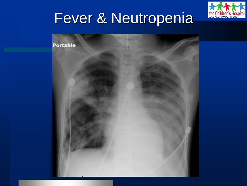
Fever & Neutropenia
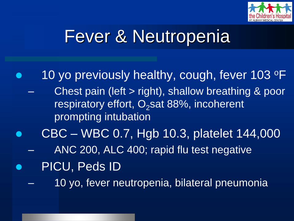
10 yo previously healthy, cough, fever 103 oF
– Chest pain (left > right), shallow breathing & poor
respiratory effort, O2sat 88%, incoherent
prompting intubation
CBC – WBC 0.7, Hgb 10.3, platelet 144,000
– ANC 200, ALC 400; rapid flu test negative
PICU, Peds ID
– 10 yo, fever neutropenia, bilateral pneumonia
Fever & Neutropenia
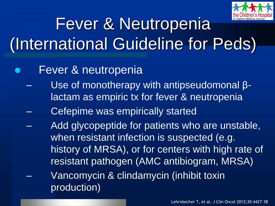
Fever & neutropenia
– Use of monotherapy with antipseudomonal β-
lactam as empiric tx for fever & neutropenia
– Cefepime was empirically started
– Add glycopeptide for patients who are unstable,
when resistant infection is suspected (e.g.
history of MRSA), or for centers with high rate of
resistant pathogen (AMC antibiogram, MRSA)
– Vancomycin & clindamycin (inhibit toxin
production)
Fever & Neutropenia
(International Guideline for Peds)
Lehrnbecher T, et al. J Clin Oncol 2012;30:4427-38
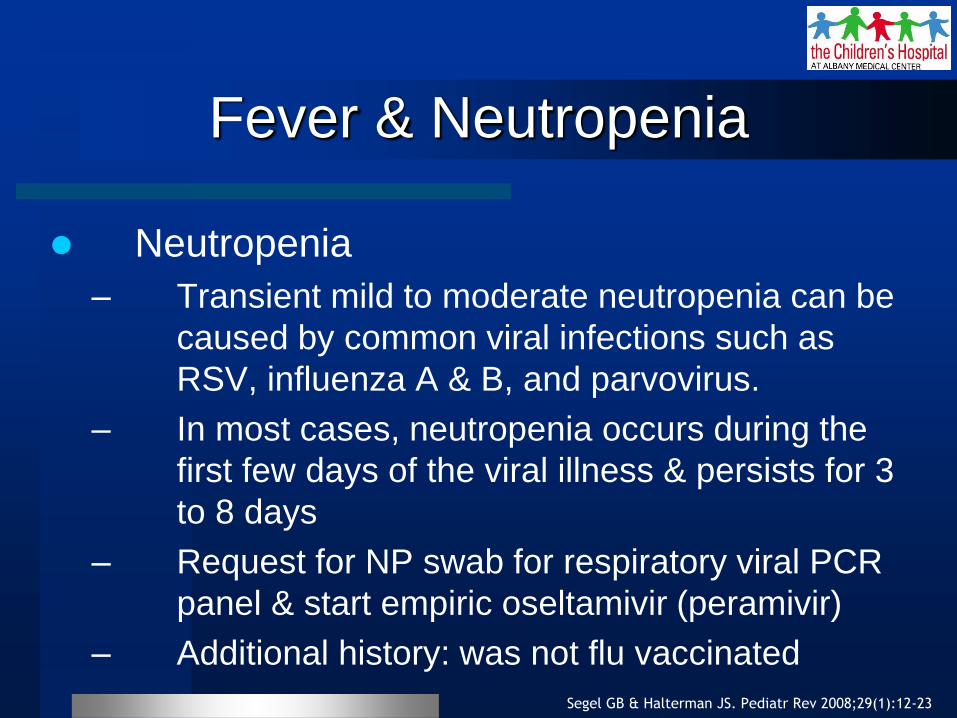
Neutropenia
– Transient mild to moderate neutropenia can be
caused by common viral infections such as
RSV, influenza A & B, and parvovirus.
– In most cases, neutropenia occurs during the
first few days of the viral illness & persists for 3
to 8 days
– Request for NP swab for respiratory viral PCR
panel & start empiric oseltamivir (peramivir)
– Additional history: was not flu vaccinated
Fever & Neutropenia
Segel GB & Halterman JS. Pediatr Rev 2008;29(1):12-23
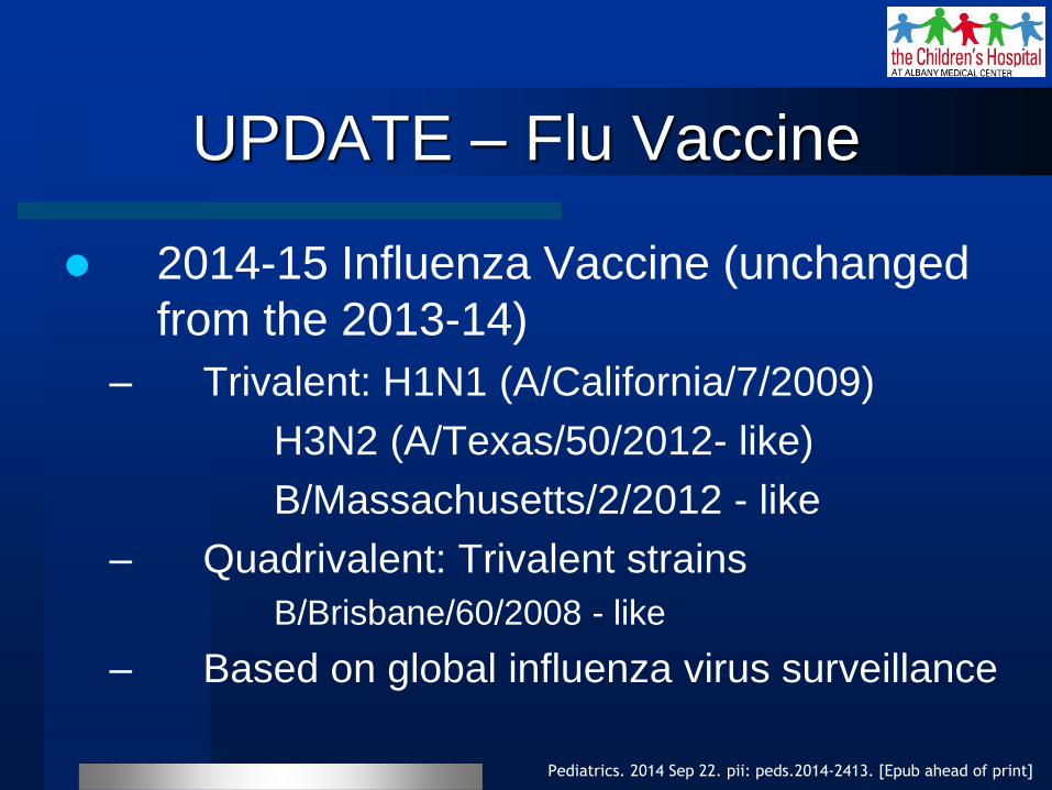
UPDATE – Flu Vaccine
2014-15 Influenza Vaccine (unchanged
from the 2013-14)
– Trivalent: H1N1 (A/California/7/2009)
H3N2 (A/Texas/50/2012- like)
B/Massachusetts/2/2012 - like
– Quadrivalent: Trivalent strains
B/Brisbane/60/2008 - like
– Based on global influenza virus surveillance
Pediatrics. 2014 Sep 22. pii: peds.2014-2413. [Epub ahead of print]
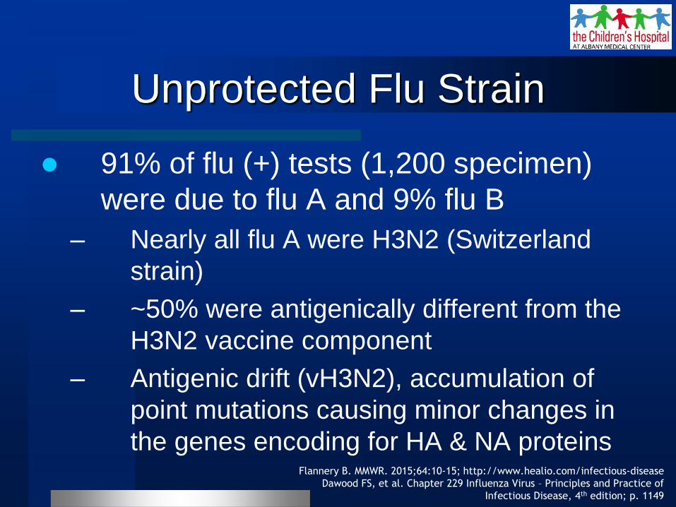
Unprotected Flu Strain
91% of flu (+) tests (1,200 specimen)
were due to flu A and 9% flu B
– Nearly all flu A were H3N2 (Switzerland
strain)
– ~50% were antigenically different from the
H3N2 vaccine component
– Antigenic drift (vH3N2), accumulation of
point mutations causing minor changes in
the genes encoding for HA & NA proteins Flannery B. MMWR. 2015;64:10-15; http://www.healio.com/infectious-disease
Dawood FS, et al. Chapter 229 Influenza Virus – Principles and Practice of
Infectious Disease, 4th edition; p. 1149
http://www.cartoonstock.com/directory/i/inoculation.asp
Flu vaccine
effectiveness
is low (23%)
Flannery B. MMWR. 2015;64:10-15
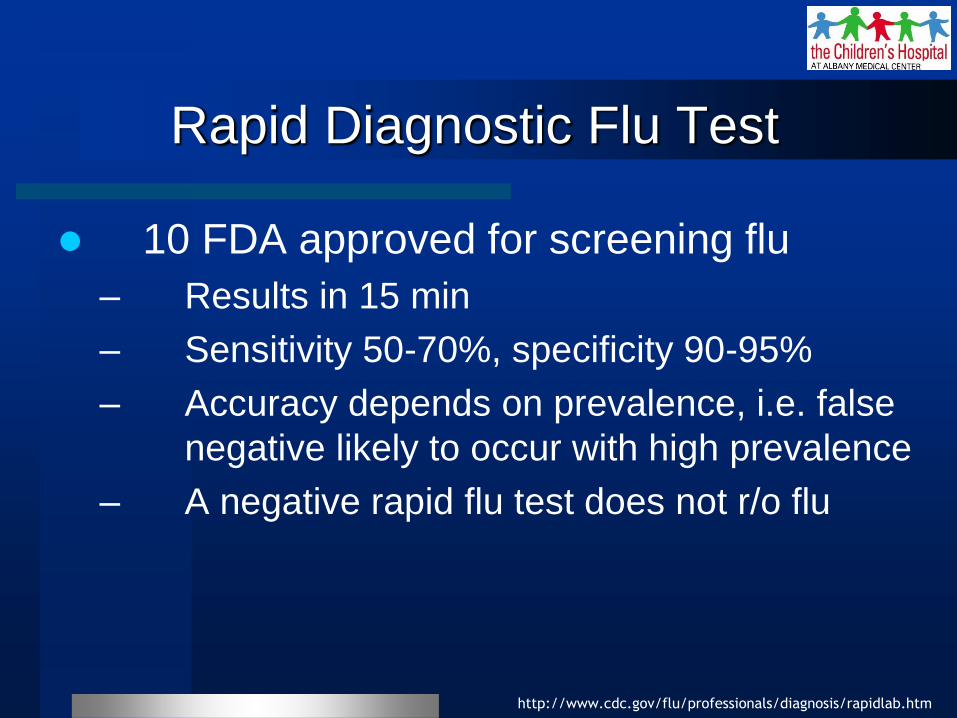
10 FDA approved for screening flu
– Results in 15 min
– Sensitivity 50-70%, specificity 90-95%
– Accuracy depends on prevalence, i.e. false
negative likely to occur with high prevalence
– A negative rapid flu test does not r/o flu
Rapid Diagnostic Flu Test
http://www.cdc.gov/flu/professionals/diagnosis/rapidlab.htm
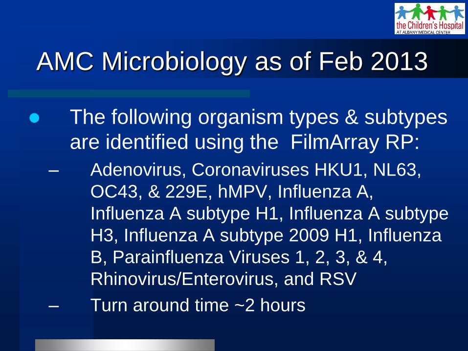
The following organism types & subtypes
are identified using the FilmArray RP:
– Adenovirus, Coronaviruses HKU1, NL63,
OC43, & 229E, hMPV, Influenza A,
Influenza A subtype H1, Influenza A subtype
H3, Influenza A subtype 2009 H1, Influenza
B, Parainfluenza Viruses 1, 2, 3, & 4,
Rhinovirus/Enterovirus, and RSV
– Turn around time ~2 hours
AMC Microbiology as of Feb 2013
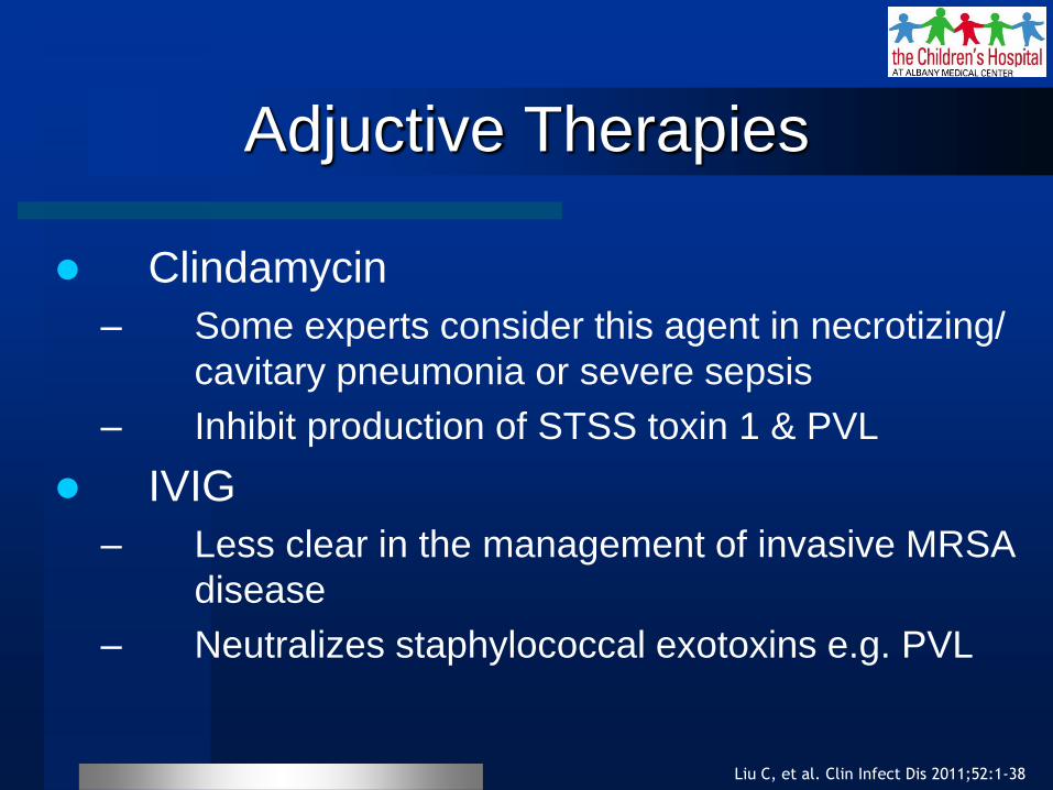
Clindamycin
– Some experts consider this agent in necrotizing/
cavitary pneumonia or severe sepsis
– Inhibit production of STSS toxin 1 & PVL
IVIG
– Less clear in the management of invasive MRSA
disease
– Neutralizes staphylococcal exotoxins e.g. PVL
Adjuctive Therapies
Liu C, et al. Clin Infect Dis 2011;52:1-38
Emergency use authorization 2009-10 H1N1
FDA approved - 1st neuraminidase inhibitor
for IV administration
Similar efficacy to PO oseltamivir
Off label use in hospitalized patients with
severe influenza
Indication: Severe bilateral pneumonia, ANC
200, with concerns for malabsorption
IV Peramivir
The Medical Letter 2015;57(1461): 17-19 Available at
http://secure.medicalletter.org/TML-article-1461b
10 yo previously healthy, bilateral severe
pneumonia, febrile & neutropenia associated
with flu B & MRSA
– Cefepime & vancomycin
– Clindamycin & IVIG
– Oseltamivir & peramivir
– Transferred to a hospital in Boston for ECMO
regarding ARDS
Fever & Neutropenia
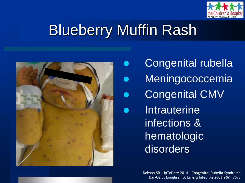
Blueberry Muffin Rash
Congenital rubella
Meningococcemia
Congenital CMV
Intrauterine
infections &
hematologic
disorders
Dobson SR. UpToDate 2014 – Congenital Rubella Syndrome
Bar-Oz B, Loughran B. Emerg Infec Dis 2003;9(6): 7578
Initially described with congenital rubella due
to extramedullary dermal erythropoiesis
– In the setting of profound anemia
Pathophysiology is not clear
– Hematopoietic stem cells migrate from the bone
marrow & settle in the skin
– Or dermal mesenchymal cells differentiate in situ
into blood-producing cells
Blueberry Muffin Rash
http://neoreviews.aappublications.org/site/case49/experts.xhtml
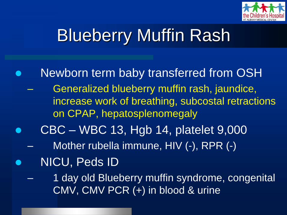
Newborn term baby transferred from OSH
– Generalized blueberry muffin rash, jaundice,
increase work of breathing, subcostal retractions
on CPAP, hepatosplenomegaly
CBC – WBC 13, Hgb 14, platelet 9,000
– Mother rubella immune, HIV (-), RPR (-)
NICU, Peds ID
– 1 day old Blueberry muffin syndrome, congenital
CMV, CMV PCR (+) in blood & urine
Blueberry Muffin Rash
Ganciclovir 6 mg/kg IV q12h, 21 days
Valganciclovir 16 mg/kg PO q12 hr, up to 6
mos.
– Treatment recommended for symptomatic
congenital CMV disease, with or without CNS
involvement
– Treatment should start in the 1st month of life
– Benefit for hearing loss and neurodevelopmental
outcomes
Blueberry Muffin Rash
Bradley JS (Eds). Nelson’s Pediatric Antimicrobial Therapy 2015; 21st ed.:21
Most common side effects: neutropenia
– 68% in IV ganciclovir
– 20% in PO valganciclovir
Some patients responds to G-CSF or
discontinuation of therapy
Blueberry Muffin Rash
Bradley JS (Eds). Nelson’s Pediatric Antimicrobial Therapy 2015; 21st ed.:21
IV GCV x 6 weeks improves audiologic
outcomes at 6 months but the benefits wane
over time
RCT PO VGC with symptomatic congenital
CMV comparing 6 months vs. 6 weeks of tx
– 1o end point change in hearing in the best ear
from baseline to 6 months
– 2o end point change in hearing in the best ear
from baseline to 12 – 24 months, neuro-
development
6 months versus 6 weeks
Kimberlin D, et al. N Engl J Med 2015; 372:933-43
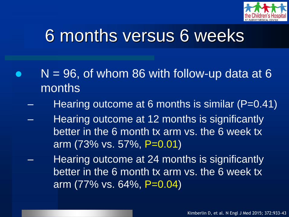
N = 96, of whom 86 with follow-up data at 6
months
– Hearing outcome at 6 months is similar (P=0.41)
– Hearing outcome at 12 months is significantly
better in the 6 month tx arm vs. the 6 week tx
arm (73% vs. 57%, P=0.01)
– Hearing outcome at 24 months is significantly
better in the 6 month tx arm vs. the 6 week tx
arm (77% vs. 64%, P=0.04)
6 months versus 6 weeks
Kimberlin D, et al. N Engl J Med 2015; 372:933-43
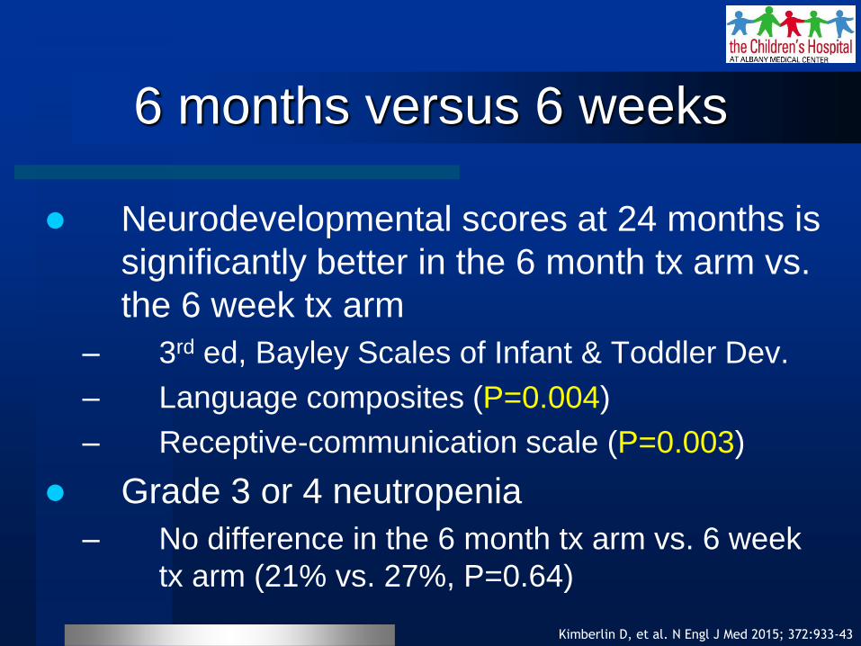
Neurodevelopmental scores at 24 months is
significantly better in the 6 month tx arm vs.
the 6 week tx arm
– 3rd ed, Bayley Scales of Infant & Toddler Dev.
– Language composites (P=0.004)
– Receptive-communication scale (P=0.003)
Grade 3 or 4 neutropenia
– No difference in the 6 month tx arm vs. 6 week
tx arm (21% vs. 27%, P=0.64)
6 months versus 6 weeks
Kimberlin D, et al. N Engl J Med 2015; 372:933-43
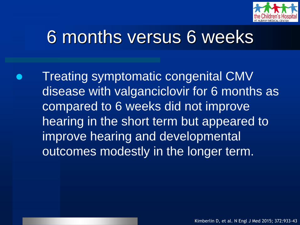
Treating symptomatic congenital CMV
disease with valganciclovir for 6 months as
compared to 6 weeks did not improve
hearing in the short term but appeared to
improve hearing and developmental
outcomes modestly in the longer term.
6 months versus 6 weeks
Kimberlin D, et al. N Engl J Med 2015; 372:933-43
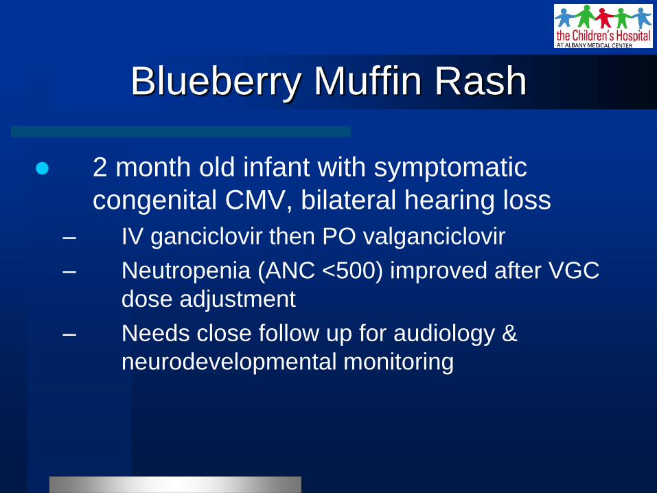
2 month old infant with symptomatic
congenital CMV, bilateral hearing loss
– IV ganciclovir then PO valganciclovir
– Neutropenia (ANC <500) improved after VGC
dose adjustment
– Needs close follow up for audiology &
neurodevelopmental monitoring
Blueberry Muffin Rash
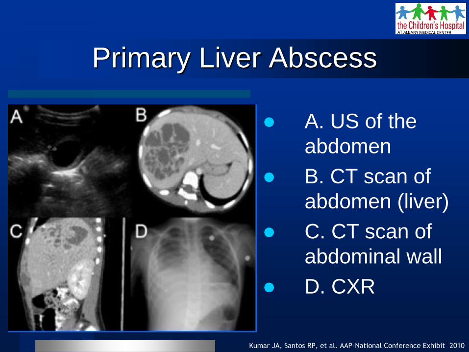
Primary Liver Abscess
Kumar JA, Santos RP, et al. AAP-National Conference Exhibit 2010
A. US of the
abdomen
B. CT scan of
abdomen (liver)
C. CT scan of
abdominal wall
D. CXR
7 yo previously healthy female with nausea,
diarrhea, severe abdominal pain, fever, &
hypotension hospitalized with concern for
sepsis.
– No history of travel to endemic areas in Asia
US of the abdomen showed a multiloculated
mass in the right hepatic lobe.
– A CT guided drainage obtained 200-mL of
purulent material which yielded glistening mucoid
colonies.
Primary Liver Abscess
Kumar JA, Santos RP, et al. AAP-National Conference Exhibit 2010
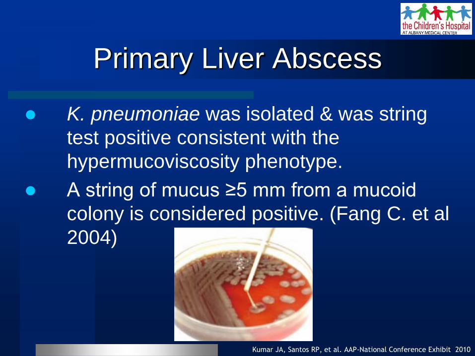
K. pneumoniae was isolated & was string
test positive consistent with the
hypermucoviscosity phenotype.
A string of mucus ≥5 mm from a mucoid
colony is considered positive. (Fang C. et al
2004)
Primary Liver Abscess
Kumar JA, Santos RP, et al. AAP-National Conference Exhibit 2010
Her hospital course was complicated with
bacteremia, cholecystitis status post open
cholecystectomy, & right pleural effusion
requiring drainage & chest tube placement.
– Improved while on antimicrobial regimen (≥3
weeks)
Primary Liver Abscess
Kumar JA, Santos RP, et al. AAP-National Conference Exhibit 2010
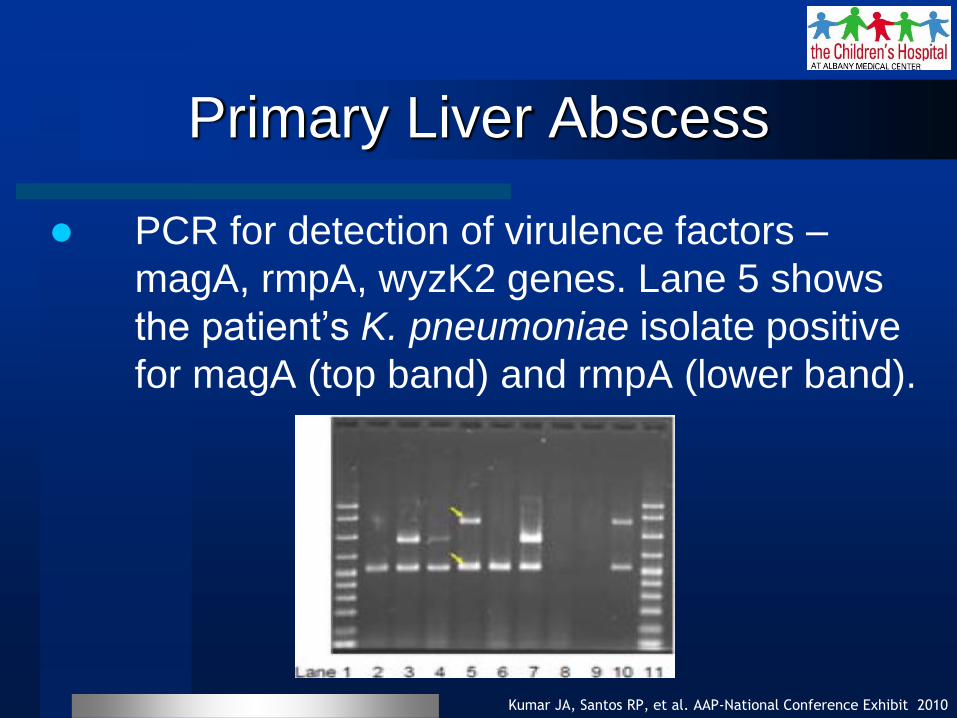
PCR for detection of virulence factors –
magA, rmpA, wyzK2 genes. Lane 5 shows
the patient’s K. pneumoniae isolate positive
for magA (top band) and rmpA (lower band).
Primary Liver Abscess
Kumar JA, Santos RP, et al. AAP-National Conference Exhibit 2010
Molecular studies showed the presence of
both magA associated with virulence through
K1 serotype expression & rmpA, a regulator
of capsular polysaccharide synthesis.
– Consistent with hypermucoviscosity phenotype.
– Hypermucoviscosity phenotype (e.g. glistening
mucoid colonies, string test positive) should
prompt clinicians to look for other foci of
infections.
Primary Liver Abscess
Kumar JA, Santos RP, et al. AAP-National Conference Exhibit 2010
This is an emerging disease due to the
absence of traditional risk factors such as
chronic medical conditions & exposure to
endemic areas.
This is the first case report of K. pneumoniae
isolate with genotypic characteristics similar
to those reported in Asia (magA+ & rmpA+)
causing invasive disease in a previously
healthy child.
Primary Liver Abscess
Kumar JA, Santos RP, et al. AAP-National Conference Exhibit 2010
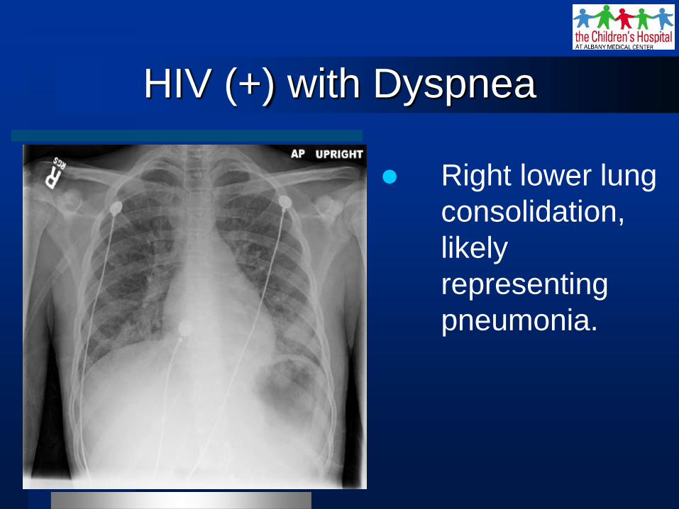
HIV (+) with Dyspnea
Right lower lung
consolidation,
likely
representing
pneumonia.
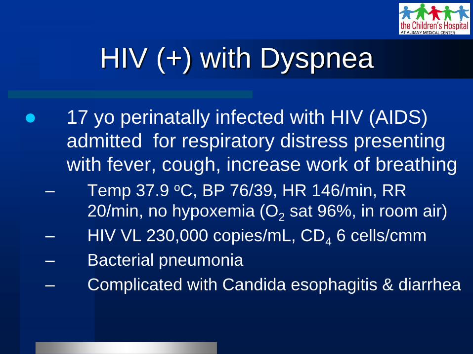
17 yo perinatally infected with HIV (AIDS)
admitted for respiratory distress presenting
with fever, cough, increase work of breathing
– Temp 37.9 oC, BP 76/39, HR 146/min, RR
20/min, no hypoxemia (O2 sat 96%, in room air)
– HIV VL 230,000 copies/mL, CD4 6 cells/cmm
– Bacterial pneumonia
– Complicated with Candida esophagitis & diarrhea
HIV (+) with Dyspnea
HIV (+) with Dyspnea
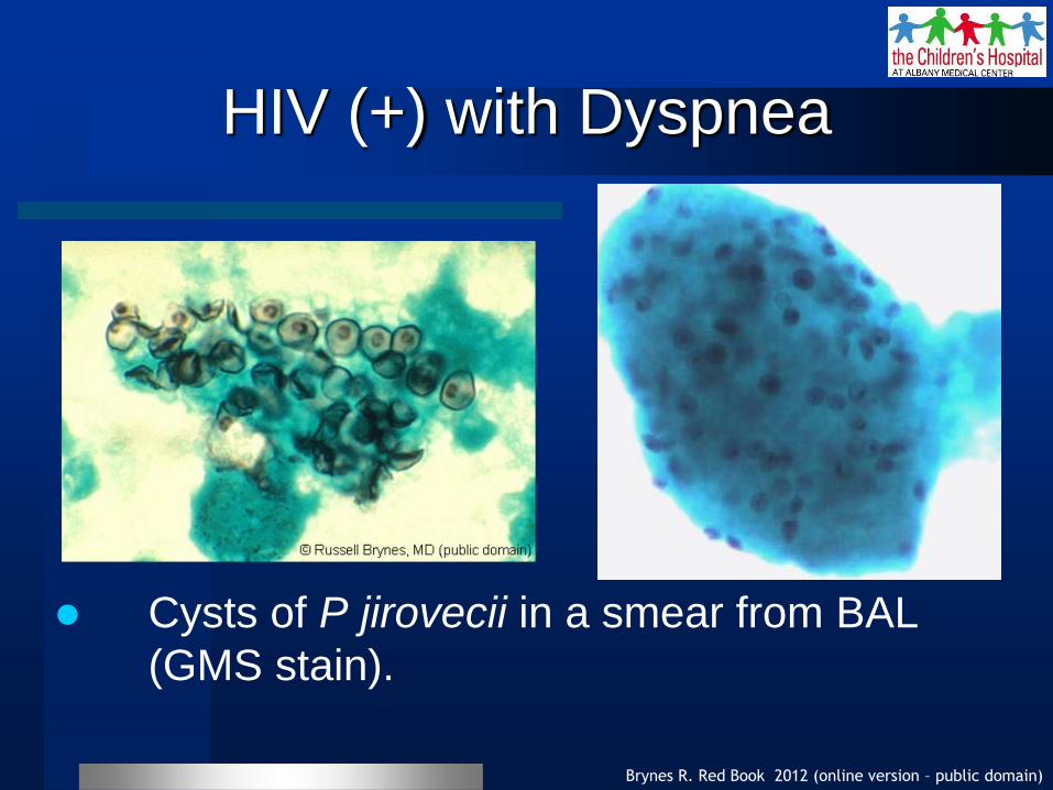
Brynes R. Red Book 2012 (online version – public domain)
Cysts of P jirovecii in a smear from BAL
(GMS stain).
Most children with PneumoCystis pneumonia
(PCP) are hypoxic with low arterial O2.
Characteristic syndrome of subacute diffuse
pneumonitis with dyspnea, tachypnea, O2
desaturation, nonproductive cough, & fever.
Mortality rate in immunocompromised
patients ranges from 5%-40% with tx &
approaches 100% without tx.
PCP
AAP. 2015 Red Book – Pneumocystis jirovecii; 30th ed:638-44
CXR often show bilateral diffuse interstitial or
alveolar disease; rarely, lobar, cavitary,
miliary, & nodular lesions or even no lesions
are seen.
A definitive diagnosis of PCP is made by
visualization of organisms in lung tissue or
respiratory tract secretion specimens.
PCP
AAP. 2015 Red Book – Pneumocystis jirovecii; 30th ed:638-44
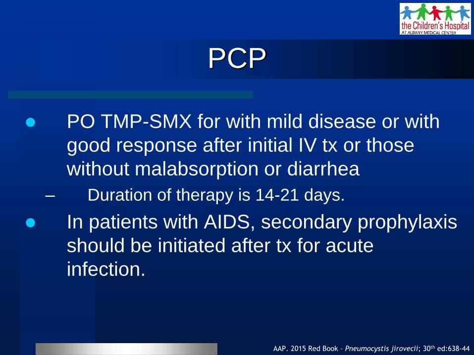
PO TMP-SMX for with mild disease or with
good response after initial IV tx or those
without malabsorption or diarrhea
– Duration of therapy is 14-21 days.
In patients with AIDS, secondary prophylaxis
should be initiated after tx for acute
infection.
PCP
AAP. 2015 Red Book – Pneumocystis jirovecii; 30th ed:638-44

17 yo perinatally infected with HIV with AIDS
– TMP-SMX for 3 weeks for PCP then 2nd
prophylaxis with TMP-SMX SS
– Fluconazole for candida esophagitis for 3
weeks then suppressive regimen
– MAC prophylaxis was offered
– cART – RPV/TDF-FTC QD + RAL BID
HIV (+) with Dyspnea
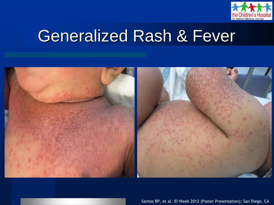
Generalized Rash & Fever
Santos RP, et al. ID Week 2012 (Poster Presentation); San Diego, CA
Generalized Rash & Fever
Santos RP, et al. ID Week 2012 (Poster Presentation); San Diego, CA
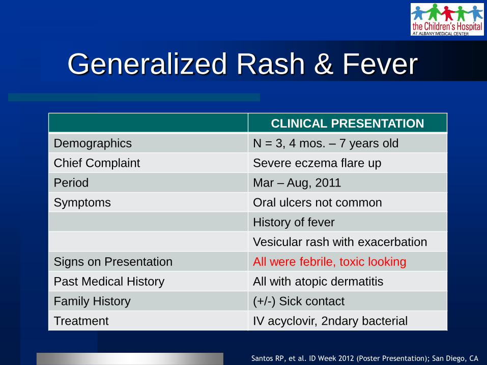
CLINICAL PRESENTATION
Demographics N = 3, 4 mos. – 7 years old
Chief Complaint Severe eczema flare up
Period Mar – Aug, 2011
Symptoms Oral ulcers not common
History of fever
Vesicular rash with exacerbation
Signs on Presentation All were febrile, toxic looking
Past Medical History All with atopic dermatitis
Family History (+/-) Sick contact
Treatment IV acyclovir, 2ndary bacterial
Generalized Rash & Fever
Santos RP, et al. ID Week 2012 (Poster Presentation); San Diego, CA
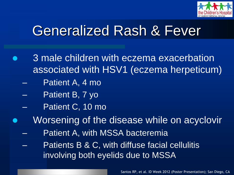
3 male children with eczema exacerbation
associated with HSV1 (eczema herpeticum)
– Patient A, 4 mo
– Patient B, 7 yo
– Patient C, 10 mo
Worsening of the disease while on acyclovir
– Patient A, with MSSA bacteremia
– Patients B & C, with diffuse facial cellulitis
involving both eyelids due to MSSA
Generalized Rash & Fever
Santos RP, et al. ID Week 2012 (Poster Presentation); San Diego, CA
All patients improved after completing
acyclovir regimen & ~10 days of antibiotics
– Patient A, cefazolin
– Patient B, clindamycin
– Patient C, cefazolin
No recurrence of EH while on suppressive
acyclovir regimen (10 mg/kg PO BID) &
vitamin D supplements
Eczema Herpeticum (HSV1)
Santos RP, et al. ID Week 2012 (Poster Presentation); San Diego, CA
Stollery N. The Practitioner 2011;255(1738):32-3
Diamond C, et al. Pediatr Infect Dis J 1999;18(6):487-9
Stanberry LR, et al. Clin Infect Dis 1994;18:401-7
Eczema herpeticum (EH) is a dermatologic
emergency associated with herpes simplex
virus (HSV) type 1 viremia.
HSV viremia had been rarely described in
immunocompetent and more commonly
among immunocompromised children.
Eczema Herpeticum (HSV1)
Santos RP, et al. ID Week 2012 (Poster Presentation); San Diego, CA
Aronson PL, et al. Pediatrics 2011;128:1161-7
Improved clinical outcome requires prompt
recognition of EH since delayed (>1 day)
acyclovir initiation is associated with
prolonged length of hospital stay.
– In our patients, systemic antiviral agents were
given within 24-48 hours of clinical presentation.
Another Rash & Fever
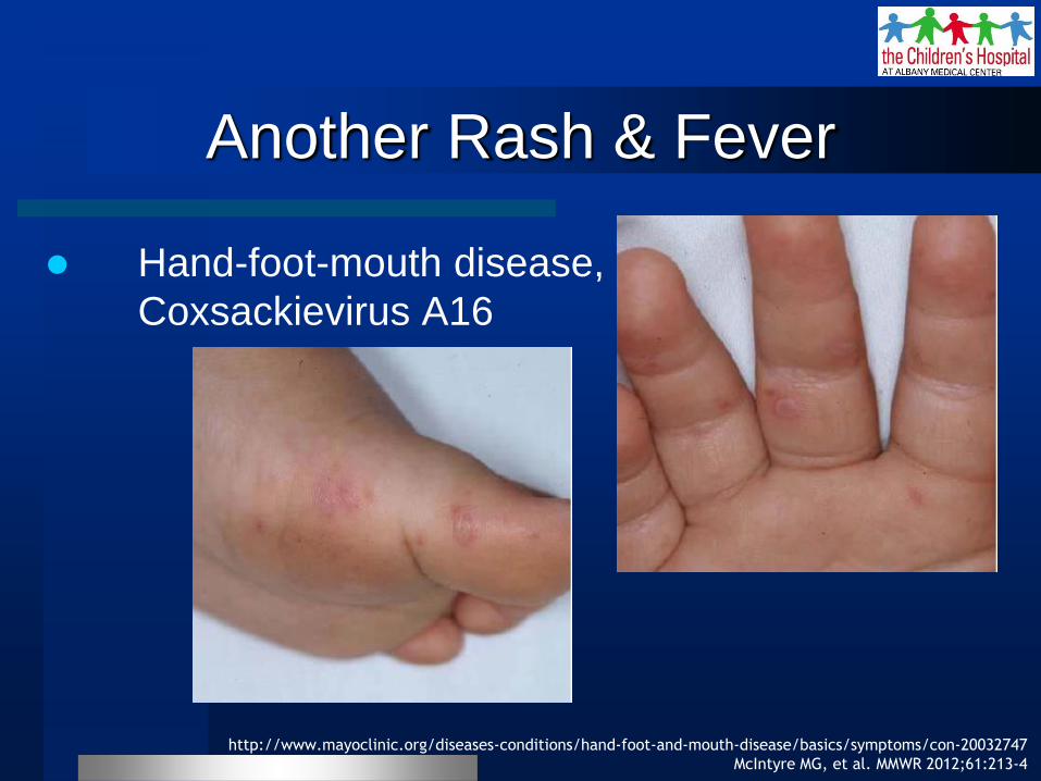
Hand-foot-mouth disease,
Coxsackievirus A16
http://www.mayoclinic.org/diseases-conditions/hand-foot-and-mouth-disease/basics/symptoms/con-20032747
McIntyre MG, et al. MMWR 2012;61:213-4
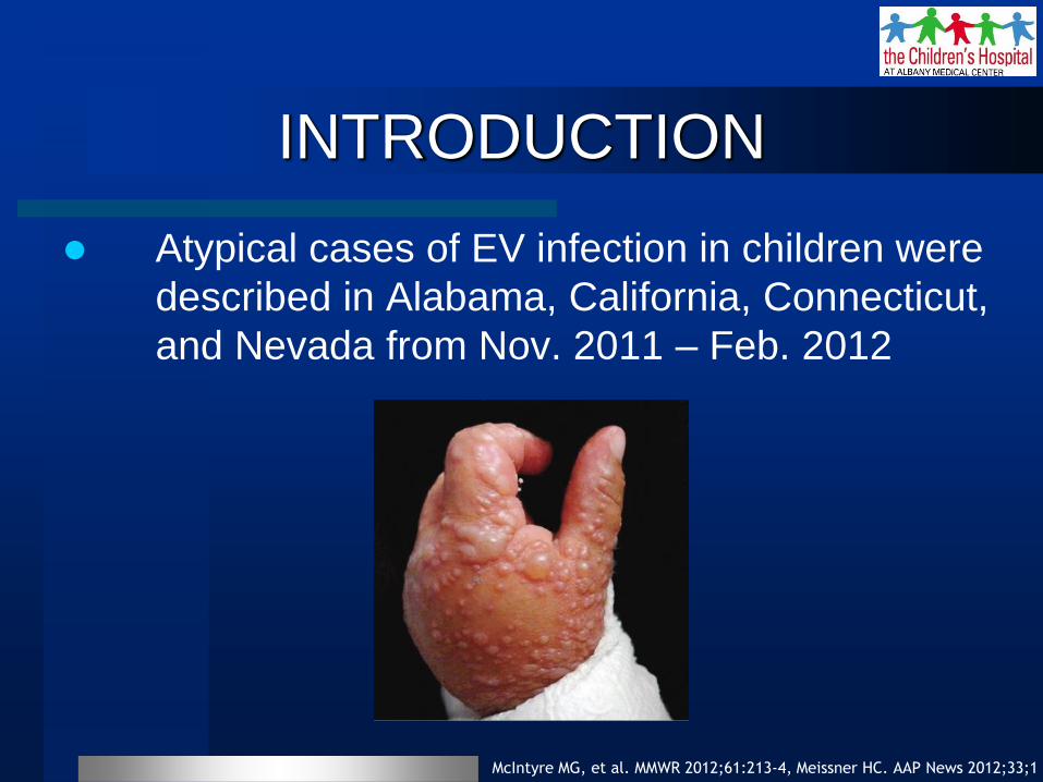
INTRODUCTION
Atypical cases of EV infection in children were
described in Alabama, California, Connecticut,
and Nevada from Nov. 2011 – Feb. 2012
McIntyre MG, et al. MMWR 2012;61:213-4, Meissner HC. AAP News 2012;33;1
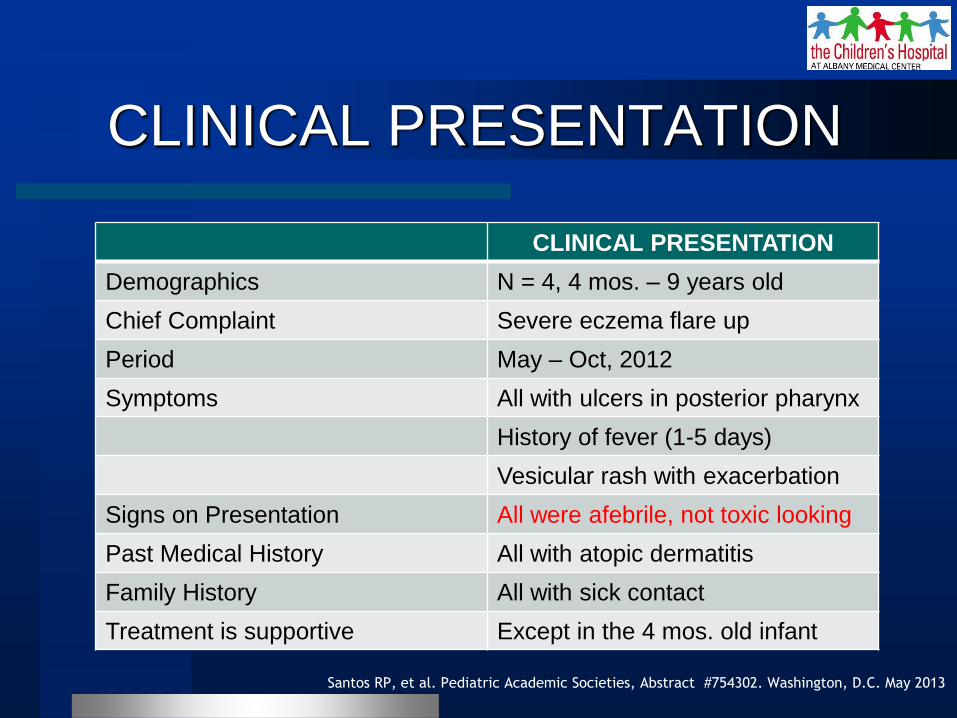
CLINICAL PRESENTATION
Santos RP, et al. Pediatric Academic Societies, Abstract #754302. Washington, D.C. May 2013
CLINICAL PRESENTATION
Demographics N = 4, 4 mos. – 9 years old
Chief Complaint Severe eczema flare up
Period May – Oct, 2012
Symptoms All with ulcers in posterior pharynx
History of fever (1-5 days)
Vesicular rash with exacerbation
Signs on Presentation All were afebrile, not toxic looking
Past Medical History All with atopic dermatitis
Family History All with sick contact
Treatment is supportive Except in the 4 mos. old infant
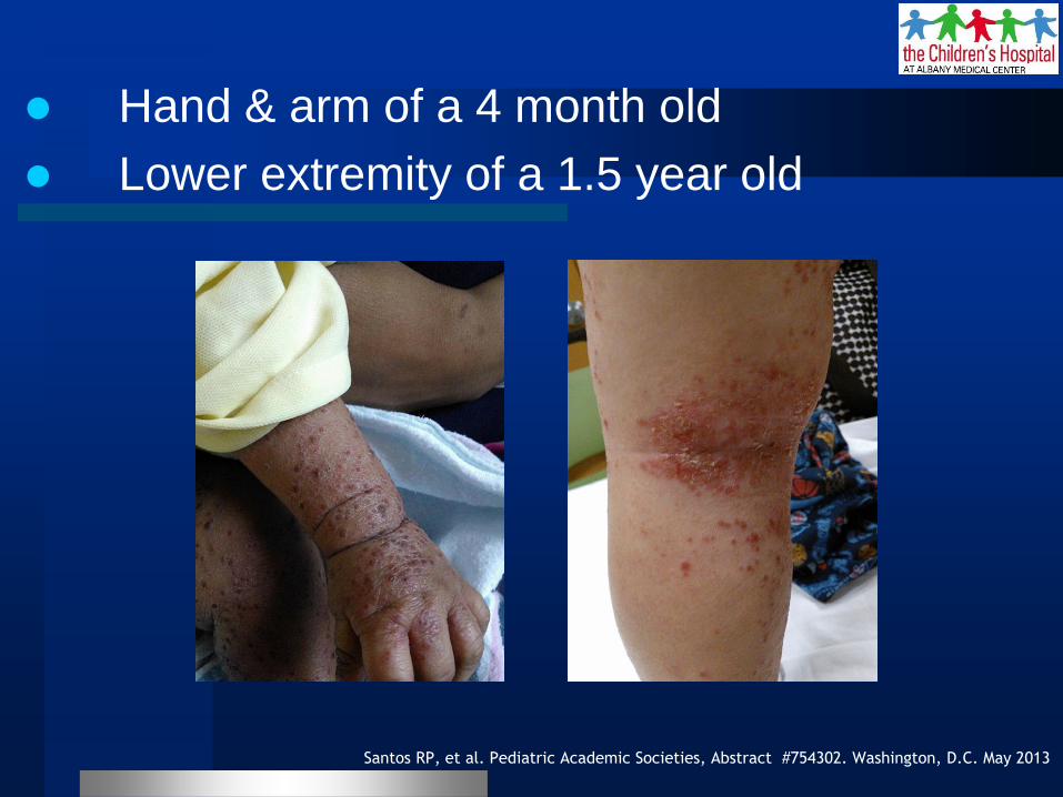
Hand & arm of a 4 month old
Lower extremity of a 1.5 year old
Santos RP, et al. Pediatric Academic Societies, Abstract #754302. Washington, D.C. May 2013
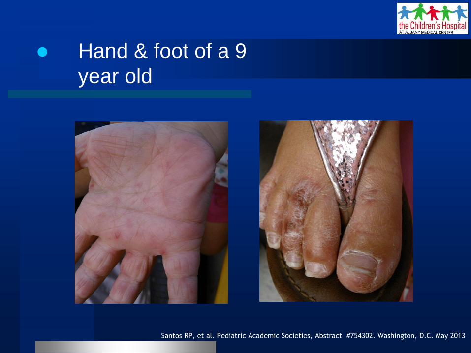
Hand & foot of a 9
year old
Santos RP, et al. Pediatric Academic Societies, Abstract #754302. Washington, D.C. May 2013
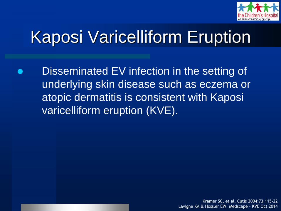
Kaposi Varicelliform Eruption
Disseminated EV infection in the setting of
underlying skin disease such as eczema or
atopic dermatitis is consistent with Kaposi
varicelliform eruption (KVE).
Kramer SC, et al. Cutis 2004;73:115-22
Lavigne KA & Hossler EW. Medscape – KVE Oct 2014
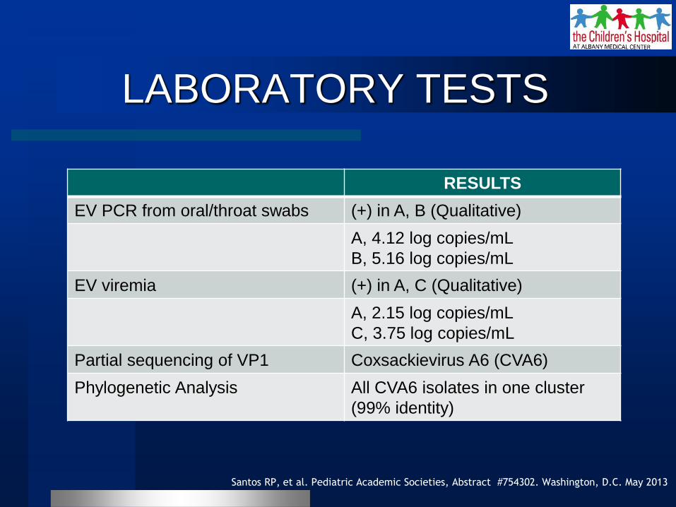
LABORATORY TESTS
RESULTS
EV PCR from oral/throat swabs (+) in A, B (Qualitative)
A, 4.12 log copies/mL
B, 5.16 log copies/mL
EV viremia (+) in A, C (Qualitative)
A, 2.15 log copies/mL
C, 3.75 log copies/mL
Partial sequencing of VP1 Coxsackievirus A6 (CVA6)
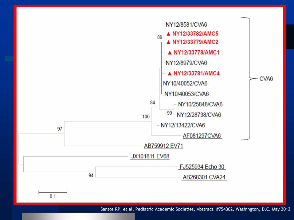
Phylogenetic Analysis All CVA6 isolates in one cluster
(99% identity)
Santos RP, et al. Pediatric Academic Societies, Abstract #754302. Washington, D.C. May 2013
Santos RP, et al. Pediatric Academic Societies, Abstract #754302. Washington, D.C. May 2013
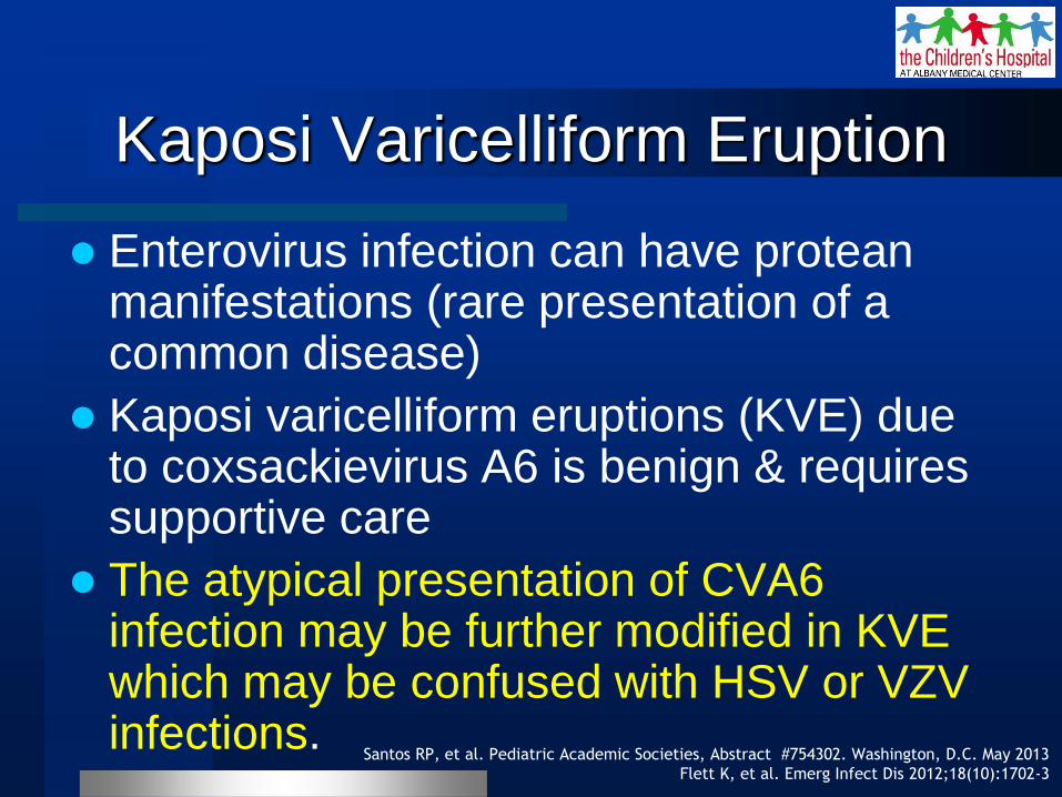
Kaposi Varicelliform Eruption
Enterovirus infection can have protean manifestations (rare presentation of a common disease)
Kaposi varicelliform eruptions (KVE) due to coxsackievirus A6 is benign & requires supportive care
The atypical presentation of CVA6 infection may be further modified in KVE which may be confused with HSV or VZV infections.
Santos RP, et al. Pediatric Academic Societies, Abstract #754302. Washington, D.C. May 2013
Flett K, et al. Emerg Infect Dis 2012;18(10):1702-3
TAKE HOME MESSAGE
Identified clinically emergent infectious
diseases through visual diagnosis.
Described the clinical course of emergent
infectious diseases.
Reviewed evidenced-based
recommendations in the management of
emergent infectious diseases.
When in doubt, call Peds ID