Diagnostic performance of multidetector computed ... fileORIGINAL ARTICLE Diagnostic performance of...
9
ORIGINAL ARTICLE Diagnostic performance of multidetector computed tomography in the evaluation of esophageal varices Tarek ELKammash a , Inas ELFiky b , Fatma Zaiton b, * , Soha E. Khorshed c a Department of Diagnostic Radiology, Suez Canal University, Egypt b Department of Diagnostic Radiology, Zagazig University, Egypt c Department of Tropical Medicine, Zagazig University, Egypt Received 4 June 2015; accepted 3 November 2015 Available online 19 November 2015 KEYWORDS Esophageal varices; Multidetector CT (MDCT); Portosystemic collaterals; Red color sign Abstract Objective: Our purpose was to evaluate the role of multidetector computed tomography (MDCT) in evaluation of esophageal varices (EV). Patients and methods: 112 patients with liver cirrhosis were included, EV was evaluated for grades, presence of collateral, palisade vein dilatation and also patient acceptability. Results: The sensitivity of MDCT for radiologist A was 94.8%, specificity 98.5%, Accuracy 97.8%, PPV 94.8%, NPV 98.5% and for radiologist B, 99.4%, 99.6%, 99.6%, 99.3% and 99.7% respectively. MDCT detected para esophageal varices in 38 cases, gastric fundus varices in 47 cases and splenorenal collaterals were seen in 14 cases, palisade vein dilatation was +ve in 58 cases, ve in 47 cases and (±) in 7 cases. 3 cases of HCC and 1 liver cyst were incidentally found during exam- ination. There was a highly significant correlation between degree of palisade vein dilatation, increasing grade of esophageal varices and Red color sign with p value <0.01. MDCT was more accepted than endoscopy in 83%. The preference of CT was statistically significant p < 0.01. Conclusion: MDCT is a fast, well tolerable, non-invasive procedure and accepted from most of the examined patients for evaluation and grading of EV, detection of other portosystemic collaterals and hepatobiliary pathologies. Ó 2015 The Authors. The Egyptian Society of Radiology and Nuclear Medicine. Production and hosting by Elsevier B.V. This is an open access article under the CC BY-NC-ND license (http://creativecommons. org/licenses/by-nc-nd/4.0/). Introduction Portal hypertension is a serious complication of cirrhosis. It is defined as a hepatic venous pressure gradient (HVPG) above 5 mmHg. Development of significant complication for portal hypertension as ascites and/or esophageal and gastric varices generally develops when HVPG increases above 10 mmHg (1,2). Gastroesophageal varices were the most recognized por- tosystemic collaterals because their rupture results in danger- * Corresponding author. Mobile: +20 1060052849. E-mail addresses: [email protected] (T. ELKammash), [email protected] (I. ELFiky), [email protected] (F. Zaiton), [email protected] (S.E. Khorshed). Peer review under responsibility of Egyptian Society of Radiology and Nuclear Medicine. The Egyptian Journal of Radiology and Nuclear Medicine (2016) 47, 43–51 Egyptian Society of Radiology and Nuclear Medicine The Egyptian Journal of Radiology and Nuclear Medicine www.elsevier.com/locate/ejrnm www.sciencedirect.com http://dx.doi.org/10.1016/j.ejrnm.2015.11.003 0378-603X Ó 2015 The Authors. The Egyptian Society of Radiology and Nuclear Medicine. Production and hosting by Elsevier B.V. This is an open access article under the CC BY-NC-ND license (http://creativecommons.org/licenses/by-nc-nd/4.0/).
Transcript of Diagnostic performance of multidetector computed ... fileORIGINAL ARTICLE Diagnostic performance of...
The Egyptian Journal of Radiology and Nuclear Medicine (2016) 47, 43–51
Egyptian Society of Radiology and Nuclear Medicine
The Egyptian Journal of Radiology andNuclearMedicine
www.elsevier.com/locate/ejrnmwww.sciencedirect.com
ORIGINAL ARTICLE
Diagnostic performance of multidetector computed
tomography in the evaluation of esophageal varices
* Corresponding author. Mobile: +20 1060052849.
E-mail addresses: [email protected] (T. ELKammash),
[email protected] (I. ELFiky), [email protected]
(F. Zaiton), [email protected] (S.E. Khorshed).
Peer review under responsibility of Egyptian Society of Radiology and
Nuclear Medicine.
http://dx.doi.org/10.1016/j.ejrnm.2015.11.0030378-603X � 2015 The Authors. The Egyptian Society of Radiology and Nuclear Medicine. Production and hosting by Elsevier B.V.This is an open access article under the CC BY-NC-ND license (http://creativecommons.org/licenses/by-nc-nd/4.0/).
Tarek ELKammash a, Inas ELFiky b, Fatma Zaiton b,*, Soha E. Khorshed c
aDepartment of Diagnostic Radiology, Suez Canal University, EgyptbDepartment of Diagnostic Radiology, Zagazig University, EgyptcDepartment of Tropical Medicine, Zagazig University, Egypt
Received 4 June 2015; accepted 3 November 2015Available online 19 November 2015
KEYWORDS
Esophageal varices;
Multidetector CT (MDCT);
Portosystemic collaterals;
Red color sign
Abstract Objective: Our purpose was to evaluate the role of multidetector computed tomography
(MDCT) in evaluation of esophageal varices (EV).
Patients and methods: 112 patients with liver cirrhosis were included, EV was evaluated for grades,
presence of collateral, palisade vein dilatation and also patient acceptability.
Results: The sensitivity of MDCT for radiologist A was 94.8%, specificity 98.5%, Accuracy
97.8%, PPV 94.8%, NPV 98.5% and for radiologist B, 99.4%, 99.6%, 99.6%, 99.3% and 99.7%
respectively. MDCT detected para esophageal varices in 38 cases, gastric fundus varices in 47 cases
and splenorenal collaterals were seen in 14 cases, palisade vein dilatation was +ve in 58 cases, �ve
in 47 cases and (±) in 7 cases. 3 cases of HCC and 1 liver cyst were incidentally found during exam-
ination. There was a highly significant correlation between degree of palisade vein dilatation,
increasing grade of esophageal varices and Red color sign with p value <0.01. MDCT was more
accepted than endoscopy in 83%. The preference of CT was statistically significant p< 0.01.
Conclusion: MDCT is a fast, well tolerable, non-invasive procedure and accepted from most of the
examined patients for evaluation and grading of EV, detection of other portosystemic collaterals
and hepatobiliary pathologies.� 2015 The Authors. The Egyptian Society of Radiology and Nuclear Medicine. Production and hosting
by Elsevier B.V. This is an open access article under the CC BY-NC-ND license (http://creativecommons.
org/licenses/by-nc-nd/4.0/).
Introduction
Portal hypertension is a serious complication of cirrhosis. It isdefined as a hepatic venous pressure gradient (HVPG) above5 mmHg. Development of significant complication for portalhypertension as ascites and/or esophageal and gastric varices
generally develops when HVPG increases above 10 mmHg(1,2).
Gastroesophageal varices were the most recognized por-
tosystemic collaterals because their rupture results in danger-

Case 1 50 years old male presented with posthepatitis cirrhosis; (a) CT post-contrast portal venous phase axial image shows single
enhanced vascular structure in the esophageal wall projecting inside the lumen measuring 3.2 mm in diameter representing single
esophageal varix (black arrow) (Score I); (b) CT multiplannar reformatted image shows the enhanced esophageal varix (black arrow); (c)
upper GI endoscopy shows single intraluminal tubular shaped esophageal varix not risky (grade 1) (white arrow) (CT Score 1, endoscopy
grade 1, RC 0).
44 T. ELKammash et al.
ous variceal bleeding, which is considered as the commonestlethal complication of portal hypertension (3).
Reports from the 1940s to the 1980s demonstrate mortality
rates 30–60% from variceal bleeding. Although mortality froma variceal bleeding has been reduced to be 20–30% with pro-gression of endoscopic and radiological techniques simultane-ously with new pharmacologic therapies, it remains of clinical
significance (4,5).Early diagnosis of gastroesophageal varices before the
onset of first bleed is highly recommended as many studies
showed that the risk of variceal bleeding can be reduced from50% to 15% for large esophageal varices (6).
Esophagogastroduodenoscopy (EGD) is the gold standard
in the diagnosis of gastroesophageal varices; however, the useof endoscopy as a method of screening is limited, due to itsinvasive, expensive, need sedation, and patients poor accep-
tance of the procedure (7–9).To limit the number of patients who should undergo
endoscopic screening, a noninvasive, less expensive and welltolerated test for diagnosis of varices with high sensitivity
and specificity has been studied, such as platelet count andprothrombin time; as well as radiological criteria such asspleen size, but found to be not highly accurate predictors of
high-risk varices (10). Ultrasound imaging also was noninva-sive, nonexpensive and well tolerated but it has limited
specificity and cannot substitute endoscopy as a screening toolfor large esophageal varices (11,12).
Computed tomography (CT) imaging is noninvasive, does
not necessitate sedation, and allows accurate assessment ofvariceal site and size, and it is also better tolerated by mostof the patients than endoscopy. With rapid evolution of CTtechnology especially the introduction of multi-detector com-
puted tomography (MDCT) imaging with its multiplanarcapabilities, esophageal, paraesophageal and gastric varicesas well as other portosystemic shunts was progressively recog-
nized in patients with liver cirrhosis (13–15).Magnetic resonance (MR) imaging is probably as accurate
as CT but is more expensive and less accessible; in addition,
some of the rarest pathways (eg, pleuropericardial or thoracicwall varices) may be missed at MR imaging (16).
The purpose of this study was to prospectively evaluate the
effectiveness of MDCT in evaluation of esophageal varices incirrhotic patients compared with finding of the gold standardEsophagogastroscopy.
Patients and methods
112 patients with liver cirrhosis were included in this study.Patients were examined at the period between 2011 and 2014
in Suez Canal and Zagazig University hospitals in radiology
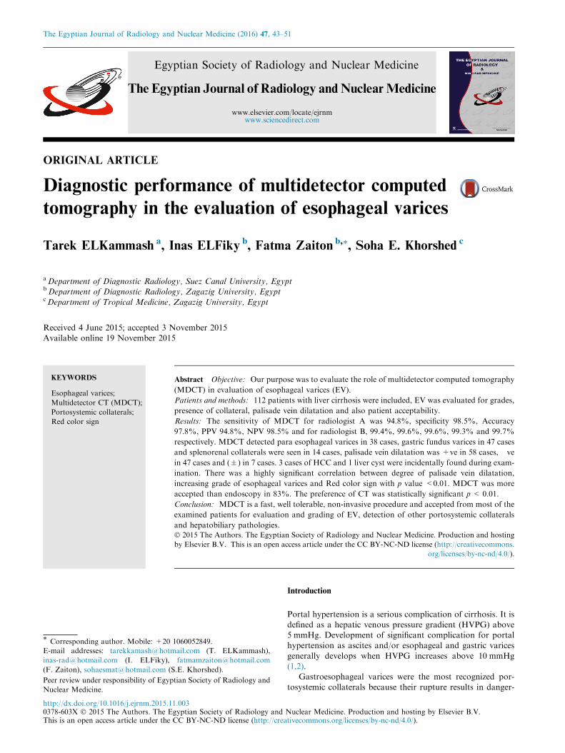
Case 2 A 47 years old male with posthepatitis liver cirrhosis. (a) CT axial post-contrast portal venous phase image shows multiple small
enhanced esophageal varices on the inner surface of the esophagus projecting inside the lumen measuring in between 1 and 2.7 mm in
diameter {white arrow}(Score 2), and there are associated enhanced para-esophageal varices. (b) Axial CT at different level shows the
enhanced paraesophageal varices (white arrow). (c) CT Coronal reformatted image shows the esophageal and paraesophageal (two white
arrows) with splenomegaly and splenic hilar varices. (d) CT Coronal reformatted image of the arterial phase of triphasic examination
shows ill defined rapidly enhancing hepatic focal mass involving segment VI with related neovascularity representing HCC. (e) Upper GIT
endoscopy shows multiple lobulated intraluminal esophageal varices not risky (white arrows) (grade 2) (CT Score 2, endoscopy grade 2,
RC 0).
Diagnostic performance of multidetector computed tomography 45
departments and endoscopy units. 77 patients were male and45 patients were females, and their ages ranged between 38and 72 years with mean age 51.4 years. Approval from com-
mittee board was obtained for the study and written consentwas taken from all patients after explanation of the procedureand any possible complications for the patients. All patients
had CT study with IV. Contrast injection followed by upperGIT endoscopy within 2 weeks from CT study.
Exclusion criteria were Patients with active gastrointestinal
hemorrhage, those with a history of endoscopic variceal liga-tion, those with a history of adverse reactions to iodinated con-trast agent, patients with known congenital anomalies of the
portal vein, and those who refused to do endoscopy after CTangiography were excluded.
CT examination for the abdomen
Plain CT examination including the lower chest and the upperabdomen was done first to demonstrate calcification and com-
pare pattern of enhancement, followed by triphasic examina-tion after injection of contrast media; 100 ml of iopamidol300, was injected using automatic injector (Medrad Stellant
injector, Indianola PA, USA), at a rate of 4.0 ml/s through a18-gauge IV catheter inserted into an antecubital vein.
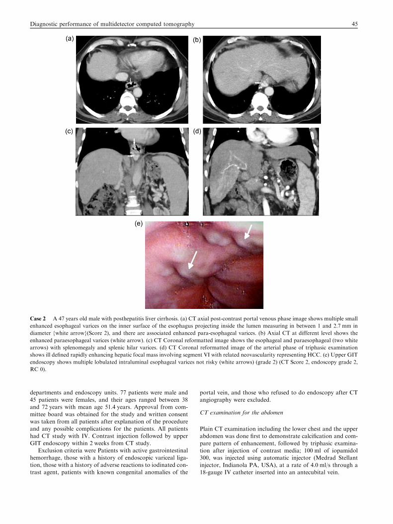
Case 3 A 38-years-old male with history of hepatitis. (a) CT axial post-contrast portal venous phase image shows multiple enhanced
vascular structures involving the whole circumference of the inner surface of lower esophagus measuring between 1.2 and 2.3 mm (white
arrow) (Score 3). (b) CT coronal reformatted image shows the multiple enhanced esophageal varices affecting lower esophagus (white
arrow). (c) Upper GIT endoscopy shows multiple tortuous tubular intraluminal esophageal varices (grade 3) with mucosal red spot seen
(white arrow) representing (RC 1) (CT Score 3, endoscopy grade 3, RC 1).
46 T. ELKammash et al.
Three sets of images were acquired in a craniocaudal direc-
tional at 25, 65, and 180 s after injection of the contrast medium.The first acquisitionwas used for hepatic arterial phase imaging;the second acquisition for portal venous phase imaging, and the
3rd acquisition to image the hepatic venous phase. Images wereobtained during single breath holding. All scans were performedutilizing a 64-slice CT scanner (Somatom Definition AS, Sie-mens Medical, Forchheim, Germany) and utilizing the high-
quality scan mode, at 1.25-mm slice thickness, and reconstruc-tion Intervals of 0.625 mm for portal venous phase imaging.Images were transferred to aworkstation andmultiplanar refor-
mation (MPR) images were obtained in coronal and sagittal sec-tions at 0.5- or 1-mm thickness, and a 5-mm interval in theregion where varices were detected. The second set of triphasic
enhanced CT images was used for evaluation of the entire eso-phageal varices in detail. All CT images were interpreted bytwo independent radiologists (A and B). Utilizing the informa-tion obtained from MDCT, images were analyzed for the
following:
I. Size of the varices; CT-Visualized esophageal varices
were classified into 4 groups by MDCT according toclassification proposed by Shimizu et al. (17) whereasScore 0: no varices visualization on the inner surface
of the esophagus, Score 1: one varix less than 5 mm indiameter detected on inner surface from the esophagus,Score 2: several varices less than 5 mm detected on the
inner surface from the esophagus, and Score 3: one varix5 mm or greater or several varices occupy more thanhalf of the circumference of the esophagus.
II. The presence or absence of palisade vein dilatation. Pal-isade vein was defined as visualization of vessels that tra-versed between the lower esophagus and the cardiac
region according to the criteria proposed by Japan soci-ety of portal hypertension (18).
III. Visualized porto-systemic collaterals. The prevalence of
the various routes of Porto systemic shunts seen byMDCT was recorded.
IV. Acceptance and tolerability of the patients for either
MDCT or upper GIT endoscopy were assessed bypatient questionnaire after doing both techniques.
Upper GIT endoscopy was performed within 2 weeks fol-
lowing CT study; esophageal varices were evaluated for loca-tion and form, and presence or absence of RC sign.Classification system of the Japanese Society for Portal Hyper-
tension and esophageal varices (18) was used such as Score 1(small straight), Score 2 (enlarged tortuous) and Score 3 (largecoiled shaped).
Red color sign (RC), defined as endoscope-detected darkred spots on the mucosa of the lower esophagus, was used toevaluate the risk of hemorrhage and provide a rough estimateof intravascular pressure within the esophageal varices (EV),
and RC was classified into four grades: RC 0: no mucosal col-oring; RC 1: a few localized red spots; RC 2: between RC 1and RC 3; and RC 3: several mucosal red spots throughout
the circumference of the lower esophagus. Upper GI endo-scopy was done by experienced doctor of 11 years of experi-ence in performing upper GI endoscopy. Results were
recorded, tabulated and statistically analyzed.

Case 4 A 40-years-old male with esophageal and gastric fundal varices. (a) and (b) CT axial post-contrast portal venous phase image
shows enhanced intraluminal esophageal varices involving the whole circumference of the inner surface of lower esophagus with large
varix measuring 6.2 mm in diameter (white arrow) (Score 3). (c) CT coronal reformat shows enhanced vascular structures at the gastric
fundus (white arrow). (d) Upper GIT endoscopy shows multiple lobulated submucosal esophageal varices (grade 3) and mucosal red spots
(black arrow heads) representing (RC 2). (e) associated gastric endoscopy shows fundal multiple lobulated submucosal gastric varices (CT
Score 3, endoscopy grade 3, RC 2).
Diagnostic performance of multidetector computed tomography 47
Statistical analysis
The categorical variables were expressed as a number (percent-age). Comparison between percent of paired categorical vari-
ables was done by McNemar (v2) test with exact correctionif number of discordant pairs was fewer than 20, while Pear-son’s Chi-square (v2) test was used for unpaired categoricalvariables. Inter-rater agreement in detection and grading of
esophageal varices between MDCT and endoscopy was ana-lyzed using McNemar, and Kappa (K) statistic. Agreementwas obtained if the McNemar was not significant and the
Kappa statistic was significant, and criteria to qualify forstrength of agreement were as follows: K< 0.2: poor;K 0.21–0.40: fair; K 0.41–0.60: moderate; K 0.61–0.80: good;
K 0.81–1.00: very good. All tests were two sided, and
p-value < 0.05 was considered significant. All statistics wereperformed using SPSS 22.0 for windows (SPSS Inc., Chicago,IL, USA) and MedCalc 13 for windows (MedCalc Software
bvba, Ostend, Belgium).
Results
One hundred and twelve patients with liver cirrhosis wereinvestigated in this study (77 males, 45 females, age 38–72 years; mean 51.4. with SD 8.4). There were no significant
differences in age and sex distribution regarding detectionand grading of esophageal varices (p> 0.05). The cause of cir-rhosis was Hepatitis B in 52 (46%) patients, Hepatitis C in 49(44%) patients and Bilharziasis in 11 (10%) patients. The
diagnosis of cirrhosis for the involved patients was based on

Case 5 A 44-years old male with liver cirrhosis, esophageal and gastric fundal varices. (a) CT axial post-contrast portal venous phase
image shows multiple enhanced intraluminal esophageal varices involving the whole circumference of the inner surface of lower esophagus
(white arrow) (Score 3), and there is associated liver cirrhosis and ascites. (b) CT coronal reformatted image shows the enhanced lower
esophageal varices (white arrow) and gastric fundal varices (black arrow). (c) Upper GIT endoscopy shows multiple tubular elongated
submucosal esophageal varices (grade 3), and there is multiple mucosal red spots with active bleeding (black arrow heads) representing
(RC 3) (CT Score 3, endoscopy grade 3, RC 3).
48 T. ELKammash et al.
liver histologic findings (22 cases) or the combination oftypical clinical features (symptoms and signs of cirrhosis and
its complications), laboratory results (viral marker, hyper-bilirubinemia, hypoalbuminemia, coagulopathy, and cytope-nia testing), and imaging findings (liver configuration, border
irregularity, splenomegaly, ascites, and collateral vessels) (90cases). Hepatocellular carcinoma was diagnosed in 3 casesfrom the involved patients with liver cirrhosis and one case
of hepatic cyst was differentiated from HCC as well utilizingMDCT.
Table 1 Performance of both radiologists in grading of
esophageal varices compared to upper GIT endoscopy.
Esophageal varices
grades
Radiologist
(A)
Radiologist
(B)
Upper GIT
Endoscopy
Grade 0 15 (13%) 13 (12%) Score
0
13
(12%)
Grade I 44 (39%) 46 (41%) Score
I
47
(42%)
Grade II 41 (37%) 39 (35%) Score
II
38
(34%)
Grade III 12 (11%) 14 (12%) Score
III
14
(12%)
According to MDCT finding the esophageal varices weregraded into Score 0, Grade I (Case 1), Grade II (Case 2) and
Grade III (Cases 3–5). The detection and scoring of esophagealvarices by MDCT as obtained from each radiologist (A and B)were compared with the endoscopy results which were used as
gold slandered (Table 1), and the sensitivity, specificity, accu-racy, positive and negative predictive value of radiologist Aand radiologists B for each grade from EV were recorded
and tabulated as well (Table 2).No statistically significant difference was detected between
radiologists A and B in detecting and grading esophageal
varices with p-value = 0.563, 0.503, 0.563, 0.250 for grade 0,I, II and III respectively (Table 3).
Good agreement was detected between radiologists A, Band upper GIT endoscopy regarding detection and grading
of esophageal varices (Table 4) with Kappa coefficient equalto 0.953, 0.987 and 0.895 for Radiologist A vs. Upper GITendoscopy, Radiologist B vs. Upper GIT endoscopy and
Radiologist A vs. Radiologist B respectively which was highlysignificant with p value < 0.001 for all.
Other portosystemic collaterals; among the examined cases
were detected by MDCT as para esophageal varices were seenin 38 cases, gastric fundus varices in 47 cases and splenorenalcollaterals were seen in 14 cases. Yet the direction of blood
flow within the vasculature could not be determined utilizingmultidetector CT.
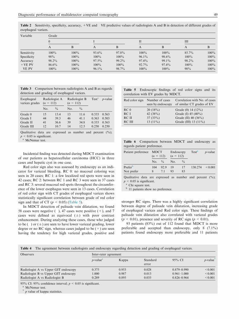
Table 2 Sensitivity, specificity, accuracy, +VE and �VE predictive values of radiologists A and B in detection of different grades of
esophageal varices.
Variable Grade
0 I II III
A B A B A B A B
Sensitivity 100% 100% 93.6% 97.8% 100% 100% 85.7% 100%
Specificity 98% 100% 100% 100% 96.1% 98.6% 100% 100%
Accuracy 98.2% 100% 97.5% 99.2% 97.4% 99.1% 98.2% 100%
+VE PV 86.6% 100% 100% 100% 92.7% 97.4% 100% 100%
�VE PV 100% 100% 96.1% 98.7% 100% 100% 98% 100%
Table 3 Comparison between radiologists A and B as regards
detection and grading of esophageal varices.
Esophageal
varices grades
Radiologist A
(n = 112)
Radiologist B
(n= 112)
Testa p-value
No. % No. %
Grade 0 15 13.4 13 11.6 0.333 0.563
Grade I 44 39.3 46 41.1 0.363 0.503
Grade II 41 36.6 39 34.8 0.333 0.563
Grade III 12 10.7 14 12.5 0.250 0.250
Qualitative data are expressed as number and percent (%);
p< 0.05 is significant.a McNemar test.
Table 5 Endoscopic findings of red color signs and its
correlation with EV grades by MDCT.
Red color sign Number of cases
seen by endoscopy
Correlation with No. of cases
of similar CT grades of EV
RC 0 20 (18%) Grade (0) 14 (12%)
RC I 42 (38%) Grade (I) 45 (40%)
RC II 37 (33%) Grade (II) 40 (36%)
RC III 13 (11%) Grade (III) 13 (11%)
Table 6 Comparison between MDCT and endoscopy as
regards patient preference.
Patient preference MDCT
(n = 112)
Endoscopy
(n= 112)
Testa p-value
No. % No. %
Preferb 104 92.9 19 17 130.274 <0.001
Not prefer 8 7.1 93 83
Qualitative data are expressed as number and percent (%);
p< 0.05 is significant.a Chi square test.b 11 patients show no preference.
Diagnostic performance of multidetector computed tomography 49
Incidental finding was detected during MDCT examinationof our patients as hepatocellular carcinoma (HCC) in three
cases and hepatic cyst in one case.Red color sign also was assessed by endoscopy as an indi-
cator for variceal bleeding. RC 0: no mucosal coloring was
seen in 20 cases; RC 1: a few localized red spots were seen in42 cases; RC 2: between RC 1 and RC 3 were seen in 37 casesand RC 3: several mucosal red spots throughout the circumfer-
ence of the lower esophagus were seen in 13 cases. Correlationof red color sign with CT grades of esophageal varices showsstatistically significant correlation between grade of red colorsign and that of CT (p < 0.05) (Table 5).
In MDCT detection of palisade vein dilatation, we found58 cases were negative (�), 47 cases were positive (+), and 7cases were defined as equivocal (±) with poor contrast
enhancement. During analyzing these cases, those who judgedto be (�) or (±) are seen to have lower variceal grading, lowerdegree or no RC sign, whereas cases judged to be (+) are seen
having the tendency for high variceal grades, positive and
Table 4 The agreement between radiologists and endoscopy regard
Observers Inter-rater agreement
p-valuea K
Radiologist A vs Upper GIT endoscopy 0.375 0.
Radiologist B vs Upper GIT endoscopy 1.000 0.
Radiologist A vs Radiologist B 0.289 0.
95% CI: 95% confidence interval; p< 0.05 is significant.a McNemar test.* p value of kappa statistics.
stronger RC signs. There was a highly significant correlationbetween degree of palisade vein dilatation, increasing grade
of esophageal varices and Red color sign. These findings ofpalisade vein dilatation also correlated with variceal grades(p< 0.01), presence and severity of RC sign (p< 0.01).
93 patients (83%) out of 112 found that MDCT is morepreferable and accepted than endoscopy, only 8 (7.1%)patients found endoscopy more preferable and 11 patients
ing detection and grading of esophageal varices.
appa Standard
error
95% CI p-value*
953 0.028 0.879–0.990 <0.001
987 0.013 0.961–1.000 <0.001
895 0.035 0.826–0.964 <0.001
50 T. ELKammash et al.
(0.8%) show no preference between both techniques. The pref-erence of CT as imaging modality from the patient side wasstatistically significant p < 0.01 (Table 6).
Illustrative cases represent MDCT scoring of esophagealvarices.
Discussion
Variceal bleeding is a serious adverse event in patients withliver cirrhosis. Patients survive the 1st episodes of variceal
bleeding have a greater than 60% risk of recurrent hemorrhagewithin 1st year of recurrent episode (19). We tried in this studyto detect the value of MDCT in diagnosis of esophageal
varices as a newly evolving, non-invasive procedure and itsacceptance to the patients.
112 patients with liver cirrhosis were involved in this study
(77 males, 45 females, age 38–72 years; mean 51.4. SD 8.4.In this study, utilizing MDCT, the scanning series take very
short time and most of the patients can withstand single breathhold which makes the procedures and diagnostic quality much
better. This was mentioned by Rydberg et al. (20) who clarifiedthat the rapid scanning capability of MDCT allows increasedcraniocaudal scanning range and thinner slice acquisition in
a single breath hold. This results in high spatial resolutionand better depictions of fine vasculature. We found also theavailability of precise MIP in sagittal and coronal planes raise
the diagnostic performance in visualization of esophagealvarices, differentiating it from paraesophageal varices as wellas visualization of other portosystemic collaterals and thiswas reported by Nakayama et al. (21) and Ishikawa et al. (22).
Using multidetector CT in detection of esophageal varicesshows high sensitivity, specificity, accuracy, positive and nega-tive predictive values. Our recorded sensitivity, specificity,
accuracy, +ve and �ve predictive value of CT in detectionof EV for radiologist A were 94.8%, 98.5%, 97.8%, 94.8%and 98.5% and for radiologist B were 99.4%, 99.6%, 99.6%,
99.3% and 99.7% respectively. In our study, the differencebetween radiologists A and B in detecting different grades ofesophageal varices was insignificant. Also there is a good
agreement between radiologists A, B and upper GIT endo-scopy regarding detection and grading of esophageal varices.This statistically proved high performance of multidetectorCT in our study was in agreement with Perri et al. (23) who
reported sensitivity and specificity of 75%, 62% and 85%,75% for radiologist 1 and radiologist 2 respectively. AlsoKim et al. (24) recorded 90–93.3% for sensitivity, and 81.7–
96.7% for specificity for radiologists 1 and 2 respectively.The higher sensitivity and specificity in our study may bedue to the fact that we use 64 slice CT while in study of Perri
et al. (23) they used 4 detectors and in Also Kim et al. (24) theyused 16 detectors in their studies.
In endoscopic findings, particularly cases with erythrogenicfindings (red color sign), we try to see whether there is relation
between the degree of red color sign and the grades of EVdetected by CT and we found that there was a significant cor-relation between grades of esophageal varices seen by multide-
tector CT and grades of red color sign (p< 0.05). This resultwas similar to that mentioned by Dessouky and Abdel Aal(25).
In examining presence or absence of palisade vein dilata-tion, we found 58 negative cases and 47 positive cases and 7
cases were recognized as equivocal (±) showing poor con-trast opacification. When we correlate negative and equivo-cal cases with the degree of RC sign, we found these cases
either do not get or have low grade RC sign. In contrarywe found cases which were evaluated as positive tended tohave positive and stronger RC signs (p < 0.01). Also we
observed that there is increase in the degree of vein dilata-tion with increasing grades of EV (p < 0.01). These highlysignificant correlation results were in agreement with those
Dessouky and Abdel Aal (25).In the incidental detection of other portosystemic collat-
erals, we have 38 cases of para esophageal varices and 47cases of gastric fundus varices whereas splenorenal collater-
als were seen in 14 cases utilizing high speed multidetectorCT which was able to identify them and differentiates itfrom esophageal varices, having the advantage more than
endoscopy which shows EV only and this was in agreementwith Kodama et al. (13) and Mifune et al. (15) who clarifiedthe important advantage of multi-detector row CT over
single-detector row helical CT and conventional portographyis the increased speed of scanning, which permits routine useof very thin collimation for imaging the portosystemic
collateral vessels whereas collateral vessels can now bedemonstrated without the risk, discomfort and invasivenessof catheterization.
In our study, multidetector CT was able to detect the feed-
ing and draining variceal vessels, yet it could not detect thedirection of blood flow within the portosystemic collateralswhich is considered a drawback compared to the conventional
portography. This limitation of multidetector CT wasmentioned also by Chen et al. (26).
Also MDCT was able to diagnose 3 cases of HCC within
cirrhotic liver patients and differentiate those from anothercase of simple hepatic cyst during its routine protocolscanning for esophageal varices and this gives other advan-
tage of MDCT over endoscopy and other invasive proce-dures. And this was in agreement with Kim et al. (27),who stated that considering the high cost of performingmultiple tests and the relative invasiveness of endoscopy, a
single noninvasive surveillance tool for both varices andHCC may be important.
When we compare the acceptance of both techniques (CT
and endoscopy) from the patient side, 93 patients (83%) outof 112 found that MDCT is more preferable and accepted thanendoscopy, only 8 (7.1%) patients found endoscopy more
preferable and 11 patients (0.8%) show no preference betweenboth techniques. The preference of CT as imaging modalityfrom the patient side was statistically significant p< 0.01. Thiswas in agreement with Perri et al. (23), Kim et al. (24) and also
Dessouky and Abdel Aal (25), who found multidetector CTmore tolerable and cost-effective compared to upper GITendoscopy and patients are more willing to utilize it for
followup.
Conclusion
Multidetector CT with MIP facilities is a reliable noninvasive,highly tolerable examination in evaluation of esophagealvarices with ability to detect other portosystemic collaterals;
in addition, evaluation of the whole MDCT examinationallows the detection of other associated pathologies.
Diagnostic performance of multidetector computed tomography 51
Conflict of interest
The authors declare that there are no conflict of interests.
References
(1) Biecker Erwin. Gastrointestinal bleeding in cirrhotic patients with
portal hypertension. ISRN Hepatol 2013;2013, 20p 541836.
(2) Groszmann RJ, Garcia-Tsao G, Bosch J, et al. Beta-blockers to
prevent gastroesophageal varices in patients with cirrhosis. New
Engl J Med 2005;353(21):2254–61.
(3) Garcia-Tsao G, Sanyal AJ, Grace ND, Carey W. Prevention and
management of gastroesophageal varices and variceal hemor-
rhage in cirrhosis. Hepatology 2007;46(3):922–38.
(4) Rye Kara, Scott Robert, Mortimore Gerri, Lawson Adam,
Austin Andrew, Freeman Jan. Towards noninvasive detection
of oesophageal varices. Int J Hepatol 2012;2012, 9p 343591.
(5) Garcia-Tsao G. Current management of the complications of
cirrhosis and portal hypertension: variceal hemorrhage, ascites,
and spontaneous bacterial peritonitis. Gastroenterology
2001;120:726–48.
(6) De Franchis R. Evolving consensus in portal hypertension report
of the Baveno IV consensus workshop on methodology of
diagnosis and therapy in portal hypertension. J Hepatol 2005;43
(1):167–76.
(7) Eisen GM, Eliakim R, Zaman A, Schwartz J, Faigel D,
Rondonotti E, et al. The accuracy of PillCam ESO capsule
endoscopy versus conventional upper endoscopy for the diagnosis
of esophageal varices: a prospective three-center pilot study.
Endoscopy 2006;38:31–5.
(8) Terayama N, Matsui O, Kobayashi S, Sanada J, Gabata T, Koda
W, et al. Portosystemic shunt on CT during arterial portography:
prevalence in patients with and without liver cirrhosis, Abdom.
Imaging 2008;33(1):80–6.
(9) Lapalus MG, Dumortier J, Fumex F, Roman S, Lot M, Prost B,
et al. Esophageal capsule endoscopy versus esophagogastroduo-
denoscopy for evaluating portal hypertension: a prospective
comparative study of performance and tolerance. Endoscopy
2006;38:36–41.
(10) Schepis F, Camma C, Niceforo D, Magnano A, Pallio S,
Cinquegrani M, et al. Which patients with cirrhosis should
undergo endoscopic screening for esophageal varices detection?
Hepatology 2001;33:333–8.
(11) Riggio O, Angeloni S, Nicolini G, Merli M, Merkel C.
Endoscopic screening for esophageal varices in cirrhotic patients.
Hepatology 2002;35:501–2.
(12) Kim YJ, Raman SS, Yu NC, To’o KJ, Jutabha R, Lu DS.
Esophageal varices in cirrhotic patients: evaluation with liver CT.
AJR Am J Roentgenol 2007;188:139–44.
(13) Kodama H, Aikata H, Takaki S, Azakami T, Katamura Y,
Kawaoka T, et al. Evaluation of portosystemic collaterals by
MDCT-MPR imaging for management of hemorrhagic esopha-
geal varices. Eur J Radiol 2010;76(2):239–45.
(14) Kang Heoung Keun, Jeong Yong Yeon, Choi Jun Ho, et al.
Three-dimensional multi-detector row CT portal venography in
the evaluation of portosystemic collateral vessels in liver cirrhosis.
RadioGraphics 2002;22:1053–61.
(15) H. Mifune, S. Akaki, K. Ida, T. Sei, S. Kanazawa, H. Okada.
Evaluation of esophageal varices by multidetector – raw CT,
correlation with endoscopic red color sign. Acta Med Okayama
2007;61(5):247–54.
(16) Lipp MJ, Broder A, Hudesman D, Suwandhi P, Okon SA,
Horowitz M, et al. Detection of esophageal varices using CT and
MRI. Dig Dis Sci 2011;56(9):2696–700.
(17) Shimizu T, Namba R, Matsuoka T, et al. Esophageal varices
before and after endoscopic variceal ligation: evaluation using
helical CT. Eur Radiol 1999;9:1546–9.
(18) The Japan Society for Portal Hypertension: the general rules for
study of portal hypertension. 2nd ed. Tokyo: Kanehara; 2004. p.
37–38.
(19) DeFranchis R. Evolving consensus in portal hypertension report of
the Banevo. IV. Consensus workshop onmethodology of diagnosis
and therapy in portal hypertension. J Hepatol 2005;43:67–76.
(20) Rydberg J, Buckwalter KA, Caldemeyer KS, et al. Multisection
CT: scanning techniques and clinical applications. Radiographics
2000;20:1787–806.
(21) Nakayama Y, Imuta M, Funama Y, Kadota M, Utsunomiya D,
Shiraishi S, et al. CT portography by multidetector helical CT:
comparison of three rendering models. Radit Med 2002;20:273–9.
(22) Ishikawa T, Ushiki T, Mizuno Ki, Togashi T, Watanabe K, Seki
Ki, et al. CT-maximum intensity projection is a clinically useful
modality for the detection of gastric varices. World J Gastroen-
terol 2005;11(47):7515–9.
(23) Perri RE, Chiorean MV, Fidler JL, Fletcher JG, Talwalkar JA,
Stadheim L, et al. A prospective evaluation of computerized
tomographic (CT) scanning as a screening modality for esopha-
geal varices. Hepatology 2008;47(5):1587–94.
(24) Kim SH, Kim YJ, Lee JM, Choi KD, Chung YJ, Han JK, et al.
Esophageal varices in patients with cirrhosis: multidetector CT
esophagography—comparison with endoscopy. Radiology
2007;242(3):759–68.
(25) Dessouky BA, Abdel Aal SM. Multidetector CT oesophagogra-
phy: an alternative screening method for endoscopic diagnosis of
oesophageal varices and bleeding risk. Arab J Gastroenterol
2013;14(3):99–108.
(26) Chen Tian-wu, Yang Zhi-gang, Li Xiao, et al. Evaluation of
entire gastric fundic and esophageal varices secondary to
posthepatitic cirrhosis: portal venography using 64-row MDCT.
Abdom Imaging 2010;35:1–7.
(27) Kim SH, Han JK, Lee KH, et al. Computed tomography
gastrography with volume-rendering technique: correlation with
double-contrast barium study and conventional gastroscopy. J
Comput Assist Tomogr 2003;27:140–9.