DIAGNOSTIC & CULTIVATION of VIRUSES€¦ · Cultivation of Virus Since they are obligate...
22
10/14/2019 1 Lecture 6 DIAGNOSTIC & CULTIVATION of VIRUSES 1 A. Laboratory viral diagnosis involves one of three basic approaches: o antigens in clinical specimens o serologic testing of viral specific antibodies. o virus isolation; direct demonstration of virus, viral nucleic acid, 2
Transcript of DIAGNOSTIC & CULTIVATION of VIRUSES€¦ · Cultivation of Virus Since they are obligate...

10/14/2019
1
Lecture 6
DIAGNOSTIC & CULTIVATION
of VIRUSES1
A. Laboratory viral diagnosis involves one of three
basic approaches:
o antigens in clinical specimens
o serologic testing of viral specific antibodies.
o virus isolation; direct demonstration of virus, viral
nucleic acid,
2
10/14/2019
2
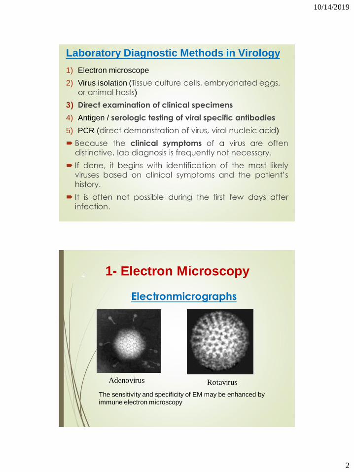
Laboratory Diagnostic Methods in Virology
1) Electron microscope
2) Virus isolation (Tissue culture cells, embryonated eggs,
or animal hosts)
3) Direct examination of clinical specimens
4) Antigen / serologic testing of viral specific antibodies
5) PCR (direct demonstration of virus, viral nucleic acid)
Because the clinical symptoms of a virus are often
distinctive, lab diagnosis is frequently not necessary.
If done, it begins with identification of the most likely
viruses based on clinical symptoms and the patient’s
history.
It is often not possible during the first few days after
infection.
3
Electronmicrographs
Adenovirus Rotavirus
1- Electron Microscopy
The sensitivity and specificity of EM may be enhanced by immune electron microscopy
4
10/14/2019
3
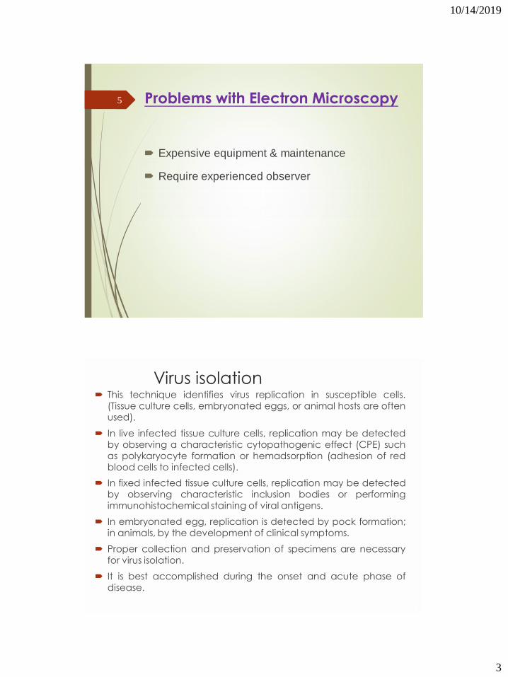
Problems with Electron Microscopy
Expensive equipment & maintenance
Require experienced observer
5
Virus isolation This technique identifies virus replication in susceptible cells.
(Tissue culture cells, embryonated eggs, or animal hosts are oftenused).
In live infected tissue culture cells, replication may be detectedby observing a characteristic cytopathogenic effect (CPE) suchas polykaryocyte formation or hemadsorption (adhesion of redblood cells to infected cells).
In fixed infected tissue culture cells, replication may be detectedby observing characteristic inclusion bodies or performingimmunohistochemical staining of viral antigens.
In embryonated egg, replication is detected by pock formation;in animals, by the development of clinical symptoms.
Proper collection and preservation of specimens are necessaryfor virus isolation.
It is best accomplished during the onset and acute phase ofdisease.
6

10/14/2019
4
2- Cell culture
Cell Cultures are most widely used for virus isolation,
there are 3 types of cell cultures:
1. Primary cells - Monkey Kidney
2. Semi-continuous cells - Human embryonic kidney and skin, fibroblasts
3. Continuous cells - HeLa, Vero, Hep2, LLC-MK2, MDCK
7
Tissue culture: Tissue culture of human or animal cells are frequently used for
the cultivation of viruses. There are mainly three types of
tissue culture:
1. Organ culture, e.g. tracheal ring organ culture is employedfor the isolation of coronavirus.
2. Explant culture: Minced tissue may be grown as explantembedded in plasma clots. This is not useful in virology. In thepast adenoid tissue explant culture were used for adenovirus.
3. Cell culture: This is very popular and useful techniqueroutinely used for cultivation of viruses. From tissue, fragmentscells are dispersed by proteolytic enzymes like trypsin andmechanical shake. After washing the cells, they aresuspended in growth medium and distributed in petridishes,test tubes or bottles.
The cells adhere to glass surface and grow out to form amonolayer sheet and can be seen in situ under low power.
8
10/14/2019
5
Cell Cultures
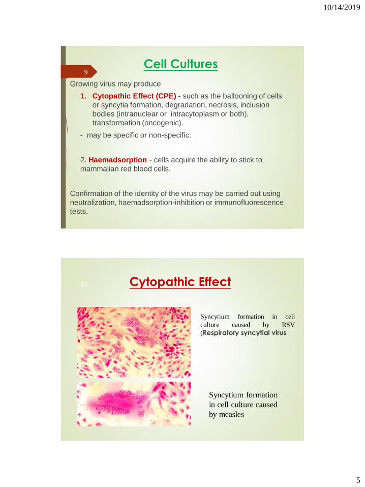
Growing virus may produce
1. Cytopathic Effect (CPE) - such as the ballooning of cells
or syncytia formation, degradation, necrosis, inclusion
bodies (intranuclear or intracytoplasm or both),
transformation (oncogenic).
- may be specific or non-specific.
2. Haemadsorption - cells acquire the ability to stick to
mammalian red blood cells.
Confirmation of the identity of the virus may be carried out using
neutralization, haemadsorption-inhibition or immunofluorescence
tests.
9
Cytopathic Effect
Syncytium formation in cell
culture caused by RSV
(Respiratory syncytial virus
Syncytium formation
in cell culture caused
by measles
10
10/14/2019
6
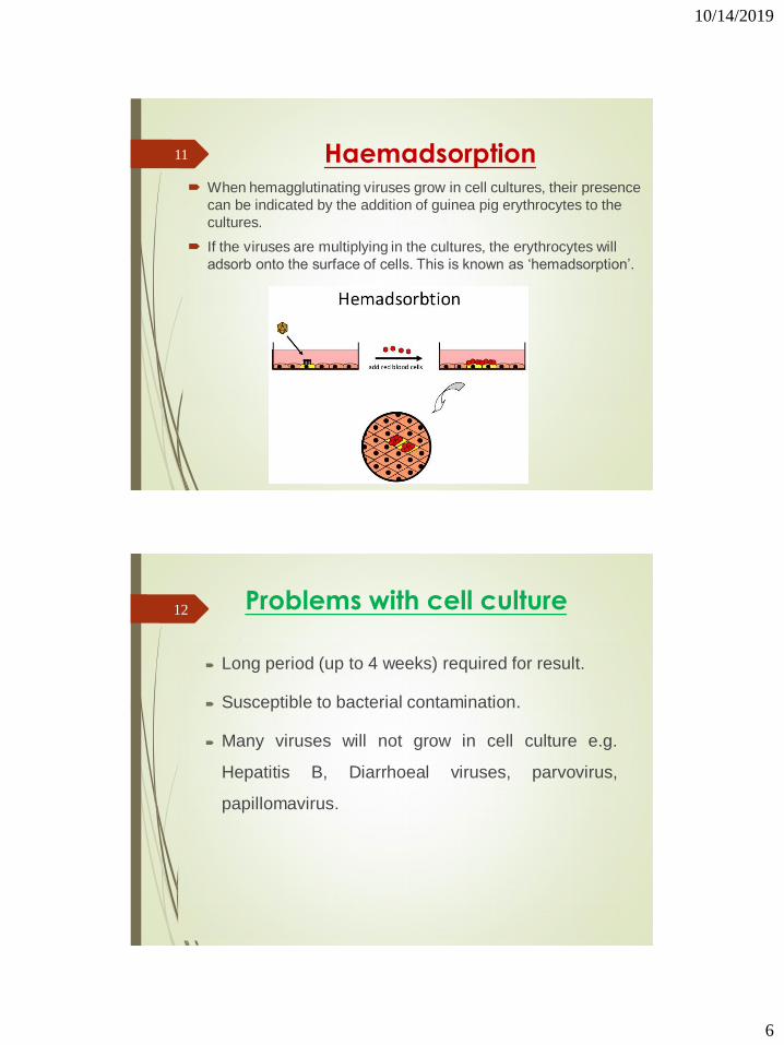
Haemadsorption When hemagglutinating viruses grow in cell cultures, their presence
can be indicated by the addition of guinea pig erythrocytes to the
cultures.
If the viruses are multiplying in the cultures, the erythrocytes will
adsorb onto the surface of cells. This is known as ‘hemadsorption’.
11
Problems with cell culture
Long period (up to 4 weeks) required for result.
Susceptible to bacterial contamination.
Many viruses will not grow in cell culture e.g.
Hepatitis B, Diarrhoeal viruses, parvovirus,
papillomavirus.
12

10/14/2019
7
3. Direct examination of clinical specimens
General characteristics
Specimens may include sections of tissue biopsies,
tissue imprints or smears, blood, cerebrospinal fluid,
urine, throat swabs, feces, or saliva.
Only those specimens likely to contain the virus (e.g.,
throat swabs for respiratory tract infection) should be
examined.
13
4- Antigen / Antibody tests
Antibodies
(also known as immunoglobulins abbreviated Ig) are gamma
globulin proteins that are found in blood and are used by the
immune system to identify and neutralize foreign objects, such
as bacteria and viruses.
14

10/14/2019
8
Antigen / Antibody tests
Antigens
A substance that when introduced into the body
stimulates the production of an antibody
Immunoassay
A laboratory technique that makes use of the binding
between an antigen and its homologous antibody in
order to identify and quantify the specific antigen or
antibody in a sample
15
Serologic tests1. General characteristics
Serologic tests are used to determine the titer of specific antiviral antibodies.
Paired blood samples are taken (one sample at the onset and one sample during the recovery phase of the illness); at least a fourfold increase in titer between the samples must be present to indicate a current infection.
The test may be diagnostic without the use of paired samples if significant levels of IgM antiviral antibodies are obtained.
Techniques include virus neutralization, complement fixation, hemagglutination inhibition tests, and solid-phase immunoassays.
These tests are expensive to perform and must be standardized for each virus.
16

10/14/2019
9
Antigen / Antibody tests
Viral Serology
Criteria for diagnosing Primary Infection
Presence of IgM
Criteria for diagnosing Reinfection
4 fold or more increase in titer of IgG or total antibody between
acute and convalescent sera
Absence or slight increase in IgM
17
18

10/14/2019
10
Western Blot
Western blotting, also known as immunoblotting or
protein blotting, is a technique used to detect the
presence of a specific protein in a complex protein
mixture
It is a core technique in cell biology, molecular biology,
virology and others
19
Western Blot
HIV-1 Western Blot
Lane1: Positive Control
Lane 2: Negative Control
Sample A: Negative
Sample B: Indeterminate
Sample C: Positive
20
10/14/2019
11
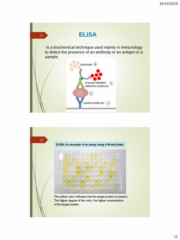
ELISA
Is a biochemical technique used mainly in immunology
to detect the presence of an antibody or an antigen in a
sample.
21
22

10/14/2019
12
Direct ELISA 23
Sandwich ELISA
The ELISA plate is coated with Antibody to detect specific antigen
24

10/14/2019
13
Sandwich ELISA25
Indirect ELISA 26

10/14/2019
14
ELISA :SUMMARY27
Immunofluorescense
Immunofluorescence is a serological test where the labeling of antibodies or antigens is done with fluorescent dyes (fluorochromes).
Fluorochromes are dyes which have the ability to absorb the short wavelength UV radiation and emit light of longer wavelength fluorescence (visible green light).
Examples : FITC, Rhodamine , Acridine orange
There are two ways of doing IF staining
Direct immunofluorescence
Indirect immunofluorescence
28

10/14/2019
15
Direct immunofluorescenceUse: Direct detection of Pathogens or their Ag’s in tissues or in pathological
samples
Ag is fixed on the slide
Fluorescein labeled Ab’s are layered over it
Slide is washed to remove unattached Ab’s
Examined under UV light in an fluorescent microscope
The site where the Ab attaches to its specific Ag will show apple green fluorescence
Indirect immunofluorescence Use: Indirect detection of antibodies against viruses in serum
Indirect test is a double-layer technique
The unlabelled antibody is applied directly to the tissue substrate
Treated with a fluorochrome-conjugated anti-immunoglobulin serum
29
30

10/14/2019
16
5- Polymerase Chain Reaction
PCR allows the in vitro amplification of specific target DNA
sequences by a factor of 106 and is thus an extremely sensitive
technique.
Detection and identification of the PCR product is usually carried
out by agarose gel electrophoresis, hybridization with a specific
oligonucleotide probe, restriction enzyme analysis, or DNA
sequencing.
31
32

10/14/2019
17
What is qPCR
“quantitative Polymerase Chain Reaction”
A method that allows to follow in real time (that is why is also called
Real-Time PCR) the amplification of a target.
The target can be nucleic acids (RNA or DNA).
Taq polymerase can only synthesize DNA, so how do we study RNA
using qPCR?
33
Reverse Transcription
mRNA can be copied to
complementary DNA sequence
(cDNA) using reverse
transcriptase—a DNA polymerase
that uses ssRNA as template.
Processed mRNA will match
protein coding sequence while
unprocessed (nuclear) mRNA will
contain intron sequences.
34

10/14/2019
18
Uses of qPCR
Precise quantitation of DNA or RNA in samples
Estimation of gene number
Gene expression studies by quantification of messenger RNA
Mnay technqiues are used for qPCR :
1- SYBRGreen
2- Taqman
35
SYBRGreen36

10/14/2019
19
TaqMan (quencher & reporter)37
CULTIVATION of VIRUSES
38

10/14/2019
20
Cultivation of Virus
Since they are obligate intracellular parasites
and cannot grow on inanimate culture
medium, 3 methods are used for their
cultivation:
a. Animals inoculation.
b. Chick embryo.
c. Tissue culture.
39
Animal inoculation: It is one of the oldest methods for the cultivation of viruses.
The poliomyelitis virus after intraspinal or intracerebralinoculation in monkeys causes typical paralytic disease andso isolation of viruses.
Suckling mice is susceptible to Cox-sackie viruses withmanifestation of severe myositis and paralysis.
Smallpox virus may be inoculated in the scarfied skin orcornea of rabbit. Brain tissue of rabied dog when inoculatedintracerebrally in mice or rabbit develop encephalitis.
Growth of virus in animals may be known by the disease,visible classical lesions or death.
Sometimes immunity in experimental animal may interferewith the growth of viruses in that animal.
40
10/14/2019
21
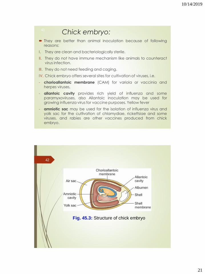
Chick embryo: They are better than animal inoculation because of following
reasons:
I. They are clean and bacteriologically sterile.
II. They do not have immune mechanism like animals to counteractvirus infection.
III. They do not need feeding and caging.
IV. Chick embryo offers several sites for cultivation of viruses, i.e.
- chorioallantoic membrane (CAM) for variola or vaccinia andherpes viruses,
- allantoic cavity provides rich yield of influenza and someparamyxoviruses, also Allantoic inoculation may be used forgrowing influenza virus for vaccine purposes. Yellow fever
- amniotic sac may be used for the isolation of influenza virus andyolk sac for the cultivation of chlamydiae, rickettsiae and someviruses. and rabies are other vaccines produced from chickembryo.
41
42
Fig. 45.3: Structure of chick embryo

10/14/2019
22
Disadvantages of Egg Inoculation
1. Eggs may be contaminated with mycoplasma
and latent fowl viruses which may interfere with the
growth of other viruses.
2. The susceptibility of chick embryo is limited to a
few viruses only.
3. Even slight amount of bacterial contamination in
the inoculum may kill the embryo.
43
Tissue culture: Tissue culture of human or animal cells are frequently used
for the cultivation of viruses. There are mainly three types of
tissue culture:
1. Organ culture, e.g. tracheal ring organ culture isemployed for the isolation of coronavirus.
2. Explant culture: Minced tissue may be grown as explantembedded in plasma clots. This is not useful in virology. Inthe past adenoid tissue explant culture were used foradenovirus.
3. Cell culture: This is very popular and useful techniqueroutinely used for cultivation of viruses. From tissue,fragments cells are dispersed by proteolytic enzymes liketrypsin and mechanical shake. After washing the cells,they are suspended in growth medium and distributed inpetridishes, test tubes or bottles.
The cells adhere to glass surface and grow out to form amonolayer sheet and can be seen in situ under low power.
44