Diagnosis and Treatment of Coronary Heart Disease in Women
144
Evidence Report/Technology Assessment Number 8 1 Diagnosis and Treatment of Coronary Heart Disease in Women: Systematic Reviews of Evidence on Selected Topics Prepared for: Agency for Healthcare Research and Quality U.S. Department of Health and Huma n Services www.ahrq.gov Contract No. 290-97-0013 Prepared by : University of California, San Francisco-Stanford University Evidence-based Practice Center, Stanford, CA Principal Investigator Deborah Grady, MD, MPH EPC Coordinator Kathryn McDonald, MM EPC Staff Kara Bischoff Aurelie Cabou Christopher Gehrman Kate Hoerster Lily Chaput, MD, MPH Margaret Kristof, MS, RN Kathryn Melsop, MS Dan H. Moore, PhD AHRQ Publication No. 03-E037 May 2003 Topic Team Staff Noninvasive Diagnostic Tests Mary Beattie, MD Louise Metz, MD Kirsten Fleishmann, MD Rita Redberg, MD Lipid Lowering Treatment Judith Walsh, MD, MPH Michael Pignone, MD, MPH Paul Varosy, MD Diabetes as Risk Factor Alka Kanaya, MD Lily Chaput, MD, MPH Troponins Paul Heindenreich, MD Kathy McDonald, MM UCSF/Mt. Zion Fishbon Library Staff Gail Sorrough, MLIS Gloria Won, MLIS
Transcript of Diagnosis and Treatment of Coronary Heart Disease in Women

Evidence Report/Technology Assessment Number 81
Diagnosis and Treatment of Coronary Heart Disease in Women: Systematic Reviews of Evidence on Selected Topics
Prepared for: Agency for Healthcare Research and Quality U.S. Department of Health and Human Services www.ahrq.gov Contract No. 290-97-0013 Prepared by : University of California, San Francisco-Stanford University Evidence-based Practice Center, Stanford, CA
Principal Investigator Deborah Grady, MD, MPH EPC Coordinator Kathryn McDonald, MM EPC Staff Kara Bischoff Aurelie Cabou Christopher Gehrman Kate Hoerster Lily Chaput, MD, MPH Margaret Kristof, MS, RN Kathryn Melsop, MS Dan H. Moore, PhD AHRQ Publication No. 03-E037 May 2003
Topic Team Staff Noninvasive Diagnostic Tests Mary Beattie, MD Louise Metz, MD Kirsten Fleishmann, MD Rita Redberg, MD Lipid Lowering Treatment Judith Walsh, MD, MPH Michael Pignone, MD, MPH Paul Varosy, MD
Diabetes as Risk Factor Alka Kanaya, MD Lily Chaput, MD, MPH Troponins Paul Heindenreich, MD Kathy McDonald, MM UCSF/Mt. Zion Fishbon Library Staff Gail Sorrough, MLIS Gloria Won, MLIS

ii
This document is in the public domain and may be used and reprinted without permission except those copyrighted materials noted for which further reproduction is prohibited without the specific permission of copyright holders. Suggested Citation: Grady D, Chaput L, Kristof M. Diagnosis and Treatment of Coronary Heart Disease in Women: Systematic Reviews of Evidence on Selected Topics. Evidence Report/Technology Assessment No. 81. (Prepared by the University of California, San Francisco-Stanford Evidence-based Practice Center under Contract No 290-97-0013.) AHRQ Publication No. 03-E037. Rockville, MD: Agency for Healthcare Research and Quality. May 2003.
This report may be used, in whole or in part, as the basis for development of clinical practice guidelines and other quality enhancement tools, or a basis for reimbursement and coverage policies. AHRQ or U.S. Department of Health and Human Services endorsement of such derivative products may not be stated or implied. AHRQ is the lead Federal agency charged with supporting research designed to improve the quality of health care, reduce its cost, address patient safety and medical errors, and broaden access to essential services. AHRQ sponsors and conducts research that provides evidence-based information on health care outcomes; quality; and cost, use, and access. The information helps health care decisionmakers—patients and clinicians, health system leaders, and policymakers—make more informed decisions and improve the quality of health care services.

iii
Preface The Agency for Healthcare Research and Quality (AHRQ), through its Evidence-Based Practice Centers (EPCs), sponsors the development of evidence reports and technology assessments to assist public- and private-sector organizations in their efforts to improve the quality of health care in the United States. The reports and assessments provide organizations with comprehensive, science-based information on common, costly medical conditions and new health care technologies. The EPCs systematically review the relevant scientific literature on topics assigned to them by AHRQ and conduct additional analyses when appropriate prior to developing their reports and assessments. To bring the broadest range of experts into the development of evidence reports and health technology assessments, AHRQ encourages the EPCs to form partnerships and enter into collaborations with other medical and research organizations. The EPCs work with these partner organizations to ensure that the evidence reports and technology assessments they produce will become building blocks for health care quality improvement projects throughout the Nation. The reports undergo peer review prior to their release. AHRQ expects that the EPC evidence reports and technology assessments will inform individual health plans, providers, and purchasers as well as the health care system as a whole by providing important information to help improve health care quality. We welcome written comments on this evidence report. They may be sent to: Director, Center for Practice and Technology Assessment, Agency for Healthcare Research and Quality, 6010 Executive Blvd., Suite 300, Rockville, MD 20852. Carolyn M. Clancy, M.D. Director Agency for Healthcare Research and Quality
The authors of this report are responsible for its content. Statements in the report should not be construed as endorsement by the Agency for Healthcare Research and Quality or the U.S. Department of Health and Human Services of a particular drug, device, test, treatment, or other clinical service.
Jean Slutsky, P.A., M.S.P.H. Acting Director, Center for Practice and
Technology Assessment Agency for Healthcare Research and Quality


v
Structured Abstract
Objectives. The Agency for Healthcare Research and Quality (AHRQ) and the National Institutes of Health Office of Research on Women's Health funded the University of California, San Francisco-Stanford Evidence-based Practice Center to perform systematic reviews and meta-analyses on four key topics related to coronary heart disease (CHD) in women: (1) accuracy of exercise myocardial perfusion imaging and echocardiography for diagnosis of CHD in women, (2) lipid lowering treatment to reduce risk of CHD in women, (3) diabetes as a risk factor for CHD in women, and (4) troponin as a prognostic factor for CHD in women. For each question, we also attempted to provide evidence stratified by race or ethnicity. We used standard methods to systematically review the medical literature to address each topic. The evidence identified was reviewed and graded, data were abstracted and the findings summarized for each topic. Search Strategy. We developed specific search terms for each of the four key topics and performed standardized searches of electronic databases. We also reviewed bibliographies and sought suggestions from our peer reviewers. Authors of studies that met selection criteria but did not report findings by gender were contacted and asked to provide gender-specific outcomes. Selection Criteria. Inclusion and exclusion criteria were defined for each of the four systematic reviews. Titles and abstracts were reviewed by investigators who coded each for eligibility. The full text of eligible articles was reviewed independently by two physician investigators using standardized forms to classify eligibility, rate quality and abstract data. The findings of all eligible studies rated good or fair quality were included in the summary estimates. Data Collection and Analysis. Titles and abstracts were entered and coded using EndNote® (Niles Software, Inc ). Data from the standardized review forms were entered in an Access (Microsoft ® Corporation) database to allow tracking of eligibility, quality and study design. Abstracted data were also stored electronically in a database (EXCEL, Microsoft® Corporation). Main Results
Overall
• Findings from 82 otherwise eligible studies could not be included in the systematic reviews
because data were not stratified by gender. We contacted the author of these studies twice requesting data for women and received data from 19 studies (23 percent).
• Little evidence was available regarding the key questions as they pertain to women of different races/ethnicities. For this reason, only the review of diabetes as a risk factor for CHD provides summary findings by ethnicity.

vi
Accuracy of exercise myocardial perfusion imaging and echocardiography for diagnosis of CHD in women • We found 14 eligible studies that provided data on the accuracy of noninvasive tests in 893
women. Ten studies examined the accuracy of myocardial perfusion imaging and four examined the accuracy of exercise echocardiography.
• In women, the overall accuracy of both exercise myocardial perfusion imaging and exercise echocardiography for diagnosis of CHD is low with positive likelihood ratios of 2.5 to 3 and negative likelihood ratios of about 0.3.
• The accuracy of exercise myocardial perfusion imaging for diagnosis of CHD is not clinically different in women compared to men.
• There is little difference in the accuracy of exercise myocardial perfusion imaging and exercise echocardiography for diagnosis of CHD in women.
• The accuracy of exercise myocardial perfusion imaging for diagnosis of CHD is similar whether thallium or sestamibi is used as the imaging agent.
Efficacy of lipid lowering to reduce risk of CHD in women
• Although 20 clinical trials of the effects of lipid lowering therapy included women, only nine
published results by gender. By contacting study investigators, we were successful in obtaining data on women from two additional trials. Thus, we were able to analyze results from 11 trials that included 15,917 women.
• In women with known CHD, treatment with lipid lowering therapy reduces risk of CHD mortality 26 percent, nonfatal myocardial infarction (MI) 36 percent and major CHD events 21 percent. There was insufficient evidence to show that lipid lowering reduces rates of revascularization procedures and no evidence of a reduction of risk in total mortality.
• For women without CHD, there is insufficient evidence to determine whether lipid lowering reduces risk for any clinical outcome.
Diabetes as a risk factor for CHD in women • We found 17 eligible studies that included 43,944 women (4,522 with diabetes and 39,422
without diabetes). • Adjusted summary odds ratios (ORs) for CHD mortality and nonfatal MI due to diabetes are
higher among women than men, but summary ORs for all-cause mortality are slightly higher in men than women. All of the differences between men and women are modest and not statistically significant.
• The summary odds ratio for CHD mortality due to diabetes is 2.9 (95% confidence interval [CI], 2.2-3.8) for women and 2.3 (95% CI, 1.9-2.8) for men. The summary OR for nonfatal MI due to diabetes is 1.7 (95% CI, 1.3-2.3) for women and 1.6 (95% CI, 1.1-2.2) for men. The summary OR for all-cause mortality due to diabetes is 1.9 (95% CI, 1.7-2.3) for women and 2.1 (95% CI, 1.7-2.7) for men.

vii
• Summary estimates for risk of CHD mortality due to diabetes for nonwhite men and women are similar to those for whites.
• The difference in relative risk for CHD outcomes between men and women is progressively attenuated with adjustment for major cardiovascular risk factors. This finding may be due to the fact that women with diabetes have more risk factors or more severe risk factor abnormalities in comparison to women without diabetes than is the case for men with and without diabetes.
Prognostic value of troponin for CHD in women • We identified eight eligible cohort studies that provided data on 3,169 women and 4,070
men. • Elevated troponin was observed in 35 percent of women and 39 percent of men with non-ST
elevation acute coronary syndromes. • Women with acute coronary syndromes were older and more likely to have diabetes and
hypertension than men. • An elevated troponin indicates a similar increase in risk of death for both women (summary
OR 2.63; 95% CI 1.75-3.95) and men (OR 2.83; 95% CI 1.92-4.17). • An elevated troponin indicates a greater increase in risk of nonfatal MI for women (summary
OR 1.80; 95% CI 1.28-2.54) than men (OR 1.06; 95% CI 0.8-1.41). Conclusions . The major problem in performing these systematic reviews was that data stratified by sex and race/ethnicity from completed studies are often not available. We recommend that, in addition to requiring participation of women and minorities in research, the National Institutes of Health, U.S. Food and Drug Administration, and other funding and regulatory agencies insist that outcome data by subgroup be published or archived and made easily available to meta-analysts.

ix
Contents Summary .........................................................................................................................1 Evidence Report..........................................................................................................9 Chapter 1. Introduction................................................................................................. 11 Organization ............................................................................................................ 11
Key Questions.......................................................................................................... 12 References for Introduction..................................................................................... 15
Chapter 2. Systematic Review of the Accuracy of Exercise Myocardial Perfusion Imaging and Echocardiography for Diagnosis of Coronary Heart Disease in Women......................................................................................................... 17 Introduction.............................................................................................................. 17 Methodology............................................................................................................ 18 Data Sources ...................................................................................................... 18 Search Terms ..................................................................................................... 18 Inclusion Criteria ............................................................................................... 19
Article Identification.......................................................................................... 19 Obtaining Unpublished Results ......................................................................... 20 Quality Assessment ........................................................................................... 20 Data Abstraction................................................................................................ 21 Data Management and Archive ......................................................................... 21 Data Analysis ..................................................................................................... 22 Results...................................................................................................................... 22 Study Identification ........................................................................................... 22 Description of Eligible Studies .......................................................................... 22 Findings ............................................................................................................. 23 Conclusions .............................................................................................................. 26 Future Research ....................................................................................................... 28 References................................................................................................................ 29 Chapter 3. Systematic Review of Lipid Lowering Treatment to Reduce Risk of Coronary Heart Disease in Women .................................................................. 35 Introduction.............................................................................................................. 35 Methodology............................................................................................................ 36 Data Sources ...................................................................................................... 36 Search Terms ..................................................................................................... 36
Inclusion Criteria ............................................................................................... 36 Article Identification.......................................................................................... 37
Obtaining Unpublished Results ......................................................................... 37 Quality Assessment ........................................................................................... 37 Data Abstraction................................................................................................ 38 Data Management and Archive ......................................................................... 38 Data Analysis ..................................................................................................... 38 Results...................................................................................................................... 39 Results of Study Identification .......................................................................... 39

x
Description of Eligible Studies .......................................................................... 39 Findings ............................................................................................................. 40 Conclusions .............................................................................................................. 43 Future Research ....................................................................................................... 45 References................................................................................................................ 47 Chapter 4. Systematic Review of Diabetes as a Risk Factor for Coronary Heart Disease in Women......................................................................................................... 51 Introduction.............................................................................................................. 51
Methodology............................................................................................................ 51 Data Sources ...................................................................................................... 51 Search Terms ..................................................................................................... 52 Inclusion Criteria ............................................................................................... 52 Definition of Outcomes ..................................................................................... 52
Article Identification.......................................................................................... 52 Obtaining Unpublished Results in Women ....................................................... 53 Quality Assessment ........................................................................................... 53 Data Abstraction................................................................................................ 54 Data Management and Archive ......................................................................... 54 Data Analysis ..................................................................................................... 54 Results...................................................................................................................... 55 Results of Study Identification .......................................................................... 55 Description of Eligible Studies .......................................................................... 56 Summary of Results........................................................................................... 56 Conclusions .............................................................................................................. 57 Future Research ....................................................................................................... 59 References................................................................................................................ 61 Chapter 5. Systematic Review of Troponin as a Prognostic Factor for CHD in Women....................................................................................................................... 67 Introduction.............................................................................................................. 67 Methodology............................................................................................................ 68 Data Sources ...................................................................................................... 68 Search Terms ..................................................................................................... 68 Inclusion and Exclusion Criteria ....................................................................... 69
Article Identification.......................................................................................... 69 Data Abstraction................................................................................................ 69
Obtaining Unpublished Results ......................................................................... 69 Quality Assessment ........................................................................................... 69 Data Management and Archive ......................................................................... 69 Data Analysis ..................................................................................................... 70 Results...................................................................................................................... 70 Results of Study Identification .......................................................................... 70 Patient Characteristics ....................................................................................... 71 Study Report Characteristics ............................................................................. 71 Quality of Study Reports ................................................................................... 71 Findings ............................................................................................................. 71 Conclusions .............................................................................................................. 73

xi
Future Research ....................................................................................................... 74 References................................................................................................................ 75 Evidence Tables ............................................................................................................. 89 Evidence Table 1. Diagnostic Testing: Characteristics of studies of exercise myocardial perfusion imaging............................................ 91
Evidence Table 2. Diagnostic Testing: Characteristics of studies of exercise echocardiography............................................................................................... 92
Evidence Table 3. Diagnostic Testing: Findings of exercise myocardial perfusion imaging and summary estimates of accuracy................. 93 Evidence Table 4. Diagnostic Testing: Summary estimates of accuracy of myocardial perfusion imaging from studies using sestamibi compared to studies using thallium................................................... 95 Evidence Table 5. Diagnostic Testing: Findings of studies of
exercise echocardiography and summary estimates of accuracy ...................... 96 Evidence Table 6. Lipids: Characteristics of included trials ............................ 97 Evidence Table 7. Lipids: Outcomes of eligible studies .................................. 99
Evidence Table 8. Lipids: Summary results .................................................... 100 Evidence Table 9. Diabetes: Characteristics of studies of coronary heart disease risk in diabetics vs. nondiabetics.................................................. 101 Evidence Table 10. Diabetes: Adjusted odds ratios for CHD mortality, nonfatal MI, and all-cause mortality by sex...................................... 103 Evidence Table 11. Diabetes: Summary odds ratios for CHD mortality, nonfatal myocardial infarction, and all-cause mortality by sex................................................................................................................. 104
Evidence Table 12. Diabetes: Summary odds ratios for CHD mortality by sex, quality of studies and sensitivity analysis.......................................................... 105 Evidence Table 13. Troponin: Characteristics of the study population by gender ...................................................................................... 106 Evidence Table 14. Troponin: Characteristics of the study reports.................. 107 Evidence Table 15. Troponin: Outcomes for women....................................... 108 Evidence Table 16. Troponin: Outcomes for men............................................ 109
Bibliography .................................................................................................................. 111 Appendixes .................................................................................................................... 125 Appendix A. Peer Reviewers.................................................................................. 127 Appendix B. Quality Evaluation Forms ................................................................. 131 Appendix C. Data Abstraction Forms .................................................................... 137 Appendix D. Abbreviations and Acronyms............................................................ 157 Figures Figure 1. Troponin: Death for women with and without an elevated troponin ...... 79 Figure 2. Troponin: Death for men with and without an elevated troponin ............ 80 Figure 3. Troponin: Death or myocardial infarction for women with and without an elevated troponin .......................................................................... 81 Figure 4. Troponin: Death or myocardial infarction for men with and
without an elevated troponin .......................................................................... 82

xii
Figure 5. Troponin: Nonfatal infarction for women with and without an elevated troponin ............................................................................................ 83
Figure 6. Troponin: Nonfatal infarction for men with and without an elevated troponin........................................................................................................... 84
Figure 7. Troponin: Odds ratios and their summary for death for both men and women............................................................................................................. 85
Figure 8. Troponin: Odds ratios and their summary for death or myocardial infarction for both men and women................................................................ 86 Figure 9. Troponin: Odds ratios and their summary for nonfatal myocardial infarction for both men and women................................................................ 87

Overview
Coronary heart disease (CHD) is a commondisease and cause of death in women,accounting for over 250,000 deaths in womenper year. Over the last two decades, multipleimportant studies have helped define accurateclinical tests, risk factors, preventiveinterventions, and effective therapies for CHD.Unfortunately, many of these studies have eitherexcluded women entirely or included onlylimited numbers of women and minorities.Thus, much of the evidence supportingcontemporary recommendations for testing,prevention, and treatment of coronary disease inwomen is extrapolated from studies conductedpredominantly in middle-aged men. The twobest approaches to obtain additional evidence ondiagnosis and treatment of CHD in women areto conduct large studies that include adequatenumbers of women and minorities to answer theresearch question or to perform systematicreviews and meta-analyses summarizing effectestimates by subgroup.
The Agency for Healthcare Research andQuality (AHRQ) and the National Institutes ofHealth Office of Research on Women's Healthfunded the University of California, SanFrancisco (UCSF)-Stanford Evidence-basedPractice Center (EPC) to review the evidenceregarding prevention, diagnosis, andmanagement of coronary heart disease inwomen and minorities. In an initial phase ofthis work, the UCSF-Stanford EPC conducted a
preliminary review of evidence on 42 topicsrelated to CHD in women, titled Results ofSystematic Review of Research on Diagnosis andTreatment of Coronary Heart Disease in Women.1
Based on these reviews, we identified four keyquestions for systematic review and meta-analysis. The results of these four reviews arepresented in this report.
Key Questions
1. What is the accuracy of noninvasive testsfor diagnosis of CHD in women: exercisemyocardial perfusion imaging (MPI) andexercise echocardiography?a. What are the summary estimates of
sensitivity, specificity and likelihoodratios for exercise MPI and exerciseechocardiography in women?
b. What is the accuracy of exercise MPIand exercise echocardiography inwomen compared to men?
2. What is the effectiveness of treatment withlipid lowering drugs for reducing CHD riskin women with and without CHD?a. What is the effectiveness of drug
treatment in reducing total mortality,CHD mortality, CHD events or CHDprocedures in women with knownCHD and those without known CHD?
3. What is the relative risk for CHD inwomen with type 2 diabetes?a. What is the relative risk for CHD in
women with type 2 diabetes comparedto women without diabetes?
Evidence Report/Technology AssessmentNumber 81
Diagnosis and Treatment of Coronary HeartDisease in Women: Systematic Reviews of
Evidence on Selected TopicsSummary
Agency for Healthcare Research and Quality
U.S . DEPARTMENT OF HEALTH AND HUMAN SERV ICES • Pub l i c Hea l t h Se r v i ce
1 Grady D, Chaput L, Kristof M. Results of Systematic Review of Research on Diagnosis and Treatment of Coronary HeartDisease in Women. Evidence Report/Technology Assessment No. 80. (Prepared by the University of California, SanFrancisco-Stanford Evidence-based Practice Center under Contract No 290-97-0013.) AHRQ Publication No. 03-0035.Rockville, MD: Agency for Healthcare Research and Quality. May 2003.

b. Does the relative risk for CHD differ between womenand men with type 2 diabetes?
4. What is the prognostic value of troponin for CHD inwomen?a. What is the impact of troponin on risk for death
among women with non-ST elevation acute coronarysyndromes?
b. Does the prognostic value of troponin for mortalitydiffer between men and women?
c. What is the impact of troponin on risk for death ormyocardial infarction for women with non-STelevation acute coronary syndromes?
d. Does the prognostic value of troponin for mortalityor myocardial infarction differ between men andwomen?
For each of the four questions, we also attempted toidentify and summarize evidence stratified by race or ethnicity.
Methodology
We performed standardized searches of electronic databasesof publications relevant to the topic areas. We developedspecific search terms for each of the four key topics andconducted a separate search for evidence regarding each. Wealso reviewed the bibliographies of retrieved articles andsought suggestions for additional articles from our expert peerreviewers. For each topic area, we established clear inclusioncriteria that required that studies provide data regarding theresearch question specific to women.
For three of the key questions (noninvasive diagnostic tests,lipid lowering and diabetes), two UCSF-Stanford EPCinvestigators reviewed all identified titles and excluded thosethat did not meet inclusion criteria. The abstracts of remainingarticles were reviewed by two UCSF-Stanford EPC physicianinvestigators, who independently classified eligibility. The fulltext of the remaining eligible articles was reviewedindependently by two UCSF-Stanford EPC physicianinvestigators using standardized abstraction forms to classifyeligibility, rate quality as fair or good based on predefinedcriteria, and abstract data for eligible studies. For the keyquestion regarding troponin, titles and abstracts were reviewedby one UCSF-Stanford EPC investigator. Data wereabstracted from each eligible article by two independentreviewers and entered on standardized electronic data forms.
Accuracy of exercise myocardial perfusionimaging and echocardiography for diagnosisof CHD in women
We searched PubMed®, the Cochrane Database, andDARE for articles in English and other languages publishedfrom 1990 through January 2002. We used the following
search terms to identify cross-sectional studies in which theaccuracy of the exercise MPI or exercise echocardiography wascompared to angiographic findings:(Note: An asterisk indicates truncation of the search term.)• Exercise MPI: thallium radioisotopes,
radiopharmaceuticals, tomography emission-computedsingle-photon, technetium TC 99M sestamibi,organotechnetium compounds, Spect, Cardiolite, MibiAND exercise, exercise test, exercise tolerance, exercise*,exercising, "stress test" AND diagnosis, diagnoses,diagnostic, diagnosing, predictive values of test
• Exercise echocardiography: echocardio*, ultrasound,ultrasonographyAND exercise, exercise test, exercise tolerance, exercise*,exercising, "stress test"AND diagnosis, diagnoses, diagnostic, diagnosing,predictive values of test
• Outcomes: cardiovascular diseases, heart diseases,myocardial ischemia, coronary disease
Searches for noninvasive diagnostic tests identified 3,136titles. After eliminating ineligible studies by review of titlesand abstracts, we reviewed the full text of 326 articles andfound 14 eligible cross-sectional studies with data on womenthat were included in the systematic review. Ten studiesexamined the accuracy of MPI and four examined theaccuracy of exercise echocardiography.
Efficacy of lipid lowering to reduce risk ofCHD in women
We searched PubMed®, the Cochrane Database, andDARE for articles in English and other languages publishedfrom 1966 through January 2002. We used the followingsearch terms to identify clinical trials: • Lipid lowering: hyperlipidemia and anticholesteremic
agents, antilipemic agents, simvastatin, lovastatin,pravastatin, atorvastatin, fluvastatin, gemfibrozil,cholestyramine, cholestpol, niacin
• Outcomes: cardiovascular diseases, heart diseases,myocardial ischemia, coronary disease
Searches for clinical trials of lipid lowering treatmentidentified 1,335 titles. After eliminating ineligible studies byreview of titles and abstracts, we reviewed the full text of 120articles and found 11 eligible randomized trials that provideddata on women and were included in the systematic review.
Diabetes as a risk factor for CHD in women
We searched PubMed®, the Cochrane Database, andDARE for articles in English and other languages publishedfrom 1966 through January 2002. We used the followingsearch terms to identify cohort and cross-sectional studies:
2

• Diabetes: diabetes• Outcomes: cardiovascular disease, myocardial infarction,
ischemic heart diseaseSearches for diabetes as a risk factor for CHD in women
identified 4,578 titles. After eliminating ineligible studies byreview of titles and abstracts, we reviewed the full text of 233articles. We found 17 studies that fulfilled all inclusioncriteria; 12 were prospective cohort studies and five were cross-sectional analyses.
Prognostic value of troponin for CHD inwomen
We searched MEDLINE® for articles in English and otherlanguages published from 1966 through January 2002. Weused the following search terms to identify clinical trials orcohort studies:• The text word troponin, and• The text words angina or unstable or myocardial infarction
or ischemia. We also performed a search of EMBASE from 1990-1998,
but did not find any additional articles fulfilling the studycriteria.
Searches identified 1,049 articles. We excluded 878 articlesbased on title or abstracts and reviewed the full text of 171articles. Of these, eight eligible studies provided data onwomen and were included in the systematic review; six wereclinical trials and two were cohort studies.
Findings
Overall
• Data from many otherwise eligible studies could not beincluded in the systematic reviews because the findingswere not stratified by sex. We identified 82 studies thatincluded women, but did not stratify the data by sex. Wecontacted authors of these studies twice requesting data onwomen but received data from only 19 studies (23percent).
• Little evidence was available regarding the key questions asthey pertain to women of different races/ethnicities. Forthis reason, only the review of diabetes as a risk factor forCHD provides summary findings by ethnicity.
Accuracy of exercise myocardial perfusionimaging and echocardiography for diagnosisof CHD in women
• Although 34 eligible studies of the accuracy of exercisemyocardial perfusion imaging or exercise echocardiographyincluded women, only nine published results by sex. Bycontacting study investigators, we were successful inobtaining data on women from five additional studies.
Thus, we were able to analyze results from 14 studies thatincluded 893 women. Ten studies examined the accuracyof myocardial perfusion imaging and four examined theaccuracy of exercise echocardiography.
• In women, the overall accuracy of both exercise myocardialperfusion imaging and exercise echocardiography fordiagnosis of CHD is low with positive likelihood ratios of2.5 to 3 and negative likelihood ratios of about 0.3.
• The accuracy of exercise myocardial perfusion imaging fordiagnosis of CHD is not clinically different in womencompared to men.
• There is little difference in the accuracy of exercisemyocardial perfusion imaging and exerciseechocardiography for diagnosis of CHD in women.
• The accuracy of exercise myocardial perfusion imaging fordiagnosis of CHD is similar whether thallium or sestamibiis used as the imaging agent.
Efficacy of lipid lowering to reduce risk ofCHD in women
• Although 20 clinical trials of the effects of lipid loweringtherapy included women, only nine published results bysex. By contacting study investigators, we were successful inobtaining data on women from two additional trials. Thus,we were able to analyze results from 11 trials that included15,917 women.
• In women with known CHD, treatment with lipidlowering therapy reduces risk of CHD mortality 26percent, nonfatal myocardial infarction (MI) 36 percentand major CHD events 21 percent. There was insufficientevidence to show that lipid lowering reduces rates ofrevascularization procedures and no evidence of areduction of risk in total mortality.
• For women without CHD, there is insufficient evidence todetermine whether lipid lowering reduces risk for anyclinical outcome.
Diabetes as a risk factor for CHD in women
• Although 36 eligible studies included women, only 10published results by sex. By contacting study investigators,we were successful in obtaining data on women from sevenadditional studies. Thus, we were able to analyze resultsfrom 17 studies that included 43,944 women (4,522 withdiabetes and 39,422 without diabetes).
• Adjusted summary odds ratios (ORs) for CHD mortalityand nonfatal MI due to diabetes are higher among womenthan men, but summary ORs for all-cause mortality areslightly higher in men than women. All of the differencesare modest and not statistically significant.
• The summary OR for CHD mortality due to diabetes is2.9 (95% confidence interval [CI], 2.2-3.8) for womenand 2.3 (95% CI, 1.9-2.8) for men. The summary OR fornonfatal MI due to diabetes is 1.7 (95% CI, 1.3-2.3) forwomen and 1.6 (95% CI, 1.1-2.2) for men. The summary
3

OR for all-cause mortality due to diabetes is 1.9 (95% CI,1.7-2.3) for women and 2.1 (95% CI, 1.7-2.7) for men.
• Summary estimates for risk of CHD mortality due todiabetes for white men and women are similar to those forall ethnicities combined.
• The difference in relative risk for CHD outcomes betweenmen and women is progressively attenuated withadjustment for major cardiovascular risk factors. Thisfinding may be due to the fact that women with diabeteshave more risk factors or more severe risk factorabnormalities in comparison to women without diabetesthan is the case for men with and without diabetes.
Prognostic value of troponin for CHD inwomen
• We reviewed the full text of 171 articles and found threeeligible studies with data on women. Nine additional largestudies of the prognostic value of troponin includedwomen, but did not provide data stratified by sex. Aftercontacting authors, we obtained data for women from fiveof these studies. Thus, we identified eight eligible studiesthat provided data on 3,169 women and 4,070 men.
• Elevated troponin was observed in 35 percent of womenand 39 percent of men with non-ST elevation acutecoronary syndromes.
• Women with acute coronary syndromes were older andmore likely to have diabetes and hypertension than menwith acute coronary syndromes.
• Elevated troponin indicates a similar increase in risk ofdeath for both women (summary OR 2.63; 95% CI, 1.75-3.95) and men (summary OR 2.83; 95% CI, 1.92-4.17).
• Elevated troponin indicates a greater increase in risk ofnonfatal MI for women (summary OR 1.80; 95% CI,1.28-2.54) than men (summary OR 1.06; 95% CI, 0.8-1.41).
Future Research
The major problem in performing these systematic reviewswas lack of availability of data on women and minoritypopulations. Many studies that include women did notprovide estimates stratified by sex. Attempts to obtainunpublished data from women were time-consuming and onlymodestly successful.
Recommendations for future research follow.
Overall
• Future studies that include women should publish or makeavailable outcomes stratified by sex and ethnicity.
Accuracy of exercise myocardial perfusionimaging and echocardiography for diagnosisof CHD in women
• The quality of future studies of the accuracy of noninvasivetests for the diagnosis of CHD should be improved byexcluding persons with known CHD, performing both thenoninvasive test and angiography in all participants andassuring that the outcome of the noninvasive test is assessedby personnel blinded to the results of angiography.
• Future research should address ways to improve accuracy ofnoninvasive tests for CHD in both men and women.
Efficacy of lipid lowering to reduce risk ofCHD in women
• Future clinical trials should include adequate numbers ofwomen to determine the effect of lipid lowering in womenat high risk but without known CHD.
Diabetes as a risk factor for CHD in women
• Future prospective studies should present sex- andrace/ethnicity-specific fatal and nonfatal coronary diseaseendpoints before and after adjustment for establishedCHD risk factors.
• Future studies should attempt to clarify the effect ofestablished risk factors, which cluster in women withdiabetes, compared to the effect of diabetes itself inincreasing risk for CHD among women with diabetes.
Prognostic value of troponin for CHD inwomen
• Future studies are needed to verify and explore why theprognostic value of elevated troponin results for nonfatalMI is different in women compared to men.
Availability of the Full Report
The full evidence report from which this summary wastaken was prepared for the Agency for Healthcare Researchand Quality (AHRQ) by the University of California, SanFrancisco-Stanford Evidence-based Practice Center, underContract No. 290-97-0013. It is expected to be available inMay 2003. At that time, printed copies may be obtained freeof charge from the AHRQ Publications Clearinghouse bycalling 800-358-9295. Requesters should ask for EvidenceReport/Technology Assessment No. 81, Diagnosis andTreatment of Coronary Heart Disease in Women: SystematicReviews of Evidence on Selected Topics. In addition, Internetusers will be able to access the report and this summary onlinethrough AHRQ’s Web site at www.ahrq.gov.
AHRQ Pub. No. 03-E036May 2003
ISSN 1530-440X

Evidence Report

11
Chapter 1: Introduction Coronary heart disease (CHD) is the most common cause of death in women. Approximately 1 in 2 women develop CHD and 1 in 3 die from it,1 accounting for over 250,000 deaths in women per year.2 Despite the high prevalence of CHD in women, it has traditionally been thought of as a disease of middle-aged men, perhaps because women tend to develop CHD about a decade later in life than men.3 During the last two decades, multiple important studies have helped define accurate clinical tests, important risk factors, preventive interventions and effective therapies for CHD. Unfortunately, the majority of these studies have either excluded women entirely or included only limited numbers of women.4 Thus, much of the evidence that supports contemporary recommendations for testing, prevention and treatment of coronary disease in women is extrapolated from studies conducted predominantly in middle-aged men. Applying the findings of studies in men to management of CHD in women may not be appropriate since the symptoms of CHD, natural history and response to therapy in women differ from that in men.5 Because large studies that include adequate numbers of women and minorities to answer the research question are generally not feasible, systematic reviews of the literature may be the best option for maximizing management of CHD in women. The Agency for Healthcare Research and Quality (AHRQ) and the National Institutes of Health Office of Research on Women's Health funded the University of California, San Francisco (UCSF)-Stanford Evidence-based Practice Center (EPC) for the development of an initial review of evidence-based research on five key topics, including 42 subtopic areas related to the diagnosis and management of coronary heart disease in women and minority race/ethnic groups.6 Based on the results of the initial report, four key questions were identified for systematic review and meta-analysis: (1) the accuracy of exercise myocardial perfusion imaging and exercise echocardiography for diagnosis of CHD in women; (2) the efficacy of lipid lowering to reduce risk of CHD in women; (3) the strength of diabetes as a risk factor for CHD in women, and (4) the prognostic value of elevated troponin for CHD in women. This report presents the results of these four systematic reviews. Organization The methods of conducting these systematic reviews were similar. However, the appropriate study designs, inclusion criteria, clinical outcomes and statistical methods differed. In addition, the audience for each of these systematic reviews will likely differ. For these reasons, we present the four systematic reviews sequentially to allow each systematic review to stand alone.

12
Key Questions Recognizing the importance of the issues raised above, multiple groups have requested evidence-based research pertinent to diagnosis and management of CHD in women and minority populations. The groups include an ad hoc women's health coalition (American Heart Association, American College of Cardiology, American College of Obstetricians and Gynecologists, American Society of Echocardiography, Association of Black Cardiologists, Jacobs Institute of Women's Health, Mayo Clinic Women's Heart Clinic, Society for Women's Health Research, and WomenHeart: National Coalition for Women with Heart Disease), the American Association for Clinical Chemistry and the National Institutes of Health Office of Research on Women's Health. The Centers for Medicare & Medicaid Services and Harvard Pilgrim Health Services have also expressed interest. Concern about sex and gender-based differences in diagnosis and treatment of CHD was also noted in the U.S. Senate Appropriations Committee's report accompanying the FY 2000 Departments of Labor, Health and Human Services, and Education and Related Agencies Appropriations bill. Specifically, these groups have requested evidence related to the following four key questions: 1. What is the accuracy of noninvasive tests for diagnosis of CHD in women:
exercise myocardial perfusion-imaging (MPI) and exercise echocardiography? a. What are the summary estimates of sensitivity, specificity and likelihood ratios
for exercise MPI and exercise echocardiography in women? b. What is the accuracy of exercise MPI and exercise echocardiography in
women compared to men? 2. What is the effectiveness of treatment with lipid lowering drugs for reducing
CHD risk in women with and without CHD? a. What is the effectiveness of drug treatment in reducing total mortality, CHD
mortality, CHD events or CHD procedures in women with known CHD and those without known CHD?
3. What is the relative risk for CHD in women with type 2 diabetes?
a. What is the relative risk for CHD in women with type 2 diabetes compared to women without diabetes?
b. Does the relative risk for CHD differ between women and men with type 2 diabetes?
4. What is the prognostic value of troponin for CHD in women?
a. What is the impact of troponin on risk for death among women with non-ST elevation acute coronary syndromes?
b. Does the prognostic value of troponin for mortality differ between men and women?
c. What is the impact of troponin on risk for death or myocardial infarction for women with non-ST elevation acute coronary syndromes?

13
d. Does the prognostic value of troponin for mortality or myocardial infarction differ between men and women?
For each of the four questions, we also attempted to identify and summarize evidence stratified by race or ethnicity.

14

15
References for Introduction 1. Grady D, Rubin SM, Petitti DB, et al. Hormone therapy to prevent disease and
prolong life in postmenopausal women. Ann Intern Med 1992;117(12):1016-37. 2. American Heart Association. Heart Disease and Stroke Statistics--2002 Update.
Dallas, Tex.: American Heart Association; 2001. 3. Lerner DJ, Kannel WB. Patterns of coronary heart disease morbidity and
mortality in the sexes: a 26-year follow-up of the Framingham population. Am Heart J 1986;111(2):383-90.
4. Healy B. The Yentl syndrome. N Engl J Med 1991;325(4):274-6. 5. Wenger NK, Speroff L, Packard B. Cardiovascular health and disease in women.
N Engl J Med 1993;329(4):247-56. 6. Grady D, Chaput L, Kristof M. Results of Systematic Review of Research on the
Diagnosis and Treatment of Coronary Heart Disease in Women. Evidence Report/Technology Assessment No. 80. (Prepared by the University of California, San Francisco-Stanford Evidence-based Practice Center under Contract No 290-97-0013.) AHRQ Publication No. 03-0035. Rockville, MD: Agency for Healthcare Research and Quality. May 2003.

17
Chapter 2. Systematic Review of the Accuracy of Exercise Myocardial Perfusion Imaging and Echocardiography for Diagnosis of Coronary Heart Disease in Women Introduction Multiple studies suggest that the accuracy of diagnostic testing for coronary heart disease (CHD) may be different in women compared to men.1-6 Many factors may account for a differential accuracy, including differences in the pre-test probability of disease, chest wall anatomy, left ventricular chamber size, ability to exercise maximally, catecholamine response to exercise or hormone levels. One systematic review of the studies of the diagnostic accuracy of exercise electrocardiogram (ECG), exercise thallium and exercise echocardiogram in women included literature published up to 1995. The review examined five myocardial perfusion imaging (MPI) studies that included 842 women and three echocardiography studies that included 296 women.7 MPI studies all used thallium as the radionuclide; two studies used planar imaging and three used single photon emission computed tomography (SPECT). Weighted mean sensitivity and specificity for exercise ECG in women were 61 and 70 percent; for exercise MPI 78 and 64 percent; and for exercise echocardiography 86 and 79 percent. The findings suggested that exercise stress testing without imaging has limited accuracy in women and that planar MPI is more specific than SPECT. Exercise echocardiography appeared to be the most accurate test, but data were available from only three studies. This systematic review is now outdated and provides little information on the accuracy of currently used MPI techniques that almost universally employ SPECT with technetium or technetium plus thallium imaging. Another systematic review examined the accuracy of exercise echocardiography and exercise SPECT imaging in men and women based on literature published up to 1997.8 Weighted mean sensitivity and specificity for exercise MPI were 87 and 64 percent and for exercise echocardiography 85 and 77 percent. The authors concluded that exercise echocardiography and exercise SPECT have similar sensitivities for the detection of coronary artery disease, but exercise echocardiography has slightly higher specificity. The total number of subjects in this study was 5,436; 70 percent were men and separate estimates for accuracy in women were not provided. The purpose of this systematic review is to evaluate the accuracy of exercise echocardiography and MPI in women, to determine if there are differences in accuracy of these tests in men and women, and to assess test characteristics of exercise MPI with thallium compared to technitium sestamibi imaging.

18
Methodology Data sources We searched PubMed®, the Cochrane Database, and DARE for articles in English and other languages published from 1990 through January 2002. We also reviewed bibliographies and asked peer reviewers (Appendix A) to identify additional articles. The date limits of the search were chosen because both exercise echocardiography and exercise MPI using SPECT with thallium and sestamibi were in widespread use during this period. Search Terms We used the following search terms to identify cross-sectional studies in which the accuracy of the diagnostic tests of interest were compared to angiographic findings: Limits publication dates 1990 to January 2002, human
Not: practice guideline, letter, editorial, review, meta-analysis Infant newborn, infant, preschool child, child
Predictor 1: thallium radioisotopes, radiopharmaceuticals, tomography emission-computed single-photon, technetium TC 99M sestamibi, organotechnetium compounds, Spect, Cardiolite, Mibi AND exercise, exercise test, exercise tolerance, exercise*, exercising, "stress test" AND diagnosis, diagnoses, diagnostic, diagnosing, predictive values of test
Predictor 2: echocardio*, ultrasound, ultrasonography AND exercise, exercise test, exercise tolerance, exercise*, exercising, "stress test" AND diagnosis, diagnoses, diagnostic, diagnosing, predictive values of test Note -- all of the commas represent "OR" statements.
Outcomes cardiovascular diseases, heart diseases, myocardial ischemia, coronary disease

19
Inclusion Criteria To be included, articles were required to fit the following criteria: 1) Contained primary data on at least 10 women who underwent exercise ECG with
radionuclide injection and SPECT imaging or exercise echocardiography. 2) Estimated accuracy of noninvasive tests using angiographic evidence of CHD as the
gold standard. 3) Provided data to calculate true positives (TP), true negatives (TN), false positives
(FP) and false negatives (FN) for the noninvasive tests. 4) Clear definition of positive noninvasive test and positive angiogram provided. 5) Published between 1990 and January 2002. Articles published outside this date range
that were recommended by peer reviewers were included. We excluded studies that met the following criteria: 1) Noninvasive tests performed exclusively in patients after myocardial infarction (MI),
percutaneous angioplasty, coronary artery bypass surgery or hospitalization for an unstable coronary syndrome. In these patients, noninvasive tests are done for the purpose of risk assessment rather than diagnosis.
2) Tests in which pharmacologic agents rather than exercise were used as the stressor. Use of pharmacologic stressors may significantly affect the accuracy of noninvasive testing; many different agents are used and protocols for their use vary substantially.
Article Identification An initial search using the terms listed above identified articles that potentially provided evidence. Two University of California, San Francisco (UCSF)-Stanford Evidence-based Practice Center (EPC) investigators reviewed the titles and excluded those that clearly did not provide data on humans or clearly did not address the question. The abstracts of the remaining articles were reviewed independently by two UCSF-Stanford EPC physician investigators and coded using the categories listed below. Disagreements were discussed and consensus codes were entered into a database (Access, Microsoft Corporation).
T Test – the study clearly does not include data on exercise ECG with imaging or exercise echocardiography.
A Angiogram - the study clearly does not compare the results of the noninvasive test with the results of angiography.
ND Not diagnostic - The study assesses noninvasive tests performed exclusively in patients after myocardial infarction, percutaneous angioplasty, coronary artery bypass surgery or hospitalization for an unstable coronary syndrome.
R Review – the study is a review that does not contain primary data.

20
NH No humans - the study clearly does not include data on humans. E1 Eligible – the study may contain primary evidence regarding the research
questions in women and will be reviewed in full-text. Articles coded E1 were retrieved and the full text was reviewed independently by two UCSF-Stanford EPC physician investigators. Names of authors and titles of journals were obscured before articles were reviewed. Obtaining Unpublished Results in Women Some eligible studies included women in the study population, but did not report findings separately by gender. In these instances we attempted to contact authors of these studies to obtain estimates in women. If we did not receive a response after the first contact, a second attempt was made. We contacted 34 authors2, 9-41 and received data from five.11, 15, 17, 18, 24 Quality Assessment The full text of each eligible study was reviewed independently by two UCSF-Stanford EPC physician investigators who completed a quality evaluation form (Appendix B). The studies included in this systematic review are cross-sectional. The three major quality issues affecting these studies are verification bias, biased outcome measurement and spectrum effect. Verification bias occurs when the decision to proceed to the gold standard is in part dependent on the results of the noninvasive test. Since positive noninvasive test results are more likely to be followed by an invasive test, this tends to increase the chance of detecting a true positive (TP) relative to a false negative (FN) and tends to increase the chance of detecting a false positive (FP) relative to a true negative (TN). Therefore, sensitivity may appear to be higher and specificity lower in the verified sample. Biased outcome measurement occurs when personnel performing or reading the results of the noninvasive test already know the results of angiography. Spectrum effect refers to the variation in test performance depending on the severity of disease in the population studied. Sensitivity and specificity appear higher when the persons studied either have severe disease or are healthy. For instance, in participants with significant coronary disease and healthy volunteers, the spectrum of disease is clear-cut, and both sensitivity and specificity will be higher compared to a population with intermediate prior probability of coronary disease, such as those with angina. Our quality assessment addresses verification bias and biased outcome measurement, and we recorded spectrum of disease to allow subgroup analyses.

21
To be categorized as good quality, articles were required to meet the following parameters: • All participants who had the noninvasive test also had angiography. • The diagnosis of coronary artery disease on angiography was made by investigators
blinded to the results of the noninvasive test Studies that did not meet these criteria were considered fair quality. Data Abstraction Two UCSF-Stanford EPC physician investigators independently reviewed the full text of each eligible study and completed a data abstraction form (Appendix C). Data abstracted included characteristics of the study (design, inclusion and exclusion criteria, noninvasive tests performed, and setting), participant characteristics (number of women and men, mean age of participants, number with prior MI, number with revascularization, cardiac risk factors in the population, and indications for cardiac testing), and test characteristics (type of exercise, average duration of exercise, percent with adequate exercise, radionuclide and imaging protocols used, criteria for positive noninvasive test, and criteria for positive coronary angiogram). For each eligible study, the numbers of true positive, true negative, false positive and false negative tests were recorded or calculated as necessary. We also abstracted accuracy measures for all subgroups evaluated. Disagreements between abstractors were discussed and decided by consensus. For studies with multiple publications, only data from the most comprehensive or recent publication were used. Data Management and Archive We entered all identified titles and abstracts in an EndNote® file (Niles Software, Inc.) that includes searchable key words as codes for eligibility. Information on all articles that were reviewed in full text was transferred from EndNote® to a database (Access, Microsoft® Corporation) that allows us to categorize each article by reason for exclusion. Quality assessment data for each eligible study were also entered in the database, allowing us to categorize eligible articles by quality. Abstracted data were entered into a database (EXCEL, Microsoft® Corporation) for preparation of evidence tables and calculation of summary estimates, confidence intervals and tests of heterogeneity. The full- text articles that were retrieved, and the abstraction forms for each article are filed in Dr. Grady's offices at the UCSF Mt. Zion Women's Health Clinical Research Center.

22
Data Analyses The primary outcomes of each study were expressed as sensitivity, specificity, positive likelihood ratio and negative likelihood ratio comparing the results of the noninvasive test to angiographic findings. Summary results were calculated as the mean of the appropriate proportion (sensitivity, specificity, likelihood ratios) weighted by the sample size of each individual study. The significance level for all p-values for the weighted means was set at 0.05. All findings were assessed for heterogeneity using Z-tests. The significance level for tests of heterogeneity was 0.10. To avoid calculation problems associated with zero cells, 0.5 was added to all cells to calculate variances and standard deviations.42 Results for women vs. men were compared using the Q* statistic, the point on the summary ROC curve where sensitivity equals specificity.43 Publication bias usually occurs if small studies with unremarkable findings (poor accuracy) are not published while small studies with markedly positive findings (high accuracy) are published. We calculated the correlation between individual study sample size and sensitivity using Kendall’s Tau to assess potential publication bias.
Results Study Identification Our searches identified 3,136 titles. After eliminating ineligible studies by review of titles and abstracts, we reviewed the full text of 326 articles and found 14 eligible for inclusion in the systematic review.2, 6, 17, 18, 23, 24, 41, 44-50 Of the 10 studies included that examined exercise MPI as the noninvasive test, five used thallium,17, 41, 44-46 four used sestamibi18, 23, 47, 48 and one used both6 as radionuclide agents. Nine MPI studies that provided accuracy estimates in women were excluded from the systematic review: four used pharmacologic agents in addition to exercise;51-54 four reported some measures of accuracy, but did not report adequate data to allow calculation of all required estimates,5, 55-57 and one did not provide definitions of a positive noninvasive test or positive coronary angiogram.58 Four eligible studies examined the accuracy of exercise echocardiography as the noninvasive test.2, 24, 49, 50 One study of exercise echocardiography that provided accuracy estimates for women was excluded because it was published outside our date range. 59 Description of Eligible Studies The characteristics of the 10 studies assessing the accuracy of MPI that were included in the systematic review are shown in Evidence Table 1. The number of participants in each

23
study ranged from 41 to 371 and 14 to 100 percent were women. The total number of participants was 1,249, including 549 women and 700 men. Three of the studies included only women. One study included both men and women, but provided data that allowed calculation of accuracy estimates only in women.48 The mean age of participants ranged from 51 to 62 years. Eight of the studies included participants with prior MI. All 10 studies used SPECT imaging; five used thallium only, four sestamibi only and one used both radionuclides. The definition of an abnormal test was very similar in nine of the studies (fixed or reversible perfusion defects, perfusion defects at rest or after exercise or decreased uptake at rest or after exercise). One study defined a positive test as reversible uptake defects in more than one of 22 coronary segments. All but one of the 10 studies defined 50 percent stenosis of one or more major coronary artery at angiography as the gold standard for the presence of coronary artery disease. Eight studies used treadmill exercise and two used bicycle exercise. Six of the MPI studies were judged fair quality and four were judged good quality. The characteristics of the four studies assessing the accuracy of echocardiography that were included in the systematic review are shown in Evidence Table 2. The number of participants in each study ranged from 70 to 340 and 15 to 100 percent were women. The total number of participants was 689, including 344 women and 345 men. Two of the studies included both men and women and 2 included only women.2, 49 The mean age of participants ranged from 55 to 66 years. One of the studies included participants with prior MI. Two of the studies defined an abnormal test as new or worse regional wall motion abnormalities after exercise and two defined a positive as regional wall motion abnormalities at rest or after exercise. All studies defined 50 percent stenosis of at least one major coronary artery at angiography as the gold standard for the presence of coronary artery disease. Two of the echocardiography studies were judged fair quality and two were judged good quality. Findings Exercise Myocardial Perfusion Imaging In women, sensitivity of exercise MPI using either thallium or sestamibi ranged from 0.61 to 1.0 with a mean weighted sensitivity of 0.77 (95% CI 0.72-0.83) (Evidence Table 3). Specificity of exercise MPI in women ranged from 0.40-1.0, with a mean weighted specificity of 0.69 (95% CI 0.62-0.75). The mean weighted positive likelihood ratio for exercise MPI in women was 2.46 (95% CI 2.00-3.04) and the mean weighted negative likelihood ratio was 0.33 (95% CI 0.26-0.41). Based on the findings of the six studies that included men, the mean weighted sensitivity of exercise MPI in men was 0.93 (95% CI 0.90-0.95) (Evidence Table 3) and the mean weighted specificity was 0.57 (95% CI 0.47-0.67). The mean weighted positive likelihood ratio for exercise MPI in men was 2.17 (95% CI 1.73-2.73) and the mean weighted negative likelihood ratio was 0.13 (95% CI 0.09-0.19).
24
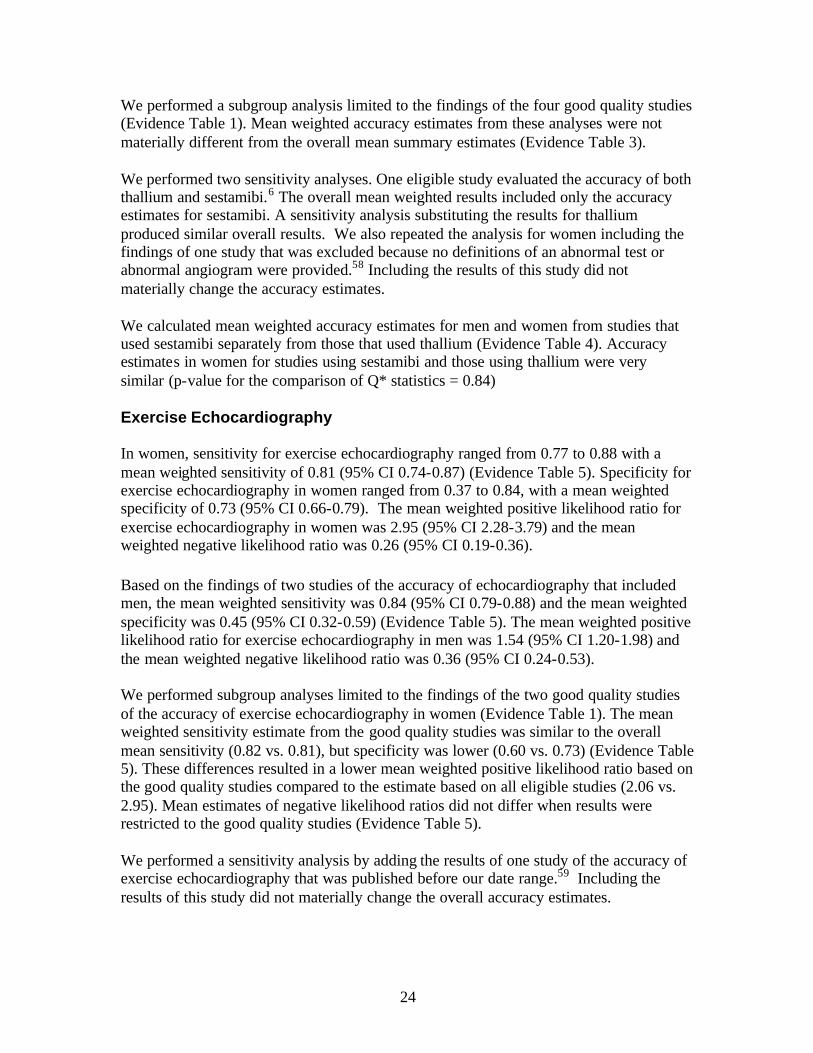
We performed a subgroup analysis limited to the findings of the four good quality studies (Evidence Table 1). Mean weighted accuracy estimates from these analyses were not materially different from the overall mean summary estimates (Evidence Table 3). We performed two sensitivity analyses. One eligible study evaluated the accuracy of both thallium and sestamibi.6 The overall mean weighted results included only the accuracy estimates for sestamibi. A sensitivity analysis substituting the results for thallium produced similar overall results. We also repeated the analysis for women including the findings of one study that was excluded because no definitions of an abnormal test or abnormal angiogram were provided.58 Including the results of this study did not materially change the accuracy estimates. We calculated mean weighted accuracy estimates for men and women from studies that used sestamibi separately from those that used thallium (Evidence Table 4). Accuracy estimates in women for studies using sestamibi and those using thallium were very similar (p-value for the comparison of Q* statistics = 0.84) Exercise Echocardiography In women, sensitivity for exercise echocardiography ranged from 0.77 to 0.88 with a mean weighted sensitivity of 0.81 (95% CI 0.74-0.87) (Evidence Table 5). Specificity for exercise echocardiography in women ranged from 0.37 to 0.84, with a mean weighted specificity of 0.73 (95% CI 0.66-0.79). The mean weighted positive likelihood ratio for exercise echocardiography in women was 2.95 (95% CI 2.28-3.79) and the mean weighted negative likelihood ratio was 0.26 (95% CI 0.19-0.36). Based on the findings of two studies of the accuracy of echocardiography that included men, the mean weighted sensitivity was 0.84 (95% CI 0.79-0.88) and the mean weighted specificity was 0.45 (95% CI 0.32-0.59) (Evidence Table 5). The mean weighted positive likelihood ratio for exercise echocardiography in men was 1.54 (95% CI 1.20-1.98) and the mean weighted negative likelihood ratio was 0.36 (95% CI 0.24-0.53). We performed subgroup analyses limited to the findings of the two good quality studies of the accuracy of exercise echocardiography in women (Evidence Table 1). The mean weighted sensitivity estimate from the good quality studies was similar to the overall mean sensitivity (0.82 vs. 0.81), but specificity was lower (0.60 vs. 0.73) (Evidence Table 5). These differences resulted in a lower mean weighted positive likelihood ratio based on the good quality studies compared to the estimate based on all eligible studies (2.06 vs. 2.95). Mean estimates of negative likelihood ratios did not differ when results were restricted to the good quality studies (Evidence Table 5). We performed a sensitivity analysis by adding the results of one study of the accuracy of exercise echocardiography that was published before our date range.59 Including the results of this study did not materially change the overall accuracy estimates.

25
Accuracy of Exercise MPI vs. Echocardiography in Women Based on 10 studies of the accuracy of exercise MPI and four of exercise echocardiography, the accuracy of each test for the diagnosis of CHD in women is similar (Evidence Tables 3 and 5). The mean weighted sensitivity, specificity and positive likelihood ratio for MPI are slightly lower than for echocardiography (sensitivity 0.77 vs. 0.81; specificity 0.69 vs. 0.73, positive likelihood ratio 2.46 vs. 2.95), but the differences are small and not statistically different (p-value for the Q* statistic = 0.10). The accuracy of the two tests is also similar when analyses are restricted to good quality studies (Evidence Tables 3 and 5). The use of sestamibi instead of thallium also did not change the accuracy of MPI studies in women (Evidence Table 4). Accuracy of Noninvasive Testing in Women Compared to Men The mean weighted sensitivity of MPI in women is somewhat lower than in men (0.77 vs. 0.93), but the specificity is higher (0.69 vs. 0.57) (Evidence Table 3). The positive likelihood ratio is slightly higher in women compared to men (2.46 vs. 2.17) as is the negative likelihood ratio (0.33 vs. 0.13). These differences in the accuracy of MPI between men and women were statistically significant (p-value for the comparison of Q* statistics 0.028), but it is not clear whether higher sensitivity or higher specificity is preferable. Comparison of mean weighted accuracy estimates between women and men may be biased if these data are derived from different studies that may have used somewhat different methods and definitions of positive tests. To avoid this problem, we calculated mean weighted accuracy estimates for men and women restricted to the findings of studies that included both genders (Evidence Table 3). Based on the findings of these studies, sensitivity of MPI is lower in women than in men (0.86 vs. 0.93), but specificity is the same (0.57 for both genders; p-value for the comparison of Q* statistics 0.012). This analysis suggests that exercise MPI is more accurate in men than in women, but the differences are small and not clinically meaningful. The mean weighted sensitivity of echocardiography in women is similar to that in men (0.81 vs. 0.84), but the specificity is higher (0.73 vs. 0.45) (Evidence Table 5). The positive likelihood ratio was substantially higher in women than in men (2.95 vs. 1.54), and the negative likelihood ratio was slightly lower (0.26 vs. 0.36). However, given the small numbers of men included in the analyses, we could not calculate a Q* statistic or determine any statistically significant differences between men and women with regard to exercise echocardiography. Assessments for Heterogeneity and Publication Bias There was no heterogeneity in any of the mean weighted estimates of accuracy. Publication bias usually occurs if small studies with unremarkable findings (poor accuracy) are not published while small studies with markedly positive findings (high

26
accuracy) are published. We calculated the correlation between individual study sample size and sensitivity using Kendall’s Tau to assess potential publication bias. There was no evidence of publication bias in any of the summary estimates of accuracy.
Conclusions In the last decade, both exercise echocardiography and exercise MPI have become widely available and commonly used for noninvasive diagnosis of coronary disease. It is important for both patients and providers to understand the accuracy of these tests and their limitations. We obtained results from 14 studies published between 1990 and 2002 on the accuracy of these tests in women. Based on these data, the overall accuracy of both tests in women is low with positive likelihood ratios of 2.5 to 3 and negative likelihood ratios of about 0.3. There are several advantages of estimating accuracy of a diagnostic test using likelihood ratios rather than sensitivity and specificity. First, it is possible to achieve a high sensitivity for most diagnostic tests by accepting a low specificity; similarly, high specificity can be achieved by accepting low sensitivity. In contrast, both sensitivity and specificity must be high to achieve good likelihood ratios (positive LR = sensitivity/(1-specificity) and negative LR = (1-sensitivity)/specificity). Secondly, likelihood ratios are a powerful tool to apply clinically using Bayes’ theorem; the post-test odds that a patient has the disease are estimated by multiplying the pre-test odds by the positive likelihood ratio. For instance, in a 55 year old woman with probable angina, the prior probability of CHD is about 30 percent.60 If her exercise MPI is positive, her posterior probability of CHD would be about 50 percent (prior odds 1:2.3 multiplied by positive LR of 2.5 equal posterior odds of 2.5:2.3 which is equivalent to posterior probability of about 50 percent). Similarly, if her exercise echocardiogram is positive, her posterior probability of having CHD would be about 55 percent. If either of these studies were negative, her posterior probability would be about 10 percent. Small differences in the posterior probabilities based on exercise MPI or echocardiogram do not have different clinical implications and suggest that the value of these tests is equivalent. The common conception that exercise testing in women should always be combined with imaging may not be true. A prior meta-analysis that evaluated the accuracy of exercise EKG in women found a mean weighted positive likelihood ratio of 2.25 and a negative likelihood ratio of 0.55.7 These accuracy estimates are very similar to those that we calculated for exercise MPI and echocardiogram and would result in very similar estimates of posterior probability of CHD. However, women who receive exercise EKG testing without imaging are more likely to have a normal EKG at baseline and thus may be less likely to have significant CHD. Thus, comparison of the accuracy of exercise EKG with exercise imaging studies or exercise echocardgiography may be biased unless patients are randomized to receive the different tests.

27
The value of a diagnostic test result depends on the accuracy of the test, the prior probability of disease and the threshold for treatment. In women with low to intermediate prior probability of CHD, a positive exercise MPI or echocardiogram result in similar posterior probabilities that may warrant further testing. Our 55 year-old woman with angina, for example, has about a 50 percent probability of having CHD if she has a positive noninvasive test. Before labeling her as having CHD and beginning treatment, many clinicians may want to pursue angiography. An older woman in her mid sixties with angina has about a 50 percent prior probability of having CHD. If she has a negative noninvasive test, her posterior probability of having CHD is about 25 percent. Many clinicians may prefer a more accurate test (such as angiography) before declaring that this woman does not have CHD and forgoing treatment. Our analysis found that the sensitivity of exercise MPI in women was lower than in men. While this difference was statistically significant, it was small and not clinically meaningful. Most of the studies included in our review reported a higher prevalence of CHD in men than in women. The prevalence of prior MI also was higher in men compared to women. This spectrum effect could account for the apparent lower sensitivity of MPI in women compared to men. Alternatively, the lower sensitivity in women may be due to differences in chest wall anatomy, left ventricular chamber size, ability to exercise maximally, catecholamine response to exercise or hormone levels. Comparison of the accuracy of exercise echocardiography in women and men was limited by the small number of studies that reported separate data for men. Evaluation of the accuracy of noninvasive tests requires a dichotomous outcome. The conventional “gold standard” for the presence of CHD is 50 percent or more stenosis of one or more of the major coronary arteries at angiography, and this is the definition that we used. However, coronary heart disease represents a continuum of disease that may not be best measured by angiography. In addition to diagnosis of CHD, exercise MPI and exercise echocardiography are also performed to localize disease and to determine the extent of disease. Our analysis provides no data on the accuracy of noninvasive tests for these purposes. The value of our review is limited by the quality of the studies included and the number of persons included. Many of the studies included in this systematic review were rated only fair quality, often because it was not clear that personnel who interpreted the noninvasive test were blinded to the results of the angiogram. Prior knowledge of angiographic results could falsely increase the accuracy of the noninvasive test. In many of the studies included, only persons with a positive noninvasive test went on to have angiography. This verification bias may result in higher sensitivity and lower specificity than in studies in which all subjects undergo both tests. Unfortunately, most studies did not report the percent of maximum predicted heart rate achieved by subjects. Failure to achieve 85 percent of maximum predicted heart rate would likely result in a higher number of false negatives and lower sensitivity. Sensitivity might be lower in women if they are less likely than men to exercise adequately.

28
Most of the studies of MPI and one of the studies of echocardiography included a substantial proportion of persons with prior myocardial infarction. Including persons with diagnosed disease likely increases sensitivity at the cost of specificity. Finally, we identified only 14 studies that met our inclusion criteria and the number of women included was limited. We identified many additional studies that included some women, but were unable to obtain data stratified by gender from the authors. Future Research Studies of the accuracy of noninvasive tests should publish all estimates of accuracy stratified by sex, or make these estimates available to public access. Stratification by sex would allow more precise estimates of the accuracy of noninvasive tests for CHD in women, but it is unlikely that this would result in a substantial improvement in the estimated accuracy of the tests. The major finding of this systematic review is that the accuracy of noninvasive tests for CHD in both men and women is low and that future research should address ways to improve accuracy. The quality of future studies of the accuracy of noninvasive tests for the diagnosis of CHD would be improved by excluding persons with known CHD, performing both the noninvasive test and angiography in all participants and assuring that the outcome of the noninvasive test is assessed by personnel blinded to the results of angiography. Finally, assessment of the value of diagnostic tests for estimating risk of future CHD events would have important long-term clinical implications.

29
References 1. Barolsky SM, Gilbert CA, Faruqui A, et al. Differences in electrocardiographic response
to exercise of women and men: a non-Bayesian factor. Circulation 1979;60(5):1021-7. 2. Marwick TH, Torelli J, Harjai K, et al. Influence of left ventricular hypertrophy on
detection of coronary artery disease using exercise echocardiography. J Am Coll Cardiol 1995;26(5):1180-6.
3. Sketch MH, Mohiuddin SM, Lynch JD, et al. Significant sex differences in the correlation of electrocardiographic exercise testing and coronary arteriograms. Am J Cardiol 1975;36(2):169-73.
4. Weiner DA, Ryan TJ, McCabe CH, et al. Exercise stress testing. Correlations among history of angina, ST-segment response and prevalence of coronary-artery disease in the Coronary Artery Surgery Study (CASS). N Engl J Med 1979;301(5):230-5.
5. Hansen CL, Crabbe D, Rubin S. Lower diagnostic accuracy of thallium-201 SPECT myocardial perfusion imaging in women: an effect of smaller chamber size. J Am Coll Cardiol 1996;28(5):1214-9.
6. Taillefer R, DePuey EG, Udelson JE, et al. Comparative diagnostic accuracy of Tl-201 and Tc-99m sestamibi SPECT imaging (perfusion and ECG-gated SPECT) in detecting coronary artery disease in women. J Am Coll Cardiol 1997;29(1):69-77.
7. Kwok Y, Kim C, Grady D, et al. Meta-analysis of exercise testing to detect coronary artery disease in women. Am J Cardiol 1999;83(5):660-6.
8. Fleischmann KE, Hunink MG, Kuntz KM, et al. Exercise echocardiography or exercise SPECT imaging? A meta-analysis of diagnostic test performance. JAMA 1998;280(10):913-20.
9. Beleslin BD, Ostojic M, Stepanovic J, et al. Stress echocardiography in the detection of myocardial ischemia. Head-to-head comparison of exercise, dobutamine, and dipyridamole tests. Circulation 1994;90(3):1168-76.
10. Berman DS, Kiat H, Friedman JD, et al. Separate acquisition rest thallium-201/stress technetium-99m sestamibi dual- isotope myocardial perfusion single-photon emission computed tomography: a clinical validation study. J Am Coll Cardiol 1993;22(5):1455-64.
11. Bjornstad K, Aakhus S, Hatle L. Comparison of digital dipyridamole stress echocardiography and upright bicycle stress echocardiography for identification of coronary artery stenosis. Cardiology 1995;86(6):514-20.
12. Christian TF, Miller TD, Bailey KR, et al. Noninvasive identification of severe coronary artery disease using exercise tomographic thallium-201 imaging. Am J Cardiol 1992;70(1):14-20.
13. Crouse LJ, Harbrecht JJ, Vacek JL, et al. Exercise echocardiography as a screening test for coronary artery disease and correlation with coronary arteriography. Am J Cardiol 1991;67(15):1213-8.

30
14. Dagianti A, Penco M, Agati L, et al. Stress echocardiography: comparison of exercise, dipyridamole and dobutamine in detecting and predicting the extent of coronary artery disease. J Am Coll Cardiol 1995;26(1):18-25.
15. Fleming RM, Kirkeeide RL, Taegtmeyer H, et al. Comparison of technetium-99m teboroxime tomography with automated quantitative coronary arteriography and thallium-201 tomographic imaging. J Am Coll Cardiol 1991;17(6):1297-302.
16. Galanti G, Sciagra R, Comeglio M, et al. Diagnostic accuracy of peak exercise echocardiography in coronary artery disease: comparison with thallium-201 myocardial scintigraphy. Am Heart J 1991;122(6):1609-16.
17. Gupta NC, Esterbrooks DJ, Hilleman DE, et al. Comparison of adenosine and exercise thallium-201 single-photon emission computed tomography (SPECT) myocardial perfusion imaging. The GE SPECT Multicenter Adenosine Study Group. J Am Coll Cardiol 1992;19(2):248-57.
18. Hambye AS, Vervaet A, Lieber S, et al. Diagnostic value and incremental contribution of bicycle exercise, first-pass radionuclide angiography, and 99mTc-labeled sestamibi single- photon emission computed tomography in the identification of coronary artery disease in patients without infarction. J Nucl Cardiol 1996;3(6 Pt 1):464-74.
19. Hecht HS, DeBord L, Sotomayor N, et al. Supine bicycle stress echocardiography: peak exercise imaging is superior to postexercise imaging. J Am Soc Echocardiogr 1993;6(3 Pt 1):265-71.
20. Heiba SI, Hayat NJ, Salman HS, et al. Technetium-99m-MIBI myocardial SPECT: supine versus right lateral imaging and comparison with coronary arteriography. J Nucl Med 1997;38(10):1510-4.
21. Ho YL, Wu CC, Huang PJ, et al. Dobutamine stress echocardiography compared with exercise thallium-201 single-photon emission computed tomography in detecting coronary artery disease-effect of exercise level on accuracy. Cardiology 1997;88(4):379-85.
22. Hoffmann R, Lethen H, Kleinhans E, et al. Comparative evaluation of bicycle and dobutamine stress echocardiography with perfusion scintigraphy and bicycle electrocardiogram for identification of coronary artery disease. Am J Cardiol 1993;72(7):555-9.
23. Kiat H, Van Train KF, Maddahi J, et al. Development and prospective application of quantitative 2-day stress-rest Tc-99m methoxy isobutyl isonitrile SPECT for the diagnosis of coronary artery disease. Am Heart J 1990;120(6 Pt 1):1255-66.
24. Luotolahti M, Saraste M, Hartiala J. Exercise echocardiography in the diagnosis of coronary artery disease. Ann Med 1996;28(1):73-7.
25. Mahmarian JJ, Boyce TM, Goldberg RK, et al. Quantitative exercise thallium-201 single photon emission computed tomography for the enhanced diagnosis of ischemic heart disease. J Am Coll Cardiol 1990;15(2):318-29.
26. Marangelli V, Iliceto S, Piccinni G, et al. Detection of coronary artery disease by digital stress echocardiography: comparison of exercise, transesophageal atrial pacing and dipyridamole echocardiography. J Am Coll Cardiol 1994;24(1):117-24.

31
27. Marwick TH, D'Hondt AM, Mairesse GH, et al. Comparative ability of dobutamine and exercise stress in inducing myocardial ischaemia in active patients. Br Heart J 1994;72(1):31-8.
28. Minoves M, Garcia A, Magrina J, et al. Evaluation of myocardial perfusion defects by means of "bull's eye" images. Clin Cardiol 1993;16(1):16-22.
29. Nguyen T, Heo J, Ogilby JD, et al. Single photon emission computed tomography with thallium-201 during adenosine-induced coronary hyperemia: correlation with coronary arteriography, exercise thallium imaging and two-dimensional echocardiography. J Am Coll Cardiol 1990;16(6):1375-83.
30. Ozdemir K, Kisacik HL, Oguzhan A, et al. Comparison of exercise stress testing with dobutamine stress echocardiography and radionuclide ventriculography for diagnosis of coronary artery disease. Jpn Heart J 1999;40(6):715-27.
31. Palmas W, Friedman JD, Diamond GA, et al. Incremental value of simultaneous assessment of myocardial function and perfusion with technetium-99m sestamibi for prediction of extent of coronary artery disease. J Am Coll Cardiol 1995;25(5):1024-31.
32. Quinones MA, Verani MS, Haichin RM, et al. Exercise echocardiography versus 201Tl single-photon emission computed tomography in evaluation of coronary artery disease. Analysis of 292 patients. Circulation 1992;85(3):1026-31.
33. Roger VL, Pellikka PA, Oh JK, et al. Identification of multivessel coronary artery disease by exercise echocardiography. J Am Coll Cardiol 1994;24(1):109-14.
34. Rubello D, Zanco P, Candelpergher G, et al. Usefulness of 99mTc-MIBI stress myocardial SPECT bull's-eye quantification in coronary artery disease. Q J Nucl Med 1995;39(2):111-5.
35. Salustri A, Pozzoli MM, Hermans W, et al. Relationship between exercise echocardiography and perfusion single-photon emission computed tomography in patients with single-vessel coronary artery disease. Am Heart J 1992;124(1):75-83.
36. Solot G, Hermans J, Merlo P, et al. Correlation of 99Tcm-sestamibi SPECT with coronary angiography in general hospital practice. Nucl Med Commun 1993;14(1):23-9.
37. Sylven C, Hagerman I, Ylen M, et al. Variance ECG detection of coronary artery disease--a comparison with exercise stress test and myocardial scintigraphy. Clin Cardiol 1994;17(3):132-40.
38. Tawa CB, Baker WB, Kleiman NS, et al. Comparison of adenosine echocardiography, with and without isometric handgrip, to exercise echocardiography in the detection of ischemia in patients with coronary artery disease. J Am Soc Echocardiogr 1996;9(1):33-43.
39. Van Train KF, Areeda J, Garcia EV, et al. Quantitative same-day rest-stress technetium-99m-sestamibi SPECT: definition and validation of stress normal limits and criteria for abnormality. J Nucl Med 1993;34(9):1494-502.
40. Van Train KF, Garcia EV, Maddahi J, et al. Multicenter trial validation for quantitative analysis of same-day rest-stress technetium-99m-sestamibi myocardial tomograms. J Nucl Med 1994;35(4):609-18.
41. Van Train KF, Maddahi J, Berman DS, et al. Quantitative analysis of tomographic stress thallium-201 myocardial scintigrams: a multicenter trial. J Nucl Med 1990;31(7):1168-79.

32
42. Haldane J. The estimation and significance of the logarithm of a ratio of frequencies. Ann Hum Genet 1955;20:309-314.
43. Moses LE, Shapiro D, Littenberg B. Combining independent studies of a diagnostic test into a summary ROC curve: data-analytic approaches and some additional considerations. Stat Med 1993;12(14):1293-316.
44. Chae SC, Heo J, Iskandrian AS, et al. Identification of extensive coronary artery disease in women by exercise single-photon emission computed tomographic (SPECT) thallium imaging. J Am Coll Cardiol 1993;21(6):1305-11.
45. Laurienzo JM, Cannon RO, 3rd, Quyyumi AA, et al. Improved specificity of transesophageal dobutamine stress echocardiography compared to standard tests for evaluation of coronary artery disease in women presenting with chest pain. Am J Cardiol 1997;80(11):1402-7.
46. Astarita C, Nicolai E, Liguori E, et al. [Dipyridamole-echocardiography and thallium exercise myocardial scintigraphy in the diagnosis of obstructive coronary or microvascular disease in hypertensive patients with left ventricular hypertrophy and angina]. G Ital Cardiol 1998;28(9):996-1004.
47. Sciammarella MG, Fragasso G, Gerundini P, et al. 99Tcm-MIBI single photon emission tomography (SPET) for detecting myocardial ischaemia and necrosis in patients with significant coronary artery disease. Nucl Med Commun 1992;13(12):871-8.
48. Mak KH, Ang ES, Goh AS, et al. Myocardial perfusion imaging with technetium-99m sestamibi SPECT in the evaluation of coronary artery disease. Australas Radiol 1995;39(2):112-7.
49. Williams MJ, Marwick TH, O'Gorman D, et al. Comparison of exercise echocardiography with an exercise score to diagnose coronary artery disease in women. Am J Cardiol 1994;74(5):435-8.
50. Roger VL, Pellikka PA, Bell MR, et al. Sex and test verification bias. Impact on the diagnostic value of exercise echocardiography. Circulation 1997;95(2):405-10.
51. Cecil MP, Kosinski AS, Jones MT, et al. The importance of work-up (verification) bias correction in assessing the accuracy of SPECT thallium-201 testing for the diagnosis of coronary artery disease. J Clin Epidemiol 1996;49(7):735-42.
52. Kang X, Berman DS, Lewin H, et al. Comparative ability of myocardial perfusion single-photon emission computed tomography to detect coronary artery disease in patients with and without diabetes mellitus. Am Heart J 1999;137(5):949-57.
53. Santana-Boado C, Candell-Riera J, Castell-Conesa J, et al. Diagnostic accuracy of technetium-99m-MIBI myocardial SPECT in women and men. J Nucl Med 1998;39(5):751-5.
54. Niemeyer MG, Van der Wall EE, Kuyper AF, et al. Discordance of visual and quantitative ana lysis regarding false negative and false positive test results in thallium-201 myocardial perfusion scintigraphy. Am J Physiol Imaging 1991;6(1):34-43.
55. Takeuchi M, Sonoda S, Miura Y, et al. Comparative diagnostic value of dobutamine stress echocardiography and stress thallium-201 single-photon-emission computed tomography for detecting coronary artery disease in women. Coron Artery Dis 1996;7(11):831-5.

33
56. Morise AP, Diamond GA, Detrano R, et al. Incremental value of exercise electrocardiography and thallium-201 testing in men and women for the presence and extent of coronary artery disease. Am Heart J 1995;130(2):267-76.
57. Sharir T, Germano G, Waechter PB, et al. A new algorithm for the quantitation of myocardial perfusion SPECT. II: validation and diagnostic yield. J Nucl Med 2000;41(4):720-7.
58. Iskandrian AE, Heo J, Nallamothu N. Detection of coronary artery disease in women with use of stress single-photon emission computed tomography myocardial perfusion imaging. J Nucl Cardiol 1997;4(4):329-35.
59. Sawada SG, Ryan T, Fineberg NS, et al. Exercise echocardiographic detection of coronary artery disease in women. J Am Coll Cardiol 1989;14(6):1440-7.
60. Chaitman BR, Bourassa MG, Davis K, et al. Angiographic prevalence of high-risk coronary artery disease in patient subsets (CASS). Circulation 1981;64(2):360-7.

35
Chapter 3. Systematic Review of Lipid Lowering Treatment to Reduce Risk of Coronary Heart Disease in Women Introduction Coronary heart disease (CHD) is the leading cause of death in the United States and half of all deaths from CHD occur in women.1, 2 Among white women, the cumulative risk of developing CHD between 50 and 94 years of age is 46 percent and the cumulative risk of dying from CHD is 31 percent.3 Elevated total cholesterol, low density lipoprotein-C (LDL-C) and triglycerides, and low high density lipoprotein-C (HDL-C) are risk factors for CHD in women.4-6 Lipid lowering may be achieved with either diet or drugs, but few studies have addressed the effects of dietary interventions on clinical outcomes. Several randomized clinical trials have evaluated the effect of lipid lowering with drugs on risk of CHD events, both in persons with known cardiovascular disease and in those without CHD.7-11 Unfortunately, many of the clinical trials of lipid lowering treatments did not include women and others did not include adequate numbers of women to allow sex-specific analyses. Finally, some of the trials that did include women reported aggregate events (e.g. major coronary events) but did not report specific outcomes such as CHD death or nonfatal myocardial infarction (MI) separately. A previous systematic review of lipid lowering therapy in women included only studies published before 1995 and is now outdated.12 A more recent systematic review that included only trials of statin drugs found that both women and men treated with statins had a 30 percent reduction in risk of major CHD events.13 However, this review did not address outcomes other than major CHD events in women, did not stratify by primary or secondary prevention and did not include data from recent large trials. The goal of this systematic review is to critically assess the available clinical trial evidence regarding drug treatment of hyperlipidemia for the prevention of CHD events and death in women. We will assess the effects of lipid lowering on total mortality, CHD mortality, nonfatal myocardial infarction (MI), CHD events and revascularization procedures in women with and without prior CHD.

36
Methodology Data sources We searched PubMed®, the Cochrane Database, and DARE for articles published in English and other languages from 1966 through January 2002. We also reviewed bibliographies and asked peer reviewers (Appendix A) to identify additional articles. Search Terms Search terms were developed in collaboration with a medical librarian and included the following: Limits publication dates 1966 to January 2002, human
Not: practice guideline, letter, editorial, review, meta-analysis, infant, newborn, preschool child, child
Predictor hyperlipidemia and anticholesteremic agents, antilipemic agents, simvastatin, lovastatin, pravastatin, atorvastatin, fluvastatin, gemfibrozil, cholestyramine, cholestpol, niacin
Outcomes cardiovascular diseases, heart diseases, myocardial ischemia, coronary disease
Inclusion Criteria To be included, articles were required to fit the following criteria: 1) Randomized clinical trials of outpatients with or without known CHD. 2) Treatment duration of at least one year. 3) Study population classified as either primary (participants without prior CHD) or
secondary prevention (participants with prior CHD). 4) Data on women provided. 5) Impact of lipid lowering with drug treatment assessed for at least one of the following clinical outcomes: total mortality, CHD mortality, nonfatal MI, CHD events or revascularization procedures. Coronary events included ischemic coronary syndromes and nonfatal myocardial infarction. CHD procedures included coronary bypass graft surgery and percutaneous coronary angioplasty or stenting. 6) Published between January 1, 1966 and January 30, 2002. Articles published outside this date range recommended by peer reviewers were included. We excluded studies that only provided evidence on the effect of treatment on changes in lipids, angiographic findings or other intermediate outcomes. For studies with multiple publications, only data from the most comprehensive or most recent publication were used.

37
Article Identification An initial search using the terms listed above identified articles that potentially provided evidence. Two University of California, San Francisco (UCSF)-Stanford Evidence-based Practice Center (EPC) physician investigators reviewed the titles and excluded those that clearly did not provide data on humans or clearly did not address the question. The abstracts of the remaining articles were reviewed independently by two UCSF-Stanford EPC physician investigators and coded using the categories listed below. Disagreements were discussed and the following consensus codes were entered into a database (Access, Microsoft Corporation): RQ Research question - the article clearly does not address the research question. R Review – the study is a review that does not contain primary data. NSD Not appropriate study design - The article is not a randomized clinical trial. NH No humans - the study clearly does not include data on humans. E1 Eligible – the study may contain primary evidence regarding the research
question in women and will be reviewed in full-text. Articles coded E1 were retrieved and the full text was reviewed independently by two UCSF-Stanford EPC investigators. Names of authors and titles of journals were obscured before articles were reviewed. Obtaining Unpublished Results Some eligible studies included women in the study population, but did not report findings separately by gender. In these instances we attempted to contact authors to obtain these data. If we did not receive a response after the first contact, a second attempt was made. We contacted 13 authors11, 14-27 and received data from two.16, 25, 26 Quality Assessment The full text of each eligible study was reviewed independently by two UCSF-Stanford physician investigators, who completed a quality evaluation form (Appendix B). All of the studies included in this systematic review are randomized clinical trials. To be categorized as good quality, articles were required to meet the following additional parameters: • Inclusion/exclusion criteria clear and appropriate. • Randomization allocation concealed. • Control group received placebo. • Participants and research staff blinded to intervention. • More than 75 percent complete followup.

38
All other trials were considered fair quality. Disagreements between reviewers regarding quality parameters were decided by discussion and consensus. Data Abstraction Two UCSF-Stanford EPC physician investigators independently reviewed the full text of each eligible study and completed a data abstraction form (Appendix C). We abstracted information on the study population (primary prevention trials were defined as those that included individuals without known CHD; secondary prevention trials included individuals with known CHD), inclusion criteria, length of followup, numbers of men and women, participant characteristics such as age, other cardiovascular risk factors and cardiac medication use, baseline and followup lipoprotein values and all clinical outcomes that were measured. When possible, data were abstracted for men and women separately. Disagreements were discussed and decided by consensus. Data Management and Archive We entered all identified titles and abstracts in an EndNote® file (Niles Software, Inc.) that includes searchable key words as codes for eligibility. Information on all articles that were reviewed in full text was transferred from EndNote® to a relational database (Access, Microsoft® Corporation) that allows us to categorize each article by reason for exclusion. Quality assessment data for each eligible study were also entered in the Access database, allowing us to categorize eligible articles by quality. Selected data were transferred to a flatfile database (EXCEL, Microsoft® Corporation) for preparation of evidence tables and calculation of summary estimates, confidence intervals and tests of heterogeneity. The full- text articles that were retrieved, and the abstraction forms for each article are filed by topic and question in Dr. Grady's offices at the UCSF Mt. Zion Women's Health Clinical Research Center. Data Analyses The primary outcome of each clinical trial was expressed as the relative risk (RR) among treated compared to untreated study participants. Summary estimates of RR and 95 percent confidence intervals (CI) were calculated using the Mantel-Haenszel method and both fixed and random effects models. Results of the fixed and random effects models were similar, and we report only the findings of the random effects model. To avoid calculation problems associated with zero cells, 0.5 was added to all cells to calculate variances and standard deviations.28 The significance level for all tests of outcome was set at 0.05. All findings were assessed for heterogeneity using a standard Chi-square test and Q statistic with critical value set at 0.10. All analyses were performed separately for the findings of primary and secondary prevention studies. Subgroup analyses were performed by type of drug treatment (statins vs. others), and by good vs. fair quality.

39
Publication bias usually occurs if small studies with unremarkable findings (relative risks about 1.0) are not published while small studies with markedly positive findings (in this case, low relative risks) are published. We calculated the correlation between individual study weight (1/variance) and relative risk using a nonparametric correlation coefficient (Kendall’s Tau) with critical value set at 0.10 to assess potential publication bias. Statistically significant correlation of study weight and relative risk suggests publication bias. Results Results of Study Identification Our searches identified 1,335 titles. After eliminating ineligible studies by review of titles and abstracts, we reviewed the full text of 120 articles. We identified 20 studies that fit all inclusion criteria, but only 9 provided outcomes stratified by sex.7-9, 11, 27, 29-39 We contacted the principal investigators of the studies that did not provide data for women to request this information.11, 14-27 We received data on women from two investigators.16, 25,
26 Thus, 11 studies (represented by 19 articles) were found to be both eligible and to contain data stratified by sex for inclusion in the systematic review.7-9, 11, 16, 25-27, 29-39 One additional study did not meet inclusion criteria because it did not provide data on any of the clinical outcomes of interest and the study population was equally divided between persons with prior CHD and those without prior CHD and separate estimates for the effects of lipid lowering in primary and secondary prevention were not published.40 Description of Eligible Studies Characteristics of the 11 eligible trials are described in Evidence Table 6. The numbers of participants in each trial ranged from 151 to 20,536 and 15 to 50 percent of participants were women. The total number of women included in the trials was 15,917, but almost two thirds were from two studies.11, 39Information on the ethnicity of participants was not provided in most trials. Duration of treatment ranged from 2.8 to 6.1 years and averaged 4.7 years. Seven of the trials were classified as secondary prevention and four were classified as primary prevention (Evidence Table 6). Eligibility criteria for 5 trials required at least mild hyperlipidemia,7, 16, 25, 26, 31, 39 four required a range of cholesterol levels that would include some participants in the normal range9, 11, 27, 34 and 2 included participants regardless of cholesterol levels.29, 30 Three trials assessed the effects of clofibrate29, 30 or colestipol31 and eight assessed the efficacy of treatment with statins (lovastatin,9, 25, 26 simvastatin,7, 11, 39pravastatin16, 27, 34). All but one of the 11 trials included a placebo control group39 and all but two were adequately blinded.31, 39 In all but one of the trials,31 followup was greater than 75 percent complete. Overall, seven of the trials were rated good quality and four were rated fair (Evidence Table 6).

40
The clinical outcomes evaluated were total mortality, CHD mortality, nonfatal MI, CHD events and revascularization (Evidence Tables 7 and 8). Most trials were designed to address clinical outcomes, but two were designed to evaluate change in intimal medial thickness of the carotid artery16, 25, 26 and included clinical events only as secondary outcomes. For studies with mixed populations (e.g. some participants had CHD and some did not), the trial was classified as primary or secondary prevention based on the status of the majority of participants. Participants in most of the trials classified as primary prevention were at high risk for CHD outcomes due to presence of CHD risk factors. Three trials included participants with and without CHD. In the colestipol trial, only 20 percent of participants had CHD and this trial was classified as a primary prevention study.31 In the Heart Protection Study, 65 percent of participants had known CHD and remaining 35 percent had peripheral vascular disease, cerebrovascular disease or diabetes.11 Because the majority of participants had CHD and those without CHD were also at very high risk for CHD events, this trial was classified as a secondary prevention study. A recently published trial included older participants with CHD and those at high risk for CHD in approximately equal numbers40 and did not present results stratified by history of CHD. Because of the equal distribution, we are unable to classify this study as either primary or secondary prevention. In addition, we could not include the results of this trial for any specific coronary disease endpoint because the results for women were only given for the composite outcome of cardiovascular events (CHD mortality, nonfatal MI, fatal stroke and nonfatal stroke). Findings For each outcome, we assessed the effects of lipid lowering separately for primary and secondary prevention studies. We also calculated summary estimates based on the findings of all eligible studies, those that used a statin as the lipid lowering agent and those that were rated good quality (Evidence Table 8). Seven trials assessed the effects of lipid lowering among women with CHD (secondary prevention)7, 8, 11, 16, 27, 29, 30, 32-37 and included a total of 8,244 women. Two of these trials used clofibrate as the intervention,29, 30 while five used a statin.7, 8, 11, 16, 27, 32-37 Both of the trials of clofibrate were rated fair,29, 30 while all of the statin trials were rated good quality. While seven trials provided data, three were small (22 to 124 women),16, 29, 30 two were mid-sized (5768, 34, 35 and 827 women32, 33) and only two included more than 1,000 women (1,51627, 36, 37 and 5,08211). Evidence was also limited because several of the trials reported results among women for only one or two of the five outcomes of interest (total mortality, CHD mortality, nonfatal MI, CHD events and revascularization).
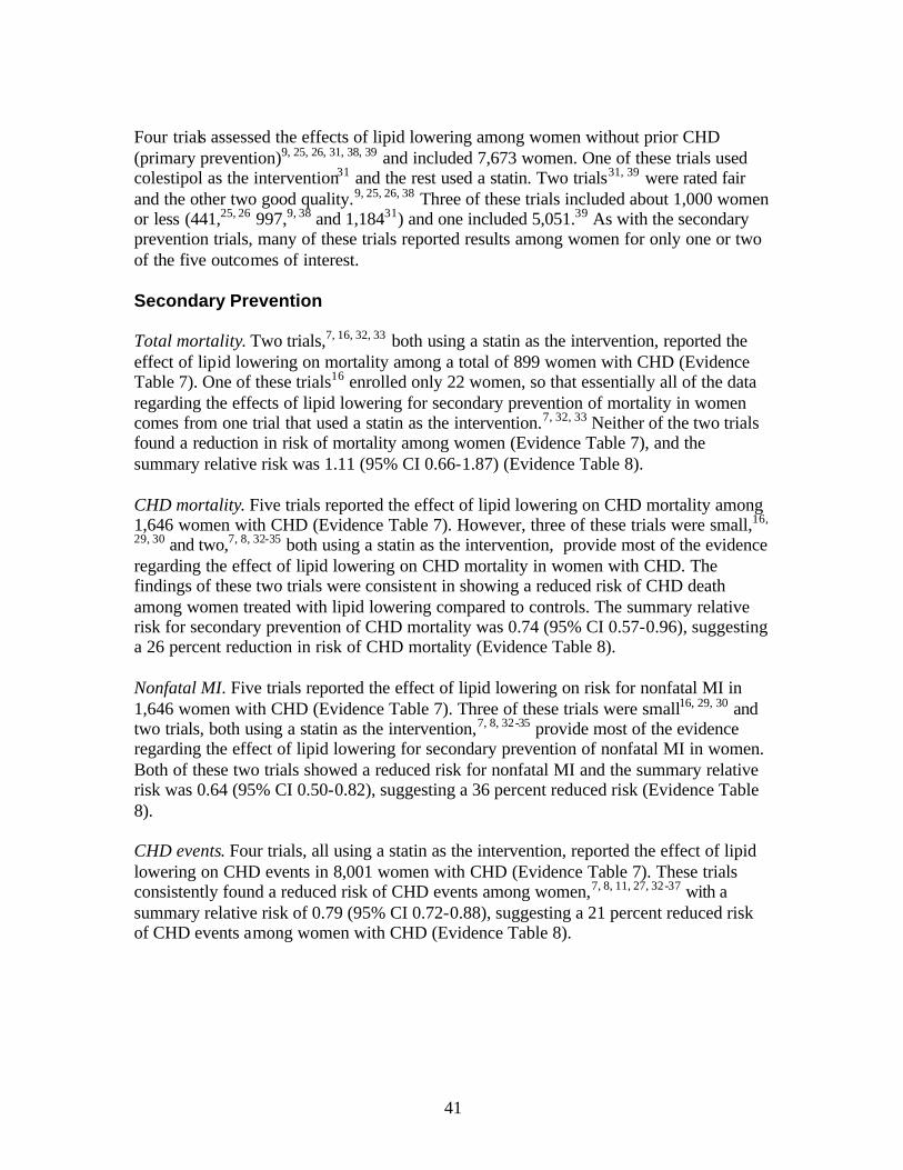
41
Four trials assessed the effects of lipid lowering among women without prior CHD (primary prevention)9, 25, 26, 31, 38, 39 and included 7,673 women. One of these trials used colestipol as the intervention31 and the rest used a statin. Two trials31, 39 were rated fair and the other two good quality.9, 25, 26, 38 Three of these trials included about 1,000 women or less (441,25, 26 997,9, 38 and 1,18431) and one included 5,051.39 As with the secondary prevention trials, many of these trials reported results among women for only one or two of the five outcomes of interest. Secondary Prevention Total mortality. Two trials,7, 16, 32, 33 both using a statin as the intervention, reported the effect of lipid lowering on mortality among a total of 899 women with CHD (Evidence Table 7). One of these trials16 enrolled only 22 women, so that essentially all of the data regarding the effects of lipid lowering for secondary prevention of mortality in women comes from one trial that used a statin as the intervention.7, 32, 33 Neither of the two trials found a reduction in risk of mortality among women (Evidence Table 7), and the summary relative risk was 1.11 (95% CI 0.66-1.87) (Evidence Table 8). CHD mortality. Five trials reported the effect of lipid lowering on CHD mortality among 1,646 women with CHD (Evidence Table 7). However, three of these trials were small,16,
29, 30 and two,7, 8, 32-35 both using a statin as the intervention, provide most of the evidence regarding the effect of lipid lowering on CHD mortality in women with CHD. The findings of these two trials were consistent in showing a reduced risk of CHD death among women treated with lipid lowering compared to controls. The summary relative risk for secondary prevention of CHD mortality was 0.74 (95% CI 0.57-0.96), suggesting a 26 percent reduction in risk of CHD mortality (Evidence Table 8). Nonfatal MI. Five trials reported the effect of lipid lowering on risk for nonfatal MI in 1,646 women with CHD (Evidence Table 7). Three of these trials were small16, 29, 30 and two trials, both using a statin as the intervention,7, 8, 32-35 provide most of the evidence regarding the effect of lipid lowering for secondary prevention of nonfatal MI in women. Both of these two trials showed a reduced risk for nonfatal MI and the summary relative risk was 0.64 (95% CI 0.50-0.82), suggesting a 36 percent reduced risk (Evidence Table 8). CHD events. Four trials, all using a statin as the intervention, reported the effect of lipid lowering on CHD events in 8,001 women with CHD (Evidence Table 7). These trials consistently found a reduced risk of CHD events among women,7, 8, 11, 27, 32-37 with a summary relative risk of 0.79 (95% CI 0.72-0.88), suggesting a 21 percent reduced risk of CHD events among women with CHD (Evidence Table 8).

42
Revascularization. Two trials, both using a statin as the intervention, reported the effect of lipid lowering for secondary prevention of revascularization procedures in 1,403 women with CHD (Evidence Table 7). Both of these trials found a reduction in risk among treated women and the summary relative risk was 0.70 (95% CI 0.42-1.16). Although the summary relative risk suggests a 30 percent reduction in risk of revascularization, this finding was not statistically significant (Evidence Table 8). Drug class and study quality. Only two studies, including a total of 221 women, addressed the impact of lipid lowering drugs other than statins.29, 30 Thus, evidence on the effect of non-statin drugs is limited. However, the summary ORs were similar for all outcomes when findings were restricted to those studies using a statin. Both of the studies that used a non-statin drug were rated fair quality,29, 30and all five of the trials that used a statin were rated good quality. Thus, the summary ORs are also unchanged when the results are restricted to good quality studies. Sensitivity analyses. One trial that we included with the secondary prevention studies enrolled a mixed population of persons with and without CHD and reported the effect of statin treatment on risk for CHD events.11 Because 65 percent of the participants had prior CHD and the rest had vascular disease or diabetes, we included the results of this trial as secondary prevention. We also performed a sensitivity analysis excluding the results of this trial. The summary relative risk for secondary prevention of CHD events excluding the results of this trial was essentially unchanged (summary relative risk 0.74; 95% CI 0.61-0.91). We also performed a sensitivity analysis by adding the results of the cardiovascular disease outcomes from the PROSPER trial to the summary results for secondary prevention of CHD events.40 The summary relative risk for secondary prevention of CHD events including the results of this trial was essentially unchanged (summary relative risk 0.76; 95% CI 0.72-0.87). Primary Prevention Total mortality. Four trials9, 25, 26, 31, 38, 39 reported the effect of lipid lowering on mortality among 7,673 women without prior CHD (Evidence Table 7). One of these trials31 used colestipol as the intervention, while the rest used a statin. One of the trials reported a lower risk of mortality in women treated with lipid lowering compared to controls, but the other three did not (Evidence Table 7). The summary relative risk for primary prevention of mortality was 0.95 (95% CI 0.62-1.46) (Evidence Table 8). CHD mortality. Three trials9, 25, 26, 31, 38 reported the effect of lipid lowering on CHD mortality among 2,622 women without prior CHD (Evidence Table 7). One of these trials31 used colestipol as the intervention, while the other two used a statin. One of the three trials reported a lower risk of CHD mortality in women treated with lipid lowering compared to controls,25, 26 but the others did not. The summary relative risk for primary prevention of CHD mortality was 1.07 (95% CI 0.47-2.40).

43
Nonfatal MI. Two trials9, 25, 26, 38 reported the effect of lipid lowering on risk for nonfatal MI in 1,646 women without prior CHD (Evidence Table 7). Both of these trials used a statin as the intervention, and both found a reduced risk of nonfatal MI among women treated with lipid lowering. The summary relative risk for primary prevention of nonfatal MI was 0.61 (95% CI 0.22-1.68). Although the summary relative risk suggests a 39 percent reduction in risk of nonfatal MI among treated women, this finding was not statistically significant. CHD events. Two trials, both using a statin as the intervention,9, 38, 39 reported the effect of lipid lowering on risk for CHD events in 6,048 women without prior CHD (Evidence Table 7). The results of these trials are inconsistent, and the summary relative risk for primary prevention of CHD events was 0.87 (95% CI 0.50-1.49). Revascularization. Only one trial reported the effect of statin therapy for primary prevention of revascularization procedures in women9, 38 (Evidence Table 7). This trial found a relative risk of 0.87 (95% CI 0.33-2.31). Drug class and study quality. Evidence on the primary prevention effects of drugs other than statins is limited as only one trial addressed the impact of a non-statin drug.31 The summary ORs were similar for all outcomes when findings were restricted to those studies using a statin or to studies rated good quality. Sensitivity analyses. We performed a sensitivity analysis by adding the results of the cardiovascular disease outcomes from the PROSPER trial to the summary results for primary prevention of CHD events.40 The summary relative risk for primary prevention of CHD events including the results of this trial was essentially unchanged (summary relative risk 0.97; 95% CI 0.84-1.12). Assessments for Heterogeneity and Publication Bias There was no statistical evidence of heterogene ity in any of the overall summary estimates of the effect of lipid lowering on any outcome except for secondary prevention of revascularization (Evidence Table 8). There was no evidence of publication bias in any of the summary estimates. Conclusions Although 20 clinical trials of the effects of lipid lowering therapy included women, only nine published results by gender. By contacting study investigators, we were successful in obtaining data on women from two additional trials. Thus, we were able to analyze results from 11 trials that included 15,917 women. However, complete data on the five outcomes of interest were not available from each trial, limiting our ability to assess the effect of lipid lowering on some outcomes. Only three studies, including a total of 1,405 women, addressed the impact of lipid lowering drugs other than statins. Thus, evidence on the effect of non-statin drugs is limited.

44
In the secondary prevention setting, treatment with lipid lowering therapy reduced risk of CHD mortality, nonfatal MI and CHD events in women. Summary estimates suggest a 26 percent reduction in risk of CHD mortality, a 36 percent reduction in risk of nonfatal MI and a 21 percent reduction in risk of a CHD event. There was no evidence of a reduction in risk of total mortality and insufficient evidence to document a reduction in risk of revascularization procedures. In the primary prevention setting, there was insufficient evidence of reduced risk of any clinical outcome in women. The summary relative risk for nonfatal MI was similar to that for secondary prevention (39 percent reduction vs. 36 percent reduction for secondary prevention), but was not statistically significant. A prior systematic review of the findings of clinical trials of the effects of lipid lowering among persons without CHD used inclusion criteria and methods very similar to ours, but did not stratify the results by gender.41 Since 90 percent of the participants included in that review were men, the results primarily reflect the effects of lipid lowering in men. Among (mostly) men, primary prevention with lipid lowering resulted in about a 30 percent reduced risk for both CHD events and CHD mortality41 Our findings suggest that, among persons without CHD, lipid lowering may not be as effective in women as in men without CHD. However, our power to observe a modest reduction in CHD risk was limited because the findings of only four primary prevention trials were available for inclusion in the meta-analysis. We were unable to include findings from a recently published clinical trial of the effect of lipid lowering among 2,804 men and 3,000 women aged 70 to 82 years randomized to pravastatin or placebo and followed for a mean of 3.2 years.40 About half of the participants in this trial had vascular disease and the others had vascular risk factors. Results were reported for the effect of lipid lowering on cardiovascular events in women (CHD mortality, nonfatal MI, fatal stroke and nonfatal stroke); the relative risk among women treated with pravastatin was 0.96 (95% CI 0.79-1.18). We could not include these data because we could not categorize the trial as primary or secondary prevention and results in women were only given for cardiovascular events. Given the timeline for this review, we did not have time to contact the authors to request findings stratified by sex, primary vs. secondary prevention and clinical outcomes. However, in sensitivity analyses that included the results of this trial as either primary or secondary prevention of CHD events did not alter the findings. There were no clinical differences in the summary odds ratios when studies included were restricted to those that used a statin as the intervention or to good quality studies. This is likely because eight of the 11 included trials used a statin as the intervention, and seven of the 11 trials were rated good quality. In summary, lipid lowering therapy appears to reduce risk of CHD mortality, nonfatal MI and CHD events 25 to 35 percent in women with prior CHD. There was inadequate evidence to document a reduction in risk of any clinical outcome among women without prior CHD. Data were limited, but the risk for total mortality was not lower in women

45
treated with lipid lowering, regardless of whether they had prior CHD or not. The lack of reduction in risk for mortality in either primary or secondary prevention settings may be because lipid lowering does not affect total mortality in women or because there were few deaths, even after summarizing study findings. Future Research Future randomized trials should include women in adequate numbers to assess the effects of lipid lowering on clinical outcomes. Studies that include women should report the effects of lipid lowering on all clinical outcomes stratified by sex and primary vs. secondary prevention.


47
References 1. Thom TJ. Cardiovascular disease mortality among United States women. In:
Eaker ED, Packard B, Wenger NK, et al., editors. Coronary Heart Disease in Women. New York: Haymarket Doyma; 1987. p. 270.
2. American Heart Association. Heart Disease and Stroke Statistics--2002 Update. Dallas, Tex.: American Heart Association; 2001.
3. Grady D, Rubin SM, Petitti DB, et al. Hormone therapy to prevent disease and prolong life in postmenopausal women. Ann Intern Med 1992;117:1016-1037.
4. Manolio TA, Pearson TA, Wenger NK, et al. Cholesterol and heart disease in older persons and women: review of an NHLBI workshop. Ann Epidemiol 1992; 2: 161-176 1992.
5. LaRosa JC. Triglycerides and coronary risk in women and the elderly. Arch Intern Med 1997;157:961-968.
6. NIH. Consensus Conference. Triglyceride, high density lipoprotein and coronary heart disease. JAMA 1993;269:505-510.
7. Randomised trial of cholesterol lowering in 4444 patients with coronary heart disease: the Scandinavian Simvastatin Survival Study (4S). Lancet 1994;344(8934):1383-9.
8. Sacks FM, Pfeffer MA, Moye LA, et al. The effect of pravastatin on coronary events after myocardial infarction in patients with average cholesterol levels. Cholesterol and Recurrent Events Trial investigators. N Engl J Med 1996;335(14):1001-9.
9. Downs JR, Clearfield M, Weis S, et al. Primary prevention of acute coronary events with lovastatin in men and women with average cholesterol levels: results of AFCAPS/TexCAPS. Air Force/Texas Coronary Atherosclerosis Prevention Study. JAMA 1998;279(20):1615-22.
10. Shepherd J, Cobbe SM, Ford I, et al. Prevention of coronary heart disease with pravastatin in men with hypercholesterolemia. N Engl J Med 1995;333:1301-7.
11. MRC/BHF Heart Protection Study of cholesterol lowering with simvastatin in 20,536 high-risk individuals: a randomised placebo-controlled trial. Lancet 2002;360(9326):7-22.
12. Walsh J, Grady D. Treatment of hyperlipidemia in women. JAMA 1995;274:1152-2258.
13. LaRosa JC, He J, Vupputuri S. Effect of statins on risk of coronary disease: a meta-analysis of randomized controlled trials. JAMA 1999;282:2340-2346.
14. Brensike JF, Levy RI, Kelsey SF, et al. Effects of therapy with cholestyramine on progression of coronary arteriosclerosis: results of the NHLBI Type II Coronary Intervention Study. Circulation 1984;69(2):313-24.
15. The Pravastatin Multinational Study Group for Cardiac Risk Patients. Effects of pravastatin in patients with serum total cholesterol levels from 5.2 to 7.8 mmol/liter (200 to 300 mg/dl) plus two additional atherosclerotic risk factors. Am J Cardiol 1993;72(14):1031-7.
16. Byington RP, Furberg CD, Crouse JR, 3rd, et al. Pravastatin, Lipids, and Atherosclerosis in the Carotid Arteries (PLAC- II). Am J Cardiol 1995;76(9):54C-59C.

48
17. Post Coronary Artery Bypass Graft Trial Investigators. The effect of aggressive lowering of low-density lipoprotein cholesterol levels and low-dose anticoagulation on obstructive changes in saphenous-vein coronary-artery bypass grafts. N Engl J Med 1997;336(3):153-62.
18. Herd JA, Ballantyne CM, Farmer JA, et al. Effects of fluvastatin on coronary atherosclerosis in patients with mild to moderate cholesterol elevations (Lipoprotein and Coronary Atherosclerosis Study [LCAS]). Am J Cardiol 1997;80(3):278-86.
19. Campeau L, Hunninghake DB, Knatterud GL, et al. Aggressive cholesterol lowering delays saphenous vein graft atherosclerosis in women, the elderly, and patients with associated risk factors. NHLBI post coronary artery bypass graft clinical trial. Post CABG Trial Investigators. Circulation 1999;99(25):3241-7.
20. Riegger G, Abletshauser C, Ludwig M, et al. The effect of fluvastatin on cardiac events in patients with symptomatic coronary artery disease during one year of treatment. Atherosclerosis 1999;144(1):263-70.
21. Arntz HR, Agrawal R, Wunderlich W, et al. Beneficial effects of pravastatin (+/-colestyramine/niacin) initiated immediately after a coronary event (the randomized Lipid-Coronary Artery Disease [L-CAD] Study). Am J Cardiol 2000;86(12):1293-8.
22. Teo KK, Burton JR, Buller CE, et al. Long-term effects of cholesterol lowering and angiotensin-converting enzyme inhibition on coronary atherosclerosis: The Simvastatin/Enalapril Coronary Atherosclerosis Trial (SCAT). Circulation 2000;102(15):1748-54.
23. Brown BG, Zhao XQ, Chait A, et al. Simvastatin and niacin, antioxidant vitamins, or the combination for the prevention of coronary disease. N Engl J Med 2001;345(22):1583-92.
24. Pitt B, Mancini GB, Ellis SG, et al. Pravastatin limitation of atherosclerosis in the coronary arteries (PLAC I): reduction in atherosclerosis progression and clinical events. PLAC I investigation. J Am Coll Cardiol 1995;26(5):1133-9.
25. ACAPS Group. Rationale and design for the Asymptomatic Carotid Artery Plaque Study (ACAPS). Control Clin Trials 1992;13(4):293-314.
26. Furberg CD, Adams HP, Jr., Applegate WB, et al. Effect of lovastatin on early carotid atherosclerosis and cardiovascular events. Asymptomatic Carotid Artery Progression Study (ACAPS) Research Group. Circulation 1994;90(4):1679-87.
27. Long-Term Intervention with Pravastatin in Ischaemic Disease (LIPID) Study Group. Prevention of cardiovascular events and death with pravastatin in patients with coronary heart disease and a broad range of initial cholesterol levels. N Engl J Med 1998;339(19):1349-57.
28. Haldane JBS. The estimation and significance of the logarithm of a ratio of frequencies. Ann Hum Genet 1955 1955;20:309-314.
29. Research Committee of the Scottish Society of Physicians. Ischemic Heart Disease: a secondary prevention trial using clofibrate. BMJ 1971;4:775-784.
30. Group of Physicians of the Newcastle Upon Tyne Region. Trial of clofibrate in the treatment of ischemic heart disease. BMJ 1971;4:767-775.

49
31. Dorr AE, Gundersen K, Schneider JC, Jr., et al. Colestipol hydrochloride in hypercholesterolemic patients--effect on serum cholesterol and mortality. J Chronic Dis 1978;31(1):5-14.
32. Miettinen TA, Pyorala K, Olsson AG, et al. Cholesterol- lowering therapy in women and elderly patients with myocardial infarction or angina pectoris: findings from the Scandinavian Simvastatin Survival Study (4S). Circulation 1997;96(12):4211-8.
33. Pedersen TR, Berg K, Cook TJ, et al. Safety and tolerability of cholesterol lowering with simvastatin during 5 years in the Scandinavian Simvastatin Survival Study. Arch Intern Med 1996;156(18):2085-92.
34. Lewis SJ, Sacks FM, Mitchell JS, et al. Effect of pravastatin on cardiovascular events in women after myocardial infarction: the cholesterol and recurrent events (CARE) trial. J Am Coll Cardiol 1998;32(1):140-6.
35. Plehn JF, Davis BR, Sacks FM, et al. Reduction of stroke incidence after myocardial infarction with pravastatin: the Cholesterol and Recurrent Events (CARE) study. The Care Investigators. Circulation 1999;99(2):216-23.
36. Tonkin AM, Colquhoun D, Emberson J, et al. Effects of pravastatin in 3260 patients with unstable angina: results from the LIPID study. Lancet 2000;356(9245):1871-5.
37. White HD, Simes RJ, Anderson NE, et al. Pravastatin therapy and the risk of stroke. N Engl J Med 2000;343(5):317-26.
38. Clearfield M, Downs JR, Weis S, et al. Air Force/Texas Coronary Atherosclerosis Prevention Study (AFCAPS/TexCAPS): efficacy and tolerability of long-term treatment with lovastatin in women. J Womens Health Gend Based Med 2001;10(10):971-81.
39. Antihypertensive and Lipid-Lowering Treatment to Prevent Heart Attack Trial (ALLHAT) Officers and Coordinators for the ALLHAT Collaborative Research Group. Major outcomes in moderately hypercholesterolemic, hypertensive patients randomized to pravastatin vs usual care: The Antihypertensive and Lipid-Lowering Treatment to Prevent Heart Attack Trial (ALLHAT-LLT). JAMA 2002;288(23):2998-3007.
40. Shepherd J, Blauw GJ, Murphy MB, et al. Pravastatin in elderly individuals at risk of vascular disease (PROSPER): a randomised controlled trial. Lancet 2002;360(9346):1623-30.
41. Pignone M, Phillips C, Mulrow C. Use of lipid lowering drugs for primary prevention of coronary heart disease: meta-analysis of randomised trials. BMJ 2000;321:1-5.
Chapter 4. Systematic Review of Diabetes as a Risk Factor for Coronary Heart Disease in Women Introduction Studies suggest that there may be a stronger association between type 2 diabetes and coronary heart disease (CHD) risk in women than in men. Estimates of coronary heart disease mortality in diabetic men have varied from 1 to 3-fold the rate in nondiabetic men,1-10 while estimates in diabetic women have ranged from 2 to 5-fold the rate in nondiabetic women.2, 5, 8, 10-12 Variations in study population, design, quality and findings make it difficult to evaluate the strength of diabetes as a risk factor for CHD in either sex. Two previous meta-analyses that included studies that did not adjust for major cardiovascular risk factors concluded that diabetes is a stronger risk factor for CHD mortality in women than in men.13, 14 However, it is unclear whether these reported sex differences are real or attributable to differences in other major risk factors for CHD between diabetic men and women. The goal of this systematic review is to establish an accurate estimate of CHD risk among women with type 2 diabetes and to compare the risk of CHD in diabetic women to that in diabetic men. Our main analyses will include only studies that provide multivariate-adjusted comparisons to determine the independent association between diabetes and coronary disease outcomes. Methodology Data Sources We searched PubMed®, the Cochrane Database, and DARE for studies in English or other languages published from 1966 through January 2002 . We also reviewed bibliographies and asked peer reviewers (Appendix A) to identify additional articles. In the case of multiple publications from a single study, we used the most comprehens ive or recent publication.

52
Search Terms Search terms were developed in collaboration with a medical librarian and include the following: Limits publication dates 1966 to 2002, peer-reviewed articles Predictor diabetes Outcomes cardiovascular disease, myocardial infarction, ischemic heart disease Inclusion Criteria To be included, articles were required to fit the following criteria: 1) Include both men and women and provide an estimate of the CHD risk associated
with diabetes in both sexes. 2) Followup of the cohort for at least six months. 2) Data on one of the following outcomes: total mortality, CHD mortality,
cardiovascular disease (CVD) mortality or nonfatal myocardial infarction (MI). 3) Inclusion of primarily type 2 diabetic participants (defined by self- report, use of
diabetic medication, medical record diagnosis, positive oral glucose tolerance test or an elevated fasting glucose).
4) Inclusion of multivariate adjustment for confounders, including at least age, hypertension, hypercholesterolemia and smoking.
5) Inclusion of a nondiabetic, concurrent control group. 6) Published between January 1, 1966 and January 1, 2002. Articles published outside
this date range that were recommended by peer reviewers (Appendix A) were included.
Definition of Outcomes All included studies defined CHD mortality by the International Classification of Diseases, Ninth Revision (ICD-9) codes of 410 through 414 or by physician documentation of sudden cardiac death. Nonfatal MI was defined by definite electrocardiographic criteria using the Minnesota code, enzyme levels consistent with MI, self-report (with or without Rose questionnaire criteria), or medical record documentation. Article Identification An initial search using the terms listed above identified articles that potentially provided evidence. Two University of California, San Francisco (UCSF)-Stanford Evidence-based Practice Center (EPC) investigators reviewed the titles and excluded those that clearly did not provide data on humans or clearly did no t address the question.

53
The abstracts of the remaining articles were reviewed independently by two UCSF-Stanford EPC physician investigators and coded using the categories listed below. Disagreements were discussed and consensus codes were entered into a database (Access, Microsoft Corporation).
RQ Research question: the article clearly does not address the research question R Review – the study is a review that does not contain primary data NH No humans - the study clearly does not include data on humans O Outcome- the study clearly does not address the outcomes of interest P Predictor- the study clearly does not include type 2 diabetics E1 Eligible – the study may contain primary evidence regarding the research
questions in women and will be reviewed in full-text Articles coded E1 were retrieved and the full text was reviewed independently by two UCSF-Stanford EPC physician investigators. Names of authors and titles of journals were obscured before articles were reviewed. Obtaining Unpublished Results in Women Some eligible studies included women in the study population, but did not report findings separately by gender. In these instances we twice attempted to contact the authors of these studies to obtain these data. We contacted the authors of 26 articles, requesting the required information.4, 5, 8, 11, 15-36 Authors of seven studies15-21 provided the data necessary to satisfy inclusion criteria. Some authors were unable to recreate their original analyses 4, 11, 22, 23 or did not have the necessary variables in the dataset,24-26 and others did not provide the requested data.5, 8, 27-36 Quality Assessment The full text of each eligible study was reviewed independently by two UCSF-Stanford physician investigators who completed a quality evaluation form (Appendix B). Most of the studies included in this systematic review are prospective cohort studies. The major quality issues with this study design are lack of information on potential confounders, inadequate duration of followup, non-blinded outcome adjudication and loss to followup. To be categorized as good quality, articles were required to meet the following parameters: • Prospective cohort design (vs. retrospective cohort or cross-sectional design) • Type 2 diabetes defined by fasting plasma glucose or oral glucose-tolerance test (vs.
other definitions of diabetes) • Multivariate adjustment for potential confounders in addition to age, hypertension,
hypercholesterolemia, and smoking • At least 14 years of followup time (the median length of followup of all studies) • Less than 10 percent loss to followup

54
Studies were considered to be of fair quality if they met the following parameters: • Retrospective or cross-sectional study design • Criteria other than fasting plasma glucose or oral glucose-tolerance test used to define diabetes • Adjusted for age, hypertension, hypercholesterolemia, and smoking only • Follow-up time of less than 14 years • More than 10 percent loss to followup Data Abstraction Two UCSF-Stanford EPC physician investigators independently reviewed the full text of each eligible study and completed a data abstraction form (Appendix C). One author reviewed titles and abstracts of articles retrieved from the search and excluded case reports, letters, comments, reviews, and reports without primary data. Two UCSF-Stanford EPC physician investigators reviewed the 50 remaining manuscripts to determine study eligibility. Data were extracted on study quality, participant characteristics, length of followup, and outcomes (CHD mortality, nonfatal MI, and cardiovascular or all-cause mortality). Discrepancies between reviewers were resolved by consensus. For studies with multiple publications, only data from the most comprehensive or recent publication were used. Data Management and Archive We entered all identified titles and abstracts in an EndNote® (Niles Software, Inc) file that includes searchable key words as codes for eligibility. Information on all articles that were reviewed in full text was transferred from EndNote® (Niles Software, Inc) to a database (Access, Microsoft® Corporation) that allows us to categorize each article by reason for exclusion. Quality assessment data for each eligible study were also entered in the database (Access, Microsoft® Corporation), allowing us to categorize eligible articles by quality. Abstracted data were entered into a database (EXCEL, Microsoft® Corporation) for preparation of evidence tables and calculation of summary estimates, confidence intervals and tests of heterogeneity. The full- text articles that were retrieved, and the abstraction forms for each article are filed by topic and question in Dr. Grady's offices at the UCSF Mt. Zion Women's Health Clinical Research Center. Data Analysis The primary outcome of each study was expressed as the most adjusted odds ratio (and 95 percent confidence interval) for CHD events among persons with diabetes compared to those without diabetes. Summary estimates of odds ratio and 95 percent confidence

55
intervals were calculated using a general variance-based (confidence interval) method37 that retains adjustment for confounding. We calculated summary odds ratios using both a fixed and random effects model.38 Results were similar using both models and we report only summary odds ratios based on the random effects model. The significance level for all summary relative risks was set at 0.05. All estimates were assessed for heterogeneity using a Chi square test with the significance level set at 0.10. Publication bias usually occurs if small studies with unremarkable findings (odds ratios for the association of diabetes and CHD risk around 1.0) are not published while small studies with markedly positive findings (high odds ratios) are published. We calculated the correlation between individual study weight (1/variance) and odds ratio using Kendall’s Tau (a nonparametric correlation coefficient) to assess potential publication bias. Summary estimates for men and women were compared using the Z-test, with a two-tailed five percent level of significance. The main comparisons were repeated in subgroups defined by race/ethnicity (white, black, Latino, Japanese American, and Native American) and by study design (prospective cohort and cross-sectional analyses). Sensitivity analyses were performed to assess the effects of study quality and degree of adjustment for confounding on the outcome. Results
Results of Study Identification Our searches identified 4,578 titles. Of the 233 articles that contained primary data, 50 were duplicative publications, 46 did not include a nondiabetic control group, 44 did not provide information about the outcomes of interest and 26 did not perform analyses based on diabetes status. Eight studies did not provide data stratified by sex and used ineligible study designs,39-46 seven were hospital-based studies with followup less than six months,47-53 nine included only patients with prior MI.54-62 Seven studies were excluded because the study population consisted of a single sex only.63-69 Of the 36 remaining studies, ten met all inclusion criteria.12, 70-78 Twenty-two did not publish adequately adjusted risk estimates,4, 5, 15-31, 33-35 two did not report 95 percent confidence intervals or p-values for adjusted results,2, 8 and two provided only combined outcomes of nonfatal and fatal CHD.11, 36 We contacted the authors of these 26 articles twice requesting the required information.4, 5, 8, 11, 15-36 Authors of seven studies15-21 provided the data necessary to satisfy inclusion criteria. Some authors were unable to recreate their original analyses 4, 11, 22, 23 or did not have the necessary variables in the dataset,24-26 and others did not provide the requested data.5, 8, 27-36

56
Description of Eligible Studies After receiving additional information from authors, 17 studies fulfilled all inclusion criteria (Evidence Table 9); 12 were prospective cohort studies12, 15, 20, 70-78 and five were cross-sectional analyses.16-19, 21 More than one publication provided data from the same cohort 70, 77 such that the meta-analysis includes adjusted findings from 14 distinct study populations. Followup time in the 12 prospective cohort studies ranged from 5 to 32 years (mean approximately 14 years). Most of the studies enrolled middle-aged participants; one study enrolled only subjects older than 65.19 The 14 study populations included 6,235 diabetic participants (48 percent women) and 71,306 nondiabetic control subjects (52 percent women). In 7 of the 17 studies,12, 15, 16, 18, 19, 71, 79 all diabetics were type 2; the remainder of the studies included a few type 1 diabetics, but the majority were type 2.20, 21, 70, 72, 144, 73-78 Summary of Results
Evidence Table 10 presents the multivariate-adjusted odds ratios (OR) by gender and ethnicity for CHD mortality, nonfatal MI, and all-cause mortality for each included study. Most of the studies show a higher OR for CHD mortality and for nonfatal MI due to diabetes among women compared to men. Outcomes for cardiovascular and all-cause mortality are mixed with approximately half of the studies showing a higher odds ratio for women than men. CHD Mortality The overall summary OR for CHD mortality due to diabetes was 2.3 (95% CI, 1.9 - 2.8) for men and 2.9 (95% CI, 2.2 - 3.8) for women for all race/ethnic groups combined (Evidence Table 11). Although the overall summary OR for CHD mortality from diabetes for women was somewhat higher than for men, the summary estimates were not statistically different (p=0.19 for the comparison of ORs). In sensitivity analyses that included only studies of whites, a trend to statistically significant differences between the summary odds ratios for men and women was observed only when outcomes were inadequately adjusted for potential confounders (Evidence Table 12). For example, based on the results of studies that provided age-adjusted estimates, the summary OR was higher in women compared to men (3.42 vs. 2.07; p-value for difference = .05). Most studies that reported CHD mortality were performed in white subjects, limiting subgroup analyses by race to whites. Summary estimates for CHD mortality from eligible studies for white men and women were similar to those for all ethnicities combined 2.2 (95% CI, 1.8-2.7) for men and 2.8 (95% CI, 2.1–3.7) for women.

57
Nonfatal Myocardial Infarction and All-Cause Mortality The summary OR for nonfatal MI due to diabetes was 1.6 (95% CI, 1.1-2.2) for men and 1.7 (95% CI, 1.3-2.3) for women, a difference that was not statistically significant (p=.68 for comparison of ORs in men and women) (Evidence Table 11). The summary OR for all-cause mortality due to diabetes was 2.1 (95% CI, 1.7-2.7) for men and 1.9 (95% CI, 1.7-2.3) for women, a difference that was not statistically significant (p=.50 for comparison of ORs in men and women) (Evidence Table 11). Despite summarizing estimates from 14 distinct study populations, we lacked power to perform subgroup analyses by race/ethnicity for CHD mortality and total mortality. We were able to derive summary estimates for nonfatal MI for Latinos only from two cross-sectional analyses.18, 19 Diabetes did not significantly increase risk of nonfatal myocardial infarction for Latino men (summary OR 1.2; 95% CI, 0.6-2.4) or for Latina women (1.4; 95% CI, 0.9-2.1). The summary estimates for Latino men and women were lower than those for non-Latino whites (OR 1.7; 95% CI 1.1-2.6 for men and 2.8; 95% CI 1.7-4.4 for women). Assessments for Heterogeneity and Publication Bias There was no heterogeneity in the findings of the individual studies for CHD death, nonfatal MI and total mortality in women. There was no hetereogeneity in the findings of the studies for CHD mortality in men, but there was significant heterogeneity of the findings among men for nonfatal MI and total mortality (Evidence Table 11) that was not explained in subgroup analyses. There was no evidence of publication bias in any of the summary odds ratios. Conclusions Using estimates adjusted for age, hypertension, hypercholesterolemia and smoking, summary ORs for CHD mortality and nonfatal MI due to diabetes were higher among women than men, but ORs for all-cause mortality were slightly higher in men than women. All of the differences were modest and not statistically significant. Two prior meta-analyses have addressed the question of whether there is a sex-specific difference in risk for coronary outcomes related to diabetes.13, 14 The first meta-analysis included the results of 25 prospective, population-based studies that provided unadjusted data to examine gender differences in relative risk of CHD mortality and myocardial infarction associated with type 2 diabetes.13 The risk of CHD death was higher for diabetic women compared to men. However, many of the cohort studies included in this meta-analysis did not control for established risk factors for coronary disease. The second and more recent meta-analysis included the results of 10 studies and found that women

58
with diabetes were at significantly higher risk of CHD mortality compared with men (2.58 vs. 1.85, p=.045 for the comparison of ORs)14. This meta-analysis included studies that adjusted only for age and included subjects with prior coronary disease. In a subgroup analysis excluding studies of patients with existing coronary disease, there was no significant difference between summary ORs for CHD death between men and women (1.9 in men vs. 2.4 in women, p=0.18). A third systematic review based on this evidence report was recently published.80 These results of the two prior systematic reviews are consistent with our findings, except that we found no statistically significant differences between summary ORs for CHD for men and women. This disparity is likely due to the fact that the prior reviews included studies in which outcomes were unadjusted, while our inclusion critieria required adjustment for major CHD risk factors. Our subgroup analyses suggest that the difference in relative risk for CHD mortality between men and women is attenuated with adjustment for major cardiovascular risk factors. This may be due to the fact that diabetic women have more risk factors or more severe risk factor abnormalities compared to nondiabetic women than do diabetic men compared to nondiabetic men.81 Alternatively, cardiac risk factors may have a stronger impact on CHD risk in women than in men or risk factors may be managed less aggressively in women than in men.82, 83Adjustment for additional risk factors that were not included in most of the analyses in studies in our meta-analysis, (HDL cholesterol, triglycerides, exercise, body mass index) or more specific adjustment using continuous measures of risk rather than risk categories, might eliminate the remaining disparity between men and women. These data suggest that most of the observed difference in risk for CHD due to diabetes in men and women is mediated by traditional cardiac risk factors that are likely modifiable. Four large prospective cohort studies did not meet criteria for inclusion in our meta-analysis.4, 8, 11, 36 These four studies had conflicting results; one showed a higher diabetes-associated relative risk for CHD mortality in men compared to women,36 another showed a higher relative risk among women,4 and the two remaining studies found no difference between the sexes.8, 11 It is unlikely that the addition of the results of these four studies would have changed our summary estimates significantly. The results of one large prospective cohort study in the United States was not included, since participants were all women.66 In a sensitivity analysis, we added the results of this study to our summary estimate for CHD mortality in white women. The resulting summary OR for CHD mortality was 2.83 (95% CI, 2.27-3.53), very similar to the summary estimate restricted to the results of studies that included both men and women (OR = 2.79; 95% CI, 2.11-3.69). It is now recommended that cardiovascular risk factors be treated as aggressively in diabetic patients without a history of CHD as in nondiabetic patients with a prior myocardial infarction.84 Based on the results of the present review, diabetes independently increases the risk of fatal CHD in both men and women without pre-existing CHD by 2- to 3-fold. The fact that the summary OR for CHD mortality is

59
attenuated more with adjustment for major risk factors in women than in men diabetics suggests that women with diabetes might benefit more from aggressive risk factor management than diabetic men. As with any systematic review, we are limited to the variables measured and endpoints reported in each eligible study. We required that outcomes be adjusted for major CHD risk factors, but these variables were defined differently in the studies. Likewise, there were differences in definition of outcomes among studies. Some studies differentiated patients with impaired glucose tolerance from those with frank diabetes, while others included those with impaired glucose tolerance with nondiabetic subjects. Some studies did not completely distinguish participants with type 1 diabetes from those with type 2. These errors of misclassification may have caused us to underestimate summary ORs. Lastly, we were unable to analyze results based on race/ethnicity for most of the outcomes due to the absence of studies meeting our inclusion criteria in nonwhite populations. The advantage of the present systematic review is that it is restricted to the findings of studies controlled for age, hypertension, hypercholesterolemia, and smoking. The most accurate adjusted summary odds ratio for coronary heart disease mortality due to diabetes for all race/ethnic groups combined is 2.3 for men and 2.9 for women. The difference in odds ratios between men and women is modest and not statistically significant. Future Research Future prospective studies should present sex- and ethnicity-specific fatal and nonfatal coronary disease endpoints before and after adjustment with established CHD risk factors. Analyzing the effect of specific risk factors separately and in combination will help to clarify their role in the cardiovascular protection observed in women without diabetes. In addition, much remains to be learned about coronary outcomes among ethnic minority groups with diabetes.

60

61
References 1. Pell S, D'Alonzo CA. Factors associated with long-term survival of diabetics. JAMA
1970;214(10):1833-40. 2. Kannel WB, McGee DL. Diabetes and cardiovascular disease. The Framingham study.
JAMA 1979;241(19):2035-8. 3. Herman JB, Medalie JH, Goldbourt U. Differences in cardiovascular morbidity and
mortality between previously known and newly diagnosed adult diabetics. Diabetologia 1977;13(3):229-34.
4. Heyden S, Heiss G, Bartel AG, et al. Sex differences in coronary mortality among diabetics in Evans County, Georgia. J Chronic Dis 1980;33(5):265-73.
5. Jarrett RJ, McCartney P, Keen H. The Bedford survey: ten year mortality rates in newly diagnosed diabetics, borderline diabetics and normoglycaemic controls and risk indices for coronary heart disease in borderline diabetics. Diabetologia 1982;22(2):79-84.
6. Fuller JH, Shipley MJ, Rose G, et al. Mortality from coronary heart disease and stroke in relation to degree of glycaemia: the Whitehall study. Br Med J (Clin Res Ed) 1983;287(6396):867-70.
7. Yano K, Kagan A, McGee D, et al. Glucose intolerance and nine-year mortality in Japanese men in Hawaii. Am J Med 1982;72(1):71-80.
8. Butler WJ, Ostrander LD, Jr., Carman WJ, et al. Mortality from coronary heart disease in the Tecumseh study. Long-term effect of diabetes mellitus, glucose tolerance and other risk factors. Am J Epidemiol 1985;121(4):541-7.
9. Eschwege E, Richard JL, Thibult N, et al. Coronary heart disease mortality in relation with diabetes, blood glucose and plasma insulin levels. The Paris Prospective Study, ten years later. Horm Metab Res Suppl 1985;15:41-6.
10. Reunanen A. Mortality in type 2 diabetes. Ann Clin Res 1983;15(Suppl 37):26-8. 11. Folsom AR, Szklo M, Stevens J, et al. A prospective study of coronary heart disease in
relation to fasting insulin, glucose, and diabetes. The Atherosclerosis Risk in Communities (ARIC) Study. Diabetes Care 1997;20(6):935-42.
12. Vilbergsson S, Sigurdsson G, Sigvaldason H, et al. Coronary heart disease mortality amongst non- insulin-dependent diabetic subjects in Iceland: the independent effect of diabetes. The Reykjavik Study 17-year follow up. J Intern Med 1998;244(4):309-16.
13. Orchard TJ. The impact of gender and general risk factors on the occurrence of atherosclerotic vascular disease in non- insulin-dependent diabetes mellitus. Ann Med 1996;28(4):323-33.
14. Lee WL, Cheung AM, Cape D, et al. Impact of diabetes on coronary artery disease in women and men: a meta-analysis of prospective studies. Diabetes Care 2000;23(7):962-8.
15. Niskanen L, Turpeinen A, Penttila I, et al. Hyperglycemia and compositional lipoprotein abnormalities as predictors of cardiovascular mortality in type 2 diabetes: a 15-year followup from the time of diagnosis. Diabetes Care 1998;21(11):1861-9.

62
16. Fujimoto WY, Leonetti DL, Kinyoun JL, et al. Prevalence of complications among
second-generation Japanese-American men with diabetes, impaired glucose tolerance, or normal glucose tolerance. Diabetes 1987;36(6):730-9.
17. Fujimoto WY, Leonetti DL, Bergstrom RW, et al. Glucose intolerance and diabetic complications among Japanese-American women. Diabetes Res Clin Pract 1991;13(1-2):119-29.
18. Rewers M, Shetterly SM, Baxter J, et al. Prevalence of coronary heart disease in subjects with normal and impaired glucose tolerance and non- insulin-dependent diabetes mellitus in a biethnic Colorado population. The San Luis Valley Diabetes Study. Am J Epidemiol 1992;135(12):1321-30.
19. Lindeman RD, Romero LJ, Hundley R, et al. Prevalences of type 2 diabetes, the insulin resistance syndrome, and coronary heart disease in an elderly, biethnic population. Diabetes Care 1998;21(6):959-66.
20. Sievers ML, Nelson RG, Knowler WC, et al. Impact of NIDDM on mortality and causes of death in Pima Indians. Diabetes Care 1992;15(11):1541-9.
21. Howard BV, Lee ET, Cowan LD, et al. Coronary heart disease prevalence and its relation to risk factors in American Indians. The Strong Heart Study. Am J Epidemiol 1995;142(3):254-68.
22. Mitchell BD, Hazuda HP, Haffner SM, et al. Myocardial infarction in Mexican-Americans and non-Hispanic whites. The San Antonio Heart Study. Circulation 1991;83(1):45-51.
23. Simons LA, McCallum J, Friedlander Y, et al. Diabetes, mortality and coronary heart disease in the prospective Dubbo study of Australian elderly. Aust N Z J Med 1996;26(1):66-74.
24. de Grauw WJ, van den Hoogen HJ, van de Lisdonk EH, et al. Control group characteristics and study outcomes: empirical data from a study on mortality of patients with type 2 diabetes mellitus in Dutch general practice. J Epidemiol Community Health 1998;52(Suppl 1):9S-12S.
25. Fraser GE, Strahan TM, Sabate J, et al. Effects of traditional coronary risk factors on rates of incident coronary events in a low-risk population. The Adventist Health Study. Circulation 1992;86(2):406-13.
26. Seeman T, Mendes de Leon C, Berkman L, et al. Risk factors for coronary heart disease among older men and women: a prospective study of community-dwelling elderly. Am J Epidemiol 1993;138(12):1037-49.
27. Cruz-Vidal M, Garcia-Palmieri MR, Costas R, Jr., et al. Abnormal blood glucose and coronary heart disease: the Puerto Rico Heart Health Program. Diabetes Care 1983;6(6):556-61.
28. DeStefano F, Ford ES, Newman J, et al. Risk factors for coronary heart disease mortality among persons with diabetes. Ann Epidemiol 1993;3(1):27-34.
29. Feskens EJ, Kromhout D. Glucose tolerance and the risk of cardiovascular disease: the Zutphen Study. J Clin Epidemiol 1992;45(11):1327-34.
30. Fitzgerald AP, Jarrett RJ. Are conventional risk factors for mortality relevant in type 2 diabetes? Diabet Med 1991;8(5):475-80.

63
31. Hoy W, Light A, Megill D. Cardiovascular disease in Navajo Indians with type 2
diabetes. Public Health Rep 1995;110(1):87-94. 32. Kannel WB, McGee DL. Diabetes and glucose tolerance as risk factors for cardiovascular
disease: the Framingham study. Diabetes Care 1979;2(2):120-6. 33. Laakso M, Ronnemaa T, Pyorala K, et al. Atherosclerotic vascular disease and its risk
factors in non- insulin-dependent diabetic and nondiabetic subjects in Finland. Diabetes Care 1988;11(6):449-63.
34. Laakso M, Ronnemaa T, Lehto S, et al. Does NIDDM increase the risk for coronary heart disease similarly in both low- and high-risk populations? Diabetologia 1995;38(4):487-93.
35. Kuusisto J, Mykkanen L, Pyorala K, et al. NIDDM and its metabolic control predict coronary heart disease in elderly subjects. Diabetes 1994;43(8):960-7.
36. Kannel WB, Wilson PW. Risk factors that attenuate the female coronary disease advantage. Arch Intern Med 1995;155(1):57-61.
37. Greenland S. Invited commentary: a critical look at some popular meta-analytic methods. Am J Epidemiol 1994;140(3):290-6.
38. DerSimonian R, Laird N. Meta-analysis in clinical trials. Control Clin Trials 1986;7(3):177-88.
39. Mak KH, Moliterno DJ, Granger CB, et al. Influence of diabetes mellitus on clinical outcome in the thrombolytic era of acute myocardial infarction. GUSTO-I Investigators. Global Utilization of Streptokinase and Tissue Plasminogen Activator for Occluded Coronary Arteries. J Am Coll Cardiol 1997;30(1):171-9.
40. Melchior T, Kober L, Madsen CR, et al. Accelerating impact of diabetes mellitus on mortality in the years following an acute myocardial infarction. TRACE Study Group. Trandolapril Cardiac Evaluation. Eur Heart J 1999;20(13):973-8.
41. OConnor PJ, Crabtree BF, Nakamura RM. Mortality experience of Navajos with type 2 diabetes mellitus. Ethn Health 1997;2(3):155-62.
42. Yusuf S, Sleight P, Pogue J, et al. Effects of an angiotensin-converting-enzyme inhibitor, ramipril, on cardiovascular events in high-risk patients. The Heart Outcomes Prevention Evaluation Study Investigators [see comments] [published errata appear in N Engl J Med 2000 Mar 9;342(10):748 and 2000 May 4;342(18):1376]. N Engl J Med 2000;342(3):145-53.
43. Barbash GI, White HD, Modan M, et al. Significance of diabetes mellitus in patients with acute myocardial infarction receiving thrombolytic therapy. Investigators of the International Tissue Plasminogen Activator/Streptokinase Mortality Trial. J Am Coll Cardiol 1993;22(3):707-13.
44. de Vegt F, Dekker JM, Ruhe HG, et al. Hyperglycaemia is associated with all-cause and cardiovascular mortality in the Hoorn population: the Hoorn Study. Diabetologia 1999;42(8):926-31.
64
45. UK Prospective Diabetes Study (UKPDS) Group. Intensive blood-glucose control with
sulphonylureas or insulin compared with conventional treatment and risk of complications in patients with type 2 diabetes (UKPDS 33). U.K. Prospective Diabetes Study (UKPDS) Group [published erratum appears in Lancet 1999 Aug 14;354(9178):602] [see comments]. Lancet 1998;352(9131):837-53.
46. UK Prospective Diabetes Study (UKPDS) Group. Effect of intensive blood-glucose control with metformin on complications in overweight patients with type 2 diabetes (UKPDS 34). U.K. Prospective Diabetes Study (UKPDS) Group [see comments] [published erratum appears in Lancet 1998 Nov 7;352(9139):1557]. Lancet 1998;352(9131):854-65.
47. Cooper RS, Pacold IV, Ford ES. Age-related differences in case-fatality rates among diabetic patients with myocardial infarction. Findings from National Hospital Discharge Survey, 1979-1987. Diabetes Care 1991;14(10):903-8.
48. Fava S, Azzopardi J, Muscat HA, et al. Factors that influence outcome in diabetic subjects with myocardial infarction. Diabetes Care 1993;16(12):1615-8.
49. Granger CB, Califf RM, Young S, et al. Outcome of patients with diabetes mellitus and acute myocardial infarction treated with thrombolytic agents. The Thrombolysis and Angioplasty in Myocardial Infarction (TAMI) Study Group. J Am Coll Cardiol 1993;21(4):920-5.
50. Lomuscio A, Castagnone M, Vergani D, et al. Clinical correlation between diabetic and non diabetic patients with myocardial infarction. Acta Cardiol 1991;46(5):543-54.
51. Seyoum B, Abdulkadir J, Berhanu P, et al. Profile of coronary artery risk factors in Ethiopian diabetic patients. East Afr Med J 1999;76(2):105-7.
52. Singer DE, Moulton AW, Nathan DM. Diabetic myocardial infarction. Interaction of diabetes with other preinfarction risk factors. Diabetes 1989;38(3):350-7.
53. Tansey MJ, Opie LH, Kennelly BM. High mortality in obese women diabetics with acute myocardial infarction. Br Med J 1977;1(6077):1624-6.
54. Abbott RD, Donahue RP, Kannel WB, et al. The impact of diabetes on survival following myocardial infarction in men vs women. The Framingham Study [published erratum appears in JAMA 1989 Apr 7;261(13):1884] [see comments]. JAMA 1988;260(23):3456-60.
55. Abbud ZA, Shindler DM, Wilson AC, et al. Effect of diabetes mellitus on short- and long-term mortality rates of patients with acute myocardial infarction: a statewide study. Myocardial Infarction Data Acquisition System Study Group. Am Heart J 1995;130(1):51-8.
56. Behar S, Boyko V, Reicher-Reiss H, et al. Ten-year survival after acute myocardial infarction: comparison of patients with and without diabetes. SPRINT Study Group. Secondary Prevention Reinfarction Israeli Nifedipine Trial. Am Heart J 1997;133(3):290-6.
57. Donahue RP, Goldberg RJ, Chen Z, et al. The influence of sex and diabetes mellitus on survival following acute myocardial infarction: a community-wide perspective. J Clin Epidemiol 1993;46(3):245-52.

65
58. Liao Y, Cooper RS, Ghali JK, et al. Sex differences in the impact of coexistent diabetes
on survival in patients with coronary heart disease. Diabetes Care 1993;16(5):708-13. 59. Miettinen H, Lehto S, Salomaa V, et al. Impact of diabetes on mortality after the first
myocardial infarction. The FINMONICA Myocardial Infarction Register Study Group [see comments]. Diabetes Care 1998;21(1):69-75.
60. Orlander PR, Goff DC, Morrissey M, et al. The relation of diabetes to the severity of acute myocardial infarction and post-myocardial infarction survival in Mexican-Americans and non-Hispanic whites. The Corpus Christi Heart Project. Diabetes 1994;43(7):897-902.
61. Rytter L, Troelsen S, Beck-Nielsen H. Prevalence and mortality of acute myocardial infarction in patients with diabetes. Diabetes Care 1985;8(3):230-4.
62. Zuanetti G, Latini R, Maggioni AP, et al. Influence of diabetes on mortality in acute myocardial infarction: data from the GISSI-2 study. J Am Coll Cardiol 1993;22(7):1788-94.
63. Adlerberth AM, Rosengren A, Wilhelmsen L. Diabetes and long-term risk of mortality from coronary and other causes in middle-aged Swedish men. A general population study. Diabetes Care 1998;21(4):539-45.
64. Balkau B, Eschwege E, Papoz L, et al. Risk factors for early death in non- insulin dependent diabetes and men with known glucose tolerance status [see comments]. BMJ 1993;307(6899):295-9.
65. Lapidus L. Ischaemic heart disease, stroke and total mortality in women--results from a prospective population study in Gothenburg, Sweden. Acta Med Scand Suppl 1985;705:1-42.
66. Manson JE, Colditz GA, Stampfer MJ, et al. A prospective study of maturity-onset diabetes mellitus and risk of coronary heart disease and stroke in women. Arch Intern Med 1991;151(6):1141-7.
67. Rodriguez BL, Lau N, Burchfiel CM, et al. Glucose intolerance and 23-year risk of coronary heart disease and total mortality: the Honolulu Heart Program. Diabetes Care 1999;22(8):1262-5.
68. Stamler J, Vaccaro O, Neaton JD, et al. Diabetes, other risk factors, and 12-yr cardiovascular mortality for men screened in the Multiple Risk Factor Intervention Trial. Diabetes Care 1993;16(2):434-44.
69. Stengard JH, Tuomilehto J, Pekkanen J, et al. Diabetes mellitus, impaired glucose tolerance and mortality among elderly men: the Finnish cohorts of the Seven Countries Study. Diabetologia 1992;35(8):760-5.
70. Barrett-Connor EL, Cohn BA, Wingard DL, et al. Why is diabetes mellitus a stronger risk factor for fatal ischemic heart disease in women than in men? The Rancho Bernardo Study. JAMA 1991;265(5):627-31.
71. Collins VR, Dowse GK, Ram P, et al. Non- insulin-dependent diabetes and 11-year mortality in Asian Indian and Melanesian Fijians. Diabet Med 1996;13(2):125-32.

66
72. Jousilahti P, Vartiainen E, Tuomilehto J, et al. Sex, age, cardiovascular risk factors, and
coronary heart disease: a prospective followup study of 14 786 middle-aged men and women in Finland. Circulation 1999;99(9):1165-72.
73. Keil JE, Sutherland SE, Knapp RG, et al. Mortality rates and risk factors for coronary disease in black as compared with white men and women. N Engl J Med 1993;329(2):73-8.
74. Kleinman JC, Donahue RP, Harris MI, et al. Mortality among diabetics in a national sample. Am J Epidemiol 1988;128(2):389-401.
75. Lowe LP, Liu K, Greenland P, et al. Diabetes, asymptomatic hyperglycemia, and 22-year mortality in black and white men. The Chicago Heart Association Detection Project in Industry Study. Diabetes Care 1997;20(2):163-9.
76. Pan WH, Cedres LB, Liu K, et al. Relationship of clinical diabetes and asymptomatic hyperglycemia to risk of coronary heart disease mortality in men and women. Am J Epidemiol 1986;123(3):504-16.
77. Scheidt-Nave C, Barrett-Connor E, Wingard DL. Resting electrocardiographic abnormalities suggestive of asymptomatic ischemic heart disease associated with non-insulin-dependent diabetes mellitus in a defined population. Circulation 1990;81(3):899-906.
78. Wei M, Gaskill SP, Haffner SM, et al. Effects of diabetes and level of glycemia on all-cause and cardiovascular mortality. The San Antonio Heart Study. Diabetes Care 1998;21(7):1167-72.
79. Fujioka S, Matsuzawa Y, Tokunaga K, et al. Improvement of glucose and lipid metabolism associated with selective reduction of intra-abdominal visceral fat in premenopausal women with visceral fat obesity. Int J Obes 1991;15(12):853-9.
80. Kanaya AM, Grady D, Barrett-Connor E. Explaining the sex difference in coronary heart disease mortality among patients with type 2 diabetes mellitus: a meta-analysis. Arch Intern Med 2002;162(15):1737-45.
81. Goldschmid MG, Barrett-Connor E, Edelstein SL, et al. Dyslipidemia and ischemic heart disease mortality among men and women with diabetes. Circulation 1994;89(3):991-7.
82. Chandra NC, Ziegelstein RC, Rogers WJ, et al. Observations of the treatment of women in the United States with myocardial infarction: a report from the National Registry of Myocardial Infarction-I. Arch Intern Med 1998;158(9):981-8.
83. Steingart RM, Packer M, Hamm P, et al. Sex differences in the management of coronary artery disease. Survival and Ventricular Enlargement Investigators. N Engl J Med 1991;325(4):226-30.
84. American Diabetes Association. Standards of medical care for patients with diabetes mellitus. Diabetes Care 2001;24(Supp. 1):S33-43.

67
Chapter 5. Systematic Review of Troponin as a Prognostic Factor for CHD in Women Introduction Patients with acute coronary syndromes (defined as myocardial infarction (MI) or unstable angina) are at increased risk for subsequent acute myocardial infarction and death. Managing patients with known or suspected acute coronary syndromes consumes a large amount of resources. Approximately five million people undergo evaluation for acute coronary syndromes in emergency departments annually in the United States at an estimated cost of over six billion dollars.1 Several tests have been used to identify patients at high risk of a major cardiac event, including the electrocardiogram, blood tests of proteins (cardiac markers) released with myocardial injury and clinical characteristics obtained from the history and physical exam. Several characteristics were recently combined in a risk score by the Thrombolysis in Myocardial Infarction (TIMI) study group.2 These characteristics include age of 65 or greater, known coronary artery disease, at least three risk factors for coronary artery disease (family history, hypertension, diabetes, hypercholesterolemia, current smoker), ST-segment deviation of at least 0.5 mm, recent severe angina, aspirin use in the last seven days and elevated cardiac markers (troponin, creatine kinase-MB fraction). In a recently published study based on data from Evidence Report Number 31 (Prediction of Risk for Patients with Unstable Angina),3 we found that troponin cardiac markers indicate substantial risk for death or subsequent myocardial infarction.4
Cardiac troponin immunoassays (troponin T and I) were approved in 1994 by the Food and Drug Administration as markers of acute myocardial infarction and risk stratification. The troponin complex is comprised of three proteins (C, I, and T) which together regulate the contraction of striated muscle (cardiac and non-cardiac). Troponin C binds calcium and regulates contraction, troponin I inhibits actomyosin adenosine triphosphatase, and troponin T binds the troponin complex to tropomyosin. Because cardiac troponin C has the same amino acid sequence as skeletal muscle it is not a specific marker for cardiac injury. In contrast, cardiac troponins I and T are easily distinguished from skeletal troponin I and T, and the detection of cardiac troponin in serum is highly specific for cardiac injury. Both I and T have a small molecular mass and are thus released rapidly following cellular injury. They typically are detected four to six hours following injury and peak at 12 to 18 hours. Troponin I assays are produced by multiple companies and there is no standard threshold for an elevated test. Although the troponin T assay is standardized (produced by a single company), there are several generations of assays that are progressively more sensitive. The American College of Cardiology currently recommends that each lab report a positive troponin if the value is greater than the 99th percentile for normal controls.

68
Although all elevated troponin levels are now considered diagnostic of myocardial infarction in the appropriate clinical setting (per the American College of Cardiology and European Society of Cardiology), little is known about the prognostic value of an elevated troponin level for women. Because women with acute coronary syndromes are often older than men, they may be more likely to have congestive heart failure which often results in elevated troponin levels independent of acute coronary syndromes. Thus, the prognostic value of troponin for women may differ from the value for men.5 One recently published study found that women suspected of acute coronary syndromes but with a negative troponin test (<0.06 ng/ml) had a very low six month risk (1 percent) of future death or myocardial infarction.6 This was not the case for a similar group of men whose risk of events was 9 percent in those with a negative troponin test.
Thus we sought to answer the following question: • What is the impact of troponin on risk for death or myocardial infarction for women
and men with non-ST elevation acute coronary syndromes? Women with suspected acute coronary syndromes are often older than men and are likely to have more risk factors for coronary disease.6, 7 Thus, it is possible that the prognostic value of troponin will be different for men and women. If substantial differences between men and women exist, then different risk assessments should be considered for men and women. Methodology
Data Sources We used the results of a previous search of troponin articles in unstable angina (through 1999)3 and supplemented this with a second search (through 2002) to identify gender specific rates of cardiac outcome (death or myocardial infarction) for patients with non-ST elevation acute coronary syndromes with and without elevated troponin levels. Because few published studies provided sex specific data, we also contacted a selected group of study authors directly. Peer reviewers (Appendix A) were asked to submit articles that provide evidence to address the questions. Search Terms We searched MEDLINE® (1966-2002) and reviewed cited references of retrieved articles to identify relevant published studies. Our search criteria were (1) the text word troponin, and (2) the text words angina or unstable or myocardial infarction or ischemia. We also performed a search of the EMBASE database from 1990-1998, but did not find any additional articles fulfilling the study criteria. We contacted experts in the field of cardiac markers to identify large unpublished cohort studies.

69
Inclusion and Exclusion Criteria To be included, articles were required to fit the following inclusion criteria: 1) Clinical trial or cohort study 2) Evaluate patients with suspected myocardial ischemia 3) Evaluate the prognostic value of troponin levels in patients with non-ST elevation
acute coronary syndromes 4) Published between January 1, 1966 and January 30, 2002. We excluded studies that only included patients with myocardial infarction. We also excluded case-control studies, articles that did not report mortality, and articles with followup limited to hospitalization. Article Identification Study selection was performed initially by title review (PAH). Candidate abstracts were then reviewed and selected for data retrieval.
Data Abstraction Two independent reviewers abstracted data for each article on standardized electronic data forms. A third reviewer compared their results and settled any differences. At least one reviewer of the pair had clinical cardiology expertise and one had experience in critical appraisal. We recorded the outcomes of nonfatal myocardial infarction and death. These were combined to form the outcome of death or myocardial infarction. If outcomes at more than one time period were reported, we used the value closest to 30 days following presentation. Obtaining Unpublished Results A number of eligible studies included women in the study population, but did not report findings separately by sex. In these instances we attempted to contact authors of all large studies (defined as >300 patients or >10 deaths during followup) to obtain this data. We contacted ten authors regarding nine studies8-17 and received data from five studies.8-10, 12, 13, 16 Quality Assessment We performed double abstractions for each article. For data obtained directly from authors we asked for confirmation of the data we received. We determined if the following quality indicators were present for the studies: clear listing of exclusion criteria, statement of whether providers were or were not blinded to the troponin results (for clinical trials), clear definition of myocardial infarction, classification of death outcome as cardiac death or total death. If less than three of these indicators (or the two applicable to cohort studies) were present, a study was classified as poor; otherwise it was considered to be good quality.

70
Data Management and Archive All abstracted and author provided data were entered and stored electronically (EXCEL, Microsoft® Corporation). A citation of each article reviewed was archived using EndNote® (Niles Software Inc.). Data Analysis We used standard random (DerSimonian-Laird) and fixed (Peto) effects methods to estimate summary odds ratios for the outcomes of death and myocardial infarction.18, 19 Because both fixed and random-effects summary estimates were similar, we report only the random-effects results. For studies with no events in a patient group, we added 0.5 to each cell of the study for the random-effects calculation. We tested homogeneity of study effect size using a standard Chi-square test with the Q statistic.19 Summary estimates for men and women were compared using the z statistic. Data are presented as summary odds ratios with 95% confidence intervals, with two-tailed P-values and statistical significance set at P < 0.05. Results Results of study identification A total of 1,049 articles were identified with the MEDLINE® and EMBASE databases and citation reviews. We excluded 878 articles based on title or abstract because they did not evaluate the prognostic value of troponin in patients with non-ST elevation acute coronary syndromes. The remaining 171 articles were retrieved and reviewed, and 78 of these articles met all of the inclusion/exclusion criteria. Eligible articles were then reviewed to determine whether they reported relevant data for women. Only three of the 78 articles reported sex and troponin specific outcomes of death or myocardial infarction following hospitalization. The three included studies reported data for 407 women and 774 men. Since so few studies reported data for an analysis of prognostic value of troponin by sex, we contacted the authors of the nine largest of the 78 studies to request outcomes data partitioned by sex and troponin test results. We obtained unpublished gender specific

71
data for 2,762 women and 3,296 men from the authors of five of the nine large studies. 8-10, 12, 13, 16 Two investigators reported on different topics for the same population.10, 16 Patient Characteristics Patient characteristics are displayed by sex in Evidence Table 13. Women were consistently older than men. Women were less likely to be smokers and have a history of myocardial infarction, but were more likely to have hypertension and diabetes than men. Study Report Characteristics Evidence Table 14 shows characteristics of the studies and the source of data used in the analysis (i.e., abstraction of data from publication or supplied directly by a study author). Most studies used the highest troponin value to determine if the threshold was reached. The thresholds used for troponin T ranged from 0.1 to 0.2 ng/ml. The majority of studies were clinical trials where the troponin evaluation was a sub-study. Quality of Study Reports All eight included studies were rated as “good” quality. One study did not clearly list exclusion criteria.20 All trials noted that health care providers were blinded to the troponin results. All studies stated how myocardial infarction was defined and all reported whether deaths referred to total or cardiac deaths. Findings Troponin Values The frequency of elevated troponin and outcomes for women and men for each study are listed in Evidence Tables 15 and 16. Among 3,169 women, troponin was elevated in 1,118 (35 percent). This value ranged from 18 percent in the 1998 study report by Antman9 to 49 percent for the study by Safstrom.6 Among 4,070 men, the troponin was elevated in 1,571 (39 percent). This value ranged from 18 percent in the study by Galvani21 to 64 percent for the study by Safstrom.6 Death There were 103 deaths among 3,169 women (3.3 percent). Among 2,051 women with a negative troponin, 42 died (2.0 percent) compared to 61 (5.5 percent) who died following a positive troponin value (Figure 1). There were 129 deaths among 4,070 men (3.2 percent). Among 2,499 men with a negative troponin, 47 died (1.9 percent) compared to 82 (5.2 percent) who died following a positive troponin value (Figure 2). The summary odds ratio for death with an elevated troponin was 2.63 (95 % CI 1.75-3.95) for women and 2.83 (95% CI 1.92-4.17) for men (p=0.8 for difference in odds ratio between men and women).

72
Death or Myocardial Infarction There were 256 deaths or nonfatal myocardial infarctions among 3,169 women (8.1 percent). Among women with a negative troponin, 117 died or had a myocardial infarction (5.7 percent) compared to 139 (12.4 percent) who died or had a myocardial infarction following a positive troponin value (Figure 3). There were 366 deaths or myocardial infarctions among 4,070 men (9.0 percent). Among men with a negative troponin, 180 died or had a myocardial infarction (7.2 percent) compared to 186 (11.8 percent) who died or had a myocardial infarction following a positive troponin value (Figure 4). The summary odds ratio for the combined endpoint of death or MI for patients with an elevated troponin was for 2.16 (95% CI 1.65-2.81) women and 1.50 (95% CI 1.20-1.88) for men (p=0.04 for difference in odds ratio between men and women). Nonfatal Myocardial Infarction There were 153 nonfatal myocardial infarctions among 3,169 women (4.8 percent). Among women with a negative troponin, 75 had a nonfatal myocardial infarction (3.7 percent) compared to 78 (7.0 percent) who had a nonfatal myocardial infarction following a positive troponin value (Figure 5). There were 237 nonfatal myocardial infarctions among 4,070 men (5.8 percent). Among men with a negative troponin, 133 had a nonfatal myocardial infarction (5.3 percent) compared to 104 (6.6 percent) who had a nonfatal myocardial infarction following a positive troponin value (Figure 6). The odds ratio for death with an elevated troponin was for 1.80 (95% CI 1.28-2.54) women and 1.06 (95% CI 0.8-1.41) for men (p=0.02 for difference in odds ratio between men and women). Comparisons Between Men and Women The summary odds ratios for death (Figure 7), death or myocardial infarction (Figure 8) and nonfatal myocardial infarction (Figure 9) were computed separately for women and men. There was no significant heterogeneity (p<0.05) for any of the six summary estimates (three per gender). These results show that women and men had a similar increase in risk of death associated with a positive troponin. However, the relationship between death or myocardial infarction and elevated troponin was stronger for women (p=0.05). This was due to a stronger risk of nonfatal myocardial infarction with a positive troponin for women than for men (p=0.02).

73
Conclusions Few published data are available comparing the prognostic value of troponin for men and women. Although many analyses of troponin have included a large number of women, we identified only three studies that reported sex specific outcome data. Because several of the investigators from the larger studies provided sex specific data, we were able to calculate a more robust estimate of the impact of troponin on outcome for men and women.
Our study is consistent with prior investigations that found that women with acute coronary syndromes are older and have more comorbidities (hypertension, diabetes) than men. In addition, we found that women were less likely to have a prior history of myocardial infarction and less likely to be smokers. Unlike other studies we found that the frequency of elevated troponin levels was similar for men and women.
We found that the prognostic value of troponin for predicting death was the same for both men and women (odds ratio near 3). However, for the combined outcome of death or MI, troponin had greater prognostic value in women than men. This was due to a smaller number of nonfatal myocardial infarctions in troponin negative women than troponin negative men.
How does one reconcile the similar troponin prognostic value for death but a difference in prognostic value for nonfatal MI? One possibility is that men and women are treated differently. In the study by Safstrom,6 low risk women (troponin <0.06 ng/ml) were less likely to undergo revascularization with percutaneous coronary interventions or bypass grafting than men (22 percent vs. 46 percent, p<0.01). However for higher levels of troponin, the rate of revascularization in men and women was similar. In fact, revascularization was more common for women than for men (though not significantly) with a troponin value >= 0.2 ng/ml (39 percent vs. 36 percent). If revascularization is frequently complicated by small myocardial infarctions, there will be more nonfatal myocardial infarctions in men than in women due to higher rates of revascularization in men, yet fatal events would be similar. Unfortunately, we do not have data on revascularization by gender and troponin level for most of the included studies.
Another possibility is that men are at an increased risk for nonfatal myocardial infarction (but not fatal infarction) compared to women. Men are known to have more severe coronary artery disease than women among those presenting with chest pain. Men may be more likely to develop myocardial infarction at a site unrelated to the culprit lesion responsible for the initial coronary syndrome. These infarctions would not be preceded by a positive troponin level. Our findings could be explained if these new infarctions are more likely to be survived than the initial coronary syndrome. If men are simply at higher risk of events in general, we would have expected to observe similar gender specific risk with elevated troponin for death and nonfatal myocardial infarction.

74
Our ability to observe differences between men and women in their risk associated with an elevated troponin would not have been possible without the data provided directly from authors of past studies. Less than a third of the patients’ data used in the analysis were available from published studies. Because each study had limited power to detect differences between men and women, the authors may be reluctant to use limited resources to analyze and publish inconclusive sub-group data. Thus, obtaining data directly from authors is often critical to determine results for sub-populations.
Limitations
Although we observed a difference between men and women in the relationship between troponin and risk of nonfatal myocardial infarction, the cause of this difference could not be determined. The borderline statistical significance indicates that this difference may have occurred by chance. Future Research Future studies will be needed to verify and explore possible causes for the finding that troponin results prognosticate nonfatal MI differently in women compared to men. In addition, authors should be encouraged to report outcomes data by sex and ethnic sub-groups or to make these analyses easily available.

75
References 1. Barish RA, Doherty RJ, Browne BJ. Reengineering the emergency evaluation of
chest pain. J Healthc Qual 1997;19(5):6-12. 2. Antman EM, Cohen M, Bernink PJ, et al. The TIMI risk score for unstable
Angina/Non-ST elevation MI: A method for prognostication and therapeutic decision making [In Process Citation]. JAMA 2000;284(7):835-42.
3. Heidenreich P, Go A, Melsopo KA, et al. Prediction of risk for patients with unstable angina. Evidence Report No. 31 (prepared by the UCSF-Stanford Evidence-based Practice Center under Contract No. 290-97-0013). AHRQ Publication No. 01-E001. Rockville, MD: Agency for Healthcare Research and Quality. December 2000.
4. Heidenreich PA, Alloggiamento T, Melsop K, et al. The prognostic value of troponin in patients with non-ST elevation acute coronary syndromes: a meta-analysis. J Am Coll Cardiol 2001;38(2):478-85.
5. Alpert JS, Thygesen K, Antman E, et al. Myocardial infarction redefined--a consensus document of The Joint European Society of Cardiology/American College of Cardiology Committee for the redefinition of myocardial infarction. J Am Coll Cardiol 2000;36(3):959-69.
6. Safstrom K, Lindahl B, Swahn E. Risk stratification in unstable coronary artery disease--exercise test and troponin T from a gender perspective. FRISC-Study Group. Fragmin during InStability in Coronary artery disease. J Am Coll Cardiol 2000;35(7):1791-800.
7. Lagerqvist B, Safstrom K, Stahle E, et al. Is early invasive treatment of unstable coronary artery disease equally effective for both women and men? FRISC II Study Group Investigators. J Am Coll Cardiol 2001;38(1):41-8.
8. Antman EM, Tanasijevic MJ, Thompson B, et al. Cardiac-specific troponin I levels to predict the risk of mortality in patients with acute coronary syndromes. N Engl J Med 1996;335(18):1342-9.
9. Antman EM, Sacks DB, Rifai N, et al. Time to positivity of a rapid bedside assay for cardiac-specific troponin T predicts prognosis in acute coronary syndromes: a Thrombolysis in Myocardial Infarction (TIMI) 11A substudy. J Am Coll Cardiol 1998;31(2):326-30.
10. Christenson RH, Duh SH, Newby LK, et al. Cardiac troponin T and cardiac troponin I: relative values in short-term risk stratification of patients with acute coronary syndromes. GUSTO-IIa Investigators Clin Chem 1998;44(3):494-501.
11. Hamm CW, Goldmann BU, Heeschen C, et al. Emergency room triage of patients with acute chest pain by means of rapid testing for cardiac troponin T or troponin I. N Engl J Med 1997;337(23):1648-53.
12. Hamm CW, Heeschen C, Goldmann B, et al. Benefit of abciximab in patients with refractory unstable angina in relation to serum troponin T levels. c7E3 Fab Antiplatelet Therapy in Unstable Refractory Angina (CAPTURE) Study Investigators. N Engl J Med 1999;340(21):1623-9.

76
13. Heeschen C, Hamm CW, Goldmann B, et al. Troponin concentrations for stratification of patients with acute coronary syndromes in relation to therapeutic efficacy of tirofiban. PRISM Study Investigators. Platelet Receptor Inhibition in Ischemic Syndrome Management. Lancet 1999;354(9192):1757-62.
14. Lindahl B, Venge P, Wallentin L. Relation between troponin T and the risk of subsequent cardiac events in unstable coronary artery disease. The FRISC study group. Circulation 1996;93(9):1651-7.
15. Luscher MS, Thygesen K, Ravkilde J, et al. Applicability of cardiac troponin T and I for early risk stratification in unstable coronary artery disease. TRIM Study Group. Thrombin Inhibition in Myocardial ischemia. Circulation 1997;96(8):2578-85.
16. Ohman EM, Armstrong PW, Christenson RH, et al. Cardiac troponin T levels for risk stratification in acute myocardial ischemia. GUSTO IIA Investigators. N Engl J Med 1996;335(18):1333-41.
17. Stubbs P, Collinson P, Moseley D, et al. Prospective study of the role of cardiac troponin T in patients admitted with unstable angina. BMJ 1996;313(7052):262-4.
18. DerSimonian R, Laird N. Meta-analysis in clinical trials. Controlled Clin Trials 1986;7(3):177-88.
19. Petitti D. Statistical Methods in Meta-Analysis. In: Meta-Analysis, Decision Analysis and Cost-Effectiveness Analysis. New York: Oxford University Press; 1994. p. 90-114.
20. Ravkilde J, Horder M, Gerhardt W, et al. Diagnostic performance and prognostic value of serum troponin T in suspected acute myocardial infarction. Scand J Clin Lab Invest 1993;53(7):677-85.
21. Galvani M, Ottani F, Ferrini D, et al. Prognostic influence of elevated values of cardiac troponin I in patients with unstable angina. Circulation 1997;95(8):2053-9.
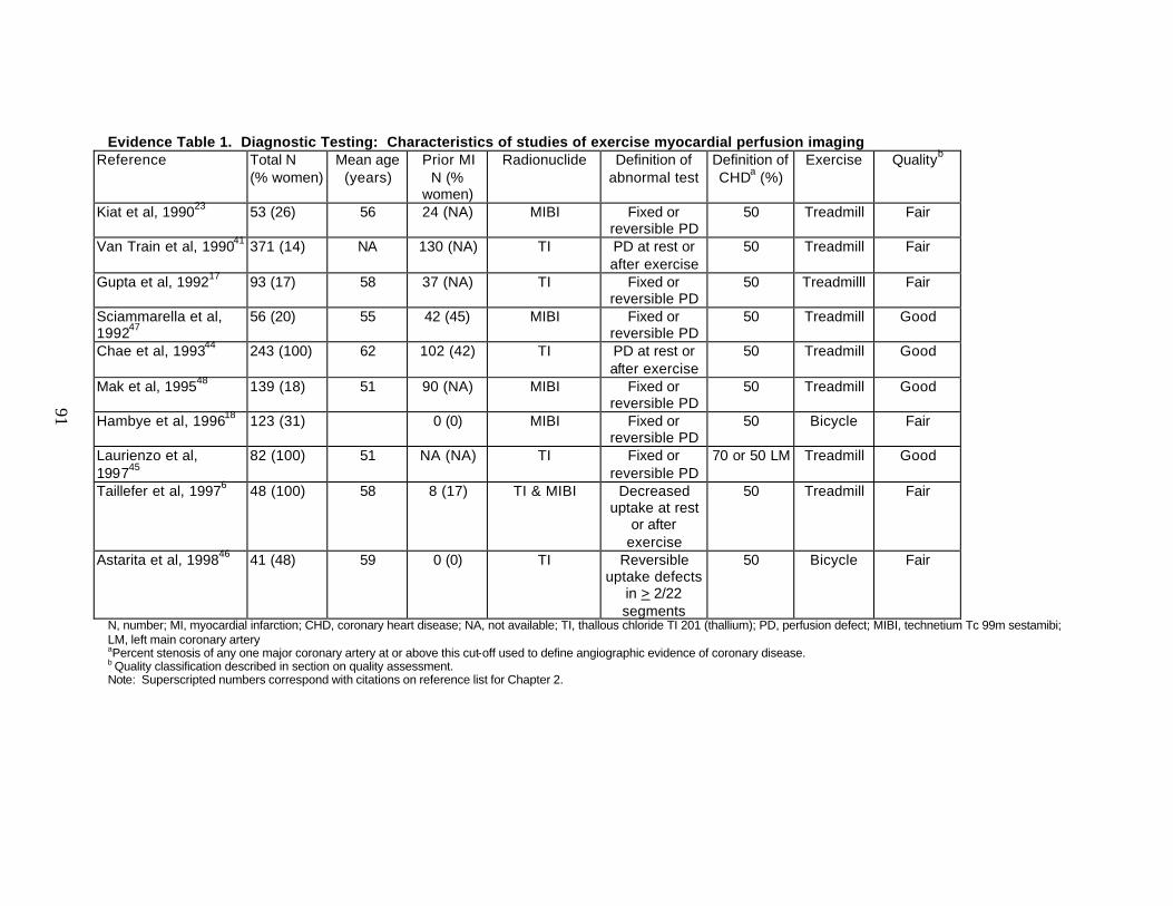
Evidence Table 1. Diagnostic Testing: Characteristics of studies of exercise myocardial perfusion imaging Reference Total N
(% women) Mean age
(years) Prior MI
N (% women)
Radionuclide Definition of abnormal test
Definition of CHDa (%)
Exercise Qualityb
Kiat et al, 199023
53 (26) 56 24 (NA) MIBI Fixed or reversible PD
50 Treadmill Fair
Van Train et al, 199041 371 (14) NA 130 (NA) TI PD at rest or after exercise
50 Treadmill Fair
Gupta et al, 199217
93 (17) 58 37 (NA) TI Fixed or reversible PD
50 Treadmilll Fair
Sciammarella et al, 199247
56 (20) 55 42 (45) MIBI Fixed or reversible PD
50 Treadmill Good
Chae et al, 199344
243 (100) 62 102 (42) TI PD at rest or after exercise
50 Treadmill Good
Mak et al, 199548
139 (18) 51 90 (NA) MIBI Fixed or reversible PD
50 Treadmill Good
Hambye et al, 199618
123 (31) 0 (0) MIBI Fixed or reversible PD
50 Bicycle Fair
Laurienzo et al, 199745
82 (100) 51 NA (NA) TI Fixed or reversible PD
70 or 50 LM Treadmill Good
Taillefer et al, 19976 48 (100) 58 8 (17) TI & MIBI Decreased uptake at rest
or after exercise
50 Treadmill Fair
Astarita et al, 199846 41 (48) 59 0 (0) TI Reversible uptake defects
in > 2/22 segments
50 Bicycle Fair
N, number; MI, myocardial infarction; CHD, coronary heart disease; NA, not available; TI, thallous chloride TI 201 (thallium); PD, perfusion defect; MIBI, technetium Tc 99m sestamibi; LM, left main coronary artery aPercent stenosis of any one major coronary artery at or above this cut-off used to define angiographic evidence of coronary disease. b Quality classification described in section on quality assessment. Note: Superscripted numbers correspond with citations on reference list for Chapter 2.
91

Evidence Table 2. Diagnostic Testing: Characteristics of studies of exercise echocardiography Reference Total N
(% women) Mean age
(years) Prior MI
N (% women)
Definition of abnormal test
Definition of CHD (%)a
Exercise Used
Qualityb
Williams et al, 199449
70 (100) 60 0 (0) New or worse RWMA after
exercise
50 Bicycle Good
Marwick et al, 19952
161 (100) 60 0 (0) RWMA at rest or after exercise
50 Treadmill & bicycle
Fair
Luotolahti et al, 199624
118 (15) 55 57 (39) New or worse RWMA after
exercise
50 Bicycle Fair
Roger et al, 199750 340 (28) 66 0 (0) RWMA at rest or after exercise
50 Treadmill Good
N, number; MI, myocardial infarction; CHD, coronary heart disease; RWMA, regional wall motion abnormality a Percent stenosis of any one major coronary artery at or above this cut-off used to define angiographic evidence of coronary disease. b Quality classification described in section on quality assessment. Note: Superscripted numbers correspond with citations on reference list for Chapter 2.
92

Evidence Table 3. Diagnostic Testing: Findings of studies of exercise myocardial perfusion imaging and summary estimates of accuracy.
Women Men
Reference Sensitivity
(95% CI)
Specificity
(95% CI)
LR+
(95% CI)
LR-
(95% CI)
Sensitivity
(95% CI)
Specificity
(95% CI)
LR+
(95% CI)
LR-
(95% CI)
Kiat et al, 199023
1.00 (0.72-1.00)
0.67 (0.09-0.99)
3.00 (0.82-11.00)
a
0.92 (0.78-0.98)
0.50 (0.01-0.99)
1.84 (0.58-5.86)
0.16 (0.03-0.75)
Van Train et al, 199041
0.95 (0.83-1.00)
0.62 (0.32-0.87)
2.47 (1.27-4.82)
0.08 (0.02-0.30)
0.95 (0.92-0.97)
0.43 (0.29-0.58)
1.67 (1.31-2.13)
0.12 (0.06-0.22)
Gupta et al, 199217
0.85 (0.55-0.99)
0.67 (0.09-0.99)
2.54 (0.68-9.46)
0.23 (0.06-0.92)
0.81 (0.69-0.90)
0.86 (0.57-0.99)
5.67 (1.78-18.1)
0.22 (0.12-0.39)
Sciammarella et al, 199247
1.00 (0.54-1.00)
0.40 (0.05-0.96)
1.67 (0.81-3.43)
a
0.95 (0.83-1.00)
0.33 (0.04-0.78)
1.42 (0.80-2.51)
0.15 (0.03-0.75)
Chae et al, 199344
0.71 (0.63-0.78)
0.65 (0.54-0.75)
2.03 (1.47-2.79)
0.44 (0.33-0.59)
b
b
b
b
Mak et al, 199548
0.83 (0.62-0.95)
1.00 (0.02-1.00)
0.17 (0.05-0.55)
b
b
b
b
Hambye et al, 199618
0.61 (0.39-0.80)
0.67 (0.39-0.89)
1.83 (0.85-3.93)
0.59 (0.30-1.10)
0.94 (0.85-0.99)
0.82 (0.60-0.95)
5.15 (2.20-12.0)
0.08 (0.03-0.20)
Laurienzo et al, 199745
0.86 (0.65-0.97)
0.80 (0.68-0.89)
4.32 (2.53-7.37)
0.17 (0.06-0.46)
b
b
b
b
Taillefer et al, 19976 (using sestamibi) (using thallium)
0.72 (0.53-0.86)
0.75 (0.57-0.89)
0.81 (0.54-0.96)
0.50 (0.25-0.50)
3.83 (1.44-10.2)
1.50
(0.89-2.54)
0.35 (0.19-0.64)
0.50 (0.23-1.07)
b
b
b b
b b
b
b
93

Evidence Table 3. Diagnostic Testing: Findings of studies of exercise myocardial perfusion imaging and summary estimate s of accuracy. (continued)
Women Men
Reference Sensitivity
(95% CI)
Specificity
(95% CI)
LR+
(95% CI)
LR-
(95% CI)
Sensitivity
(95% CI)
Specificity
(95% CI)
LR+
(95% CI)
LR-
(95% CI)
Astarita et al, 199846
1.00
(0.29-1.00)
0.47
(0.23-0.72)
1.89
(1.05-3.39)
a
1.00
(0.77-1.00)
0.50
(0.16-0.84)
2.00
(1.02-3.92)
a
SUMMARY All eligible studiesc Good quality studies Studies including men and women
0.77
(0.72-0.83)
0.75 (0.69-0.81)
0.86
(0.77-0.92)
0.69
(0.62-0.75)
0.71 (0.69-0.78)
0.57
(0.43-0.70)
2.46
(2.00-3.04)
2.54 (1.95-3.32)
2.01
(1.46-2.76)
0.33
(0.26-0.41)
0.36 (0.28-0.46)
0.24
(0.14-0.42)
0.93
(0.90-0.95)
d
0.93 (0.90-0.95)
0.57
(0.47-0.67) d
0.57 (0.47-0.67)
2.17
(1.73-2.73) d
2.17 (1.73-2.37)
0.13
(0.09-0.19)
d
0.13 (0.09-0.19)
CI, confidence interval; LR+, likelihood ratio for a positive test; LR- likelihood ratio for a negative test a Estimates not calculable because sensitivity or specificity = 100%. bEstimates not available because these studies did not include men. cIncludes results from the study by Taillefer for sestamibi.6 dEstimates not available because only one good quality study included men. Note: Superscripted numbers correspond with citations on reference list for Chapter 2.
94

Evidence Table 4. Diagnostic Testing: Summary estimates of accuracy of myocardial perfusion imaging from studies using sestamibi compared to studies using thallium
Women Men
Radionuclide Sensitivity
(95% CI)
Specificity
(95% CI)
LR+
(95% CI)
LR-
(95% CI)
Sensitivity
(95% CI)
Specificity
(95% CI)
LR+
(95% CI)
LR-
(95% CI)
Sestamibi6, 18, 23, 47, 48 0.77 (0.67-0.85)
0.70 (0.53-0.83)
2.57 (1.58-4.17)
0.33 (0.22-0.50)
0.94 (0.89-0.97)
0.70 (0.51-0.85)
3.12 (1.82-5.37)
0.09 (0.05-0.18)
Thallium6, 17, 41, 44-46 0.77 (0.72-0.82)
0.67 (0.60-0.74)
2.32 (1.87-2.88)
0.34 (0.27-0.44)
0.92 (0.89-0.95)
0.52 (0.40-0.64)
1.93 (1.51-2.46)
0.14 (0.09-0.22)
CI, confidence interval; LR+ = likelihood ratio for a positive test; LR- = likelihood ratio for a negative test Note: Superscripted numbers correspond with citations on reference list for Chapter 2.
95

Evidence Table 5. Diagnostic Testing: Findings of studies of exercise echocardiography and summary estimates of accuracy
Women Men
Reference Sensitivity
(95% CI)
Specificity
(95% CI)
LR+
(95% CI)
LR-
(95% CI)
Sensitivity
(95% CI)
Specificity
(95% CI)
LR+
(95% CI)
LR-
(95% CI)
Williams et al, 199449
0.88 (0.72-0.97)
0.84 (0.68-0.94)
5.42 (2.62-11.2)
0.14 (0.06-0.35)
a
a
a
a
Marwick et al, 19952
0.80 (0.68-0.89)
0.81 (0.72-0.88)
4.28 (2.79-6.57)
0.25 (0.15-0.42)
a
a
a
a
Luotolahti et al, 199624
0.77 (0.46-0.95)
0.80 (0.28-0.99)
3.85 (0.91-16.4)
0.29 (0.10-0.82)
0.96 (0.90-0.99)
0.60 (0.14-0.95)
2.39 (0.91-6.29)
0.07 (0.02-0.22)
Roger et al, 199750
0.79 (0.66-0.89)
0.37 (0.22-0.54)
1.26 (0.95-1.67)
0.56 (0.32-1.08)
0.78 (0.71-0.84)
0.44 (0.30-0.59)
1.39 (1.07-1.80)
0.50 0.33-0.76)
SUMMARY All eligible studies Good quality studies Studies including men and women
0.81
(0.74-0.87)
0.82 (0.73-0.89)
0.79
(0.68-0.88)
0.73
(0.66-0.79)
0.60 (0.48-0.71)
0.42
(0.27-0.58)
2.95
(2.28-3.79)
2.06 (1.53-2.77)
1.36
(1.02-1.81)
0.26
(0.19-0.36)
0.29 (0.18-0.47)
0.50
(0.28-0.89)
0.84
(0.79-0.88)
b
0.84 (0.79-0.88)
0.45
(0.32-0.59)
b
0.45 (0.32-0.59)
1.54
(1.20-1.98)
b
1.54 (1.20-1.98)
0.36
(0.24-0.53)
b
0.36 (0.24-0.53)
CI, confidence interval; LR+ = likelihood ratio for a positive test; LR- = likelihood ratio for a negative test aEstimates not available because these studies did not include men. bEstimates not available because only one good quality study included men and women. Note: Superscripted numbers correspond with citations on reference list for Chapter 2.
96

Evidence Table 6. Lipids: Characteristics of Included Clinical Trials
Study Name, Year
N Women/ Total
(% Women)
Mean Age of Women
Percent with CHDa
Lipid Entry Criterion
Intervention
Mean Follow Up
(years)
Outcomes in Women
Quality Ratingb
Scottish Society of Physicians, 197129
124/717 (17)
54
100% None Clofibrate 6 CHD mortality nonfatal MI
Fair
Newcastle Upon Tyne, 197130
97/497 (20)
54 100% None Clofibrate
5 CHD mortality nonfatal MI
Fair
Colestipol Study, 197831
1184/2278 (52)
57 20% TC >250 mg/dl Colestipol 3 total mortality CHD mortality
Fair
4S, 19947, 32, 33 827/4444 (19)
61 100% TC 213-309 mg/dl Simvastatin 5.4 total mortality CHD mortality nonfatal MI revascularization CHD eventsc
Good
ACAPS, 199425, 26 441/919 (48)
61 0% LDL 130-159 mg/dl with any number of risk factors; LDL 160-189 mg/dl with no or one risk factor
Lovastatin 2.8 total mortality CHD mortality nonfatal MI
Good
PLAC II, 199416 22/151 (15)
NA 100% LDL in 60-90th percentile for age and gender
Pravastatin 3 total mortality CHD mortality nonfatal MI
Good
CARE, 19988, 34, 35
576/4159 (14)
61 100% TC <240 mg/dl and LDL-C 115-174 mg/dl
Pravastatin 5 total mortality CHD mortality nonfatal MI revascularization CHD eventsc
Good
LIPID, 199827, 36,
37 1516/9014
(17) 62 100% TC 155-271 mg/dl Pravastatin 6.1 CHD eventsc Good
97

Evidence Table 6. Lipids: Characteristics of Included Clinical Trials (continued)
Study Name, Year
N Women/ Total
(% Women)
Mean Age of Women
Percent with CHDa
Lipid Entry Criterion
Intervention
Mean Follow Up
(years)
Outcomes in Women
Quality Ratingb
AFCAPS/ TEXCAPS, 19989,
38
997/6605 (15)
62 0% TC 180-264 mg/dl LDL 130-190 mg/dl and HDL <47 mg/dl
Lovastatin 5.2 total mortality CHD mortality nonfatal MI revascularization CHD eventsc
Good
HPS, 200211 5082/20,536 (25)
NA 65% TC >135 mg/dl Simvastatin 5 CHD eventsc Good
ALLHAT, 200239 5051/10,355 (49)
NA 14% LDL 100-189 mg/dl
Pravastatin 4.8 total mortality CHD eventsc
Fair
4S, Scandinavian Simvastatin Survival Study; ACAPS, Asymptomatic Carotid Artery Progression Study; PLAC II, Pravastatin, Lipids and Atherosclerosis in the Carotid Arteries; CARE,
Cholesterol and Recurrent Events trial; LIPID, Long-term Intervention with Pravastatin in Ischaemic Disease; AFCAPS/TEXCAPS, Air Force/Texas Coronary Atherosclerosis Prevention Study; HPS, Heart Protection Study; ALLHAT, Antihypertensive and Lipid-Lowering Treatment to Prevent heart Attack Trial.
N, number; CHD, coronary heart disease; mg/dl, milligrams per deciliter; MI, myocardial infarction; NA, not available; CVD, cardiovascular disease; TC total cholesterol; LDL, low density lipoprotein
aCHD defined as history of myocardial infarction or angina. bsee Quality Assessment section in text. c CHD events in, CARE, LIPID and ALLHAT defined as CHD mortality or nonfatal MI; in 4S as CHD mortality, non fatal MI or resuscitated cardiac arrest and in AFCAPS as CHD
mortality, nonfatal MI or unstable angina or sudden cardiac death; in HPS as CHD mortality, nonfatal MI, stroke or revascularization. Note: Superscripted numbers correspond with citations on reference list for Chapter 3
98

99
Evidence Table 7. Lipids: Outcomes of Eligible Studies
Outcome Study
RR (95% CI)
Secondary Prevention
Total Mortality 4S7, 32, 33 PLAC II16
1.11 1.18
(0.66, 1.5) (0.03, 54.81)
CHD Mortality Scottish Society of Physicians29 Newcastle Upon Tyne30 4S7, 32, 33 PLAC II16 CARE8, 34, 35
0.17 0.20 0.79 1.18 0.80
(0.02, 1.34) (0.04, 1.13) (0.39, 1.6) (0.03, 54.81) (0.61, 1.05)
Nonfatal MI Scottish Society of Physicians29 Newcastle Upon Tyne30 4S7, 32, 33 PLAC II16 CARE8, 34, 35
0.75 0.43 0.66 1.18 0.51
(0.42, 1.33) (0.08, 2.25) (0.48, 0.90) (0.48, 54.81) (0.27, 0.94)
CHD eventsa 4S7, 32, 33 CARE8, 34, 35 LIPID27, 36, 37 HPS11
0.68 0.60 0.87 0.81
(0.51, 0.91) (0.37, 0.97) (0.67, 1.13) (0.72, 0.92)
Revascularizaion 4S7, 32, 33 CARE8, 34, 35
0.52 0.82
(0.31, 0.86) (0.64, 1.20)
Primary Prevention
Total Mortality Colestipol31 ACAPS25, 26 AFCAPS/TEXCAPS9, 38 ALLHAT39
0.92 0.09 1.53 0.98
(0.51, 1.69) (0.01, 1.7) (0.62, 3.81) (0.83, 1.17)
CHD Mortality Colestipol31 ACAPS25, 26 AFCAPS/TEXCAPS9, 38
1.08 0.35 2.99
(0.44, 2.63) (0.01, 8.47) (0.12, 73.3)
Nonfatal MI ACAPS25, 26 AFCAPS/TEXCAPS9, 38
0.35 0.69
(0.04, 3.31) (0.21, 2.28)
CHD eventsa AFCAPS/TEXCAPS9, 38 ALLHAT39
0.54 1.02
(0.22, 1.34) (0.81, 1.28)
Revascularization AFCAPS/TEXCAPS9, 38 0.87 (0.33,2.31) CHD, coronary heart disease; CI, confidence interval; MI, myocardial infarction; NA not available; RR, relative
risk; 4S, Scandinavian Simvastatin Survival Study; ACAPS, Asymptomatic Carotid Artery Progression Study; PLAC II, Pravastatin, Lipids and Atherosclerosis in the Carotid Arteries; CARE, Cholesterol and Recurrent Events trial; LIPID, Long-term Intervention with Pravastatin in Ischaemic Disease; AFCAPS/TEXCAPS, Air Force/Texas Coronary Atherosclerosis Prevention Study; HPS, Heart Protection Study; ALLHAT, Antihypertensive and Lipid-Lowering Treatment to Prevent Heart Attack Trial.
aCHD events in CARE, LIPID and ALLHAT defined as CHD mortality or nonfatal MI; in 4S as CHD mortality, non fatal MI or resuscitated cardiac arrest; in AFCAPS as CHD mortality, nonfatal MI, unstable angina or sudden cardiac death, and in HPS as CHD mortality, nonfatal MI, stroke or revascularization. Note: Superscripted numbers correspond with citations on reference list for Chapter 3.

100
Evidence Table 8. Lipids: Summary Results
Outcome
Secondary Prevention References RR (95% CI)
Primary Prevention References RR (95% CI)
Total mortality All studies Statin drugs Good quality
7, 16, 32, 33 7, 16, 32, 33 7, 16, 32, 33
1.11 (0.66 1.87) 1.11 (0.66 1.87) 1.11 (0.66 1.87)
9, 25, 26, 31, 38, 39 9, 25, 26, 38, 39 9, 25, 26, 38
0.95 (0.62, 1.46) 0.87 (0.37, 2.00)a 0.45 (0.03, 7.00)a
CHD mortality All studies
Statin drugs Good quality
7, 8, 16, 29, 30, 32-35 7, 8, 16, 32-35 7, 8, 16, 32-35
0.74 (0.57, 0.96) 0.78 (0.60, 1.01) 0.78 (0.60, 1.01)
7, 16, 32, 33 7, 16, 32, 33 7, 16, 32, 33
1.07 (0.47, 2.40) 1.02 (0.11, 9.76) 1.02 (0.11, 9.76)
Nonfatal MI All studies
Statin drugs Good quality
7, 8, 16, 29, 30, 32-35 7, 8, 16, 32-35 7, 8, 16, 32-35
0.64 (0.50, 0.82) 0.63 (0.48, 0.83) 0.63 (0.48, 0.83)
9, 25, 26, 38 9, 25, 26, 38 9, 25, 26, 38
0.61 (0.22, 1.68) 0.61 (0.22, 1.68) 0.61 (0.22, 1.68)
CHD eventsb
All studies Statin drugs Good quality
7, 8, 11, 27, 32-37 7, 8, 11, 27, 32-37 7, 8, 11, 27, 32-37
0.79 (0.72, 0.88) 0.79 (0.72, 0.88) 0.79 (0.72, 0.88)
9, 38, 39
9, 38, 39 9, 38
0.87 (0.50, 1.49) 0.87 (0.50, 1.49) 0.54 (0.22, 1.30)
Revascularization All studies
Statin drugs Good quality
7, 8, 32-35 7, 8, 32-35 7, 8, 32-35
0.70 (0.42, 1.16)a
0.70 (0.42, 1.16)a
0.70 (0.42, 1.16)a
9, 38 9, 38 9, 38
0.87 (0.33,2.31)c 0.87 (0.33,2.31)c 0.87 (0.33,2.31)c
RR, relative risk; CI, confidence interval; CHD, coronary heart disease; MI, myocardial infarction; statin drugs included lovastatin,
pravastatin or simvastatin; good quality is defined in the methods section. aP-value for heterogeneity <0.10 b CHD events in CARE, LIPID and ALLHAT defined as CHD mortality or nonfatal MI; in 4S as CHD mortality, non fatal MI or
resuscitated cardiac arrest; in AFCAPS as CHD mortality, nonfatal MI, unstable angina or sudden cardiac death, and in HPS as CHD mortality, nonfatal MI, stroke or revascularization. See Tables 1 and 2 for full names of studies.
cOnly one trial provided data on this outcome9, 38 Note: Superscripted numbers correspond with citations on reference list for Chapter 3.

Evidence Table 9. Diabetes: Characteristics of studies of coronary heart disease risk in diabetics vs. nondiabetic subjects Mean Number of Subjects Quality Followup Mean Age Diabetic Nondiabetic Source Study Name or Location Score (years) (Range), y M F M F Barrett-Connor et al, 70 1991a Rancho Bernardo, CA good 14.4 63(50-89) 207 127 893 1244 Collins et al, 71 1996 Asian Indian, Fiji fair 11 20+ 102 129 1077 1236 Fujimoto et al, 16, 17 1987, 1991 King County, WA fair NA 62- 64 78 52 79 72 Howard et al, 21 1995 Strong Heart Study fair 4.8 55-57 797 1412 1049 1291 Jousilahti et al, 72 1999 WHO-MONICA fair 7-12 25-64 262 254 6828 7442 Keil et al, 73 1993 Charleston Heart Study fair 30 50(35-74) 30 42 956 1153 Kleinman et al, 74 1988 NHANES I fair 9 57,63b(40-77) 189 218 3151 3823 Lindeman et al, 19 1998 Bernalillo County, NM fair NA > 65 115 73 330 321
101

Evidence Table 9. Diabetes: Characteristics of studies of coronary heart disease risk in diabetics vs. nondiabetic subjects (continued) Mean Number of Subjects Quality Followup Mean Age Diabetic Nondiabetic Source Study Name or Location Score (years) (Range), y M F M F Lowe et al, 75 1997a and Pan et al, 761986a Chicago, IL fair 9-22 45,52b(35-64) 926 170 7975 7860 Niskanen et al, 15 1998 Finland-Kuopio fair 15 55(45-64) 70 63 62 82 Rewers et al, 18 1992 San Luis Valley, CO fair NA 25-74 186 521 243 571 Scheidt-Nave et al, 77 1990a Rancho Bernardo, CA good 14.4 50-89 159 157 591 732 Sievers et al, 20 1998 Gila River, AZ fair 12.1 > 15 630 813 1542 1552 Vilbergsson et al, 12 1998 Reykjavik Study good 17 52(34-79) 267 210 8861 9549 Wei et al, 78 1998 San Antonio Heart Study fair 7.5 43,53b (25-64) 190 281 1910 2494 CI indicates confidence interval; NA indicates not available; WHO-MONICA indicates World Health Organization-Monitoring of Trends and Determinants in Cardiovascular Disease; NHANES I indicates National Health and Nutrition Examination Survey I aDuplicate publication, but separate outcomes or subgroups reported in each article. bValues represent mean age for nondiabetic and diabetic subjects, respectively. Note: Superscripted numbers correspond with citations on reference list for Chapter 4.
102

Evidence Table 10. Diabetes: Adjusted odds ratios (95% confidence intervals) for coronary heart disease mortality, nonfatal myocardial infarction (NFMI), and all-cause mortality by sex
Source Race/
Ethnicity
CHD Mortality Male Female
OR (95% CI)
Nonfatal MI
Male Female
OR (95% CI)
All-Cause Mortality Male Female
OR (95% CI)
Barrett-Connor, 701991 W 1.9(1.3-2.8) 3.3(2.0-5.6) Scheidt-Nave, 771990 W 1.3 (0.8-2.1) 1.8 (1.0-3.0) Collins, 711996 I
Me 3.2(1.3-2.0) 1.6(0.4-6.0)
20.7 (2.5-171) 5.4(1.2-24.3)
4.4 (2.7-6.9) 1.3 (0.7-2.5)
3.1 (1.7-5.8) 2.3 (1.2-4.6)
Fujimotoa, 161987 JA 2.1 (1.3-3.3) Fujimotoa, 171991 JA 1.2 (0.8-1.9) Howarda, 211995 NA 1.59 (.74-3.21) 7.11(2.24-
22.58)
Jousilahti, 721999 W 2.4 (1.6-3.4) 4.3 (2.4-7.6) Keil, 731993 W
B 0.8 (0.5-2.4) 2.5(0.3-18.7)
1.3 (0.4-4.5) 2.0 (0.9-4.5)
1.2 (0.7-2.0) 3.1 (1.4-7.2)
2.2 (1.1-4.2) 1.8 (1.1-2.8)
Kleinman,741988 W 2.8 (2.0-3.8) 2.5 (1.6-3.8) 2.3 (1.8-2.8) 2.0 (1.6-2.6) Lindemana, 191998 W 3.4 (1.2-5.5) 3.6 (1.3-10) Lindemana, 191998 L 1.7 (0.9-3.3) 1.4 (0.5-3.7) Lowe, 751997 Pan, 761986
W B W
3.8 (1.7-8.4)
4.7 (1.9-11.9)
1.9 (1.6-2.2) 1.8 (1.0-3.3)
Niskanena, 151998 W 5.4(1.7-14.7) 5.2(1.8-15) Rewersa, 181992 W
L 1.7 (1.0-2.8)
0.9 (0.5-1.4) 2.7 (1.5-4.7) 1.4 (0.9-2.1)
Sieversa, 201992 P 5.3 (2.2-13) 6.3 (0.8-50) 1.8 (1.4-2.4) 1.4 (1.0-2.0) Vilbergsson, 121998 W 2.0 (1.5-2.6) 2.2 (1.7-2.8) 1.9 (1.6-2.3) 1.7 (1.3-2.1) Wei, 781998 M 2.1 (1.3-3.5) 3.2 (1.9-5.4)
aMultivariate results received via personal communication with authors; OR indicates odds ratio; CI indicates confidence interval; W=white, B=black, I=East Indian, Me=Melanesian, JA=Japanese- American, L=Latino, P=Pima Indian, M=multiple races, INA=Native American Note: Superscripted numbers correspond with citations on reference list for Chapter 4.
103

Evidence Table 11. Diabetes: Summary odds ratios for coronary heart disease (CHD) mortality, nonfatal myocardial infarction (NFMI), and all-cause mortality by sex (comparing diabetics to nondiabetics)
Outcome Men
Summary OR (95%CI)
Women Summary OR
(95%CI)
References p-valuea
OR indicates odds ratio; CI indicates confidence interval. aP Value for comparison of odds ratio estimates between men and women. bP Value for heterogeneity < .10. Race-ethnicity groups with only one study (n=1), were not meta-analyzed. Note: Superscripted numbers correspond with citations on reference list for Chapter 4.
CHD Mortality: (n=8) 2.33 (1.91-2.85) 2.92 (2.22-3.84) 12, 20, 70-74, 76 0.19 White (n=6) 2.22 (1.79-2.75) 2.79 (2.11-3.69) 12, 70, 72-74, 76 0.20 Black (n=1) 2.5 (0.3-18.7) 2.0 (0.9-4.5) 73 0.87 East Indian (n=1) 3.2 (1.3-2.0) 20.7 (2.5-171) 71 0.11 Melanesian (n=1) 1.6 (0.4-6.0) 5.4 (1.2-24.3) 71 0.24 Pima India (n=1) 5.3 (2.2-13) 6.3 (0.8-50) 20 0.88 Nonfatal MI: (n=5) 1.55 (1.11-2.17)b 1.70 (1.27-2.28) 16-19, 21, 77 0.68 White (n=3) 1.65 (1.06-2.56) 2.29 (1.59-3.30) 18,19,77 0.26 Native American (n=1) 1.59 (0.74-3.21) 7.11 (2.24-22.58) 21 0.03 Japanese American (n=1) 2.1 (1.3-3.3) 1.2 (0.8-1.9) 16,17 0.08 Latino (n=2) 1.2 (0.6-2.4) 1.4 (0.9-2.1) 18,19 0.71 Mortality: (n=7) 2.13 (1.71-2.65)b 1.94 (1.65-2.28) 12,15, 20, 71, 73, 74,75, 78 0.50 White (n=5) 2.01 (1.59-2.54) 1.98 (1.65-2.38) 12,15, 73-75 0.92 Black (n=2 male; n=1 female) 2.19 (1.28-3.73) 1.8 (1.1-2.8) 73,75 0.60 East Indian (n=1) 4.4 (2.7-6.9) 3.1(1.7-5.8) 71 0.37 Melanesian (n=1) 1.3 (0.7-2.5) 2.3 (1.2-4.6) 71 0.21 Pima Indian (n=1) 1.8 (1.4-2.4) 1.4 (1.0-2.0) 20 0.24
104

Evidence Table 12. Diabetes: Summary odds ratios (95% confidence intervals) for coronary heart disease mortality by sex (diabetes vs. no diabetes) quality of studies and sensitivity analysis (white race only) Summary Odds Ratios (95% Confidence Interval) Subgroup Men Women P-valuea References Quality of Studiesb Good (n=2) 1.96 (1.56-2.47) 2.54 (1.73-3.72) .26 12,70 Fair (n=4) 2.42 (1.71-3.42) 3.07 (1.97-4.80) .41 72,73,74,76 Level of Adjustment for Potential Confoundingb Age Adjusted only (n=4) 2.07 (1.39-3.08) 3.42 (2.55-4.59) .05 12,70,73,76
Unadjusted only (n=3) 2.16 (1.56-2.98) 3.22 (2.39-4.34) .08 70,73,74 OR indicates odds ratio; CI indicates confidence interval aP value for comparison of odds ratio estimates between men and women. b The studies inc luded in these subgroup analyses were eligible for inclusion because they provided outcomes adjusted for at least age, hypertension, hyperlipidemia and smoking;
these studies also provided unadjusted and/or age-adjusted estimates which are used to calculate these summary estimates. Note: Superscripted numbers correspond with citations on reference list for Chapter 4.
105

Evidence Table 13. Troponin: Characteristics of the Study Populations by Gender
Author
Year
Patient Cohort
Agea
(years)
History of MI (%)
Hypertension (%)
Diabetes Mellitus
(%)
Smoking (%)
M F M F M F M F M F
Antman 19968
1996 ACS 58 61 45 30 35 56 12 20 39 34
Antman 19989
1998 ACS 61 64 42 35 59 68 29 41 31 19
Christenson10, 16
1996 ACS 62 69 25 25 39 60 16 26 38 23
Galvani21
1997 Unstable Angina
66 48 na na na
Hamm12
1999 Unstable Angina
61 20 37 12 41
Heeschen13
1999 ACS 64 46 55 21 69
Ravkilde20
1993 Unstable Angina
68 28 9 9 na
Safstrom6 2000 ACS 69 72 29 29 30 37 12 16 22 16 na=not available; ACS=suspected acute coronary syndrome (non-ST elevation); UA=unstable angina (MI excluded). MI=myocardial Infarction. aMean or median. Note: Superscripted numbers correspond with citations on reference list for Chapter 5.
106
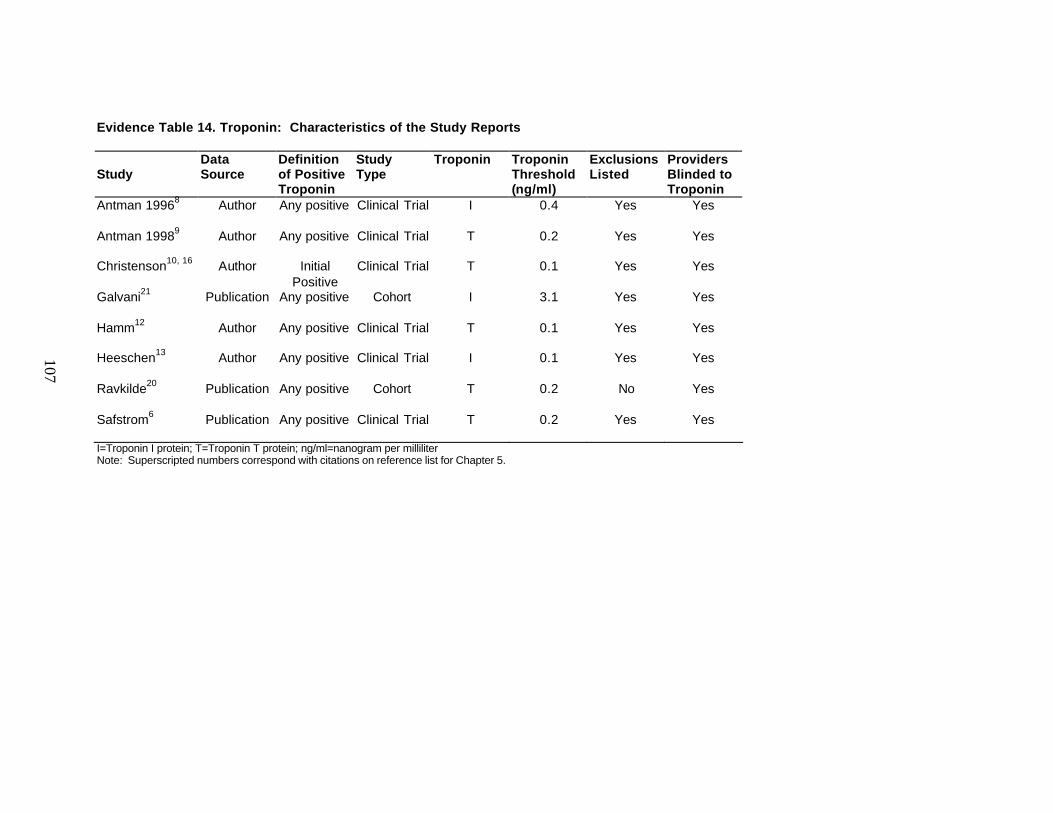
Evidence Table 14. Troponin: Characteristics of the Study Reports
Study
Data Source
Definition of Positive Troponin
Study Type
Troponin Troponin Threshold (ng/ml)
Exclusions Listed
Providers Blinded to Troponin
Antman 19968
Author Any positive Clinical Trial I 0.4 Yes Yes
Antman 19989
Author Any positive Clinical Trial T 0.2 Yes Yes
Christenson10, 16
Author Initial Positive
Clinical Trial T 0.1 Yes Yes
Galvani21
Publication Any positive Cohort I 3.1 Yes Yes
Hamm12
Author Any positive Clinical Trial T 0.1 Yes Yes
Heeschen13
Author Any positive Clinical Trial I 0.1 Yes Yes
Ravkilde20
Publication Any positive Cohort T 0.2 No Yes
Safstrom6
Publication Any positive Clinical Trial T 0.2 Yes Yes
I=Troponin I protein; T=Troponin T protein; ng/ml=nanogram per milliliter Note: Superscripted numbers correspond with citations on reference list for Chapter 5.
107

Evidence Table 15. Troponin: Outcomes for Women
Death (%) Death or MI (%)
Author
Total Troponin Positive (%)
Troponin Negative Positive
Troponin Negative Positive
Antman 19968
471
179 (38)
5 (1.7)
10 (5.6)
18 (6.2)
19 (10.6)
Antman 19989
194 35 (18) 3 (1.9) 2 (5.7) 6 (3.8) 7 (20.0)
Christenson10, 16
251 102 (41) 8 (5.4) 13 (12.7) 14 (9.4) 15 (14.7)
Galvani21
29 11 (38) 0 (0) 1 (9) 2 (11) 2 (18)
Hamm12
333 149 (45) 2 (1.1) 3 (2.0) 16 (8.7) 26 (17.4)
Heeschen13
1513 464 (31) 20 (1.9) 19 (4.1) 51 (4.9) 30 (8.2)
Ravkilde20
36 12 (33) 0 (0) 0 (0) 0 (0) 2 (17)
Safstrom6
342 166 (49) 4 (2.3) 13 (7.8) 10 (5.7) 30 (18.1)
Total 3169 1118 (35) 42 (2.0) 61 (5.5) 117 (5.7) 139 (12.4) MI = myocardial infarction Note: Superscripted numbers correspond with citations on reference list for Chapter 5.
108
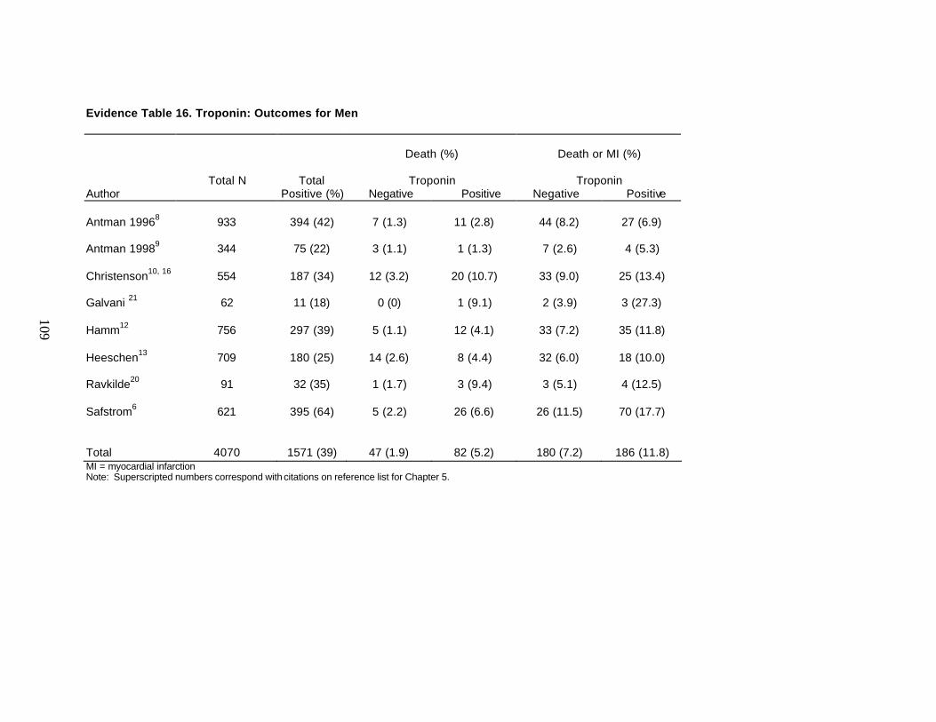
Evidence Table 16. Troponin: Outcomes for Men
Death (%) Death or MI (%)
Author
Total N Total Positive (%)
Troponin Negative Positive
Troponin Negative Positive
Antman 19968
933
394 (42)
7 (1.3)
11 (2.8)
44 (8.2)
27 (6.9)
Antman 19989
344 75 (22) 3 (1.1) 1 (1.3) 7 (2.6) 4 (5.3)
Christenson10, 16
554 187 (34) 12 (3.2) 20 (10.7) 33 (9.0) 25 (13.4)
Galvani 21
62 11 (18) 0 (0) 1 (9.1) 2 (3.9) 3 (27.3)
Hamm12
756 297 (39) 5 (1.1) 12 (4.1) 33 (7.2) 35 (11.8)
Heeschen13
709 180 (25) 14 (2.6) 8 (4.4) 32 (6.0) 18 (10.0)
Ravkilde20
91 32 (35) 1 (1.7) 3 (9.4) 3 (5.1) 4 (12.5)
Safstrom6
621 395 (64) 5 (2.2) 26 (6.6) 26 (11.5) 70 (17.7)
Total 4070 1571 (39) 47 (1.9) 82 (5.2) 180 (7.2) 186 (11.8) MI = myocardial infarction Note: Superscripted numbers correspond with citations on reference list for Chapter 5.
109

111
Bibliography Abbott RD, Donahue RP, Kannel WB, et al. The impact of diabetes on survival following myocardial infarction in men vs women. The Framingham Study [published erratum appears in JAMA 1989 Apr 7;261(13):1884] [see comments]. JAMA 1988;260(23):3456-60.
Abbud ZA, Shindler DM, Wilson AC, et al. Effect of diabetes mellitus on short- and long-term mortality rates of patients with acute myocardial infarction: a statewide study. Myocardial Infarction Data Acquisition System Study Group. Am Heart J 1995;130(1):51-8.
ACAPS Group. Rationale and design for the Asymptomatic Carotid Artery Plaque Study
(ACAPS). Control Clin Trials 1992;13(4):293-314. Adlerberth AM, Rosengren A, Wilhelmsen L. Diabetes and long-term risk of mortality from coronary and other causes in middle-aged Swedish men. A general population study. Diabetes Care 1998;21(4):539-45. Alpert JS, Thygesen K, Antman E, et al. Myocardial infarction redefined--a consensus document of The Joint European Society of Cardiology/American College of Cardiology Committee for the redefinition of myocardial infarction. J Am Coll Cardiol 2000;36(3):959-69.
American Diabetic Association. Standards of medical care for patients with diabetes mellitus. Diabetes Care 2001;24(Supp. 1):S33-43. American Heart Association. Heart Disease and Stroke Statistics--2002 Update. Dallas, Tex.: American Heart Association; 2001.
Antihypertensive and Lipid-Lowering Treatment to Prevent Heart Attack Trial (ALLHAT) Officers and Coordinators for the ALLHAT Collaborative Research Group. Major outcomes in moderately hypercholesterolemic, hypertensive patients randomized to pravastatin vs usual care: The Antihypertensive and Lipid-Lowering Treatment to Prevent Heart Attack Trial (ALLHAT-LLT). JAMA 2002;288(23):2998-3007.
Antman EM, Cohen M, Bernink PJ, et al. The TIMI risk score for unstable Angina/Non-ST elevation MI: A method for prognostication and therapeutic decision making [In Process Citation]. JAMA 2000;284(7):835-42. Antman EM, Sacks DB, Rifai N, et al. Time to positivity of a rapid bedside assay for cardiac-specific troponin T predicts prognosis in acute coronary syndromes: a Thrombolysis in Myocardial Infarction (TIMI) 11A substudy. J Am Coll Cardiol 1998;31(2):326-30. Antman EM, Tanasijevic MJ, Thompson B, et al. Cardiac-specific troponin I levels to predict the risk of mortality in patients with acute coronary syndromes. N Engl J Med 1996;335(18):1342-9.
Arntz HR, Agrawal R, Wunderlich W, et al. Beneficial effects of pravastatin (+/-colestyramine/niacin) initiated immediately after a coronary event (the randomized Lipid-Coronary Artery Disease [L-CAD] Study). Am J Cardiol 2000;86(12):1293-8.
Astarita C, Nicolai E, Liguori E, et al. [Dipyridamole-echocardiography and thallium exercise myocardial scintigraphy in the diagnosis of obstructive coronary or microvascular disease in hypertensive patients with left ventricular hypertrophy and angina]. G Ital Cardiol 1998;28(9):996-1004. Balkau B, Eschwege E, Papoz L, et al. Risk factors for early death in non-insulin dependent diabetes and men with known glucose tolerance status [see comments]. BMJ 1993;307(6899):295-9.

112
Barbash GI, White HD, Modan M, et al. Significance of diabetes mellitus in patients with acute myocardial infarction receiving thrombolytic therapy. Investigators of the International Tissue Plasminogen Activator/Streptokinase Mortality Trial. J Am Coll Cardiol 1993;22(3):707-13. Barish RA, Doherty RJ, Browne BJ. Reengineering the emergency evaluation of chest pain. J Healthc Qual 1997;19(5):6-12. Barolsky SM, Gilbert CA, Faruqui A, et al. Differences in electrocardiographic response to exercise of women and men: a non-Bayesian factor. Circulation 1979;60(5):1021-7. Barrett-Connor EL, Cohn BA, Wingard DL, et al. Why is diabetes mellitus a stronger risk factor for fatal ischemic heart disease in women than in men? The Rancho Bernardo Study. JAMA 1991;265(5):627-31. Behar S, Boyko V, Reicher-Reiss H, et al. Ten-year survival after acute myocardial infarction: comparison of patients with and without diabetes. SPRINT Study Group. Secondary Prevention Reinfarction Israeli Nifedipine Trial. Am Heart J 1997;133(3):290-6. Beleslin BD, Ostojic M, Stepanovic J, et al. Stress echocardiography in the detection of myocardial ischemia. Head- to-head comp arison of exercise, dobutamine, and dipyridamole tests. Circulation 1994;90(3):1168-76. Berman DS, Kiat H, Friedman JD, et al. Separate acquisition rest thallium-201/stress technetium-99m sestamibi dual-isotope myocardial perfusion single-photon emission computed tomography: a clinical validation study. J Am Coll Cardiol 1993;22(5):1455-64. Bjornstad K, Aakhus S, Hatle L. Comparison of digital dipyridamole stress echocardiography and upright bicycle stress echocardiography for identification of coronary artery stenosis. Cardiology 1995;86(6):514-20. Brensike JF, Levy RI, Kelsey SF, et al. Effects of therapy with cholestyramine on progression of coronary arteriosclerosis: results of the NHLBI Type II Coronary Intervention Study. Circulation 1984;69(2):313-24. Brown BG, Zhao XQ, Chait A, et al. Simvastatin and niacin, antioxidant vitamins, or the combination for the prevention of coronary disease. N Engl J Med 2001;345(22):1583-92. Butler WJ, Ostrander LD, Jr., Carman WJ, et al. Mortality from coronary heart disease in the Tecumseh study. Long-term effect of diabetes mellitus, glucose tolerance and other risk factors. Am J Epidemiol 1985;121(4):541-7. Byington RP, Furberg CD, Crouse JR, 3rd, et al. Pravastatin, Lipids, and Atherosclerosis in the Carotid Arteries (PLAC- II). Am J Cardiol 1995;76(9):54C-59C. Campeau L, Hunninghake DB, Knatterud GL, et al. Aggressive cholesterol lowering delays saphenous vein graft atherosclerosis in women, the elderly, and patients with associated risk factors. NHLBI post coronary artery bypass graft clinical trial. Post CABG Trial Investigators. Circulation 1999;99(25):3241-7. Cecil MP, Kosinski AS, Jones MT, et al. The importance of work-up (verification) bias correction in assessing the accuracy of SPECT thallium-201 testing for the diagnosis of coronary artery disease. J Clin Epidemiol 1996;49(7):735-42. Chae SC, Heo J, Iskandrian AS, et al. Identification of extensive coronary artery disease in women by exercise single-photon emission computed tomographic (SPECT) thallium imaging. J Am Coll Cardiol 1993;21(6):1305-11.

113
Chaitman BR, Bourassa MG, Davis K, et al. Angiographic prevalence of high-risk coronary artery disease in patient subsets (CASS). Circulation 1981;64(2):360-7. Chandra NC, Ziegelstein RC, Rogers WJ, et al. Observations of the treatment of women in the United States with myocardial infarction: a report from the National Registry of Myocardial Infarction-I. Arch Intern Med 1998;158(9):981-8. Christenson RH, Duh SH, Newby LK, et al. Cardiac troponin T and cardiac troponin I: relative values in short-term risk stratification of patients with acute coronary syndromes. GUSTO-IIa Investigators Clin Chem 1998;44(3):494-501. Christian TF, Miller TD, Bailey KR, et al. Noninvasive identification of severe coronary artery disease using exercise tomographic thallium-201 imaging. Am J Cardiol 1992;70(1):14-20. Clearfield M, Downs JR, Weis S, et al. Air Force/Texas Coronary Atherosclerosis Prevention Study (AFCAPS/TexCAPS): efficacy and tolerability of long-term treatment with lovastatin in women. J Womens Health Gend Based Med 2001;10(10):971-81. Collins VR, Dowse GK, Ram P, et al. Non-insulin-dependent diabetes and 11-year mortality in Asian Indian and Melanesian Fijians. Diabet Med 1996;13(2):125-32. Cooper RS, Pacold IV, Ford ES. Age-related differences in case-fatality rates among diabetic patients with myocardial infarction. Findings from National Hospital Discharge Survey, 1979-1987. Diabetes Care 1991;14(10):903-8. Crouse LJ, Harbrecht JJ, Vacek JL, et al. Exercise echocardiography as a screening test for coronary artery disease and correlation with coronary arteriography. Am J Cardiol 1991;67(15):1213-8. Cruz-Vidal M, Garcia -Palmieri MR, Costas R, Jr., et al. Abnormal blood glucose and coronary heart disease: the Puerto Rico Heart Health Program. Diabetes Care 1983;6(6):556-61. Dagianti A, Penco M, Agati L, et al. Stress echocardiography: comparison of exercise, dipyridamole and dobutamine in detecting and predicting the extent of coronary artery disease. J Am Coll Cardiol 1995;26(1):18-25. de Grauw WJ, van den Hoogen HJ, van de Lisdonk EH, et al. Control group characteristics and study outcomes: empirical data from a study on mortality of patients with type 2 diabetes mellitus in Dutch general practice. J Epidemiol Community Health 1998;52(Suppl 1):9S-12S. de Vegt F, Dekker JM, Ruhe HG, et al. Hyperglycaemia is associated with all-cause and cardiovascular mortality in the Hoorn population: the Hoorn Study. Diabetologia 1999;42(8):926-31. DerSimonian R, Laird N. Meta-analysis in clinical trials. Control Clin Trials 1986;7(3):177-88. DeStefano F, Ford ES, Newman J, et al. Risk factors for coronary heart disease mortality among persons with diabetes. Ann Epidemiol 1993;3(1):27-34. Donahue RP, Goldberg RJ, Chen Z, et al. The influence of sex and diabetes mellitus on survival following acute myocardial infarction: a community-wide perspective. J Clin Epidemiol 1993;46(3):245-52. Dorr AE, Gundersen K, Schneider JC, Jr., et al. Colestipol hydrochloride in hypercholesterolemic patients --effect on serum cholesterol and mortality. J Chronic Dis 1978;31(1):5-14.

114
Downs JR, Clearfield M, Weis S, et al. Primary prevention of acute coronary events with lovastatin in men and women with average cholesterol levels: results of AFCAPS/TexCAPS. Air Force/Texas Coronary Atherosclerosis Prevention Study. JAMA 1998;279(20):1615-22. Eschwege E, Richard JL, Thibult N, et al. Coronary heart disease mortality in relation with diabetes, blood glucose and plasma insulin levels. The Paris Prospective Study, ten years later. Horm Metab Res Suppl 1985;15:41-6. Fava S, Azzopardi J, Muscat HA, et al. Factors that influence outcome in diabetic subjects with myocardial infarction. Diabetes Care 1993;16(12):1615-8. Feskens EJ, Kromhout D. Glucose tolerance and the risk of cardiovascular disease: the Zutphen Study. J Clin Epidemiol 1992;45(11):1327-34. Fitzgerald AP, Jarrett RJ. Are conventional risk factors for mortality relevant in type 2 diabetes? Diabet Med 1991;8(5):475-80. Fleischmann KE, Hunink MG, Kuntz KM, et al. Exercise echocardiography or exercise SPECT imaging? A meta-analysis of diagnostic test performance. JAMA 1998;280(10):913-20. Fleming RM, Kirkeeide RL, Taegtmeyer H, et al. Comparison of technetium-99m teboroxime tomography with automated quantitative coronary arteriography and thallium-201 tomographic imaging. J Am Coll Cardiol 1991;17(6):1297-302. Folsom AR, Szklo M, Stevens J, et al. A prospective study of coronary heart disease in relation to fasting insulin, glucose, and diabetes. The Atherosclerosis Risk in Communities (ARIC) Study. Diabetes Care 1997;20(6):935-42. Fraser GE, Strahan TM, Sabate J, et al. Effects of traditional coronary risk factors on rates of incident coronary events in a low-risk population. The Adventist Health Study. Circulation 1992;86(2):406-13. Fujimoto WY, Leonetti DL, Bergstrom RW, et al. Glucose intolerance and diabetic complications among Japanese-American women. Diabetes Res Clin Pract 1991;13(1-2):119-29. Fujimoto WY, Leonetti DL, Kinyoun JL, et al. Prevalence of complications among second-generation Japanese-American men with diabetes, impaired glucose tolerance, or normal glucose tolerance. Diabetes 1987;36(6):730-9. Fujioka S, Matsuzawa Y, Tokunaga K, et al. Improvement of glucose and lipid metabolism associated with selective reduction of intra-abdominal visceral fat in premenopausal women with visceral fat obesity. Int J Obes 1991;15(12):853-9. Fuller JH, Shipley MJ, Rose G, et al. Mortality from coronary heart disease and stroke in relation to degree of glycaemia: the Whitehall study. BMJ (Clin Res Ed) 1983;287(6396):867-70. Furberg CD, Adams HP, Jr., Applegate WB, et al. Effect of lovastatin on early carotid atherosclerosis and cardiovascular events. Asymptomatic Carotid Artery Progression Study (ACAPS) Research Group. Circulation 1994;90(4):1679-87. Galanti G, Sciagra R, Comeglio M, et al. Diagnostic accuracy of peak exercise echocardiography in coronary artery disease: comparison with thalliu m-201 myocardial scintigraphy. Am Heart J 1991;122(6):1609-16.
Galvani M, Ottani F, Ferrini D, et al. Prognostic influence of elevated values of cardiac troponin I in patients with unstable angina. Circulation 1997;95(8):2053-9.

115
Goldschmid MG, Barrett-Connor E, Edelstein SL, et al. Dyslipidemia and ischemic heart disease mortality among men and women with diabetes. Circulation 1994;89(3):991-7. Grady D, Chaput L, Kristof M. Results of Systematic Review of Research on the Diagnosis and Treatment of Coronary Heart Disease in Women. Evidence Report/Technology Assessment No. 7. (Prepared by the University of California, San Francisco-Stanford Evidence-based Practice Center under Contract #290-97-0013.): Agency for Healthcare Research and Quality. October 2002. Grady D, Rubin SM, Petitti DB, et al. Hormone therapy to prevent disease and prolong life in postmenopausal women. Ann Intern Med 1992;117(12):1016-37. Granger CB, Califf RM, Young S, et al. Outcome of patients with diabetes mellitus and acute myocardial infarction treated with thrombolytic agents. The Thrombolysis and Angioplasty in Myocardial Infarction (TAMI) Study Group. J Am Coll Cardiol 1993;21(4):920-5. Greenland S. Invited commentary: a critical look at some popular meta-analytic methods. Am J Epidemiol 1994;140(3):290-6. Group of Physicians of the Newcastle upon Tyne Region. Trial of clofibrate in the treatment of ischemic heart disease. BMJ 1971;4:767-775. Gupta NC, Esterbrooks DJ, Hilleman DE, et al. Comparison of adenosine and exe rcise thallium-201 single -photon emission computed tomography (SPECT) myocardial perfusion imaging. The GE SPECT Multicenter Adenosine Study Group. J Am Coll Cardiol 1992;19(2):248-57. Haldane J. The estimation and significance of the logarithm of a ratio of frequencies. Ann Hum Genet 1955;20:309-314. Hambye AS, Vervaet A, Lieber S, et al. Diagnostic value and incremental contribution of bicycle exercise, first-pass radionuclide angiography, and 99mTc-labeled sestamibi single - photon emission computed tomography in the identification of coronary artery disease in patients without infarction. J Nucl Cardiol 1996;3(6 Pt 1):464-74. Hamm CW, Goldmann BU, Heeschen C, et al. Emergency room triage of patients with acute chest pain by means of rapid testing for cardiac troponin T or troponin I. N Engl J Med 1997;337(23):1648-53. Hamm CW, Heeschen C, Goldmann B, et al. Benefit of abciximab in patients with refractory unstable angina in relation to serum troponin T levels. c7E3 Fab Antiplatelet Therapy in Unstable Refractory Angina (CAPTURE) Study Investigators. N Engl J Med 1999;340(21):1623-9. Hansen CL, Crabbe D, Rubin S. Lower diagnostic accuracy of thallium-201 SPECT myocardial perfusion imaging in women: an effect of smaller chamber size. J Am Coll Cardiol 1996;28(5):1214-9. Healy B. The Yentl syndrome. N Engl J Med 1991;325(4):274-6.
Heart Protection Study Collaborative Group. Medical Research Council/British Heart Foundation (MRC/BHF) Heart Protection Study of cholesterol lowering with simvastatin in 20,536 high-risk individuals: a randomised placebo-controlled trial. Lancet 2002;360(9326):7-22. Hecht HS, DeBord L, Sotomayor N, et al. Supine bicycle stress echocardiography: peak exercise imaging is superior to postexercise imaging. J Am Soc Echocardiogr 1993;6(3 Pt 1):265-71. Heeschen C, Hamm CW, Goldmann B, et al. Troponin concentrations for stratification of patients with acute coronary syndromes in relation to therapeutic efficacy of tirofiban. PRISM Study Investigators. Platelet Receptor Inhibition in Ischemic Syndrome Management. Lancet 1999;354(9192):1757-62.

116
Heiba SI, Hayat NJ, Salman HS, et al. Technetium-99m-MIBI myocardial SPECT: supine versus right lateral imaging and comparison with coronary arteriography. J Nucl Med 1997;38(10):1510-4. Heidenreich P, Go A, Melsop K, et al. Prediction of risk for patients with unstable angina. Evidence Report/Technology Assessment No. 31 (prepared by the UCSF Stanford Evidence-based Practice Center under Contract No. 290-97-0013). AHRQ Publication No. 01-E001. Rockville, MD: Agency for Healthcare Research and Quality. December 2000. Heidenreich PA, Alloggiamento T, Melsop K, et al. The prognostic value of troponin in patients with non-ST elevation acute coronary syndromes: a meta-analysis. J Am Coll Cardiol 2001;38(2):478-85. Herd JA, Ballantyne CM, Farmer JA, et al. Effects of fluvastatin on coronary atherosclerosis in patients with mild to moderate cholesterol elevations (Lipoprotein and Coronary Atherosclerosis Study [LCAS]). Am J Cardiol 1997;80(3):278-86. Herman JB, Medalie JH, Goldbourt U. Differences in cardiovascular morbidity and mortality between previously known and newly diagnosed adult diabetics. Diabetologia 1977;13(3):229-34. Heyden S, Heiss G, Bartel AG, et al. Sex differences in coronary mortality among diabetics in Evans County, Georgia. J Chronic Dis 1980;33(5):265-73. Ho YL, Wu CC, Huang PJ, et al. Dobutamine stress echocardiography compared with exercise thallium-201 single -photon emission computed tomography in detecting coronary artery disease-effect of exercise level on accuracy. Cardiology 1997;88(4):379-85. Hoffmann R, Lethen H, Kleinhans E, et al. Comparative evaluation of bicycle and dobutamine stress echocardiography with perfusion scintigraphy and bicycle electrocardiogram for identification of coronary artery disease. Am J Cardiol 1993;72(7):555-9. Howard BV, Lee ET, Cowan LD, et al. Coronary heart disease prevalence and its relation to risk factors in American Indians. The Strong Heart Study. Am J Epidemiol 1995;142(3):254-68. Hoy W, Light A, Megill D. Cardiovascular disease in Navajo Indians with type 2 diabetes. Public Health Rep 1995;110(1):87-94. Iskandrian AE, Heo J, Nallamothu N. Detection of coronary artery disease in women with use of stress single-photon emission computed tomography myocardial perfusion imaging. J Nucl Cardiol 1997;4(4):329-35. Jarrett RJ, McCartney P, Keen H. The Bedford survey: ten year mortality rates in newly diagnosed diabetics, borderline diabetics and normoglycaemic controls and risk indices for coronary heart disease in borderline diabetics. Diabetologia 1982;22(2):79-84. Jousilahti P, Vartiainen E, Tuomilehto J, et al. Sex, age, cardiovascular risk factors, and coronary heart disease: a prospective follow-up study of 14 786 middle -aged men and women in Finland. Circulation 1999;99(9):1165-72. Kanaya AM, Grady D, Barrett-Connor E. Explaining the sex difference in coronary heart disease mortality among patients with type 2 diabetes mellitus: a meta-analysis. Arch Intern Med 2002;162(15):1737-45. Kang X, Berman DS, Lewin H, et al. Comparative ability of myocardial perfusion single-photon emission computed tomography to detect coronary artery disease in patients with and without diabetes mellitus. Am Heart J 1999;137(5):949-57.

117
Kannel WB, McGee DL. Diabetes and glucose tolerance as risk factors for cardiovascular disease: the Framingham study. Diabetes Care 1979;2(2):120-6. Kannel WB, McGee DL. Diabetes and cardiovascular disease. The Framingham study. JAMA 1979;241(19):2035-8. Kannel WB, Wilson PW. Risk factors that attenuate the female coronary disease advantage. Arch Intern Med 1995;155(1):57-61. Keil JE, Sutherland SE, Knapp RG, et al. Mortality rates and risk factors for coronary disease in black as compared with white men and women [see comments]. N Engl J Med 1993;329(2):73-8. Kiat H, Van Train KF, Maddahi J, et al. Development and prospective application of quantitative 2-day stress-rest Tc-99m methoxy isobutyl isonitrile SPECT for the diagnosis of coronary artery disease. Am Heart J 1990;120(6 Pt 1):1255-66. Kleinman JC, Donahue RP, Harris MI, et al. Mortality among diabetics in a national sample. Am J Epidemiol 1988;128(2):389-401. Kuusisto J, Mykkanen L, Pyorala K, et al. NIDDM and its metabolic control predict coronary heart disease in elderly subjects. Diabetes 1994;43(8):960-7. Kwok Y, Kim C, Grady D, et al. Meta-analysis of exercise testing to detect coronary artery disease in women. Am J Cardiol 1999;83(5):660-6. Laakso M, Ronnemaa T, Lehto S, et al. Does NIDDM increase the risk for coronary heart disease similarly in both low- and high-risk populations? Diabetologia 1995;38(4):487-93. Laakso M, Ronnemaa T, Pyorala K, et al. Atherosclerotic vascular disease and its risk factors in non-insulin-dependent diabetic and nondiabetic subjects in Finland. Diabetes Care 1988;11(6):449-63. Lagerqvist B, Safstrom K, Stahle E, et al. Is early invasive treatment of unstable coronary artery disease equally effective for both women and men? FRISC II Study Group Investigators. J Am Coll Cardiol 2001;38(1):41-8. Lapidus L. Ischaemic heart disease, stroke and total mortality in women--results from a prospective population study in Gothenburg, Sweden. Acta Med Scand Suppl 1985;705:1-42. LaRosa JC. Triglycerides and coronary risk in women and the elderly. Arch Intern Med 1997;157:961-968. LaRosa JC, He J, Vupputuri S. Effect of statins on risk of coronary disease: a meta-analysis of randomized controlled trials. JAMA 1999;282:2340-6. Laurienzo JM, Cannon RO, 3rd, Quyyumi AA, et al. Improved specificity of transesophageal dobutamine stress echocardiography compared to standard tests for evaluation of coronary artery disease in women presenting with chest pain. Am J Cardiol 1997;80(11):1402-7. Lee WL, Cheung AM, Cape D, et al. Impact of diabetes on coronary artery disease in women and men: a meta-analysis of prospective studies. Diabetes Care 2000;23(7):962-8. Lerner DJ, Kannel WB. Patterns of coronary heart disease morbidity and mortality in the sexes: a 26-year follow-up of the Framingham population. Am Heart J 1986;111(2):383-90.

118
Lewis SJ, Sacks FM, Mitchell JS, et al. Effect of pravastatin on cardiovascular events in women after myocardial infarction: the cholesterol and recurrent events (CARE) trial. J Am Coll Cardiol 1998;32(1):140-6. Liao Y, Cooper RS, Ghali JK, et al. Sex differences in the impact of coexistent diabetes on survival in patients with coronary heart disease. Diabetes Care 1993;16(5):708-13. Lindahl B, Venge P, Wallentin L. Relation between troponin T and the risk of subsequent cardiac events in unstable coronary artery disease. The FRISC study group. Circulation 1996;93(9):1651-7. Lindeman RD, Romero LJ, Hundley R, et al. Prevalences of type 2 diabetes, the insulin resistance syndrome, and coronary heart disease in an elderly, biethnic population. Diabetes Care 1998;21(6):959-66. Lomuscio A, Castagnone M, Vergani D, et al. Clinical correlation between diabetic and non diabetic patients with myocardial infarction. Acta Cardiol 1991;46(5):543-54.
Long-Term Intervention with Pravastatin in Ischaemic Disease (LIPID) Study Group. Prevention of cardiovascular events and death with pravastatin in patients with coronary heart disease and a broad range of initial cholesterol levels. N Engl J Med 1998;339(19):1349-57.
Lowe LP, Liu K, Greenland P, et al. Diabetes, asymptomatic hyperglycemia, and 22-year mortality in black and white men. The Chicago Heart Association Detection Project in Industry Study. Diabetes Care 1997;20(2):163-9. Luotolahti M, Saraste M, Hartiala J. Exercise echocardiography in the diagnosis of coronary artery disease. Ann Med 1996;28(1):73-7. Luscher MS, Thygesen K, Ravkilde J, et al. Applicability of cardiac troponin T and I for early risk stratification in unstable coronary artery disease. TRIM Study Group. Thrombin Inhibition in Myocardial ischemia. Circulation 1997;96(8):2578-85. Mahmarian JJ, Boyce TM, Goldberg RK, et al. Quantitative exercise thallium-201 single photon emission computed tomography for the enhanced diagnosis of ischemic heart disease. J Am Coll Cardiol 1990;15(2):318-29. Mak KH, Ang ES, Goh AS, et al. Myocardial perfusion imaging with technetium-99m sestamibi SPECT in the evaluation of coronary artery disease. Australas Radiol 1995;39(2):112-7. Mak KH, Moliterno DJ, Granger CB, et al. Influence of diabetes mellitus on clinical outcome in the thrombolytic era of acute myocardial infarction. GUSTO-I Investigators. Global Utilization of Streptokinase and Tissue Plasminogen Activator for Occluded Coronary Arteries. J Am Coll Cardiol 1997;30(1):171-9. Manolio TA, Pearson TA, Wenger NK, et al. Cholesterol and heart disease in older persons and women: review of an NHLBI workshop. Ann Epidemiol 1992; 2: 161-176 1992.
Manson JE, Colditz GA, Stampfer MJ, et al. A prospective study of maturity-onset diabetes mellitus and risk of coronary heart disease and stroke in women. Arch Intern Med 1991;151(6):1141-7.
Marangelli V, Iliceto S, Piccinni G, et al. Detection of coronary artery disease by digital stress
echocardiography: comparison of exercise, transesophageal atrial pacing and dipyridamole echocardiography. J Am Coll Cardiol 1994;24(1):117-24.
Marwick TH, D'Hondt AM, Mairesse GH, et al. Comparative ability of dobutamine and exercise
stress in inducing myocardial ischaemia in active patients. Br Heart J 1994;72(1):31-8.

119
Marwick TH, Torelli J, Harjai K, et al. Influence of left ventricular hypertrophy on detection of coronary artery disease using exercise echocardiography. J Am Coll Cardiol 1995;26(5):1180-6.
Melchior T, Kober L, Madsen CR, et al. Accelerating impact of diabetes mellitus on mortality in
the years following an acute myocardial infarction. TRACE Study Group. Trandolapril Cardiac Evaluation. Eur Heart J 1999;20(13):973-8.
Miettinen H, Lehto S, Salomaa V, et al. Impact of diabetes on mortality after the first myocardial infarction. The FINMONICA Myocardial Infarction Register Study Group [see comments]. Diabetes Care 1998;21(1):69-75.
Miettinen TA, Pyorala K, Olsson AG, et al. Cholesterol-lowering therapy in women and elderly patients with myocardial infarction or angina pectoris: findings from the Scandinavian Simvastatin Survival Study (4S). Circulation 1997;96(12):4211-8.
Minoves M, Garcia A, Magrina J, et al. Evaluation of myocardial perfusion defects by means of
"bull's eye" images. Clin Cardiol 1993;16(1):16-22.
Mitchell BD, Hazuda HP, Haffner SM, et al. Myocardial infarction in Mexican-Americans and non-Hispanic whites. The San Antonio Heart Study. Circulation 1991;83(1):45-51.
Morise AP, Diamond GA, Detrano R, et al. Incremental value of exercise electrocardiography and thallium-201 testing in men and women for the presence and extent of coronary artery disease. Am Heart J 1995;130(2):267-76.
Moses LE, Shapiro D, Littenberg B. Combining independent studies of a diagnostic test into a summary ROC curve: data-analytic approaches and some additional considerations. Stat Med 1993;12(14):1293-316.
National Institutes of Health Consensus Conference. Triglyceride, high density lipoprotein and
coronary heart disease. JAMA 1993;269:505-510
Nguyen T, Heo J, Ogilby JD, et al. Single photon emission computed tomography with thallium-201 during adenosine-induced coronary hyperemia: correlation with coronary arteriography, exercise thallium imaging and two-dimensional echocardiography. J Am Coll Cardiol 1990;16(6):1375-83.
Niemeyer MG, Van der Wall EE, Kuyper AF, et al. Discordance of visual and quantitative analysis regarding false negative and false positive test results in thallium-201 myocardial perfusion scintigraphy. Am J Physiol Imaging 1991;6(1):34-43.
Niskanen L, Turpeinen A, Penttila I, et al. Hyperglycemia and compositional lipoprotein abnormalities as predictors of cardiovascular mortality in type 2 diabetes: a 15-year follow-up from the time of diagnosis. Diabetes Care 1998;21(11):1861-9.
OConnor PJ, Crabtree BF, Nakamura RM. Mortality experience of Navajos with type 2 diabetes mellitus. Ethn Health 1997;2(3):155-62.
Ohman EM, Armstrong PW, Christenson RH, et al. Cardiac troponin T levels for risk stratification in acute myocardial ischemia. GUSTO IIA Investigators. N Engl J Med 1996;335(18):1333-41.
Orchard TJ. The impact of gender and general risk factors on the occurrence of atherosclerotic vascular disease in non-insulin-dependent diabetes mellitus. Ann Med 1996;28(4):323-33.
Orlander PR, Goff DC, Morrissey M, et al. The relation of diabetes to the severity of acute myocardial infarction and post-myocardial infarction survival in Mexican-Americans and non-Hispanic whites. The Corpus Christi Heart Project. Diabetes 1994;43(7):897-902.

120
Ozdemir K, Kisacik HL, Oguzhan A, et al. Comparison of exercise stress testing with dobutamine stress echocardiography and radionuclide ventriculography for diagnosis of coronary artery disease. Jpn Heart J 1999;40(6):715-27.
Palmas W, Friedman JD, Diamond GA, et al. Incremental value of simultaneous assessment of myocardial function and perfusion with technetium-99m sestamibi for prediction of extent of coronary artery disease. J Am Coll Cardiol 1995;25(5):1024-31.
Pan WH, Cedres LB, Liu K, et al. Relationship of clinical diabetes and asymptomatic hyperglycemia to risk of coronary heart disease mortality in men and women. Am J Epidemiol 1986;123(3):504-16.
Pedersen TR, Berg K, Cook TJ, et al. Safety and tolerability of cholesterol lowering with simvastatin during 5 years in the Scandinavian Simvastatin Survival Study. Arch Intern Med 1996;156(18):2085-92.
Pell S, D'Alonzo CA. Factors associated with long-term survival of diabetics. JAMA
1970;214(10):1833-40.
Petitti D. Statistical Methods in Meta-Analysis. In: Meta-Analysis, Deicion Analysis and Cost-Effectiveness Analysis. New York: Oxford University Press; 1994. p. 90-114.
Pignone M, Phillips C, Mulrow C. Use of lipid lowering drugs for primary prevention of coronary
heart disease: meta-analysis of randomised trials. BMJ 2000;321:1-5.
Pitt B, Mancini GB, Ellis SG, et al. Pravastatin limitation of atherosclerosis in the coronary arteries (PLAC I): reduction in atherosclerosis progression and clinical events. PLAC I investigation. J Am Coll Cardiol 1995;26(5):1133-9.
Plehn JF, Davis BR, Sacks FM, et al. Reduction of stroke incidence after myocardial infarction with pravastatin: the Cholesterol and Recurrent Events (CARE) study. The Care Investigators. Circulation 1999;99(2):216-23.
Post Coronary Artery Bypass Graft Trial Investigators. The effect of aggressive lowering of low-
density lipoprotein cholesterol levels and low-dose anticoagulation on obstructive changes in saphenous-vein coronary-artery bypass grafts. N Engl J Med 1997;336(3):153-62.
The Pravastatin Multinational Study Group for Cardiac Risk Patients.Effects of pravastatin in
patients with serum total cholesterol levels from 5.2 to 7.8 mmol/liter (200 to 300 mg/dl) plus two additional atherosclerotic risk factors. Am J Cardiol 1993;72(14):1031-7.
Quinones MA, Verani MS, Haichin RM, et al. Exercise echocardiography versus 201Tl single -
photon emission computed tomography in evaluation of coronary artery disease. Analysis of 292 patients. Circulation 1992;85(3):1026-31.
Ravkilde J, Horder M, Gerhardt W, et al. Diagnostic performance and prognostic value of serum
troponin T in suspected acute myocardial infarction. Scand J Clin Lab Invest 1993;53(7):677-85.
Research Committee of the Scottish Society of Physicians: Ischemic Heart Disease: a secondary prevention trial using clofibrate. BMJ 1971;4:775-784.
Reunanen A. Mortality in type 2 diabetes. Ann Clin Res 1983;15(Suppl 37):26-8.
Rewers M, Shetterly SM, Baxter J, et al. Prevalence of coronary heart disease in subjects with
normal and impaired glucose tolerance and non-insulin-dependent diabetes mellitus in a biethnic Colorado population. The San Luis Valley Diabetes Study. Am J Epidemiol 1992;135(12):1321-30.

121
Riegger G, Abletshauser C, Ludwig M, et al. The effect of fluvastatin on cardiac events in patients with symptomatic coronary artery disease during one year of treatment. Atherosclerosis 1999;144(1):263-70.
Rodriguez BL, Lau N, Burchfiel CM, et al. Glucose intolerance and 23-year risk of coronary heart disease and total mortality: the Honolulu Heart Program. Diabetes Care 1999;22(8):1262-5.
Roger VL, Pellikka PA, Bell MR, et al. Sex and test verification bias. Impact on the diagnostic value of exercise echocardiography. Circulation 1997;95(2):405-10.
Roger VL, Pellikka PA, Oh JK, et al. Identification of multivessel coronary artery disease by exercise echocardiography. J Am Coll Cardiol 1994;24(1):109-14.
Rubello D, Zanco P, Candelpergher G, et al. Usefulness of 99mTc-MIBI stress myocardial SPECT bull's-eye quantification in coronary artery disease. Q J Nucl Med 1995;39(2):111-5.
Rytter L, Troelsen S, Beck-Nielsen H. Prevalence and mortality of acute myocardial infarction in patients with diabetes. Diabetes Care 1985;8(3):230-4.
Sacks FM, Pfeffer MA, Moye LA, et al. The effect of pravastatin on coronary events after myocardial infarction in patients with average cholesterol levels. Cholesterol and Recurrent Events Trial investigators. N Engl J Med 1996;335(14):1001-9.
Safstrom K, Lindahl B, Swahn E. Risk stratification in unstable coronary artery disease--exercise test and troponin T from a gender perspective. FRISC-Study Group. Fragmin during InStability in Coronary artery disease. J Am Coll Cardiol 2000;35(7):1791-800.
Salustri A, Pozzoli MM, Hermans W, et al. Relationship between exercise echocardiography and perfusion single-photon emission computed tomography in patients with single-vessel coronary artery disease. Am Heart J 1992;124(1):75-83.
Santana-Boado C, Candell-Riera J, Castell-Conesa J, et al. Diagnostic accuracy of technetium-99m-MIBI myocardial SPECT in women and men. J Nucl Med 1998;39(5):751-5.
Sawada SG, Ryan T, Fineberg NS, et al. Exercise echocardiographic detection of coronary artery
disease in women. J Am Coll Cardiol 1989;14(6):1440-7. Scandinavian Simvastatin Survival Study Group. Randomised trial of cholesterol lowering in 4444
patients with coronary heart disease: the Scandinavian Simvastatin Survival Study (4S). Lancet 1994;344(8934):1383-9.
Scheidt-Nave C, Barrett-Connor E, Wingard DL. Resting electrocardiographic abnormalities suggestive of asymptomatic ischemic heart disease associated with non-insulin-dependent diabetes mellitus in a defined population. Circulation 1990;81(3):899-906.
Sciammarella MG, Fragasso G, Gerundini P, et al. 99Tcm-MIBI single photon emission
tomography (SPET) for detecting myocardial ischaemia and necrosis in patients with significant coronary artery disease. Nucl Med Commun 1992;13(12):871-8.
Seeman T, Mendes de Leon C, Berkman L, et al. Risk factors for coronary heart disease among
older men and women: a prospective study of community-dwelling elderly. Am J Epidemiol 1993;138(12):1037-49.
Seyoum B, Abdulkadir J, Berhanu P, et al. Profile of coronary artery risk factors in Ethiopian diabetic patients. East Afr Med J 1999;76(2):105-7.

122
Sharir T, Germano G, Waechter PB, et al. A new algorithm for the quantitation of myocardial perfusion SPECT. II: validation and diagnostic yield. J Nucl Med 2000;41(4):720-7.
Shepherd J, Blauw GJ, Murphy MB, et al. Pravastatin in elderly individuals at risk of vascular disease (PROSPER): a randomised controlled trial. Lancet 2002;360(9346):1623-30.
Shepherd J, Cobbe SM, Ford I. Prevention of coronary heart disease with pravastatin in men with hypercholesterolemia. N Engl J Med 1995;333:1301-7.
Sievers ML, Nelson RG, Knowler WC, et al. Impact of NIDDM on mortality and causes of death in Pima Indians. Diabetes Care 1992;15(11):1541-9.
Simons LA, McCallum J, Friedlander Y, et al. Diabetes, mortality and coronary heart disease in the prospective Dubbo study of Australian elderly. Aust N Z J Med 1996;26(1):66-74.
Singer DE, Moulton AW, Nathan DM. Diabetic myocardial infarction. Interaction of diabetes with other preinfarction risk factors. Diabetes 1989;38(3):350-7.
Sketch MH, Mohiuddin SM, Lynch JD, et al. Significant sex differences in the correlation of electrocardiographic exercise testing and coronary arteriograms. Am J Cardiol 1975;36(2):169-73.
Solot G, Hermans J, Merlo P, et al. Correlation of 99Tcm-sestamibi SPECT with coronary angiography in general hospital practice. Nucl Med Commun 1993;14(1):23-9.
Stamler J, Vaccaro O, Neaton JD, et al. Diabetes, other risk factors, and 12-year cardiovascular mortality for men screened in the Multiple Risk Factor Intervention Trial. Diabetes Care 1993;16(2):434-44.
Steingart RM, Packer M, Hamm P, et al. Sex differences in the management of coronary artery
disease. Survival and Ventricular Enlargement Investigators. N Engl J Med 1991;325(4):226-30.
Stengard JH, Tuomilehto J, Pekkanen J, et al. Diabetes mellitus, impaired glucose tolerance and mortality among elderly men: the Finnish cohorts of the Seven Countries Study. Diabetologia 1992;35(8):760-5.
Stubbs P, Collinson P, Moseley D, et al. Prospective study of the role of cardiac troponin T in patients admitted with unstable angina. BMJ 1996;313(7052):262-4.
Sylven C, Hagerman I, Ylen M, et al. Variance ECG detection of coronary artery disease--a comparison with exercise stress test and myocardial scintigraphy. Clin Cardiol 1994;17(3):132-40.
Taillefer R, DePuey EG, Udelson JE, et al. Comparative diagnostic accuracy of Tl-201 and Tc-99m sestamibi SPECT imaging (perfusion and ECG-gated SPECT) in detecting coronary artery disease in women. J A m Coll Cardiol 1997;29(1):69-77.
Takeuchi M, Sonoda S, Miura Y, et al. Comparative diagnostic value of dobutamine stress
echocardiography and stress thallium-201 single-photon-emission computed tomography for detecting coronary artery disease in women. Coron Artery Dis 1996;7(11):831-5.
Tansey MJ, Opie LH, Kennelly BM. High mortality in obese women diabetics with acute myocardial infarction. BMJ 1977;1(6077):1624-6.
Tawa CB, Baker WB, Kleiman NS, et al. Comparison of adenosine echocardiography, with and without isometric handgrip, to exercise echocardiography in the detection of ischemia in patients with coronary artery disease. J Am Soc Echocardiogr 1996;9(1):33-43.

123
Teo KK, Burton JR, Buller CE, et al. Long-term effects of cholesterol lowering and angiotensin-converting enzyme inhibition on coronary atherosclerosis: The Simvastatin/Enalapril Coronary Atherosclerosis Trial (SCAT). Circulation 2000;102(15):1748-54.
Thom TJ. Cardiovascular disease mortality among United States women. In: Eaker ED, Packard B, Wenger NK, et al., editors. Coronary Heart Disease in Women. New York: Haymarket Doyma; 1987. p. 270.
Tonkin AM, Colquhoun D, Emberson J, et al. Effects of pravastatin in 3260 patients with unstable angina: results from the LIPID study. Lancet 2000;356(9245):1871-5.
UK Prospective Diabetes Study (UKPDS) Group. Intensive blood-glucose control with sulphonylureas or insulin compared with conventional treatment and risk of complications in patients with type 2 diabetes (UKPDS33). [published erratum appears in Lancet 1999 Aug 14;354(9178):602] [see comments]. Lancet 1998;352(9131):837-53.
UK Prospective Diabetes Study (UKPDS) Group. Effect of intensive blood-glucose control with metformin on complications in overweight patients with type 2 diabetes (UKPDS34). [see comments] [published erratum appears in Lancet 1998 Nov 7;352(9139):1557]. Lancet 1998;352(9131):854-65.
Van Train KF, Areeda J, Garcia EV, et al. Quantitative same-day rest-stress technetium-99m-sestamibi SPECT: definition and validation of stress normal limits and criteria for abnormality. J Nucl Med 1993;34(9):1494-502.
Van Train KF, Garcia EV, Maddahi J, et al. Multicenter trial validation for quantitative analysis of same-day rest-stress technetium-99m-sestamibi myocardial tomograms. J Nucl Med 1994;35(4):609-18.
Van Train KF, Maddahi J, Berman DS, et al. Quantitative analysis of tomographic stress thallium-201 myocardial scintigrams: a multicenter trial. J Nucl Med 1990;31(7):1168-79.
Vilbergsson S, Sigurdsson G, Sigvaldason H, et al. Coronary heart disease mortality amongst non-insulin-dependent diabetic subjects in Iceland: the independent effect of diabetes. The Reykjavik Study 17-year follow up. J Intern Med 1998;244(4):309-16.
Walsh J, Grady D. Treatment of hyperlipidemia in women. JAMA 1995;274:1152-2258.
Wei M, Gaskill SP, Haffner SM, et al. Effects of diabetes and level of glycemia on all-cause and cardiovascular mortality. The San Antonio Heart Study. Diabetes Care 1998;21(7):1167-72.
Weiner DA, Ryan TJ, McCabe CH, et al. Exercise stress testing. Correlations among history of angina, ST-segment response and prevalence of coronary-artery disease in the Coronary Artery Surgery Study (CASS). N Engl J Med 1979;301(5):230-5.
Wenger NK, Speroff L, Packard B. Cardiovascular health and disease in women. N Engl J Med 1993;329(4):247-56.
White HD, Simes RJ, Anderson NE, et al. Pravastatin therapy and the risk of stroke. N Engl J Med 2000;343(5):317-26.
Williams MJ, Marwick TH, O'Gorman D, et al. Comparison of exercise echocardiography with an exercise score to diagnose coronary artery disease in women. Am J Cardiol 1994;74(5):435-8.
Yano K, Kagan A, McGee D, et al. Glucose intolerance and nine-year mortality in Japanese men in Hawaii. Am J Med 1982;72(1):71-80.

124
Yusuf S, Sleight P, Pogue J, et al. Effects of an angiotensin-converting-enzyme inhibitor, ramipril, on cardiovascular events in high-risk patients. The Heart Outcomes Prevention Evaluation Study Investigators [see comments] [published errata appear in N Engl J Med 2000 Mar 9;342(10):748 and 2000 May 4;342(18):1376]. N Engl J Med 2000;342(3):145-53.
Zuanetti G, Latini R, Maggioni AP, et al. Influence of diabetes on mortality in acute myocardial infarction: data from the GISSI-2 study. J Am Coll Cardiol 1993;22(7):1788-94.

Appendix A Peer Reviewers

128
129
Appendix A: Peer Reviewers
Expert Peer Reviewers Overall Reviewers
Elizabeth Barrett-Connor, MD University of California, San Diego
JoAnn Manson, MD, DrPH Harvard Medical School, Brigham and Women's Hospital
Lori Mosca, MD, MPH, PhD Columbia University
Noninvasive Diagnostics:
C. Noel Bairey Merz, MD Cedars-Sinai Medical Center
Pamela S. Douglas, MD University of Wisconsin, Madison
Lipid lowering:
Vera Bittner, MD, MSPH University of Alabama at Birmingham
Robert H. Knopp, MD Northwest Lipid Research Clinic, University of Washington School of Medicine
Diabetes as a risk factor:
Trevor Orchard, MBBCh, MMedSi University of Pittsburgh
Troponin:
E. Magnus Ohman, MD University of North Carolina at Chapel Hill
Advice was also solicited from representatives of Partner Organizations, including:
Robert Christenson, PhD
American Association for Clinical Chemistry
Sharonne N. Hayes, M.D., F.A.C.C. American College of Cardiology
Rosalind Fabunmi, Ph.D. American Heart Association
Mary Norine Walsh, MD
Mary Winston, Ed.D.
American College of Cardiology
American Heart Association

Appendix B Quality Assessment Forms

132

133
Appendix B: Quality Assessment Forms Noninvasive Diagnostics: Exercise MPI and Echocardiography Quality Evaluation
Form
Reviewer 1: «Reviewer1» Reviewer 2: «Reviewer2» Topic Team: «Question» Article: «Article» Name: INCLUSION/EXCLUSION CRITERIA If answer is “NO” for any question 1-5 or "YES" for Q 6, skip to Q 7. (Note: you may not be able to complete Q 4 until later if PI needs to be contacted)
YES NO
1. a Is the study design cross sectional? b Does it address the research question?
2. Does the article contain primary data on > 10 women who underwent exercise EKG with radionuclide injection and SPECT imaging or exercise echocardiography.
3. Does the article include coronary angiography as the "gold standard" for measuring the accuracy of these tests?
4A. Does the article present data on the accuracy of these tests that allow calculation of sensitivity, specificity, or likelihood ratios in women (one measure acceptable if all are not given, but raw data TP, TN, FP, FN preferred) (If YES, go to Q.5)
If NO to 4A: 4B. Will PI be contacted, or has PI already been contacted? (if NO, go to Q.7) If YES to 4B: 4C. Was data received from PI? (if YES, include in review) 5. Was the paper published between January 1, 1990 and January 1, 2002? 6. Were non-invasive tests performed exclusively in patients after myocardial infarction, percutaneous angioplasty, coronary artery surgery or hospitalization for an unstable coronary syndrome? (If yes, exclude)
7. Based on your responses to questions 1-6, is this article eligible for the systematic review? IF YES, complete Question 8 - 10 and Quality Evaluation. IF NO, complete Questions 8 - 10 only.
MISCELLANEOUS 8. Does this article contain data on women only?
9. Does the investigator need to be contacted to receive further data on women? 10. Comments (continue on other side)
QUALITY EVALUATION Yes No Unclear Likely 11. Did all women who undergo the non-invasive test also undergo angiography? 12. Did only women with abnormal non-invasive tests undergo angiography? 13. Was the diagnosis of coronary artery disease on angiography made by investigators blinded to results of the non-invasive test?

134
Appendix B: Quality Assessment Forms (continued)
Treatment: Lipid Lowering Quality Evaluation Form
Reviewer 1: «Reviewer1» Reviewer 2: «Reviewer2» Topic Team: «Question» Article: «Article» INCLUSION/EXCLUSION CRITERIA If answer is “NO” for any question 1-5, STOP after completing Q 6, article does not qualify for review. One "no" response will disqualify article.
YES NO
1. Does the article assess the impact of lipid lowering on clinical outcomes: total mortality, CHD mortality, CHD events or CHD procedures?
• Is the study a randomized clinical trial? • Is this publication a sub-study? __yes __no • IF YES, which paper will be reviewed for this systematic review?
__substudy __main __ both __ neither • What is the ID# (or author/year/journal) of main paper? _________________
3. Does the article contain data on women that address the research question? 4. Does the study include treatment duration of at least one year? 5. Was the article published between January 1, 1966 and January 1, 2002? 6. Based on your responses to questions 1-5, is this article eligible for the systematic review? IF YES, proceed to Question 7. IF NO, stop here.
MISCELLANEOUS 7. Is the study: (select one only) ( ) primary prevention ( ) mixed ( ) secondary prevention ( ) cannot determine
8. Were any references found in this article that need to be reviewed? IF YES: list information on back of this page so that article can be obtained.
9. Does the investigator need to be contacted to receive further data on women? 10. Comments: (continue on other side)
QUALITY EVALUATION YES NO NOT CLEAR
11. Was there a control group that received placebo? 12. Were the participants blinded to the intervention? 13. Were the providers blinded to intervention? 14. Were the staff who assessed outcome blinded to intervention? 15. Was randomization allocation concealed? 16. Were the inclusion/exclusion criteria clear? 17. Was there > 75% complete follow up?

135
Appendix B: Quality Assessment Forms (continued)
Risk Factors: Diabetes Quality Evaluation Form
Reviewer 1: «Reviewer1» Reviewer 2: «Reviewer2» Topic Team: «Question» Article: «Article» INCLUSION/EXCLUSION CRITERIA YES NO
1. Is the study design a prospective or retrospective cohort, or cross sectional study?
2. Does the article address the risk/rates of CHD mortality, a CHD event, or angiographic evidence of CHD in diabetic persons?
3. Does the article include data specific to Type II diabetic participants? 4.a. Does the article include data on women, with outcomes specified by gender?
4. b. If NO, should PI be contacted?
5. Does the article include multivariate adjustment for confounders, including age, hypertension, hypercholesterolemia and tobacco use?
6. Does the article include a nondiabetic control group (studies with historical controls or standardized mortality ratios will be excluded)?
7. Was the paper published between January 1, 1966 and January 1, 2002? If the answer to any of the above questions is NO, STOP.
QUALITY EVALUATION YES NO NA 8. Were the inclusion/exclusion criteria clear? 9. If article is a prospective cohort study, was there less than 25% loss to follow-up?
10. Did the analysis include multivariate adjustment for potential confounders beyond the specified 4 variables: age, hypertension, hypercholesterolemia, tobacco?
11. Was the outcome adjudicated blindly? 12. If a prospective cohort, was the length of follow up time at least 6 months?
13. Was diabetes defined based on any formal glucose testing (fasting plasma glucose or OGTT)?

Appendix C Data Abstraction Forms

138

139
Appendix C: Data Abstraction Forms (continued)
DATA ABSTRACTION FORM: DIAGNOSTICS
REVIEWER DATE ARTICLE NUMBER Author Title Journal Year Country Institution Is there another article from this same database? Y or N STUDY CHARACTERISTICS Setting (University/Community/Both/Emergency Room?/Pre-op?) Study design ( prospective, retrospective) Exercise Echo done (Y/N) Exercise SPECT done (Y/N) Other tests done (Y/N), if Y, which?__________ POPULATION Consecutive? Y N Unclear Total n______ Number of Women_____ % follow up, all_____% follow up, women______ Mean age of all ______and of women________ Clinical indications, all: angina_____atypical chest pain____ asymptomatic____other_ Clinical indic, women: angina_____atypical CP_____asymptomatic______other_____ Inclusion criteria: Exclusion criteria: BL Characteristics Women Men All Age Range Cardiac Risks (%) Diabetes Smokers HTN Dyslipidemia (definition)
Cardiac Hx (%) Angina Prior MI Prior CABG Prior PTCA
TEST FACTORS Exercise preformed: Bruce____Other treadmill_____Bike_____Step____Other_____ Percent with Adequate exercise: Women_____ All_____ Radionuclide used (if applicable): Tl______MIBI______Both______Other______

140
Imaging used: SPECT_____Echo_____Planar______Other_______ Criteria for positivity_____________________

141
Appendix C: Data Abstraction Forms (continued) For Nuclear med Women Men All + ECG Criteria + Imaging Criteria
# w/ +ECG/+image #w/+ECG/ -image # w/-ECG/+image # w/-ECG/ -image
_ Positive reported at rest (Y/N) at exercise (Y/N) If echo, WMA new (Y/N) or worse (Y/N) If echo, cine loop or videotape reading? If SPECT, visual (Y/N) quantitative (Y/N) Number of non-diagnostic tests, all________ women______ Reasons for non-diagnostic tests, all: inadequate HR______technically difficult_______ Inadequate double product____ image uninterpretable (rest/stress/both)______ Reasons for non-diag, women: inadequate HR_____technically difficult______ Inadequate double product____image ininterpretable (rest/stress/both)_____ Reversibility examined (Y/N) Protocol: Time _______Return for repeat images (Y/N) and if Y, when? Baseline ECG abnormalities: LBBB______RBBB____dig effect_____LVH____ST/T__ GOLD STANDARD Cath done on all (Y/N), if N # who received cath_____# of women who received cath___ Angiographic definition of dz: < 50%___>50%____>70%____>50% LM____other____ Time between exercise test and cath_____ Cath data Women Men All % normal % w/ single vessel
% w/ 2 vessel % w/3 vessel % w/ L Main
SUBGROUPS Was severe disease a subgroup (Y/N), and if so, how was it defined?__________ Other subgroups and definitions? Data for Sens/Spec Women Men All Participants # with cath results
TP (+cath+test) FP (-cath+test)
TN (-cath-test) FN (-cath-test)

142
Appendix C: Data Abstraction Forms (continued) Subgroup: Data for Sens/Spec Women Men All
Participants # with cath results
TP (+cath+test) FP (-cath+test)
TN (-cath-test) FN (-cath-test)
Subgroup: Data for Sens/Spec Women Men All Participants # with cath results
TP (+cath+test) FP (-cath+test)
TN (-cath-test) FN (-cath-test)
Comments:

143
Appendix C: Data Abstraction Forms (continued)
Data Abstraction Form: Lipid Lowering
Reviewer_____________________________________________________________ First Author ___________________________________________________________ Study Number__________________________________________________________ Year of Publication_______________________________________________________ Directions: Complete this form after completing the quality review form which assesses study eligibility. Form does not need to be filled out on studies that are not eligible. METHODS
Study Design Randomized Clinical Trial Y ___________ N___________________
Participants Source (eg community, hospital) ________________________________ Inclusion criteria Age range___________________________________________ Total cholesterol _________________________________ LDL cholesterol__________________________________ HDL cholesterol__________________________________ Known CHD?________________________________________ Other________________________________________________ Exclusion criteria (eg cancer, dementia)___________________________________
Primary/secondary prevention Primary _____________________ Secondary ____________________ Mixed (if mixed, what is the % of each) ____________________________ Location (city, state, country) __________________________________________ Recruitment Start Date ___________________________________________
Intervention Drug Name(s)____________________________________ Starting dose________________________________ Was the dose titratable?_________________________ Diet Type of diet(s)_______________________________ ______________________________ Interventions common to control and intervention group
_________________________________________

144
Appendix C: Data Abstraction Forms (continued)
Outcomes Primary outcome IF INDIVIDUAL Total mortality ______________________
CHD mortality ______________________ Non-fatal MI _______________________ Angina ____________________________ Revascularization ____________________ Cardiovascular death_________________ Stroke_____________________________ Other ______________________________
Primary Composite Outcome(s) MARK ALL THAT APPLY Total mortality ______________________
CHD mortality ______________________ Non-fatal MI _______________________ Angina ____________________________ Revascularization ____________________ Cardiovascular death_________________ Stroke_____________________________ Other ______________________________
Secondary outcomes Total mortality ______________________
CHD mortality ______________________ Non-fatal MI _______________________ Angina ____________________________ Revascularization ____________________ Cardiovascuar death__________________ Stroke_____________________________ Other (Individual) ______________________________ Other (Composite) ______________________________
Follow-up strategy and how often Frequency Patient examination/clinic visit _________ Questionnaire _________ Telephone contact _________ Medical records _________ Death registry _________ Other____________________________ _________ Over what period of time were outcomes assessed (mean, range) Exact dates of follow-up__________________________ Mean length of follow-up_________________________ Follow up rate or percent__________________________ RESULTS
Participant Information Women Men Intervention Control Total number of patients

145
Appendix C: Data Abstraction Forms (continued) Age
Average age intervention (I) ___________ Avg age I women ________ Average age I men___________
Average age placebo (P) ________________ Average age P women___________ Average age P men____________
Angina Total (%) Women Men Intervention Control
Hypertension Total (%) Women Men Intervention Control
Anti-hypertensives Total (%) Women Men Intervention Control
Diabetes Total (%) Women Men Intervention Control
Current smokers Total (%) Women Men Intervention Control
Aspirin use Total (%) Women Men Intervention Control
ACE inhibitor use Total (%) Women Men Intervention Control
Beta blocker use Total (%) Women Men Intervention Control

146
Appendix C: Data Abstraction Forms (continued) Baseline Lipids
Total LDL HDL Triglycerides Intervention Control
Baseline Lipids in women Total LDL HDL Triglycerides Intervention Control Outcomes
Follow-up Lipids Total LDL HDL Triglycerides Intervention Control
Follow-up Lipids in women Total LDL HDL Triglycerides Intervention Control
All cause mortality OVERALL Number of events Event rate (eg # per 1000 pt
years) Number at risk
Intervention Control WOMEN Number of events Event rate (eg # per 1000 pt
years) Number at risk
Intervention Control MEN Number of events Event rate (eg # per 1000 pt
years) Number at risk
Intervention Control Relative Risk________________ Relative risk in women_________ Absolute risk reduction__________ Absolute risk reduction in women__________ NNT________________

147
Appendix C: Data Abstraction Forms (continued)
CHD death OVERALL Number of events Event rate (eg # per 1000 pt
years) Number at risk
Intervention Control WOMEN Number of events Event rate (eg # per 1000 pt
years) Number at risk
Intervention Control MEN Number of events Event rate (eg # per 1000 pt
years) Number at risk
Intervention Control Relative Risk________________ Relative risk in women_________ Absolute risk reduction__________ Absolute risk reduction in women__________ NNT________________
Nonfatal MI OVERALL Number of events Event rate (eg # per 1000 pt
years) Number at risk
Intervention Control WOMEN Number of events Event rate (eg # per 1000 pt
years) Number at risk
Intervention Control MEN Number of events Event rate (eg # per 1000 pt
years) Number at risk
Intervention Control Relative Risk________________ Relative risk in women_________ Absolute risk reduction__________ Absolute risk reduction in women__________ NNT________________

148
Appendix C: Data Abstraction Forms (continued)
Angina OVERALL Number of events Event rate (eg # per 1000 pt
years) Number at risk
Intervention Control WOMEN Number of events Event rate (eg # per 1000 pt
years) Number at risk
Intervention Control MEN Number of events Event rate (eg # per 1000 pt
years) Number at risk
Intervention Control Relative Risk________________ Relative risk in women_________ Absolute risk reduction__________ Absolute risk reduction in women__________ NNT________________
Revascularization OVERALL Number of events Event rate (eg # per 1000 pt
years) Number at risk
Intervention Control WOMEN Number of events Event rate (eg # per 1000 pt
years) Number at risk
Intervention Control MEN Number of events Event rate (eg # per 1000 pt
years) Number at risk
Intervention Control Relative Risk________________ Relative risk in women_________ Absolute risk reduction__________ Absolute risk reduction in women__________ NNT________________
Cardiovascular death OVERALL Number of events Event rate (eg # per 1000 pt
years) Number at risk
Intervention Control

149
Appendix C: Data Abstraction Forms (continued) WOMEN Number of events Event rate (eg # per 1000 pt
years) Number at risk
Intervention Control MEN Number of events Event rate (eg # per 1000 pt
years) Number at risk
Intervention Control Relative Risk________________ Relative risk in wo men_________ Absolute risk reduction__________ Absolute risk reduction in women__________ NNT________________
Stroke OVERALL Number of events Event rate (eg # per 1000 pt
years) Number at risk
Intervention Control WOMEN Number of events Event rate (eg # per 1000 pt
years) Number at risk
Intervention Control MEN Number of events Event rate (eg # per 1000 pt
years) Number at risk
Intervention Control Relative Risk________________ Relative risk in women_________ Absolute risk reduction__________ Absolute risk reduction in women__________ NNT________________
Other OVERALL Number of events Event rate (eg # per 1000 pt
years) Number at risk
Intervention Control WOMEN Number of events Event rate (eg # per 1000 pt
years) Number at risk
Intervention Control

150
Appendix C: Data Abstraction Forms (continued) MEN Number of events Event rate (eg # per 1000 pt
years) Number at risk
Intervention Control Relative Risk________________ Relative risk in women_________ Absolute risk reduction__________ Absolute risk reduction in women__________ NNT________________
Other OVERALL Number of events Event rate (eg # per 1000 pt
years) Number at risk
Intervention Control WOMEN Number of events Event rate (eg # per 1000 pt
years) Number at risk
Intervention Control MEN Number of events Event rate (eg # per 1000 pt
years) Number at risk
Intervention Control Relative Risk________________ Relative risk in women_________ Absolute risk reduction__________ Absolute risk reduction in women__________ NNT________________ Adverse event(s) LFT abnormality How defined_________________________________ Number (%) Intervention________________________ Number (%) Control____________________________ Liver failure How defined_________________________________ Number (%) Intervention________________________ Number (%) Control____________________________ CK abnormality How defined_________________________________ Number (%) Intervention________________________ Number (%) Control____________________________ Rhabdomyolysis How defined_________________________________ Number (%) Intervention________________________ Number (%) Control____________________________

151
Appendix C: Data Abstraction Forms (continued) Renal failure How defined_________________________________ Number (%) Intervention________________________ Number (%) Control____________________________ Breast cancer How defined_________________________________ Number (%) Intervention________________________ Number (%) Control____________________________ Other How defined_________________________________ Number (%) Intervention________________________ Number (%) Control____________________________ Other How defined_________________________________ Number (%) Intervention________________________ Number (%) Control____________________________

152

153
Appendix C: Data Abstraction Forms (continued)
Data Abstraction Form: Diabetes Reviewer: ____________________________ First Author: __________________ Study #: ______________________________ Year of Publication: ____________ METHODS: Study design: _____ Prospective cohort study _____ Retrospective cohort study _____ Cross-sectional analysis _____ Case-Control study _____ Randomized Clinical Trial, intervention type: ________________ Study name (acronym):_____________________________________________________ Patient Population (clinic, survey, volunteers…): ________________________________ Location (City/State/Country): ______________________________________________ Recruitment start date: ____________________________________________________ Type of diabetics studied: _____ Type 1 (Insulin-Dependent DM) _____ Type 2 (NIDDM) Type 2 diabetes defined by (check all that apply):
_____ self-report _____ use of diabetic medication _____ medical record diagnosis _____ Positive Oral Glucose Tolerance Test, cut-off: ________________ _____ Elevated fasting Glucose, cut-off: __________________________ _____ Elevated HbA1c or glycohemoglobin, cut-off: ________________ _____ other:
Outcomes: Primary endpoint(s): 1. 2. Secondary endpoints(s): 1. 3. 2. 4. Endpoint Definition: (check all that apply) • Coronary heart disease death defined by: Cardiovascular disease death defined by:
_____ ICD-9 code 410-414 _____ ICD-9 code 400-459 _____ sudden CHD death _____ physician documentation _____ Other: _____ other:
• MI defined by: Stroke death defined by: _____ EKG changes _____ ICD-9 code 430-438
_____ Enzyme levels consistent with MI _____ physician documentation _____ Other: _____ Other: Follow-up strategy (Check all that apply):
_____ patient examination/clinic visit _____ questionnaire returned _____ telephone contact _____ medical records search _____ death registry _____ other:

154
Appendix C: Data Abstraction Forms (continued) Within what period of time were outcomes assessed (mean, range): _________________ Exact dates of follow-up: ________________________________ Follow-up rate or percent: ________________________________ RESULTS: Participant Information: Women Men Number of (type 2) diabetics Number of nondiabetic patients Total number of patients Race: (list all: # with DM, # without DM) • •
Age (mean, range): Diabetics Non-diabetics
• Are outcomes reported with crude numbers (y/n): • Are outcomes adjusted by age alone (y/n)? • Are outcomes adjusted for other variables (y/n)? List all variables: Outcome: Coronary heart disease death Race: ____________
WOMEN MEN With Diabetes Without Diabetes
Women: Crude RR: ____________________ 95% CI or p-value: _________________________ Age-adjusted RR: __________________ “ _________________________ Multiply-adjusted RR: _______________ “ _________________________ Men: Crude RR: ____________________ 95% CI or p-value: _________________________ Age-adjusted RR: ___________________ “ _________________________ Multiply-adjusted RR: _______________ “ _________________________ Outcome: Non-fatal Myocardial Infarction
WOMEN MEN With Diabetes Without Diabetes
Women: Crude RR: ____________________ 95% CI or p-value: _________________________ Age-adjusted RR: __________________ “ _________________________ Multiply-adjusted RR: _______________ “ _________________________ Men: Crude RR: ____________________ 95% CI or p-value: _________________________ Age-adjusted RR: ___________________ “ _________________________ Multiply-adjusted RR: _______________ “ _________________________

155
Appendix C: Data Abstraction Forms (continued) Outcome: Cardiovascular Death
WOMEN MEN With Diabetes Without Diabetes
Women: Crude RR: ____________________ 95% CI or p-value: _________________________ Age-adjusted RR: __________________ “ _________________________ Multiply-adjusted RR: _______________ “ _________________________ Men: Crude RR: ____________________ 95% CI or p-value: _________________________ Age-adjusted RR: ___________________ “ _________________________ Multiply-adjusted RR: _______________ “ _________________________ Outcome: All-cause mortality
WOMEN MEN With Diabetes Without Diabetes
Women: Crude RR: ____________________ 95% CI or p-value: _________________________ Age-adjusted RR: __________________ “ _________________________ Multiply-adjusted RR: _______________ “ _________________________ Men: Crude RR: ____________________ 95% CI or p-value: _________________________ Age-adjusted RR: ___________________ “ _________________________ Multiply-adjusted RR: _______________ “ _________________________ Other outcome: ______________________________________
WOMEN MEN With Diabetes Without Diabetes
Women: Crude RR: ____________________ 95% CI or p-value: _________________________ Age-adjusted RR: __________________ “ _________________________ Multiply-adjusted RR: _______________ “ _________________________ Men: Crude RR: ____________________ 95% CI or p-value: _________________________ Age-adjusted RR: ___________________ “ _________________________ Multiply-adjusted RR: _______________ “ _________________________

156
Appendix C: Data Abstraction Forms (continued) Other outcome: ______________________________________
WOMEN MEN With Diabetes Without Diabetes
Women: Crude RR: ____________________ 95% CI or p-value: _________________________ Age-adjusted RR: __________________ “ _________________________ Multiply-adjusted RR: _______________ “ _________________________ Men: Crude RR: ____________________ 95% CI or p-value: _________________________ Age-adjusted RR: ___________________ “ _________________________ Multiply-adjusted RR: _______________ “ _________________________ If Race information is provided, enter each outcome with crude number and RR per race: Race: _________________________________________ Outcome: ______________________________________
WOMEN MEN With Diabetes Without DM
Women: Men: Age-adj RR(CI): _____________________ Age-adj RR(CI): _______________________ Mult.-adj RR(CI): _____________________ Mult-adj RR(CI): _______________________ Race: _________________________________________ Outcome: ______________________________________
WOMEN MEN With Diabetes Without DM
Women: Men: Age-adj RR(CI): _____________________ Age-adj RR(CI): _______________________ Mult.-adj RR(CI): _____________________ Mult-adj RR(CI): _______________________

Appendix D Abbreviations and Acronyms

158

159
Appendix D: Abbreviations and Acronyms
ACE angiotensin converting enzyme AHRQ Agency for Healthcare Research and Quality CABG coronary artery bypass grafting CHD coronary heart disease CKMB creatine kinase myocardial bands CHF congestive heart failure CVD cardiovascular disease CI confidence interval DTS Duke Treadmill Score ECHO echocardiography EKG electrocardiogram EPC Evidence-based Practice Center ETT exercise treadmill test HDL High density lipoprotein HDL-C High density lipoprotein cholesterol ICD-9 International Classification of Diseases, Ninth Revision LDL Low density lipoprotein LDL-C Low density lipoprotein cholesterol LM Left main coronary artery Lp(a) lipoprotein a mg milligrams MI myocardial infarction MIBI technetium Tc 99m sestamibi mm millimeter mmol millimole MPI myocardial perfusion- imaging MRC/BHF Medical Research Council/British Heart Foundation MB myocardial bands N number NA not available NFMI nonfatal myocardial infarction ng nanograms NHLBI National Heart Lung and Blood Institute OR odds ratio PCI percutaneous coronary intervention PD perfusion defect PTCA percutaneous coronary angioplasty RWMA regional wall motion abnormality RR relative risk SPECT single photon emission computed tomography TI thallous chloride TI 201 (thallium)

160
Appendix D: Abbreviations and Acronyms (continued) Names of studies: 4S Scandinavian Simvastatin Survival Study ACAPS Asymptomatic Carotid Artery Progression Study AFCAPS/TEXCAPS Air Force/Texas Coronary Artherosclerosis Prevention Study ALLHAT-LLT Antihypertensive and Lipid-Lowering treatment to prevent Heart
Attack Trial - Lipid-Lowering Trial CARE Cholesterol and Recurrent Events trial HPS Heart Protection Study LIPID Long-term Intervention with Pravastatin in Ischaemic Disease NHANES I National Health and Nutrition Examination Survey I PLAC I Pravastatin Limitation of Atherosclerosis in the Coronary
Arteries PLAC II Pravastatin, Lipids and Atherosclerosis in the Carotid Arteries PROSPER PROspective Study of Pravastatin in the Elderly at Risk TIMI Thrombolysis in Myocardial Infarction WHO-MONICA World Health Organization-Monitoring of Trends and
Determinants in Cardiovascular Disease