Determination of the effects of cinnamon bark fractions on ...
12
RESEARCH ARTICLE Open Access Determination of the effects of cinnamon bark fractions on Candida albicans and oral epithelial cells Marie-Pier Veilleux and Daniel Grenier * Abstract Background: Candida albicans is an opportunistic pathogen that causes oral candidiasis and denture stomatitis. It has also been reported to infect oral mucositis lesions in patients who suffer from cancer affecting the head and neck and who receive chemotherapy and radiotherapy treatments. This study aimed to investigate the effects of two cinnamon bark fractions, i.e., an essential oil and an aqueous extract enriched in proanthocyanidins (Cinnulin PF®) on growth, biofilm formation, and adherence properties of C. albicans as well as on oral epithelial cells (barrier integrity, inflammatory response). Methods: A microplate dilution assay was used to determine antifungal and anti-biofilm properties. A fluorescent assay was used to determine C. albicans adherence to oral epithelial cells. Cytotoxicity toward oral epithelial cells was assessed by determination of cell metabolic activity. Tight junction integrity of gingival keratinocytes was assessed by determination of transepithelial electrical resistance. IL-6 and IL-8 secretion by TNFα-stimulated oral epithelial cells was quantified by ELISA. Results: While Cinnulin PF® did not reduce C. albicans growth, the cinnamon bark oil exhibited high antifungal activity with minimum inhibitory concentrations and minimum fungicidal concentrations in the range of 0.039 to 0.078%. The cinnamon oil was also active against a pre-formed C. albicans biofilm. Interestingly, Cinnulin PF® prevented biofilm formation by C. albicans and attenuated its adherence to oral epithelial cells. At their effective concentrations, the cinnamon oil and the Cinnulin PF® displayed no significant cytotoxicity against oral epithelial cells. In an in vitro model, both cinnamon fractions reinforced the integrity of the oral epithelial barrier. Lastly, Cinnulin PF® inhibited the secretion of interleukin-6 and interleukin-8 by oral epithelial cells stimulated with TNF-α. Conclusion: By their ability to attenuate growth, biofilm formation and adherence property of C. albicans, to reinforce the epithelial barrier function, and to exert anti-inflammatory properties the two cinnamon fractions (essential oil, Cinnulin PF®) investigated in the present study may be promising agents for treating oral infections involving C. albicans. Keywords: Biofilm, Candida albicans, Cinnamon, Essential oil, Epithelial cells, Polyphenols Background Candida albicans is a commensal fungus that colonizes oral mucosal surfaces and that is normally harmless in healthy individuals as it is maintained at low levels by spe- cific and non-specific salivary and mucosal defense mech- anisms as well as by competitive inhibition by oral bacteria [1]. However, under certain circumstances, this opportunistic microorganism can cause a superficial infection called candidiasis. Oral candidiasis is character- ized by the appearance of white plaques on inflamed and red mucosa (inner cheeks, tongue, throat) and by pain when eating or swallowing [1, 2]. If the infection becomes invasive, which can occur in immunologically and medic- ally compromised individuals, it can cause septicemia leading to organ failure and eventually death [3]. C. albi- cans has also been reported to infect oral mucositis lesions [4, 5], causing inflammation of the oropharyngeal mucosa [6, 7]. Patients who suffer from cancer affecting the head © The Author(s). 2019 Open Access This article is distributed under the terms of the Creative Commons Attribution 4.0 International License (http://creativecommons.org/licenses/by/4.0/), which permits unrestricted use, distribution, and reproduction in any medium, provided you give appropriate credit to the original author(s) and the source, provide a link to the Creative Commons license, and indicate if changes were made. The Creative Commons Public Domain Dedication waiver (http://creativecommons.org/publicdomain/zero/1.0/) applies to the data made available in this article, unless otherwise stated. * Correspondence: [email protected] Oral Ecology Research Group, Faculty of Dentistry, Université Laval, 2420 Rue de la Terrasse, Quebec City, QC G1V 0A6, Canada Veilleux and Grenier BMC Complementary and Alternative Medicine (2019) 19:303 https://doi.org/10.1186/s12906-019-2730-2
Transcript of Determination of the effects of cinnamon bark fractions on ...

Veilleux and Grenier BMC Complementary and Alternative Medicine (2019) 19:303 https://doi.org/10.1186/s12906-019-2730-2
RESEARCH ARTICLE Open Access
Determination of the effects of cinnamon
bark fractions on Candida albicans and oralepithelial cells Marie-Pier Veilleux and Daniel Grenier*Abstract
Background: Candida albicans is an opportunistic pathogen that causes oral candidiasis and denture stomatitis. Ithas also been reported to infect oral mucositis lesions in patients who suffer from cancer affecting the head andneck and who receive chemotherapy and radiotherapy treatments. This study aimed to investigate the effects oftwo cinnamon bark fractions, i.e., an essential oil and an aqueous extract enriched in proanthocyanidins (CinnulinPF®) on growth, biofilm formation, and adherence properties of C. albicans as well as on oral epithelial cells (barrierintegrity, inflammatory response).
Methods: A microplate dilution assay was used to determine antifungal and anti-biofilm properties. A fluorescentassay was used to determine C. albicans adherence to oral epithelial cells. Cytotoxicity toward oral epithelial cellswas assessed by determination of cell metabolic activity. Tight junction integrity of gingival keratinocytes wasassessed by determination of transepithelial electrical resistance. IL-6 and IL-8 secretion by TNFα-stimulated oralepithelial cells was quantified by ELISA.
Results: While Cinnulin PF® did not reduce C. albicans growth, the cinnamon bark oil exhibited high antifungalactivity with minimum inhibitory concentrations and minimum fungicidal concentrations in the range of 0.039 to0.078%. The cinnamon oil was also active against a pre-formed C. albicans biofilm. Interestingly, Cinnulin PF®prevented biofilm formation by C. albicans and attenuated its adherence to oral epithelial cells. At their effectiveconcentrations, the cinnamon oil and the Cinnulin PF® displayed no significant cytotoxicity against oral epithelialcells. In an in vitro model, both cinnamon fractions reinforced the integrity of the oral epithelial barrier. Lastly,Cinnulin PF® inhibited the secretion of interleukin-6 and interleukin-8 by oral epithelial cells stimulated with TNF-α.Conclusion: By their ability to attenuate growth, biofilm formation and adherence property of C. albicans, to reinforcethe epithelial barrier function, and to exert anti-inflammatory properties the two cinnamon fractions (essential oil,Cinnulin PF®) investigated in the present study may be promising agents for treating oral infections involving C. albicans.
Keywords: Biofilm, Candida albicans, Cinnamon, Essential oil, Epithelial cells, Polyphenols
BackgroundCandida albicans is a commensal fungus that colonizesoral mucosal surfaces and that is normally harmless inhealthy individuals as it is maintained at low levels by spe-cific and non-specific salivary and mucosal defense mech-anisms as well as by competitive inhibition by oralbacteria [1]. However, under certain circumstances, thisopportunistic microorganism can cause a superficial
© The Author(s). 2019 Open Access This articInternational License (http://creativecommonsreproduction in any medium, provided you gthe Creative Commons license, and indicate if(http://creativecommons.org/publicdomain/ze
* Correspondence: [email protected] Ecology Research Group, Faculty of Dentistry, Université Laval, 2420 Ruede la Terrasse, Quebec City, QC G1V 0A6, Canada
infection called candidiasis. Oral candidiasis is character-ized by the appearance of white plaques on inflamed andred mucosa (inner cheeks, tongue, throat) and by painwhen eating or swallowing [1, 2]. If the infection becomesinvasive, which can occur in immunologically and medic-ally compromised individuals, it can cause septicemialeading to organ failure and eventually death [3]. C. albi-cans has also been reported to infect oral mucositis lesions[4, 5], causing inflammation of the oropharyngeal mucosa[6, 7]. Patients who suffer from cancer affecting the head
le is distributed under the terms of the Creative Commons Attribution 4.0.org/licenses/by/4.0/), which permits unrestricted use, distribution, andive appropriate credit to the original author(s) and the source, provide a link tochanges were made. The Creative Commons Public Domain Dedication waiverro/1.0/) applies to the data made available in this article, unless otherwise stated.

Veilleux and Grenier BMC Complementary and Alternative Medicine (2019) 19:303 Page 2 of 12
and neck and who receive chemotherapy and radiotherapytreatments are almost all affected by oral mucositis [6, 7].C. albicans produces several virulence factors that play
critical roles in the pathogenic process leading to super-ficial or systemic infections [8]. The cell surface adhesinsof C. albicans allow initial adhesion to oral epithelialcells, a key step prior to subsequent tissue invasion anddamage [8–10]. C. albicans can form biofilms on bioticand abiotic oral surfaces; this increases the resistance ofthe fungus to antimicrobial agents and the host immunesystem [2, 11, 12]. Additional virulence factors producedby C. albicans include its ability to switch from the yeastform to an invasive hyphae morphotype and to secreteproteolytic and lipolytic enzymes [8]. These pathogenicdeterminants may be potential targets for new antifungalagents that may limit the appearance of strains resistantto conventional antifungals.Despite the availability of antifungal agents to treat C.
albicans-associated oral infections, treatment failures areincreasingly common due to the emergence of resistantstrains [13–15]. Given this, investigations of the antifun-gal potential of new molecules are highly relevant. In re-cent years, plant-derived compounds with antifungalpotential have attracted the interest of researchers [16].Cinnamon, a spice derived from the inner bark of thecinnamon tree, has been reported to possess a numberof therapeutic properties, including antimicrobial activity[17, 18]. In the present study, we investigated the effectsof two cinnamon bark fractions, an essential oil and anaqueous extract enriched in proanthocyanidins, on thegrowth, biofilm formation, and adherence properties ofC. albicans. In addition, an oral epithelial cell model wasused to study the effects of the two fractions on the in-tegrity of the epithelial barrier and the host inflamma-tory response.
MethodsSource of cinnamon fractionsA cinnamon extract commercialized as Cinnulin PF® (Lot#: CNCP 1604003) was kindly provided by IN IngredientsInc. (Spring Hill, TN, USA). The aqueous extract, whichwas prepared from the bark of Cinnamomum burmannii,contains 531.9mg/g of proanthocyanidins according tothe datasheet provided by the company. Cinnamon bark isrelatively unusual as it contains proanthocyanidins with ahigh number of A-type bonds [19]. A 20mg/mL stock so-lution of the extract was prepared in 50% (v/v) dimethyl-sulfoxide and was sterilized by filtration (0.22-μm poresize). Carrier solvent was used as a control in all assays. Acinnamon bark essential oil (Lot #: BHC09A4) extractedfrom Cinnamomum verum, was purchased from Hunzar-oma (Longueuil, QC, Canada). The chromatographic ana-lysis performed by the company showed that thecinnamon oil contained cinnamaldehyde (71.35% [v/v]),
eugenol (6.18%), linolol (6.02), β-caryophyllene (6.02%),cinnamyle acetate (4.04%), benzyle benzoate (0.96%), p-cymene (0.56%), and 1,8-cineol (0.55%).
C. albicans and culture conditionsC. albicans ATCC 28366 (reference strain) and LAM-1(clinical strain from a case of systemic candidiasis) werecultivated in Sabouraud dextrose medium (BBL Microbiol-ogy Systems, Cockeysville, MD, USA) at pH 7 and 37 °C.
Determination of the minimum inhibitory and minimumfungicidal concentrationsThe minimum inhibitory concentration (MIC) and mini-mum fungicidal concentration (MFC) were determinedusing a microplate dilution assay. To determine the MICvalue, a 24-h culture of C. albicans was diluted in freshculture medium (Sabouraud dextrose medium) to an op-tical density at 660 nm (OD660) of 0.2 corresponding to a1 McFarland standard. Aliquots (100 μL) of C. albicanswere added to an equal volume of serial dilutions in cul-ture medium of cinnamon oil (1.25 to 0.0195%) or Cin-nulin PF® (1000 to 62.5 μg/mL) in 96-well microplates.Wells without C. albicans or without the cinnamon frac-tions were used as controls. When testing the cinnamonoil, the microplate was covered with an adhesive film toavoid evaporation of the volatile compounds. After anincubation at 37 °C for 24 h (stationary growth phase),growth was monitored by recording the OD660 using amicroplate reader (Bio-Rad Laboratories, Mississauga,ON, Canada). The MIC value corresponded to the low-est concentration of the cinnamon fractions that com-pletely inhibited growth. To determine the MFC, 5 μLfrom wells showing no visible growth was spotted onSabouraud dextrose agar plates, which were incubated at37 °C for 3 days. The MFC value corresponded to thelowest concentration of the cinnamon fractions whereno colony formation was observed. The antifungal agentnystatin was used as a reference antifungal. The MFC/MIC ratio was calculated, and a compound or fractionwas considered fungicidal when the ratio was ≤4 andfungistatic when the ratio was > 4 [20]. All assays wereperformed in triplicate to ensure reproducibility.
Membrane permeabilityThe ability of the cinnamon oil at MFC to permeabilizethe membrane of C. albicans ATCC 28366 was evalu-ated using SYTOX Green dye (Life Technologies Inc.,Burlington, ON, Canada), which binds to DNA once themembrane has been compromised. The assay was per-formed as previously described [21]. The fluorescenceresulting from the binding of the dye to DNA was re-corded using a Synergy 2 microplate reader (BioTek In-struments, Winooski, VT, USA) every 15 min for 2 hwith the excitation wavelength set at 485 nm and the
Veilleux and Grenier BMC Complementary and Alternative Medicine (2019) 19:303 Page 3 of 12
emission wavelength set at 528 nm. A reaction mixturewithout essential oil was used as a negative control.
Biofilm formation and killingThe effect of the cinnamon fractions on biofilm forma-tion by C. albicans ATCC 28366 was determined bygrowing microorganisms in Sabouraud dextrosemedium in a 96-well plate in the presence of two-foldserial dilutions of the compounds. Following a 24-h in-cubation at 37 °C, the medium and free-floating micro-organisms were removed by aspiration using a 26 gneedle, and the wells were washed three times withdistilled water. Biofilms were stained with 100 μL of0.01% crystal violet for 15 min. The wells were thenwashed three times with distilled water and were driedat 37 °C overnight, after which 100 μL of 75% ethanol(v/v) was added to each well to release the dye from thebiofilm. Absorbance at 550 nm (A550) was then mea-sured using a microplate reader. The effect of the cin-namon fractions on biofilm formation was alsoexamined by scanning electron microscopy using theprotocol previously described by Lagha et al. [22]. Sam-ples were examined using a JEOL JSM6360LV scanningelectron microscope operating at 30 kV. The ability ofthe cinnamon oil to kill a pre-formed C. albicans bio-film was also investigated. Biofilms were prepared in a96-well plate by cultivating C. albicans in Sabourauddextrose medium for 24 h prior to treatment (1 h) withthe cinnamon oil at the MFC value. Biofilm viabilitywas then measured with an XTT [2,3-bis(2-methoxy-4-nitro-sulfophenyl)-2H-tetrazolium-5-carboxanilide so-dium salt] assay, as described previously [23].
Epithelial cell culture conditions and viability assaysThe human oral epithelial cell line B11, which waskindly provided by S. Groeger (Justus Liebig UniversityGiessen, Germany) and has already been characterized[24], was cultured in keratinocyte serum-free medium(K-SFM; Life Technologies Inc.) supplemented withgrowth factors (50 μg/mL of bovine pituitary extract and5 ng/mL of human epidermal growth factor) and 100 μg/mL of penicillin G-streptomycin. The human oral epi-thelial cell line GMSM-K [25] was kindly provided by V.Murrah (University of North Carolina, Chapel Hill, NC,USA) and was cultivated in Dulbecco’s Modified Eagle’sMedium (DMEM) supplemented with 10% heat-inactivated inactivated fetal bovine serum (FBS) and100 μg/mL of penicillin G-streptomycin. The cell cul-tures were incubated at 37 °C in a 5% CO2 atmosphere.Epithelial cells (1 × 10⁵ cells in 200 μL) were seeded intothe wells of a 96-well tissue culture plate and were culti-vated until they reached confluence. The cells were thentreated with either Cinnulin PF® (0, 125, 250, 500,1000 μg/mL) or cinnamon oil (0, 0.0078, 0.0156, 0.0313,
0.0625, 0.125%) in the appropriate culture medium for24 h. Their viability was then determined using an MTT(3-[4,5-diethylthiazol-2-yl]-2,5diphenyltetrazolium brom-ide) colorimetric assay according to the manufacturer’sprotocol (Roche Diagnostics, Laval, QC, Canada).
Adherence to epithelial cellsThe effect of the cinnamon fractions on the adherenceof C. albicans ATCC 28366 to oral epithelial cells wasassessed using the human GMSM-K cell line. Epithelialcells were seeded (5 × 10⁴ cells/well) in a 96-well clearbottom black microplate (Greiner Bio One, Frickenhau-sen, Germany) and were incubated at 37 °C in a 5% CO2
atmosphere until they reached confluence. The wellswere then washed with DMEM-1% heat-inactivated FBSand were blocked with 1% bovine serum albumin (BSA)to prevent non-specific fungal adherence, and the cinna-mon fractions diluted in DMEM-1% heat-inactivatedFBS medium were added. Wells without the cinnamonfractions were used as controls. In parallel, cells from anovernight culture of C. albicans were labeled with fluor-escein isothiocyanate (FITC; Sigma-Aldrich Canada Co.)according to a protocol routinely used in our laboratory[26]. FITC-labeled C. albicans was added at a multipli-city of infection (MOI) of 100 to wells containing an epi-thelial cell monolayer (in the absence or presence of thecinnamon fractions). Following an incubation for 4 h at37 °C, unbound C. albicans were aspirated, and the wellswere washed three times with 50mM phosphate-buffered saline (pH 7; PBS). Adhered C. albicans weredetermined by monitoring fluorescence using a Synergy2 microplate reader with the excitation and emissionwavelengths set at 488 and 522 nm, respectively. Ad-hered FITC-labeled C. albicans were also observed usingan Olympus FSX100 fluorescence microscope (OlympusCanada Inc., Richmond Hill, ON, Canada).
Oral epithelial barrier integrityThe effect of the cinnamon fractions on the integrity ofthe epithelial barrier was assessed using the human B11cell line described above and the protocol previouslydescribed by Ben Lagha and Grenier [27]. Briefly, epi-thelial cells (3.5 × 10⁵ cells/insert) were seeded in Co-star Transwell™ plates with clear polyester membraneinserts (6.5-mm diameter, 0.4-μm pore size; CorningCo., Cambridge, MA, USA). The basolateral and apicalcompartments were filled with 0.6 mL and 0.1 mL ofculture medium, respectively. Following a 3-day incuba-tion to allow the cells to form tight junctions, theconditioned medium was replaced with antibiotic-freeK-SFM, and the cells were incubated for a further 16 h.The cinnamon fractions were then added, and the in-tegrity of the epithelial tight junctions was determinedby monitoring the transepithelial electrical resistance

Veilleux and Grenier BMC Complementary and Alternative Medicine (2019) 19:303 Page 4 of 12
(TER) using an ohmmeter (EVOM2, World PrecisionInstruments, Sarasota, FL, USA) after 2 and 4 h of incu-bation at 37 °C in a 5% CO2 atmosphere. Resistancevalues were calculated in Ohms (Ω)/cm2 by multiplyingthe resistance values by the surface area of the mem-brane filter. Results are expressed as a percentage ofthe basal control value measured at time 0 (100%value).
Secretion of cytokines by oral epithelial cellsThe effect of the cinnamon fractions on the secretion ofthe pro-inflammatory cytokines interleukin 6 (IL-6) andinterleukin 8 (IL-8) was investigated using the GMSM-Kepithelial cell line. Cells were seeded in a 6-well plate(106 cells/well in 2 mL) and were cultured overnight at37 °C in a 5% CO2 atmosphere to allow cell adhesion.The epithelial cells were pre-treated with the cinnamonfractions for 30 min prior to stimulating them with 1 ng/mL of recombinant human TNF-α (AnaSpec, Fremont,CA, USA). After a 24-h incubation, cell-free superna-tants were collected and were stored at − 20 °C untilused. Commercial enzyme-linked immunosorbent assay(ELISA) kits (R&D Systems, Minneapolis, MN, USA)were used to quantify IL-6 and IL-8 concentrations ac-cording to the manufacturer’s protocols.
Statistical analysisUnless indicated otherwise, all assays were performed intriplicate in two independent experiments, and themeans ± standard deviations were calculated. Statisticalanalyses were performed using a one-way analysis ofvariance with a post hoc Bonferroni multiple compari-son (GraphPad Software Inc.; La Jolla, CA, USA). All re-sults were considered statistically significant at p < 0.01.
ResultsThe antifungal activity of the cinnamon fractions is re-ported in Table 1. While Cinnulin PF® at concentrationsup to 1000 μg/mL did not reduce the growth of eitherstrain of C. albicans, the cinnamon bark oil displayedhigh antifungal activity, with MIC and MFC values inthe range of 0.039 to 0.078% (v/v). Nystatin, which was
Table 1 Minimum inhibitory concentrations (MIC) and minimum funalbicans
Candida albicans
ATCC 28366
Compounds MIC MFC MF
Cinnulin PF® (μg/mL) > 1000 > 1000 –
Cinnamon bark oil (% [v/v]) 0.039 0.078 2
Nystatin (μg/mL) 50 200 4
* MFC/MIC Ratio: > 4 means fungistatic activity; ≤ 4 means fungicidal activity
used as a reference antifungal agent, had a MIC of50 μg/mL and an MFC of 200 μg/mL. Cinnamon oil isfungicidal rather than fungistatic, with an MFC/MIC ra-tio in the range of 1 to 2.SYTOX® Green dye is a fluorescent molecule that pen-
etrates impaired cytoplasmic membranes, binds to DNA,and emits fluorescence. When the C. albicans cells weretreated with cinnamon oil, a time-dependent increase influorescence occurred, suggesting that their membraneshad been permeabilized due to the fungicidal activity ofthe cinnamon oil (Fig. 1). No significant increase influorescence occurred in the negative control over the 2-h incubation period. Cinnulin PF®, which had no anti-microbial effect on C. albicans, also did not cause anincrease in fluorescence (data not shown).The effect of Cinnulin PF® and cinnamon bark oil on
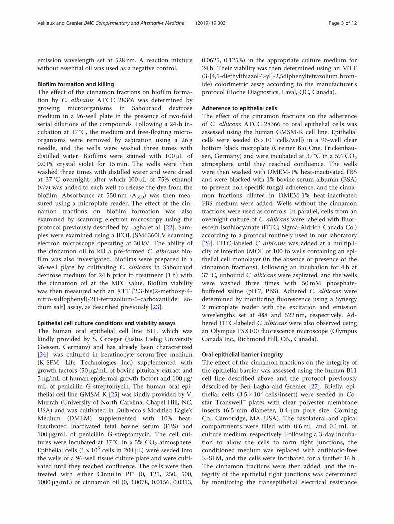
biofilm formation by C. albicans was then investigated.Although Cinnulin PF® did not reduce the growth of C.albicans, it significantly attenuated biofilm formation asdetermined by crystal violet staining (Fig. 2a). More spe-cifically, at a Cinnulin PF® concentration of 62.5 μg/mL,biofilm formation was reduced by 91%. The effect ofCinnulin PF® on biofilm formation by C. albicans wasalso visualized by scanning electron microscopy. Thecontrol biofilm of C. albicans appeared dense, and hy-phae were an important structural component (Fig. 3aand b). Electron micrographs clearly showed the markedreduction in mature biofilm when C. albicans was grownin the presence of 62.5 μg/mL of Cinnulin PF® (Fig. 3cand d). In addition, no hyphae were observed. The cin-namon bark oil also attenuated biofilm formation by C.albicans at concentrations that did not inhibit growth.The formation of biofilm was reduced by 86% when C.albicans was grown in the presence of 0.0049% cinna-mon oil (Fig. 2b).Given the fungicidal activity of the cinnamon bark oil,
we determined whether it could kill C. albicans biofilms.Since Cinnulin PF® did not show any antimicrobial effectagainst C. albicans, it was not tested in this analysis. A24-h pre-formed C. albicans biofilm was treated for 60min with cinnamon oil at its MFC. Residual viability wasdetermined using an XTT assay that measures metabolic
gicidal concentrations (MFC) of cinnamon fractions against C.
LAM-1
C/MIC* Ratio MIC MFC MFC/MIC Ratio
> 1000 > 1000 –
0.078 0.078 1
50 200 4

Fig. 1 Effect of cinnamon bark oil on the membrane integrity of C.albicans ATCC 28266 as determined using SYTOX® Green dye, whichpenetrates damaged cytoplasmic membranes. C. albicans cells wereincubated with cinnamon oil at its MFC and fluorescence wasrecorded for 2 h
Fig. 2 Effect of Cinnulin PF® (panel a) and cinnamon bark oil (panel b) onof 100% was assigned to the growth and biofilm obtained in the absencetriplicate assays from two independent experiments. *: significantly differen
Veilleux and Grenier BMC Complementary and Alternative Medicine (2019) 19:303 Page 5 of 12
activity. This treatment reduced biofilm viability by 48%,but did not cause any desorption of the biofilm biomass(Fig. 4).The effect of the cinnamon fractions on the adherence
of C. albicans to oral epithelial cells (GMSM-K cell line)was then tested. Cinnulin PF® dose-dependently reducedthe adherence of FITC-labeled C. albicans to epithelialcells (Fig. 5a). More specifically, in the presence of1000 μg/mL of Cinnulin PF®, adherence was inhibited by59%. The ability of Cinnulin PF® to reduce the adherenceof C. albicans to oral epithelial cells was confirmed byfluorescence microscopy (Fig. 5b). Cinnamon bark oilhad no inhibitory effect on the adherence of C. albicansto oral epithelial cells (data not shown).In order to investigate the biocompatibility of the cin-
namon fractions, we tested their effects on the viabilityof two oral epithelial cell lines. Up to 1000 μg/mL ofCinnulin PF® had no cytotoxic effect on B11 epithelialcells (Fig. 6a). However, 500 μg/mL of Cinnulin PF®
the growth and biofilm formation of C. albicans ATCC 28266. A valueof the cinnamon fractions. Results are expressed as the means ± SD oft from the control (p < 0.01)
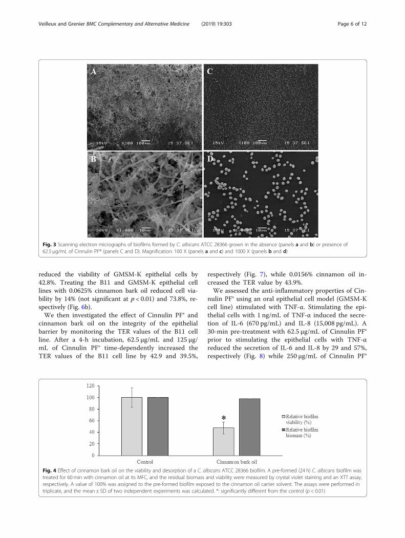
Fig. 3 Scanning electron micrographs of biofilms formed by C. albicans ATCC 28366 grown in the absence (panels a and b) or presence of62.5 μg/mL of Cinnulin PF® (panels C and D). Magnification: 100 X (panels a and c) and 1000 X (panels b and d)
Veilleux and Grenier BMC Complementary and Alternative Medicine (2019) 19:303 Page 6 of 12
reduced the viability of GMSM-K epithelial cells by42.8%. Treating the B11 and GMSM-K epithelial celllines with 0.0625% cinnamon bark oil reduced cell via-bility by 14% (not significant at p < 0.01) and 73.8%, re-spectively (Fig. 6b).We then investigated the effect of Cinnulin PF® and
cinnamon bark oil on the integrity of the epithelialbarrier by monitoring the TER values of the B11 cellline. After a 4-h incubation, 62.5 μg/mL and 125 μg/mL of Cinnulin PF® time-dependently increased theTER values of the B11 cell line by 42.9 and 39.5%,
Fig. 4 Effect of cinnamon bark oil on the viability and desorption of a C. atreated for 60 min with cinnamon oil at its MFC, and the residual biomassrespectively. A value of 100% was assigned to the pre-formed biofilm expotriplicate, and the mean ± SD of two independent experiments was calcula
respectively (Fig. 7), while 0.0156% cinnamon oil in-creased the TER value by 43.9%.We assessed the anti-inflammatory properties of Cin-
nulin PF® using an oral epithelial cell model (GMSM-Kcell line) stimulated with TNF-α. Stimulating the epi-thelial cells with 1 ng/mL of TNF-α induced the secre-tion of IL-6 (670 pg/mL) and IL-8 (15,008 pg/mL). A30-min pre-treatment with 62.5 μg/mL of Cinnulin PF®prior to stimulating the epithelial cells with TNF-αreduced the secretion of IL-6 and IL-8 by 29 and 57%,respectively (Fig. 8) while 250 μg/mL of Cinnulin PF®
lbicans ATCC 28366 biofilm. A pre-formed (24 h) C. albicans biofilm wasand viability were measured by crystal violet staining and an XTT assay,sed to the cinnamon oil carrier solvent. The assays were performed inted. *: significantly different from the control (p < 0.01)

Fig. 5 Effect of Cinnulin PF® on the adherence of C. albicans ATCC 28366 to GMSM-K oral epithelial cells. Panel A: FITC-labeled C. albicans cellsadhered to epithelial cells were quantified by measuring fluorescence using a microplate reader. A value of 100% was assigned to C. albicansadhered to epithelial cells in the absence of Cinnulin PF®. Results are expressed as the means ± SD of triplicate assays from two independentexperiments. *: significantly different from the control (p < 0.01). Panel B: Fluorescence micrograph of FITC-labeled C. albicans cells adhered toepithelial cells
Veilleux and Grenier BMC Complementary and Alternative Medicine (2019) 19:303 Page 7 of 12
almost totally inhibited the secretion of the two cyto-kines. The cinnamon bark oil did not reduce the se-cretion of IL-6 or IL-8 at non-cytotoxic concentrations(≤ 0.0078%; data not shown).
DiscussionC. albicans can be isolated from various sites inhumans. It is an opportunistic pathogen and has beenassociated with superficial and systemic infections, espe-cially in immunologically or medically compromised in-dividuals [3, 11]. C. albicans causes oral candidiasis anddenture stomatitis, and may also be involved in dentalcaries, periodontal diseases, and refractory endodonticinfections [2]. Ulcerative oral lesions (oral mucositis)resulting from chemotherapy and radiotherapy treat-ments are susceptible to secondary infections by oral mi-croorganisms, including C. albicans [4, 5]. For instance,Belazi et al. [28] isolated Candida spp. from oral muco-sitis lesions in 77% of patients undergoing radiotherapyfor head and neck cancer.
C. albicans infections can generally be successfullytreated with conventional antifungal agents. However,the emergence of resistance to these therapeutic agentsis of increasing concern [13–15], which is why investiga-tions of the antifungal potential of new molecules arehighly relevant. Plants and their derivatives are an im-portant source of bioactive molecules. Essential oils ex-tracted from different parts of certain plants (leaves,flowers, seeds, bark, etc.) possess numerous therapeuticproperties, including antimicrobial activities [29, 30].Moreover, proanthocyanidins, a family of polyphenolsconsisting of flavan-3-ol oligomers and polymers, havebeen proposed as promising molecules for treating oralinfections given their anti-adherence and anti-inflammatory properties [31]. The present study was de-signed to evaluate the effects of two cinnamon fractions,an essential oil and an aqueous extract enriched inproanthocyanidins, on both C. albicans (growth, biofilmformation, adherence properties) and oral epithelial cells(barrier integrity, inflammatory response).
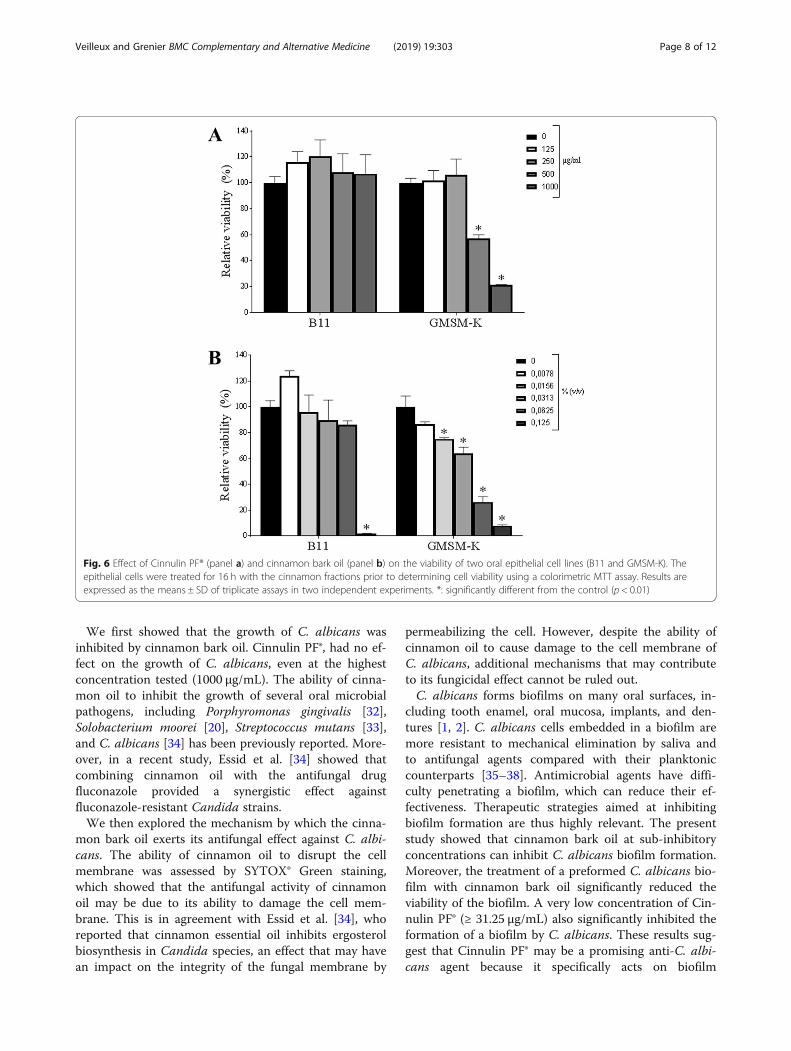
Fig. 6 Effect of Cinnulin PF® (panel a) and cinnamon bark oil (panel b) on the viability of two oral epithelial cell lines (B11 and GMSM-K). Theepithelial cells were treated for 16 h with the cinnamon fractions prior to determining cell viability using a colorimetric MTT assay. Results areexpressed as the means ± SD of triplicate assays in two independent experiments. *: significantly different from the control (p < 0.01)
Veilleux and Grenier BMC Complementary and Alternative Medicine (2019) 19:303 Page 8 of 12
We first showed that the growth of C. albicans wasinhibited by cinnamon bark oil. Cinnulin PF®, had no ef-fect on the growth of C. albicans, even at the highestconcentration tested (1000 μg/mL). The ability of cinna-mon oil to inhibit the growth of several oral microbialpathogens, including Porphyromonas gingivalis [32],Solobacterium moorei [20], Streptococcus mutans [33],and C. albicans [34] has been previously reported. More-over, in a recent study, Essid et al. [34] showed thatcombining cinnamon oil with the antifungal drugfluconazole provided a synergistic effect againstfluconazole-resistant Candida strains.We then explored the mechanism by which the cinna-
mon bark oil exerts its antifungal effect against C. albi-cans. The ability of cinnamon oil to disrupt the cellmembrane was assessed by SYTOX® Green staining,which showed that the antifungal activity of cinnamonoil may be due to its ability to damage the cell mem-brane. This is in agreement with Essid et al. [34], whoreported that cinnamon essential oil inhibits ergosterolbiosynthesis in Candida species, an effect that may havean impact on the integrity of the fungal membrane by
permeabilizing the cell. However, despite the ability ofcinnamon oil to cause damage to the cell membrane ofC. albicans, additional mechanisms that may contributeto its fungicidal effect cannot be ruled out.C. albicans forms biofilms on many oral surfaces, in-
cluding tooth enamel, oral mucosa, implants, and den-tures [1, 2]. C. albicans cells embedded in a biofilm aremore resistant to mechanical elimination by saliva andto antifungal agents compared with their planktoniccounterparts [35–38]. Antimicrobial agents have diffi-culty penetrating a biofilm, which can reduce their ef-fectiveness. Therapeutic strategies aimed at inhibitingbiofilm formation are thus highly relevant. The presentstudy showed that cinnamon bark oil at sub-inhibitoryconcentrations can inhibit C. albicans biofilm formation.Moreover, the treatment of a preformed C. albicans bio-film with cinnamon bark oil significantly reduced theviability of the biofilm. A very low concentration of Cin-nulin PF® (≥ 31.25 μg/mL) also significantly inhibited theformation of a biofilm by C. albicans. These results sug-gest that Cinnulin PF® may be a promising anti-C. albi-cans agent because it specifically acts on biofilm

Fig. 7 Effect of Cinnulin PF® (panel a) and cinnamon bark oil (panel b) on the integrity of the epithelial barrier (B11 cell line). The TER values weredetermined after a 6-h incubation. A value of 100% was assigned to the TER values at time 0. Results are expressed as the means ± SD oftriplicate assays. *: significantly different from the control (p < 0.01)
Veilleux and Grenier BMC Complementary and Alternative Medicine (2019) 19:303 Page 9 of 12
formation, a critical step of the infectious process. Pre-liminary assays showed that Cinnulin PF® had no effecton hyphae formation (data not shown). In vivo, biofilmformation by C. albicans requires initial adherence tothe oral mucosa. Interestingly, Cinnulin PF® significantlyattenuated the adherence of C. albicans while no sucheffect was observed with cinnamon oil.The oral epithelium protects the underlying tissues
from microbial invasion and thus actively contributes tothe maintenance of oral health [39]. This barrier effect ismediated by the tight junctions that seal the epithelialcells together. We thus investigated the ability of thecinnamon fractions to strengthen the epithelial barrier.Our results demonstrated that electrical resistance in-creased when the epithelial cells were cultivated in thepresence of either cinnamon bark oil or Cinnulin PF®.These results suggest that these cinnamon fractions, by
reinforcing the epithelium, may potentially prevent theinvasion of the oral mucosa by oral pathogens.Although the host inflammatory response is key to
maintaining oral health, an acute and exacerbated in-flammatory reaction as observed in oral candidiasisand oral mucositis may be deleterious by causing tis-sue damage. More specifically, the development oforal mucositis in patients receiving chemotherapy andradiotherapy treatments involves the stimulation ofinfiltrating macrophages, resulting in the activation ofNF-κB [6, 7]. This process is associated with the se-cretion of inflammatory cytokines, including TNF-α,that promote inflammation and tissue destruction. Inthe present study, when epithelial cells were chal-lenged with TNF-α, they secreted a large quantity ofIL-6 and IL-8. These two pro-inflammatory cytokinesare known to play a critical role for the recruitment

Fig. 8 Effect of Cinnulin PF® on TNF-α-induced IL-6 (panel a) and IL-8 (panel b) secretion by oral epithelial cells (GMSM-K cell line). Results areexpressed as the means ± SD of triplicate assays in two independent experiments. *: significantly different from the control (p < 0.01)
Veilleux and Grenier BMC Complementary and Alternative Medicine (2019) 19:303 Page 10 of 12
and activation of neutrophils and macrophages at thesite of infection [40, 41]. However, due to this pro-tecting reaction of the host against fungal pathogens,an accumulation of inflammatory mediators occurs toinduce a chronic and persistent inflammation, and ul-timately tissue destruction. Therefore, preventing anexcessive activation of innate immunoeffectors may beassociated with resolution of the inflammatoryprocess. In this study, we showed a dose-dependentinhibitory effect of Cinnulin PF® on TNF-α-inducedsecretion of IL-6 and IL-8 by oral epithelial cells.In this study, we showed that the two cinnamon
fractions under investigation share a number of
common properties (anti-biofilm, tight junction pro-motion) but also exhibit some distinct features. Morespecifically, cinnamon essential oil inhibited C. albi-cans growth while Cinnulin PF® attenuated the epithe-lial cell inflammatory response. Therefore, combiningthe two cinnamon fractions may be a valuable thera-peutic approach for the treatment of C. albicans in-fections through their effects on different targets.
ConclusionBy their ability to attenuate growth, biofilm formationand adherence property of C. albicans, to reinforcethe epithelial barrier function, and to attenuate the

Veilleux and Grenier BMC Complementary and Alternative Medicine (2019) 19:303 Page 11 of 12
inflammatory response of epithelial cells, the two cin-namon fractions (essential oil, Cinnulin PF®) investi-gated in the present study may be promising agentsfor controlling C. albicans infections such as oralcandidiasis, denture stomatitis, and Candida-infectedoral mucositis lesions.
AbbreviationsDMEM: Dulbecco’s Modified Eagle’s Medium; ELISA: Enzyme-linkedimmunosorbent assay (ELISA); FBS: Fetal bovine serum; FITC: Fluoresceinisothiocyanate; IL-6: Interleukin-6; IL-8: Interleukin-8; K-SFM: Keratinocyteserum-free medium; MFC: Minimum fungicidal concentration; MIC: Minimuminhibitory concentration; MTT: 3-[4,5-diethylthiazol-2-yl]-2,5diphenyltetrazolium bromide; OD: Optical density; TER: Transepithelialelectrical resistance; TNF-α: Tumor necrosis factor-alpha; XTT: 2,3-bis(2-methoxy-4-nitro-sulfophenyl)-2H-tetrazolium-5-carboxanilide sodium salt
AcknowledgementsWe are grateful to IN Ingredients Inc. (Spring Hill, TN, USA) for providing theCinnulin PF®, S. Groeger (Department of Periodontology, Justus LiebigUniversity Giessen, Germany) for providing the B11 cell line, and V. Murrah(University of North Carolina, Chapel Hill, NC, USA) for providing the GMSM-Kcell line.
Authors’ contributionsM-PV conducted the experiments, M-PV and DG analyzed the results. DGcontributed reagents/materials. The manuscript was written by M-PV andDG. Both authors read and approved the final manuscript.
FundingThis work was supported by the Laboratoire de Contrôle Microbiologique del’Université Laval (grant number 2019-03-21). This funding body had no rolein the study design, data collection and analysis, or submission of the manu-script for publication.
Availability of data and materialsThe datasets used and/or analysed during the current study are availablefrom the corresponding author on reasonable request.
Ethical approval and consent to participateNo ethics approval was required because no experiments on humans oranimals were carried out.
Consent for publicationNot applicable.
Competing interestsWe wish to confirm that there are no known conflicts of interest associatedwith this publication and there has been no financial support for this workthat could have influenced its outcome.
Received: 7 June 2019 Accepted: 24 October 2019
References1. Rautemaa R, Ramage G. Oral candidosis – clinical challenges of a biofilm
disease. Crit Rev Microbiol. 2011;37:328–36.2. O’Donnell LE, Millhouse E, Sherry L, Kean R, Malcolm J, Nile CJ, Ramage G.
Polymicrobial Candida biofilms: friends and foe in the oral cavity. FEMSYeast Res. 2015:15–fov077.
3. Pappas PG. Invasive candidiasis. Infect Dis Clin N Am. 2006;20:485–506.4. Stringer AM, Logan RM. The role of oral flora in the development of
chemotherapy-induced oral mucositis. J Oral Pathol Med. 2015;44:81–7.5. Vanhoecke B, De Ryck T, Stringer A, Van de Wiele T, Keefe D. Microbiota and
their role in the pathogenesis of oral mucositis. Oral Dis. 2015;21:17–30.6. Al-Ansari S, Zecha JA, Barasch A, de Lange J, Rozema FR, Raber-Durlacher JE.
Oral mucositis induced by anticancer therapies. Curr Oral Health Rep. 2015;2:202–11.
7. Lalla RV, Saunders DP, Peterson DE. Chemotherapy or radiation-induced oralmucositis. Dent Clin N Am. 2014;58:341–9.
8. Höfs S, Mogavero S, Hube B. Interactions of Candida albicans with host cells:virulence factors, host defense, escape strategies, and the microbiota. JMicrobiol. 2016;54:149–69.
9. Wachtler B, Wilson D, Haedicke K, Dalle F, Hube B. From attachment todamage: defined genes of Candida albicans mediate adhesion, invasion anddamage during interaction with oral epithelial cells. PLoS One. 2011;6:e17046.
10. Williams DW, Jordan RP, Wei XQ, Alves CT, Wise MP, Wilson MJ, Lewis MA.Interactions of Candida albicans with host epithelial surfaces. J OralMicrobiol. 2013;5:22434.
11. Douglas LJ. Candida biofilms and their role in infection. Trends Microbiol.2003;11:30–6.
12. Tsui C, Kong EF, Jabra-Rizk MA. Pathogenesis of Candida albicans biofilm.Pathog Dis. 2016;74:ftw018.
13. Cleveland AA, Harrison LH, Farley MM, Hollick R, Stein B, Chiller TM, LockhartSR, Park BJ. Declining incidence of candidemia and the shiftingepidemiology of Candida resistance in two US metropolitan areas, 2008-2013: results from population based surveillance. PLoS One. 2015;10:e0120452.
14. Ford CB, Funt JM, Abbey D, Issi L, Guiducci C, Martinez DA, Delorey T, Li BY,White TC, Cuomo C, Rao RP, Merman J, Thaompso DA, Regev A. Theevolution of drug resistance in clinical isolates of Candida albicans. eLife.2015;4:e00662.
15. Sanguinetti M, Posteraro B, Lass-Florl C. Antifungal drug resistance amongCandida species: mechanisms and clinical impact. Mycoses. 2015;58:2–13.
16. Lu M, Li T, Wan J, Li X, Yuan L, Sun S. Antifungal effects ofphytocompounds on Candida species alone and in combination withfluconazole. Int J Antimicrob Agents. 2017;4:125–36.
17. Ranasinghe P, Pigera S, Premakumara GAS, Galappaththy P, Constantine GR,Katulanda P. Medicinal properties of ‘true’ cinnamon (Cinnamomumzeylanicum): a systematic review. BMC Complement Altern Med. 2013;13:275.
18. Rao PV, Gan GH. Cinnamon: a multifaceted medicinal plant. Evid BasedComplement Alternat Med. 2014;642942.
19. Mateos-Martín M, Fuguet E, Quero C, Pérez-Jiménez J, Torres J. Newidentification of proanthocyanidins in cinnamon (Cinnamomum zeylanicumL.) using MALDI-TOF/TOF mass spectrometry. Anal Bioanal Chem. 2012;402:1327–36.
20. Pfaller MA, Sheehan DJ, Rex JH. Determination of fungicidal activitiesagainst yeasts and molds: lessons from bactericidal testing and the need forstandardization. Clin Microbiol Rev. 2004;17:268–80.
21. LeBel G, Haas B, Adam AA, Veilleux MP, Lagha AB, Grenier D. Effect ofcinnamon (Cinnamomum verum) bark essential oil on the halitosis-associated bacterium Solobacterium moorei and in vitro cytotoxicity. ArchOral Biol. 2017;83:97–104.
22. Lagha AB, Dudonné S, Desjardins Y, Grenier D. Wild blueberry (Vacciniumangustifolium Ait.) polyphenols target Fusobacterium nucleatum and thehost inflammatory response: Potential innovative molecules for treatingperiodontal diseases. J Agric Food Chem. 2015;63:6999–7008.
23. Morici P, Fais R, Rizzato C, Tavanti A, Lupetti A. Inhibition of Candidaalbicans biofilm formation by the synthetic lactoferricin derived peptidehLF1-11. PLoS One. 2016;11:e0167470.
24. Groeger S, Michel J, Meyle J. Establishment and characterization ofimmortalized human gingival keratinocyte cell lines. J Periodontal Res. 2008;43:604–14.
25. Gilchrist EP, Moyer MP, Shillitoe EJ, Clare N, Murrah VA. Establishment of ahuman polyclonal oral epithelial cell line. Oral Surg Oral Med Oral PatholOral Radiol Endod. 2000;90:340–7.
26. Marquis A, Genovese S, Epifano F, Grenier D. The plant coumarinsauraptene and lacinartin as potential multifunctional therapeutic agents fortreating periodontal disease. BMC Complement Altern Med. 2012;12:80.
27. Ben Lagha A, Grenier D. Black tea theaflavins attenuate Porphyromonasgingivalis virulence properties, modulate gingival keratinocyte tight junctionintegrity and exert anti-inflammatory activity. J Periodontal Res. 2017;52:458–70.
28. Belazi M, Velegraki A, Koussidou-Eremondi T, Andreadis D, Hini S, Arsenis G,Eliopoulou C, Destouni E, Antoniades D. Oral Candida isolates in patientsundergoing radiotherapy for head and neck cancer: prevalence, azolesusceptibility profiles and response to antifungal treatment. Oral MicrobiolImmunol. 2004;19:347–51.
29. Bakkali F, Averbeck S, Averbeck D, Idaomar M. Biological effects of essentialoils – a review. Food Chem Toxicol. 2008;46:446–75.
Veilleux and Grenier BMC Complementary and Alternative Medicine (2019) 19:303 Page 12 of 12
30. Swamy MK, Akhtar MS, Sinniah UR. Antimicrobial properties of plantessential oils against human pathogens and their mode of action: anupdated review. Evid Based Complement Alternat Med. 2016;3012462.
31. Feghali K, Feldman M, La VD, Santos J, Grenier D. Cranberryproanthocyanidins: natural weapons against periodontal diseases. J AgricFood Chem. 2012;60:5728–35.
32. Wang Y, Zhang Y, Shi YQ, Pan XH, Lu YH, Cao P. Antibacterial effects ofcinnamon (Cinnamomum zeylanicum) bark essential oil on Porphyromonasgingivalis. Microb Pathog. 2018;116:26–32.
33. Chaudhari LK, Jawale BA, Sharma S, Sharma H, Kumar CD, Kulkarni PA.Antimicrobial activity of commercially available essential oils againstStreptococcus mutans. J Contemp Dent Pract. 2012;13:71–4.
34. Essid R, Hammami M, Gharbi D, Karkouch I, Ben Hamouda T, Elkahoui S,Limam F, Tabbene O. Antifungal mechanism of the combination ofCinnamomum verum and Pelargonium graveolens essential oils withfluconazole against Candida strains. Appl Microbiol Biotechnol. 2017;101:6993–7006.
35. Chandra J, Mukherjee PK, Leidich SD, Faddoul FF, Hoyer LL, Douglas LJ,Ghannoum MA. Antifungal resistance of candidal biofilms formed ondenture acrylic in vitro. J Dent Res. 2001;80:903–8.
36. Hawser SP, Douglas LJ. Resistance of Candida albicans biofilms to antifungalagents in vitro. Antimicrob Agents Chemother. 1995;39:2128–31.
37. Ramage G, Wickes BL, Lopez-Ribot JL. Biofilms of Candida albicans and theirassociated resistance to antifungal agents. Am Clin Lab. 2001;20:42–4.
38. Ramage G, VandeWalle K, Bachmann SP, Wickes BL, Lopez-Ribot JL. In vitropharmacodynamic properties of three antifungal agents against preformedCandida albicans biofilms determined by time-kill studies. AntimicrobAgents Chemother. 2002;46:3634–6.
39. Groeger SE, Meyle J. Epithelial barrier and oral bacterial infection.Periodontol 2000. 2015;69:46–67.
40. Jones SA. Directing transition from innate to acquired immunity: defining arole for IL-6. J Immunol. 2005;175:3463–8.
41. Rossi D, Zlotnik A. The biology of chemokines and their receptors. Annu RevImmunol. 2000;18:217–42.
Publisher’s NoteSpringer Nature remains neutral with regard to jurisdictional claims inpublished maps and institutional affiliations.