DETECTION OF FILARIAL ANTIGEN USING ANTIBODIES …Polyclonal antibodies raised in mouse ascitic...
6
DETECTION OF FILARIAL ANTIGEN USING ANTIBODIES RAISED AGAINST WUCHERERIA BANCROFTI MICROFILARIAL SDS SOLUBLE ANTIGEN K Cheirmaraj, MVR Reddy and BC Harinath* Department of Biochemistry, Mahatma Gandhi Institute of Medical Sciences, Sevagram 442 102, India. Abstract. Polyclonal antibodies raised in mouse ascitic fluid against Wuchereria bancrofti microfilarial antigens (Wb Mf SDS SAg) were studied for their diagnostic use in bancroftian filariasis using a dip stick, enzyme-linked immunosorbent assay. In sandwich ELISA, 100% ofmicrofilaremic sera (30 out of 30) 53% of acute filarial sera (7/13), 40% of subacute filarial sera (6 out of IS), 13% of chronic filarial sera (VIS) and 20% of endemic area normal sera (3/15) showed the presence of filarial antigen. Determination of filarial antigen titer in microfilaremic sera showed an apparent positive correlation between microfilarial density and antigen titer. The antibody raised against Wb Mf SDS S Ag was found to be cross reactive with phosphory1choline epitopes. The filarial antigen detected by anti Wb Mf SDS S Ag antibodies in sandwich ELISA is possibly associated with the active stage (microfilaremia) of infection. INTRODUCTION Specific diagnosis of human filarial infection is not always possible with currently employed para- sitological tests due to their limited sensitivity for the detection of low microfilaremia, pre-patent and occult filarial infection (Hamilton, 1985). The detection of parasite antigen in biological fluids such as serum or urine is more reliable and useful in the early diagnosis of filariasis (Harinath, 1986). Several antigen detection assays have been reported for bancroftian filariasis. But in most of these assays antibodies raised against the hetero- logous filarial parasites such as Litomosoides cari- nii (Das Gupta et ai, 1984). Brugia pahangi (Au et ai, 1981) and B. malayi (Hamilton et ai, 1984) have been used to detect the cross reacting antigen. Homologous filarial antigens have been reported to be more specific and reliable than heterologous filarial antigens in the diagnostic assays (Kharat et ai, 1989). Simonson (1985) showed that the microfilarial surface antigens are useful in the immunodiagnosis of filariasis. Detergent extracted B. pahangi adult antigens showed elevated humoral reactivity in W bancrofti infected humans when compared to saline extracted antigens (Lammie et ai, 1990). Production of polyclonal antibodies in mouse ascitic fluid for immunological studies is * Corresponding author particularly useful when the available antigen material is limited (Overcamp et ai, 1988). This communication reports the use of polyclonal anti- bodies raised against W bancrofti microfilariae detergent extracted antigens in sera of filarial patients by enzyme-linked immunosorbent assay. MATERIALS AND METHODS Serum specimens Human sera belonging to different groups, viz normal (from endemic and non-endemic areas of filariasis), filarial (bancroftian, brugian and oncho- cercal filariasis) and other helminthic sera were screened in this study. Endemic area normal blood samples were from healthy individuals living in an endemic region having no history of filariasis. Non-endemic area normal blood samples were collected from students coming to this Institute from places like Chandigarh and Kashmir in India where there is no filariasis. Bancroftian filarial (microfilaremic and clinical filarial) and brugian filarial (microfilaremic) serum samples were col- lected from endemic regions in Maharashtra and Orissa states in India. Microfilaremia was confirmed by night blood (wet smear) examination. Clinical disease refers to acute filariasis (lymphangitis and lymphadenitis), sub-acute filariasis (hydrocele and soft edema of limbs persisting over 2-5 years) and Vol 23 No 3 September 1992 444
Transcript of DETECTION OF FILARIAL ANTIGEN USING ANTIBODIES …Polyclonal antibodies raised in mouse ascitic...
DETECTION OF FILARIAL ANTIGEN USING ANTIBODIES RAISED AGAINST WUCHERERIA BANCROFTI MICROFILARIAL
SDS SOLUBLE ANTIGEN
K Cheirmaraj, MVR Reddy and BC Harinath*
Department of Biochemistry, Mahatma Gandhi Institute of Medical Sciences, Sevagram 442 102, India.
Abstract. Polyclonal antibodies raised in mouse ascitic fluid against Wuchereria bancrofti microfilarial antigens (Wb Mf SDS SAg) were studied for their diagnostic use in bancroftian filariasis using a dip stick, enzyme-linked immunosorbent assay. In sandwich ELISA, 100% ofmicrofilaremic sera (30 out of 30) 53% of acute filarial sera (7/13), 40% of subacute filarial sera (6 out of IS), 13% of chronic filarial sera (VIS) and 20% of endemic area normal sera (3/15) showed the presence of filarial antigen. Determination of filarial antigen titer in microfilaremic sera showed an apparent positive correlation between microfilarial density and antigen titer. The antibody raised against Wb Mf SDS S Ag was found to be cross reactive with phosphory1choline epitopes. The filarial antigen detected by anti Wb Mf SDS S Ag antibodies in sandwich ELISA is possibly associated with the active stage (microfilaremia) of infection.
INTRODUCTION
Specific diagnosis of human filarial infection is not always possible with currently employed parasitological tests due to their limited sensitivity for the detection of low microfilaremia, pre-patent and occult filarial infection (Hamilton, 1985). The detection of parasite antigen in biological fluids such as serum or urine is more reliable and useful in the early diagnosis of filariasis (Harinath, 1986). Several antigen detection assays have been reported for bancroftian filariasis. But in most of these assays antibodies raised against the heterologous filarial parasites such as Litomosoides carinii (Das Gupta et ai, 1984). Brugia pahangi (Au et ai, 1981) and B. malayi (Hamilton et ai, 1984) have been used to detect the cross reacting antigen. Homologous filarial antigens have been reported to be more specific and reliable than heterologous filarial antigens in the diagnostic assays (Kharat et ai, 1989). Simonson (1985) showed that the microfilarial surface antigens are useful in the immunodiagnosis of filariasis. Detergent extracted B. pahangi adult antigens showed elevated humoral reactivity in W bancrofti infected humans when compared to saline extracted antigens (Lammie et ai, 1990). Production of polyclonal antibodies in mouse ascitic fluid for immunological studies is
* Corresponding author
particularly useful when the available antigen material is limited (Overcamp et ai, 1988). This communication reports the use of polyclonal antibodies raised against W bancrofti microfilariae detergent extracted antigens in sera of filarial patients by enzyme-linked immunosorbent assay.
MATERIALS AND METHODS
Serum specimens
Human sera belonging to different groups, viz normal (from endemic and non-endemic areas of filariasis), filarial (bancroftian, brugian and onchocercal filariasis) and other helminthic sera were screened in this study. Endemic area normal blood samples were from healthy individuals living in an endemic region having no history of filariasis. Non-endemic area normal blood samples were collected from students coming to this Institute from places like Chandigarh and Kashmir in India where there is no filariasis. Bancroftian filarial (microfilaremic and clinical filarial) and brugian filarial (microfilaremic) serum samples were collected from endemic regions in Maharashtra and Orissa states in India. Microfilaremia was confirmed by night blood (wet smear) examination. Clinical disease refers to acute filariasis (lymphangitis and lymphadenitis), sub-acute filariasis (hydrocele and soft edema of limbs persisting over 2-5 years) and
Vol 23 No 3 September 1992 444
DETECTION OF FILARIAL ANTIGEN
chronic diseases (elephantiasis of limbs/genitalia with evidence of gross fibrosis). Human sera from persons with onchocerciasis, strongyloidiasis, toxocariasis and hydatidosis were generously provided by Dr N Weiss from the Swiss Tropical Institute.
Wuchereria bancrofti microfilarial SOS soluble antigen (Wb Mf SOS SAg)
Detergent extracted antigen from W. bancrofti microfilariae was prepared as described by Maizels et al (1986). Briefly, W. bancrofti microfilariae, separated from microfilaremic blood samples by nUcleopore membrane filtration (Kharat et aI, 1980) were homogenized and extracted with phosphate buffered saline (PBS pH 7.2) overnight at 4°C. Proteins soluble in PBS were recovered by centrifugation and the pellets were suspended in 5% sodium dodecyl sulphate (SDS), 5% 2-mercaptoethanol and 8M urea in O.OIM sodium phosphate buffer (SPB pH 7.2) and extracted overnight at 4T. The SDS soluble fraction was recovered by centrifugation (13,000 rpm) at 4°C for 30 minutes and the supernatant was separated, extensively dialyzed (48 hours at 4°C) against O.OIM SPB and labeled as SDS soluble antigen (Wb Mf SDS S Ag). The protein content in the antigenic preparation was determined by Lowry's (1951) method.
Polyclonal antibodies against Wb Mf SOS S Ag
Polyclonal antibodies were raised against Wb Mf SDS S Ag in Balblc mice ascitic fluid as described by Overcamp et al (I988). The globulin fraction was separated from ascitic fluid by 33% ammonium sulphate saturation. The precipitate was resuspended in O.OIM SPB, dialyzed overnight against SPB and the protein content was estimated (Lowry et aI, 1951).
Sandwich ELISA
Sandwich ELISA was carried out using cellulose acetate membrane sticks (Parkhe et aI, 1988). Conjugation of anti Wb Mf SDS S Ag antibody with penicillinase (Sigma Chemical Co, USA) was achieved by the method of A vrameas (1969). The substrate consisted of soluble starch (150 mg) in 27.5 ml of 0.25M SPB containing 10.64 mg of penicillin V and 100 fll of 0.08M iodine in 3.2M potassium iodide solution. The substrate was prepared freshly before use.
Vol 23 No 3 September 1992
Optimally diluted anti Wb Mf SDS S Ag antibody (100 nglstick) in 0.05M SPB (pH 7.2) was coated onto the cellulose acetate membrane squares (5 x 5 mm) fixed on plastic sticks and air dried. The unbound sites were saturated with 3% gelatin in SPB (0.05M, pH 7.2). After washing with PBS (0.01 M, pH 7.2) containing 0.05% (v/v) Tween 20 (PBSII'), the sticks were incubated with 0.5 ml of serially diluted (I : 75 and 2 fold) test sera in PBSII' with 0.5% bovine serum albumin (PBSII'BSA) at 37"C for 2 hours. After washing as above, the sticks were incubated with 0.5 ml of anti Wb Mf SDS S Ag Ab-penicillinase conjugate (I : 2,000) diluted in PBSII'-BSA at 37"C for 2 hours. Following the final washing, the immune reaction was observed by incubating the sticks with starchiodine-penicillin V substrate. The complete decolorization or decolorization with a slight tinge of substrate color denoted a positive reaction, while negative reaction was confirmed by the persistence of blue color.
Indirect ELISA
The reaction pattern of anti Wb Mf SDS S Ag anti-body with phosphorylcholine (PC) was studied by indirect ELISA (Reddy et aI, 1989) and the results were compared with the reaction pattern to Wb Mf SDS SAg. PC-BSA and anti PC IgM monoclonal antibody (MAb) were kind gifts by Dr Mario Philip, New England Biolabs, USA. Polyvinylchloride microtiter plates (Dynatech, Singapore) were sensitized by incubating with 100 fll of Wb Mf SDS S Ag (1.0 flglml) or PC-BSA (1 : 105 times diluted) in 0.06M carbonate buffer pH 9.6 at 37"C for 3 hours. The wells were drained and were further incubated with 200 fll of 3% BSA in carbonate buffer for 2 hours. Then the plate was washed and incubated with serial dilutions of ascitic fluids collected from immunized mice along with control ascitic fluid. Anti PC-IgM MAb was included as a positive control and incubated at 37°C for 2 hours. After washing with PBSIT, 100 fll of appropriate dilution of peroxidase conjugated anti-mouse IgM or IgG was added to each well and incubated at 37°C for 2 hours. After final washing, the immune reaction was observed after adding O-phenylenediamine (OPD) substrate and the color developed was read after 15 minutes at 490 nm with an ELISA reader. The results were expressed as the highest dilution of ascitic fluid that resulted in an OD greater than
445
- -
SOUTHEAST ASEAN J TROP MED PUBLIC HEALTH
the mean + 3 SO of the control ascitic fluids tested in the assay.
RESULTS ANO DISCUSSION
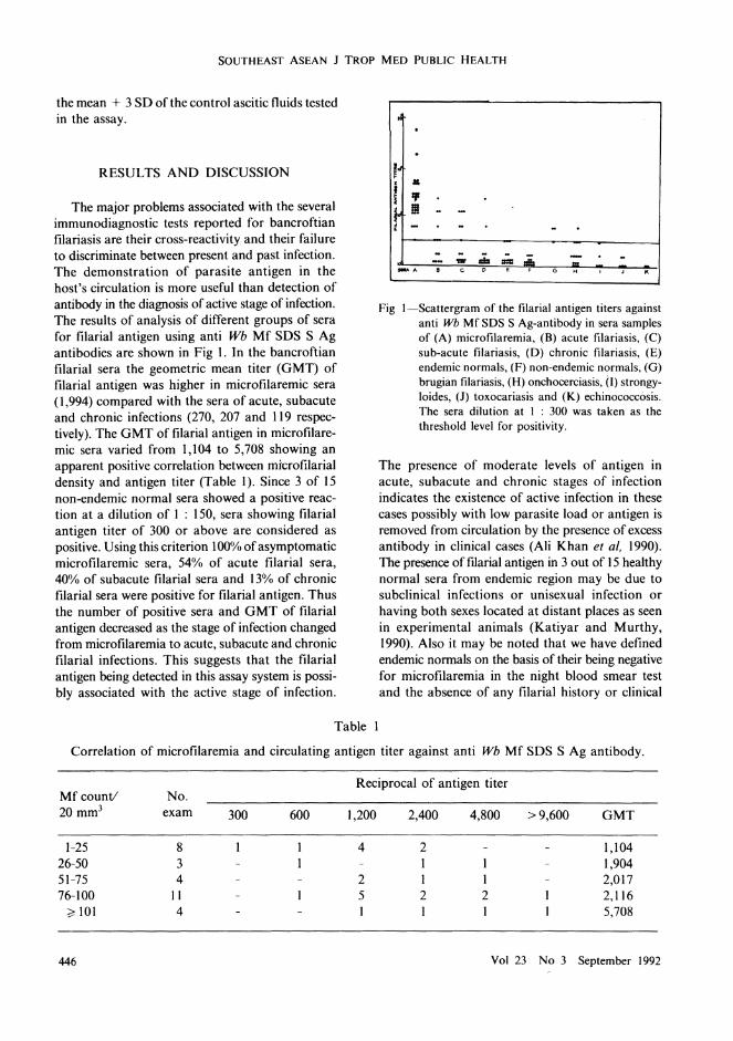
The major problems associated with the several immunodiagnostic tests reported for bancroftian filariasis are their cross-reactivity and their failure to discriminate between present and past infection. The demonstration of parasite antigen in the host's circulation is more useful than detection of antibody in the diagnosis of active stage of infection. The results of analysis of different groups of sera for filarial antigen using anti Wb Mf SOS S Ag antibodies are shown in Fig I. In the bancroftian filarial sera the geometric mean titer (GMT) of filarial antigen was higher in microfilaremic sera (1,994) compared with the sera of acute, subacute and chronic infections (270, 207 and 119 respectively). The GMT of filarial antigen in microfilaremic sera varied from 1,104 to 5,708 showing an apparent positive correlation between microfilarial density and antigen titer (Table I). Since 3 of 15 non-endemic normal sera showed a positive reaction at a dilution of 1 : 150, sera showing filarial antigen titer of 300 or above are considered as positive. Using this criterion 100"10 ofasymptomatic microfilaremic sera, 54% of acute filarial sera, 40% of subacute filarial sera and 13% of chronic filarial sera were positive for filarial antigen. Thus the number of positive sera and GMT of filarial antigen decreased as the stage of infection changed from microfilaremia to acute, subacute and chronic filarial infections. This suggests that the filarial antigen being detected in this assay system is possibly associated with the active stage of infection.
Table
• . .
r· •.. ~ III - t
- .-- -... .;;" .. -:: _A c D . F G , J KH•
Fig I-Scattergram of the filarial antigen titers against anti Wb Mf SDS SAg-antibody in sera samples of (A) microfilaremia, (8) acute filariasis, (C) sub-acute filariasis, (D) chronic filariasis, (E) endemic normals, (F) non-endemic normals, (0) brugian filariasis, (H) onchocerciasis, (I) strongyloides, (J) toxocariasis and (K) echinococcosis. The sera dilution at I : 300 was taken as the threshold level for positivity.
The presence of moderate levels of antigen in acute, subacute and chronic stages of infection indicates the existence of active infection in these cases possibly with low parasite load or antigen is removed from circulation by the presence of excess antibody in clinical cases (Ali Khan et ai, 1990). The presence of filarial antigen in 3 out of 15 healthy normal sera from endemic region may be due to subclinical infections or unisexual infection or having both sexes located at distant places as seen in experimental animals (Katiyar and Murthy, 1990). Also it may be noted that we have defined endemic normals on the basis of their being negative for microfilaremia in the night blood smear test and the absence of any filarial history or clinical
Correlation of microfilaremia and circulating antigen titer against anti Wb Mf SOS S Ag antibody.
Reciprocal of antigen titer MfcounV No. 20mm3 exam 300 600 1,200 2,400 4,800 >9,600 GMT
1-25 8 4 2 1,104 26-50 3 1 1 1,904 51-75 4 2 1 1 2,017 76-100 11 5 2 2 2,116
;;dOl 4 1 1 1 5,708
446 Vol 23 No 3 September 1992
DETECTION OF FILARIAL ANTIGEN
symptoms suggestive of filarial infection. The possible failure to detect microfilariae present in very low concentration by this test in these cases can not be ruled out. Long term follow up of these cases is necessary to determine whether they are due to prepatent infection or occult infection.
In earlier studies from our laboratory Ali Khan et al (1990) used IgG fraction of human filarial serum immunoglobulins (FSIgG) along with Wb Mf excretory-secretory antigen in inhibition ELISA and detected filarial antigen in 81 % of microfilaremic sera and also in a significantly higher number of acute and subacute clinical filarial sera (85% and 88% respectively). Using FSIgG in sandwich ELISA Reddy et al (1984) reported 82% of microfilaremic cases and 63% of clinical filarial cases positive for filarial antigen. -By uSing anti B. malayi adult soluble antigen antibody raised in mouse ascitic fluid, filarial antigen was detected in about 93% of micro filariae carriers and 20-40% of acute and chronic filarial sera by sandwich ELISA (Cheirmaraj et ai, 1990). A monoclonal antibody raised against Wb Mf excretory-secretory antigen (Wb E34) was used in sandwich ELISA (Reddy et ai, 1986) to detect filarial antigen in 68% of microfilaremic sera and 12% of clinical filarial sera. However, using the same monoclonal antibody along with a polyclonal rabbit anti B. malayi antibody, Zheng et al (1987) reported 94% and 53% of microfilarial and clinical filarial sera respectively positive for filarial antigen.
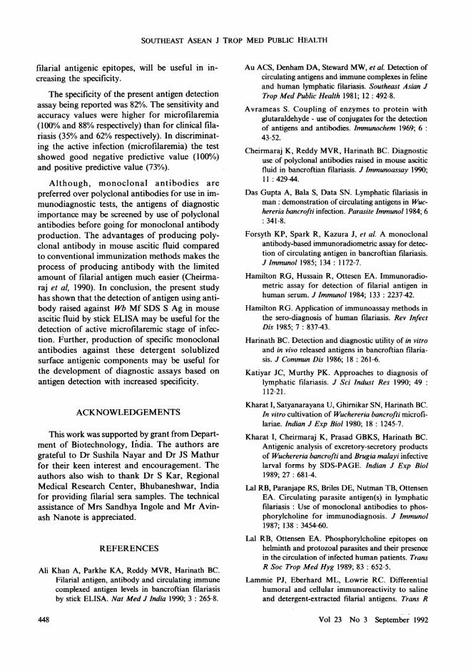
The reaction pattern of anti Wb Mf SDS S Ag antibody with filarial antigen and the cross-reacting PC epitope was carried out by indirect ELISA. PC-BSA detected high titers of IgM antibody whereas Wb Mf ES Ag detected high titers of filarial IgG antibody in immune ascitic fluid (Fig 2). Most of the monoclonal antibodies raised against filarial antigen which were cross-reacting with PC epitopes were found to be ofIgM isotype (Forsyth et ai, 1985; Lal et ai, 1987). Wb Mf SDS S Ag also showed the presence of PC epitopes by direct binding studies with anti-PC antibodies. Immunoassay based on the detection of antigens containing the PC epitopes currently appear to be more sensitive for the diagnosis of filarial infection (Forsyth et ai, 1985; Lal et ai, 1987; Maizels et ai, 1987). Positive correlations between levels of circulating PC antigen and Mf density (Forsyth et ai, 1985) and adult worm burden (Wenger et ai, 1988)
Vol 23 No 3 September 1992
21)00 ,-------------
1500
It: LI.I !:: r
10006 2 i= ~ -l sao ~ ~ Cl..
fa a: 0
19M '9 G
Fig 2-IgM and IgO antibody level against Wb MfSDS S Ag 0 and PC-BSA • in ascitic fluid collected from mouse immunized with Wb Mf SDS SAg. Results shown are mean ± SO of 4 samples.
have been reported. In such immunoassays for the detection of W bancrofti infection, most microfilaremic individuals and a variable proportion of clinical filarial patients are positive for filarial antigen, similar to the results obtained in the present study. Even though detection of circulating PC antigen showed good sensitivity for the diagnosis of filarial infection, another problem of specificity that has to be overcome is the wide distribution of PC epitopes in nature (Lal and Ottesen, 1989) which may lead to false positive reactions. In present study all the 3 brugian filarial sera, 3 out of 15 onchocercal sera, lout of 5 strongyloidal sera and none of the 5 toxocariasis and 2 hydatidosis sera also showed positive reactions for filarial antigen. The cross-reactivity with brugian filarial, onchocercal and strongyloides infections may be due to sharing of antigens among these parasites or the presence of PC epitopes. The PC reactive monoclonal antibodies showing potential for the diagnosis of filarial infection (Lal et aI, 1987) were found to be cross-reactive to a certain extent (50-72%) with onchocercal and strongyloidal sera (Lal and Ottesen, 1989). Further studies, whether anti Wb Mf SDS S Ag antibody detects predominantly PC bearing antigenic epitopes or specific
447
SOUTHEAST ASEAN J TROP MED PUBLIC HEALTH
filarial antigenic epitopes, wiIl be useful in In
creasing the specificity.
The specificity of the present antigen detection assay being reported was 82%. The sensitivity and accuracy values were higher for microfilaremia (100% and 88% respectively) than for clinical filariasis (35% and 62% respectively). In discriminating the active infection (microfilaremia) the test showed good negative predictive value (100%) and positive predictive value (73%).
Although, monoclonal antibodies are preferred over polyclonal antibodies for use in immunodiagnostic tests, the antigens of diagnostic importance may be screened by use of polyclonal antibodies before going for monoclonal antibody production. The advantages of producing polyclonal antibody in mouse ascitic fluid compared to conventional immunization methods makes the process of producing antibody with the limited amount of filarial antigen much easier (Cheirmaraj et ai, 1990). In conclusion, the present study has shown that the detection of antigen using antibody raised against Wb Mf SDS S Ag in mouse ascitic fluid by stick ELISA may be useful for the detection of active microfilaremic stage of infection. Further, production of specific monoclonal antibodies against these detergent solublized surface antigenic components may be useful for the development of diagnostic assays based on antigen detection with increased specificity.
ACKNOWLEDGEMENTS
This work was supported by grant from Department of Biotechnology, India. The authors are grateful to Dr Sushila Nayar and Dr JS Mathur for their keen interest and encouragement. The authors also wish to thank Dr S Kar, Regional Medical Research Center, Bhubaneshwar, India for providing filarial sera samples. The technical assistance of Mrs Sandhya Ingole and Mr Avinash Nanote is appreciated.
REFERENCES
Ali Khan A, Parkhe KA, Reddy MVR, Harinath BC. Filarial antigen, antibody and circulating immune complexed antigen levels in bancroftian filariasis by stick ELISA. Nat Med J India 1990; 3 : 265-8.
Au ACS, Denham DA, Steward MW, et al. Detection of circulating antigens and immune complexes in feline and human lymphatic filariasis. Southeast Asian J Trop Med Public Health 1981; 12: 492-8.
Avrameas S. Coupling of enzymes to protein with glutaraldehyde - use of conjugates for the detection of antigens and antibodies. Immunochem 1969; 6 : 43-52.
Cheirmaraj K, Reddy MVR, Harinath Be. Diagnostic use of polyclonal antibodies raised in mouse ascitic fluid in bancroftian filariasis. J Immunoassay 1990; II : 429-44.
Das Gupta A, Bala S, Data SN. Lymphatic filariasis in man: demonstration of circulating antigens in Wuchereria bancrofti infection. Parasite Immuno/1984; 6 : 341-8.
Forsyth KP, Spark R, Kazura J, et al. A monoclonal antibody-based immunoradiometric assay for detection of circulating antigen in bancroftian filariasis. J Immuno/1985; 134: 1172-7.
Hamilton RG, Hussain R, Ottesen EA. Immunoradiometric assay for detection of filarial antigen in human serum. J Immunol 1984; 133 : 2237-42.
Hamilton RG. Application of immunoassay methods in the sero-diagnosis of human filariasis. Rev Infect Dis 1985; 7 : 837-43.
Harinath BC. Detection and diagnostic utility of in vitro and in vivo released antigens in bancroftian filariasis. J Commun Dis 1986; 18 : 261-6.
Katiyar JC, Murthy PK. Approaches to diagnosis of lymphatic filariasis. J Sci Indust Res 1990; 49 : 112-21.
Kharat I, Satyanarayana U, Ghirnikar SN, Harinath BC. In vitro cultivation of Wuchereria bancrofti microfilariae. Indian J Exp Bioi 1980; 18 : 1245-7.
Kharat I, Cheirmaraj K, Prasad GBKS, Harinath BC. Antigenic analysis of excretory-secretory products of Wuchereria bancrofti and Brugia malayi infective larval forms by SDS-PAGE. Indian J Exp Bioi 1989; 27 : 681-4.
Lal RB, Paranjape RS, Briles DE, Nutman TB, Ottensen EA. Circulating parasite antigen(s) in lymphatic filariasis: Use of monoclonal antibodies to phosphorylcholine for immunodiagnosis. J Immunol 1987; 138 : 3454-60.
Lal RB, Ottensen EA. Phosphorylcholine epitopes on helminth and protozoal parasites and their presence in the circulation of infected human patients. Trans R Soc Trop Med Hyg 1989; 83 : 652-5.
Lammie PJ, Eberhard ML, Lowrie Re. Differential humoral and cellular immunoreactivity to saline and detergent-extracted filarial antigens. Trans R
Vol 23 No 3 September 1992 448
DETECfION OF FILARIAL ANTIGEN
Soc Trop Med Hyg 1990; 84 : 407-10.
Lowry OH, Rosenbrough NJ, Farr AL, Randall RJ. Protein measurement with the Folin-phenol reagent. J Bioi Chem 1951; 193 : 265-75.
Maizels RM, Burke JA, Sutanto I, Purnomo. Partons F. Secreted and surface antigens from larval stages of Wuchereria bancrofti the major human lymphatic filarial parasite. Mol Biochem Parasitol1986; 19 : 27-34.
Maizels RM, Burke JA, Denham DA. Phosphorylcholinebearing antigens in filarial nematode parasites : analysis of somatic extracts, in vitro secretions and infection sera from Brugia malayi and B. pahangi. Parasite Immuno11987; 9 : 49-66.
Overcamp D, Mohammed Ali S, Cartledge C, Landon J. Production of polyc1onal antibodies in ascitic fluid of mice : Techniques and applications. J Immunoassay 1988; 9 : 51-68.
Parkhe KA. Ramaprasad P, Harinath BC. Stick enzyme linked immunosorbent assay using the avidin-biotin system for detection of circulating antigen in bancroftian filariasis. J Biosci 1988; 13 : 229-33.
Reddy MVR, Malhotra A, Harinath BC. Detection of circulating antigen in bancroftian filariasis by sand
wich ELISA using filarial serum IgG. J Helminthol 1984; 58 : 259-62.
Reddy MVR, Ramaprasad P, Piessens WF, Harinath Be. Diagnostic utility of monoclonal antibodies raised against microfilarial excretory-secretory antigens in bancroftian filariasis. J Biosci 1986; 10 : 461-6.
Reddy MVR, Parkhe KA, Piessens WF, Harinath Be. Wb E34 monoclonal antibody: Further characterization and diagnostic use in bancroftian filariasis. J Clin Lab Analysis 1989; 3 : 277-81.
Simonson PE. Wuchereria bancrofti in Tanzania: immune reactions to the microfilarial surface and the effect of diethylcarbamazine upon these reactions. Trans R Soc Trop Med Hyg 1985; 79 : 852-8.
Wenger JD, Forsyth KP, Kazura JW. Identification of phosphorylcholine epitope - containing antigens in Brugia malayi and relation of serum epitope levels to infection status of jirds with brugian filariasis. Am J Trop Med Hyg 1988; 38 : 133-41.
Zheng H, Zhenghou T, Reddy MVR, Harinath BC, Piessens WF. Parasite antigens in sera and urine of patients with bancroftian and brigian filariasis detected by sandwich ELISA with monoclonal antibodies. Am J Trop Med Hyg 1987; 36 : 554-60.
Vol 23 No 3 September 1992 449