Diagnostic Ascitic Aspiration...Diagnostic ascitic aspiration (also known as an ascitic tap) is a...
29
Diagnostic Ascitic Aspiration August 2019
Transcript of Diagnostic Ascitic Aspiration...Diagnostic ascitic aspiration (also known as an ascitic tap) is a...
Diagnostic Ascitic Aspiration August 2019
Dr Alex Thompson Specialty Trainee in Gastroenterology, NHS Lothian Dr Tom Manship Specialty Trainee in Gastroenterology, NHS Lothian Dr Aaron McGowan Specialty Trainee in Gastroenterology, NHS Lothian Dr Tim Cartlidge Specialty Trainee in Cardiology, NHS Lothian Dr Anna Stout Clinical Teaching Fellow, NHS Lothian Dr Oliver Daly Consultant Anaesthetist, NHS Lothian Dr James Tiernan Consultant Physician, NHS Lothian Dr Simon Edgar Director of Medical Education, NHS Lothian Dr Caroline Bates Consultant Acute and GI Physician, NHS Lothian Dr Mike Williams Consultant Hepatologist, NHS Lothian
Diagnostic ascitic aspiration (also known as an ascitic tap) is a frequently performed
procedure, used to obtain a sample of fluid from the peritoneal cavity.
It is a key investigation in the assessment of new ascites and should be performed urgently
(within 6 hours) in all unwell patients with cirrhosis and ascites to exclude infection.
This is a procedural competency requirement for several postgraduate curricula.
Indications
1. Todeterminetheaetiologyofnew-onsetascites
2. Toexcludespontaneousbacterialperitonitisinanycirrhoticpatientwithascitesand
aclinicaldeterioration
Risk Assessment: Ascitic aspiration
Potential Contraindications:
1. Anticoagulation
2. Patient factors increasing technical difficulty
Risk Assessment: Ascitic aspiration (cont.)
Anticoagulation:
• Aprolongedprothrombin time (PT) is a common finding in cirrhoticpatientsasa
resultofimpairedsyntheticliverfunctionandreducedproductionofclottingfactors.
However,asthesepatientsalsohavereducedproductionofanticoagulantfactors,a
prolongedPTdoesnotequatetoan increasedbleedingrisk.There is thereforeno
needtocorrectaprolongedPTpriortoanascitictap.
• Similarly, low platelets are common in cirrhosis and ascitic taps can safely be performed
in patients with platelets >20. If the platelet count is less than 50 you should seek senior
advice about the risks and benefits of a platelet transfusion.
• In contrast, anticoagulation with warfarin or direct-acting oral anticoagulants
(DOACs) does increase bleeding risk and these should bewithheld or reversed if
possible.Anascitictapshouldbeavoidedinpatientswithdisseminatedintravascular
coagulation.
Patient Factors:
• A confused/agitated patient increases the technical challenge and should prompt
consideration of whether a more experienced practitioner is available to perform the
procedure.
• Other physical patient factors can increase the technical challenge of performing an
ascitic tap. These include obesity, significant organomegaly, and previous abdominal
surgery with the risk of adhesions. Again, seek an experienced practitioner.
• Pregnancy, abdominal skin infection at the proposed puncture sites, and severe bowel
distension are usually contraindications to an ascitic tap.
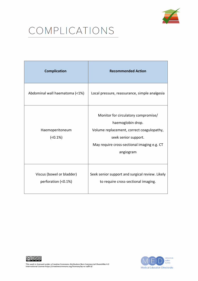
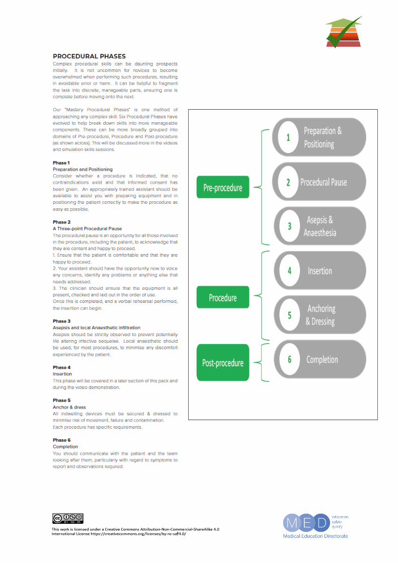
Complication
Recommended Action
Abdominal wall haematoma (<1%)
Local pressure, reassurance, simple analgesia
Haemoperitoneum
(<0.1%)
Monitor for circulatory compromise/
haemoglobin drop.
Volume replacement, correct coagulopathy,
seek senior support.
May require cross-sectional imaging e.g. CT
angiogram
Viscus (bowel or bladder)
perforation (<0.1%)
Seek senior support and surgical review. Likely
to require cross-sectional imaging.
Ascites is the accumulation of excess fluid within the peritoneal cavity. This usually becomes
clinically detectable when there is more than 1L of fluid although >20L can ultimately collect
within the peritoneal cavity.
Abdominal distension should be investigated by percussion for flank dullness, and assessment
for “shifting dullness” by rolling the patient onto their side.
Upon suspicion of first presentation with ascites, the person should undergo abdominal
imaging, in the form of Ultrasound or CT, to confirm presence of ascites before interventional
procedures.
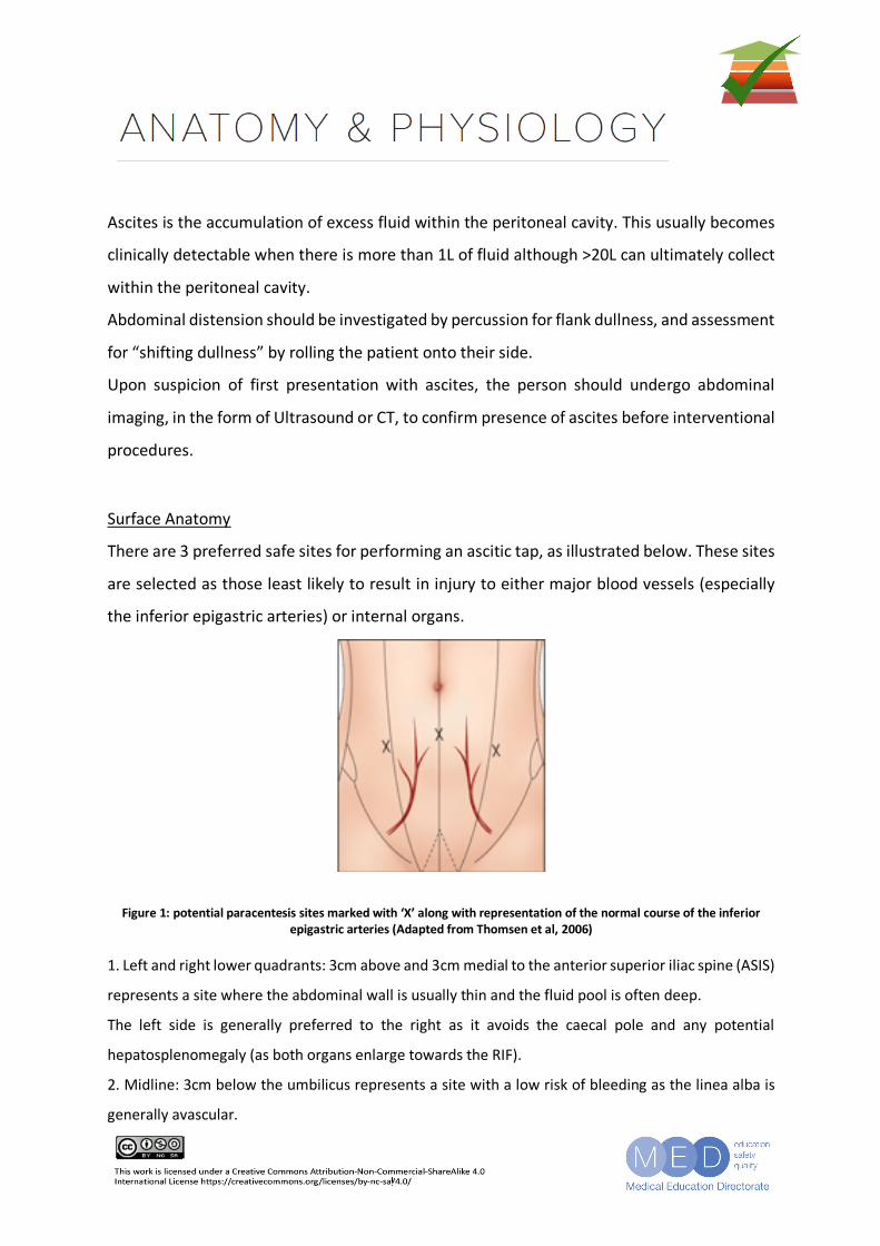
Surface Anatomy
There are 3 preferred safe sites for performing an ascitic tap, as illustrated below. These sites
are selected as those least likely to result in injury to either major blood vessels (especially
the inferior epigastric arteries) or internal organs.
Figure 1: potential paracentesis sites marked with ‘X’ along with representation of the normal course of the inferior epigastric arteries (Adapted from Thomsen et al, 2006)
1. Left and right lower quadrants: 3cm above and 3cm medial to the anterior superior iliac spine (ASIS)
represents a site where the abdominal wall is usually thin and the fluid pool is often deep.
The left side is generally preferred to the right as it avoids the caecal pole and any potential
hepatosplenomegaly (as both organs enlarge towards the RIF).
2. Midline: 3cm below the umbilicus represents a site with a low risk of bleeding as the linea alba is
generally avascular.
Pathophysiology
Cirrhosis is the most common cause of ascites and accounts for approximately 80% of cases.
The onset of ascites is a major landmark in the natural history of cirrhosis and is associated
with a worsening in prognosis.
In cirrhotic ascites, there is also a risk of bacterial translocation across the gut barrier resulting
in spontaneous bacterial peritonitis (SBP). This may present in a non-specific manner, and
some patients may not have either fever or abdominal pain. SBP is often associated with an
acute kidney injury and carries a relatively high mortality. For these reasons, it is crucial to
assess for SBP in a cirrhotic patient with ascites and any clinical deterioration. The British
Society of Gastroenterology cirrhosis care bundle recommends that an ascitic tap is
performed within 6 hours of admission in all patients with cirrhosis and ascites.
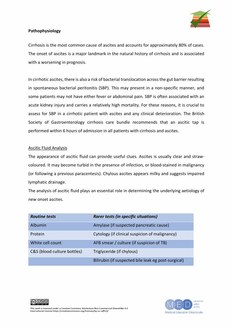
Ascitic Fluid Analysis
The appearance of ascitic fluid can provide useful clues. Ascites is usually clear and straw-
coloured. It may become turbid in the presence of infection, or blood-stained in malignancy
(or following a previous paracentesis). Chylous ascites appears milky and suggests impaired
lymphatic drainage.
The analysis of ascitic fluid plays an essential role in determining the underlying aetiology of
new onset ascites.
Routine tests Rarer tests (in specific situations)
Albumin Amylase (if suspected pancreatic cause)
Protein Cytology (if clinical suspicion of malignancy)
White cell count AFB smear / culture (if suspicion of TB)
C&S (blood culture bottles) Triglyceride (if chylous)
Bilirubin (if suspected bile leak eg post-surgical)
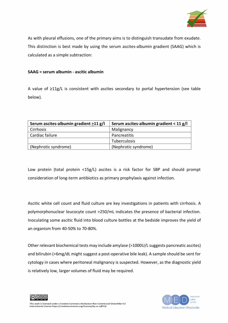
As with pleural effusions, one of the primary aims is to distinguish transudate from exudate.
This distinction is best made by using the serum ascites-albumin gradient (SAAG) which is
calculated as a simple subtraction:
SAAG = serum albumin - ascitic albumin
A value of ≥11g/L is consistent with ascites secondary to portal hypertension (see table
below).
Serum ascites-albumin gradient >11 g/l Serum ascites-albumin gradient < 11 g/l Cirrhosis Malignancy Cardiac failure Pancreatitis Tuberculosis (Nephrotic syndrome) (Nephrotic syndrome)
Low protein (total protein <15g/L) ascites is a risk factor for SBP and should prompt
consideration of long-term antibiotics as primary prophylaxis against infection.
Ascitic white cell count and fluid culture are key investigations in patients with cirrhosis. A
polymorphonuclear leucocyte count >250/mL indicates the presence of bacterial infection.
Inoculating some ascitic fluid into blood culture bottles at the bedside improves the yield of
an organism from 40-50% to 70-80%.
Other relevant biochemical tests may include amylase (>1000U/L suggests pancreatic ascites)
and bilirubin (>6mg/dL might suggest a post-operative bile leak). A sample should be sent for
cytology in cases where peritoneal malignancy is suspected. However, as the diagnostic yield
is relatively low, larger volumes of fluid may be required.
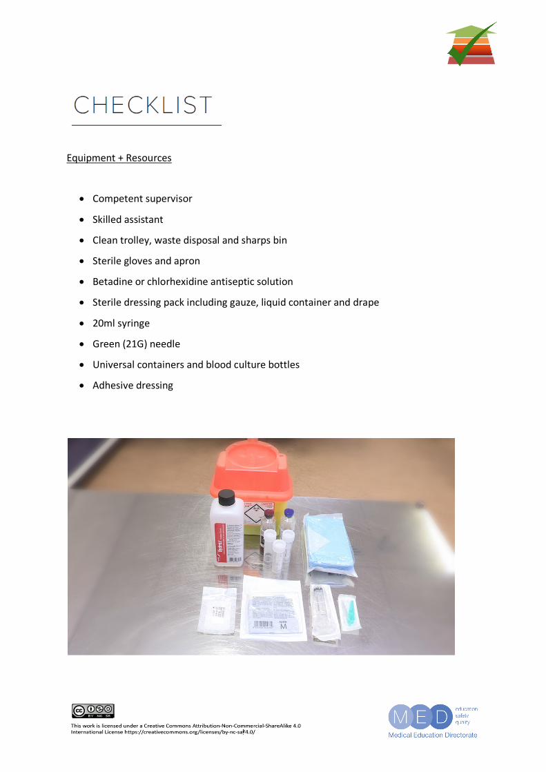
Equipment + Resources
• Competent supervisor
• Skilled assistant
• Clean trolley, waste disposal and sharps bin
• Sterile gloves and apron
• Betadine or chlorhexidine antiseptic solution
• Sterile dressing pack including gauze, liquid container and drape
• 20ml syringe
• Green (21G) needle
• Universal containers and blood culture bottles
• Adhesive dressing
Exclude contraindications
Check whether known allergies
Examine patient – note organomegaly, scars of previous surgery
Review platelet count and any recent imaging
Check for anticoagulant medications
Preparation
Ensure patient has emptied bladder
Lie patient supine, head elevated 20-30 degrees
Identify and mark planned site for tap (e.g. with blunt needle cap)
(3cm above and 3cm medial to Left ASIS)
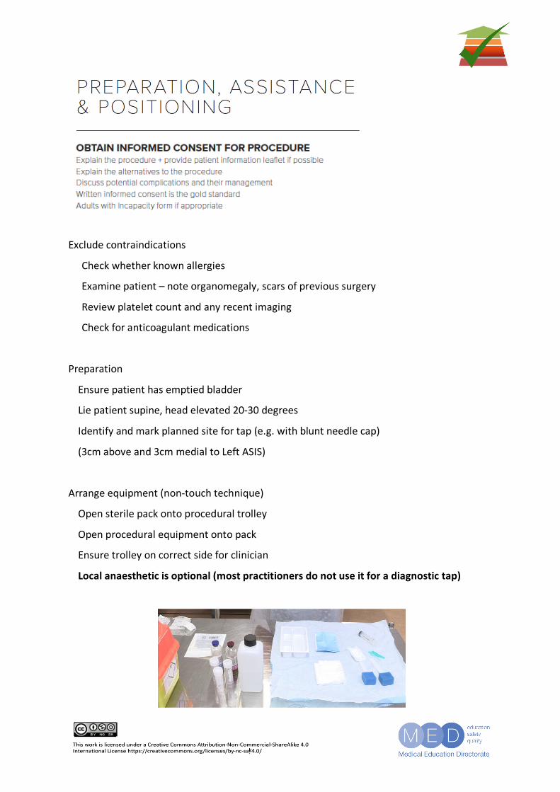
Arrange equipment (non-touch technique)
Open sterile pack onto procedural trolley
Open procedural equipment onto pack
Ensure trolley on correct side for clinician
Local anaesthetic is optional (most practitioners do not use it for a diagnostic tap)
Aseptic Conditions
Wearing apron, wash hands and put on sterile gloves
Apply antiseptic skin preparation to patient
Drape the area
Allow skin to dry
Local anaesthetic is optional for a diagnostic ascitic aspiration
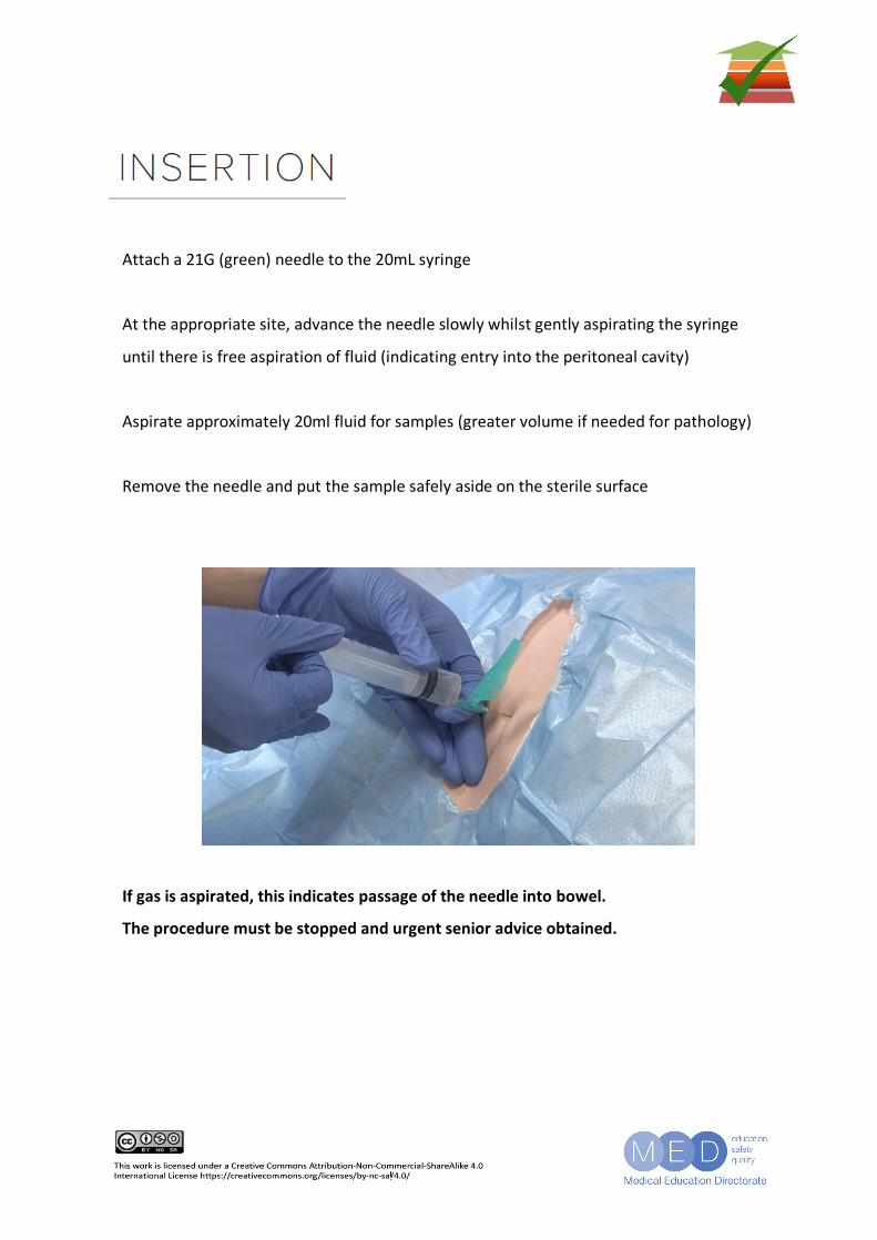
Attach a 21G (green) needle to the 20mL syringe
At the appropriate site, advance the needle slowly whilst gently aspirating the syringe
until there is free aspiration of fluid (indicating entry into the peritoneal cavity)
Aspirate approximately 20ml fluid for samples (greater volume if needed for pathology)
Remove the needle and put the sample safely aside on the sterile surface
If gas is aspirated, this indicates passage of the needle into bowel.
The procedure must be stopped and urgent senior advice obtained.
No specific anchoring is required for diagnostic aspiration
Apply a simple adhesive dressing to the entry site, following removal of the needle
Inoculate fluid into blood culture bottles and universal containers
Dispose of waste and sharps appropriately
Label and send samples to:
Microbiology for cell count and culture
Biochemistry for albumin and protein
Pathology if malignancy suspected
Paired serum should be sent for albumin
Document the procedure, including any immediate complications and results
• Ideally use an electronic menu to prompt high standard documentation
Mastery Assessment Procedural Checklist References
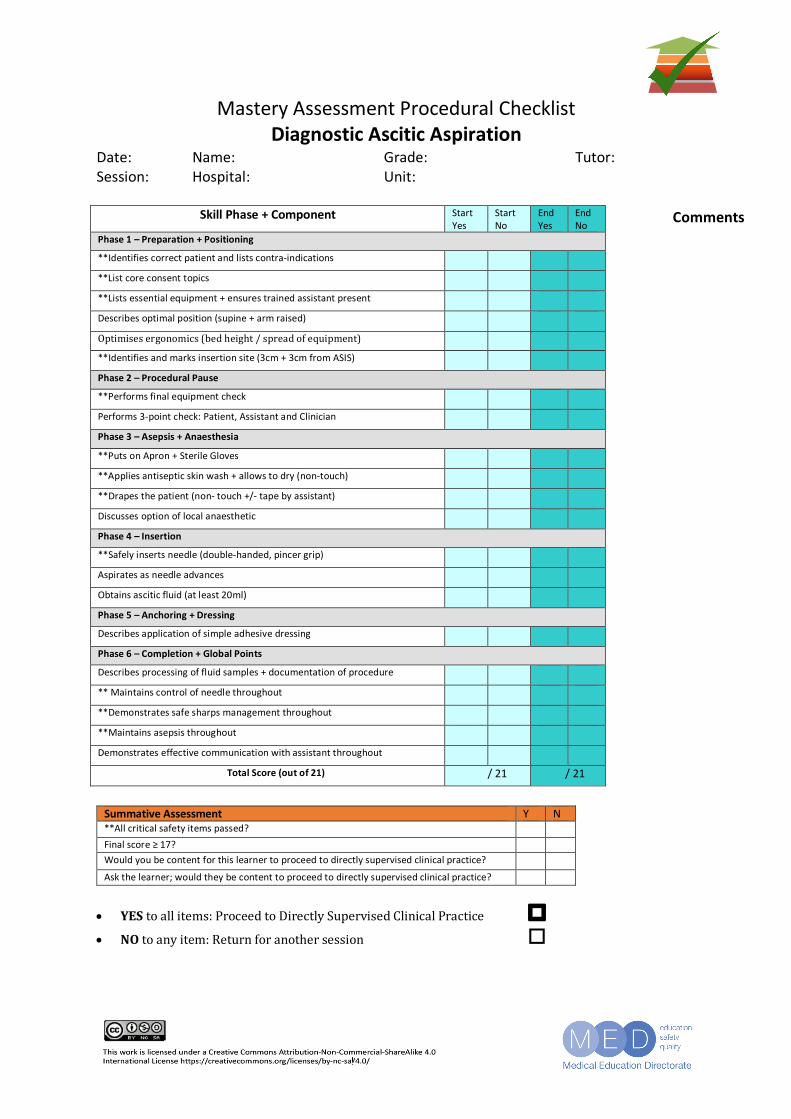
Mastery Assessment Procedural Checklist Diagnostic Ascitic Aspiration
Date: Name: Grade: Tutor: Session: Hospital: Unit:
Skill Phase + Component Start Yes
Start No
End Yes
End No
Phase 1 – Preparation + Positioning
**Identifies correct patient and lists contra-indications
**List core consent topics
**Lists essential equipment + ensures trained assistant present
Describes optimal position (supine + arm raised)
Optimisesergonomics(bedheight/spreadofequipment)
**Identifies and marks insertion site (3cm + 3cm from ASIS)
Phase 2 – Procedural Pause
**Performs final equipment check
Performs 3-point check: Patient, Assistant and Clinician
Phase 3 – Asepsis + Anaesthesia
**Puts on Apron + Sterile Gloves
**Applies antiseptic skin wash + allows to dry (non-touch)
**Drapes the patient (non- touch +/- tape by assistant)
Discusses option of local anaesthetic
Phase 4 – Insertion
**Safely inserts needle (double-handed, pincer grip)
Aspirates as needle advances
Obtains ascitic fluid (at least 20ml)
Phase 5 – Anchoring + Dressing
Describes application of simple adhesive dressing
Phase 6 – Completion + Global Points
Describes processing of fluid samples + documentation of procedure
** Maintains control of needle throughout
**Demonstrates safe sharps management throughout
**Maintains asepsis throughout
Demonstrates effective communication with assistant throughout
Total Score (out of 21) / 21 / 21
Summative Assessment Y N **All critical safety items passed? Final score ≥ 17? Would you be content for this learner to proceed to directly supervised clinical practice? Ask the learner; would they be content to proceed to directly supervised clinical practice?
• YEStoallitems:ProceedtoDirectlySupervisedClinicalPractice o
• NOtoanyitem:Returnforanothersession o
Comments
Moore KP, Aithal GP. Guidelines on the management of ascites in cirrhosis. Vol. 55 Suppl 6, Gut. 2006, pp.vi1-12. Runyon BA; Management of adult patients with ascites due to cirrhosis: an update. AASLD Practice Guidelines Committee. Hepatology. 2009 Jun;49(6):2087-107. Thomsen TW, Shaffer RW, White B, Setnik GS. Paracentesis. N Engl J Med 2006;355:e21 European Association for the Study of the Liver. EASL Clinical Practice Guidelines for the management of patients with decompensated cirrhosis. J Hepatol. 2018 Aug;69(2):406-60 McPherson S, Dyson J, Austin A, Hudson M. Response to the NCEPOD report: development of a care bundle for patients admitted with decompensated cirrhosis- the first 24h. Frontline Gastroenterol. 2016 Jan;7(1):16-23. Scottish Government (2000). Adults with incapacity (Scotland) Act 2000. Edinburgh. Available at: http://www.scotland.gov.uk/Topics/Justice/law/awi