Dental Trauma and Antemortem Tooth Loss in …pages.uoregon.edu/jrlukacs/Dr. John R. Lukacs...
17
Dental Trauma and Antemortem Tooth Loss in Prehistoric Canary Islanders: Prevalence and Contributing Factors J. R. LUKACS* Department of Anthropology University of Oregon, Eugene, OR 97403-1218, USA ABSTRACT Differential diagnosis of the aetiology of antemortem tooth loss (AMTL) may yield important insights regarding patterns of behaviour in prehistoric peoples. Variation in the consistency of food due to its toughness and to food preparation methods is a primary factor in AMTL, with dental wear or caries a significant precipitating factor. Nutritional deficiency diseases, dental ablation for aesthetic or ritual reasons, and traumatic injury may also contribute to the frequency of AMTL. Systematic observations of dental pathology were conducted on crania and mandibles at the Museo Arqueologico de Tenerife. Observations of AMTL revealed elevated frequencies and remarkable aspects of tooth crown evulsion. This report documents a 9.0% overall rate of AMTL among the ancient inhabitants of the island of Tenerife in the Canary Archipelago. Sex-specific tooth count rates of AMTL are 9.8% for males and 8.1% for females, and maxillary AMTL rates (10.2%) are higher than mandibular tooth loss rates (7.8%) Dental trauma makes a small but noticeable contribution to tooth loss among the Guanches, especially among males. In several cases of tooth crown evulsion, the dental root was retained in the alveolus, without periapical infection, and alveolar bone was in the initial stages of sequestering the dental root. In Tenerife, antemortem loss of maxillary anterior teeth is consistent with two potential causal factors: (a) accidental falls while traversing volcanic terrain; and (b) interpersonal combat, including traditional wrestling, stick-fighting and ritual combat. Steep-walled valleys (barrancos) and lava fields (malpaı´s) required agile locomotion and occasional vaulting with the aid of a wooden staff. Accidental falls involving facial injury may have contributed to AMTL. Traditional conflict resolution involved competitive wrestling (lucha canaria), stick-fighting (juego del palo), and ritualised contests involving manual combat. These activities made a small but recognisable impact on anterior dental trauma and tooth loss. Inter-personal behaviours of such intensity leave their mark on skeletal and dental remains, thereby providing insight into the lives and cultural traditions of the ancient Guanches. Copyright ß 2006 John Wiley & Sons, Ltd. Key words: dental palaeopathology; dental trauma; antemortem tooth loss; tooth fracture; manual combat; accidents; Tenerife; Canary Islands Introduction Loss of teeth from the jaws is a complex and multi- causal process (Brothwell, 1963:277; Lukacs, 1989:271; Scott et al., 1991:194). Despite the valuable insights that can be derived from the careful study of antemortem tooth loss patterns, dental palaeopathologists seem to have neglected this potentially rewarding area of investigation. Four primary causal factors contribute to pre- mature loss of teeth: (1) variations in dietary consistency (Leigh, 1925; Alexandersen, 1967a; Frayer, 1987, 1989; Beckett & Lovell, 1994; Nelson et al., 1999; Bonfiglioli et al., 2003); International Journal of Osteoarchaeology Int. J. Osteoarchaeol. (in press) Published online in Wiley InterScience (www.interscience.wiley.com). DOI: 10.1002/oa.864 * Correspondence to: Department of Anthropology, University of Oregon, Eugene, OR 97403-1218, USA. e-mail: [email protected] Copyright # 2006 John Wiley & Sons, Ltd. Received 22 November 2004 Revised 21 December 2005 Accepted 24 January 2006
Transcript of Dental Trauma and Antemortem Tooth Loss in …pages.uoregon.edu/jrlukacs/Dr. John R. Lukacs...

International Journal of OsteoarchaeologyInt. J. Osteoarchaeol. (in press)
erscience.wiley.com). DOI: 10.1002/oa.864
Published online in Wiley InterScience (www.int* Correspondence to: DepartOregon, Eugene, OR 97403-e-mail: [email protected]
Copyright # 2006 Joh
Dental Trauma and AntemortemTooth Loss in Prehistoric CanaryIslanders: Prevalence andContributing Factors
J. R. LUKACS*
Department of Anthropology University of Oregon, Eugene, OR 97403-1218, USA
ABSTRACT Differential diagnosis of the aetiology of antemortem tooth loss (AMTL) may yield importantinsights regarding patterns of behaviour in prehistoric peoples. Variation in the consistency offood due to its toughness and to food preparation methods is a primary factor in AMTL, withdental wear or caries a significant precipitating factor. Nutritional deficiency diseases, dentalablation for aesthetic or ritual reasons, and traumatic injury may also contribute to thefrequency of AMTL. Systematic observations of dental pathology were conducted on craniaand mandibles at the Museo Arqueologico de Tenerife. Observations of AMTL revealedelevated frequencies and remarkable aspects of tooth crown evulsion. This report documentsa 9.0% overall rate of AMTL among the ancient inhabitants of the island of Tenerife in theCanary Archipelago. Sex-specific tooth count rates of AMTL are 9.8% for males and 8.1% forfemales, and maxillary AMTL rates (10.2%) are higher than mandibular tooth loss rates (7.8%)Dental trauma makes a small but noticeable contribution to tooth loss among the Guanches,especially amongmales. In several cases of tooth crown evulsion, the dental root was retainedin the alveolus, without periapical infection, and alveolar bone was in the initial stages ofsequestering the dental root. In Tenerife, antemortem loss of maxillary anterior teeth isconsistent with two potential causal factors: (a) accidental falls while traversing volcanicterrain; and (b) interpersonal combat, including traditional wrestling, stick-fighting and ritualcombat. Steep-walled valleys (barrancos) and lava fields (malpaıs) required agile locomotionand occasional vaulting with the aid of a wooden staff. Accidental falls involving facial injurymay have contributed to AMTL. Traditional conflict resolution involved competitive wrestling(lucha canaria), stick-fighting (juego del palo), and ritualised contests involving manualcombat. These activities made a small but recognisable impact on anterior dental traumaand tooth loss. Inter-personal behaviours of such intensity leave their mark on skeletal anddental remains, thereby providing insight into the lives and cultural traditions of the ancientGuanches. Copyright � 2006 John Wiley & Sons, Ltd.
Key words: dental palaeopathology; dental trauma; antemortem tooth loss; tooth fracture;
manual combat; accidents; Tenerife; Canary Islands
Introduction
Loss of teeth from the jaws is a complex andmulti-causal process (Brothwell, 1963:277; Lukacs,1989:271; Scott et al., 1991:194). Despite the
ment of Anthropology, University of1218, USA.
n Wiley & Sons, Ltd.
valuable insights that can be derived from thecareful study of antemortem tooth loss patterns,dental palaeopathologists seem to have neglectedthis potentially rewarding area of investigation.Four primary causal factors contribute to pre-mature loss of teeth: (1) variations in dietaryconsistency (Leigh, 1925; Alexandersen, 1967a;Frayer, 1987, 1989; Beckett & Lovell, 1994;Nelson et al., 1999; Bonfiglioli et al., 2003);
Received 22 November 2004Revised 21 December 2005Accepted 24 January 2006

J. R. Lukacs
(2) nutritional deficiency diseases (Stuart-Macadam, 1989); (3) cultural or ritual ablation(Merbs, 1989; Pietrusewsky & Douglas, 1993;Tayles, 1996); and (4) trauma (Leigh, 1929;Merbs, 1989: 171; Lukacs & Hemphill, 1990).Variations in diet may be especially complex sinceAMTL can result from recognisably distinctiveaetiological pathways (see Lukacs, 1989, Fig. 1).Firstly abrasive foods may cause severe attrition,resulting in pulp exposure, dental abscess, andultimate tooth loss (Lukacs & Pal, 1993).Secondly, soft foods and refined diets, high incarbohydrates, may encourage development oflarge caries lesions, producing pulp exposure,abscess formation, and finally tooth loss (Lukacs,1992). Thirdly, large calculus accumulations mayserve as gingival irritants, resulting in periodontaldisease and alveolar recession, the ultimateoutcome being tooth loss (Koritzer, 1968; Clarke& Hirsch, 1991a, b; Diaz & Tayles, 1997). Thisreport, however, will focus on the role of dentaltrauma as a non-dietary factor contributing topatterns of AMTL among the Canary Islanders.My interest in dental trauma was initially
stimulated by two remarkable cases from prehistoricPakistan (Lukacs & Hemphill, 1990). The first is anadult male from aceramic levels at NeolithicMehrgarh, Baluchistan. The left central incisorcrown suffered a complicated fracture in life
Figure 1. Location map of the
Copyright # 2006 John Wiley & Sons, Ltd.
(Andraesen, 1982), as indicated by wear on theincisal edge of the broken crown and developmentof a medium-sized periapical dental lesion. Theforce causing this trauma is indicated by theorientation of the fracture plane and suggestsforceful pulling of an unknown material outwardfrom the oral cavity. The second case comes fromthe Bronze Age urban centre of Harappa, in PunjabProvince, where a robust adult male exhibits atraumatic root fracture of the right lateral incisor andantemortem loss of URI1. Consequently, as I beganmy investigation of the dental anthropology ofprehistoric human remains from the Canary Islands,in Santa Cruz de Tenerife, I had some acquaintancewith and interest in evidence of dental trauma.
Materials and methods
This study was conducted from April to October1991, at the Museo Arqueologico de Tenerife,with the collaboration of Conrado Rodrıguez-Martın. The antiquity of specimens included inthis study ranges from the 6th to the 14th centuryAD. While 90% of the specimens are from theisland of Tenerife (Figure 1), the remainder arefrom other islands in the Canary archipelago. Theoriginal inhabitants of Tenerife, known asGuanches, had close physical and linguistic
Canary Island archipelago.
Int. J. Osteoarchaeol. (in press)
Tooth Loss in Prehistoric Canary Islanders
similarities with the Berbers of northwest Africa.This relationship was initially documented bywell-known 19th century anthropologists andnatural historians (Blumenbach, 1808; von Hum-boldt & Bonpland, 1819; Vernaux, 1891). Theorigins and biological diversity of Guanches weresubsequently approached from a typologicalracial paradigm based on craniometry (Hooton,1916, 1925; Schwidetsky, 1957). Multiple inde-pendent studies of dental morphology confirmthe close affinities between Guanches and Berbersobserved by earlier investigators (Bermudez deCastro, 1985; Irish, 1993; Guatelli-Steinberg et al.,2001).Recent analysis of mtDNA and Y chromosome
polymorphisms reveal three sources for themodern Canary Islander gene pool: a majorIberian contribution (62–78%); a substantialnorthwest African component (23–38%); and aminor sub-Saharan African component (3%;Maca-Meyer et al., 2004a). The documentationof mtDNA diversity among prehistoric Guanchesimplies significant founder effects and multiplemigration events (Maca Meyer et al., 2004b). Ychromosome variation suggests that the abori-ginal genetic contribution to the modern Canar-ian gene pool exhibits a strong sexual bias (Floreset al., 2001, 2003). A colonisation scenario withthree primary components can be derived: (a)northwest Africans settled the islands from east towest following a stepping-stone model; (b) thisaboriginal population was subsequently augmen-ted by slave trade immigration from sub-Saharanand northwest Africa; and (c) in the 15th centurythe islands experienced extensive Europeanimmigration associated with the Norman con-quest and Iberian colonisation (Rando et al., 1999;Flores et al., 2001). These genetic estimatessuggest that initial colonisation of the islandsoccurred around 600 BC, significantly earlier thanthe 1st century BC, as previously believed(Mercer, 1980).The provenance of most specimens in the
study collection is known. Many were recoveredfrom ossuaries in caves located in isolatedbarrancos that are difficult to enter. Stratigraphicand chronological status of individual specimensis not always precise, and crania and mandiblesincluded in this study are isolated specimens notassociated with one another or with complete
Copyright # 2006 John Wiley & Sons, Ltd.
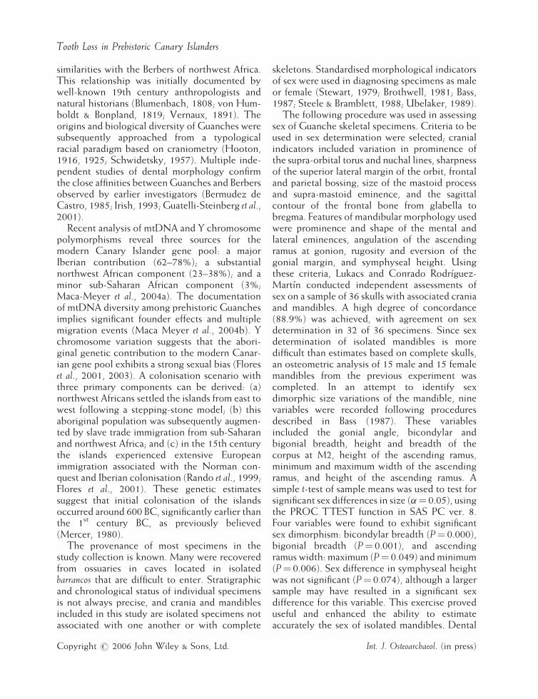
skeletons. Standardised morphological indicatorsof sex were used in diagnosing specimens as maleor female (Stewart, 1979; Brothwell, 1981; Bass,1987; Steele & Bramblett, 1988; Ubelaker, 1989).The following procedure was used in assessing
sex of Guanche skeletal specimens. Criteria to beused in sex determination were selected; cranialindicators included variation in prominence ofthe supra-orbital torus and nuchal lines, sharpnessof the superior lateral margin of the orbit, frontaland parietal bossing, size of the mastoid processand supra-mastoid eminence, and the sagittalcontour of the frontal bone from glabella tobregma. Features of mandibular morphology usedwere prominence and shape of the mental andlateral eminences, angulation of the ascendingramus at gonion, rugosity and eversion of thegonial margin, and symphyseal height. Usingthese criteria, Lukacs and Conrado Rodrıguez-Martın conducted independent assessments ofsex on a sample of 36 skulls with associated craniaand mandibles. A high degree of concordance(88.9%) was achieved, with agreement on sexdetermination in 32 of 36 specimens. Since sexdetermination of isolated mandibles is moredifficult than estimates based on complete skulls,an osteometric analysis of 15 male and 15 femalemandibles from the previous experiment wascompleted. In an attempt to identify sexdimorphic size variations of the mandible, ninevariables were recorded following proceduresdescribed in Bass (1987). These variablesincluded the gonial angle, bicondylar andbigonial breadth, height and breadth of thecorpus at M2, height of the ascending ramus,minimum and maximum width of the ascendingramus, and height of the ascending ramus. Asimple t-test of sample means was used to test forsignificant sex differences in size (a¼ 0.05), usingthe PROC TTEST function in SAS PC ver. 8.Four variables were found to exhibit significantsex dimorphism: bicondylar breadth (P¼ 0.000),bigonial breadth (P¼ 0.001), and ascendingramus width: maximum (P¼ 0.049) and minimum(P¼ 0.006). Sex difference in symphyseal heightwas not significant (P¼ 0.074), although a largersample may have resulted in a significant sexdifference for this variable. This exercise proveduseful and enhanced the ability to estimateaccurately the sex of isolated mandibles. Dental
Int. J. Osteoarchaeol. (in press)

J. R. Lukacs
status was recorded as part of a systematicanthropological analysis of the dentition of eachspecimen. Each tooth was graded as eitherpresent or absent, and if absent, loss was recordedas either antemortem or postmortem. Notes weremade regarding the degree to which alveoli weredamaged postmortem, resorbed following ante-mortem tooth loss, or damaged in life by trauma.Statistical analysis used the PROC FREQprocedure in SAS PC ver. 8. Chi-square testswere used to identify significant differences inAMTL frequency by side, sex and jaw. Fisher’sexact test was used when any cell had a samplesize of five or fewer (Zar, 1999).
Results
The overall rate of AMTL for the study sample is9.0%. Figure 2a shows variation in tooth loss byside, for males and females separately (Table 1).The dark vertical dashed lines represent the meansex-specific tooth loss rates (9.8% males; 8.1%females). Noteworthy patterns include: (1) side toside differences in AMTL are largely non-significant, the one exception being the lowerthird molar among females; (2) most molar andsome premolar teeth exceed mean values; (3) lossof anterior teeth is generally less than the mean;and (4) among males, there is a slight tendency inlower anterior teeth for the left side to exceed theright side. Tooth-specific AMTL rates arepresented separately for each sex in Figure 2b(Table 2). Only five of 16 comparisons show asignificant difference, and four of these occur inthe maxilla where male rates exceed female rates.In the mandible, sex differences in AMTL aremostly non-significant and, except for the firstmolar, male and female values display moresimilarity than in the maxilla.Sex differences in AMTL were also analysed by
determining the percentage of individuals thatlost one or more teeth from a specific tooth class(Table 3). Individual tooth classes and compositetooth classes are listed along the horizontalaxis for the maxilla and mandible separately inFigure 3a. The category ANY includes individualswith antemortem loss of one or more teeth fromany tooth class. Several points are remarkable: (1)no significant differences occur between the sexes
Copyright # 2006 John Wiley & Sons, Ltd.
for individual tooth classes, or for any of themandibular teeth; (2) significant differences areevident in two composite categories in themaxilla–Incisor and Canine combined (abbre-viated as I & C in Table 3), and Incisor, Canineand Premolar combined (I, C & P in Table 3),where male rates exceed female rates. The finalcomparison evaluates differences in AMTL ratesbetween maxillary and mandibular isomeres(Figure 3b). In this graph the upper dotted linerepresents the mean AMTL rate for maxillae(male and female, 10.2%), and the mean AMTLrate for mandibular teeth (sexes combined, 7.8%)is portrayed by the lower dotted line. Five of the16 comparisons (31%) are significant, and in fourof these maxillary teeth show higher loss ratesthan lower teeth. In males, higher maxillary ratesare evident for P3, P4 and M1, while in femalesonly P3 follows this pattern. By contrast, thefemale I1 shows significantly greater loss of thelower vs. the upper central incisor.
A small but important component in producingthis pattern of AMTL is the contribution made byloss due to traumatic fractures. Eleven specimens,five maxillae and six mandibles, display evidenceof traumatically fractured teeth (Figure 4). Thesecompound fractures are characterised by a list ofseveral shared features. The fractures consistentlyoccur at the alveolar margin; reparative dentino-genesis maintains root integrity, and therebyprevents sequelae such as focal destruction ofperiapical or alveolar bone due to infection. Inmost instances, some degree of proliferativealveolar bone growth is evident (Figure 5). Thisforms over the broken root and represents anearly stage in the sequestration and ultimateresorption of the tooth root. Incisor and premolarteeth are primarily affected, canines rarely and nomolar fractures were observed. Three maxillaryand three mandibular specimens (a total of nineteeth) exhibit evidence of proliferative bonegrowth over the broken root surface (3 incisorand 2 premolars in the maxilla, and 3 incisors and1 premolar in the mandible). Others displayevidence of dental trauma such as broken alveolarwalls or septa in association with remodelling,and only two specimens yield indications ofassociated periapical infection.
The fact that these fractures are found towardthe anterior region of the mouth, that six of nine
Int. J. Osteoarchaeol. (in press)

Figure 2. (a) Percentage antemortem tooth loss by side; (b) frequency of antemortem tooth loss by sex.
Tooth Loss in Prehistoric Canary Islanders
teeth with proliferative bone development are leftincisor teeth, and that other forms of alveolar bonedamage and repair are evident, leads to theconclusion that traumatic injury of incisors, canines
Copyright # 2006 John Wiley & Sons, Ltd.
and premolar teeth contributed to the observedrates of antemortem tooth loss documented for theGuanches. Two explanatory hypotheses can beoffered for the patterns of antemortem tooth loss
Int. J. Osteoarchaeol. (in press)

Table 1. Antemortem tooth loss among Guanches by side
Tooth Female Male
Left P � Right Left P � Right
AMTL n % AMTL n % AMTL n % AMTL n %
UI1 7 153 4.6 0.980 7 151 4.6 9 105 8.6 1.000 9 105 8.6UI2 7 151 4.6 0.990 7 150 4.7 7 104 6.7 0.324 11 104 10.6UC 3 147 2.0 0.722 4 145 2.8 4 104 3.9 0.748 6 104 5.8UP3 16 152 10.5 0.331 11 150 7.3 17 105 16.2 0.952 17 107 15.9UP4 16 152 10.5 0.583 13 150 8.7 19 106 17.9 0.513 23 107 21.5UM1 20 152 13.2 0.484 24 150 16.0 21 108 19.4 0.735 23 108 21.3UM2 20 150 13.3 0.629 17 148 11.5 13 105 12.4 0.567 16 106 15.1UM3 8 108 7.4 0.052 18 114 15.8 11 88 12.5 0.884 11 94 11.7Maxilla 97 1165 8.3 0.733 101 1158 8.7 101 825 12.2 0.319 116 835 13.9LI1 12 159 7.6 0.569 15 161 9.3 14 134 10.5 0.709 12 132 9.1LI2 3 159 1.9 0.336 7 160 4.4 10 133 7.5 0.310 6 132 4.6LC 5 158 3.2 0.770 7 160 4.4 5 132 3.8 0.447 2 131 1.5LP3 6 161 3.7 0.777 7 161 4.4 8 134 6.0 0.217 3 133 2.3LP4 11 161 6.8 0.829 12 161 7.5 10 130 7.7 0.972 10 132 7.6LM1 27 161 16.8 0.902 28 162 17.3 17 133 12.8 0.706 15 133 11.3LM2 15 162 9.3 0.986 15 161 9.3 13 133 9.8 0.541 16 132 12.1LM3 15 137 11.0 0.321 10 134 7.5 9 114 7.9 0.546 12 118 10.2Mandible 94 1258 7.5 0.610 101 1260 8.0 86 1043 8.2 0.413 76 1043 7.3Total 191 2423 7.9 0.548 202 2418 8.4 187 1868 10.0 0.829 192 1828 10.2
�P¼probability associated with x2 values; probabilities in italics associated with Fisher’s exact test.
Table 2. Antemortem tooth loss among Guanches, by sex
Tooth Female P Male
AMTL n % AMTL n %
UI1 14 304 4.6 0.067 18 210 8.6UI2 14 301 4.7 0.067 18 208 8.7UC 7 292 2.4 0.143 10 208 4.8UP3 27 302 8.9 0.000� 34 212 16.3UP4 29 302 9.6 0.001� 42 213 19.7UM1 44 302 14.6 0.083 44 216 20.4UM2 37 298 12.4 0.660 29 211 13.7UM3 26 222 11.7 0.907 22 182 12.1Maxilla 198 2323 8.1 0.000� 217 1660 13.1LI1 27 320 8.4 0.574 26 266 9.8LI2 10 319 3.1 0.090 16 265 6.0LC 12 318 3.8 0.453 7 263 2.7LP3 13 322 4.0 0.960 11 267 4.1LP4 23 322 7.1 0.821 20 262 7.6LM1 55 323 17.0 0.024� 32 266 12.0LM2 30 323 9.3 0.506 29 265 10.9LM3 25 271 9.2 0.946 21 232 9.1Mandible 195 2518 7.7 0.978 162 2086 7.8Total 393 4841 8.1 0.000� 379 3746 9.8
� ¼Significant difference at P< 0.05.
J. R. Lukacs
and traumatic injury of anterior teeth amongprehistoric Canary Islanders: (1) accidental fallswhile traversing steep and uneven volcanic terrain;and (2) injury during traditional forms of inter-
Copyright # 2006 John Wiley & Sons, Ltd.
personal combat. The latter included bothcompetitive wrestling and stick-fighting, as wellas conflict resolution through ritualised hand-to-hand combat. The ways in which these factors
Int. J. Osteoarchaeol. (in press)

Table 3. Percentage of Guanche specimens with AMTL, by tooth class
Sex (n) Incisor Canine Premolar Molar I & C I, C & P P & M ANY
n % n % n % n % n % n % n % n %
MaxillaFemale (153) 18 11.8 6 3.9 29 19.0 45 29.4 20 13.1 36 23.5 54 35.3 55 36.0P 0.194 0.137 0.141 0.532 0.056 0.045 0.592 0.078Male (109) 19 17.4 9 8.3 29 26.6 36 33.0 24 22.0 38 34.9 42 38.5 51 46.8Total (262) 37 14.1 15 5.7 58 22.1 81 30.9 44 16.8 74 28.2 96 36.6 106 40.5
MandibleFemale (163) 19 11.7 10 6.1 20 12.3 52 31.9 26 16.0 38 23.3 58 35.6 71 43.6P 0.247 0.725 0.784 0.489 0.674 0.943 0.498 0.312Male (135) 22 16.3 7 5.2 18 13.3 38 28.2 24 17.8 31 23.0 43 31.9 51 37.8Total (298) 41 13.8 17 5.7 38 12.8 90 30.2 50 16.8 69 23.1 101 33.9 122 40.9
Key: I¼ Incisor; C¼Canine; P¼premolar; M¼molar; I & C¼ incisor and canine combined; I, C & P¼ incisor, canineand premolar combined; P & M¼premolar and molar combined; ANY¼ antemortem loss observed in any tooth class;P¼probability associated with x2 test.
Tooth Loss in Prehistoric Canary Islanders
would potentially contribute to dental fracture andAMTL are considered in greater detail below.
Locomotion over hazardous terrain
The Canary Islands are volcanic and geologi-cally recent in origin. They represent anextremely dry western extension of the SaharaDesert (Mercer, 1980). Active volcanism isevident today on the islands of Fuerteventura,La Palma and Tenerife. All islands have recentlava flows of varying size, landforms such ascraters and calderas, and Tenerife presents themagnificent strato-volcano known as El Teidewhich reaches an altitude of 3718m (12,199 ft).Lava flows present multiple obstacles to easybipedal locomotion, including coarsely texturedrock with uneven and tenacious surfaces, rockyprominences and crevasses, as well as unstableareas and rock of crumbly composition. Thesefeatures require constant vigilance duringlocomotion, if accidental falls are to be avoided.Recently extruded lava beds of all types arereferred to by modern Tenerifenos as malpaıs,and these volcanic badlands may have made keyfeatures of Guanche life difficult. Maintaininginter-group contact for social and economicactivity, acquiring essential natural resources,and gaining access to hillside caves in which thedeceased were placed required travel across lavafields or through barrancos. Many island areas notrecently blanketed with lava flows are charac-
Copyright # 2006 John Wiley & Sons, Ltd.
terised by narrow, steep-sided valleys referredto as barrancos. They present many of the samechallenges to easy locomotion as malpaıs.Navigating volcanic landscapes required skill,
diligence and effective use of a staff or stave,which in pre-conquest times was called magodo oramodeghe. A general, multipurpose tool, staffs weremade from branches of the membrillo tree, wereapproximately 1.8m long and 2.5 cm thick, andafter removing the bark were seasoned andtreated with pig fat or linseed oil for resiliency.Guanches used staffs for two main purposes: (a) aslocomotor aides for support in traversing malpaısand barrancos; and (b) as weapons, for protectingflocks, for interpersonal defence or combat, andfor competitive sporting events (see below). Incrossing volcanic landscapes, staffs would havebeen used in vaulting over crevasses and forsupport in ascending or descending steepobstacles. Guanche monuments typically depictadult male Guanches with staffs, either at rest—standing with the support of a staff, as in thecentral plaza of Santa Cruz (Tenerife)—orengaged in actively traversing rocky terrain, asin Las Palmas (Gran Canaria). The lattermonument shows Guanches vaulting over, andaccidentally falling from, rocky promontories.Accidental falls were probably common occur-rences while traversing such a treacherouslandscape. Falls during locomotion would undou-btedly have had greater impact on the postcranialskeleton, increasing the prevalence of traumasuch as sprains, dislocations and fractures, than on
Int. J. Osteoarchaeol. (in press)

* = < 0.05
white = female
* *ANYP/MI-PI/CMPCIANYP/MI-PI/CMPCI
% S
pec
imen
s w
ith
AM
TL
0
5
10
15
20
25
30
35
40
45
50
MaleFemale
M a x i l l a M a n d i b l e
F e m a l e M a l e M3M2M1P4P3CI2I1M3M2M1P4P3CI2I1
Fre
quen
cy o
f AM
TL
(%)
20
15
10
5
0
5
10
15
20
25
M a x i l l a
M a n d i b l e
*
*
*
** *
**
*
*
* < 0.05
(a)
(b)
Figure 3. (a) Percentage of specimens with AMTL by tooth class; (b) frequency of antemortem tooth loss by jaw and bysex.
Copyright # 2006 John Wiley & Sons, Ltd. Int. J. Osteoarchaeol. (in press)
J. R. Lukacs

Figure 4. Maxillary antemortem tooth loss and dental trauma. (a) MAT-822 (adult, male): note resorbed alveolus of URI1and remanent of ULI1 root with crown missing (neg. no. CANIS-2-91-9). (b) MAT-867 (adult, male): complete resorptionof the URC alveolus is visible (neg. no. CANIS-2-91-31). (c) MAT-1026 (adult, female): note the roots of URP3 and URP4are present, but the crowns of these teeth are broken away, and in RP4 proliferative bone growth over the root is visibledistally. Also, resorption of the ULC alveolus is in active progress (neg. no. CANIS-2-91-20).
Tooth Loss in Prehistoric Canary Islanders
cranio-facial or dental structures. Nevertheless, itis probable that a small yet significant percentageof the antemortem loss of anterior teeth amongthe Guanches is attributable to accidental fallsthat resulted in either dental fractures orevulsions.
Copyright # 2006 John Wiley & Sons, Ltd.
Interpersonal combat: ethnohistoryand palaeopathology
Today, the most common form of Canariancombat is traditional wrestling or lucha canaria. Inpre-conquest times, wrestling matches assumed
Int. J. Osteoarchaeol. (in press)

Figure 5. Mandibular antemortem tooth loss and dental trauma. (a) MAT-582 (adult, male): lower left central and lateralincisor tooth crowns are missing, and proliferative bone growth has begun to seal off the roots of these teeth (neg. no.CANIS-1-91-22). (b) MAT-1011 (adult, male): The crown of the lower LI2 is missing and proliferative growth of alveolarbone across the root surface can be observed (neg. no. CANIS-1-91-10). (c) MAT-346 (adult, female): crowns of lowercentral incisor teeth are missing and root fragments of these teeth are in tahe process of resorption (neg. no. CANIS-1-91-23).
J. R. Lukacs
the form of duels or trials of combat, andfunctioned to settle land disputes or grazingrights between combatants. Wrestling also servedas training for war and as entertainment atfestivities and weddings. Early European visitorsdescribe large stone circle wrestling areas situatedon high ground where conflict was conducted in ahand-to-hand manner without weapons of anykind. While loss of teeth may have occasionallyresulted from wrestling bouts, it was probablyrather infrequent.Although less common than wrestling among
modern Canary Islanders, a considerably moredangerous and potentially injurious form of
Copyright # 2006 John Wiley & Sons, Ltd.
combat, known as juego del palo or stick-fighting,was practised in pre-conquest times. Today thetechnique of stick-fighting varies from island toisland, as does the name of the staff used incombat (garrote—Gran Canaria; banot—Tenerife;lata—Fuerteventura). In pre-conquest days, stick-fighting comprised one part of a much moreelaborate and ritualised duel, as depicted in 16thcentury watercolours by the Italian engineerLeonardo Torriani (Heyerdahl, 1952: PlateXXXIII). Challengers met at an arena and initiallytook positions opposite one another on large flatstones approximately 2–3 metres apart. Eachcombatant was armed with three stones (for
Int. J. Osteoarchaeol. (in press)

Tooth Loss in Prehistoric Canary Islanders
throwing at the opponent), three more stones (forwounding during hand-to-hand combat) and astaff or stave (in pre-conquest times known asmagodo or amodeghe). After throwing three stonesfrom their initial positions, opponents engaged inmanual combat using small stones held betweenthe knuckles as a vicious ‘brass-knuckle’ or‘knuckle-duster’. In the final stage of a match,contestants employed staves in their attempt todefeat the opponent. Contemporary CanaryIslanders omit the stone-throwing and manualstone weapons of earlier times, and have retainedonly the stick-fighting component of thisgenerally hazardous form of sportive combat.Thus ethnohistorical descriptions and artwork ofstick-fighting portray the use of stone andwooden weapons as an integral component ofthe contest, every phase of which displays highpotential for bodily injury. The recognition ofanterior dental trauma as one contributing factorin the rate of anterior tooth loss amongprehistoric Canary Islanders is plausiblyexplained by the longstanding and injurioustraditions of stick-fighting and wrestling. That sixout of nine anterior dental fractures are on the leftside suggests that interpersonal combat may havebeen a contributing factor. Further support forthis interpretation comes from the prevalence ofcranio-facial trauma among the Guanches, apattern of trauma that has been observed bymultiple investigators and is consistent with theidea that anterior dental trauma may result, inpart, from interpersonal violence.
Discussion: the multifactorialnature of AMTL
Anthropological analyses of AMTL are undoubt-edly complicated by the difficulty of attributing aparticular case of tooth loss to a specificaetiological factor. The purpose of this discussionis to provide a broader context for this analysis ofdental trauma and AMTL among the Guanches,to emphasise the multifactorial nature of AMTL,and thereby to encourage the collection andreporting of evidence that bears upon theaetiology of AMTL in prehistoric human skeletalremains (Armelagos & Rose, 1972).
Copyright # 2006 John Wiley & Sons, Ltd.
While AMTL provides an important measureof dental status, it is difficult to interpret andcannot be used effectively in isolation of othermeasures of dental status (Tal & Tau, 1984; Hallet al., 1986). The complex and multicausal natureof AMTL in prehistoric human populations is wellillustrated by the native American inhabitants ofTexas (Hartnady & Rose, 1991) and by humanskeletal series from the Arabian Gulf states(Littleton & Frohlich, 1993). Abnormally earlyand high AMTL rates were reported amongArchaic Period inhabitants of the Lower PecosRegion, Texas, by Hartnady & Rose (1991), whodescribed a two-stage model of dental loss.Firstly, dietary components including grit, bone,nut hulls and fibre contribute to high attrition,and simultaneously, sticky high-carbohydratefoods result in a high caries rates (14% toothcount) for a non-agricultural subsistence pattern.In concert, these two primary factors account forthe excessively high rates of AMTL in the LowerPecos people. These factors are further augmen-ted by non-dietary use of anterior teeth as tools inbasket weaving and producing woven goods.Littleton & Frohlich (1993) recognised fourbasic patterns of AMTL in skeletal series of theArabian Gulf: (1) absent—low, as in the marinedependent Ras el-Hamra series; (2) low andattrition-induced, at Umm an Nar, Bronze AgeMaysar, and Failaka; (3) moderate to severe andinfluenced by calculus deposition, as at BronzeAge Shimal, Iron Age Galilah, Ras el-Khaimah 5,and Islamic Bahrain; and (4) high, severe, earlyonset, and caries-related, as at Bronze andIron Age Bahrain, Iron Age Maysar, and Rasel-Khaimah 3. Pattern 4 AMTL, characterised byhigh prevalence and early onset, was furtherdocumented among Iron Age remains fromOman(Nelson et al., 1999). The association of differentAMTL patterns and frequencies with distinctivesubsistence strategies and levels of techno-cultural development provides useful insightsinto the aetiology of AMTL.
Cultural and ritual factors in AMTL
A comprehensive review of tooth ablation byPietrusewsky & Douglas (1993), focused onvariations in type and incidence among
Int. J. Osteoarchaeol. (in press)

J. R. Lukacs
Hawaiians. In north Africa, ablation of maxillaryincisor teeth during late childhood or adoles-cence was reported by Ferembach et al. (1962) forremains from Taforalt and Afalou. The pattern ofevulsion among these epipalaeolithic culturesfocused on removal of maxillary central incisors,and males were affected twice as often as females(Ferembach et al., 1962: 60). The diagnosis ofritual ablation is often problematic and subject tocontroversy. Hrdlicka (1940), for example,attributed antemortem loss of front teeth inpopulations of Siberia and North America to‘ritual ablation’. However, Merbs’ (1968:29)review of Hrdlicka’s work led to the conclusionthat Arctic peoples have an extremely high rate ofAMTL ‘. . .not because of any ritual removal ofthese teeth, but because of trauma occasioned byuse of the anterior dentition as a tool’. Cook(1981) documented 12 cases of traumatic loss ofdeciduous teeth among Koniag Eskimo youth,and favoured purposive ablation. Costa (1980)and Mayhall (1977) have also reported on toothloss among Arctic peoples.
Dietary variations and AMTL
Dietary factors are a primary causal agent in theaetiology of antemortem tooth loss, and differ-ences in AMTL between groups practisingdistinctive subsistence patterns are not uncom-mon. In many regions the rate of AMTL isreported to increase dramatically with the onsetand intensification of agriculture. This relation-ship has been documented for the Arabian Gulf(Littleton & Frohlich, 1993), northern Chile(Kelley et al., 1991), prehistoric Iroquois ofOntario (Patterson, 1984), and the TennesseeValley area (Smith, 1987), for example. The onlyprior analysis of AMTL among prehistoricCanary Islanders was conducted by Fuste(1961), who found higher rates among skeletalsample from sites in the interior than amongcoastal sites in Gran Canaria. Since severedental caries is a major contributing factor toantemortem loss of post-canine teeth, Fuste’sobservation is in agreement with the recent reportof higher dental caries prevalence among skeletalsamples from inland sites (15.4%) on Gran
Copyright # 2006 John Wiley & Sons, Ltd.
Canaria than from coastal sites (7.6%;Delgado-Darias et al., 2005).
The pattern of AMTL among five early northChilean skeletal samples reveals different aetio-logical pathways (Kelley et al., 1991). Attrition-induced pulp exposure contributed to AMTLrates at Morro-1, whereas at Quitor-5 rapid toothloss in early childhood was probably due tosevere caries involvement. These differences wereattributed to increased dependence upon agri-culture. The pre-intensive agricultural group ofIroquois from Le Vesconte Mound (8.0%),Ontario, displays an AMTL rate less than halfthat of two neighbouring horticultural groups(Bennett site, 18.7%; Kleinberg Ossuary, 18.5%)(Patterson, 1986). Alternatively, some investi-gators have reported evidence to the contrary. Adramatic decline in AMTL rates between hunter-gatherers and agriculturalists was described forthe Tehuacan Valley, Mexico (Anderson, 1965).
A recent analysis of dental caries and ante-mortem tooth loss in the northern Peten area ofMexico documented differences in frequency bysocial status (Cucina & Tiesler, 2003).
In general, AMTL due to heavy attrition ordental caries favours postcanine teeth, andespecially molar teeth (Nelson et al., 1999).Depending on a group’s subsistence system andfood preparation practices, heavy masticatoryforces or the complexity of occlusal grooves arefactors that contribute to higher rates of dentalwear or caries, respectively, as agents of ante-mortem loss in molar teeth. By contrast, themajority of traumatic dental and facial injuriesinvolving accidental falls or interpersonal vio-lence predilect the maxillary anterior teeth(Andraesen, 1982).
Sex differences in AMTL
When AMTL is comparatively evaluated by sex,the most common pattern is for females to displayhigher rates than males (Al-Shammery et al.,1998). Among prehistoric Americans of centralCalifornia, analysis of completely resorbedalveoli revealed a sex difference, with femalesaffected more than twice as often as males( Jurmain, 1990: 337). Females are reported todisplay higher rates of AMTL than males in
Int. J. Osteoarchaeol. (in press)

Tooth Loss in Prehistoric Canary Islanders
human skeletal samples from prehistoric northernChile (Kelley et al., 1991); Iron Age, Italy(Gualandi, 1992); and 7th–9th century Ponte-cagnano, Italy (Fornaciari et al., 1985–86).However, some investigators report results tothe contrary (M> F), as in skeletal series from theMiddle Ages of Hungary (Frayer, 1984). Merbs(1968:25) described sex differences in the use ofanterior teeth as tools among Arctic peoples;however, no sex-specific figures for AMTL ratesare provided. Examples of how differences inmanipulative and occupational use of the denti-tion between the sexes and between groups mightresult in unique patterns of tooth loss areprovided. A significant, yet not widely appreci-ated, factor contributing to higher AMTL amongwomen is the female gender bias in dental cariesprevalence due to hormonal fluctuations associ-ated with pregnancy and monthly cycling(Lukacs & Largaespada, 2006). The microbiologyof oral ecology is complex, influenced by diet aswell as by saliva composition and flow rates,which in turn are influenced by female hormones.These interactions would have a more significantimpact on AMTL rates among postcanine teeth,especially the molar tooth class, since cariestypically tend to be less prevalent in anteriorteeth (Hillson, 2001; Duyar & Erdal, 2003).
Variation in AMTL by jaw and tooth class
Among prehistoric native Americans from differ-ent geoecological settings in Oregon, Hall et al.(1986) found AMTL to range from 33% to 56% infive regional samples, and to be more frequent inthe maxilla than in the mandible. While thegreater maxillary frequency of AMTL is signifi-cant, no cultural or biological explanation for thedifference is offered. Socket resorption is presentin 52% of adults from prehistoric centralCalifornia natives from site CA-Ala-329 (Jurmain,1990). The average number of sockets resorbedper specimen is 4.6 in the maxilla and 5.0 in themandible; molars and premolar teeth are prim-arily affected, and maxillae (93%) display greaterinvolvement than mandibles (71%). Among theIroquois of Ontario, there is a higher rate ofAMTL from mandibles than from maxillae, with
Copyright # 2006 John Wiley & Sons, Ltd.
rates being from 3.2% to 5.8% greater in thelower jaw.
Antemortem trauma and tooth loss
Reports on dental trauma that may influenceAMTL rates are rare in the anthropologicalliterature (Hillson, 1986; Scott & Turner, 1988).While a thorough review of dental alterationsincluded dental fractures (Milner & Larsen, 1991),their impact on AMTL rates was not addressed. Asalient early example is Leigh’s (1929) observationof 12 fractured teeth (7% of individuals) in askeletal series from prehistoric Guam. Alexander-sen (1967b, 1978:51) and Scott et al. (1991: 194)may be counted among those who have recog-nised the small but important contribution thatdental trauma make to AMTL rates. Antemortemtooth trauma, including chipped and fracturedteeth, were reported by Patterson (1986) as part ofhis evaluation of the oral heath of three pre-Iroquoian populations from southernOntario. The‘pre-maize agriculture’ hunting and gatheringgroup from Le Vesconte Mound displayed higherrates of dental chipping and fractures than either ofthe two horticultural groups (Bennett site andKleinberg Ossuary), a feature attributed to moreextensive use of the dentition in the preparationand consumption of food (Patterson, 1986: 10).Fractured teeth among the Neolithic people ofAbu Hureyra, Syria, have been attributed to thefailure to remove hard seeds and small stones fromgrain during processing (Molleson & Jones, 1991;Molleson, 1994).Dental chipping and enamel microfractures
due to hard inclusions in food, such as smallstones or bone fragments, have been documentedfor Aleuts and Eskimo (Turner & Cadien, 1969),and more recently for the Epipalaeolithic samplefrom Taforalt in eastern Morocco (Bonfiglioliet al., 2004). Small-scale dental damage fromdietary inclusions can occur throughout thedentition, and does not differentially affect aspecific tooth class. Large-scale trauma, such ascomplicated fractures that impinge upon the pulpchamber, are more prevalent in the anteriordentition and are not of dietary origin. Rather,traumatic fractures of anterior teeth are moreoften the result of either occupational use of teeth
Int. J. Osteoarchaeol. (in press)

J. R. Lukacs
as tools, accidental falls or unanticipated encoun-ters with stable or moving objects, and violentinterpersonal interactions (Pindborg, 1970;Andraesen 1982).
Conclusions
Antemortem tooth loss (AMTL) is a complex andmultifaceted process that involves dietary texture,nutritional deficiency diseases, oral health status,traumatic injury and cultural practices. Thefrequency and distribution of AMTL is describedfor the Guanches of the island of Tenerife in theCanary Archipelago. Dental trauma is implicatedas a factor contributing to the antemortem loss ofanterior maxillary teeth among the Guanches.Antemortem loss of maxillary anterior teethamong the Guanches of Tenerife is consistentwith two potential causal factors: (a) accidentalfalls while traversing volcanic landscapes andgorges; and (b) violent interpersonal combat. Thelatter includes traditional wrestling and stick-fighting competitions, as well as conflict resol-ution through ritualised hand-to-hand combat.The left side bias in antemortem fractures ofanterior teeth among males is consistent withinterpersonal combat as a factor contributing toanterior AMTL among the Guanches. Theanalysis of oral palaeopathology must go beyondthe simple reporting of AMTL rates, and seek toidentify the relative significance of multipleaetiological agents responsible for tooth losspatterns in prehistory. Recording and reportingdata that permit the partitioning of total toothloss into its multifactorial aetiological com-ponents will result in greater insight into thebehaviour of earlier human populations.
Acknowledgements
This research was made possible by an invitationfrom Rafael Gonzalez Anton (Director of theMuseum of Archaeology and Ethnography,Tenerife) to examine Guanche skeletons underhis care, and by a Quincentennial PostdoctoralFellowship in Spain, under the aegis of the Coun-cil for International Exchange of Scholars. Thefellowship was awarded by the US-Spanish Com-
Copyright # 2006 John Wiley & Sons, Ltd.
mittee for Cultural and Educational Co-operation(Madrid) and Ana Marie Mingote, Program Offi-cer, was most helpful with local arrangements andfellowship administration.
Cooperation and assistance in research wasprovided by Conrado Rodrıguez-Martın, curatorof the Museum’s human skeletal remains; hisencouragement is deeply appreciated. Dr MikeEddy and Dr Jose Juan Jimenez Gonzalez pro-vided valuable information regarding ethnohisto-rical descriptions of early forms of Canariancombat. Joel Irish directed me to the reproduc-tion of Torriani’s artwork.
Thanks are due to Shirley Lukacs for initial dataentry in Santa Cruz de Tenerife, to DebbieGuatelli-Steinberg and Ryan Johnson for sub-sequent data entry and verification at the Uni-versity of Oregon and to Lawrence Owens for thelocation map.
References
Alexandersen V. 1967a. Pathology of jaws and tem-poromandibular joint. In Diseases in Antiquity, Broth-well D, Sandison AT (eds). Charles C. Thomas:Springfield, IL; 551–595.
Alexandersen V. 1967b. The evidence for injuriesto the jaws. In Diseases in Antiquity, Brothwell D,Sandison AT (eds). Charles C. Thomas: Springfield,IL; 623–629.
Alexandersen V. 1978. Sukas V: A study of the teethand jaws from a Middle Bronze Age collective graveon Tall Sukas. Det Kongelige Danske VidenskabernesSelskab Biologiske Skrifter. 22(2): 1–56.
Al-Shammery A, El Backly M, Guile EE. 1998. Per-manent tooth loss among adults and children inSaudi Arabia. Community Dental Health. 15: 277–280.
Anderson JE. 1965. Human skeletons of Tehuacan.Science 148: 496–497.
Andraesen JO. 1982. Traumatic Injuries of the Teeth.Munksgaard: Copenhagen.
Armelagos GJ, Rose JC. 1972. Factors contributing toantemortem tooth loss in populations from ancientNubia. American Journal of Physical Anthropology 37(3):428.
Bass WM. 1987. Human Osteology: A Laboratory and FieldManual (3rd edn). Missouri Archaeological Society:Columbia (MO).
Beckett S, Lovell NC. 1994. Dental disease evidencefor agricultural intensification in the NubianC-group. International Journal of Osteoarchaeology4(3): 223–240.
Int. J. Osteoarchaeol. (in press)

Tooth Loss in Prehistoric Canary Islanders
Bermudez de Castro JM. 1985. La Denticion de losPobladores Prehistoricos de las Islas Canarias: EstudioAnthropologico. Doctoral thesis, Universidad Com-plutense de Madrid, Facultad de Ciencias Biologi-cas, Departamento de Paleontologia.
Blumenbach JF. 1808. Decas quarta collectionissuae craniorum diversarum gentium illustrata.Dieterich JC: Gottingen; p. 7. Available onlineat http://www-gdz.sub.uni-goettingen.de/cgi-bin/digbib.cgi?PPN35283028X_0014_INS
Bonfiglioli B, Brasili P, Belcastro MG. 2003. Dento-alveolar lesions and nutritional habits of a RomanImperial age population (1st–4th c. AD): Quadrella(Molise, Italy). Homo 54: 36–56.
Bonfiglioli B, Mariotti V, Facchini F, Belcastro MG,Condemi S. 2004. Masticatory and non-masticatorydental modifications in the Epipaleolithic necropo-lis of Taforalt (Morocco). International Journal ofOsteoarchaeology 14: 448–456.
Brothwell D. 1963. The macroscopic dental pathology ofsome earlier human populations. In Dental Anthropology.Brothwell D (ed.). Pergamon Press: London; 271–288.
Brothwell D. 1981. Digging Up Bones (3rd edn). CornellUniversity Press: Ithaca.
Clarke NG, Hirsch RS. 1991a. Tooth dislocation: therelationship with tooth wear and dental abscesses.American Journal of Physical Anthropology 85(3): 293–298.
Clarke NG, Hirsch RS. 1991b. Physiological, pulpal,and periodontal factors influencing alveolar bone.In Advances in Dental Anthropology, Kelley MA, LarsenCS (eds). Wiley-Liss: New York; 241–266.
Cook DC. 1981. Koniag Eskimo tooth ablation: wasHrdlicka right after all? Current Anthropology 22: 159–163.
Costa RL. 1980. Age, sex, and antemortem loss ofteeth in prehistoric Eskimo samples from PointHope and Kodiak Island, Alaska. American Journalof Physical Anthropology 53: 579–587.
Cucina A, Tiesler V. 2003. Dental caries and ante-mortem tooth loss in the northern Peten area,Mexico: a biocultural perspective on social statusdifferences among the classic Maya. American Journalof Physical Anthropology 122(1): 1–10.
Delgado-Darias T, Velasco-Vazquez J, Amay-de-la-Rosa M, Martın-Rodrıguez E, Gonzalez-Reimers E.2005. Dental caries among the prehispanic popu-lation from Gran Canaria. American Journal of PhysicalAnthropology 125: 560–568.
Diaz G, Tayles N. 1997. ‘Abscess cavity’: a misnomer.International Journal of Osteoarchaeology 7(5): 548–554.
Duyar I, Erdal YS. 2003. A new approach for calibrat-ing dental caries frequency of skeletal remains.Homo: Journal of Comparative Human Biology 54(1):57–70.
Copyright # 2006 John Wiley & Sons, Ltd.
Ferembach D, Dastugue J, Poitrat-Targowla M. 1962.La necropole epipaleolithique de Taforalt, Maroc oriental:Etude[s] des squelettes humain. Center National de laRecerche Scientifique: Rabat.
Flores C, Larruga JM, Gonzales AM, Hernandez M,Pinto FM, Cabrera VM. 2001. The origin of theCanary Island aborigines and their contribution tothe modern population: A molecular geneticperspective. Current Anthropology 42: 749–755.
Flores C, Maca-Meyer N, Perez JA, Gonzalez AM,Larruga JM, Cabrera VM. 2003. A predominantEuropean ancestry of paternal lineages from CanaryIslanders. Annals of Human Genetics 67: 138–152.
Fornaciari G, Brogi MG, Balducci E. 1985 –86. Dentalpathology of the skeletal remains of Pontecagnano,Salerno, Italy VII–IV centuries BC. Ossa 12: 9–32.
Frayer DW. 1984. Tooth size, oral pathology and classdistinctions: evidence from the Hungarian MiddleAges. Anthropologiai Kozlemenyek 28: 47–54.
Frayer DW. 1987. Caries and oral pathologies at theMesolithic sites of Muge: Cabeco da Arruda andMoita do Sebastiao. Trabahos de Antropologia e Etno-logia 27: 9–25.
Frayer DW. 1989. Oral pathologies in the EuropeanUpper Paleolithic and Mesolithic. In People andCulture in Change, Hershkovitz I (ed.). BAR Inter-national Series: Oxford; 255–281.
Fuste M. 1961. Lesiones maxilo-dentarias en craneosprehistoricos de Gran Canaria. Zeitschrift fur Morpho-logie und Anthropologie 51: 322–332.
Gualandi PB. 1992. Food habits and dental disease inan Iron Age population. Anthropologischer Anzeiger 50:67–82.
Guatelli-Steinberg D, Irish JD, Lukacs JR. 2001. Can-ary Islands–North African population affinities:measures of divergence based on dentalmorphology. Homo 52: 173–188.
Hall RL, Morrow R, Clarke JH. 1986. Dental pathol-ogy of prehistoric residents of Oregon. AmericanJournal of Physical Anthropology 69: 325–334.
Hartnady P, Rose JC. 1991. Abnormal tooth-losspatterns among Archaic-Period inhabitants of theLower Pecos Region, Texas. In Advances in DentalAnthropology, Kelley MA, Larsen CS (eds). Wiley-Liss: New York; 267–278.
Heyerdahl T. 1952. American Indians in the Pacific: TheTheory Behind the Kon-Tiki Expeditions. Rand McNally:Chicago.
Hillson S. 1986. Teeth. Cambridge Manuals inArchaeology Series. Cambridge University Press:Cambridge.
Hillson S. 2001. Recording dental caries in archae-ological human remains. International Journal ofOsteoarchaeology 11: 249–289.
Int. J. Osteoarchaeol. (in press)

J. R. Lukacs
Hooton EA. 1916. Preliminary remarks on the archae-ology and physical anthropology of Tenerife.American Anthropologist 18: 358–365.
Hooton EA. 1925. The Ancient Inhabitants of the CanaryIslands. Harvard African Studies vol. VII. PeabodyMuseum of Harvard University: Cambridge, MA.
Hrdlicka A. 1940. Ritual ablation of front teeth inSiberia and America. Smithsonian Miscellaneous Collec-tions 99(3): 1–37.
Irish JD. 1993. Dental morphometric affinity of Can-ary Islanders with North African Maghreb popu-lations (abstract). American Journal of PhysicalAnthropologysuppl.16: 114.
Jurmain R. 1990. Paleoepidemiology of a centralCalifornia prehistoric population from CA-Ala-329: dental disease. American Journal of PhysicalAnthropology 81: 333–342.
Kelley MA, Levesque DR, Weidl E. 1991. Contrastingpatterns of dental disease in five early northernChilean groups. In Advances in Dental Anthropology,Kelley MA, Larsen CS (eds). Wiley-Liss, Inc: NewYork; 203–213.
Koritzer RT. 1968. An analysis of the cause of toothloss in an ancient Egyptian population. AmericanAnthropologist 70: 550–553.
Leigh RW. 1925. Dental pathology of Indian tribes ofvaried environmental and food conditions. AmericanJournal of Physical Anthropology 8: 179–199.
Leigh RW. 1929. Dental morphology and pathologyof prehistoric Guam.Memoirs Bernice P. Bishop MuseumXI(3): 3–19.
Littleton J, Frohlich B. 1993. Fish-eaters and farmers:dental pathology in the Arabian Gulf. AmericanJournal of Physical Anthropology 92(4): 427–447.
Lukacs JR. 1989. Dental paleopathology: methods forreconstructing dietary patterns in prehistory. InReconstruction of Life from the Skeleton, Iscan MY,Kennedy KAR (eds). Alan R. Liss: New York;261–286.
Lukacs JR. 1992. Dental paleopathology and agricul-tural intensification in South Asia: new evidencefrom Bronze Age Harappa. American Journal ofPhysical Anthropology 87: 133–150.
Lukacs JR, Hemphill BE. 1990. Traumatic injuries ofprehistoric teeth: new evidence from Baluchistanand Punjab Provinces, Pakistan. AnthropologischerAnzeiger 48: 351–363.
Lukacs JR, Largaespada L. 2006. Explaining sex differ-ences in dental caries prevalence: saliva, hormonesand ‘life history’ etiologies. American Journal of HumanBiology, in press.
Lukacs JR, Pal JN. 1993. Mesolithic subsistence inNorth India: inferences from dental attributes. Cur-rent Anthropology 34: 745–765.
Copyright # 2006 John Wiley & Sons, Ltd.
Maca-Meyer N, Villar J, Perez-Mendez L, Cabrera deLeon A, Flores C. 2004a. A tale of aborigines,conquerors and slaves: Alu insertion polymorphismsand peopling of Canary Islands. Annals of HumanGenetics 68: 600–605.
Maca-Meyer N, Arnay M, Rando JC, Flores C, Gonza-lez AM, Cabrera VM, Larruga JM. 2004b. AncientmtDNA analysis and the origin of the Guanches.European Journal of Human Genetics 12: 155–162.
Mayhall JT. 1977. Cultural and environmental influ-ences on the Eskimo dentition. In Orofacial Growthand Development, Dahlberg AA, Graber TM (eds).Mouton: The Hague; 301–330.
Merbs CF. 1968. Anterior tooth loss in Arctic popu-lations. Southwestern Journal of Anthropology 24: 20–32.
Merbs CF. 1989. Trauma. In Reconstruction of Life from theSkeleton, Iscan MY, Kennedy KAR (eds). Alan R.Less: New York; 161–189.
Mercer J. 1980. The Canary Islanders: Their Prehistory,Conquest and Survival. Rex Collings: London.
Milner GR, Larsen CS. 1991. Teeth as artifacts ofhuman behavior: intentional mutilation and acci-dental modification. In Advances in Dental Anthropol-ogy, Kelley MA, Larsen CS (eds). Wiley-Liss, Inc.:New York; 357–378.
Molleson T. 1994. The eloquent bones of AbuHureyra. Scientific American 271(2): 70–75.
Molleson T, Jones K. 1991. Dental evidence fordietary change at Abu Hureyra. Journal of Archae-ological Science 18: 525–539.
Nelson GC, Lukacs JR, Yule P. 1999. Dates, caries, andearly tooth loss during the Iron Age of Oman.American Journal of Physical Anthropology 108: 333–343.
Patterson DK. 1984. A diachronic study of dentalpalaeopathology and attritional status of prehistoricOntario pre-Iroquois and Iroquois populations.Archaeological Survey of Canada, Paper 122.
Patterson DK. 1986. Changes in oral health amongprehistoric Ontario populations. Canadian Review ofPhysical Anthropology 5(2): 3–13.
Pietrusewsky M, Douglas MT. 1993. Tooth ablation inold Hawai’i. Journal of the Polynesian Society 102: 255–272.
Pindborg JJ. 1970. Pathology of the Dental Hard Tissues.W.B. Saunders: Philadelphia.
Rando JC, Cabrera VM, Larruga JM, Herandez M,Gonzalez AM, Pinto F, Bandelt H-J. 1999. Phylo-geography patterns of mtDNA reflecting the colo-nization of the Canary Islands. Annals of HumanGenetics 63: 413–428.
Schwidetsky I. 1957. In welschem Alter starben dieAltkanarier? I. Die Guanchen von Teneriffa.Homo 8:98–102.
Int. J. Osteoarchaeol. (in press)

Tooth Loss in Prehistoric Canary Islanders
Scott GR, Turner CGII. 1988. Dental anthropology.Annual Reviews in Anthropolology 17: 99–126.
Scott GR, Halffman CM, Pedersen PO. 1991. Dentalconditions of medieval Norsemen in the NorthAtlantic. Acta Archaeologica 62: 183–207.
Smith MO. 1987. Pattern of antemortem tooth lossbetween selected aboriginal populations of theTennessee Valley area. Tennessee Anthropologist 12:128–138.
Steele DG, Bramblett CA. 1988. Anatomy and Biology ofthe Human Skeleton. Texas A & M University Press:College Station.
Stewart TD. 1979. Essential of Forensic Anthropology.Charles C Thomas: Springfield.
Stuart-Macadam PL. 1989. Nutritional deficiency dis-eases: A survey of scurvy, rickets, and irondeficiency anemia. In Reconstruction of Life from theSkeleton, IscanMY, Kennedy KAR (eds). Alan R. Liss:New York; 201–222.
Tal H, Tau S. 1984. Tooth loss and tooth retention ina multitribal group of Bantu-speaking South African
Copyright # 2006 John Wiley & Sons, Ltd.
Blacks: A study of 500 dry mandibles. AmericanJournal of Physical Anthropology 64: 75–82.
Tayles N. 1996. Tooth ablation in prehistoric South-east Asia. International Journal of Osteoarchaeology 6:333–345.
Turner CGII, Cadien JD. 1969. Dental chipping inAeuts, Eskimos and Indians. American Journal ofPhysical Anthropology 31: 303–310.
Ubelaker DH. 1989. Human Skeletal Remains: Excavation,Analysis, Interpretation (2nd edn). Taraxacum:Washington.
Vernaux R. 1891. Cinq annees de sejour aux iles Canaries. A.Hennuyer: Paris.
von Humboldt A, Bonpland A. 1819. Personal Narrativeof Travels to the Equinoctial Regions of the New ContinentDuring 1799–1804. Longman, Hurst, Rees, Orme,and Brown: London. (Trans. from French by HelenMaria Williams, 1966. vol. 2. 175–183. AMS Press:New York).
Zar JH. 1999. Biostatistical Analysis (4th edn). PrenticeHall, Inc.: Upper Saddle River.
Int. J. Osteoarchaeol. (in press)


![Postmortem Autopsy-Confirmation of Antemortem &F-18&FDDNP ...€¦ · SPECIAL ARTICLE Postmortem Autopsy-Confirmation of Antemortem [F-18]FDDNP-PET Scans in a Football Player With](https://static.fdocuments.us/doc/165x107/5e167453387059582575d2ea/postmortem-autopsy-confirmation-of-antemortem-f-18fddnp-special-article.jpg)