Dengue virus infection of erythroid precursor cells is ... virus infection of erythroid precursor...
11
Dengue virus infection of erythroid precursor cells is modulated by both thalassemia trait status and virus adaptation Wannapa Sornjai a , Kornpat Khungwanmaythawee a , Saovaros Svasti b , Suthat Fucharoen b , Phitchayapak Wintachai a , Sutee Yoksan a , Sukathida Ubol c , Nitwara Wikan a , Duncan R. Smith a,n a Molecular Pathology Laboratory, Institute of Molecular Biosciences, Mahidol University, Bangkok, Thailand b Thalassemia Research Center, Institute of Molecular Biosciences, Mahidol University, Bangkok, Thailand c Department of Microbiology, Faculty of Science, Mahidol University, Bangkok, Thailand article info Article history: Received 30 September 2014 Accepted 2 October 2014 Available online 25 October 2014 Keywords: Erythroid precursor cells Dengue Thalassemia trait abstract Dengue is the most significant arthropod borne viral disease worldwide, and infection with the dengue virus causes a wide range of symptoms in humans, including bone marrow suppression. While the target cells of the virus remain poorly characterized, cells of the myeloid lineage have been shown to be important mediators of the disease. This study sought to determine whether erythroid precursor cells were susceptible to dengue virus infection, and whether erythroid cells from thalassemia trait carriers showed any protection against infection. Infection with a laboratory adapted high passage DENV-2 resulted in high levels of infection during certain stages of differentiation, and cells derived from thalassemia trait carriers showed significantly reduced susceptibility to dengue virus infection. Infection with low passage isolates resulted in only scattered cells showing evidence of infection, but high bystander apoptosis that was reduced by both a caspase 8 inhibitor and anti-tumor necrosis factor 1 receptor antibodies. & 2014 Elsevier Inc. All rights reserved. Introduction Dengue virus (DENV) is recognized as the most common arthro- pod borne human viral pathogen and is endemic in many tropical and subtropical countries. The virus belongs to family Flaviviridae, genus Flavivirus and possesses a positive sense single stranded RNA genome. Four serotypes of DENV have been classified, namely DENV-1, DENV-2, DENV-3 and DENV-4. Human infection with the virus occurs after the bite of an infected female mosquito and results in a broad range of clinical manifestations including fever, rash and headache as well as muscle and joint pain (Gubler, 1998). Early bone marrow suppression, thrombocytopenia and leukopenia are clinical hallmarks in dengue patients, but the etiology remains unclear (Bierman and Nelson, 1965; Srichaikul and Nimmannitya, 2000). Infection of the bone marrow has been suggested as a contributing factor as this compartment is the major site for hematopoiesis and bone marrow infection was found in in vivo studies of non-human primates, and it was proposed that megakaryocytes are the major target for DENV in this compartment (Noisakran et al., 2012). Pluripotent hematopoietic stem cells (HSCs) located in the bone marrow give rise to lymphoid (natural killer (NK), T and B cells) and myeloid (granulocyte, monocyte, dendritic, erythrocyte and mega- karyocyte) cells (Gunsilius et al., 2001), and several cells of the myeloid lineage including dendritic cells (Wu et al., 2000), mono- cytes (Scott et al., 1980) and megakaryocytes (Noisakran et al., 2012) have been shown to be susceptible to dengue virus infection. Mature erythroid cells arise from hematopoietic stem cells which give rise to early erythroid committed progenitor cells (burst forming units- erythroid or BFU-E) which subsequently give rise to late erythroid progenitor cells (colony forming units-erythroid or CFU-E) which further differentiate to erythroid committed precursors which con- sist of a series of erythroblastic cells (proerythroblasts, basophilic erythroblasts, polychromatic erythroblasts, orthochromatic erythro- blasts and reticulocytes) which finally develop into mature erythro- cytes (Gunsilius et al., 2001). While previous in vitro studies have shown that hematopoietic progenitor cells are susceptible to DENV infection and inhibition of cell proliferation has been observed (Murgue et al., 1997; Nakao et al., 1989), no study has to date investigated the susceptibility to DENV infection of the more diff- erentiated erythroid precursor cells. Erythroid cells have been well characterized as a target for mos- quito borne protozoan parasite Plasmodium spp., which are respon- sible for the disease malaria (Garcia, 2010) and the protective effects of hemoglobin variants or globin gene deletions has been well Contents lists available at ScienceDirect journal homepage: www.elsevier.com/locate/yviro Virology http://dx.doi.org/10.1016/j.virol.2014.10.004 0042-6822/& 2014 Elsevier Inc. All rights reserved. n Correspondence to: Molecular Pathology Laboratory, Institute of Molecular Biosciences, Mahidol University, Salaya Campus, 25/25 Phuttamonthon Sai 4, Salaya, Nakhon Pathom, Thailand 73170. Tel.: þ66 2441 9003–7; fax: þ66 2441 1013. E-mail address: [email protected] (D.R. Smith). Virology 471-473 (2014) 61–71
Transcript of Dengue virus infection of erythroid precursor cells is ... virus infection of erythroid precursor...

Dengue virus infection of erythroid precursor cells is modulatedby both thalassemia trait status and virus adaptation
Wannapa Sornjai a, Kornpat Khungwanmaythawee a, Saovaros Svasti b,Suthat Fucharoen b, Phitchayapak Wintachai a, Sutee Yoksan a, Sukathida Ubol c,Nitwara Wikan a, Duncan R. Smith a,n
a Molecular Pathology Laboratory, Institute of Molecular Biosciences, Mahidol University, Bangkok, Thailandb Thalassemia Research Center, Institute of Molecular Biosciences, Mahidol University, Bangkok, Thailandc Department of Microbiology, Faculty of Science, Mahidol University, Bangkok, Thailand
a r t i c l e i n f o
Article history:Received 30 September 2014Accepted 2 October 2014Available online 25 October 2014
Keywords:Erythroid precursor cellsDengueThalassemia trait
a b s t r a c t
Dengue is the most significant arthropod borne viral disease worldwide, and infectionwith the dengue viruscauses a wide range of symptoms in humans, including bone marrow suppression. While the target cells ofthe virus remain poorly characterized, cells of the myeloid lineage have been shown to be importantmediators of the disease. This study sought to determine whether erythroid precursor cells were susceptibleto dengue virus infection, and whether erythroid cells from thalassemia trait carriers showed any protectionagainst infection. Infection with a laboratory adapted high passage DENV-2 resulted in high levels ofinfection during certain stages of differentiation, and cells derived from thalassemia trait carriers showedsignificantly reduced susceptibility to dengue virus infection. Infection with low passage isolates resulted inonly scattered cells showing evidence of infection, but high bystander apoptosis that was reduced by both acaspase 8 inhibitor and anti-tumor necrosis factor 1 receptor antibodies.
& 2014 Elsevier Inc. All rights reserved.
Introduction
Dengue virus (DENV) is recognized as the most common arthro-pod borne human viral pathogen and is endemic in many tropicaland subtropical countries. The virus belongs to family Flaviviridae,genus Flavivirus and possesses a positive sense single stranded RNAgenome. Four serotypes of DENV have been classified, namelyDENV-1, DENV-2, DENV-3 and DENV-4. Human infection with thevirus occurs after the bite of an infected female mosquito and resultsin a broad range of clinical manifestations including fever, rash andheadache as well as muscle and joint pain (Gubler, 1998). Early bonemarrow suppression, thrombocytopenia and leukopenia are clinicalhallmarks in dengue patients, but the etiology remains unclear(Bierman and Nelson, 1965; Srichaikul and Nimmannitya, 2000).Infection of the bone marrow has been suggested as a contributingfactor as this compartment is the major site for hematopoiesis andbone marrow infection was found in in vivo studies of non-humanprimates, and it was proposed that megakaryocytes are the majortarget for DENV in this compartment (Noisakran et al., 2012).
Pluripotent hematopoietic stem cells (HSCs) located in the bonemarrow give rise to lymphoid (natural killer (NK), T and B cells) andmyeloid (granulocyte, monocyte, dendritic, erythrocyte and mega-karyocyte) cells (Gunsilius et al., 2001), and several cells of themyeloid lineage including dendritic cells (Wu et al., 2000), mono-cytes (Scott et al., 1980) and megakaryocytes (Noisakran et al., 2012)have been shown to be susceptible to dengue virus infection. Matureerythroid cells arise from hematopoietic stem cells which give rise toearly erythroid committed progenitor cells (burst forming units-erythroid or BFU-E) which subsequently give rise to late erythroidprogenitor cells (colony forming units-erythroid or CFU-E) whichfurther differentiate to erythroid committed precursors which con-sist of a series of erythroblastic cells (proerythroblasts, basophilicerythroblasts, polychromatic erythroblasts, orthochromatic erythro-blasts and reticulocytes) which finally develop into mature erythro-cytes (Gunsilius et al., 2001). While previous in vitro studies haveshown that hematopoietic progenitor cells are susceptible to DENVinfection and inhibition of cell proliferation has been observed(Murgue et al., 1997; Nakao et al., 1989), no study has to dateinvestigated the susceptibility to DENV infection of the more diff-erentiated erythroid precursor cells.
Erythroid cells have been well characterized as a target for mos-quito borne protozoan parasite Plasmodium spp., which are respon-sible for the disease malaria (Garcia, 2010) and the protective effectsof hemoglobin variants or globin gene deletions has been well
Contents lists available at ScienceDirect
journal homepage: www.elsevier.com/locate/yviro
Virology
http://dx.doi.org/10.1016/j.virol.2014.10.0040042-6822/& 2014 Elsevier Inc. All rights reserved.
n Correspondence to: Molecular Pathology Laboratory, Institute of MolecularBiosciences, Mahidol University, Salaya Campus, 25/25 Phuttamonthon Sai 4,Salaya, Nakhon Pathom, Thailand 73170.Tel.: þ66 2441 9003–7; fax: þ66 2441 1013.
E-mail address: [email protected] (D.R. Smith).
Virology 471-473 (2014) 61–71
established (Taylor et al., 2012). For example heterozygous sicklecell traits have been well characterized as providing a protectiveadvantage against malaria in endemic areas (Aidoo et al., 2002;Allison, 1954; Piel et al., 2010; Williams et al., 2005). Similarly,αþ-thalassemia traits have been proposed as another determinantthat may provide protection against severe malaria (Enevold et al.,2007; Mockenhaupt et al., 2004; Wambua et al., 2006). Thesestudies support the malaria hypothesis which proposes that it isthe selective pressure provided by the malaria parasite that main-tains the sickle cell and thalassemia traits in populations (Clegg andWeatherall, 1999). In some areas of the world, and particularly inSoutheast Asia, thalassemia traits are found in the population atextremely high levels (Fucharoen and Winichagoon, 1992). In partsof Thailand the hemoglobin variant Hb E is found in up to 50% ofthe population and other traits show similarly high penetrance(Fucharoen and Winichagoon, 1992). It is possible therefore thatother mechanisms besides malaria are providing selective pressureon maintaining these traits in the population. This study sought todetermine whether erythroid precursor cells were susceptible toDENV infection, and whether thalassemia traits resulted in anymodulation of DENV infectivity.
Materials and methods
Cells and virus
LLC-MK2 cells were grown in Dulbecco's modified Eagle medium(DMEM; Gibco BRL, Gaithesburg, MD) supplemented with 5% heatinactivated fetal bovine serum (FBS; Gibco BRL) and 100 units/mL ofpenicillin/streptomycin (PAA Laboratories GmbH, Pasching, Austria).The human erythroleukemia cell line K562 was grown in RPMI-1640 (Gibco BRL) supplemented with 10% FBS and 100 units/mL ofpenicillin/streptomycin. Both cell lines were incubated at 37 1C with5% CO2. Dengue virus serotype 2 (DENV-2; strain 16681; GenBankaccession number M84727.1), Dengue virus serotype 2 (DENV-2DHF; DENV-2/THAI/NS1-141/2006; GenBank Accession numberKM519587) and Dengue virus serotype 4 (DENV-4 DF; DENV-4/THAI/NS1-058/2006; GenBank accession number KM519591) wereused in this study. DENV-2/THAI/NS1-141/2006 and DENV-4/THAI/NS1-058/2006 were passaged 3 times through C6/36 cells prior touse in this study. All dengue viruses were propagated in C6/36 cells(ATCC CRL-1660) and chikungunya virus (CHIKV) ECSA 226Vgenotype was propagated in Vero (ATCC: CCL-81) cells as describedelsewhere (Wikan et al., 2012). The supernatants were partiallypurified by centrifugation and stored at �80 1C as viral stock. Allvirus titers were determined by standard plaque assay on LLC-MK2
cells or Vero cells as previously described (Sithisarn et al., 2003).
Sample collection and erythroid cell culture
The study was performed in accordance with the HelsinkiDeclaration and was conducted after approval by the EthicalCommittee, Mahidol University Institutional Review Board. Writ-ten informed consent was obtained from all subjects. Completeblood count (CBC) indices, hemoglobin typing and α-globin genegenotyping were used to classify thalassemia carriers and normalcontrols. Fifty milliliter of peripheral blood was taken from at leastthree individuals per group which were classified either as normalsubject, β-thalassemia trait, Hb E trait or α-thalassemia 1 trait.CD34þ hematopoietic stem cells (HSCs) were isolated from peri-pheral blood samples and cultured under conditions that pro-duced differentiation in the erythroid lineage as described inpreviously (Lithanatudom et al., 2010; Wannatung et al., 2009)and see more detail in Supplemental materials and methods.Under these growth conditions more than 95% of cells show
erythroblast morphology as established previously (Wannatunget al., 2009). Cell numbers were established by trypan blueexclusion assay and counting using a cell counting chamber.
Virus infection
Cells (K562 cells or erythroid committed progenitor or precursorcells) were cultured in 6-well culture plates or 12-well cultureplates under standard conditions and were infected with DENV-2strain 16681 at various multiplicity of infection (m.o.i.) and DENV-2DHF and DENV-4 DF at m.o.i. 50. Some normal erythroid committedprecursor cells were infected with CHIKV at m.o.i. 0.1, 1 and 5. Cellsand virus were incubated for 2 h in normal mediumwithout serumalbumin with occasional agitation after which complete mediumwas added and cells were incubated under standard conditionsuntil required. In some experiments DENV-2 was pre-incubated for1 h at 4 oC with a 1:100 dilution of a mouse monoclonal anti-DENVE protein antibody produced by hybridoma HB114 (Henchal et al.,1982) before addition to cells as described elsewhere (Klompornet al., 2011). For viral production measurement, after 2 h of incuba-tion, unbound virus was washed 4 times with IMDM mediumwithout serum albumin. Supernatant was collected at several timepoints and virus titer was measured.
Flow cytometry, indirect immunofluorescence assay andimmunocytochemical staining
Flow cytometry and indirect immunofluorescence were under-taken essentially as described elsewhere (Leecharoenkiat et al.,2011; Lithanatudom et al., 2010; Panyasrivanit et al., 2009). Imm-unocytochemical staining was performed using a Vectastain ABCkit following the manufacturer's protocol and counterstained withWright–Giemsa staining. For full details, see file Supplementalmaterials and methods.
Western blot analysis
Supernatant was collected from DENV-2 infected cells. Totalprotein was extracted from mock infected and infected cells. Pro-teins were subjected to western blot analysis exactly as describedpreviously (Leecharoenkiat et al., 2011). For further details, see fileSupplemental materials and methods.
Caspase 8 inhibition
Day 7 erythroid precursor cells were pretreated with or without10 mM of Caspase 8 inhibitor (Z-Ile-Glu(O-ME)-Thr-Asp(O-Me) fluor-omethyl ketone; Sigma-Aldrich, St. Louis, MO) and incubated at37 1C, 5% CO2 with agitation for 2 h. Incubated cells were infectedwith DENV-4 DF with or without 10 mM of Caspase 8 inhibitor. After2 h of viral incubation, complete IMDM medium with or without afinal concentration of 10 mM of Caspase 8 inhibitor was added andcells were subsequently incubated at 37 1C with 5% CO2 for 48 h.
TNF-R1 receptor blocking
Erythroid precursor cells were infected with DENV-2 DHF underthe standard protocol and at 6 h post infection the medium wassupplemented an anti-TNF-R1 (sc-7895; Santa Cruz Biotechnology Inc.,Dallas, TX) antibody to final concentrations of 5 mg⧸mL. An identicalamount of antibody was added at 24 h post infection. Treated anduntreated cells were incubated under standard conditions.
W. Sornjai et al. / Virology 471-473 (2014) 61–7162

Statistical analysis
Statistical analysis was performed by using PASW statisticsversion 18 (SPSS Inc., Chicago, IL). Data was analyzed by indepen-dent or paired sample t- tests as appropriate and a p value o0.05was considered as significant.
Results
DENV-2 infection of K562 cells and normal erythroid cells
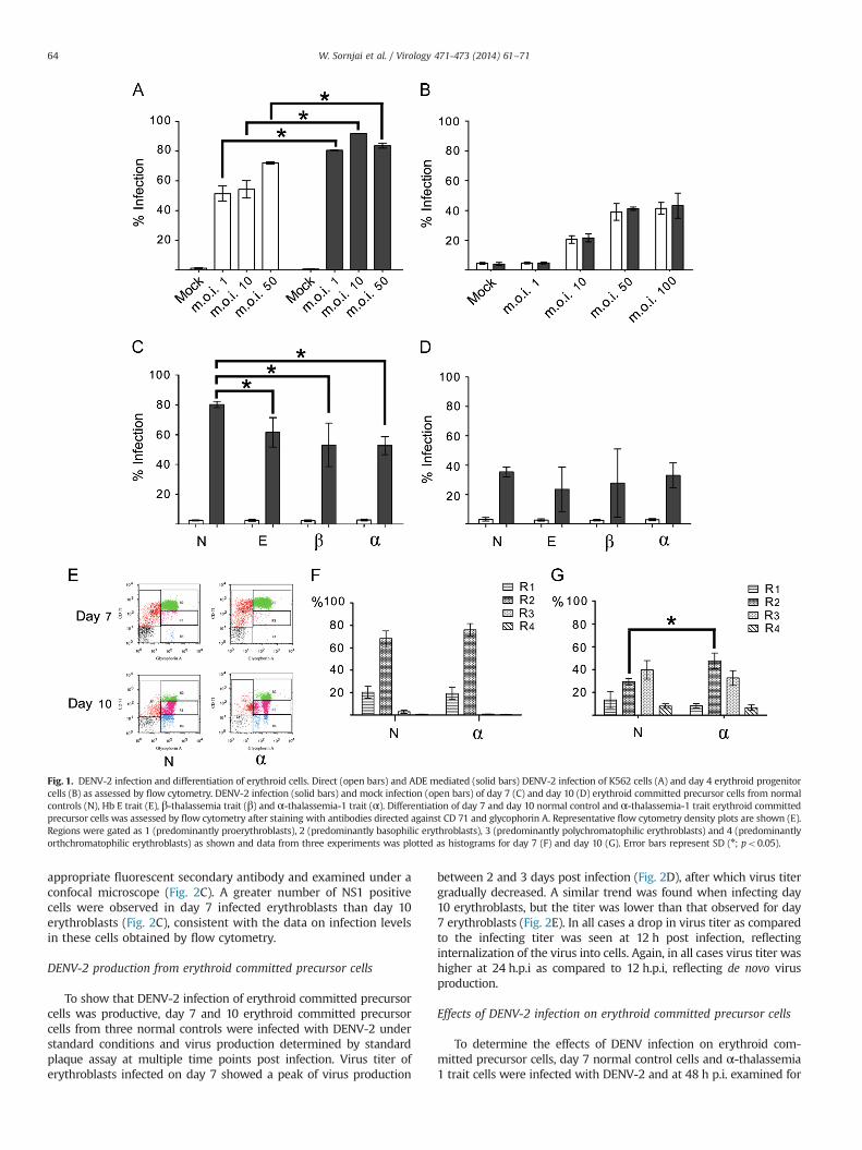
The human erythromyeloblastoid leukemia cell line K562 isfrequently used as a model for erythroid cell studies, and the cellscan give rise to erythroid cells under appropriate induction (Osti et al.,1997; Rutherford et al., 1979). We therefore initially validated thesusceptibility of this cell line to DENV infection as has been shown byothers (Goncalvez et al., 2007) under both conditions of directinfection and under conditions of ADE infection (in which entry ismediated through the Fc receptor). Cells were therefore either directlyexposed to DENV, or exposed to a DENV/antibody complex at m.o.i. of1, 10 and 50 and after 48 h the percentage of infected cells wasdetermined by flow cytometry. Results showed that K562 cells wereable to be directly infected by DENV, and that significantly increasedlevels of infection were seen when the DENV was complexed with anon-neutralizing anti-DENV E protein antibody (Fig. 1A), consistentwith the reports of others (Goncalvez et al., 2007).
We next determined the susceptibility of selected normal controlCD34þ HSCs from two donors that had been cultured for 4 days(which corresponds to the erythroid committed progenitor stage) tobe infected by DENV under conditions of direct infection and ADEinfection with infectivity determined by flow cytometry as aboveat 48 h post infection. Results (Fig. 1B) showed infection of day4 erythroid progenitor cells that increased with increasing m.o.i.between 1 and 50, and that no increase of infectivity was seenwhencells were infected under conditions of ADE, suggesting that infec-tion occurs by direct infection, and not through Fc receptor mediatedinternalization. These results are consistent with the earlier observa-tions of others (Murgue et al., 1997; Nakao et al., 1989).
DENV-2 infection of erythroid precursor cells
To investigate the susceptibility of erythroid committed pre-cursor cells, CD34þ HSCs were selected and cultured for 7 and 10days under conditions that drive erythropoiesis (Leecharoenkiatet al., 2011; Lithanatudom et al., 2010; Wannatung et al., 2009) andinfected with DENV at m.o.i. 50. At 48 h post infection (p.i.), thedegree of cell infection was determined by flow cytometry. Results(Fig. 1C and D) showed that nearly 80% of the cells infected on day7 were infected, while approximately 40% of cells cultured for 10days prior to infection were infected which is similar to the levelsseen when day 4 cells (erythroid progenitor) were infected. Weadditionally analyzed infection of erythroid committed precursorcells from thalassemia carriers to DENV-2 infection and CD34þHSCs were isolated from 3 individuals each of Hb E trait, β-thal-assemia trait and α-thalassemia 1 trait and cultured for 7 or 10days and infected with DENV-2, and samples were analyzed byflow cytometry at 48 h p.i. The results showed that erythroidcommitted precursor cells from all three traits were susceptibleto infection, and, as with control erythroblast, higher levels ofinfection were seen in cells infected at day 7 as compared to day10 (Fig. 1C and D). While no difference was seen in the levelof infection between erythroblasts infected on day 10 of cul-ture (Fig. 1D), all three traits showed significantly lower levels ofinfection (po0.05) as compared to normal control cells when day7 cells were infected (Fig. 1C), with the lowest level of infection
being seen in cells isolated from thalassemia carriers of α-thal-assemia 1. We additionally determined whether normal controlday 7 erythroid committed precursor cells were susceptible to in-fection with CHIKV using m.o.i. 0.1, 1 and 5 and the results reve-aled that these cells were not susceptible to CHIKV infection (datanot shown).
Differentiation of erythroid committed precursor cells
Culture of isolated CD34þ HSCs for 7 to 10 days results in apool of cells at different stages of differentiation (proerythroblasts,basophilic erythroblasts, polychromatic erythroblasts, orthochro-matic erythroblasts) as shown previously (Leecharoenkiat et al.,2011). To determine whether the reduced infection could resultfrom altered differentiation of erythroid committed precursor cellsfrom thalassemia carriers, the differentiation of normal controlcells was compared to cells isolated from α-thalassemia 1 carriers.Day 7 and day 10 erythroid precursor cells from three α-thal-assemia 1 carriers and three normal controls were stained withantibodies directed against CD71 and glycophorin A and analyzedby flow cytometry. Cell populations were gated to R1-R4 areasaccording to erythroid cell maturation as described by others(McGrath et al., 2008), and representative flow cytometry densityplots are shown (Fig. 1E). No difference in differentiation wasobserved between day 7 erythroblasts from normal controls andα-thalassemia 1 carriers (Fig. 1F), with the majority of cells beingproerythroblast (R1) and basophilic erythroblasts (R2). A signifi-cantly higher level of basophilic erythroblasts were seen in day 10α-thalassemia 1 trait cells as compared to normal controls(p¼ 0.013; Fig. 1G), but as no difference in infection was seen inday 10 erythroblasts, the difference is unlikely to be of significancein the infection process.
DENV-2 replication in erythroid committed precursor cells
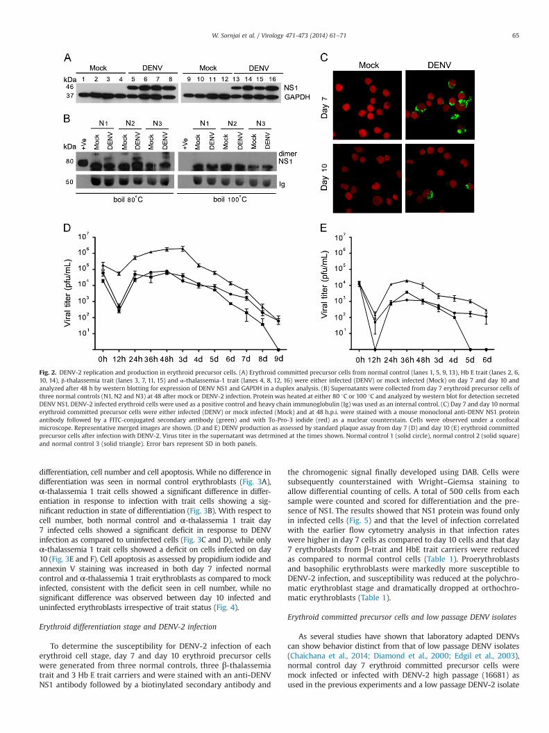
Evidence from flow cytometry is consistent with infection oferythroid committed precursor cells for both normal controls andthalassemia traits. However, the antibody used to determine thedegree of infection is directed against a structural protein (theE protein) and therefore it is possible that positive cells reflectinternalized virus without viral replication occurring. To excludethis possibility, cells were investigated for expression of denguenon-structural protein 1 (NS1) whose expression results fromtranslation of the viral genomic RNA and is absolutely requiredfor viral replication (Muller and Young, 2013). Day 7 and day 10erythroblasts from normal controls and thalassemia carriers of HbE, β-thalassemia and α-thalassemia 1 were either infected withDENV-2 or mock infected and at day 2 p.i. proteins were extractedand used in western analysis. Results (Fig. 2A) showed the clearexpression of NS1 in all DENV-2 infected cells. DENV NS1 protein isfound intracellularly and as a membrane associated and secretedhexameric form which is composed of heat labile amphipathicdimeric subunits (Gutsche et al., 2011; Pryor and Wright, 1993). Todetect secreted DENV NS1, supernatants from day 7 mock andinfected erythroblasts were separated by electrophoresis and sub-jected to western blot analysis after transfer to solid matrix support.Dimeric NS1 was observed in infected supernatants which wereheated at 80 1C and was not present in 100 1C boiled protein(Fig. 2B). Erythroblasts are cultured in medium containing humanAB serum, and thus it was not possible to observe the monomericform due to cross reaction from the secondary antibody used in thewestern blot. However, the blot was stripped and re-probed withsecondary antibody only to serve as a loading control (Ig in Fig. 2B).Moreover, expression of NS1 protein was clearly evident in DENV-2infected cells when normal infected and mock infected erythro-blasts were stained with an anti-NS1 monoclonal antibody and an
W. Sornjai et al. / Virology 471-473 (2014) 61–71 63
appropriate fluorescent secondary antibody and examined under aconfocal microscope (Fig. 2C). A greater number of NS1 positivecells were observed in day 7 infected erythroblasts than day 10erythroblasts (Fig. 2C), consistent with the data on infection levelsin these cells obtained by flow cytometry.
DENV-2 production from erythroid committed precursor cells
To show that DENV-2 infection of erythroid committed precursorcells was productive, day 7 and 10 erythroid committed precursorcells from three normal controls were infected with DENV-2 understandard conditions and virus production determined by standardplaque assay at multiple time points post infection. Virus titer oferythroblasts infected on day 7 showed a peak of virus production
between 2 and 3 days post infection (Fig. 2D), after which virus titergradually decreased. A similar trend was found when infecting day10 erythroblasts, but the titer was lower than that observed for day7 erythroblasts (Fig. 2E). In all cases a drop in virus titer as comparedto the infecting titer was seen at 12 h post infection, reflectinginternalization of the virus into cells. Again, in all cases virus titer washigher at 24 h.p.i as compared to 12 h.p.i, reflecting de novo virusproduction.
Effects of DENV-2 infection on erythroid committed precursor cells
To determine the effects of DENV infection on erythroid com-mitted precursor cells, day 7 normal control cells and α-thalassemia1 trait cells were infected with DENV-2 and at 48 h p.i. examined for
Fig. 1. DENV-2 infection and differentiation of erythroid cells. Direct (open bars) and ADE mediated (solid bars) DENV-2 infection of K562 cells (A) and day 4 erythroid progenitorcells (B) as assessed by flow cytometry. DENV-2 infection (solid bars) and mock infection (open bars) of day 7 (C) and day 10 (D) erythroid committed precursor cells from normalcontrols (N), Hb E trait (E), β-thalassemia trait (β) and α-thalassemia-1 trait (α). Differentiation of day 7 and day 10 normal control and α-thalassemia-1 trait erythroid committedprecursor cells was assessed by flow cytometry after staining with antibodies directed against CD 71 and glycophorin A. Representative flow cytometry density plots are shown (E).Regions were gated as 1 (predominantly proerythroblasts), 2 (predominantly basophilic erythroblasts), 3 (predominantly polychromatophilic erythroblasts) and 4 (predominantlyorthchromatophilic erythroblasts) as shown and data from three experiments was plotted as histograms for day 7 (F) and day 10 (G). Error bars represent SD (n; po0.05).
W. Sornjai et al. / Virology 471-473 (2014) 61–7164
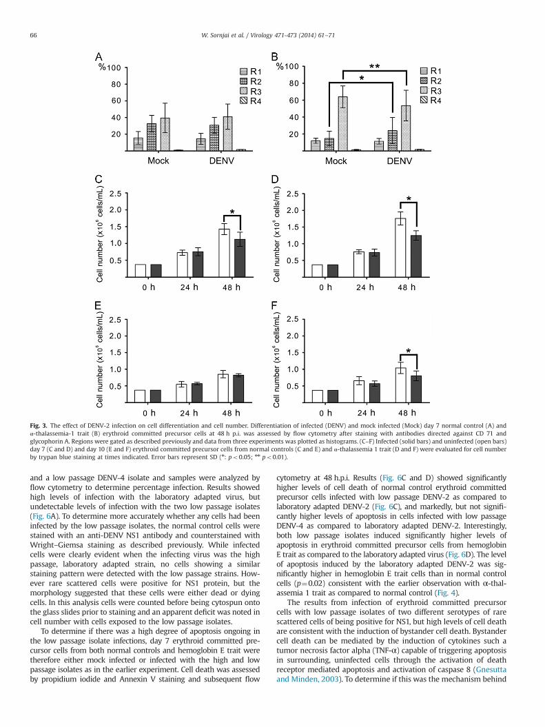
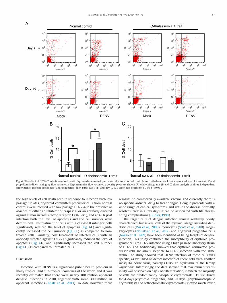
differentiation, cell number and cell apoptosis. While no difference indifferentiation was seen in normal control erythroblasts (Fig. 3A),α-thalassemia 1 trait cells showed a significant difference in differ-entiation in response to infection with trait cells showing a sig-nificant reduction in state of differentiation (Fig. 3B). With respect tocell number, both normal control and α-thalassemia 1 trait day7 infected cells showed a significant deficit in response to DENVinfection as compared to uninfected cells (Fig. 3C and D), while onlyα-thalassemia 1 trait cells showed a deficit on cells infected on day10 (Fig. 3E and F). Cell apoptosis as assessed by propidium iodide andannexin V staining was increased in both day 7 infected normalcontrol and α-thalassemia 1 trait erythroblasts as compared to mockinfected, consistent with the deficit seen in cell number, while nosignificant difference was observed between day 10 infected anduninfected erythroblasts irrespective of trait status (Fig. 4).
Erythroid differentiation stage and DENV-2 infection
To determine the susceptibility for DENV-2 infection of eacherythroid cell stage, day 7 and day 10 erythroid precursor cellswere generated from three normal controls, three β-thalassemiatrait and 3 Hb E trait carriers and were stained with an anti-DENVNS1 antibody followed by a biotinylated secondary antibody and
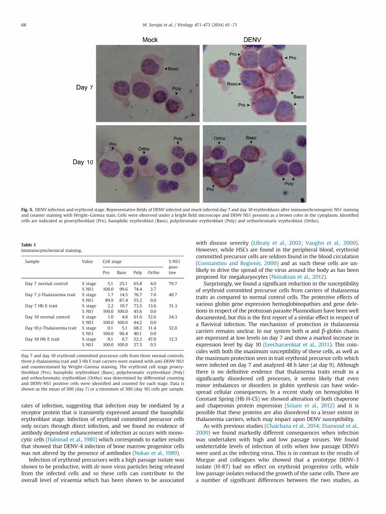
the chromogenic signal finally developed using DAB. Cells weresubsequently counterstained with Wright–Giemsa staining toallow differential counting of cells. A total of 500 cells from eachsample were counted and scored for differentiation and the pre-sence of NS1. The results showed that NS1 protein was found onlyin infected cells (Fig. 5) and that the level of infection correlatedwith the earlier flow cytometry analysis in that infection rateswere higher in day 7 cells as compared to day 10 cells and that day7 erythroblasts from β-trait and HbE trait carriers were reducedas compared to normal control cells (Table 1). Proerythroblastsand basophilic erythroblasts were markedly more susceptible toDENV-2 infection, and susceptibility was reduced at the polychro-matic erythroblast stage and dramatically dropped at orthochro-matic erythroblasts (Table 1).
Erythroid committed precursor cells and low passage DENV isolates
As several studies have shown that laboratory adapted DENVscan show behavior distinct from that of low passage DENV isolates(Chaichana et al., 2014; Diamond et al., 2000; Edgil et al., 2003),normal control day 7 erythroid committed precursor cells weremock infected or infected with DENV-2 high passage (16681) asused in the previous experiments and a low passage DENV-2 isolate
Fig. 2. DENV-2 replication and production in erythroid precursor cells. (A) Erythroid committed precursor cells from normal control (lanes 1, 5, 9, 13), Hb E trait (lanes 2, 6,10, 14), β-thalassemia trait (lanes 3, 7, 11, 15) and α-thalassemia-1 trait (lanes 4, 8, 12, 16) were either infected (DENV) or mock infected (Mock) on day 7 and day 10 andanalyzed after 48 h by western blotting for expression of DENV NS1 and GAPDH in a duplex analysis. (B) Supernatants were collected from day 7 erythroid precursor cells ofthree normal controls (N1, N2 and N3) at 48 after mock or DENV-2 infection. Protein was heated at either 80 1C or 100 1C and analyzed by western blot for detection secretedDENV NS1. DENV-2 infected erythroid cells were used as a positive control and heavy chain immunoglobulin (Ig) was used as an internal control. (C) Day 7 and day 10 normalerythroid committed precursor cells were either infected (DENV) or mock infected (Mock) and at 48 h.p.i. were stained with a mouse monoclonal anti-DENV NS1 proteinantibody followed by a FITC-conjugated secondary antibody (green) and with To-Pro-3 iodide (red) as a nuclear counterstain. Cells were observed under a confocalmicroscope. Representative merged images are shown. (D and E) DENV production as assessed by standard plaque assay from day 7 (D) and day 10 (E) erythroid committedprecursor cells after infection with DENV-2. Virus titer in the supernatant was detrmined at the times shown. Normal control 1 (solid circle), normal control 2 (solid square)and normal control 3 (solid triangle). Error bars represent SD in both panels.
W. Sornjai et al. / Virology 471-473 (2014) 61–71 65
and a low passage DENV-4 isolate and samples were analyzed byflow cytometry to determine percentage infection. Results showedhigh levels of infection with the laboratory adapted virus, butundetectable levels of infection with the two low passage isolates(Fig. 6A). To determine more accurately whether any cells had beeninfected by the low passage isolates, the normal control cells werestained with an anti-DENV NS1 antibody and counterstained withWright–Giemsa staining as described previously. While infectedcells were clearly evident when the infecting virus was the highpassage, laboratory adapted strain, no cells showing a similarstaining pattern were detected with the low passage strains. How-ever rare scattered cells were positive for NS1 protein, but themorphology suggested that these cells were either dead or dyingcells. In this analysis cells were counted before being cytospun ontothe glass slides prior to staining and an apparent deficit was noted incell number with cells exposed to the low passage isolates.
To determine if there was a high degree of apoptosis ongoing inthe low passage isolate infections, day 7 erythroid committed pre-cursor cells from both normal controls and hemoglobin E trait weretherefore either mock infected or infected with the high and lowpassage isolates as in the earlier experiment. Cell death was assessedby propidium iodide and Annexin V staining and subsequent flow
cytometry at 48 h.p.i. Results (Fig. 6C and D) showed significantlyhigher levels of cell death of normal control erythroid committedprecursor cells infected with low passage DENV-2 as compared tolaboratory adapted DENV-2 (Fig. 6C), and markedly, but not signifi-cantly higher levels of apoptosis in cells infected with low passageDENV-4 as compared to laboratory adapted DENV-2. Interestingly,both low passage isolates induced significantly higher levels ofapoptosis in erythroid committed precursor cells from hemoglobinE trait as compared to the laboratory adapted virus (Fig. 6D). The levelof apoptosis induced by the laboratory adapted DENV-2 was sig-nificantly higher in hemoglobin E trait cells than in normal controlcells (p¼0.02) consistent with the earlier observation with α-thal-assemia 1 trait as compared to normal control (Fig. 4).
The results from infection of erythroid committed precursorcells with low passage isolates of two different serotypes of rarescattered cells of being positive for NS1, but high levels of cell deathare consistent with the induction of bystander cell death. Bystandercell death can be mediated by the induction of cytokines such atumor necrosis factor alpha (TNF-α) capable of triggering apoptosisin surrounding, uninfected cells through the activation of deathreceptor mediated apoptosis and activation of caspase 8 (Gnesuttaand Minden, 2003). To determine if this was the mechanism behind
Fig. 3. The effect of DENV-2 infection on cell differentiation and cell number. Differentiation of infected (DENV) and mock infected (Mock) day 7 normal control (A) andα-thalassemia-1 trait (B) erythroid committed precursor cells at 48 h p.i. was assessed by flow cytometry after staining with antibodies directed against CD 71 andglycophorin A. Regions were gated as described previously and data from three experiments was plotted as histograms. (C–F) Infected (solid bars) and uninfected (open bars)day 7 (C and D) and day 10 (E and F) erythroid committed precursor cells from normal controls (C and E) and α-thalassemia 1 trait (D and F) were evaluated for cell numberby trypan blue staining at times indicated. Error bars represent SD (n: po0.05; nn po0.01).
W. Sornjai et al. / Virology 471-473 (2014) 61–7166
the high levels of cell death seen in response to infection with lowpassage isolates, erythroid committed precursor cells from normalcontrols were infected with low passage DENV-4 in the presence orabsence of either an inhibitor of caspase 8 or an antibody directedagainst tumor necrosis factor receptor 1 (TNF-R1), and at 48 h postinfection both the level of apoptosis and the cell number weredetermined. Pre-treatment of cells with a caspase 8 inhibitor bothsignificantly reduced the level of apoptosis (Fig. 6E) and signifi-cantly increased the cell number (Fig. 6F) as compared to non-treated cells. Similarly, post treatment of infected cells with anantibody directed against TNF-R1 significantly reduced the level ofapoptosis (Fig. 6G) and significantly increased the cell number(Fig. 6H) as compared to untreated cells.
Discussion
Infection with DENV is a significant public health problem inmany tropical and sub-tropical countries of the world and it wasrecently estimated that there were nearly 100 million apparentdengue infections in 2010, together with some 300 million inapparent infections (Bhatt et al., 2013). To date however there
remains no commercially available vaccine and currently there isno specific antiviral drug to treat dengue. Dengue presents with awide range of clinical symptoms, and while the disease normallyresolves itself in a few days, it can be associated with life threat-ening complications (Gubler, 1998).
The target cells of dengue infection remain relatively poorlycharacterized, but several cells of the myeloid lineage including den-dritic cells (Wu et al., 2000), monocytes (Scott et al., 1980), mega-karyocytes (Noisakran et al., 2012) and erythroid progenitor cells(Nakao et al., 1989) have been identified as being targets of dengueinfection. This study confirmed the susceptibility of erythroid pro-genitor cells to DENV infection using a high passage laboratory strainof DENV and additionally showed that erythroid committed pre-cursor cells are also susceptible to DENV infection with the samestrain. The study showed that DENV infection of these cells wasspecific, as we failed to detect infection of these cells with anothermosquito borne virus, namely CHIKV an Alphavirus of the familyTogaviridae. Interestingly, the data showed that maximum suscept-ibility was observed on day 7 of differentiation, in which the majorityof cells are predominantly basophilic erythroblasts. HSCs culturedfor 4 days (erythroid progenitor) and 10 days (polychromatophilicerythroblasts and orthochromatic erythroblasts) showed much lower
Fig. 4. The effect of DENV-2 infection on cell death. Erythroid committed precursor cells from normal controls and α-thalassemia 1 traits were evaluated for annexin V andpropidium iodide staining by flow cytometry. Representative flow cytometry density plots are shown (A) while histograms (B and C) show analysis of three independentexperiments. Infected (solid bars) and uninfected (open bars) day 7 (B) and day 10 (C). Error bars represent SD (n; po0.05).
W. Sornjai et al. / Virology 471-473 (2014) 61–71 67
rates of infection, suggesting that infection may be mediated by areceptor protein that is transiently expressed around the basophilicerythroblast stage. Infection of erythroid committed precursor cellsonly occurs through direct infection, and we found no evidence ofantibody dependent enhancement of infection as occurs with mono-cytic cells (Halstead et al., 1980) which corresponds to earlier resultsthat showed that DENV-4 infection of bone marrow progenitor cellswas not altered by the presence of antibodies (Nakao et al., 1989).
Infection of erythroid precursors with a high passage isolate wasshown to be productive, with de novo virus particles being releasedfrom the infected cells and so these cells can contribute to theoverall level of viraemia which has been shown to be associated
with disease severity (Libraty et al., 2002; Vaughn et al., 2000).However, while HSCs are found in the peripheral blood, erythroidcommitted precursor cells are seldom found in the blood circulation(Constantino and Bogionis, 2000) and as such these cells are un-likely to drive the spread of the virus around the body as has beenproposed for megakaryocytes (Noisakran et al., 2012).
Surprisingly, we found a significant reduction in the susceptibilityof erythroid committed precursor cells from carriers of thalassemiatraits as compared to normal control cells. The protective effects ofvarious globin gene expression hemoglobinopathies and gene dele-tions in respect of the protozoan parasite Plasmodium have beenwelldocumented, but this is the first report of a similar effect in respect ofa flaviviral infection. The mechanism of protection in thalassemiacarriers remains unclear. In our system both α and β-globin chainsare expressed at low levels on day 7 and show a marked increase inexpression level by day 10 (Leecharoenkiat et al., 2011). This coin-cides with both the maximum susceptibility of these cells, as well asthe maximum protection seen in trait erythroid precursor cells whichwere infected on day 7 and analyzed 48 h later (at day 9). Althoughthere is no definitive evidence that thalassemia traits result in asignificantly disordered cell processes, it seems likely that evenminor imbalances or disorders in globin synthesis can have wide-spread cellular consequences. In a recent study on hemoglobin HConstant Spring (Hb H-CS) we showed alteration of both chaperoneand chaperonin protein expression (Sriiam et al., 2012) and it ispossible that these proteins are also disordered to a lesser extent inthalassemia carriers, which may impact upon DENV susceptibility.
As with previous studies (Chaichana et al., 2014; Diamond et al.,2000) we found markedly different consequences when infectionwas undertaken with high and low passage viruses. We foundundetectable levels of infection of cells when low passage DENVswere used as the infecting virus. This is in contrast to the results ofMurgue and colleagues who showed that a prototype DENV-3isolate (H-87) had no effect on erythroid progenitor cells, whilelow passage isolates reduced the growth of the same cells. There area number of significant differences between the two studies, as
Fig. 5. DENV infection and erythroid stage. Representative fields of DENV infected and mock infected day 7 and day 10 erythroblasts after immunochromogenic NS1 stainingand counter staining with Wright–Giemsa stain. Cells were observed under a bright field microscope and DENV NS1 presents as a brown color in the cytoplasm. Identifiedcells are indicated as proerythroblast (Pro), basophilic erythroblast (Baso), polychromatic erythroblast (Poly) and orthochromatic erythroblast (Ortho).
Table 1Immunocytochemical staining.
Sample Value Cell stage % NS1posi-tivePro Baso Poly Ortho
Day 7 normal control % stage 5.1 25.1 65.8 4.0 79.7% NS1 100.0 99.6 74.4 3.7
Day 7 β-Thalassemia trait % stage 1.7 14.5 76.7 7.0 49.7% NS1 89.9 87.4 55.2 0.0
Day 7 Hb E trait % stage 2.2 10.7 73.5 13.6 51.3% NS1 100.0 100.0 45.6 0.0
Day 10 normal control % stage 1.0 4.8 61.6 32.6 24.3% NS1 100.0 100.0 44.2 0.0
Day 10 β-Thalassemia trait % stage 0.1 5.1 68.2 31.4 32.0% NS1 100.0 96.4 40.1 0.0
Day 10 Hb E trait % stage 0.1 0.7 52.2 47.0 12.3% NS1 100.0 100.0 27.5 0.5
Day 7 and day 10 erythroid committed precursor cells from three normal controls,three β-thalassemia trait and 3 Hb E trait carriers were stained with anti-DENV NS1and counterstained by Wright–Giemsa staining. The erythroid cell stage proery-throblast (Pro), basophilic erythroblast (Baso), polychromatic erythroblast (Poly)and orthochromatic erythroblast (Ortho) was determined by differential countingand DENV-NS1 positive cells were identified and counted for each stage. Data isshown as the mean of 500 (day 7) or a minimum of 300 (day 10) cells per sample.
W. Sornjai et al. / Virology 471-473 (2014) 61–7168
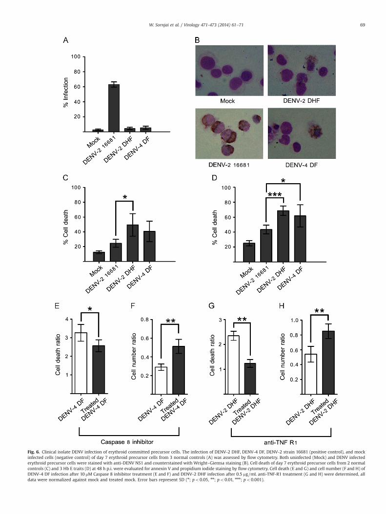
Fig. 6. Clinical isolate DENV infection of erythroid committed precursor cells. The infection of DENV-2 DHF, DENV-4 DF, DENV-2 strain 16681 (positive control), and mockinfected cells (negative control) of day 7 erythroid precursor cells from 3 normal controls (A) was assessed by flow cytometry. Both uninfected (Mock) and DENV infectederythroid precursor cells were stained with anti-DENV NS1 and counterstained with Wright–Giemsa staining (B). Cell death of day 7 erythroid precursor cells from 2 normalcontrols (C) and 3 Hb E traits (D) at 48 h p.i. were evaluated for annexin V and propidium iodide staining by flow cytometry. Cell death (E and G) and cell number (F and H) ofDENV-4 DF infection after 10 mM Caspase 8 inhibitor treatment (E and F) and DENV-2 DHF infection after 0.5 mg⧸mL anti-TNF-R1 treatment (G and H) were determined, alldata were normalized against mock and treated mock. Error bars represent SD (n; po0.05, nn; po0.01, nnn; po0.001).
W. Sornjai et al. / Virology 471-473 (2014) 61–71 69

Murgue and colleagues (Murgue et al., 1997) investigated erythroidprogenitor cells, while our study focused on erythroid committedprecursor cells, and our study period was two days post infectionwhile that of Murgue and colleagues (Murgue et al., 1997) was8 days post infection and the infecting titer was significantly lowerin the Murgue and colleagues study (Murgue et al., 1997). It is ofparticular note that the study by Murgue and colleagues reliedsolely on cell counts, and provided no direct evidence of DENVinfection of these cells (Murgue et al., 1997). While the high in-fecting titers used in this study may be of some concern, viremiastudies in dengue fever (DF) and dengue hemorrhagic fever (DHF)patients have shown median peak viremias of 10Log 9.89 and 10.27for serum of DF and DHF respectively (Tricou et al., 2011), and assuch the titers used in this study are still very far below possiblephysiological parameters.
While the low passage viruses showed only scattered cells withNS1 positive staining indicative of infection, the low passage isolatesinduced extremely high levels of cell death as compared to thelaboratory adapted virus. Our data suggests that the cell death isinduced through activation of capases 8 mediated apoptosis inbystander (uninfected) cells. In this case, although only very fewcells maybe infected, bystander apoptosis may be a significant factorin bone marrow suppression. Our results support that the bystanderapoptosis was mediated by TNF-α, and high levels of TNF-α havebeen detected in dengue patients, and levels of TNF-α are believedto be associated with disease severity (Green and Rothman, 2006;Hober et al., 1993; Kittigul et al., 2000).
Bystander apoptosis would serve to dampen infection throughremoving cells that could otherwise be productively infected addingto the viral load. It is interesting that committed erythroid precursorcells from thalassemia trait carriers both showed a reduced sus-ceptibility to the laboratory adapted virus, as well as markedlyincreased levels of cell death with low passage isolates as comparedto normal control committed erythroid precursor cells. Both ofthese observations would support that the presence of a thalasse-mia trait may protect in a small way against severe dengue disease.
Acknowledgments
This work was supported by a Research Chair Grant from theNational Science and Technology Development Agency (NSTDA),the Thailand Research Fund (Grant no. BRG5780004) and MahidolUniversity. W.S. is supported by a Thai Royal Golden JubileeResearch Scholarship, P.W. (Grant no. PHD/0101/2553) is supportedby a Thailand Graduate Institute of Science and Technology (TGIST)(Grant no. TG-22-14-55-002D) Ph.D. Scholarship and N.W. issupported by Mahidol University Post-Doctoral Scholarship (Grantno. MU-PD_2014_06).
Appendix A. Supporting information
Supplementary data associated with this article can be found inthe online version at http://dx.doi.org/10.1016/j.virol.2014.10.004.
References
Aidoo, M., Terlouw, D.J., Kolczak, M.S., McElroy, P.D., ter Kuile, F.O., Kariuki, S.,Nahlen, B.L., Lal, A.A., Udhayakumar, V., 2002. Protective effects of the sickle cellgene against malaria morbidity and mortality. Lancet 359, 1311–1312.
Allison, A.C., 1954. The distribution of the sickle-cell trait in East Africa andelsewhere, and its apparent relationship to the incidence of subtertian malaria.Trans. R. Soc. Trop. Med. Hyg. 48, 312–318.
Bhatt, S., Gething, P.W., Brady, O.J., Messina, J.P., Farlow, A.W., Moyes, C.L., Drake, J.M.,Brownstein, J.S., Hoen, A.G., Sankoh, O., Myers, M.F., George, D.B., Jaenisch, T., Wint,G.R., Simmons, C.P., Scott, T.W., Farrar, J.J., Hay, S.I., 2013. The global distributionand burden of dengue. Nature 496, 504–507.
Bierman, H.R., Nelson, E.R., 1965. Hematodepressive virus diseases of Thailand. Ann.Intern Med. 62, 867–884.
Chaichana, P., Okabayashi, T., Puiprom, O., Sasayama, M., Sasaki, T., Yamashita, A.,Ramasoota, P., Kurosu, T., Ikuta, K., 2014. Low levels of antibody-dependentenhancement in vitro using viruses and plasma from dengue patients. PLoS One9, e92173.
Clegg, J.B., Weatherall, D.J., 1999. Thalassemia and malaria: new insights into an oldproblem. Proc. Assoc. Am. Physicians 111, 278–282.
Constantino, B.T., Bogionis, B., 2000. Nucleated RBCs: significance in the PeripheralBlood Film. Lab. Med. 31, 223–229.
Diamond, M.S., Edgil, D., Roberts, T.G., Lu, B., Harris, E., 2000. Infection of humancells by dengue virus is modulated by different cell types and viral strains.J. Virol. 74, 7814–7823.
Edgil, D., Diamond, M.S., Holden, K.L., Paranjape, S.M., Harris, E., 2003. Translationefficiency determines differences in cellular infection among dengue virus type2 strains. Virology 317, 275–290.
Enevold, A., Alifrangis, M., Sanchez, J.J., Carneiro, I., Roper, C., Borsting, C., Lusingu, J.,Vestergaard, L.S., Lemnge, M.M., Morling, N., Riley, E., Drakeley, C.J., 2007.Associations between alphaþ-thalassemia and Plasmodium falciparum malar-ial infection in northeastern Tanzania. J. Infect. Dis. 196, 451–459.
Fucharoen, S., Winichagoon, P., 1992. Thalassemia in SouthEast Asia: problems andstrategy for prevention and control. Southeast Asian J. Trop Med Public Health23, 647–655.
Garcia, L.S., 2010. Malaria. Clin. Lab. Med. 30, 93–129.Gnesutta, N., Minden, A., 2003. Death receptor-induced activation of initiator
caspase 8 is antagonized by serine/threonine kinase PAK4. Mol. Cell Biol. 23,7838–7848.
Goncalvez, A.P., Engle St, R.E., Claire, M., Purcell, R.H., Lai, C.J., 2007. Monoclonalantibody-mediated enhancement of dengue virus infection in vitro and in vivoand strategies for prevention. Proc. Natl. Acad. Sci. USA 104, 9422–9427.
Green, S., Rothman, A., 2006. Immunopathological mechanisms in dengue anddengue hemorrhagic fever. Curr. Opin. Infect. Dis. 19, 429–436.
Gubler, D.J., 1998. Dengue and dengue hemorrhagic fever. Clin. Microbiol. Rev. 11,480–496.
Gunsilius, E., Gastl, G., Petzer, A.L., 2001. Hematopoietic stem cells. Biomed.Pharmacother. 55, 186–194.
Gutsche, I., Coulibaly, F., Voss, J.E., Salmon, J., d’Alayer, J., Ermonval, M., Larquet, E.,Charneau, P., Krey, T., Megret, F., Guittet, E., Rey, F.A., Flamand, M., 2011.Secreted dengue virus nonstructural protein NS1 is an atypical barrel-shapedhigh-density lipoprotein. Proc. Natl. Acad. Sci. USA 108, 8003–8008.
Halstead, S.B., Porterfield, J.S., O’Rourke, E.J., 1980. Enhancement of dengue virusinfection in monocytes by flavivirus antisera. Am. J. Trop. Med. Hyg. 29, 638–642.
Henchal, E.A., Gentry, M.K., McCown, J.M., Brandt, W.E., 1982. Dengue virus-specificand flavivirus group determinants identified with monoclonal antibodies byindirect immunofluorescence. Am. J. Trop. Med. Hyg. 31, 830–836.
Hober, D., Poli, L., Roblin, B., Gestas, P., Chungue, E., Granic, G., Imbert, P., Pecarere, J.L.,Vergez-Pascal, R., Wattre, P., et al., 1993. Serum levels of tumor necrosis factor-alpha (TNF-alpha), interleukin-6 (IL-6), and interleukin-1 beta (IL-1 beta) indengue-infected patients. Am. J. Trop. Med. Hyg. 48, 324–331.
Kittigul, L., Temprom, W., Sujirarat, D., Kittigul, C., 2000. Determination of tumornecrosis factor-alpha levels in dengue virus infected patients by sensitivebiotin-streptavidin enzyme-linked immunosorbent assay. J. Virol. Methods90, 51–57.
Klomporn, P., Panyasrivanit, M., Wikan, N., Smith, D.R., 2011. Dengue infection ofmonocytic cells activates ER stress pathways, but apoptosis is induced throughboth extrinsic and intrinsic pathways. Virology 409, 189–197.
Leecharoenkiat, A., Wannatung, T., Lithanatudom, P., Svasti, S., Fucharoen, S.,Chokchaichamnankit, D., Srisomsap, C., Smith, D.R., 2011. Increased oxidativemetabolism is associated with erythroid precursor expansion in beta0-thalas-saemia/Hb E disease. Blood Cells Mol. Dis. 47, 143–157.
Libraty, D.H., Endy, T.P., Houng, H.S., Green, S., Kalayanarooj, S., Suntayakorn, S.,Chansiriwongs, W., Vaughn, D.W., Nisalak, A., Ennis, F.A., Rothman, A.L., 2002.Differing influences of virus burden and immune activation on disease severityin secondary dengue-3 virus infections. J. Infect. Dis. 185, 1213–1221.
Lithanatudom, P., Leecharoenkiat, A., Wannatung, T., Svasti, S., Fucharoen, S., Smith,D.R., 2010. A mechanism of ineffective erythropoiesis in beta-thalassemia/Hb Edisease. Haematologica 95, 716–723.
McGrath, K.E., Bushnell, T.P., Palis, J., 2008. Multispectral imaging of hematopoieticcells: where flow meets morphology. J. Immunol. Methods 336, 91–97.
Mockenhaupt, F.P., Ehrhardt, S., Gellert, S., Otchwemah, R.N., Dietz, E., Anemana, S.D.,Bienzle, U., 2004. Alpha(þ)-thalassemia protects African children from severemalaria. Blood 104, 2003–2006.
Muller, D.A., Young, P.R., 2013. The flavivirus NS1 protein: molecular and structuralbiology, immunology, role in pathogenesis and application as a diagnosticbiomarker. Antiviral Res. 98, 192–208.
Murgue, B., Cassar, O., Guigon, M., Chungue, E., 1997. Dengue virus inhibits humanhematopoietic progenitor growth in vitro. J. Infect. Dis. 175, 1497–1501.
Nakao, S., Lai, C.J., Young, N.S., 1989. Dengue virus, a flavivirus, propagates in humanbone marrow progenitors and hematopoietic cell lines. Blood 74, 1235–1240.
Noisakran, S., Onlamoon, N., Hsiao, H.M., Clark, K.B., Villinger, F., Ansari, A.A., Perng, G.C.,2012. Infection of bone marrow cells by dengue virus in vivo. Exp. Hematol. 40,250–259 (e254).
Osti, F., Corradini, F.G., Hanau, S., Matteuzzi, M., Gambari, R., 1997. Human leukemiaK562 cells: induction to erythroid differentiation by guanine, guanosine andguanine nucleotides. Haematologica 82, 395–401.
W. Sornjai et al. / Virology 471-473 (2014) 61–7170

Panyasrivanit, M., Khakpoor, A., Wikan, N., Smith, D.R., 2009. Co-localization ofconstituents of the dengue virus translation and replication machinery withamphisomes. J. Gen. Virol. 90, 448–456.
Piel, F.B., Patil, A.P., Howes, R.E., Nyangiri, O.A., Gething, P.W., Williams, T.N.,Weatherall, D.J., Hay, S.I., 2010. Global distribution of the sickle cell gene andgeographical confirmation of the malaria hypothesis. Nat. Commun. 1, 104.
Pryor, M.J., Wright, P.J., 1993. The effects of site-directed mutagenesis on thedimerization and secretion of the NS1 protein specified by dengue virus.Virology 194, 769–780.
Rutherford, T.R., Clegg, J.B., Weatherall, D.J., 1979. K562 human leukaemic cellssynthesise embryonic haemoglobin in response to haemin. Nature 280,164–165.
Scott, R.M., Nisalak, A., Cheamudon, U., Seridhoranakul, S., Nimmannitya, S., 1980.Isolation of dengue viruses from peripheral blood leukocytes of patients withhemorrhagic fever. J. Infect. Dis. 141, 1–6.
Sithisarn, P., Suksanpaisan, L., Thepparit, C., Smith, D.R., 2003. Behavior of thedengue virus in solution. J. Med. Virol. 71, 532–539.
Srichaikul, T., Nimmannitya, S., 2000. Haematology in dengue and dengue haemor-rhagic fever. Baillieres Best Pract. Res. Clin. Haematol. 13, 261–276.
Sriiam, S., Leecharoenkiat, A., Lithanatudom, P., Wannatung, T., Svasti, S., Fucharoen, S.,Svasti, J., Chokchaichamnankit, D., Srisomsap, C., Smith, D.R., 2012. Proteomicanalysis of Hemoglobin H-Constant Spring (Hb H-CS) erythroblasts. Blood CellsMol. Dis. 48, 77–85.
Taylor, S.M., Parobek, C.M., Fairhurst, R.M., 2012. Haemoglobinopathies and theclinical epidemiology of malaria: a systematic review and meta-analysis. LancetInfect. Dis. 12, 457–468.
Tricou, V., Minh, N.N., Farrar, J., Tran, H.T., Simmons, C.P., 2011. Kinetics of viremiaand NS1 antigenemia are shaped by immune status and virus serotype in adultswith dengue. PLoS Negl. Trop. Dis. 5, e1309.
Vaughn, D.W., Green, S., Kalayanarooj, S., Innis, B.L., Nimmannitya, S., Suntayakorn, S.,Endy, T.P., Raengsakulrach, B., Rothman, A.L., Ennis, F.A., Nisalak, A., 2000. Dengueviremia titer, antibody response pattern, and virus serotype correlate with diseaseseverity. J. Infect. Dis. 181, 2–9.
Wambua, S., Mwangi, T.W., Kortok, M., Uyoga, S.M., Macharia, A.W., Mwacharo, J.K.,Weatherall, D.J., Snow, R.W., Marsh, K., Williams, T.N., 2006. The effect ofalphaþ-thalassaemia on the incidence of malaria and other diseases inchildren living on the coast of Kenya. PLoS Med. 3, e158.
Wannatung, T., Lithanatudom, P., Leecharoenkiat, A., Svasti, S., Fucharoen, S., Smith, D.R.,2009. Increased erythropoiesis of beta-thalassaemia/Hb E proerythroblasts ismediated by high basal levels of ERK1/2 activation. Br. J. Haematol. 146, 557–568.
Wikan, N., Sakoonwatanyoo, P., Ubol, S., Yoksan, S., Smith, D.R., 2012. Chikungunyavirus infection of cell lines: analysis of the East, central and South africanlineage. PLoS One 7, e31102.
Williams, T.N., Mwangi, T.W., Wambua, S., Alexander, N.D., Kortok, M., Snow, R.W.,Marsh, K., 2005. Sickle cell trait and the risk of Plasmodium falciparum malariaand other childhood diseases. J. Infect. Dis. 192, 178–186.
Wu, S.J., Grouard-Vogel, G., Sun, W., Mascola, J.R., Brachtel, E., Putvatana, R., Louder, M.K.,Filgueira, L., Marovich, M.A., Wong, H.K., Blauvelt, A., Murphy, G.S., Robb, M.L., Innes,B.L., Birx, D.L., Hayes, C.G., Frankel, S.S., 2000. Human skin Langerhans cells aretargets of dengue virus infection. Nat. Med. 6, 816–820.
W. Sornjai et al. / Virology 471-473 (2014) 61–71 71