
Defining Postpartum Uterine Disease and the Mechanisms of Infection and Immunity in the Female...
8
BIOLOGY OF REPRODUCTION 81, 1025–1032 (2009) Published online before print 13 May 2009. DOI 10.1095/biolreprod.109.077370 Minireview Defining Postpartum Uterine Disease and the Mechanisms of Infection and Immunity in the Female Reproductive Tract in Cattle 1 I. Martin Sheldon, 2,3 James Cronin, 3 Leopold Goetze, 4 Gaetano Donofrio, 5 and Hans-Joachim Schuberth 6 Institute of Life Science, 3 School of Medicine, Swansea University, Swansea, United Kingdom Pfizer Animal Health Europe, 4 Paris, France Dipartimento di Salute Animale, 5 Sezione di Malattie Infettive degli Animali, Facolta ` di Medicin a Veterina ria, Parma, Italy Immunology Unit, 6 University of Veterinary Medicine, Hannover, Germany ABSTRACT Uteri ne micro bial disease affects half of all dair y cattl e after parturition, causing infertility by disrupting uterine and ovarian function. Infe ction with Escheri chia coli , Arcanobacterium pyogenes, and bovine herpesvirus 4 causes endometrial tissue damage. Toll-like receptors on endometrial cells detect patho- gen-a ssociated molec ules such as bacterial DNA, lipids, and lipo poly sacch aride (LPS), leading to secre tion of cytoki nes, chemokines, and antimicrobial pepti des. Chemo kines attract neutrophils and macrophages to eliminate the bacteria, although persistence of neutrophils is associated with subclinical endo- metritis and infertilit y. Cows with uterine infections are less li kely to ov ul ate be cause they have sl ower gr owth of the post partu m domin ant foll icle in the ovary , lower periphe ral plasma estradiol concentrations, and perturbation of hypotha- lamic and pituitary function. The follicular fluid of animals with endometritis contains LPS, which is detected by the TLR4/CD14/ LY96 (MD2) receptor complex on granulosa cells, leading to lower aromatase expression and reduced estradiol secretion. If cows with uterine dis ease ovulate, the perip heral pl asma concentrations of progesterone are lower than those in normal animals. However, luteal phases are often extended in animals with uterine disease, probably because infection switches the endo metri al epith elial secretio n of pros taglan dins from the F series to the E series by a phospholipase A2-mediated mecha- nism, which would disrupt lut eol ysi s. The regulati on of endo metri al immun ity depen ds on stero id hormones, somat o- trophins, and local regulatory proteins. Advances in knowledge about infection and immunity in the female genital tract should be exploited to develop new therapeutics for uterine disease. bovi ne, female repr oductive tract, immunity , immunolog y, infection, inflammation, ovary, prostaglandins, toll-like receptors, uterus INTRODUCTION Mi cro bia l di sease of the femal e geni tal tr act is most common and of greatest economic importance in humans and cattle among the mammals [1, 2]. Microbial infections of the genital tract cause infertility by disrupting uterine and ovarian function. Many of the mechanisms underlying the recognition of mi cr obial path ogens by the in nate immune system in vertebrates have been identified during the past 10 years [3–5]. These mechanisms of innate immunity not only are important for classic immune cells such as neutrophils and macrophages but also are evident in the endometrial and ovarian cells of mammals [6–8]. As well as causing an immune and infl ammator y resp onse, micr obes or path ogen -ass ocia ted molecules disrupt endocrine function in the female reproduc- tive tract of rodents and cattle [6, 7, 9, 10]. Herein, we outline advanc es in scie nti fic knowle dge about how inf ection and innate immunity affect the female reproductive tract to cause infer tility in cattle. DEFINING POSTPARTUM REPRODUCTIVE TRACT DISEASES Uterine dis eas e wit hin a week of par tur iti on (me tri tis ) is present in up to 40% of dairy cows (Fig. 1). Metritis incidence depends on the definition of disease (see herein), but maximal herd rates for obvious clinical disease of 36%–50% have been reported in large surveys [16, 17], and 18.5%–21% of animals have metritis with signs of systemic illness such as pyrexia [18, 19]. Subsequen tly, 15%–20% of cattle have clinical disease that persists beyond 3 wk postpartum (endometritis), and about 30% have chronic inflammation of the uterus without clinical signs of uterine disease (subclinical endometritis) [2, 15, 20, 21]. Metritis occurs within 21 days and is most common within 10 days of parturition. Metritis is characterized by an enlarged ute rus and a wat ery red -brown fluid to vis cous off -wh ite purulent uterine discharge, which often has a fetid odor [2]. The severity of disease is categorized by the signs of health. We propose that cows are classified as having grade 1 metritis if the y have an abnormall y enl arged ute rus and a pur ule nt ute rin e dis cha rge wit hou t any sys temic sig ns of ill hea lth . Ani mals wit h add iti onal signs of systemic ill ness such as dec rease d mi lk yield, dullness, and fever .39.58C are cla ssif ied as having grade 2 cli nic al met rit is. Ani mals wit h sig ns of toxemia such as ina ppe tan ce, cold extremities, 1 Supported by a BBSRC Research Development Fellowship to I.M.S. (Grant No. BB/D02028X/1). J.C. is funded throu gh a DEFRA LINK award by Pfizer Animal Health and BBSRC (Grant No. F005121). 2 Corresponden ce: I. Martin Sheldon, Institute of Life Science, School of Medi cine , Swansea Univ ersi ty , Sing elton Park, Swansea, SA2 8PP , United Kingdom. FAX: 44 1792 602280; e-mail: [email protected] .uk Received: 6 March 2009. First decision: 8 April 2009. Accepted: 30 April 2009. Ó 2009 by the Society for the Study of Reproduction, Inc. eISSN: 1529-7268 http://www .biolrepro d.org ISSN: 0006-3363 1025
-
Upload
sempre-seu-you -
Category
Documents
-
view
221 -
download
0
Transcript of Defining Postpartum Uterine Disease and the Mechanisms of Infection and Immunity in the Female...

8/6/2019 Defining Postpartum Uterine Disease and the Mechanisms of Infection and Immunity in the Female Reproductive Tr…
http://slidepdf.com/reader/full/defining-postpartum-uterine-disease-and-the-mechanisms-of-infection-and-immunity 1/8
BIOLOGY OF REPRODUCTION 81, 1025–1032 (2009)Published online before print 13 May 2009.DOI 10.1095/biolreprod.109.077370
M i n i r e v i e w
Defining Postpartum Uterine Disease and the Mechanisms of Infection and Immunityin the Female Reproductive Tract in Cattle1
I. Martin Sheldon,2,3 James Cronin,3 Leopold Goetze,4 Gaetano Donofrio,5 and Hans-Joachim Schuberth6
Institute of Life Science,3 School of Medicine, Swansea University, Swansea, United KingdomPfizer Animal Health Europe,4 Paris, FranceDipartimento di Salute Animale,5 Sezione di Malattie Infettive degli Animali, Facolta di Medicina Veterinaria, Parma, Italy Immunology Unit,6 University of Veterinary Medicine, Hannover, Germany
ABSTRACT
Uterine microbial disease affects half of all dairy cattle afterparturition, causing infertility by disrupting uterine and ovarianfunction. Infection with Escherichia coli , Arcanobacterium
pyogenes, and bovine herpesvirus 4 causes endometrial tissuedamage. Toll-like receptors on endometrial cells detect patho-gen-associated molecules such as bacterial DNA, lipids, andlipopolysaccharide (LPS), leading to secretion of cytokines,chemokines, and antimicrobial peptides. Chemokines attractneutrophils and macrophages to eliminate the bacteria, althoughpersistence of neutrophils is associated with subclinical endo-metritis and infertility. Cows with uterine infections are lesslikely to ovulate because they have slower growth of thepostpartum dominant follicle in the ovary, lower peripheralplasma estradiol concentrations, and perturbation of hypotha-lamic and pituitary function. The follicular fluid of animals withendometritis contains LPS, which is detected by the TLR4/CD14/LY96 (MD2) receptor complex on granulosa cells, leading tolower aromatase expression and reduced estradiol secretion. If
cows with uterine disease ovulate, the peripheral plasmaconcentrations of progesterone are lower than those in normalanimals. However, luteal phases are often extended in animalswith uterine disease, probably because infection switches theendometrial epithelial secretion of prostaglandins from the Fseries to the E series by a phospholipase A2-mediated mecha-nism, which would disrupt luteolysis. The regulation of endometrial immunity depends on steroid hormones, somato-trophins, and local regulatory proteins. Advances in knowledgeabout infection and immunity in the female genital tract shouldbe exploited to develop new therapeutics for uterine disease.
bovine, female reproductive tract, immunity, immunology,infection, inflammation, ovary, prostaglandins, toll-like receptors,uterus
INTRODUCTION
Microbial disease of the female genital tract is mocommon and of greatest economic importance in humans an
cattle among the mammals [1, 2]. Microbial infections of thgenital tract cause infertility by disrupting uterine and ovariafunction. Many of the mechanisms underlying the recognitioof microbial pathogens by the innate immune system ivertebrates have been identified during the past 10 years [3–5These mechanisms of innate immunity not only are importanfor classic immune cells such as neutrophils and macrophagebut also are evident in the endometrial and ovarian cells omammals [6–8]. As well as causing an immune aninflammatory response, microbes or pathogen-associatemolecules disrupt endocrine function in the female reproductive tract of rodents and cattle [6, 7, 9, 10]. Herein, we outlinadvances in scientific knowledge about how infection aninnate immunity affect the female reproductive tract to caus
infertility in cattle.
DEFINING POSTPARTUM REPRODUCTIVETRACT DISEASES
Uterine disease within a week of parturition (metritis) ipresent in up to 40% of dairy cows (Fig. 1). Metritis incidencdepends on the definition of disease (see herein), but maximaherd rates for obvious clinical disease of 36%–50% have beereported in large surveys [16, 17], and 18.5%–21% of animalhave metritis with signs of systemic illness such as pyrexia [1819]. Subsequently, 15%–20% of cattle have clinical diseasthat persists beyond 3 wk postpartum (endometritis), and abou30% have chronic inflammation of the uterus without clinicasigns of uterine disease (subclinical endometritis) [2, 15, 20
21].Metritis occurs within 21 days and is most common withi
10 days of parturition. Metritis is characterized by an enlargeuterus and a watery red-brown fluid to viscous off-whitpurulent uterine discharge, which often has a fetid odor [2The severity of disease is categorized by the signs of healthWe propose that cows are classified as having grade 1 metritiif they have an abnormally enlarged uterus and a purulenuterine discharge without any systemic signs of ill healthAnimals with additional signs of systemic illness such adecreased milk yield, dullness, and fever .39.58C arclassified as having grade 2 clinical metritis. Animals witsigns of toxemia such as inappetance, cold extremitie
1Supported by a BBSRC Research Development Fellowship to I.M.S.(Grant No. BB/D02028X/1). J.C. is funded through a DEFRA LINKaward by Pfizer Animal Health and BBSRC (Grant No. F005121).2Correspondence: I. Martin Sheldon, Institute of Life Science, School of Medicine, Swansea University, Singelton Park, Swansea, SA2 8PP,United Kingdom. FAX: 44 1792 602280;e-mail: [email protected]
Received: 6 March 2009.First decision: 8 April 2009.Accepted: 30 April 2009.Ó 2009 by the Society for the Study of Reproduction, Inc.eISSN: 1529-7268 http://www.biolreprod.orgISSN: 0006-3363
1025

8/6/2019 Defining Postpartum Uterine Disease and the Mechanisms of Infection and Immunity in the Female Reproductive Tr…
http://slidepdf.com/reader/full/defining-postpartum-uterine-disease-and-the-mechanisms-of-infection-and-immunity 2/8
depression, and/or collapse are classified as having grade 3metritis, which has a poor prognosis.
Clinical endometritis is defined in cattle as the presence of a purulent uterine discharge detectable in the vagina 21 days or more postpartum or mucopurulent discharge detectable in thevagina after 26 days postpartum [2]. A simple grading systembased on the character of the vaginal mucus (Fig. 2A) is readilyused to evaluate cows with clinical endometritis [2]. Theendometritis grade correlates with the presence of pathogenicorganisms associated with uterine disease (Fig. 2B) and isprognostic for the likely outcome of treatment (Fig. 2C) [11,22].
Subclinical endometritis is characterized by inflammation of
the endometrium that results in a significant reduction inreproductive performance in the absence of signs of clinicalendometritis. The inflammation is presumably associated withrecovery of the tissues after clinical endometritis, trauma, or other nonmicrobial disease. Subclinical disease is defined bypolymorphonuclear neutrophils (PMNs) exceeding between5.5% of cells [23] and 10% of cells [24] in samples collectedby flushing the uterine lumen or by endometrial cytobrush, inthe absence of clinical endometritis, about 5 wk postpartum.The incidence of subclinical endometritis is dependent on thecutoff for diagnosis and the time after parturition but is in theorder of 37%–74% of animals (Fig. 1) [15].
REPRODUCTIVE AND ECONOMIC CONSEQUENCES
OF UTERINE DISEASEThe placenta should be expelled within a few hours of
parturition in cattle. During the first week postpartum, theuterus contracts rapidly, and lochia is discharged containingremnants of fetal membranes and fluids. During the second tofourth weeks, any damaged endometrial tissue regenerates, a wave of ovarian follicles develop, a dominant follicle isselected, and estradiol secretion leads to ovulation andformation of a corpus luteum to recommence ovarian cycles[25]. The genital tract should have little evidence of theprevious pregnancy by 6 wk after calving and be capable of establishing the next pregnancy. However, about 50% of dairycows have irregular ovarian cycles during the postpartum
period, and animals with abnormal vaginal discharge are morelikely than normal animals to have delayed resumption of ovarian cycles after calving (anovulatory anestrus [odds ratio,4.5]) or prolonged postpartum luteal phases (odds ratio, 4.4)[26]. Conception rates are about 20% lower for cows withendometritis, the median calving to conception interval is 30days longer, and there are 3% more animals culled for failure toconceive [20, 21]. Cows are less fertile even after successfultreatment of clinical endometritis than age-matched counter-
parts in the same herds that had no clinical uterine diseasepostpartum [20]; this is probably because subclinical endome-tritis persists after the clinical signs have resolved. Animalswith subclinical disease also have more days open, take longer to conceive, and have conception rates about half those of normal animals [24].
The financial effect of uterine disease is derived frominfertility, increased culling for failure to conceive, reducedmilk production, and the cost of treatment. The economic cost of a single case of metritis has been calculated to be about E292 [18]. Studies report 24146000 dairy cows in theEuropean Union [27] and 8 495 000 dairy cows in the UnitedStates [28]. Using a conservative incidence rate of 20% for metritis [18, 19], we calculate that the annual cost of uterinedisease in the European Union is E1.4 billion and in the UnitedStates is $650 million. The costs of endometritis are anadditional burden on the dairy industry and need to bequantified.
PATHOGENESIS OF POSTPARTUM REPRODUCTIVETRACT DISEASE
During pregnancy, the uterus is sterile, but after parturitionthe uterine lumen is almost always contaminated with a widerange of bacteria (Fig. 1). However, development of clinicaldisease is dependent on the balance between host immunityand the pathogenicity of the bacteria. This balance can betipped in favor of disease by risk factors such as retainedplacenta, dystocia, twins, and stillbirth [29, 30]. Unfortunately,these factors are not particularly amenable to intervention toreduce the incidence of disease, and the factors that could beaddressed (such as the cleanliness of the animal or environ-ment) are less important [31].
Bacterial Infection
Escherichia coli and Arcanobacterium pyogenes are themost prevalent bacteria isolated from the uterine lumen of cattle with uterine disease, followed by a range of anaerobicbacteria such as Prevotella species, Fusobacterium necropho-rum, and Fusobacterium nucleatum [10–14]. Bacteria are alsoisolated from the uterus of animals that do not develop clinicaldisease. Indeed, the presence of coagulase-negative staphylo-cocci and a-haemolytic streptococci decreases the risk of
endometritis [11], so probiotics may be considered in the futurefor prevention of disease. Infection of the uterus with E. coliappears to pave the way for subsequent infection with other bacteria or viruses [32–34]. Furthermore, E. coli infectionduring the first days or week after parturition is associated withnegative effects on the ovary, hypothalamic-pituitary axis, andgeneral health, as well as uterine disease [32]. However, themost severe endometrial lesions are caused by A. pyogenes[14]. The strains of A. pyogenes isolated from the uterus allexpress the virulence gene plo [35], which encodes a cholesterol-dependent cytotoxin called pyolysin [36]. Choles-terol-dependent cytotoxin molecules are attracted to cholester-ol-rich domains in cell membranes, where they aggregate toform a pore, leading to osmotic death of the cell [36], and
FIG. 1. The incidence of uterine bacterial infection and disease inpostpartum dairy cattle. Bacteria can be isolated from the uterus of mostcows during the postpartum period; each marker (circle) indicates thepercentage of animals with bacteria isolated from the uterine lumen [10–14]. The shaded areas represent estimates of the proportion of animalswith metritis (red), clinical endometritis (orange), or a normal uterus(blue); the remainder of animals have subclinical endometritis [15].
1026 SHELDON ET AL.

8/6/2019 Defining Postpartum Uterine Disease and the Mechanisms of Infection and Immunity in the Female Reproductive Tr…
http://slidepdf.com/reader/full/defining-postpartum-uterine-disease-and-the-mechanisms-of-infection-and-immunity 3/8
pyolysin readily kills endometrial epithelial and stromal cells ivitro [37]. Furthermore, A. pyogenes, F. necrophorum, an Prevotella species act synergistically to enhance the likelihooand severity of uterine disease [38, 39]. Fusobacteriunecrophorum produces a leukotoxin, Prevotella melaninogenicus produces a substance that inhibits phagocytosis, and A pyogenes produces a growth factor for F. necrophorumPresumably, the necrotic lochia associated with retaineplacenta provides an excellent media for bacteria. Trauma t
the tissues during parturition also likely facilitates adhesion aninvasion of microbes. Finally, suppressed or dysregulateimmune mechanisms around the time of parturition (discusseherein) probably also perturb host defense against microbes.
Viral Infection
Bovine herpesvirus 4 (BoHV-4) is the only virus consistently associated with uterine disease after parturition in cattl[40, 41]. Like other herpesviruses, BoHV-4 can establish lateninfections in cattle, particularly in macrophages [42], and thviral infection is often identified concurrent with bacteria thacause uterine disease [43, 44]. So, the association betweeBoHV-4 infection and uterine disease has been hard testablish, although the contribution of BoHV-4 to uterindisease in which the virus is endemic in cattle will becomclear when a vaccine is developed. The virus is highly tropifor endometrial cells, rapidly replicating and killing epitheliaor stromal cells [42, 45]. BoHV-4 replication is driven by hocellular factors transactivating the viral immediate early (IE2also known as UL122) gene promoter. A luciferase reporter fothe UL122 promoter was transactivated in a concentrationdependent manner when transfected bovine stromal cells wertreated with prostaglandin E
2(PGE), E. coli, or its lipopoly
saccharide (LPS [endotoxin]), and PGE and LPS actecooperatively [34]. Furthermore, viral replication was reactvated in latently infected macrophages when cocultured witstromal cells [45]. We suggest that there may be a vicious circlcomposed of bacterial endometritis, leading to secretion o
PGE and then stimulation of viral replication by PGE and LPSwhich causes further endometrial tissue damage and inflammation (Fig. 3). Identifying the specific host cellular transcription factors that transactivate the BoHV-4 UL122 gene to drivviral replication will inform strategies to prevent herpesvirugenital tract disease in cattle and other species.
Uterine Immunity
Mammalian pregnancy involves regulation of uterinimmunity to facilitate implantation and survival of thsemiallogeneic fetus. The classic view is that immunity isuppressed during gestation, although evidence is emerginthat some immune and inflammatory mechanisms are alscritical for implantation in mammals [51]. If the immunosup
pressive mechanisms associated with pregnancy persist in thendometrium after parturition, they would likely predispose tuterine disease. So, some of the roles of the uterine immunsystem during pregnancy may be at odds with the need trespond in a coordinated way to pathogenic organisms in thuterus after parturition.
During mid and late pregnancy, lymphocytes and macrophages are found in the intercaruncular endometrium, althoug
FIG. 2. Grading scheme for clinical endometritis. A) Vaginal mucuscharacter is graded as 0 (clear or translucent mucus), 1 (mucus containingflecks of white or off-white pus), 2 (exudate containing,50% white or off-white mucopurulent material), or 3 (exudate containing !50% purulentmaterial, usually white or yellow but occasionally sanguineous) [2]. B)Endometritis grades reflect the number of pathogenic (black bars) but notopportunist nonpathogenic (white bars) bacteria isolated from the uterusof cattle [11]; data are presented as semiquantitative scores of the numberof colony-forming units (CFU) from uterine swabs, where CFU is scored as0 (no growth), 1 (,10 CFUs), 2 (10–100 CFUs), 3 (101–500 CFUs), or 4(.500 CFUs). Values differ from endometritis grade 0, ** P , 0.01 and***P , 0.001. C) Endometritis grade is prognostic for treatment success[22]; treatment success rates were determined as the percentage of
3
animals (n ¼ 300) with normal vaginal mucus 2 wk after initiendometritis grading and treatment 21–28 days postpartum. Values diffebetween endometritis grades, *P , 0.05 and **P , 0.01.
POSTPARTUM GENITAL TRACT INFECTION AND IMMUNITY 102
8/6/2019 Defining Postpartum Uterine Disease and the Mechanisms of Infection and Immunity in the Female Reproductive Tr…
http://slidepdf.com/reader/full/defining-postpartum-uterine-disease-and-the-mechanisms-of-infection-and-immunity 4/8
not in the caruncular endometrium of cattle [52–54]. Thesubepithelial uterine stroma contains more CD4þ T cells, Bcells, CD14þ macrophages, and mast cells compared with other regions of the endometrium and the myometrium [55, 56].Mast cells have a prominent sensor and effector functionduring bacterial infections in mammals [57], but their role inresponse to intrauterine bacterial contamination in cattle is not clear. Specialized uterine natural killer cells are important for normal pregnancy in many mammals, but these cells are not common at the end of gestation [58] and are sparse in thebovine endometrium [59]. Whether these immune cells are still
present in the tissues after labor in cattle and whether theyregulate the inflammatory process after pathogen contact arelargely unknown.
Innate Immunity in the Endometrium
and Pathogen Recognition
The initial defense of the mammalian endometrium against microbes is dependent on innate immune systems, includingtoll-like receptors (TLRs), antimicrobial peptides (AMPs), andacute-phase proteins [60]. Bacteria are detected by pattern
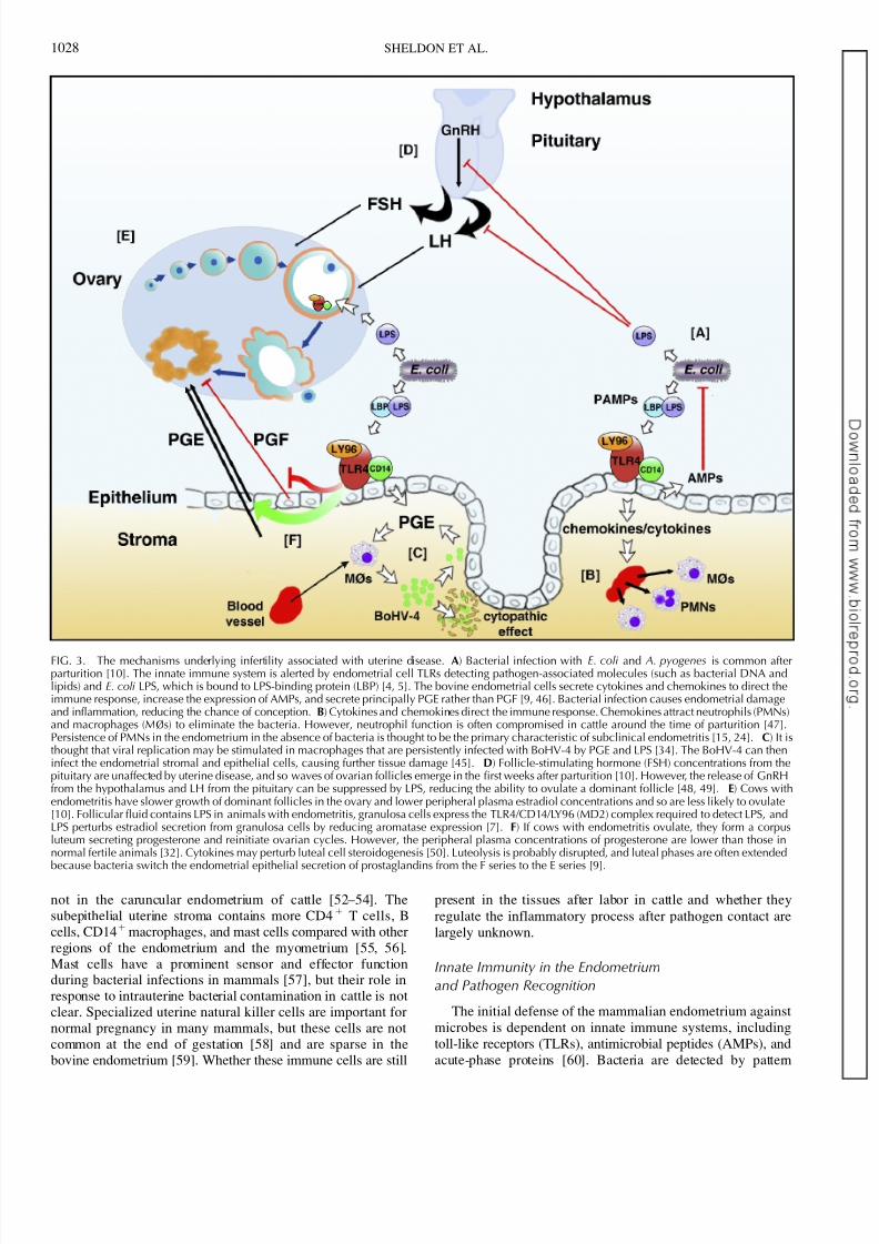
FIG. 3. The mechanisms underlying infertility associated with uterine disease. A) Bacterial infection with E. coli and A. pyogenes is common afterparturition [10]. The innate immune system is alerted by endometrial cell TLRs detecting pathogen-associated molecules (such as bacterial DNA andlipids) and E. coli LPS, which is bound to LPS-binding protein (LBP) [4, 5]. The bovine endometrial cells secrete cytokines and chemokines to direct theimmune response, increase the expression of AMPs, and secrete principally PGE rather than PGF [9, 46]. Bacterial infection causes endometrial damageand inflammation, reducing the chance of conception. B) Cytokines and chemokines direct the immune response. Chemokines attract neutrophils (PMNs)and macrophages (MØs) to eliminate the bacteria. However, neutrophil function is often compromised in cattle around the time of parturition [47].Persistence of PMNs in the endometrium in the absence of bacteria is thought to be the primary characteristic of subclinical endometritis [15, 24]. C) It isthought that viral replication may be stimulated in macrophages that are persistently infected with BoHV-4 by PGE and LPS [34]. The BoHV-4 can theninfect the endometrial stromal and epithelial cells, causing further tissue damage [45]. D) Follicle-stimulating hormone (FSH) concentrations from thepituitary are unaffected by uterine disease, and so waves of ovarian follicles emerge in the first weeks after parturition [10]. However, the release of GnRHfrom the hypothalamus and LH from the pituitary can be suppressed by LPS, reducing the ability to ovulate a dominant follicle [48, 49]. E) Cows withendometritis have slower growth of dominant follicles in the ovary and lower peripheral plasma estradiol concentrations and so are less likely to ovulate[10]. Follicular fluid contains LPS in animals with endometritis, granulosa cells express the TLR4/CD14/LY96 (MD2) complex required to detect LPS, andLPS perturbs estradiol secretion from granulosa cells by reducing aromatase expression [7]. F) If cows with endometritis ovulate, they form a corpusluteum secreting progesterone and reinitiate ovarian cycles. However, the peripheral plasma concentrations of progesterone are lower than those innormal fertile animals [32]. Cytokines may perturb luteal cell steroidogenesis [50]. Luteolysis is probably disrupted, and luteal phases are often extended
because bacteria switch the endometrial epithelial secretion of prostaglandins from the F series to the E series [9].
1028 SHELDON ET AL.

8/6/2019 Defining Postpartum Uterine Disease and the Mechanisms of Infection and Immunity in the Female Reproductive Tr…
http://slidepdf.com/reader/full/defining-postpartum-uterine-disease-and-the-mechanisms-of-infection-and-immunity 5/8
recognition receptors on mammalian cells binding moleculesspecific to microbial organisms, often called pathogen-associated molecular patterns (PAMPs) [3–5]. The most important group of such receptors comprises the TLRs, and10 members of the receptor family are widely encoded in themammalian genome and are most often found in a broad rangeof immune cells [3, 4]. TLR1, TLR2, and TLR6 recognizebacterial lipids such as lipoteichoic acid, whereas TLR3, TLR7,TLR8, and TLR9 recognize nucleic acids, often from viruses.
Lipopolysaccharide from Gram-negative bacteria such as E.coli is bound to LPS-binding protein and is recognized byTLR4 in complex with CD14 and LY96 (MD2), TLR5 bindsflagellin, and TLR9 also recognizes bacterial DNA. Activationof TLRs initiates signalling cascades, resulting in the synthesisand production of proinflammatory cytokines and chemokinesthat mobilize and activate immune cells [4, 5], which in thecase of bovine uterine disease is particularly associated with theinflux of PMNs into the uterus [61].
Whole endometrium from normal nonpregnant cattleexpresses TLR1 through TLR10 [46]. Before and after parturition, TLR2, TLR3, TLR4, TLR6, and TLR9 areexpressed in the caruncular and intercaruncular endometrium,and TLR expression was greater in the caruncular endometrium
than in the intercaruncular endometrium 4–6 h postpartum[62]. Purified populations of epithelial cells express TLR1through TLR7 and TLR9, and stromal cells express TLR1through TLR4, TLR6, TLR7, TLR9, and TLR10 [46]. TheseTLRs appear to be functional, as epithelial cells secreted PGEin response to bacterial PAMPs. Pure populations of epithelialor stromal cells (not contaminated with leukocytes, asdetermined by the lack of expression of the protein tyrosinephosphatase, receptor type, C [PTPRC, formerly CD45]panleukocyte marker) express the specific receptor complexcomprising TLR4/CD14/LY96 (MD2) to bind LPS [6, 9].Heat-killed E. coli or LPS provokes an inflammatory responseby the endometrial cells, characterized by the increasedexpression of transcripts for tumor necrosis factor, nitric oxidesynthase, and prostaglandin-endoperoxide synthase 2 (PTGS2,
formerly COX-2) and the secretion of prostaglandins F2a
(PGF) and PGE [6]. Heat-killed E. coli, LPS, A. pyogenespyolysin, BoHV-4, bacterial DNA, and lipids also influenceendometrial cell prostaglandin secretion, particularly stimulat-ing the secretion of PGE rather than PGF in cattle [9, 45, 46,63]. This may explain why animals with uterine infection havehigher concentrations than normal animals of LPS and PGE inthe uterine lumen and peripheral plasma [32, 64]. Endometrialexplants and epithelial and stromal cells also secretedpredominantly PGE in response to LPS, and this effect wasnot reversed by oxytocin [9]. This LPS-induced PGE secretionby endometrial cells is important for fertility becauseprostaglandins have multiple roles in endometrial function,and luteolysis is initiated by PGF from oxytocin-stimulated
epithelial cells [65]. In addition, PGE has an important role inthe mammalian immune response, acting through prostaglandinE receptors 2 and 4 (PTGER2 and PTGER4) to controlinflammation [66]. The bovine endometrial cells express thePTGER2 and PTGER4 necessary to respond to PGE [9, 67].The endometrial prostaglandin switch induced by LPS appearsto be early in the prostaglandin synthetic pathway. Arach-adonic acid is liberated from cell membranes by phospholipaseA2 group IV and group VI enzymes (PLA2G4 and PLA2G6)and is converted to prostaglandin H and then PGE or PGF bysynthase enzymes [68]. Treatment of endometrial cells withLPS stimulated increased levels of PLA2G6 but not PLA24Cprotein in epithelial cells but did not change the levels of PGEor PGF synthase enzymes [9].
The AMPs are an ancient component of the immune systemand the defensins family is particularly important for mucosaimmunity [69]. Bovine uterine tissue expresses lingual AM(LAP), tracheal AMP (TAP), bovine neutrophil b-defensin(BNBD4 and DEFB5), and bovine b-defensins (BBD19BBD123, and BBD124) [70]. Furthermore, pure populationof endometrial epithelial cells express LAP, TAP, BNBD4, anDEFB5, and expression was increased when cells were treatewith LPS [46]. Mucin 1 (MUC1) is an epithelial ce
glycosylated transmembrane protein that may also have a rolin microbial defense of the endometrium in mammals [71MUC1 is expressed by epithelial cells of the bovinendometrium, and expression was increased when the cellwere treated with LPS [46]. Acute-phase proteins are producein the liver in response to proinflammatory cytokines, anperipheral plasma concentrations are increased during the firsfew weeks postpartum in cattle [72]. However, no acute-phasproteins were detected in bovine endometrial cells in vitro [46]
Effector Cell Immigration into the Uterusafter Pathogen Contact
Blood-derived PMNs are the main effector cells fo
removing bacteria from the uterus after calving. Howeveendocrine and metabolic changes around the time of parturitioin cattle modulate PMN phagocytic function and genexpression [47, 73]. Furthermore, blood PMNs obtained fromcows with endometritis were significantly less phagocytic [74The process of transmigration into the uterine lumen alsmodulates PMN function. For example, interleukin 8-induceattraction of PMNs into the uterine lumen increased thgeneration of reactive oxygen species by these cells [61However, when PMNs are in the uterine lumen, their functiois further modulated by soluble factors in lochial secretionsWhereas lochial secretions of healthy cows only moderatelaffected the function of PMNs, the secretions of infected cowseverely depressed the generation of reactive oxygen specie[75].
Regulation of Uterine Immunity
Changes in hormone concentrations around the time oparturition may influence the risk of peripartal infections [76Progesterone and estrogen have immunomodulatory propertiechanging the repertoire and expression density of hormonreceptors in immune cells from cattle [77]. In additionestradiol and especially progesterone reduce the secretion oprostaglandins by epithelial or stromal cells stimulated witLPS [6]. The somatotropic axis also influences the course othe bovine puerperium, mediated by changes in plasma anendometrial levels of insulin-like growth factor 1 (IGF1) [7879]. Indeed, IGF1 has immunomodulatory properties iaddition to its growth-promoting function in mammals [80Finally, there are several proteins found in the endometriumthat could influence the immune response directly or affect thsteroid or IGF1 pathways in endometrial cells. The uterinserpins are progesterone-induced members of the serpisuperfamily of serine proteinase inhibitors and, at least in thsheep, inhibit lymphocyte proliferation to mediate the immunosuppressive effects of progesterone on uterine immunfunction [81]. A family of glycan-binding proteins, thgalectins, may also regulate uterine immunity by interactinwith multiple galactose-b1,4- N -acetylglucosamine units on cesurface glycoproteins [82, 83]. Lectin, galactoside-bindingsoluble, 1 (galectin 1 [LGALS1]) controls mammalian ceproliferation, the survival of effector T cells and neutrophils
POSTPARTUM GENITAL TRACT INFECTION AND IMMUNITY 102

8/6/2019 Defining Postpartum Uterine Disease and the Mechanisms of Infection and Immunity in the Female Reproductive Tr…
http://slidepdf.com/reader/full/defining-postpartum-uterine-disease-and-the-mechanisms-of-infection-and-immunity 6/8
and their extravasation in vivo [83–85]. One of the counter-players of galectin 1 is lectin, galactoside-binding, soluble, 3(galectin 3 [LGALS3]), which modulates the adhesion of Tcells to endothelial cells and the adhesion between T cells anddendritic cells or macrophages [86]. LGALS1 is expressed inthe murine and human female reproductive tracts, as well as byimmune cells [87, 88]. In humans, LGALS1 expression isstrongly enhanced in late-phase endometrium and in thedecidua [87], and LGALS1 is differentially expressed between
normal and pathologically altered placentas [89, 90]. In cattle,LGALS3 is detected in the ovary, oviduct, uterus, and cervixand is postulated to be involved in mucosal defense [91].However, the role of galectins in postpartum uterine diseaserequires further exploration.
UTERINE INFECTION AND OVARIAN FUNCTION
Cows with postpartum uterine infection had slower growthof the first postpartum dominant follicle and lower peripheralplasma estradiol concentrations around the time of maximalfollicle diameter, and in those animals that did ovulate,peripheral plasma progesterone concentrations were lower 5– 7 days after ovulation (,2 vs. .5 ng/ml) [10, 32]. Theseeffects of uterine microbes on ovarian function could be causedby PAMPs or inflammatory mediators acting on the hypothal-amus, pituitary, or ovary.
Hypothalamic and pituitary function is critical for directingovarian cycles. Follicle-stimulating hormone concentrations arenot affected in animals with uterine disease, so follicle wavesemerge in diseased animals as in normal animals [10].However, LPS suppresses hypothalamic release of gonadotro-pin-releasing hormone (GnRH), pituitary secretion of luteiniz-ing hormone (LH), and the sensitivity of the pituitary to GnRHin sheep [49, 92]. The consequences of these changes would bethat animals are less likely to ovulate, and this appears to be thecase in cattle administered LPS [48]. However, intrauterineinfusion of a lower concentration of LPS in cattle did not disrupt LH secretion [93].
The follicular fluid of cattle with uterine inflammation alsocontains LPS [7]. Animals with clinical disease had concen-trations of LPS that ranged up to 0.8 lg/ml; normal animals didnot have measurable concentrations of LPS in their ovarianfollicular fluid, while animals with subclinical disease hadintermediate concentrations about 40–60 days after calving.Theca cells convert cholesterol to androstenedione, which thenpasses across the basement membrane of the ovarian follicleand is converted to estradiol by the granulosa cells. Treatment of bovine theca cells from any stage of follicle development with LPS did not affect androstenedione production or cellsurvival, but granulosa cells collected from growing or dominant follicles secreted less estradiol when treated withLPS [7]. As with endometrial cells, LPS does not affect theca cell or granulosa cell survival. The effect of LPS on bovinegranulosa cells appears to be a direct one, as the granulosa cellcultures were free of contaminating leukocytes [7], and thegranulosa cell compartment within the basement membrane of the ovarian follicle is devoid of immune cells in vivo, at least inmice [94]. Furthermore, granulosa cells from cattle express theTLR4/CD14/LY96 (MD2) complex required for binding LPS[7]. Aromatase transcript expression was reduced by LPStreatment of granulosa cells collected from dominant follicles[7]. So, granulosa cells have a mechanism for direct action of LPS in the ovarian follicle to impair ovarian function andovulation. The effect of uterine disease on follicular functionmay be further enhanced by cytokines released by theendometrial cells because granulosa cell steroidogenesis is
also impaired by proinflammatory cytokines [95]. If animalsovulate, the cytokines secreted by the infected endometriummay also partly explain the reduced progesterone secretionfrom the corpus luteum because bovine luteal cells are highlyresponsive to a range of cytokines and cytokines are alsoimportant in luteolysis [50, 96].
The extended luteal phases in some cows with uterinedisease could be associated with effects on luteolysis or onluteal cell function. Certainly, the switch in endometrial
prostaglandin to PGE from PGF could disrupt the luteolyticmechanism [9]. In ruminants, PGE is luteotropic, while PGF isluteolytic [65]. Using endometrial explants, the ratio of PGE:PGF concentration was 0.45 in response to oxytocinand 2.75 following LPS treatment [9]. Furthermore, adminis-tering oxytocin after treatment of endometrial cells with LPSdid not reverse the propensity to secrete PGE [9].
CONCLUSIONS
In conclusion, uterine infections are common after parturi-tion in dairy cattle, causing infertility. The working model that links the mechanisms of infection and immunity with infertilityis summarized in Figure 3. Bacterial infection with E. coliprecedes infection with other microbes that disrupt endometrialstructure and function. The innate immune system is alerted tothe presence of pathogens by endometrial cell TLRs detectingpathogen-associated molecules such as LPS, DNA, andbacterial lipids. The endometrial cells secrete cytokines andchemokines to direct the immune response and increase theexpression of AMPs. Chemokines attract PMNs and macro-phages to eliminate the bacteria, although neutrophil function isoften perturbed in postpartum dairy cows. Persistence of PMNsin the endometrium in the absence of bacteria is thought to bethe primary characteristic of subclinical endometritis. Uterinedisease also affects ovarian function. Cows with uterinebacterial infections have slower growth of dominant folliclesin the ovary and lower peripheral plasma estradiol concentra-tions and so are less likely to ovulate. The release of GnRH
from the hypothalamus and LH from the pituitary can also besuppressed by LPS, further reducing the ability to ovulate a dominant follicle. Follicular fluid contains LPS in animals withendometritis, granulosa cells express the TLR4/CD14/LY96(MD2) complex required to detect LPS, and LPS reducesestradiol secretion. If cows with uterine infections ovulate, theperipheral plasma concentrations of progesterone are lower than those in normal fertile animals, and luteal phases are oftenextended. Luteolysis is probably disrupted because bacteria switch the endometrial epithelial secretion of prostaglandinsfrom the F series to the E series. The regulation of endometrialimmunity depends on steroid hormones, somatotrophins, andpossibly local regulatory proteins such as galectins. Advancesin knowledge about infection and immunity in the female
genital tract should be exploited to develop new treatments andprevention strategies for uterine disease.
REFERENCES
1. Ross JDC. An update on pelvic inflammatory disease. Sex Transm Infect 2002; 78:18–19.
2. Sheldon IM, Lewis GS, LeBlanc S, Gilbert RO. Defining postpartumuterine disease in cattle. Theriogenology 2006; 65:1516–1530.
3. O’Neill LA. The interleukin-1 receptor/Toll-like receptor superfamily: 10years of progress. Immunol Rev 2008; 226:10–18.
4. Akira S, Uematsu S, Takeuchi O. Pathogen recognition and innateimmunity. Cell 2006; 124:783–801.
5. Beutler B. Inferences, questions and possibilities in Toll-like receptor signalling. Nature 2004; 430:257–263.
6. Herath S, Fischer DP, Werling D, Williams EJ, Lilly ST, Dobson H,
1030 SHELDON ET AL.

8/6/2019 Defining Postpartum Uterine Disease and the Mechanisms of Infection and Immunity in the Female Reproductive Tr…
http://slidepdf.com/reader/full/defining-postpartum-uterine-disease-and-the-mechanisms-of-infection-and-immunity 7/8
Bryant CE, Sheldon IM. Expression and function of Toll-like receptor 4 inthe endometrial cells of the uterus. Endocrinology 2006; 147:562–570.
7. Herath S, Williams EJ, Lilly ST, Gilbert RO, Dobson H, Bryant CE,Sheldon IM. Ovarian follicular cells have innate immune capabilities that modulate their endocrine function. Reproduction 2007; 134:683–693.
8. Hernandez-Gonzalez I, Gonzalez-Robayna I, Shimada M, Wayne CM,Ochsner SA, White L, Richards JS. Gene expression profiles of cumuluscell oocyte complexes during ovulation reveal cumulus cells expressneuronal and immune-related genes: does this expand their role in theovulation process? Mol Endocrinol 2006; 20:1300–1321.
9. Herath S, Lilly ST, Fischer DP, Williams EJ, Dobson H, Bryant CE,
Sheldon IM. Bacterial lipopolysaccharide induces an endocrine switchfrom prostaglandin F
2a to prostaglandin E
2in bovine endometrium.
Endocrinology 2009; 150:1912–1920.10. Sheldon IM, Noakes DE, Rycroft AN, Pfeiffer DU, Dobson H. Influence
of uterine bacterial contamination after parturition on ovarian dominant follicle selection and follicle growth and function in cattle. Reproduction2002; 123:837–845.
11. Williams EJ, Fischer DP, England GCW, Dobson H, Pfeiffer DU, SheldonIM. Clinical evaluation of postpartum vaginal mucus reflects uterinebacterial infection and the inflammatory response to endometritis in cattle.Theriogenology 2005; 63:102–117.
12. Griffin JFT, Hartigan PJ, Nunn WR. Non-specific uterine infection andbovine fertility, I: infection patterns and endometritis during the first sevenweeks post-partum. Theriogenology 1974; 1:91–106.
13. Elliot L, McMahon KJ, Gier HT, Marion GB. Uterus of the cow after parturition: bacterial content. Am J Vet Res 1968; 29:77–81.
14. Bonnett BN, Martin SW, Gannon VP, Miller RB, Etherington WG.
Endometrial biopsy in Holstein-Friesian dairy cows, III: bacteriologicalanalysis and correlations with histological findings. Can J Vet Res 1991;55:168–173.
15. Gilbert RO, Shin ST, Guard CL, Erb HN, Frajblat M. Prevalence of endometritis and its effects on reproductive performance of dairy cows.Theriogenology 2005; 64:1879–1888.
16. Markusfeld O. Periparturient traits in seven high dairy herds: incidencerates, association with parity, and interrelationships among traits. J DairySci 1987; 70:158–166.
17. Zwald NR, Weigel KA, Chang YM, Welper RD, Clay JS. Geneticselection for health traits using producer-recorded data, I: incidence rates,heritability estimates, and sire breeding values. J Dairy Sci 2004; 87:4287– 4294.
18. Drillich M, Beetz O, Pfutzner A, Sabin M, Sabin HJ, Kutzer P,Nattermann H, Heuwieser W. Evaluation of a systemic antibiotic treatment of toxic puerperal metritis in dairy cows. J Dairy Sci 2001; 84:2010–2017.
19. Benzaquen ME, Risco CA, Archbald LF, Melendez P, Thatcher MJ,Thatcher WW. Rectal temperature, calving-related factors, and theincidence of puerperal metritis in postpartum dairy cows. J Dairy Sci2007; 90:2804–2814.
20. Borsberry S, Dobson H. Periparturient diseases and their effect onreproductive performance in five dairy herds. Vet Rec 1989; 124:217–219.
21. LeBlanc SJ, Duffield TF, Leslie KE, Bateman KG, Keefe GP, Walton JS,Johnson WH. Defining and diagnosing postpartum clinical endometritisand its impact on reproductive performance in dairy cows. J Dairy Sci2002; 85:2223–2236.
22. Sheldon IM, Noakes DE. Comparison of three treatments for bovineendometritis. Vet Rec 1998; 142:575–579.
23. Santos NR, Lamb GC, Brown DR, Gilbert RO. Postpartum endometrialcytology in beef cows. Theriogenology 2009; 71:739–745.
24. Kasimanickam R, Duffield TF, Foster RA, Gartley CJ, Leslie KE, WaltonJS, Johnson WH. Endometrial cytology and ultrasonography for thedetection of subclinical endometritis in postpartum dairy cows. Therio-genology 2004; 62:9–23.
25. Beam SW, Butler WR. Energy balance and ovarian follicle development
prior to the first ovulation postpartum in dairy cows receiving three levelsof dietary fat. Biol Reprod 1997; 56:133–142.
26. Opsomer G, Grohn YT, Hertl J, Coryn M, Deluyker H, de Kruif A. Riskfactors for post partum ovarian dysfunction in high producing dairy cowsin Belgium: a field study. Theriogenology 2000; 53:841–857.
27. Ataide Dias R, Mahon G, Dore G.EU cattle population in December 2007and production forecasts for 2008. Eurostat 2008. World Wide Web (URL:http://www.eds-destatis.de/de/downloads/sif/sf_08_049.pdf) . (April2009).
28. United States Department of Agriculture Economic Research Service. U.S.dairy situation at a glance USDAERS 2009; 2009. World Wide Web (URL:http://www.ers.usda.gov/publications/ldp/xlstables/DairyGLANCE.xls).(April 2009).
29. Kim IH, Kang HG. Risk factors for postpartum endometritis and the effect of endometritis on reproductive performance in dairy cows in Korea. JReprod Dev 2003; 49:485–491.
30. Grohn YT, Rajala-Schultz PJ. Epidemiology of reproductive performanin dairy cows. Anim Reprod Sci 2000; 60–61:605–614.
31. Noakes DE, Wallace L, Smith GR. Bacterial flora of the uterus of cowafter calving on two hygienically contrasting farms. Vet Rec 1991; 12440–442.
32. Williams EJ, Fischer DP, Noakes DE, England GC, Rycroft A, Dobson HSheldon IM. The relationship between uterine pathogen growth densitand ovarian function in the postpartum dairy cow. Theriogenology 20068:549–559.
33. Dohmen MJ, Joop K, Sturk A, Bols PE, Lohuis JA. Relationship betweeintra-uterine bacterial contamination, endotoxin levels and the develop
ment of endometritis in postpartum cows with dystocia or retaineplacenta. Theriogenology 2000; 54:1019–1032.
34. Donofrio G, Ravaneti L, Cavirani S, Herath S, Capocefalo A, Sheldon IMBacterial infection of endometrial stromal cells influences bovinherpesvirus 4 immediate early gene activation: a new insight into bacteriand viral interaction for uterine disease. Reproduction 2008; 136:361–366
35. Silva E, Gaivao M, Leitao S, Jost BH, Carneiro C, Vilela CL, Lopes dCosta L, Mateus L. Genomic characterization of Arcanobacteriu
pyogenes isolates recovered from the uterus of dairy cows with normpuerperium or clinical metritis. Vet Microbiol 2008; 132:111–118.
36. Jost BH, Billington SJ. Arcanobacterium pyogenes: molecular pathogenesis of an animal opportunist. Antonie Van Leeuwenhoek 2005; 88:87102.
37. Miller ANA.The effect of Arcanobacterium pyogenes in the bovine uteruLondon: University of London; 2009. PhD Thesis.
38. Olson JD, Ball L, Mortimer RG, Farin PW, Adney WS, Huffman EMAspects of bacteriology and endocrinology of cows with pyometra an
retained fetal membranes. Am J Vet Res 1984; 45:2251–2255.39. Ruder CA, Sasser RG, Williams RJ, Ely JK, Bull RC, Butler JE. Uterininfections in the postpartum cow, II: possible synergistic effect o
Fusobacterium necrophorum and Corynebacterium pyogenes. Theriognology 1981; 15:573–580.
40. Thiry E, Bublot M, Dubuisson J, Van Bressem MF, Lequarre ALomonte P, Vanderplasschen A, Pastoret PP. Molecular biology of bovinherpesvirus type 4. Vet Microbiol 1992; 33:79–92.
41. Ackermann M. Pathogenesis of gammaherpesvirus infections. VMicrobiol 2006; 113:211–222.
42. Donofrio G, van Santen VL. A bovine macrophage cell line supporbovine herpesvirus-4 persistent infection. J Gen Virol 2001; 82:11811185.
43. Monge A, Elvira L, Gonzalez JV, Astiz S, Wellenberg GJ. Bovinherpesvirus 4-associated postpartum metritis in a Spanish dairy herd. ReVet Sci 2006; 80:120–125.
44. Frazier K, Pence M, Mauel MJ, Liggett A, Hines ME II, Sangster LLehmkuhl HD, Miller D, Styer E, West J, Baldwin CA. Endometritis postparturient cattle associated with bovine herpesvirus-4 infection: 1cases. J Vet Diagn Invest 2001; 13:502–508.
45. Donofrio G, Herath S, Sartori C, Cavirani S, Flammini CF, Sheldon IMBovine herpesvirus 4 (BoHV-4) is tropic for bovine endometrial cells anmodulates endocrine function. Reproduction 2007; 134:183–197.
46. Davies D, Meade KG, Herath S, Eckersall PD, Gonzalez D, White JOConlan RS, O’Farrelly C, Sheldon IM. Toll-like receptor and antimicrobipeptide expression in the bovine endometrium. Reprod Biol Endocrino2008; 6:e53.
47. Burvenich C, Bannerman DD, Lippolis JD, Peelman L, Nonnecke BKehrli ME Jr, Paape MJ. Cumulative physiological events influence thinflammatory response of the bovine udder to Escherichia coli infectionduring the transition period. J Dairy Sci 2007; 90(suppl 1):E39–E54.
48. Peter AT, Bosu WTK, DeDecker RJ. Suppression of preovulatoluteinizing hormone surges in heifers after intrauterine infusions o
Escherichia coli endotoxin. Am J Vet Res 1989; 50:368–373.49. Karsch FJ, Battaglia DF, Breen KM, Debus N, Harris TG. Mechanisms f
ovarian cycle disruption by immune/inflammatory stress. Stress 2002; 5101–112.
50. Petroff MG, Petroff BK, Pate JL. Mechanisms of cytokine-induced deaof cultured bovine luteal cells. Reproduction 2001; 121:753–760.
51. Mor G. Inflammation and pregnancy: the role of toll-like receptors itrophoblast-immune interaction. Ann N Y Acad Sci 2008; 1127:121–12
52. Gogolin-Ewens KJ, Lee CS, Mercer WR, Brandon MR. Site-directedifferences in the immune response to the fetus. Immunology 1989; 6312–317.
53. Low BG, Hansen PJ, Drost M, Gogolin-Ewens KJ. Expression of majhistocompatibility complex antigens on the bovine placenta. J ReproFertil 1990; 90:235–243.
54. Wooding FB. Current topic: the synepitheliochorial placenta of ruminantbinucleate cell fusions and hormone production. Placenta 1992; 13:101113.
55. Leung ST, Derecka K, Mann GE, Flint AP, Wathes DC. Uterin
POSTPARTUM GENITAL TRACT INFECTION AND IMMUNITY 103

8/6/2019 Defining Postpartum Uterine Disease and the Mechanisms of Infection and Immunity in the Female Reproductive Tr…
http://slidepdf.com/reader/full/defining-postpartum-uterine-disease-and-the-mechanisms-of-infection-and-immunity 8/8
lymphocyte distribution and interleukin expression during early pregnancyin cows. J Reprod Fertil 2000; 119:25–33.
56. Kuther K, Audige L, Kube P, Welle M. Bovine mast cells: distribution,density, heterogeneity, and influence of fixation techniques. Cell TissueRes 1998; 293:111–119.
57. Marshall JS, Jawdat DM. Mast cells in innate immunity. J Allergy ClinImmunol 2004; 114:21–27.
58. Zhang J, Croy BA, Tian Z. Uterine natural killer cells: their choices, their missions. Cell Mol Immunol 2005; 2:123–129.
59. Maley SW, Buxton D, Macaldowie CN, Anderson IE, Wright SE, BartleyPM, Esteban-Redondo I, Hamilton CM, Storset AK, Innes EA.
Characterization of the immune response in the placenta of cattleexperimentally infected with Neospora caninum in early gestation. JComp Pathol 2006; 135:130–141.
60. Wira CR, Fahey JV. The innate immune system: gatekeeper to the femalereproductive tract. Immunology 2004; 111:13–15.
61. Zerbe H, Schuberth HJ, Engelke F, Frank J, Klug E, Leibold W.Development and comparison of in vivo and in vitro models for endometritis in cows and mares. Theriogenology 2003; 60:209–223.
62. Ritter N. Peripartal expression of endometrial Toll-like receptors and b-defensins in cattle. Hannover: University of Veterinary Medicine; 2007.PhD Thesis.
63. Miller AN, Williams EJ, Sibley K, Herath S, Lane EA, Fishwick J, NashDM, Rycroft AN, Dobson H, Bryant CE, Sheldon IM. The effects of
Arcanobacterium pyogenes on endometrial function in vitro, and onuterine and ovarian function in vivo. Theriogenology 2007; 68:972–980.
64. Mateus L, Lopes da Costa L, Diniz P, Ziecik AJ. Relationship betweenendotoxin and prostaglandin (PGE2 and PGFM) concentrations and
ovarian function in dairy cows with puerperal endometritis. Anim ReprodSci 2003; 76:143–154.65. Poyser NL. The control of prostaglandin production by the endometrium
in relation to luteolysis and menstruation. Prostaglandins Leukot Essent Fatty Acids 1995; 53:147–195.
66. Sugimoto Y, Narumiya S. Prostaglandin E receptors. J Biol Chem 2007;282:11613–11617.
67. Arosh JA, Banu SK, Chapdelaine P, Emond V, Kim JJ, MacLaren LA,Fortier MA. Molecular cloning and characterization of bovine prostaglan-din E2 receptors EP2 and EP4: expression and regulation in endometriumand myometrium during the estrous cycle and early pregnancy.Endocrinology 2003; 144:3076–3091.
68. Tithof PK, Roberts MP, Guan W, Elgayyar M, Godkin JD. Distinct phospholipase A2 enzymes regulate prostaglandin E2 and F2alpha production by bovine endometrial epithelial cells. Reprod Biol Endocrinol2007; 5:e16.
69. Selsted ME, Ouellette AJ. Mammalian defensins in the antimicrobialimmune response. Nat Immunol 2005; 6:551–557.
70. Cormican P, Meade KG, Cahalane S, Narciandi F, Chapwanya A, LloydAT, O’Farrelly C. Evolution, expression and effectiveness in a cluster of novel bovine beta-defensins. Immunogenetics 2008; 60:147–156.
71. Brayman M, Thathiah A, Carson DD. MUC1: a multifunctional cellsurface component of reproductive tissue epithelia. Reprod BiolEndocrinol 2004; 2:1–9.
72. Sheldon IM, Noakes DE, Rycroft A, Dobson H. Acute phase proteinresponse to postpartum uterine bacterial contamination in cattle. Vet Rec2001; 148:172–175.
73. Madsen SA, Weber PS, Burton JL. Altered expression of cellular genes inneutrophils of periparturient dairy cows. Vet Immunol Immunopathol2002; 86:159–175.
74. Kim IH, Na KJ, Yang MP. Immune responses during the peripartumperiod in dairy cows with postpartum endometritis. J Reprod Dev 2005;51:757–764.
75. Zerbe H, Ossadnik C, Leibold W, Schuberth HJ. Lochial secretions of Escherichia coli- or Arcanobacterium pyogenes-infected bovine uteri
modulate the phenotype and the functional capacity of neutrophilicgranulocytes. Theriogenology 2002; 57:1161–1177.76. Lewis GS. Steroidal regulation of uterine resistance to bacterial infection
in livestock. Reprod Biol Endocrinol 2003; 1:e117.
77. Lamote I, Meyer E, De Ketelaere A, Duchateau L, Burvenich C.Expression of the estrogen receptor in blood neutrophils of dairy cowsduring the periparturient period. Theriogenology 2006; 65:1082–1098.
78. Kawashima C, Fukihara S, Maeda M, Kaneko E, Montoya CA, Matsui M,Shimizu T, Matsunaga N, Kida K, Miyake Y, Schams D, Miyamoto A.Relationship between metabolic hormones and ovulation of dominant follicle during the first follicular wave post-partum in high-producingdairy cows. Reproduction 2007; 133:155–163.
79. Llewellyn S, Fitzpatrick R, Kenny DA, Patton J, Wathes DC. Endometrialexpression of the insulin-like growth factor system during uterineinvolution in the postpartum dairy cow. Domest Anim Endocrinol 2008;
34:391–402.80. Clark R. The somatogenic hormones and insulin-like growth factor-1:
stimulators of lymphopoiesis and immune function. Endocr Rev 1997; 18:157–179.
81. Hansen PJ. Regulation of immune cells in the uterus during pregnancy inruminants. J Anim Sci 2007; 85:E30–E31.
82. Rabinovich GA, Liu FT, Hirashima M, Anderson A. An emerging role for galectins in tuning the immune response: lessons from experimentalmodels of inflammatory disease, autoimmunity and cancer. Scand JImmunol 2007; 66:143–158.
83. Stillman BN, Hsu DK, Pang M, Brewer CF, Johnson P, Liu FT, Baum LG.Galectin-3 and galectin-1 bind distinct cell surface glycoprotein receptorsto induce T cell death. J Immunol 2006; 176:778–789.
84. Stowell SR, Karmakar S, Stowell CJ, Dias-Baruffi M, McEver RP,Cummings RD. Human galectin-1,À2, andÀ4 induce surface exposure of phosphatidylserine in activated human neutrophils but not in activated Tcells. Blood 2007; 109:219–227.
85. La M, Cao TV, Cerchiaro G, Chilton K, Hirabayashi J, Kasai K, OlianiSM, Chernajovsky Y, Perretti M. A novel biological activity for galectin-1: inhibition of leukocyte-endothelial cell interactions in experimentalinflammation. Am J Pathol 2003; 163:1505–1515.
86. Swarte VV, Mebius RE, Joziasse DH, Van den Eijnden DH, Kraal G.Lymphocyte triggering via L-selectin leads to enhanced galectin-3-mediated binding to dendritic cells. Eur J Immunol 1998; 28:2864–2871.
87. von Wolff M, Wang X, Gabius HJ, Strowitzki T. Galectin fingerprinting inhuman endometrium and decidua during the menstrual cycle and in earlygestation. Mol Hum Reprod 2005; 11:189–194.
88. Phillips B, Knisley K, Weitlauf KD, Dorsett J, Lee V, Weitlauf H.Differential expression of two beta-galactoside-binding lectins in thereproductive tracts of pregnant mice. Biol Reprod 1996; 55:548–558.
89. Maquoi E, van den Brule FA, Castronovo V, Foidart JM. Changes in thedistribution pattern of galectin-1 and galectin-3 in human placenta correlates with the differentiation pathways of trophoblasts. Placenta 1997; 18:433–439.
90. Bozic M, Petronijevic M, Milenkovic S, Atanackovic J, Lazic J, Vicovac
L. Galectin-1 and galectin-3 in the trophoblast of the gestationaltrophoblastic disease. Placenta 2004; 25:797–802.
91. Kim M, Kim S, Kim H, Joo HG, Shin T. Immunohistochemicallocalization of galectin-3 in the reproductive organs of the cow. Acta Histochemica 2008; 110:473–480.
92. Battaglia DF, Krasa HB, Padmanabhan V, Viguie C, Karsch FJ. Endocrinealterations that underlie endotoxin-induced disruption of the follicular phase in ewes. Biol Reprod 2000; 62:45–53.
93. Williams EJ, Sibley K, Miller ANA, Lane EA, Fishwick J, Nash DL,Herath S, England GC, Dobson H, Sheldon IM. The effect of Escherichiacoli lipopolysaccharide and tumour necrosis factor alpha on ovarianfunction. Am J Reprod Immunol 2008; 60:462–473.
94. Petrovska M, Dimitrov DG, Michael SD. Quantitative changes inmacrophage distribution in normal mouse ovary over the course of theestrous cycle examined with an image analysis system. Am J ReprodImmunol 1996; 36:175–183.
95. Spicer LJ, Alpizar E. Effects of cytokines on FSH-induced estradiol
production by bovine granulosa cells in vitro: dependence on size of follicle. Domest Anim Endocrinol 1994; 11:25–34.96. Okuda K, Sakumoto R. Multiple roles of TNF super family members in
corpus luteum function. Reprod Biol Endocrinol 2003; 1:e95.
1032 SHELDON ET AL.


