Vascular Malformations as a Cause of Postpartum Hemorrhage · 26 Vascular Malformations as a Cause...
7
26 Vascular Malformations as a Cause of Postpartum Hemorrhage K. Hayes INTRODUCTION Uterine vascular malformations (UVM) leading to postpartum hemorrhage (PPH) are rare. The world- wide literature consists of case reports and small case series, and while the true incidence is unknown, it is likely to represent a very small proportion of causes of PPH. UVM can be congenital but are more com- monly acquired when they tend to present with sec- ondary PPH, although in rare instances they present with primary or even tertiary bleeding 1 . As a general rule, primary PPH occurring temporally close to delivery most likely is a result of a pre-existing arterio- venous malformation (AVM), whereas secondary or tertiary PPH is more likely due to an acquired pseudoaneurysm. Secondary PPH occurs in up to 0.5–1.5% of preg- nancies 2 and the vast majority of women who present with secondary or tertiary PPH will have endometritis with or without retained products of conception (RPOC). Their management usually consists of broad spectrum antibiotics and, if necessary, an evacuation of RPOC. The vast majority of these women stop bleed- ing thereafter. Because some women receive multiple courses of antibiotics and evacuation procedures with- out evidence of infection or RPOC, clinicians finally start to consider vascular malformations rather than persevering with repeated ineffective treatments. VASCULAR MALFORMATIONS Vascular malformations may be congenital or acquired and consist of true AVMs, arteriovenous fistulae and pseudoaneurysms. Congenital arteriovenous malformations Congenital AVMs are abnormal arteriovenous con- nections that can occur anywhere in the body. These are rare as primary uterine lesions and no true estimate of their incidence is possible. The majority are usually found in the head and neck, and a 10 year review in a tertiary referral vascular center found only one uterine case out of 145 AVMs 3 . They result from abnormal development of primitive vessels that form connec- tions between pelvic arteries and veins in the uterus 4 . They are characterized by several feeding and draining vessels with an interconnecting nidus with turbulent flow 5 . When they occur in the uterus they have the potential to cause obstetric and/or gynecological bleeding. The majority of significant bleeds occur as a result of either iatrogenic intervention (uterine instru- mentation) or placental implantation involving the AVM 6 . AVMs are so rare that they are only likely to contribute truly to less than 1% of PPH. Moreover, the diagnosis is usually only made in a hysterectomy specimen or by interventional radiology on the basis of an arteriogram when bleeding is intractable and the patient remains stable enough for possible embolization. Due to their rarity, the diagnosis of an AVM is usually retrospective if it is made at all. From the clini- cal point of view, the PPH is managed as any other PPH. Not surprisingly, however, many conservative measures fail to work, as it is likely that the abnormal vascular compartment of the AVM has been opened. Acquired vascular malformations Acquired vascular malformations invariably result from iatrogenic or traumatic injury to the uterine artery vascular bed 7 . This is particularly so when sig- nificant intractable secondary PPH occurs and where uterine artery pseudoaneurysm needs to be consid- ered. A recent review of case reports and series of pseudoaneurysms 1 found 16 cases of which 10 had recently undergone cesarean section, three uterine evacuation procedures, and three had had normal spontaneous vaginal deliveries (of these, two had had previous gynecological surgery). A pseudoaneurysm is characterized by a complete lack of vascular layers (intima, media and adventitia) surrounding the blood collection which communi- cates with the parent artery through the injury. The boundary of the ‘aneurysm’ is in fact the surrounding connective tissue. In terms of etiology, cesarean section at advanced dilatation with uterine angle extension is the commonest antecedent event, where direct trauma and suturing around the uterine artery bed cause abnormal vascular connections. Failure to 218
Transcript of Vascular Malformations as a Cause of Postpartum Hemorrhage · 26 Vascular Malformations as a Cause...

26Vascular Malformations as a Cause ofPostpartum HemorrhageK. Hayes
INTRODUCTION
Uterine vascular malformations (UVM) leading topostpartum hemorrhage (PPH) are rare. The world-wide literature consists of case reports and small caseseries, and while the true incidence is unknown, it islikely to represent a very small proportion of causes ofPPH. UVM can be congenital but are more com-monly acquired when they tend to present with sec-ondary PPH, although in rare instances they presentwith primary or even tertiary bleeding1. As a generalrule, primary PPH occurring temporally close todelivery most likely is a result of a pre-existing arterio-venous malformation (AVM), whereas secondary ortertiary PPH is more likely due to an acquiredpseudoaneurysm.
Secondary PPH occurs in up to 0.5–1.5% of preg-nancies2 and the vast majority of women who presentwith secondary or tertiary PPH will have endometritiswith or without retained products of conception(RPOC). Their management usually consists of broadspectrum antibiotics and, if necessary, an evacuation ofRPOC. The vast majority of these women stop bleed-ing thereafter. Because some women receive multiplecourses of antibiotics and evacuation procedures with-out evidence of infection or RPOC, clinicians finallystart to consider vascular malformations rather thanpersevering with repeated ineffective treatments.
VASCULAR MALFORMATIONS
Vascular malformations may be congenital or acquiredand consist of true AVMs, arteriovenous fistulae andpseudoaneurysms.
Congenital arteriovenous malformations
Congenital AVMs are abnormal arteriovenous con-nections that can occur anywhere in the body. Theseare rare as primary uterine lesions and no true estimateof their incidence is possible. The majority are usuallyfound in the head and neck, and a 10 year review in atertiary referral vascular center found only one uterinecase out of 145 AVMs3. They result from abnormaldevelopment of primitive vessels that form connec-tions between pelvic arteries and veins in the uterus4.
They are characterized by several feeding and drainingvessels with an interconnecting nidus with turbulentflow5. When they occur in the uterus they have thepotential to cause obstetric and/or gynecologicalbleeding. The majority of significant bleeds occur as aresult of either iatrogenic intervention (uterine instru-mentation) or placental implantation involving theAVM6.
AVMs are so rare that they are only likely tocontribute truly to less than 1% of PPH. Moreover,the diagnosis is usually only made in a hysterectomyspecimen or by interventional radiology on the basis ofan arteriogram when bleeding is intractable and thepatient remains stable enough for possibleembolization.
Due to their rarity, the diagnosis of an AVM isusually retrospective if it is made at all. From the clini-cal point of view, the PPH is managed as any otherPPH. Not surprisingly, however, many conservativemeasures fail to work, as it is likely that the abnormalvascular compartment of the AVM has been opened.
Acquired vascular malformations
Acquired vascular malformations invariably resultfrom iatrogenic or traumatic injury to the uterineartery vascular bed7. This is particularly so when sig-nificant intractable secondary PPH occurs and whereuterine artery pseudoaneurysm needs to be consid-ered. A recent review of case reports and series ofpseudoaneurysms1 found 16 cases of which 10 hadrecently undergone cesarean section, three uterineevacuation procedures, and three had had normalspontaneous vaginal deliveries (of these, two had hadprevious gynecological surgery).
A pseudoaneurysm is characterized by a completelack of vascular layers (intima, media and adventitia)surrounding the blood collection which communi-cates with the parent artery through the injury. Theboundary of the ‘aneurysm’ is in fact the surroundingconnective tissue. In terms of etiology, cesareansection at advanced dilatation with uterine angleextension is the commonest antecedent event, wheredirect trauma and suturing around the uterine arterybed cause abnormal vascular connections. Failure to
218
completely secure the bleeding vessels at the apex ofan angular tear leads to leakage into the surroundingtissues. Recent curettage, particularly if difficult or ifthe placental tissue was very adherent, also can causedirect vascular trauma. When these abnormalitiespresent after an uncomplicated vaginal delivery, it isproposed that the myometrial vascular bed is disruptedby the mechanics of delivery or, in fact, more likelythe malformation pre-existed and only presented afterdelivery.
Due to the abnormal vascular connections, thenormal controls over hemostasis fail, and increasesin blood pressure due to activity lead to rupture ofthe fragile structure with blood draining from thelesion into the uterine cavity. This often happensspontaneously with no obvious provocation but maybe exacerbated by repeated uterine evacuation. Bleed-ing may be intermittent, and it is proposed that theboundaries of the pseudoaneurysm act intermittentlyas a valve1.
If the lesion is deeper into the myometrium anddoes not connect with the cavity, then a pelvichematoma will ensue or rarely lead to an intra-peritoneal hemorrhage. This is especially dangerous, asbleeding is covert and patients may delay seekingmedical help.
CLINICAL PRESENTATION
When should a clinician suspect this rare phenome-non? The following characteristics are most indicativeof a pseudoaneurysm as the underlying cause of aPPH:
(1) There is usually secondary heavy vaginal bleeding.
(2) There are repeated episodes of bleeding requiringmedical attention with secondary or tertiary PPH.
(3) Many women will even have received one ormore blood transfusions.
(4) Bleeding is usually painless.
(5) There is usually a history of recent emergencycesarean section (typically at advanced dilatation)or uterine curettage.
(6) There is a failure to respond to medical treatmentor uterine evacuation.
(7) There is no evidence of an alternative cause suchas infection or RPOC on transvaginalsonography.
The recent review of 16 cases of pseudoaneurysm out-lined above, reported in 15 centers, were diagnosed ata mean of 18 days postpartum with a range from 3hours to 76 days postdelivery1. In this clinical situationwhen faced with recurrent disproportionately heavybleeding that has failed to respond to ‘routine manage-ment’ with no apparent underlying cause, the need forsenior input and a search for a vascular malformationshould be apparent.
DIAGNOSIS
Diagnosis is likely only if sufficient clinical suspicionexists, as the pseudoaneurysm may be small and easy tomiss1. Although angiography is considered the goldstandard diagnostic test for vascular abnormalities,good quality transvaginal sonography with Doppler isgenerally the initial key to diagnosis. Transvaginalsonography will be diagnostic for most cases, and AbuGhazza et al.1 showed that transvaginal sonographyalone was diagnostic in 12 of the aforementioned16 cases of pseudoaneurysm. The use of transvaginalsonography not only makes the diagnosis, but alsolateralizes the lesion so that management can bedirected more selectively. The following characteris-tics establish the diagnosis of pseudoaneurysm ontransvaginal sonography:
● A discrete mass usually in the right or leftparacervical region (see Figures 1 and 2)
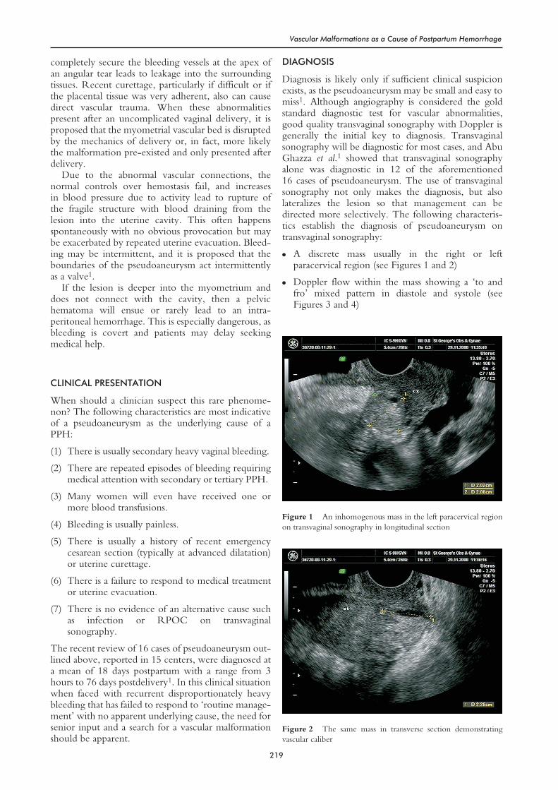
● Doppler flow within the mass showing a ‘to andfro’ mixed pattern in diastole and systole (seeFigures 3 and 4)
219
Vascular Malformations as a Cause of Postpartum Hemorrhage
Figure 1 An inhomogenous mass in the left paracervical regionon transvaginal sonography in longitudinal section
Figure 2 The same mass in transverse section demonstratingvascular caliber
● Doppler flow within the mass showing turbulentmixed flow
● No evidence of products of conception in the uter-ine cavity.
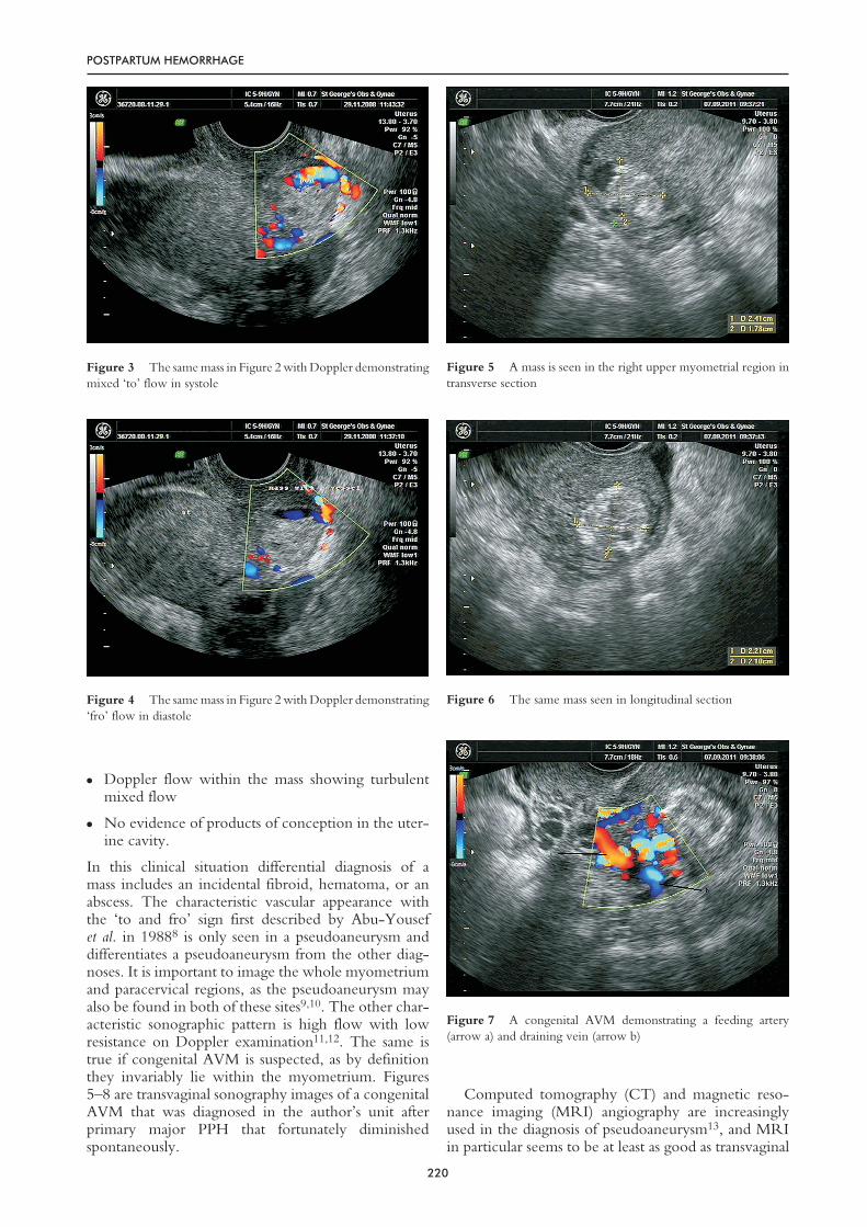
In this clinical situation differential diagnosis of amass includes an incidental fibroid, hematoma, or anabscess. The characteristic vascular appearance withthe ‘to and fro’ sign first described by Abu-Yousefet al. in 19888 is only seen in a pseudoaneurysm anddifferentiates a pseudoaneurysm from the other diag-noses. It is important to image the whole myometriumand paracervical regions, as the pseudoaneurysm mayalso be found in both of these sites9,10. The other char-acteristic sonographic pattern is high flow with lowresistance on Doppler examination11,12. The same istrue if congenital AVM is suspected, as by definitionthey invariably lie within the myometrium. Figures5–8 are transvaginal sonography images of a congenitalAVM that was diagnosed in the author’s unit afterprimary major PPH that fortunately diminishedspontaneously.
Computed tomography (CT) and magnetic reso-nance imaging (MRI) angiography are increasinglyused in the diagnosis of pseudoaneurysm13, and MRIin particular seems to be at least as good as transvaginal
220
POSTPARTUM HEMORRHAGE
Figure 3 The same mass in Figure 2 with Doppler demonstratingmixed ‘to’ flow in systole
Figure 4 The same mass in Figure 2 with Doppler demonstrating‘fro’ flow in diastole
Figure 5 A mass is seen in the right upper myometrial region intransverse section
Figure 6 The same mass seen in longitudinal section
Figure 7 A congenital AVM demonstrating a feeding artery(arrow a) and draining vein (arrow b)

sonography, though not necessarily any better interms of sensitivity4. Due to the extra cost and sparseavailability of these latter diagnostic modalities, MRIis only likely to be helpful where there is doubtover the diagnosis on transvaginal sonography andwhere invasive angiography may be avoided, wheretransvaginal sonography is negative but clinicalsuspicion is high or when the diagnosis has not beenconsidered specifically and MRI picks it up as an‘incidental finding’.
Because of the high pick up rates associated withtransvaginal sonography and other imaging tech-niques, angiography will generally be used as a confir-matory test for definitive management with the addedadvantage of mapping the vascular tree including anycollateral supply to the malformation. Endovasculartreatment can obviously be performed at the sametime.
MANAGEMENT
By the time the diagnosis is considered, the patientwill already have received antibiotics and will oftenhave had at least one evacuation of retained productsof conception (ERPC). Inevitably this fails to resolvethe problem and bleeding either continues or resumesafter an initial period of quiescence; bleeding is usuallyheavy and hemodynamic support including bloodtransfusion is common. In this situation the followingneed to be considered:
● Seek senior obstetric help
● Continue hemodynamic support if required
● Do not instrument the uterus further unless there isconvincing evidence of RPOC on ultrasound
● If pseudoaneurysm is considered, then experttransvaginal ultrasound examining the paracervicalregions, adnexae along the uterine artery, the myo-metrium in its entirety and the broad ligaments hasa high pick up rate
● In theory, uterine balloon tamponade may be effec-tive with a known pre-existing congenital AVM orpseudoaneurysm if pressure is applied to the discretearea that is bleeding. This will only be temporary tostabilize the patient before definitive treatment
● Once diagnosed, discuss the case with inter-ventional radiology – an arteriogram will confirmthe transvaginal sonography findings
● Selective uterine artery embolization is usually sim-ple to perform at this time and its efficacy can be seenimmediately with a postembolization angiogram
● In the presence of continued uterine bleedingdespite selective embolization, then bilateral uterineartery embolization or internal iliac anterior divi-sion embolization may be possible, particularly ifthe arteriogram demonstrates any collateral supplyfrom other pelvic vessels
● Postprocedural care should be routine as long asthere is no significant bleeding from the uterusand/or femoral puncture sites
● De-briefing regarding future pregnancies is impor-tant – fertility should be essentially unchanged andthere are no major obstetric issues regarding futureantenatal care or delivery
● Ultimately hysterectomy is the treatment of lastresort if uterine bleeding is intractable despite theabove measures or hemodynamic instability makesinterventional radiology inappropriate.
Embolization of the pelvic vascular system is notonly a well established treatment for PPH in general(see Chapter 49), but also has been shown to beparticularly useful in the context of a demonstrablediscrete vascular malformation. The advantages of thisprocedure are that it is minimally invasive and canbe performed under local anesthesia, it preserves theuterus and if the site of hemorrhage can be accuratelyidentified, selective embolization can be performedwith minimal disruption of the normal vascular supplyto the uterus1. The source of hemorrhage may beidentified as either an abnormality of the parent arterysuch as a pseudoaneurysm or by contrast extravasation.If such a source is not readily identified, however,empirical embolization of the uterine arteries or ante-rior divisions of the internal iliac arteries bilaterally canstill be performed. In addition angiography may iden-tify alternative, unsuspected sources of hemorrhagewhich can similarly be embolized. The literaturedescribes a high degree of success in managing vascularmalformations with embolization, though it is impos-sible to quote a success rate, as intrinsic bias existswhen failed cases leading to hysterectomy are likely tobe either not reported or at best under-reported. Fig-ures 9 and 10 demonstrate angiographically confirmedpseudoaneurysms in two different patients, and Fig-ures 11 and 12 show the contrast extravasation into theuterine cavity in the latter patient followed by itscessation after selective embolization.
221
Vascular Malformations as a Cause of Postpartum Hemorrhage
Figure 8 The same AVM as shown in Figure 7 showing adifferent pattern of flow during a single examination

Embolization is performed with percutaneouscatheterization of the femoral artery; the type of mate-rial used for embolization depends on the findings.Typically a temporary agent, gelatine sponge, is usedfor general PPH to reduce perfusion pressure, stophemorrhage and allow eventual re-canalization. How-ever, if a discrete vascular malformation such aspseudoaneursym can be identified and a catheterintroduced into it, particulate emboli, of which thereare a variety, are usually used.
Clearly, the more proximal and non-selective theembolization, the greater the risk of compromise tothe pelvic vascular supply subsequently. Pregnanciesfollowing embolization are reported including a seriesfor known AVMs14, but the long-term reproductivesequelae are not yet defined, and the procedure itselfcarries potential documented morbidity includinginfection, neurological damage and bladder necro-sis15,16. These short- and long-term risks need to beweighed against the risk of PPH on an individual basisand discussed with the patient and her partner includ-ing honesty about the uncertainties of the currentworldwide data.
A recent case report describes successful directinjection of embolization particles into a known lesionat laparotomy following failed endovascularembolizaton17, though this is a one-off case.
Ultimately hysterectomy is considered the onlytreatment if uterine bleeding is intractable despite theabove measures or hemodynamic instability makesinterventional radiology unsafe. It is clearly surgicallyimportant to get below the level of the origin of thepseudoaneurysm (usually the uterine artery) and tie offits supply or bleeding will inevitably continue.
In the rare cases where a known congenital AVMexists, PPH may be anticipated in a subsequent preg-nancy or if there is the need for gynecological inter-vention. Unfortunately there is a paucity of evidenceto guide whether expectant management or pre-pregnancy embolization is the most appropriate meansto reduce the risk of future PPH. The likelihood ofPPH is impossible to predict, but pragmatically if thereis a normal obstetric history compared with a historyof significant PPH this fact is likely to influenceindividual management. Elective embolization after adelivery can be considered to prevent future bleedingrisk, but again the pros and cons should be discussedon an individual basis.
PREVENTION OF VASCULAR MALFORMATIONS
Clearly congenital malformations cannot be pre-vented, but pseudoaneurysm is nearly always due toiatrogenic causes. The vast majority of women withthis problem have had obstetric or gynecological sur-gery as the underlying cause, many operations beingemergency lower segment cesarean section atadvanced dilatation but occasionally after curettage oropen gynecological surgery.
Failure to secure angular tears of the uterus involv-ing the uterine artery or one of its branches leads to
222
POSTPARTUM HEMORRHAGE
Figure 9 Angiographic ‘proof’ of a pseudoaneurysm of the rightuterine supply diagnosed on transvaginal sonography – arrow c
Figure 10 A second left pseudoaneurysm confirmed onangiography – arrow d

pseudoaneurysm formation. All those who work inobstetric units understand that emergency lower seg-ment cesarean section at full dilatation can lead to adifficult delivery, usually from occipital malpositionand/or deflexion of the fetal head. Greater diligencewhen delivering the fetal head at advanced dilatationto try and prevent angular tears is very important; inthe event that they occur, however, repairing themand ensuring the uterine vessels are completely securedshould reduce the risk of pseudoaneurysm formation.Also, correcting any dextro-rotation of the uterusbefore incision will also reduce the risk of inadvertentuterine artery injury. Lesions seem to occur slightlymore often on the left side, but are reported on bothsides of the uterus.
CONCLUSION
Arteriovenous malformations are a rare phenomenon,and no clinician or center will deal with large num-bers. They are undoubtedly more common than werealize, but many are likely to be clinically irrelevant,contributing only a very small proportion of thepathology causing PPH. When they do occur, how-ever, they tend to cause a disproportionate amount of
bleeding. Little or nothing can be done about congen-ital AVMs, as they cannot be predicted. Regardless,they need serious consideration when acute primaryPPH is intractable; these patients usually end up withembolization or hysterectomy as they respond poorlyto conservative treatment.
Acquired pseudoaneurysms nearly always occur fol-lowing lower segment cesarean section in advancedlabor and more rarely after gynecological surgery.Although prevention is in theory possible, it is moreimportant to have a high index of suspicion whenthere is sudden and major secondary PPH with littleevidence of RPOC, a poor response to initial therapyand following a lower segment cesarean section in thelate stages of labor.
Good quality transvaginal ultrasound with Dopplerstudies will diagnose most lesions, which can be con-firmed on arteriography. Further uterine instrumenta-tion should be avoided as this will exacerbate theproblem; selective uterine artery embolization is ahighly effective treatment with few side-effects. If
223
Vascular Malformations as a Cause of Postpartum Hemorrhage
Figure 11 Contrast extravazation into the uterine cavitydemonstrated in the pseudoaneurysm in Figure 10
Figure 12 The same patient’s angiogram demonstratingocclusion of the pseudoaneurysm after selective uterine arteryembolization

recognized in a timely fashion, the need forperipartum hysterectomy can usually be avoided withpreservation of a woman’s future fertility.
PRACTICE POINTS
● Arteriovenous malformations are a rare but impor-tant cause of PPH, usually secondary
● AVMs are usually associated with a history of emer-gency lower segment cesarean section in the latestages of labor
● AVMs are characterized by heavy bleeding with noother apparent cause that fails to respond to conser-vative and medical treatment
● High quality transvaginal ultrasound is an excellentdiagnostic tool if clinical suspicion is sufficientlyhigh
● Uterine artery embolization is the highly effectivetreatment of choice.
References
1. Abu-Ghazza O, Hayes K, Chandraharan E, Belli AM. Vascu-lar malformations in relation to obstetrics and gynaecology:diagnosis and treatment. The Obstetrician Gynaecologist2010;12:87–93
2. Uchil D. Complications of the puerperium. In: ArulkumaranS, et al. eds. The Management of Labour, 2nd edn. AndhraPradesh, India: Orient Longman, 2005
3. Kim JY, Kim DI, Do YS, et al. Surgical treatment for con-genital arteriovenous malformation: 10 years’ experience. EurJ Vasc Endovasc Surg 2006;32:101–6
4. Cura M, Martinez N, Cura A, Dalsaso TJ, Elmerhi F.Arteriovenous malformations of the uterus. Acta Radiol2009;50:823–9
5. Tanaka R, Miyasaka Y, Fujii K, Kan S, Yagashita S. Vascularstructure of arteriovenous malformations. J Clin Neurosci2000;7:24–8
6. Grivell RM, Reid KM, Mellor A. Uterine arteriovenous mal-formations: a review of the current literature. Obstet GynecolSurv 2005;60:761–7
7. Kwon JH, Kim GS. Obstetric iatrogenic arterial injuries ofthe uterus: diagnosis with US and treatment with trans-catheter arterial embolization. Radiographics 2002;22:35–46
8. Abu-Yousef M, Wiese J, Shamma A. The “to and fro” sign:duplex Doppler evidence of femoral artery pseudoaneurysm.AJR Am J Roentgenol 1988;150:632
9. Eason D, Tank R. Avoidable morbidity in a patient withpseudoaneurysm of the uterine artery after Caesarean section.J Clin Ultrasound 2006;34:407–11
10. Ho S. A case of uterine pseudoaneurysm. Singapore Med J2002;43:202–4
11. Kelly SM, Belli AM, Campbell S. Arteriovenous malforma-tion of the uterus associated with secondary postpartum hem-orrhage. Ultrasound Obstet Gynecol 2003;21:602–5
12. Müngen E, Yergök YZ, Ertekin AA, Ergür AR, Uçmakli E,Aytaçlar S. Color Doppler sonographic features of uterinearteriovenous malformations: report of two cases. UltrasoundObstet Gynecol 1997;10:215–9
13. Grivell RM, Reid KM, Mellor A. Uterine arteriovenousmalformations: a review of the current literature. ObstetGynecol Surv 2005;60:761–7
14. Delotte J, Chevallier P, Benoit B, Castillon JM, Bongain A.Pregnancy after embolization therapy for uterine arterio-venous malformation. Fertil Steril 2006;85:228
15. Hare WSC, Holland CJ. Paresis following internal iliac arteryembolization. Radiology 1983;146:47–51
16. Sibour PR. Bladder necrosis secondary to pelvic arteryembolization: case report and literature review. J Urol 1994;151:422
17. Przybojewski SJ, Sadler DJ. Novel image-guided manage-ment of a uterine arteriovenous malformation. CardiovascIntervent Radiol 2011;34(Suppl 2):S161–6
224
POSTPARTUM HEMORRHAGE