Defect-Mediated Energy Transfer between ZnO...
6
Published: December 27, 2011 r2011 American Chemical Society 3305 dx.doi.org/10.1021/jp209638g | J. Phys. Chem. C 2012, 116, 3305–3310 ARTICLE pubs.acs.org/JPCC Defect-Mediated Energy Transfer between ZnO Nanocrystals and a Conjugated Dye Gary A. Beane, † Anthony J. Morfa, † Alison M. Funston, ‡ and Paul Mulvaney* ,† † School of Chemistry & Bio21 Institute, University of Melbourne, Parkville, Victoria 3010, Australia ‡ School of Chemistry, Monash University, Clayton, Victoria 3800, Australia b S Supporting Information ’ INTRODUCTION The quenching of exciton emission in semiconducting nano- crystals by fluorophores has been studied extensively over the past decade. 17 These reports have focused overwhelmingly on energy transfer from the exciton state of the nanocrystals. To date, there have been no systematic studies of energy transfer from charge carriers in defect or trap states of nanocrystals to acceptors. Such studies are important because surface and defect recombination limits the overall radiative quantum yield of a semiconductor nanocrystal. In some cases, the defect emission may even dominate over exciton emission. For example, under UV irradiation, the semiconductor ZnO displays an intense green emission that overwhelms the UV exciton emission of the nanocrystal. 8 The decrease in intensity of the defect emission upon annealing of ZnO films suggests that vacancies within the crystals play a role in the emission, 9 whereas the dependence of the visible emission band energy on the particle size 1012 implies that a delocalized charge carrier is involved in the emission process. That, in turn, suggests that there will be a large dipole moment change associated with trap emission and that energy transfer to fluorophores should be possible. The broad visible emission observed in ZnO has been attributed variously to interstitial zinc ions, 13 oxygen vancancies, 14 chemi- sorbed oxygen, 15 divalent copper impurities, 16 and zinc vacancies. 17 Presently, oxygen vacancies are thought to be the likely cause of the visible emission. 1821 The review paper by Ozg€ ur et.al. provides an excellent overview of the main models. 22 Whereas the nature of the defect emission continues to be debated, there have been recent reports that have implicated the defect state as an energy-transfer donor species with organic fluorophores. 2327 However, calculations of the donoracceptor dipole moments (using the Forster resonance energy transfer formalism) have yielded contradictory results. For example, energy transfer from defect states located close to the nanocrystal surface were proposed to explain the unexpectedly small donor acceptor distance when the dye was covalently linked. 26,27 In contrast, when the dye was not covalently linked, surface states were not explicitly required to explain the measured donor acceptor distance, although defect states were proposed as the energy donor. 24 Hence, the general applicability of the Forster mechanism to energy transfer involving surface states must be in doubt. Another important issue is that nanocrystal defect emission is known to be sensitive to surface chemistry. 2832 For example, carboxyl-containing molecules can be photocatalytically decomposed by ZnO. 33,34 Hence it is important to separate quenching due to energy transfer from quenching due to photochemical processes of the adsorbates. In this Article, we present evidence of very efficient ener- gy transfer between ZnO and Alexa594 cadaverine (A594 cadaverine) using steady-state and time-resolved photolumines- cence (PL) measurements. We find that the observed quenching is caused by adsorbed dye molecules and that this quenching is due to energy transfer from ZnO defect states rather than the ZnO exciton state. Using the Tachiya model, we calculate that even a single dye molecule can effectively quench the emission, Received: October 6, 2011 Revised: December 16, 2011 ABSTRACT: Energy transfer from the defect state of zinc oxide nanoparticles to the fluorescent dye AlexaFluor 594 (A594) cadaverine has been studied using both steady-state and time- resolved photoluminescence (PL) measurements. The addition of five stoichiometric equivalents of A594 cadaverine comple- tely quenches the visible defect emission from zinc oxide nano crystals. We also find that the entire defect emission of ZnO is reduced without any change in the overall line shape of the emission, demonstrating that the defect emission is from a single electronic state coupled to the phonon modes of the crystal lattice. The energy transfer is modeled using the dynamic quenching model developed by Tachiya (Sadhu, S.; Tachiya, M. J. Phys. Chem. 2009, 113, 1948819492). Remarkably, there is very efficient energy transfer when there is just one adsorbed dye molecule per nanocrystal, regardless of the orientation of the dipole moment of the cadaverine molecule and the distance to the defect state.
-
Upload
phamkhuong -
Category
Documents
-
view
215 -
download
1
Transcript of Defect-Mediated Energy Transfer between ZnO...
Published: December 27, 2011
r 2011 American Chemical Society 3305 dx.doi.org/10.1021/jp209638g | J. Phys. Chem. C 2012, 116, 3305–3310
ARTICLE
pubs.acs.org/JPCC
Defect-Mediated Energy Transfer between ZnO Nanocrystals anda Conjugated DyeGary A. Beane,† Anthony J. Morfa,† Alison M. Funston,‡ and Paul Mulvaney*,†
†School of Chemistry & Bio21 Institute, University of Melbourne, Parkville, Victoria 3010, Australia‡School of Chemistry, Monash University, Clayton, Victoria 3800, Australia
bS Supporting Information
’ INTRODUCTION
The quenching of exciton emission in semiconducting nano-crystals by fluorophores has been studied extensively over thepast decade.1�7 These reports have focused overwhelmingly onenergy transfer from the exciton state of the nanocrystals. Todate, there have been no systematic studies of energy transferfrom charge carriers in defect or trap states of nanocrystals toacceptors. Such studies are important because surface and defectrecombination limits the overall radiative quantum yield of asemiconductor nanocrystal. In some cases, the defect emissionmay even dominate over exciton emission. For example, underUV irradiation, the semiconductor ZnO displays an intense greenemission that overwhelms the UV exciton emission of thenanocrystal.8 The decrease in intensity of the defect emissionupon annealing of ZnO films suggests that vacancies within thecrystals play a role in the emission,9 whereas the dependence ofthe visible emission band energy on the particle size10�12 impliesthat a delocalized charge carrier is involved in the emissionprocess. That, in turn, suggests that there will be a large dipolemoment change associated with trap emission and that energytransfer to fluorophores should be possible.
The broad visible emission observed in ZnO has been attributedvariously to interstitial zinc ions,13 oxygen vancancies,14 chemi-sorbed oxygen,15 divalent copper impurities,16 and zinc vacancies.17
Presently, oxygen vacancies are thought to be the likely cause of thevisible emission.18�21 The review paper by Ozg€ur et.al. provides anexcellent overview of the main models.22
Whereas the nature of the defect emission continues to bedebated, there have been recent reports that have implicated the
defect state as an energy-transfer donor species with organicfluorophores.23�27 However, calculations of the donor�acceptordipole moments (using the Forster resonance energy transferformalism) have yielded contradictory results. For example,energy transfer from defect states located close to the nanocrystalsurface were proposed to explain the unexpectedly small donor�acceptor distance when the dye was covalently linked.26,27
In contrast, when the dye was not covalently linked, surfacestates were not explicitly required to explain the measured donor�acceptor distance, although defect stateswere proposed as the energydonor.24 Hence, the general applicability of the Forster mechanismto energy transfer involving surface states must be in doubt. Anotherimportant issue is that nanocrystal defect emission is known to besensitive to surface chemistry.28�32 For example, carboxyl-containingmolecules canbe photocatalytically decomposedbyZnO.33,34Henceit is important to separate quenching due to energy transfer fromquenching due to photochemical processes of the adsorbates.
In this Article, we present evidence of very efficient ener-gy transfer between ZnO and Alexa594 cadaverine (A594cadaverine) using steady-state and time-resolved photolumines-cence (PL) measurements. We find that the observed quenchingis caused by adsorbed dye molecules and that this quenching isdue to energy transfer from ZnO defect states rather than theZnO exciton state. Using the Tachiya model, we calculate thateven a single dye molecule can effectively quench the emission,
Received: October 6, 2011Revised: December 16, 2011
ABSTRACT: Energy transfer from the defect state of zinc oxidenanoparticles to the fluorescent dye AlexaFluor 594 (A594)cadaverine has been studied using both steady-state and time-resolved photoluminescence (PL) measurements. The additionof five stoichiometric equivalents of A594 cadaverine comple-tely quenches the visible defect emission from zinc oxide nanocrystals. We also find that the entire defect emission of ZnO isreduced without any change in the overall line shape of theemission, demonstrating that the defect emission is from asingle electronic state coupled to the phonon modes of thecrystal lattice. The energy transfer is modeled using the dynamic quenching model developed by Tachiya (Sadhu, S.; Tachiya, M.J. Phys. Chem. 2009, 113, 19488�19492). Remarkably, there is very efficient energy transfer when there is just one adsorbed dyemolecule per nanocrystal, regardless of the orientation of the dipole moment of the cadaverine molecule and the distance to thedefect state.
3306 dx.doi.org/10.1021/jp209638g |J. Phys. Chem. C 2012, 116, 3305–3310
The Journal of Physical Chemistry C ARTICLE
although a mole ratio of five dyes per nanocrystal is needed toquench >95% of the ensemble PL due to the Poisson distributionof dye molecules among the ZnO nanocrystals. This is remark-able because the dipole moment of the adsorbed dye will notalways be favorable for transfer.
’EXPERIMENTAL SECTION
Synthesis of ZnO Nanocrystals. ZnO nanoparticles wereprepared via the method of Wood et al.12 In brief, zinc acetatedihydrate (0.955 g) was dissolved in 100 mL of absolute ethanolto make a total solute concentration of 44 mM. Upon completedissolution of zinc acetate, seven 400 μL aliquots (2.8 mL total)of 25% w/w tetramethyl ammonium hydroxide in methanol wasadded over ca. 120 s with vigorous stirring in a water bath at25 �C. The ZnO nanoparticle solution was then placed in a freezermaintained at �4 �C and stored until use. Solvents were sourcedfrom Sigma-Aldrich and were either HPLC or spectroscopic gradefor the fluorescence measurements. Alexa 594 cadaverine andrhodamine 6G (Rh6G) were purchased from Invitrogen andExciton, respectively, whereas aminopentanol (95%) was purchasedfrom Aldrich; all chemicals were used as received.Sample Preparation. The diameter of ZnO nanoparticles in
the as-synthesized solution was determined to be 3.2 nm usingthe calibration curve of Wood et al.12 Concentrations werecalculated assuming the formation of particles of this size andassuming complete conversion of precursors into particles.Samples for fluorescence measurements were all prepared inethanol (HPLC grade, Sigma-Aldrich). For steady-state emissionexperiments, the samples had a ZnO concentration of 212 nMand an A594 cadaverine concentration of 0�1060 nM; theabsorbance of the sample at the excitation wavelength was<0.1. All samples were left to equilibrate following mixing ofthe ZnO and A594 cadaverine for 5 min prior to measurementsand were not degassed. The quantum yield of ZnOwasmeasuredwith respect to a Rh6G standard under a N2 atmosphere. For PLmeasurements, the excitation wavelength was 314 nm, and thequantum yield of Rh6G at this wavelength was taken to be 0.95.35
Instrumentation. UV�visible absorbance spectra were col-lected using an Agilent 8453 UV�visible absorbance spectro-photometer system. PL spectra were obtained using a HoribaJobin Yvon Fluorolog-3 with an excitation wavelength of 314 nm.Time-resolved emission measurements were carried out withexcitation at 300 nm using a Picoquant NanoLED-300 pulseddiode laser with detection at a wavelength of 500 nm. Theinstrument response of the system was measured using acolloidal silica dispersion in water with an absorbance of 0.1 at300 nm and had a full width half-maximum (fwhm) of 2.0 ns.
’RESULTS
Optical Absorbance.The photophysical characteristics of thespecies used in this study are summarized in Table 1, whereas the
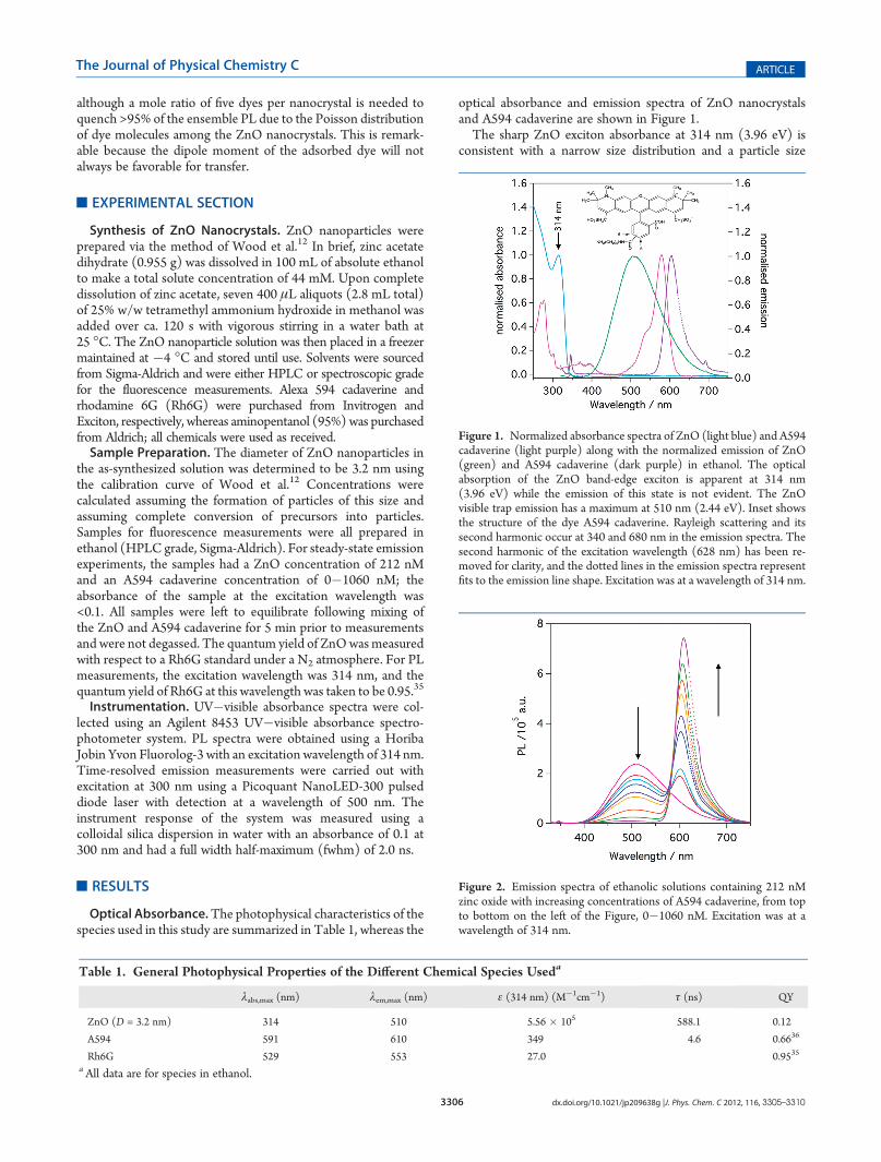
optical absorbance and emission spectra of ZnO nanocrystalsand A594 cadaverine are shown in Figure 1.The sharp ZnO exciton absorbance at 314 nm (3.96 eV) is
consistent with a narrow size distribution and a particle size
Table 1. General Photophysical Properties of the Different Chemical Species Useda
λabs,max (nm) λem,max (nm) ε (314 nm) (M�1cm�1) τ (ns) QY
ZnO (D = 3.2 nm) 314 510 5.56 � 105 588.1 0.12
A594 591 610 349 4.6 0.6636
Rh6G 529 553 27.0 0.9535
aAll data are for species in ethanol.
Figure 1. Normalized absorbance spectra of ZnO (light blue) and A594cadaverine (light purple) along with the normalized emission of ZnO(green) and A594 cadaverine (dark purple) in ethanol. The opticalabsorption of the ZnO band-edge exciton is apparent at 314 nm(3.96 eV) while the emission of this state is not evident. The ZnOvisible trap emission has a maximum at 510 nm (2.44 eV). Inset showsthe structure of the dye A594 cadaverine. Rayleigh scattering and itssecond harmonic occur at 340 and 680 nm in the emission spectra. Thesecond harmonic of the excitation wavelength (628 nm) has been re-moved for clarity, and the dotted lines in the emission spectra representfits to the emission line shape. Excitation was at a wavelength of 314 nm.
Figure 2. Emission spectra of ethanolic solutions containing 212 nMzinc oxide with increasing concentrations of A594 cadaverine, from topto bottom on the left of the Figure, 0�1060 nM. Excitation was at awavelength of 314 nm.
3307 dx.doi.org/10.1021/jp209638g |J. Phys. Chem. C 2012, 116, 3305–3310
The Journal of Physical Chemistry C ARTICLE
of 3.2 nm.12 Whereas typical semiconductor nanoparticles havesignificant exciton emission, the ZnO emission spectrum isdominated by defect emission centered at 510 nm, with noevidence of the exciton emission. The A594 cadaverine dye hasan absorbance maximum at 591 nm, whereas the emissionspectrum is Stokes shifted by 19 nm with a maximum at610 nm. The dye absorbs little around 314 nm, allowing awindow for the selective excitation of the ZnO nancrystals. Atthe highest concentration of A594 cadaverine used here, theabsorbance of the dye at 314 nmwas 3� 10�3. Resonance energytransfer requires a spectral overlap between the donor emissionand the acceptor absorbance spectra. It is evident from Figure 1that there is significant spectral overlap of the ZnO defectemission with the absorption spectrum of the A594 cadaverinedye. In contrast the overlap between the exciton emission of ZnOand the dye absorbance is negligible.Steady-State Emission. The emission spectra of ZnO solu-
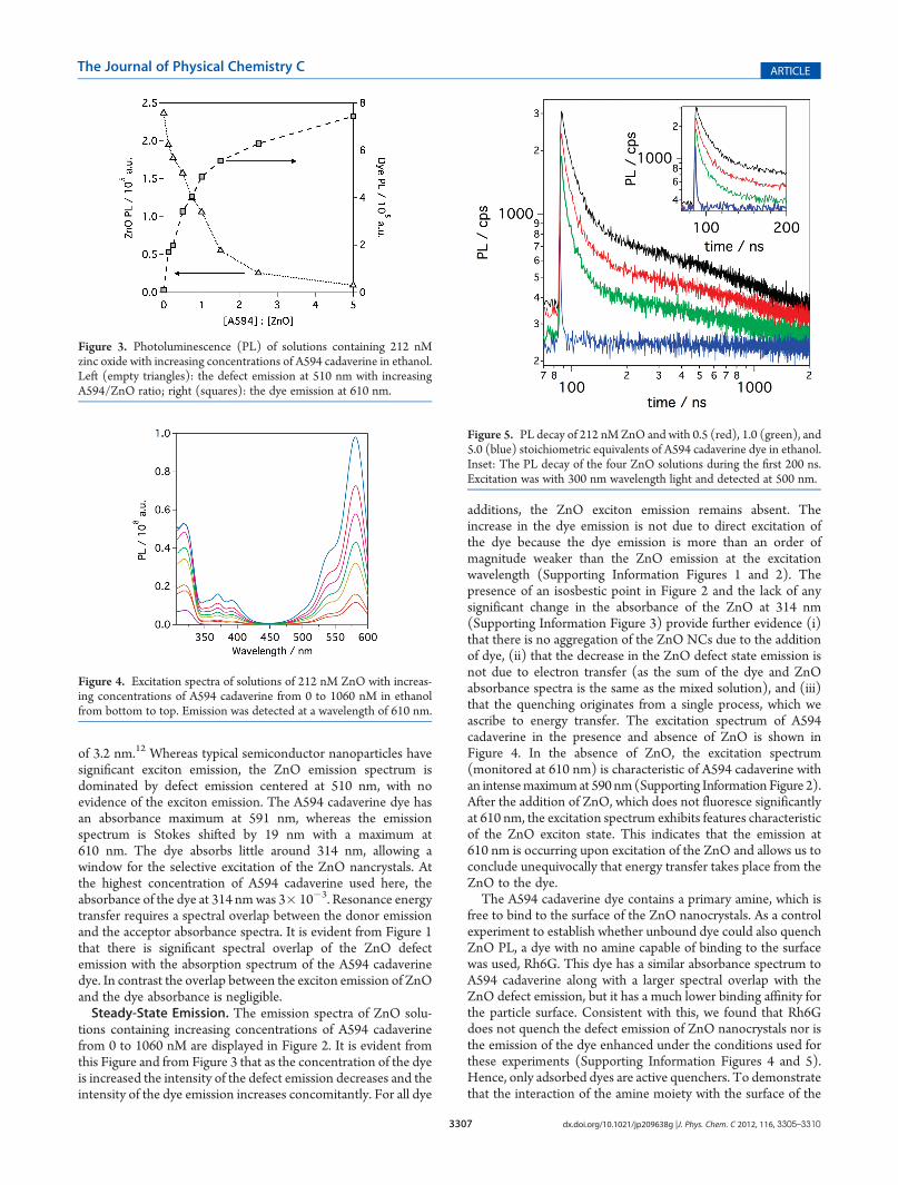
tions containing increasing concentrations of A594 cadaverinefrom 0 to 1060 nM are displayed in Figure 2. It is evident fromthis Figure and from Figure 3 that as the concentration of the dyeis increased the intensity of the defect emission decreases and theintensity of the dye emission increases concomitantly. For all dye
additions, the ZnO exciton emission remains absent. Theincrease in the dye emission is not due to direct excitation ofthe dye because the dye emission is more than an order ofmagnitude weaker than the ZnO emission at the excitationwavelength (Supporting Information Figures 1 and 2). Thepresence of an isosbestic point in Figure 2 and the lack of anysignificant change in the absorbance of the ZnO at 314 nm(Supporting Information Figure 3) provide further evidence (i)that there is no aggregation of the ZnO NCs due to the additionof dye, (ii) that the decrease in the ZnO defect state emission isnot due to electron transfer (as the sum of the dye and ZnOabsorbance spectra is the same as the mixed solution), and (iii)that the quenching originates from a single process, which weascribe to energy transfer. The excitation spectrum of A594cadaverine in the presence and absence of ZnO is shown inFigure 4. In the absence of ZnO, the excitation spectrum(monitored at 610 nm) is characteristic of A594 cadaverine withan intensemaximumat 590 nm(Supporting Information Figure 2).After the addition of ZnO, which does not fluoresce significantlyat 610 nm, the excitation spectrum exhibits features characteristicof the ZnO exciton state. This indicates that the emission at610 nm is occurring upon excitation of the ZnO and allows us toconclude unequivocally that energy transfer takes place from theZnO to the dye.The A594 cadaverine dye contains a primary amine, which is
free to bind to the surface of the ZnO nanocrystals. As a controlexperiment to establish whether unbound dye could also quenchZnO PL, a dye with no amine capable of binding to the surfacewas used, Rh6G. This dye has a similar absorbance spectrum toA594 cadaverine along with a larger spectral overlap with theZnO defect emission, but it has a much lower binding affinity forthe particle surface. Consistent with this, we found that Rh6Gdoes not quench the defect emission of ZnO nanocrystals nor isthe emission of the dye enhanced under the conditions used forthese experiments (Supporting Information Figures 4 and 5).Hence, only adsorbed dyes are active quenchers. To demonstratethat the interaction of the amine moiety with the surface of the
Figure 3. Photoluminescence (PL) of solutions containing 212 nMzinc oxide with increasing concentrations of A594 cadaverine in ethanol.Left (empty triangles): the defect emission at 510 nm with increasingA594/ZnO ratio; right (squares): the dye emission at 610 nm.
Figure 4. Excitation spectra of solutions of 212 nM ZnO with increas-ing concentrations of A594 cadaverine from 0 to 1060 nM in ethanolfrom bottom to top. Emission was detected at a wavelength of 610 nm.
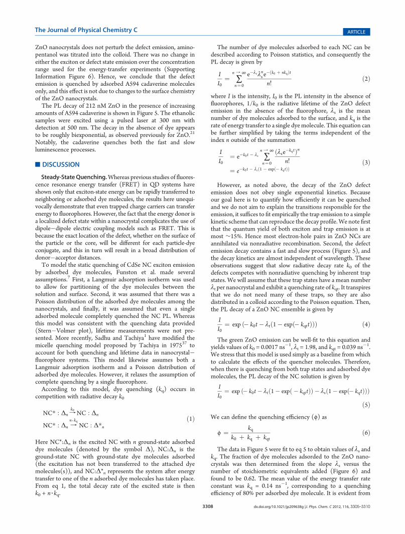
Figure 5. PL decay of 212 nMZnO and with 0.5 (red), 1.0 (green), and5.0 (blue) stoichiometric equivalents of A594 cadaverine dye in ethanol.Inset: The PL decay of the four ZnO solutions during the first 200 ns.Excitation was with 300 nm wavelength light and detected at 500 nm.
3308 dx.doi.org/10.1021/jp209638g |J. Phys. Chem. C 2012, 116, 3305–3310
The Journal of Physical Chemistry C ARTICLE
ZnO nanocrystals does not perturb the defect emission, amino-pentanol was titrated into the colloid. There was no change ineither the exciton or defect state emission over the concentrationrange used for the energy-transfer experiments (SupportingInformation Figure 6). Hence, we conclude that the defectemission is quenched by adsorbed A594 cadaverine moleculesonly, and this effect is not due to changes to the surface chemistryof the ZnO nanocrystals.The PL decay of 212 nM ZnO in the presence of increasing
amounts of A594 cadaverine is shown in Figure 5. The ethanolicsamples were excited using a pulsed laser at 300 nm withdetection at 500 nm. The decay in the absence of dye appearsto be roughly biexponential, as observed previously for ZnO.21
Notably, the cadaverine quenches both the fast and slowluminescence processes.
’DISCUSSION
Steady-StateQuenching.Whereas previous studies of fluores-cence resonance energy transfer (FRET) in QD systems haveshown only that exciton-state energy can be rapidly transferred toneighboring or adsorbed dye molecules, the results here unequi-vocally demonstrate that even trapped charge carriers can transferenergy to fluorophores. However, the fact that the energy donor isa localized defect state within a nanocrystal complicates the use ofdipole�dipole electric coupling models such as FRET. This isbecause the exact location of the defect, whether on the surface ofthe particle or the core, will be different for each particle-dyeconjugate, and this in turn will result in a broad distribution ofdonor�acceptor distances.To model the static quenching of CdSe NC exciton emission
by adsorbed dye molecules, Funston et al. made severalassumptions.7 First, a Langmuir adsorption isotherm was usedto allow for partitioning of the dye molecules between thesolution and surface. Second, it was assumed that there was aPoisson distribution of the adsorbed dye molecules among thenanocrystals, and finally, it was assumed that even a singleadsorbed molecule completely quenched the NC PL. Whereasthis model was consistent with the quenching data provided(Stern�Volmer plot), lifetime measurements were not pre-sented. More recently, Sadhu and Tachiya1 have modified themicelle quenching model proposed by Tachiya in 197537 toaccount for both quenching and lifetime data in nanocrystal�fluorophore systems. This model likewise assumes both aLangmuir adsorption isotherm and a Poisson distribution ofadsorbed dye molecules. However, it relaxes the assumption ofcomplete quenching by a single fluorophore.According to this model, dye quenching (kq) occurs in
competition with radiative decay k0
NC� : Δn fk0NC : Δn
NC� : Δn fn 3 kq
NC : Δ�nð1Þ
Here NC*:Δn is the excited NC with n ground-state adsorbeddye molecules (denoted by the symbol Δ), NC:Δn is theground-state NC with ground-state dye molecules adsorbed(the excitation has not been transferred to the attached dyemolecules(s)), and NC:Δ*n represents the system after energytransfer to one of the n adsorbed dye molecules has taken place.From eq 1, the total decay rate of the excited state is thenk0 + n 3 kq.
The number of dye molecules adsorbed to each NC can bedescribed according to Poisson statistics, and consequently thePL decay is given by
II0
¼ ∑n f ∞
n¼ 0
e�λsλns e�ðk0 þ nkqÞt
n!ð2Þ
where I is the intensity, I0 is the PL intensity in the absence offluorophores, 1/k0 is the radiative lifetime of the ZnO defectemission in the absence of the fluorophore, λs is the meannumber of dye molecules adsorbed to the surface, and kq is therate of energy transfer to a single dye molecule. This equation canbe further simplified by taking the terms independent of theindex n outside of the summation
II0
¼ e�k0t � λs ∑n f ∞
n¼ 0
ðλse�kqtÞnn!
¼ e�k0t � λsð1 � expð� kqtÞÞð3Þ
However, as noted above, the decay of the ZnO defectemission does not obey single exponential kinetics. Becauseour goal here is to quantify how efficiently it can be quenchedand we do not aim to explain the transitions responsible for theemission, it suffices to fit empirically the trap emission to a simplekinetic scheme that can reproduce the decay profile. We note firstthat the quantum yield of both exciton and trap emission is atmost ∼15%. Hence most electron-hole pairs in ZnO NCs areannihilated via nonradiative recombination. Second, the defectemission decay contains a fast and slow process (Figure 5), andthe decay kinetics are almost independent of wavelength. Theseobservations suggest that slow radiative decay rate k0 of thedefects competes with nonradiative quenching by inherent trapstates. We will assume that these trap states have a mean numberλt per nanocrystal and exhibit a quenching rate of kqt. It transpiresthat we do not need many of these traps, so they are alsodistributed in a colloid according to the Poisson equation. Then,the PL decay of a ZnO NC ensemble is given by
II0
¼ exp ð� k0t � λtð1� expð� kqttÞÞÞ ð4Þ
The green ZnO emission can be well-fit to this equation andyields values of k0 = 0.0017 ns�1, λt = 1.98, and kqt = 0.039 ns�1.We stress that this model is used simply as a baseline from whichto calculate the effects of the quencher molecules. Therefore,when there is quenching from both trap states and adsorbed dyemolecules, the PL decay of the NC solution is given by
II0
¼ exp ð� k0t � λtð1� expð � kqttÞÞ � λsð1� expð� kqtÞÞÞð5Þ
We can define the quenching efficiency (ϕ) as
ϕ ¼ kqk0 þ kq þ kqt
ð6Þ
The data in Figure 5 were fit to eq 5 to obtain values of λs andkq. The fraction of dye molecules adsorded to the ZnO nano-crystals was then determined from the slope λs versus thenumber of stoichiometric equivalents added (Figure 6) andfound to be 0.62. The mean value of the energy transfer rateconstant was kq = 0.14 ns�1, corresponding to a quenchingefficiency of 80% per adsorbed dye molecule. It is evident from

3309 dx.doi.org/10.1021/jp209638g |J. Phys. Chem. C 2012, 116, 3305–3310
The Journal of Physical Chemistry C ARTICLE
Figure 6 that the quenching efficiency is independent of thenumber of dye molecules adsorbed.In summary, the fits show that the intrinsic rates of energy
transfer from the ZnO defect state are of order 0.1 ns�1, an orderof magnitude faster than the intrinsic rate of PL decay. This valueis also significantly faster than the rate constant needed to allowfor nonradiative recombination. In agreement with the previousconclusions of Funston et al.,7 that the nanocrystal excitonemission can be quenched by a single fluorophore, we now findthat this applies to defect state emission too. This is all the moreremarkable given the wide spread in donor�acceptor distancesand dipole orientation in these systems.It is worth noting that whereas other groups have reported the
presence of two distinct defects (at approximately 500 and570 nm, respectively),38 the ZnO synthesized in this workexhibits only one defect (at 510 nm). Work by Shi et al.39 andothers40 shows that despite the visible emission from zinc oxideparticles being quite broad, it can be modeled using theHuang�Rhys equation, which describes the coupling of anelectronic state with a vibrational state. The defect emission ofthe ZnONCs synthesized in this work is also accurately fit by theHuang�Rhys model in which only the amplitude factor changeswith dye addition. (See the Supporting Information.) Thisimplies that defect emission in ZnO involves a single electronictransition that strongly couples to the phonon modes of thecrystal lattice and not a distribution of trap states.The uniform decrease in the emission of the ZnO in our
experiments upon A594 cadaverine addition, even those parts ofthe spectrum that do not overlap with the dye, demonstrates that thedefect emission behaves as a single (donor) state rather than a seriesof discrete trap states. Recent single-particle experiments by Layekand coworkers41 further support these findings and demonstrate thatthe broad emission is from coupling of the excited state and thephonon modes rather than polydispersity.
’CONCLUSIONS
We have demonstrated that very efficient energy transfer occursfrom ZnO nanocrystal defect states to adsorbed A594 cadaverinedye molecules. Steady-state observations reveal that the addition of
conjugated dyemolecules results in the reductionof defect emission,without changing the line shape. Time-resolved measurementsdemonstrate that the energy transfer in this system is from thedefect state, with the apparent lifetime of both components of theZnO fluorescence decay decreasing with increasing stoichiometricequivalents of dye. This change in the apparent lifetime was fit usingthe Sadhu kinetic model, further supporting the use of Poissonstatistics in analyzing energy transfer in the present system. Thiswork illustrates that defect states in nanocrystals must be consideredin energy transfer assays, not just exciton states, as defects them-selves can undergo energy transfer to acceptor molecules.
’ASSOCIATED CONTENT
bS Supporting Information. PL spectra and excitation spec-tra of solutions; absorbance spectra for titration of A594cadaverine; emission spectra of solutions with increasing propor-tions of Rhodamine 6G, plot of the PL of the ZnO defect at510 nm with the addition of aminopentanol (AP) into anethanolic solution, and the PL decay of a 212 nM ZnO solutionin ethanol. This material is available free of charge via the Internetat http://pubs.acs.org.
’AUTHOR INFORMATION
Corresponding Author*E-mail: [email protected].
’ACKNOWLEDGMENT
This work was supported by the Australian Research Councilvia grants LF100100117 and DP0985325. A.J.M. acknowledgesthe support of the Melbourne Materials Institute.
’REFERENCES
(1) Sadhu, S.; Tachiya, M. J. Phys. Chem. 2009, 113, 19488–19492.(2) Clapp, A. R.; Medintz, I. L.; Mauro, J. M.; Fisher, B. R.; Bawendi,
M. G.; Mattoussi, H. J. Am. Chem. Soc. 2004, 126, 301–310.(3) Clapp, A. R.; Medintz, I. L.; Mattoussi, H. ChemPhysChem 2006,
7, 47–57.(4) Medintz, I. L.; Sapsford, K. E.; Clapp, A. R.; Pons, T.; Higashiya,
S.; Welch, J. T.; Mattoussi, H. J. Phys. Chem. B 2006, 110, 10683–10690.(5) Pons, T.; Medintz, I. L.; Sykora, M.; Mattoussi, H. Phys. Rev. B
2006, 73, 245302.(6) Soujon, D.; Becker, K.; Rogach, A. L.; Feldmann, J.; Weller, H.;
Talapin, D. V.; Lupton, J. M. J. Phys. Chem. C 2007, 111, 11511–11515.(7) Funston, A. M.; Jasieniak, J. J.; Mulvaney, P. Adv. Mater. 2008,
20, 4274–4280.(8) Bera, D.; Qian, L.; Sabui, S.; Santra, S.; Holloway, P. H.Opt. Mat.
2008, 30, 1233–1239.(9) Morfa, A. J.; Beane, G.; Mashford, B.; Singh, B.; Della Gaspera,
E.; Martucci, A.; Mulvaney, P. J. Phys. Chem. C 2010, 114, 19815–19821.(10) Spanhel, L.; Anderson, M. A. J. Am. Chem. Soc. 1991, 113,
2826–2833.(11) van Dijken, A.; Meulenkamp, E. A.; Vanmaekelbergh, D.;
Meijerink, A. J. Phys. Chem. B. 2000, 104, 1715�1723.(12) Wood, A.; Giersig, M.; Hilgendorff, M.; Vilas-Campos, A.;
Liz-Marzan, L. M.; Mulvaney, P. Aust. J. Chem. 2003, 56, 1051–1057.(13) Mollwo, E. Z. Z. Phys. 1954, 138, 478–488.(14) Kroger, F. A.; Vink, H. J. J. Phys. Chem. 1954, 22, 250–252.(15) Vancraey, F.; Maenhout, W.; Dekeyser, W. Phys. Status Solidi
1965, 8, 841.(16) Dingle, R. Phys. Rev. Lett. 1969, 23, 579–581.(17) Smith, J. M.; Vehse, W. E. Phys. Lett. A 1970, 31, 147–148.
Figure 6. Analysis of the quenching data. Left axis: The mean numberof adsorbed dye molecules per ZnO nanoparticle (using eq 5) versusthe number of dye equivalents added (squares). Best fit to the datayields a slope of 0.62 ( 0.03 (dashed line). Right axis: The calculatedquenching efficiency as a function of the number of dye equivalentsadded (circles). The data yield an efficiency of 77 ( 2%.

3310 dx.doi.org/10.1021/jp209638g |J. Phys. Chem. C 2012, 116, 3305–3310
The Journal of Physical Chemistry C ARTICLE
(18) Anpo, M.; Kubokawa, Y. J. Phys. Chem. 1984, 88, 5556–5560.(19) Vanheusden, K.; Seager, C. H.; Warren, W. L.; Tallant, D. R.;
Voigt, J. A. Appl. Phys. Lett. 1996, 68, 403–405.(20) Vanheusden, K.; Warren, W. L.; Seager, C. H.; Tallant, D. R.;
Voigt, J. A.; Gnade, B. E. J. Appl. Phys. 1996, 79, 7983.(21) van Dijken, A.; Meulenkamp, E. a.; Vanmaekelbergh, D.;
Meijerink, A. J. Phys. Chem. B 2000, 104, 1715–1723.(22) Ozg€ur, U.; Alivov, Y. I.; Liu, C.; Teke, A.; Reshchikov, M. A.;
Dogan, S.; Avrutin, V.; Cho, S. J.; Morkoc, H. J. Appl. Phys. 2005, 98,041301 .(23) Yu, H.-h.; Wong,M. K. F.; Ali, E. M.; Ying, J. Y.Chem. Commun.
2008, 4912–4914.(24) Rakshit, S.; Vasudevan, S. ACS Nano 2008, 2, 1473–1479.(25) Park, J. K.; Lee, K. W.; Lee, W.; Lee, C. E. Appl. Phys. Lett. 2009,
94, 233301–233301.(26) Makhal, A.; Sarkar, S.; Bora, T.; Baruah, S.; Dutta, J.;
Raychaudhuri, a. K.; Pal, S. K. Nano. 2010, 21, 265703-1�265703-5.(27) Makhal, A.; Sarkar, S.; Bora, T.; Baruah, S.; Dutta, J.;
Raychaudhuri, a. K.; Pal, S. K. J. Phys. Chem. C 2010, 114, 10390–10395.(28) Bahnemann, D. W.; Kormann, C.; Hoffmann, M. R. J. Phys.
Chem. 1987, 91, 3789–3798.(29) Li, D.; Leung, Y. H.; Djurisic, A. B.; Liu, Z. T.; Xie, M. H.; Shi,
S. L.; Xu, S. J.; Chan, W. K. Appl. Phys. Lett. 2004, 85, 1601–1603.(30) Norberg, N. S.; Gamelin, D. R. J. Phys. Chem. B 2005, 109,
20810–20816.(31) Singla, M. L.; M, M. S.; Kumar, M. J. Lumin. 2009, 129,
434–438.(32) Bullen, C.; Mulvaney, P. Langmuir 2006, 22, 3007–3013.(33) Noto, Y.; Fukuda, K.; Onishi, T.; Tamaru, K. Trans. Faraday
Soc. 1967, 63, 3081.(34) Curri, M. L.; Comparelli, R.; Cozzoli, P. D.; Mascolo, G.;
Agostiano, A. Mater. Sci. Eng., C 2003, 23, 285–289.(35) Kubin, R. F.; Fletcher, A. N. J. Lumin. 1982, 27, 455–462.(36) Fluorescence Quantum Yields (QY) and Lifetimes (t) for Alexa
Fluor Dyes - Table 1.5. In Molecular Probes Handbook, a Guide toFluorescent Probes and Labeling Technologies, 11th ed; Invitrogen, 2010.(37) Tachiya, M. Chem. Phys. Lett. 1975, 33, 289–292.(38) Rakshit, S.; Vasudevan, S. J. Phys. Chem. C 2009, 113, 16424–
16431.(39) Shi, S. L.; Li, G. Q.; Xu, S. J.; Zhao, Y.; Chen, G. H. J. Phys. Chem.
B 2006, 110, 10475–10478.(40) Reynolds, D. C.; Look, D. C.; Jogai, B. J. Appl. Phys. 2001,
89, 6189–6191.(41) Layek, A.; De, S.; Thorat, R.; Chowdhury, A. J. Phys. Chem. Lett.
2011, 2, 1241–1247.